control of vesicle membrane permeability with catalytic
TRANSCRIPT

MaterialsHorizons
||
20
68
rsc.li/materials-horizons
ISSN 2051-6347
COMMUNICATIONStefan A. F. Bon et al.Control of vesicle membrane permeability with catalytic particles
Volume 3 Number 1 January 2016 Pages 1–84

This journal is©The Royal Society of Chemistry 2016 Mater. Horiz., 2016, 3, 41--46 | 41
Cite this:Mater. Horiz., 2016,
3, 41
Control of vesicle membrane permeability withcatalytic particles†
Ross W. Jaggers, Rong Chen and Stefan A. F. Bon*
Giant polymer vesicles which have membrane-embedded catalyti-
cally active manganese oxide particles are made using droplet-
based microfluidics. It is demonstrated that these colloidal particles
can regulate the membrane permeability of the polymersomes
upon their exposure to, and catalytic reaction with, small amounts
of dissolved hydrogen peroxide.
A vesicle is a self-contained body of liquid surrounded bya lamellar, often bilayer-type, membrane dispersed in a liquidenvironment. Compartmentalization of small and finite volumesof liquids and the consecutive emergence of membrane bio-energetics are identified as being of key importance in theevolution of cells, and hence the origins of life.1 The ability tomediate transport across a cell membrane is crucial for cellularactivity, from regulation of ion transport to uptake of nutrients.2
In eukaryotic and prokaryotic cells, functional proteins embeddedin the cell membrane regulate transport of ions and moleculesand are known as transmembrane proteins (TMPs). This conceptof having the ability to regulate and control membrane perme-ability on demand is being explored in a variety of scientific fields,using a range of both biological and chemical methods.3–29
A pioneering example where biology, in the form of TMPs,meets synthetic macromolecular chemistry, in the form ofpolymer vesicles, was reported by Meier and coworkers inwhich polymersome nanoreactors were permeabilized to smallsolutes by the inclusion of a nonspecific channel protein,bacterial porin OmpF, into the membrane wall.3 In a differentstudy Meier and coworkers demonstrated the incorporation ofAquaporin Z, a water channel protein, into the walls of poly-meric vesicles.4 Inclusion of this protein resulted in a signifi-cantly increased water permeability, almost 90 times greaterthan vesicles without. Choi and Montemagno elegantly demon-strated that several proteins working in conjunction with one
another could be inserted into a vesicle membrane, creating amulti-protein, light-driven adenosine triphosphate (ATP)engine.5 Moreover, channel proteins have also been used tomediate deoxyribonucleic acid (DNA) translocation by actingas a receptor for binding bacteriophage viruses, creating poly-meric vehicles for DNA.6
Synthetic membrane channels have also been used to modifythe permeability of polymeric vesicles, for example, Hammerand colleagues showed that the inclusion of dendritic helicalpores facilitates proton transport across a membrane.7 Perrier,Jolliffe and coworkers8 demonstrated the use of cyclic peptide–poly(N-isopropylacrylamide) conjugates as thermoresponsivechannels, whilst Gin et al.9 showed that aminocyclodextrinscan be used as anion-selective channels.
The use of membrane channels is not the only route towardsgaining control of transmembrane transport; the inherentpermeability of a vesicle membrane itself can be influencedby physical phenomena. Near their liquid-crystalline transition
Department of Chemistry, University of Warwick, Coventry, C47 7AL, UK.
E-mail: [email protected]; Web: www.bonlab.info; Tel: +44 (0)2476 574009
† Electronic supplementary information (ESI) available. See DOI: 10.1039/c5mh00093a
Received 1st June 2015,Accepted 20th August 2015
DOI: 10.1039/c5mh00093a
www.rsc.li/materials-horizons
Conceptual insightsThe ability to control membrane permeability in vesicles allows forregulated transport of matter across the vesicular wall. Vesicles can beseen as microscopic sacs containing a compartmentalized volume ofliquid dispersed in a bulk liquid environment. Nature has devisedsophisticated strategies to accomplish control of transmembrane trans-port, including endo- and exocytosis as well as the incorporation oftransmembrane proteins into cell membranes. A variety of syntheticapproaches have been explored by scientists in order to accomplish suchcontrol in manmade systems. Examples include hybrid systems wherebytransmembrane proteins were incorporated as part of synthetic vesicles,and the use of responsive macromolecular building blocks to regulatemembrane porosity upon an external trigger in polymer vesicles, alsoreferred to as polymersomes. Here we show for the first time that thepermeability of the membrane of polymer vesicles can be controlled bymembrane-embedded catalytically active manganese oxide particles. Theability to chemically trigger activity of the catalytic particle herebyinducing a temporary increase of membrane permeability offers precisetime-specific control of transmembrane transport. It is our belief that thisconcept can be applied to a wide variety of membrane-based systems.
MaterialsHorizons
COMMUNICATION
Ope
n A
cces
s A
rtic
le. P
ublis
hed
on 2
0 A
ugus
t 201
5. D
ownl
oade
d on
12/
1/20
21 1
1:03
:19
PM.
Thi
s ar
ticle
is li
cens
ed u
nder
a C
reat
ive
Com
mon
s A
ttrib
utio
n 3.
0 U
npor
ted
Lic
ence
.
View Article OnlineView Journal | View Issue

42 | Mater. Horiz., 2016, 3, 41--46 This journal is©The Royal Society of Chemistry 2016
temperatures, liposomes exhibit an enhanced leakiness of theirmembranes,10 a property exploited by Yatvin and coworkers intheir design of temperature-controlled, drug-releasing vesicles.11
Battaglia and coworkers demonstrated shear-rate induced mod-ulation of polymersome membrane permeability.12
The amphiphilic balance of the macromolecular buildingblocks of polymer vesicles can be altered to regulate permeability.Oxidative responsive polymersomes in which the hydrophobicpropylene sulfide units were oxidised by hydrogen peroxide intotheir more hydrophilic propylene sulfoxide analogues werereported by Tirelli, Hubbell and co-workers.13 A variation on thisconcept using glucose oxidase to generate hydrogen peroxidein situ was reported by Hubbell et al.14 Ahmed and Dischershowed that triggered partial hydrolysis of the block copolymerbuilding blocks of vesicular structures induced self-poratingmembranes capable of enhanced release.15 Du and Armesshowed a pH dependence in polymersome membrane perme-ability.16 Eisenberg et al. interestingly showed that an alternatingpH in the surrounding solution of a vesicle can trigger a‘‘breathing’’ behaviour, resulting in the diffusion of species inand out of the vesicle upon a reversible change in vesiclevolume.17 Van Hest and coworkers incorporated phenyl boronicacid based stimuli responsive block copolymers into vesicularstructures which allowed for pore formation at both high pH andsugar exposure.18 De Geest and coworkers created core–shellcapsules consisting of a degradable microgel core surrounded bya semi-permeable membrane. Upon degradation of the microgelcore, the accompanied increase in osmotic pressure rupturesthe surrounding membrane, and triggers the release of theencapsulated species.19
One interesting approach is the use of functional nano-particles on or in the membrane, as well as inside, of manmadevesicles. In these hybrid supracolloidal structures it has beenshown that control of membrane permeability is possible usingthe characteristic features of the nanoparticles.20–28
Oh and coworkers incorporated nanoparticles of silver20 andgold21 into the membranes of phospholipid vesicles andshowed enhanced membrane fluidity and thus permeability.Paasonen and colleagues used the concept of hyperthermia toshow that exposure to light of vesicles with gold nanoparticleson or in their membranes enhanced their permeability.22 Weitzet al. created giant polymersomes from poly(N-isopropyl acryl-amide) containing diblock copolymers and gold nanoparticlesusing microfluidics.23 These polymersomes exhibited thermo-and photo-responsive behaviour and tunable permeabilities.Lecommandoux and others prepared magneto-responsive polymervesicles by embedding iron oxide nanoparticles into the lamellarmembranes of vesicles.24–29 Upon exposure to an externallyapplied magnetic field the vesicles could undergo magnetotaxis(gradient field), deform, be used as contrast agents in magneticresonance imaging and as drug delivery vehicles that deliveredtheir payload by means of radiofrequency magnetic hyperthermia.
In this manuscript, we show that manganese oxide particlesthat are embedded into the membranes of polymer vesicles areable to enhance permeability upon activation and reaction ofthe catalytic particles with small amounts of hydrogen peroxide.
To the best of our knowledge this concept of using chemicalheterogeneous catalysis as a strategy to influence membranepermeability has not been previously reported. We believe thatour method offers an interesting tactic to control and regulaterelease from vesicular structures dispersed in a liquid.
Polymer vesicles of low dispersity in size distribution wereprepared using a microfluidics technique from water-in-oil-in-water double-emulsion droplets, with a middle phase ofchloroform containing poly(n-butyl methacrylate)94-block-poly-(N,N-dimethyl-amino ethyl methacrylate)37 and manganeseoxide particles, as shown in Fig. 1. For membrane permeabilityand release studies, which are described later, the inner aqueousphase contained either sodium sulfate, sodium fluoride, orcongo red dye.
Dark field microscopy indicates that manganese oxideparticles are part of the membrane of the vesicles as scatteringis observed (see Fig. 2a). Cryogenic scanning electron micro-scopy confirms that manganese oxide particles are embeddedinto the membrane of the polymer vesicle (see Fig. 3). Theaddition of small amounts of hydrogen peroxide (0.1 wt%) tosodium sulfate loaded vesicles dispersed in a barium chloridesolution showed a steady release of their content in the form ofobserved precipitation of barium sulfate particles (see Fig. 2b).
This observation implied a dramatic change in permeabilityof the vesicle membranes when exposed to a chemical triggerand prompted us to study the release characteristics of thecatalytic manganese oxide containing membranes quantitatively.
Fluoride ion selective electrode measurements were carriedout in the continuous phase of sodium fluoride loaded vesicles.
Fig. 1 Schematic drawings of (a) the formation of double emulsiondroplets of low dispersity in size distribution using a flow-focussingmicrofluidic device. An inner aqueous phase (pink, b) is contained withinan intermediate organic phase containing amphiphilic block-copolymerand catalyst particles (yellow, a) in an aqueous outer phase (blue, c); (b) theformation of vesicles from the double emulsion droplets. After evaporationof the volatile organics from the middle phase, vesicular polymer capsulesare formed, containing catalyst, that is manganese oxide, particles in theirwalls.
Communication Materials Horizons
Ope
n A
cces
s A
rtic
le. P
ublis
hed
on 2
0 A
ugus
t 201
5. D
ownl
oade
d on
12/
1/20
21 1
1:03
:19
PM.
Thi
s ar
ticle
is li
cens
ed u
nder
a C
reat
ive
Com
mon
s A
ttrib
utio
n 3.
0 U
npor
ted
Lic
ence
.View Article Online
This journal is©The Royal Society of Chemistry 2016 Mater. Horiz., 2016, 3, 41--46 | 43
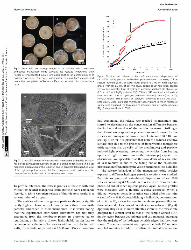
To provide reference, the release profiles of vesicles with andwithout embedded manganese oxide particles were compared(see Fig. 4, ESI†). Complete release of fluoride ions results in aconcentration of 62 ppm.
The vesicles without manganese particles showed a signifi-cantly higher release rate of fluoride ions than those withparticles embedded in their membranes. It is worth notingthat the experiments start when chloroform has not fullyevaporated from the membrane phase. Its presence led toretardation, as initially a thicker hydrophobic barrier has tobe overcome by the ions. For vesicles without particles in theirwalls, this retardation period was 20–30 min. Once chloroform
had evaporated, the release rate reached its maximum andstarted to decelerate as the concentration difference betweenthe inside and outside of the vesicles decreased. Strikingly,the chloroform evaporation process took much longer for thevesicles with manganese dioxide particles (about 100–110 min,see Fig. 4, ESI†). It is plausible that both the reduced effectivesurface area due to the presence of impermeable manganeseoxide particles (ca. 20 vol% of the membranes) and particle-induced light scattering (protecting the vesicles from heatingup due to light exposure under the microscope) explain thisobservation. We speculate that the slow down of release afterca. 300 minutes is due to the fading out of the chloroformplasticization effect, making the vesicle membranes less fluid-like.
The release behaviour of the manganese oxide vesiclesexposed to different hydrogen peroxide solutions was studied.For this we prepared water-based dispersions of ca. 6000vesicles containing 0.2 M sodium fluoride (6 mL of water outerphase, 0.1 mL of inner aqueous phase). Again, release profileswere measured with a fluoride selective electrode. When adiluted hydrogen peroxide solution was added after 150 min,0.2 mL of 3 wt% H2O2 (establishing a bulk H2O2 concentrationof ca. 0.1 wt%), a clear increase in membrane permeability andthus enhanced release rate of fluoride ions was observed (Fig. 4).Approximately 20–30 minutes after this stimulus, the release ratedropped to a similar level to that of the sample without H2O2
(in the region between 200 minutes and 250 minutes), indicatingthat the majority of the hydrogen peroxide trigger had been con-sumed. The same treatment was repeated at both 250 minutesand 340 minutes in order to confirm the initial observation.
Fig. 2 Dark field microscopy images of (a) vesicles with membraneembedded manganese oxide particles; (b) vesicles undergoing slowrelease of encapsulated sulfate ions upon addition of a small amount ofhydrogen peroxide. The outer water phase contains Ba2+ cations, andhence the precipitation of barium sulfate occurs, which is observed as ahaze.
Fig. 3 Cryo-SEM images of vesicles with membrane embedded manga-nese oxide particles: (a) overview image of a single frozen vesicle in ice; (b)magnified observation of the region in blue in panel (a); (c) magnified viewof the region in yellow in panel (a). The manganese oxide particles can beclearly observed to be part of the vesicular membrane.
Fig. 4 Fluoride ion release profiles of water-based dispersions ofca. 6000 MnO2 particle embedded polymersomes containing 0.2 Msodium fluoride (6 mL of water outer phase, 0.1 mL of inner aqueousphase) with (a) 3.0 mL of 30 wt% H2O2 added at 50 min (blue, dashedvertical line indicates time of hydrogen peroxide addition), (b) aliquots of0.2 mL of 3 wt% H2O2 added at 150, 250 and 340 min (red, solid verticallines indicate time of hydrogen peroxide addition), and (c) no H2O2
stimulus (black). This process of ‘‘catalytic’’ enhanced release was visua-lized clearly under dark field microscopy observations in which release ofsulfate ions triggered the formation of insoluble barium sulfate particles(Fig. 5, also see Movie in ESI†).
Materials Horizons Communication
Ope
n A
cces
s A
rtic
le. P
ublis
hed
on 2
0 A
ugus
t 201
5. D
ownl
oade
d on
12/
1/20
21 1
1:03
:19
PM.
Thi
s ar
ticle
is li
cens
ed u
nder
a C
reat
ive
Com
mon
s A
ttrib
utio
n 3.
0 U
npor
ted
Lic
ence
.View Article Online
44 | Mater. Horiz., 2016, 3, 41--46 This journal is©The Royal Society of Chemistry 2016
Again, a temporary increase in membrane permeability wasobserved. This shows that the catalytic reaction between hydrogenperoxide and the manganese oxide particles increases membranepermeability until the hydrogen peroxide is depleted (see Fig. 4).The exact mechanism of this temporary increased permeability isnot fully understood. We hypothesise that the catalytic break-down of hydrogen peroxide to dioxygen and water at the site ofthe particles induces the formation of transient openings at theinterface between the particles and the membrane, thus increas-ing membrane permeability. It is important to note that thedriving force for release depends on the difference between theconcentrations of fluoride ions inside and outside of the polymervesicles. Therefore, we also carried out blank experiments inwhich aliquots of water were added. Indeed, small enhancementsof release rates were observed in line with the induced concen-tration change. However, this effect was minor in comparisonwith the hydrogen peroxide catalytic trigger. Another point worthnoting, which could plausibly explain enhanced release, is thepotential self-destruction of a sub set of vesicles upon addition ofthe hydrogen peroxide trigger. We therefore carried out smallerscale experiments with 200 vesicles and counted the vesicles beforeand after our release experiments. Less than 4 vesicles (2%) werefound to be destroyed in each of these runs. The majority of vesiclessurvive the peroxide trigger and therefore we can conclude that thatenhanced membrane permeability was indeed achieved by thecatalytic activity of the manganese oxide particles.
One could argue that the manganese oxide particles act asstoppers, which upon peroxide activation, could pop out,hereby creating a temporary pore allowing for the observedenhancement in release of fluoride ions. ICP-OES measure-ments of the outer water phase post peroxide treatment showedno sign of presence of manganese. We therefore can confirmthat the manganese oxide particles remained associated withthe vesicle membranes (see ESI† for details).
Triggered release of the full vesicle content from hybridvesicles is possible upon exposure to high concentrations ofhydrogen peroxide (3.0 mL, 30 wt%). The formation of oxygenbubbles upon catalytic reaction causes imminent membranedeformation and rupture, followed by immediate release andself-destruction of the vesicles (Fig. 4, blue, and Fig. 5).
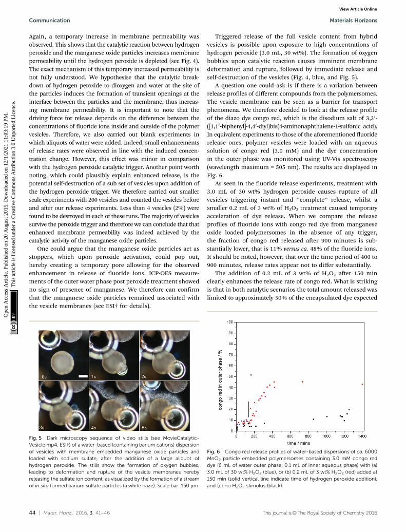
A question one could ask is if there is a variation betweenrelease profiles of different compounds from the polymersomes.The vesicle membrane can be seen as a barrier for transportphenomena. We therefore decided to look at the release profileof the diazo dye congo red, which is the disodium salt of 3,30-([1,10-biphenyl]-4,40-diyl)bis(4-aminonaphthalene-1-sulfonic acid).In equivalent experiments to those of the aforementioned fluoriderelease ones, polymer vesicles were loaded with an aqueoussolution of congo red (3.0 mM) and the dye concentrationin the outer phase was monitored using UV-Vis spectroscopy(wavelength maximum = 505 nm). The results are displayed inFig. 6.
As seen in the fluoride release experiments, treatment with3.0 mL of 30 wt% hydrogen peroxide causes rupture of allvesicles triggering instant and ‘‘complete’’ release, whilst asmaller 0.2 mL of 3 wt% of H2O2 treatment caused temporaryacceleration of dye release. When we compare the releaseprofiles of fluoride ions with congo red dye from manganeseoxide loaded polymersomes in the absence of any trigger,the fraction of congo red released after 900 minutes is sub-stantially lower, that is 11% versus ca. 48% of the fluoride ions.It should be noted, however, that over the time period of 400 to900 minutes, release rates appear not to differ substantially.
The addition of 0.2 mL of 3 wt% of H2O2 after 150 minclearly enhances the release rate of congo red. What is strikingis that in both catalytic scenarios the total amount released waslimited to approximately 50% of the encapsulated dye expected
Fig. 6 Congo red release profiles of water-based dispersions of ca. 6000MnO2 particle embedded polymersomes containing 3.0 mM congo reddye (6 mL of water outer phase, 0.1 mL of inner aqueous phase) with (a)3.0 mL of 30 wt% H2O2 (blue), or (b) 0.2 mL of 3 wt% H2O2 (red) added at150 min (solid vertical line indicate time of hydrogen peroxide addition),and (c) no H2O2 stimulus (black).
Fig. 5 Dark microscopy sequence of video stills (see MovieCatalytic-Vesicle.mp4, ESI†) of a water-based (containing barium cations) dispersionof vesicles with membrane embedded manganese oxide particles andloaded with sodium sulfate, after the addition of a large aliquot ofhydrogen peroxide. The stills show the formation of oxygen bubbles,leading to deformation and rupture of the vesicle membranes herebyreleasing the sulfate ion content, as visualized by the formation of a streamof in situ formed barium sulfate particles (a white haze). Scale bar: 150 mm.
Communication Materials Horizons
Ope
n A
cces
s A
rtic
le. P
ublis
hed
on 2
0 A
ugus
t 201
5. D
ownl
oade
d on
12/
1/20
21 1
1:03
:19
PM.
Thi
s ar
ticle
is li
cens
ed u
nder
a C
reat
ive
Com
mon
s A
ttrib
utio
n 3.
0 U
npor
ted
Lic
ence
.View Article Online

This journal is©The Royal Society of Chemistry 2016 Mater. Horiz., 2016, 3, 41--46 | 45
to be released. We found that the reason for this was the strongphysisorption of the dye to the amphiphilic block copolymer,that is poly(n-butyl methacrylate)94-block-poly(N,N-dimethyl-amino ethyl methacrylate)37. This strong adsorption, and there-fore accumulation of congo red into the vesicle membrane wall,was confirmed by confocal microscopy (see Fig. 7). For thiswe purposely buckled a polymer vesicle using an NaCl solution.As can be seen from the image, a higher intensity and thus,concentration, of the dye was observed in the wrinkled parts ofthe membrane.
Conclusions
We have demonstrated that the permeability of vesicular struc-tures containing ‘‘active’’ colloidal particles as part of theirmembrane can be regulated by use of a chemical trigger. Thismodel system offers an exciting method of regulating andcontrolling transmembrane transport, which may be of interestto a range of scientific fields.
One comment we would like to make and relates to the areaof the evolution of life is that a hybrid vesicular structure, whichhas ‘‘active’’ colloidal particles either adhered to the surface ofthe membrane, or embedded into it, can achieve chemicallyactivated control of membrane permeability. This is what wehave shown here.
The origin of biological membranes remains ambiguous.Wachtershauser30,31 and Russell and coworkers,32 for example,argue that porous pyrite (FeS) structures found in deep-seahydrothermal vents contain small pockets of water and havethe ability to accumulate organic matter due to the relativehydrophobic nature of pyrite. The hypothesis is that thesecompartmentalized structures are suitable microenvironmentsfor life to evolve and, therefore, can be seen as semi-permeablepredecessors to protocells.‡ They are formed through precipita-tion and assembly of metal sulfides on the surface of hydrogensulfide rich gas bubbles, another example being based on zincsulfide.33 A modern synthetic analogue of such semipermeable
compartmentalized structures would be a colloidosome,34
which can be built from colloidal particles that adhere toliquid–liquid, or liquid–gas interfaces, so-called Pickeringstabilizers.35 The next step in evolution of these early com-partmentalised structures describes the formation of a cellmembrane in the form of liposomes, and here is where theproblem lies. How would a primitive cell get its nutrients whenmodern model lipid-based cell membranes are effectively non-permeable for desired molecules on an acceptable time scale,if membrane-proteins are left out?36 Mansy and coworkerssuggested that protocells made with fatty acid componentswere able to take up nucleotides through concerted flipping,37
fast at high temperatures, and that in addition DNA strandseparation processes benefited from thermal fluctuations.38 Wewould like to suggest here that a hybrid vesicular structure,which has ‘‘active’’ colloidal particles as part of its membrane,may have controlled permeability in primitive cells.
Notes and References‡ Protocells, the predecessors to modern cells, possess a self-replicatinggenome encapsulated by a membrane that can grow and divide, thusallowing it replicate. Unlike a cell, however, a protocell mediates its celluptake not with sophisticated membrane transport control, but bydiffusion through the membrane alone. The evolution of these primitivecell analogues is a hot topic of debate amongst evolutionary scientists,with particular focus on the requirements of self-replication and cellfunction.32,33
1 Origins of Life: The Primal Self-Organization, ed. R. Egel,D. H. Lankenau, A. Y. Mulkidjanian and Y. Armen, Springer,Berlin, 2011, ch. 1.
2 S. J. Singer and G. L. Nicolson, Science, 1972, 175, 720–731.3 C. Nardin, S. Thoeni, J. Widmer, M. Winterhalter and
W. Meier, Chem. Commun., 2000, 1433–1434.4 M. Kumar, M. Grzelakowski, J. Zilles, M. Clark and W. Meier,
Proc. Natl. Acad. Sci. U. S. A., 2007, 104, 20719–20724.5 H. Choi and C. D. Montemagno, Nano Lett., 2005, 5, 2538–2542.6 A. Graff, M. Sauer, P. Van Gelder and W. Meier, Proc. Natl.
Acad. Sci. U. S. A., 2002, 99, 5064–5068.7 A. J. Kim, M. S. Kaucher, K. P. Davis, M. Peterca, M. R.
Imam, N. A. Christian, D. H. Levine, F. S. Bates, V. Percecand D. A. Hammer, Adv. Funct. Mater., 2009, 19, 2930–2936.
8 M. Danial, C. M. N. Tran, K. A. Jolliffe and S. Perrier, J. Am.Chem. Soc., 2014, 136, 8018–8026.
9 N. Madhavan, E. C. Robert and M. S. Gin, Angew. Chem.,Int. Ed., 2005, 44, 7584–7587.
10 D. Papahadjopoulos, K. Jacobson, S. Nir and T. Isac,Biochim. Biophys. Acta, 1973, 311, 330–348.
11 M. B. Yatvin, J. N. Weinstein, W. H. Dennis andR. Blumenthal, Science, 1978, 202, 1290–1293.
12 J. Gaitzsch, D. Appelhans, L. Wang, G. Battalgia and B. Voit,Angew. Chem., 2012, 51, 4448–4451.
13 A. Napoli, M. Valentini, N. Tirelli, M. Muller andJ. A. Hubbell, Nat. Mater., 2004, 3, 183–189.
14 A. Napoli, M. J. Boerakker, N. Tirelli, R. J. M. Nolte,N. A. J. M. Sommerdijk and J. A. Hubbell, Langmuir, 2004,20, 3487–3491.
Fig. 7 Confocal microscope image of a partially buckled polymer vesicleloaded with 0.003 M congo red dye solution, the polymersome beingdispersed in an aqueous NaCl environment. Scale bar: 50 mm.
Materials Horizons Communication
Ope
n A
cces
s A
rtic
le. P
ublis
hed
on 2
0 A
ugus
t 201
5. D
ownl
oade
d on
12/
1/20
21 1
1:03
:19
PM.
Thi
s ar
ticle
is li
cens
ed u
nder
a C
reat
ive
Com
mon
s A
ttrib
utio
n 3.
0 U
npor
ted
Lic
ence
.View Article Online

46 | Mater. Horiz., 2016, 3, 41--46 This journal is©The Royal Society of Chemistry 2016
15 F. Ahmed and D. E. Discher, J. Controlled Release, 2004, 96,37–53.
16 J. Du and S. P. Armes, J. Am. Chem. Soc., 2005, 127,12800–12801.
17 S. Yu, T. Azzam, I. Rouiller and A. Eisenberg, J. Am. Chem.Soc., 2009, 131, 10557–10566.
18 K. T. Kim, J. J. L. M. Cornelissen, R. J. M. Nolte andJ. C. M. van Hest, Adv. Mater., 2009, 21, 2787–2791.
19 B. G. De Geest, S. De Koker, J. Demeester, S. C. De Smedtand W. E. Hennink, Polym. Chem., 2010, 1, 137–148.
20 S. H. Park, S. G. Oh, J. Y. Mun and S. S. Han, Colloids Surf.,B, 2005, 44, 117–122.
21 S. H. Park, S. G. Oh, J. Y. Mun and S. S. Han, Colloids Surf., B,2006, 48, 112–118.
22 L. Paasonen, T. Laaksonen, C. Johans, M. Yliperttula,K. Kontturi and A. Urtti, J. Controlled Release, 2007, 122,86–93.
23 E. Amstad, S. H. Kim and D. A. Weitz, Angew. Chem., Int. Ed.,2012, 51, 12499–12503.
24 S. Lecommandoux, O. Sandre, F. Checot, J. Rodriguez-Hernandez and R. Perzynski, Adv. Mater., 2005, 17,712–718.
25 S. Lecommandoux, O. Sandre, F. Checot and R. Perzynski,Prog. Solid State Chem., 2006, 34, 171–179.
26 M. Krack, H. Hohenberg, A. Kornowski, P. Lindner, H. Wellerand S. Forster, J. Am. Chem. Soc., 2008, 130, 7315–7320.
27 C. Sanson, O. Diou, J. Thevenot, E. Ibarboure, A. Soum,A. Brulet, S. Miraux, E. Thiaudiere, S. Tan, A. Brisson,V. Dupuis, O. Sandre and S. Lecommandoux, ACS Nano,2011, 5, 1122–1140.
28 E. Amstad, J. Kohlbrecher, E. Muller, T. Schweizer,M. Textor and E. Reimhult, Nano Lett., 2011, 11, 1664–1670.
29 Y. Chen, A. Bose and G. D. Bothun, ACS Nano, 2010, 4,3215–3221.
30 G. Wachtershauser, Syst. Appl. Microbiol., 1988, 10, 207–210.31 G. Wachtershauser, Proc. Natl. Acad. Sci. U. S. A., 1994, 91,
4283–4287.32 M. J. Russell, A. J. Hall, A. G. Cairns-Smith and
P. S. Braterman, Nature, 1988, 336, 117.33 A. Y. Mulkidjanian, Biol. Direct, 2009, 4, 26.34 A. D. Dinsmore, M. F. Hsu, M. G. Nikolaides, M. Marquez,
A. R. Bausch and D. A. Weitz, Science, 2002, 298, 1006–1009.35 S. U. Pickering, J. Chem. Soc., Trans., 1907, 91, 2001–2021.36 D. W. Deamer, Nature, 2008, 454, 37–38.37 S. S. Mansy, J. P. Schrum, M. Krishnamurthy, S. Tobe,
D. A. Treco and J. W. Szostak, Nature, 2008, 454, 122–125.38 S. S. Mansy and J. W. Szostak, Proc. Natl. Acad. Sci. U. S. A.,
2008, 105, 13351–13355.
Communication Materials Horizons
Ope
n A
cces
s A
rtic
le. P
ublis
hed
on 2
0 A
ugus
t 201
5. D
ownl
oade
d on
12/
1/20
21 1
1:03
:19
PM.
Thi
s ar
ticle
is li
cens
ed u
nder
a C
reat
ive
Com
mon
s A
ttrib
utio
n 3.
0 U
npor
ted
Lic
ence
.View Article Online