convalescent plasma for the treatment of patients with
TRANSCRIPT
Received: 8 July 2020 | Revised: 10 August 2020 | Accepted: 16 September 2020
DOI: 10.1002/jmv.26537
R E S E A RCH AR T I C L E
Convalescent plasma for the treatment of patients withsevere coronavirus disease 2019: A preliminary report
Ali S. Omrani1,2 | Ahmed Zaqout1,2 | Anas Baiou3 | Joanne Daghfal2 |
Naser Elkum4 | Rand A. Alattar2 | Dana Bakdach5 | Hatem Abusriwil6 |
Abdalrahman M. Mostafa6 | Bassem Alhariri6 | Naseem Ambra6 |
Mohamed Khatib7 | Ali M. Eldeeb8 | Zeyd Merenkov9 | Zeinab Fawzi9 |
Saloua M. Hmissi9 | Ali A. Hssain5 | Peter V. Coyle10 | Hussam Alsoub1,2 |
Muna A. Almaslamani1,2 | Abdullatif Alkhal1,2
1Division of Infectious Diseases, Department
of Medicine, Hamad Medical Corporation,
Doha, Qatar
2Communicable Diseases Center, Hamad
Medical Corporation, Doha, Qatar
3Division of Critical Care, Department of
Medicine, Hamad Medical Corporation,
Doha, Qatar
4Research Department, Sidra Medical,
Doha, Qatar
5Medical Intensive Care Unit, Hamad Medical
Corporation, Doha, Qatar
6Department of Internal Medicine, Hazm
Mebaireek General Hospital, Hamad Medical
Corporation, Doha, Qatar
7Critical Care and Pulmonary Medicine, Hazm
Mebaireek General Hospital, Hamad Medical
Corporation, Doha, Qatar
8Department of Clinical Imaging, Hamad
Medical Corporation, Doha, Qatar
9Transfusion Medicine, Department of
Pathology and Laboratory Medicine, Hamad
Medical Corporation, Doha, Qatar
10Division of Virology, Department of
Pathology and Laboratory Medicine, Hamad
Medical Corporation, Doha, Qatar
Correspondence
Ali S. Omrani, Communicable Diseases
Center, Hamad Medical Corporation,
PO Box 3050, Doha, Qatar.
Email: [email protected]
Abstract
Background: The role of convalescent plasma therapy for patients with coronavirus
disease 2019 (COVID‐19) is unclear.
Methods: We retrospectively compared outcomes in a cohort of critical COVID‐19patients who received standard care (SC Group) and those who, in addition,
received convalescent plasma (CP Group).
Results: In total, 40 patients were included in each group. The median patient age
was 53.5 years (interquartile range [IQR] 42–60.5), and the majority of patients
required invasive ventilation (69, 86.2%). Plasma was harvested from donors after a
median of 37 days (IQR 31–46) from the first positive severe acute respiratory
syndrome coronavirus 2 (SARS‐CoV‐2) polymerase chain reaction (PCR) result and
26 days (IQR 21–32) after documented viral clearance; it was administered after a
median of 10 days (IQR 9–10) from the onset of symptoms and 2.5 days (IQR 2–4)
from admission to intensive care unit. The primary endpoint of improvement in
respiratory support status within 28 days was achieved in 26 patients (65%) in the
SC Group and 31 patients (77.5%) in the CP Group (p = .32). The 28‐day all‐causemortality (12.5% vs. 2.5%; p = .22) and viral clearance (65% vs. 55%; p = .49) were
not significantly different between the two groups. Convalescent plasma was not
significantly associated with the primary endpoint (adjusted hazard ratio 0.87; 95%
confidence interval 0.51–1.49; p = .62). Adverse events were balanced between the
two study groups.
Conclusion: In severe COVID‐19, convalescent plasma therapy was not associated
with clinical benefits. Randomized trials are required to confirm our findings.
K E YWORD S
convalescent plasma, coronavirus, COVID‐19, passive immunotherapy, SARS‐CoV‐2
J Med Virol. 2021;93:1678–1686.1678 | wileyonlinelibrary.com/journal/jmv
This is an open access article under the terms of the Creative Commons Attribution‐NonCommercial License, which permits use, distribution and reproduction in
any medium, provided the original work is properly cited and is not used for commercial purposes.
© 2020 The Authors. Journal of Medical Virology published by Wiley Periodicals LLC

1 | INTRODUCTION
The total global number of individuals diagnosed with coronavirus
disease 2019 (COVID‐19), caused by the novel betacoronavirus se-
vere acute respiratory syndrome coronavirus 2 (SARS‐CoV‐2), hassurpassed 10million, with more than 500,000 associated deaths.1 Up
to 10% of COVID‐19 patients may develop organ failure and may
require admission to an intensive care unit (ICU).2 Several investiga-
tional antiviral agents are currently being evaluated for the treatment
of patients with severe and critical COVID‐19.2 Thus far, hydroxy-
chloroquine, with or without azithromycin, lopinavir–ritonavir, and
remdesivir have not been shown to be associated with a clear clinical
benefit in patients with severe COVID‐19.3–5
In patients with severe influenza, convalescent plasma was asso-
ciated with improved viral clearance and reduced mortality.6 Similarly,
convalescent plasma therapy for patients with severe acute respiratory
syndrome (SARS) shortened hospital stays and lowered overall mortal-
ity.7 Convalescent plasma may be useful in the treatment of severe
COVID‐19. However, peer‐reviewed clinical data are limited.8 Using a
cohort of patients with severe COVID‐19, we aimed to assess the clinical
benefits of convalescent plasma over and above standard care.
2 | METHODS
2.1 | Study design and patients
Hamad Medical Corporation (HMC) provides medical care for all
COVID‐19 patients in Qatar. SARS‐CoV‐2 infection was confirmed
on respiratory samples using reverse transcription polymerase chain
reaction (RT‐PCR) assays TaqPath COVID‐19 Combo Kit (Thermo
Fisher Scientific) or Cobas SARS‐CoV‐2 Test (Roche Diagnostics).
COVID‐19 is classified as severe if any one or more of the fol-
lowing is present: respiratory rate >30/min, oxygen saturation ≤90%
while in ambient room air, partial pressure of oxygen–oxygen con-
centration (PaO2/FiO2) ≤ 300mmHg, hypotension, or any organ
failure.2 Standard care for patients with severe COVID‐19 in HMC
involved supportive care and antiviral therapy with hydroxy-
chloroquine, azithromycin, and/or lopinavir–ritonavir. Tocilizumab
could be added for those with evidence of significant systemic in-
flammation and methylprednisolone for acute respiratory distress
syndrome (ARDS). Individual regimens were selected by the at-
tending clinical teams based on patients' characteristics and needs.
From April 13, 2020, convalescent plasma derived from re-
covered COVID‐19 patients was available for clinical use for patients
with severe COVID‐19. Plasma donors are adults whose COVID‐19symptoms have resolved more than 2 weeks ago, with documented
negative upper airway SARS‐CoV‐2 RT‐PCR and negative serological
tests for syphilis and blood‐borne viruses.
We retrospectively included patients from HMC COVID‐19database. The convalescent plasma group (CP Group) included
patients who received SARS‐CoV‐2 convalescent plasma therapy
within the first 7 days of admission to ICU, required mechanical
ventilation, and completed 28 days of follow‐up by June 1, 2020.
Criteria for inclusion in the comparator group, the standard care
(SC Group), included laboratory‐confirmed SARS‐CoV‐2 infec-
tion, need for mechanical ventilation in ICU, and completion of
28 days of follow‐up by June 1, 2020. Starting from the earliest
case of SARS‐CoV‐2 in Qatar, consecutive patients eligible for
inclusion in the SC Group were selected until one control was
identified for each individual in the CP Group.
2.2 | Procedures
Donor apheresis was performed using Trima Accel Automated Blood
Collection System (Terumo BCT) and inactivated using Mirasol PRT
System (Terumo BCT). Each recipient received a total of 400ml of
ABO‐compatible convalescent plasma. Donation and receipt of
plasma were subject to HMC standard clinical policies. SARS‐CoV‐2antibody titers in the donated plasma were not available. The date on
which a patient was admitted to ICU was designated as study day 1.
All outcomes were recorded on the basis of the patients' disposition
up to study day 28. Data including outcomes and adverse events
were retrieved retrospectively from the electronic healthcare re-
cords. Radiological findings were reviewed and reported by a single
radiologist who was blinded to the group allocation.
The results are reported according to the recommendations of
STROBE initiative.9 The study was approved by HMC's Institutional
Review Board (MRC0120191), with a waiver of informed consent.
2.3 | Outcomes
The primary outcome was improvement in the respiratory status,
defined as two‐category improvement on a six‐level ordinal scale.4
The scale in a descending order was death, invasive mechanical
ventilation, noninvasive ventilation (NIV) or high‐flow nasal oxygen
(HFNO) therapy, oxygen therapy other than NIV or HFNO, hospi-
talization without need for oxygen therapy, and discharge home or a
community isolation facility. Secondary outcomes were as fol-
lows: discharged alive from ICU by study day 28 and viral clearance,
defined as two consecutive negative RT‐PCR tests on airway samples
taken within a gap of more than 24 hours.
2.4 | Statistical analysis
In the absence of data on treatment effect, the sample size for
the study was arbitrarily set at 40 patients in each group. This
sample size provides approximately 80% power to detect an increase
in the primary endpoint from 30% in the SC Group to 65% in the CP
Group, at a two‐sided significance level of .05.
Categorical data were summarized as numbers (percentages)
and compared using Fisher's exact test, whereas continuous data
were presented as medians and interquartile ranges (IQR), and
OMRANI ET AL. | 1679

compared using Wilcoxon rank‐sum test. Cox proportional hazard
was used to identify covariables associated with time to improve-
ment in respiratory support status. To avoid overfitting the model,
given the sample size and the number of events in the study, the
number of variables included in the multivariate model was limited to
four. In addition to receipt of convalescent plasma therapy and need
for invasive mechanical ventilation, Acute Physiology and Chronic
Health Evaluation (APACHE II) score was included due to its es-
tablished role as a prognostic tool in critically ill patients.10 The
fourth variable in the multivariable Cox model was receipt of sys-
temic corticosteroid therapy, which was recently reported to be
associated with survival benefit in patients with severe COVID‐19.11
Kaplan–Meier survival curve with log‐rank p value was used to
compare time to improvement in respiratory status between the two
study groups.
All p values were two‐sided with a threshold of <.05 for statis-
tical significance. Statistical analyses were performed using Stata
Statistical Software Release 15.1 (StataCorp LLC).
3 | RESULTS
Eighty individuals were included, with 40 in each group. The majority
were males (69, 86.3%) and the median age was 53.5 years (IQR
42–60.5). Diabetes mellitus (40, 50%) and hypertension (32, 40%)
were common, and median body mass index (BMI) was 27.4 kg/m2
(IQR 25–32.4). The median duration between the onset of symptoms
and admission to ICU was 7 days (IQR 6–9). Cough (73, 91.2%) and
fever (68, 85.0%) were the most frequent presenting symptoms. The
median APACHE II score was 12 (IQR 9–12), and all patients had
pulmonary infiltrates on their chest radiographic images (Table 1).
During their ICU stay, the majority of patients required invasive
mechanical ventilation (69, 86.2%), and/or vasopressor support (42,
52.5%). All patients received hydroxychloroquine, azithromycin, and
lopinavir–ritonavir, whereas tocilizumab was given to 73 patients
(91.2%) and intravenous methylprednisolone to 66 patients (82.5%).
Compared with the CP Group, the SC Group had higher median
serum creatinine at admission to ICU (90 vs. 81 µmol/L, p = .009).
There were no other significant baseline differences between the
two groups (Table 1).
The median donor age was 36.5 years (IQR 26.3–41). All donors
had infiltrates in their chest X‐rays. Seventeen donors (42.5%) re-
quired oxygen via nasal canulae, nine (22.5%) required noninvasive
ventilation, and three (7.5%) required invasive mechanical ventilation.
Plasma was harvested after a median of 37 days (IQR 31–46, range
26–56) from the first positive SARS‐CoV‐2 PCR result and 26 days
(IQR 21–32, range 14–45) after documented viral clearance; it was
administered after a median of 10 days (IQR 9–10) from the onset of
symptoms and 2.5 days (IQR 2–4, range 1–7) from admission to ICU.
Adverse events occurred in 77 patients (96.3%), including grade 4
in eight, seven of which were from the SC Group. The frequency of
adverse events was comparable between the two groups (Table 1).
Overall, the primary endpoint of improvement in respiratory
support status was achieved in 57 patients (71.3%): 26 (65%) in the
SC Group and 31 (77.5%) in the CP Group (p = .32). There were no
significant differences between the SC Group and CP Group in the
proportions of patients who were discharged alive from ICU within
28 days (65% for both groups; p > .99), viral clearance (65% vs. 55%;
p = .49), or the 28‐day all‐cause mortality (12.5% vs. 2.5%; p = .22;
Table 1).
Compared with patients in whom improvement in respiratory
status was documented within 28 days, patients in whom it was not
achieved were significantly older (median age 60 vs. 48 years;
p < .001), had higher APACHE II scores (13 vs. 11; p = .017), and were
more likely to have required hemodialysis (21.7% vs. 2 (3.5%;
p = .019). Convalescent plasma recipients constituted 54.4% of those
patients in whom the primary endpoint was achieved, compared with
39.1% of those in whom it was not achieved (p = .32; Table 2).
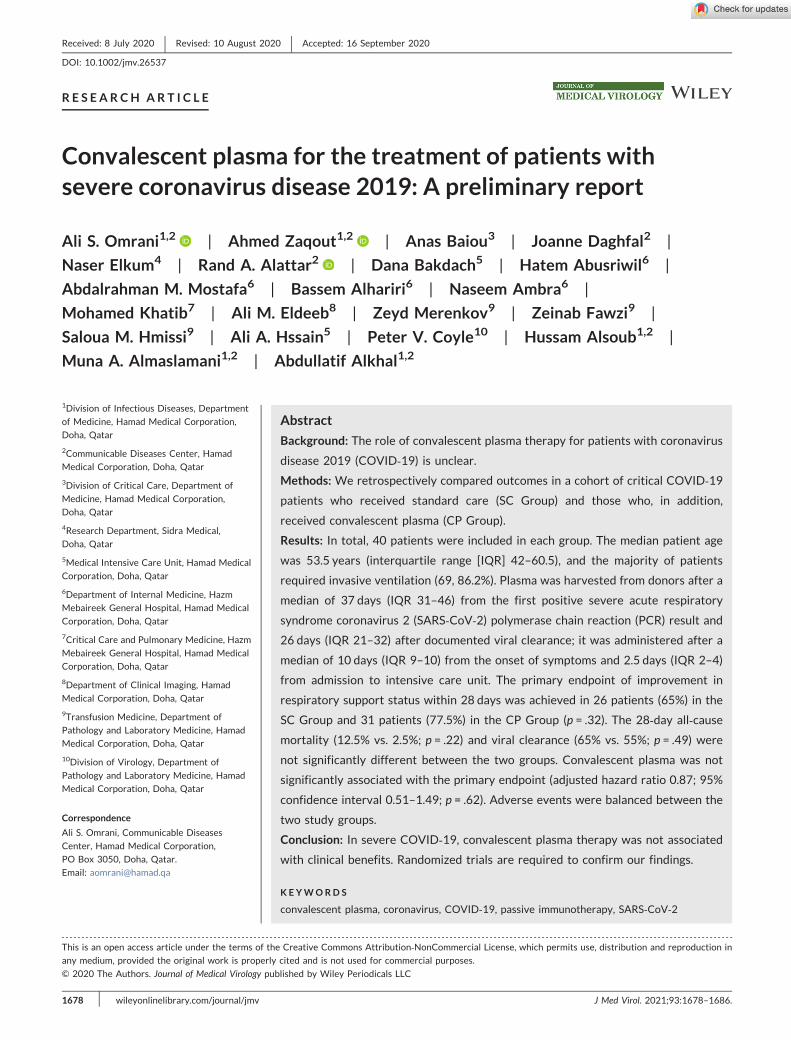
Time to improvement in the respiratory support status was not
significantly different between the two study groups (p = .99;
Figure 1). In the multivariable Cox model, higher APACHE II score
(adjusted hazard ratio [aHR], 0.89; 95% confidence interval [CI],
0.83–0.95; p = .001) and receipt of methylprednisolone (aHR, 0.23;
95% CI 0.11–0.50; p < .001) were independently associated with a
lower likelihood of achieving improvement in respiratory support
status within 28 days. Receipt of convalescent plasma was not as-
sociated with time to improvement in the respiratory support status
(aHR, 0.87; 95% CI, 0.51–1.49; p = .622; Table 3).
4 | DISCUSSION
We report 28‐day outcomes for patients with severe COVID‐19requiring mechanical ventilation in ICU. The administration of con-
valescent plasma after a median of 10 days from the symptom onset
was not associated with an increased likelihood of improvement in
the respiratory support status. Our results are consistent with those
recently reported in an underpowered randomized clinical trial
where the administration of convalescent plasma did not improve
time to clinical improvement in patients with critical COVID‐19.12
There are several possible explanations for our findings. First,
the majority of patients in this study (86.2%) required invasive
ventilation. Severe COVID‐19 is largely driven by a dysregulated
inflammatory response.13 SARS‐CoV‐2‐neutralizing antibodies may
not be adequate to reverse the immunological cascade and result in
clinical improvement.14 Indeed, earlier administration of con-
valescent plasma for patients with SARS was associated with fa-
vorable clinical outcomes.7 Furthermore, a meta‐analysis of eight
studies (1703 patients) found that convalescent plasma for severe
influenza within less than 4 days of pneumonia complications was
associated with all‐cause mortality rate of 19%, as opposed to 59% in
those who started treatment at a later point in their illness.15
However, five out of six patients with critical COVID‐19 who re-
ceived convalescent plasma after a median of 21.5 days from
1680 | OMRANI ET AL.

TABLE 1 Baseline characteristics of study groups
Variable Total (n = 80)
Standard care
group (n = 40)
Convalescent plasma
group (n = 40) p value
Demographics and comorbidities
Male sex 69 (86.3%) 35 (87.5%) 34 (85%) >.99
Arab ethnicity 22 (27.5%) 15 (37.5%) 7 (17.5%) .078
Age (years) 53.5 (42–60.5) 55.5 (46.5–60.5) 47.5 (39–60.5) .073
Body mass index (kg/m2) 27.4 (25–32.4) 28.5 (24.7–32.4) 27.2 (25.6–29.3) .68
Diabetes mellitus 40 (50%) 20 (50%) 20 (50%) >.99
Hypertension 32 (40%) 19 (47.5%) 13 (32.5%) .25
Chronic kidney disease 6 (7.5%) 4 (10%) 2 (5%) .68
Chronic lung disease 5 (6.2%) 2 (5%) 3 (7.5%) >.99
Current or past smokera 12 (15%) 9 (22.5%) 3 (7.5%) .12
Presenting symptoms
Symptoms at ICU admission (days) 7 (6–9) 7 (5.5–9) 7 (6–8.5) .85
Fever 68 (85%) 36 (90%) 32 (80%) .35
Cough 73 (91.2%) 35 (87.5%) 38 (95%) .43
Sore throat 20 (25%) 9 (22.5%) 11 (27.5%) .80
Dyspnea 58 (72.5%) 26 (65%) 32 (80%) .21
Oxygen saturation at admission to ICU 91% (85–95%) 94% (85–96%) 89% (86–94%) .059
APACHE II Score 12.0 (9.0–14.0) 12.0 (9.0–15.0) 11.5 (9.0–13.0) .23
Laboratory results at admission to ICU
Peripheral white cell count (×109/L) 8.0 (5.3–10.4) 8.0 (6.3–11.5) 7.2 (5.0–9.6) .28
Lymphocyte count (×109/L) 0.9 (0.6–1.2) 0.90 (0.7–1.3) 0.9 (0.6–1.1) .43
Platelet count (×109/L) 234 (190–300) 248.5 (200–300) 219.5 (185–281.5) .34
Serum creatinine (µmol/L) 86 (70.5–106) 90 (80–119) 81.0 (63.5–91.5) .009
CRP (mg/L) 180 (103–259) 180 (110.0–300.0) 172.5 (93–231.4) .16
Serum ferritin (µg/L)b 940 (671–1484) 1200 (595.5–1571.5) 920.5 (697.5–1288) .61
ALT (IU/L) 38.5 (24–60) 40 (24–69) 37 (21.5–55) .48
Management
Invasive mechanical ventilation 69 (86.2%) 36 (90%) 33 (82.5%) .52
Vasopressor support 42 (52.5%) 22 (55%) 20 (50%) .82
Hemodialysis 7 (8.7%) 5 (12.5%) 2 (5.0%) .43
Methylprednisolone 66 (82.5%) 31 (78%) 35 (88%) .38
Tocilizumab 73 (91.2%) 34 (85%) 39 (98%) .11
Adverse events
Acute kidney injury 29 (36.2%) 16 (40%) 13 (32.5%) .64
Anemia 48 (60.0%) 20 (50%) 28 (70%) .11
ALT rise 66 (82.5%) 35 (87.5%) 31 (77.5%) .38
Bilirubin rise 28 (35.0%) 13 (32.5%) 15 (37.5%) .81
Hypernatremia 31 (38.7%) 13 (32.5%) 18 (45.0%) .36
Hypokalemia 16 (20%) 7 (17.5%) 9 (22.5%) .78
QTc prolongation 13 (16.2%) 5 (12.5%) 8 (20%) .55
Outcomes
Improvement in respiratory support status 57 (71.3%) 26 (65%) 31 (77.5%) .32
Discharged alive from ICU within 28 days 52 (65%) 26 (65%) 26 (65%) >.99
The 28‐day all‐cause mortality 6 (7.5%) 5 (12.5%) 1 (2.5%) .22
Viral clearance 48 (60%) 26 (65%) 22 (55%) .49
(Continues)
OMRANI ET AL. | 1681

TABLE 1 (Continued)
Variable Total (n = 80)
Standard care
group (n = 40)
Convalescent plasma
group (n = 40) p value
Respiratory support at outcome .42
Ambient room air 9 (11.3%) 3 (7.5%) 6 (15%)
Supplemental oxygen 50 (62.5%) 25 (62.5%) 25 (62.5%)
Noninvasive ventilation 1 (1.3%) 0 1 (2.5%)
Invasive ventilation 20 (25%) 12 (30%) 8 (20%)
Note: Data are presented as numbers (percentages) or median (interquartile range). p values are based on Fisher's exact or Wilcoxon rank‐sum tests.
Abbreviations: ALT, alanine transaminase; AST, aspartate transaminase; CRP, C‐reactive protein; ICU, intensive care unit; IQR, interquartile range;
LDH, lactate dehydrogenase.aData missing for three patients.bData missing for eight patients.
TABLE 2 Baseline characteristics with respiratory status improvement at day 28 of admission to ICU
Variable
No
improvement (n = 23) Improvement (n = 57) p value
Demographics and comorbidities
Male sex 20 (87%) 49 (86%) >.99
Arab ethnicity 6 (26.1%) 16 (28.1%) >.99
Age (years) 60 (55–66) 48 (41–59) <.001
Body mass index (kg/m2) 27 (25–32) 27.8 (24.91–32.87) .84
Diabetes mellitus 14 (60.9%) 26 (45.6%) .32
Hypertension 12 (52.2%) 20 (35.1%) .21
Chronic kidney disease 1 (4.3%) 5 (8.8%) .67
Chronic lung disease 1 (4.3%) 4 (7.0%) >.99
Current of past smokera 4 (17.4%) 8 (14.0%) .73
Onset of symptoms at ICU
admission (days)
7 (4–8) 7 (6–9) .085
Oxygen saturation at admission to ICU 90% (85%–95%) 90% (86–95%) .77
APACHE II Score 13 (10–20) 11 (8–13) .017
Laboratory results on the day of
admission to ICU
Peripheral white cell count (×109/L) 8.8 (6.4–13.8) 7.2 (5.10–9.6) .079
Lymphocyte count (×109/L) 1.0 (0.6–1.5) 0.9 (0.6–1.0) .39
Platelet count (×109/L) 238 (190–300) 233 (190–300) .86
Serum creatinine (µmol/L) 103 (73–130) 84 (70–95) .10
CRP (mg/L) 186.3 (129.1–300) 175 (92.8–240.9) .11
Serum ferritin (µg/L)b 1293 (858.5–1847) 842 (657.5–1304) .025
ALT (IU/L) 31.5 (20–59) 39 (29.5–60) .52
Management
Convalescent plasma 9 (39.1%) 31 (54.4%) .32
Invasive mechanical ventilation 21 (91.3%) 49 (86.0%) .72
Vasopressor support 14 (60.9%) 28 (49.1%) .46
Hemodialysis 5 (21.7%) 2 (3.5%) .019
Methylprednisolone 21 (91.3%) 45 (78.9%) .33
Tocilizumab 21 (91.3%) 52 (91.2%) >.99
Note: Data are presented as numbers (percentages) or median (interquartile range). p values are based on Fisher's exact or Wilcoxon rank‐sum tests.
Abbreviations: ALT, alanine transaminase; AST, aspartate transaminase; CRP, C‐reactive protein; ICU, intensive care unit; LDH, lactate dehydrogenase.aData missing for three patients.bData missing for eight patients.
1682 | OMRANI ET AL.
molecular confirmation could not survive.16 To maximize potential
clinical benefit, SARS‐CoV‐2 convalescent plasma may need to be
administered earlier in the clinical course. To facilitate patient se-
lection, robust COVID‐19 severity prediction tools are required.
Examples exist, but none of them have been widely validated.17
A second possible explanation for the apparent lack of clinical
benefit in association with SARS‐CoV‐2 convalescent plasma in this
study is the patients' characteristics. The median patient age in this
study was 53.5 years, and only 7.5% of patients died within 28 days
of follow‐up. This contrasts with experiences reported elsewhere.
For example, an Italian report described 1591 patients with critical
COVID‐19. The median age was 63 (IQR 56–70) and the overall ICU
mortality was 26%.18 Another study from the United States reported
50% all‐cause mortality in 24 patients with critical COVID‐19 who
had a mean age of 64 years.19 Our substantially lower mortality rate
suggests that any clinical benefit with convalescent plasma, if one
exists, may be too small to be detected in our setting using our
relatively small sample size.
Third, we were unable to establish that our donors' plasma
contained adequate SARS‐CoV‐2‐neutralizing antibody titers, a pre-
requisite for effective convalescent plasma therapy.20 However,
donations were obtained after a median of 37 days (IQR 31–46) after
the date of COVID‐19 confirmation by RT‐PCR. It has been shown
that immunoglobulin M (IgM) and IgG antibodies against the
receptor‐binding domain (anti‐RBD) of SARS‐CoV‐2 are detectable in
nearly all COVID‐19 patients after 14 days of the clinical onset.21
Moreover, the presence of such antibodies strongly correlated
with SARS‐CoV‐2‐neutralizing antibody titers.21,22 In one study,
SARS‐CoV‐2‐neutralizing antibodies were undetectable in only 5.7%
of 175 individuals who recovered from mild COVID‐19.22 Con-
sidering this low percentage and the extended time frame between
infection and donation in our study, it is reasonable to suggest that
the probability that our donors did not have adequate neutralizing
SARS‐CoV‐2 antibodies is low. However, this possibility was not fully
excluded.
In this study, the baseline APACHE II score, a well‐establishedprognostic tool for critically ill patients, was negatively associated
with the primary endpoint. In addition, the use of methylpredni-
solone was independently associated with a lower likelihood of
treatment success. Current COVID‐19 management guidelines do
not recommend the routine use of systemic corticosteroids.2 This
recommendation is the result of consistent evidence of lack of
clinical benefit in settings of viral pneumonia and possible harm
including delayed viral clearance and increased mortality.23 How-
ever, dexamethasone at a dose of 6 mg daily for up to 10 days was
recently reported to be associated with significant survival benefit
in patients with moderate‐to‐severe COVID‐19.11 It has been sug-
gested that systemic steroids are probably beneficial in severe
COVID‐19 when administered at lower doses, but are potentially
harmful at higher doses.24 Our finding emphasizes the importance
of carefully selecting COVID‐19 patients for systemic steroid
therapy and the need for judicious selection of the specific treat-
ment agent and dosing regimen.
Adverse events occurred in the majority of patients in this
study. This is not surprising, given their critical status and the
multitude of medical interventions employed for both groups.
F IGURE 1 The Kaplan–Meier survival curve for time to improvement in the respiratory support status
OMRANI ET AL. | 1683

However, convalescent plasma appeared to be safe and was not
associated with excess adverse events. Modern transfusion medicine
techniques have substantially improved the safety of blood product
therapies. However, there remain some potential risks with con-
valescent plasma therapy that require careful weighing against any
possible benefits. It remains unexplored whether passive im-
munotherapy for COVID‐19 results in an attenuated natural
SARS‐CoV‐2 immune response and post‐recovery susceptibility to
SARS‐CoV‐2 re‐infection, and the potential risk of antibody‐dependent enhancement, a detrimental immunological phenomenon
previously described with other viral infections.20,25 In addition, the
risk of blood product‐derived infections, including currently
unrecognized agents, can never be completely eliminated.26
The limitations of this study include its observational nature. We
used multivariate analyses to assess independent associations with
the study endpoints, but the risk of residual confounding by indica-
tion cannot be eliminated. Nonetheless, existing peer‐reviewed data
in this area are mostly limited to uncontrolled case series of two to
ten patients and a single prematurely halted randomized trial.12,27–31
We, therefore, believe that our report, despite the limitations dis-
cussed above, provides useful insight for physicians considering the
use of SARS‐CoV‐2 convalescent plasma for the treatment of
patients with COVID‐19.In conclusion, in this retrospective cohort study of critically ill
COVID‐19 patients, convalescent plasma therapy was not asso-
ciated with clinical benefit in terms of improvement in the re-
spiratory support status within 28 days. It is not clear if the
administration of convalescent plasma at an earlier stage may
prevent the clinical progression of COVID‐19 and result in better
clinical outcomes. Randomized clinical trials are urgently required
to address these questions.
ACKNOWLEDGMENTS
Open access publication of this article was funded by Qatar National
Library. No other funding was required.
CONFLICT OF INTERESTS
The authors declare that there are no conflict of interests.
TABLE 3 Cox proportional hazard for associations with improvement in the respiratory support status
Variable
Unadjusted hazard ratio (95%
confidence interval; p value)
Adjusted hazard ratio (95%
confidence interval; p value)
Convalescent plasma therapy 0.99 (0.59–1.68; >.999) 0.87 (0.51–1.49; .622)
Male sex 1.12 (0.53–2.38; .749)
Non‐Arab ethnicity 1.02 (0.57–1.83; .936)
Body mass index (kg/m2) 1.00 (0.96–1.04; .856)
Diabetes mellitus 0.75 (0.44–1.26; .276)
Hypertension 0.71 (0.41–1.22; .217)
Current or past smoker 1.09 (0.52–2.32; .808)
Onset of symptoms at ICU
admission (days)
1.08 (0.99–1.18; .083)
Oxygen saturation 5.11 (0.9–299.44; .432)
APACHE II Score 0.92 (0.87–0.98; .006) 0.89 (0.83–0.95; .001)
Invasive mechanical ventilation 0.99 (0.47–2.11; .995) 2.37 (1.01–5.57; .048)
Methylprednisolone 0.41 (0.21–0.79; .007) 0.23 (0.11–0.50; <.001)
Tocilizumab 0.63 (0.25–1.59; .331)
Lymphocyte count (×109/L) 0.74 (0.48–1.12; 153)
Platelet count (×109/L) 1.00 (0.99–1.00; .506)
CRP (mg/L) 0.99 (0.99–1.00; .351)
ALT (IU/L) 1.00 (0.99–1.01; .798)
LDH 1.00 (0.99–1.00; .576)
D‐dimer 0.99 (0.97–1.01; .494)
Serum ferritin 0.99 (0.99–1.00; .991)
Vasopressor support 0.98 (0.58–1.65; .932)
Abbreviations: ALT, alanine transaminase; AST, aspartate transaminase; CRP, C‐reactive protein; ICU, intensive care unit; IQR, interquartile range;
LDH, lactate dehydrogenase.
1684 | OMRANI ET AL.

AUTHOR CONTRIBUTIONS
Conceptualization, methodology, formal analysis, and original draft pre-
paration: Ali S. Omrani. Data curation: Ahmed Zaqout, Anas Baiou,
Rand A. Alattar, Dana Bakdach, Hatem Abusriwil, Abdalrahman M.
Mostafa, Bassem Alhariri, Naseem Ambra, Ali M. Eldeeb, and Hussam
Alsoub. Formal analysis: Joanne Daghfal and Naser Elkum. Resources:
Mohamed Khatib, Zeyd Merenkov, Zeinab Fawzi, Saloua M. Hmissi,
Ali A. Hssain, Peter V. Coyle, Muna A. Almaslamani, and Abdullatif
Alkhal. All authors approved the manuscript and agreed for its
submission.
DATA AVAILABILITY STATEMENT
The data that support the findings of this study are available from
the corresponding author upon reasonable request.
ORCID
Ali S. Omrani https://orcid.org/0000-0001-5309-6358
Ahmed Zaqout http://orcid.org/0000-0002-0173-9697
Rand A. Alattar https://orcid.org/0000-0002-5161-4155
REFERENCES
1. World Health Organization. Coronavirus disease 2019 (COVID‐19) Si-tuation report—165; 3 July 2020. World Health Organization. https://
www.who.int/docs/default-source/coronaviruse/situation-reports/
20200703-covid-19-sitrep-165.pdf?sfvrsn=b27a772e_2. Accessed July
5, 2020.
2. World Health Organization. Clinical management of COVID‐19—interim guidance; 27 May 2020. World Health Organization. https://
apps.who.int/iris/rest/bitstreams/1278777/retrieve. Accessed July
5, 2020.
3. Cao B, Wang Y, Wen D, et al. A trial of lopinavir–ritonavir in adults
hospitalized with severe Covid‐19. N Engl J Med. 2020;382(19):
1787‐1799. https://doi.org/10.1056/NEJMoa2001282
4. Wang Y, Zhang D, Du G, et al. Remdesivir in adults with severe
COVID‐19: a randomised, double‐blind, placebo‐controlled, multi-
centre trial. Lancet. 2020;395(10236):1569‐1578. https://doi.org/
10.1016/S0140-6736(20)31022-9
5. Rosenberg ES, Dufort EM, Udo T, et al. Association of treatment
with hydroxychloroquine or azithromycin with in‐hospital mortality
in patients with COVID‐19 in New York State. JAMA. 2020;323:
2493. https://doi.org/10.1001/jama.2020.8630
6. Hung IF, To KK, Lee CK, et al. Convalescent plasma treatment re-
duced mortality in patients with severe pandemic influenza A
(H1N1) 2009 virus infection. Clin Infect Dis. 2011;52(4):447‐456.https://doi.org/10.1093/cid/ciq106
7. Cheng Y, Wong R, Soo YOY, et al. Use of convalescent plasma
therapy in SARS patients in Hong Kong. Eur J Clin Microbiol
Infect Dis. 2005;24(1):44‐46. https://doi.org/10.1007/s10096-
004-1271-9
8. Rajendran K, Krishnasamy N, Rangarajan J, Rathinam J, Natarajan M,
Ramachandran A. Convalescent plasma transfusion for the treatment
of COVID‐19: systematic review. J Med Virol. 2020;92(9):1475‐1483.https://doi.org/10.1002/jmv.25961
9. von Elm E, Altman DG, Egger M, Pocock SJ, Gøtzsche PC,
Vandenbroucke JP. The Strengthening the Reporting of
Observational Studies in Epidemiology (STROBE) statement:
guidelines for reporting observational studies. Ann Intern Med.
2007;147(8):573‐577. https://doi.org/10.7326/0003-4819-147-
8-200710160-00010
10. Knaus WA, Draper EA, Wagner DP, Zimmerman JE. APACHE II: a
severity of disease classification system. Crit Care Med. 1985;13(10):
818‐829.11. The RECOVERY Collaborative Group. Dexamethasone in hospita-
lized patients with Covid‐19—preliminary report. N Engl J Med.
2020. https://doi.org/10.1056/NEJMoa2021436
12. Li L, Zhang W, Hu Y, et al. Effect of convalescent plasma therapy
on time to clinical improvement in patients with severe and
life‐threatening COVID‐19: a randomized clinical trial. JAMA. 2020;
324(5):460‐470. https://doi.org/10.1001/jama.2020.10044
13. Huang C, Wang Y, Li X, et al. Clinical features of patients infected with
2019 novel coronavirus in Wuhan, China. Lancet. 2020;395(10223):
497‐506. https://doi.org/10.1016/S0140-6736(20)30183-514. Casadevall A, Pirofski LA. The convalescent sera option for con-
taining COVID‐19. J Clin Invest. 2020;130(4):1545‐1548. https://doi.org/10.1172/JCI138003
15. Luke TC, Kilbane EM, Jackson JL, Hoffman SL. Meta‐analysis: con-valescent blood products for Spanish influenza pneumonia: a future
H5N1 treatment? Ann Intern Med. 2006;145(8):599‐609. https://doi.org/10.7326/0003-4819-145-8-200610170-00139
16. Zeng QL, Yu ZJ, Gou JJ, et al. Effect of convalescent plasma therapy
on viral shedding and survival in COVID‐19 patients. J Infect Dis.
2020;222(1):38‐43. https://doi.org/10.1093/infdis/jiaa22817. Liang W, Liang H, Ou L, et al. Development and validation of a clinical
risk score to predict the occurrence of critical illness in hospitalized
patients with COVID‐19. JAMA Intern Med. 2020;180(8):1081‐1089.https://doi.org/10.1001/jamainternmed.2020.2033
18. Grasselli G, Zangrillo A, Zanella A, et al. Baseline characteristics and
outcomes of 1591 patients infected with SARS‐CoV‐2 admitted to
ICUs of the Lombardy Region, Italy. JAMA. 2020;323(16):
1574‐1581. https://doi.org/10.1001/jama.2020.5394
19. Richardson S, Hirsch JS, Narasimhan M, et al. Presenting char-
acteristics, comorbidities, and outcomes among 5700 patients hos-
pitalized with COVID‐19 in the New York City Area. JAMA. 2020;
323(20):2052‐2059. https://doi.org/10.1001/jama.2020.6775
20. Iwasaki A, Yang Y. The potential danger of suboptimal antibody
responses in COVID‐19. Nat Rev Immunol. 2020;20(6):339‐341.https://doi.org/10.1038/s41577-020-0321-6
21. To KKW, Tsang OTY, Leung WS, et al. Temporal profiles of viral load
in posterior oropharyngeal saliva samples and serum antibody re-
sponses during infection by SARS‐CoV‐2: an observational cohort
study. Lancet Infect Dis. 2020;20(5):565‐574. https://doi.org/10.
1016/S1473-3099(20)30196-1
22. Wu F, Wang A, Liu M, et al. Neutralizing antibody responses to SARS‐CoV‐2 in a COVID‐19 recovered patient cohort and their implica-
tions. medRxiv. 2020. https://doi.org/10.1101/2020.03.30.20047365
23. Yang Z, Liu J, Zhou Y, Zhao X, Zhao Q, Liu J. The effect of corti-
costeroid treatment on patients with coronavirus infection: a sys-
tematic review and meta‐analysis. J Infect. 2020;81(1):e13‐e20.https://doi.org/10.1016/j.jinf.2020.03.062
24. Mahase E. Covid‐19: low dose steroid cuts death in ventilated pa-
tients by one third, trial finds. BMJ. 2020;369:m2422. https://doi.
org/10.1136/bmj.m2422
25. Crowe JE Jr, Firestone CY, Murphy BR. Passively acquired antibodies
suppress humoral but not cell‐mediated immunity in mice immunized
with live attenuated respiratory syncytial virus vaccines. J Immunol.
2001;167(7):3910‐3918. https://doi.org/10.4049/jimmunol.167.7.3910
26. Busch MP, Bloch EM, Kleinman S. Prevention of transfusion‐transmitted infections. Blood. 2019;133(17):1854‐1864. https://doi.org/10.1182/blood-2018-11-833996
27. Ahn JY, Sohn Y, Lee SH, et al. Use of convalescent plasma therapy in
two COVID‐19 patients with acute respiratory distress syndrome in
Korea. J Korean Med Sci. 2020;35(14). https://doi.org/10.3346/jkms.
2020.35.e149
OMRANI ET AL. | 1685

28. Duan K, Liu B, Li C, et al. Effectiveness of convalescent plasma
therapy in severe COVID‐19 patients. Proc Natl Acad Sci U S A. 2020;
117(17):9490‐9496. https://doi.org/10.1073/pnas.200416811729. Shen C, Wang Z, Zhao F, et al. Treatment of 5 critically ill patients
with COVID‐19 with convalescent plasma. JAMA. 2020;323(16):
1582‐1589. https://doi.org/10.1001/jama.2020.4783
30. Ye M, Fu D, Ren Y, et al. Treatment with convalescent plasma for
COVID‐19 patients in Wuhan, China. J Med Virol. 2020;92(10):
1890‐1901. https://doi.org/10.1002/jmv.25882
31. Zhang B, Liu S, Tan T, et al. Treatment with convalescent plasma for
critically ill patients with severe acute respiratory syndrome
coronavirus 2 infection. Chest. 2020;158(1):e9‐e13. https://doi.org/10.1016/j.chest.2020.03.039
How to cite this article: Omrani AS, Zaqout A, Baiou A, et al.
Convalescent plasma for the treatment of patients with severe
coronavirus disease 2019: A preliminary report. J Med Virol.
2021;93:1678‐1686. https://doi.org/10.1002/jmv.26537
1686 | OMRANI ET AL.