copyright © 2004 pearson education, inc. publishing as benjamin cummings cell size is limited...
TRANSCRIPT

Copyright © 2004 Pearson Education, Inc. publishing as Benjamin Cummings
Cell Size is Limited• Surface to Volume Ratio limits upper size
• Larger cells have less surface area relative to volume

Copyright © 2004 Pearson Education, Inc. publishing as Benjamin Cummings
A Generic Animal Cell

Copyright © 2004 Pearson Education, Inc. publishing as Benjamin Cummings
• An idealized plant cell

Cell Membrane

Function
1. Separates cytoplasm from external environment
2. Regulates what enters and leaves cell
3. Cell identification
4. Cell-cell communication

Plasma membrane regulates movement of
materials due to its chemical
composition:Phospholipids
CholesterolProteins

Phospholipids
• Phospholipids BilayerPhospholipids have:1. Hydrophilic head
2.Nonpolar hydrophobic tail

Hydrophilic "heads" faces the watery environment inside and outside cell
Hydrophobic tail forced to face inward – away from water.

The phospholipids are
not bound together –
gives the membrane a
fluid nature
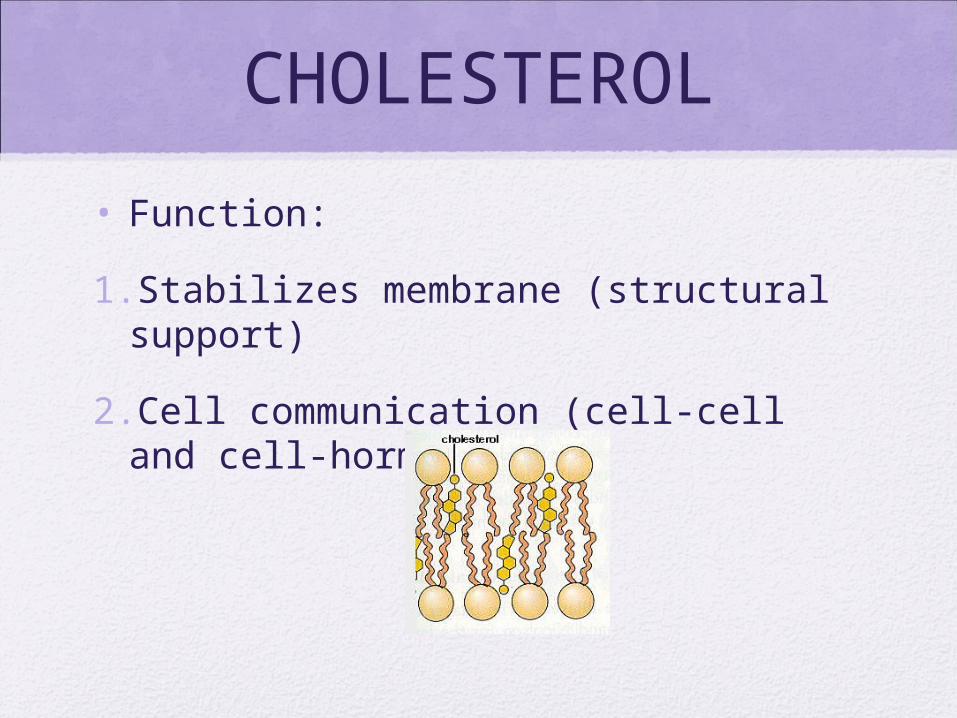
CHOLESTEROL
• Function:
1.Stabilizes membrane (structural support)
2.Cell communication (cell-cell and cell-hormone)

Some interesting facts
• Most common molecule in membrane (>50%)
• Due small size and weight – only 20% of membrane mass
• Is an amphipathic molecule Homework – find out what this means

Proteins
• Are embedded in membrane
• Serve different functions

Functions
• 1. Channel Proteins - small openings for molecules to diffuse through
• 2. Carrier Proteins- binding site on protein "grabs” molecules, pulls them into cell
• 3. Receptor Proteins – act as triggers that set off cell responses (such as release of hormones or opening of channel proteins)
• 4. Cell Recognition Proteins - ID tags, identify cell to immune system
• 5. Enzymatic Proteins - carry out metabolic reactions

LABEL YOUR DIAGRAM WITH THE FOLLOWING
DETAILS

Task: Due Tuesday Oct. 4,
2011• Read pages 47 -49 in your text
• Answer questions 1 – 5 in practice section
• Explain the structure and function of glycoproteins.
• How are liposomes used in cancer treatment and gene therapy?
• What role might receptor proteins have in medical disorders such as Hypertension?
Copyright © 2004 Pearson Education, Inc. publishing as Benjamin Cummings
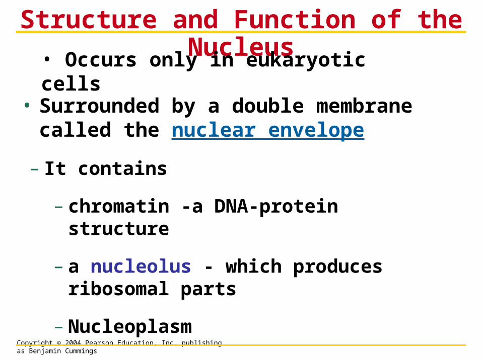
• Surrounded by a double membrane called the nuclear envelope
Structure and Function of the Nucleus
– It contains
– chromatin -a DNA-protein structure
– a nucleolus - which produces ribosomal parts
– Nucleoplasm
• Occurs only in eukaryotic cells

Copyright © 2004 Pearson Education, Inc. publishing as Benjamin Cummings
• Animation of nuclear membrane system
• http://users.uma.maine.edu/SBaker/nucleus_endo.html
• Animation of nuclear membrane system
• http://users.uma.maine.edu/SBaker/nucleus_endo.html

Copyright © 2004 Pearson Education, Inc. publishing as Benjamin Cummings

Copyright © 2004 Pearson Education, Inc. publishing as Benjamin Cummings
Nuclear Pore Complex
Allows movement of material into and out of nucleus

Copyright © 2004 Pearson Education, Inc. publishing as Benjamin Cummings
• Ribosomes build all the cell’s proteins
– Are not membrane bound
Ribosomes

Copyright © 2004 Pearson Education, Inc. publishing as Benjamin Cummings
• DNA controls the cell by transferring its coded information into RNA
How DNA Controls the Cell
– The information in the RNA is used to make proteins
Synthesis ofmRNA in thenucleus
1
2 Movement ofmRNA intocytoplasm vianuclear pore
3 Synthesis ofprotein in thecytoplasm
DNA
mRNA
Nucleus
Cytoplasm
mRNA
Ribosome
Protein

Copyright © 2004 Pearson Education, Inc. publishing as Benjamin Cummings
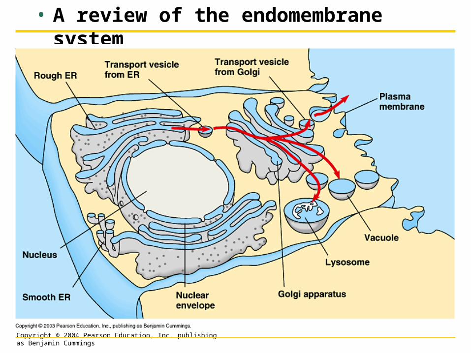
• Many of the membranous organelles in the cell belong to the endomembrane system
– Endoplasmic reticulum - rough and smooth
– Golgi Apparatus
– Lysosomes
– Vacuoles
THE ENDOMEMBRANE SYSTEM: MANUFACTURING AND DISTRIBUTING
CELLULAR PRODUCTS

Copyright © 2004 Pearson Education, Inc. publishing as Benjamin Cummings
• The endoplasmic reticulum (ER)
The Endoplasmic Reticulum
– Greek for ‘network within a cell’
– Produces an enormous variety of molecules
– Is composed of smooth and rough ER
Nuclearenvelope
Ribosomes
Rough ERSmooth ER

Copyright © 2004 Pearson Education, Inc. publishing as Benjamin Cummings
• The “roughness” of the rough ER is due to ribosomes that stud the outside of the ER membrane
Rough ER

Copyright © 2004 Pearson Education, Inc. publishing as Benjamin Cummings
• The functions of the rough ER include
– Producing proteins
– Producing new membrane

Copyright © 2004 Pearson Education, Inc. publishing as Benjamin Cummings
• After the rough ER synthesizes a molecule it packages the molecule into transport vesicles
1

Copyright © 2004 Pearson Education, Inc. publishing as Benjamin Cummings
Smooth ER• The smooth ER lacks
the surface ribosomes of ER
• Produces lipids, including steroids and sex hormones
• Regulates sugar
• Detoxifies drugs
• Stores calcium

Copyright © 2004 Pearson Education, Inc. publishing as Benjamin Cummings
• The Golgi apparatus
The Golgi Apparatus
– Works in partnership with the ER
– Refines, stores, and distributes the products of cells
Transportvesiclefrom ER
“Receiving” side ofGolgi apparatus
Golgi apparatus
New vesicle forming
Transport vesiclefrom the Golgi
“Shipping” side ofGolgi apparatus
Plasma membrane

Copyright © 2004 Pearson Education, Inc. publishing as Benjamin Cummings
• A lysosome is a membrane-enclosed sac
Lysosomes
– Greek for ‘breakdown body’
– It contains digestive enzymes
• Isolated by membrane
– The enzymes break down
• Macromolecules
• Old organelles

Copyright © 2004 Pearson Education, Inc. publishing as Benjamin Cummings
• Lysosomes have several types of digestive functions
• They exit the Golgi apparatus

Copyright © 2004 Pearson Education, Inc. publishing as Benjamin Cummings
– They fuse with food vacuoles to digest the food

Copyright © 2004 Pearson Education, Inc. publishing as Benjamin Cummings
– They fuse with old organelles to recycle parts
– Digest bacteria in white blood cells

Copyright © 2004 Pearson Education, Inc. publishing as Benjamin Cummings
Lysosomal diseasesGenetic disorders
Recipe is messed up
Enzyme doesn’t work
what should get broken down doesn’t
Tay-Sachs
lipids aren’t broken down
build up occurs
death by age 5
Pompe’s disease
glycogen builds up

Copyright © 2004 Pearson Education, Inc. publishing as Benjamin Cummings
• Vacuoles are membranous sacs
Vacuoles
– Two types are the contractile vacuoles of protists and the central vacuoles of plants
Figure 4.15
Contractilevacuoles
Centralvacuole
(a) Contractile vacuoles in a protist (b) Central vacuole in a plant cell
Copyright © 2004 Pearson Education, Inc. publishing as Benjamin Cummings
• A review of the endomembrane system

Copyright © 2004 Pearson Education, Inc. publishing as Benjamin Cummings
• Cells require a constant energy supply to do all the work of life
CHLOROPLASTS AND MITOCHONDRIA: ENERGY CONVERSION

Copyright © 2004 Pearson Education, Inc. publishing as Benjamin Cummings
• Chloroplasts are the sites of photosynthesis, the conversion of light energy to chemical energy
CHLOROPLASTS
Figure 4.17
Inner and outermembranes ofenvelope
Space betweenmembranes
Stroma (fluid inchloroplast)
Granum

Copyright © 2004 Pearson Education, Inc. publishing as Benjamin Cummings
Chloroplasts• Double membrane
• Grana
– Stacks of thylakoids
• Hollow disks
• Sunlight energy is coverted to chemical energy
• Stroma- fluid filling chloroplast
• Contains some DNA

Copyright © 2004 Pearson Education, Inc. publishing as Benjamin Cummings
• Mitochondria are the sites of cellular respiration, which involves the production of ATP from food molecules
Mitochondria
Figure 4.18
Outermembrane
Innermembrane
Cristae
Matrix
Space betweenmembranes

Copyright © 2004 Pearson Education, Inc. publishing as Benjamin Cummings
Mitochondria• Double membrane
– Big bag stuffed into smaller bag
– Folds of inner bag called cristae
• Matrix -space inside inner bag
• Contains some DNA

Copyright © 2004 Pearson Education, Inc. publishing as Benjamin Cummings
• The cytoskeleton is an infrastructure of the cell consisting of a network of fibers
– Microfilaments - small threads
– Intermediate filaments - ropelike
– Microtubules - small tubes
THE CYTOSKELETON:CELL SHAPE AND MOVEMENT

Copyright © 2004 Pearson Education, Inc. publishing as Benjamin Cummings
• One function of the cytoskeleton
Maintaining Cell Shape
– Provide mechanical support to the cell and maintain its shape

Copyright © 2004 Pearson Education, Inc. publishing as Benjamin Cummings
Figure4.9x

Copyright © 2004 Pearson Education, Inc. publishing as Benjamin Cummings
• The cytoskeleton can change the shape of a cell
– This allows cells like amoebae to move

Copyright © 2004 Pearson Education, Inc. publishing as Benjamin Cummings
• Cilia and flagella are motile appendages
Cilia and Flagella

Copyright © 2004 Pearson Education, Inc. publishing as Benjamin Cummings
• Flagella propel the cell in a whiplike motion
• Cilia move in a coordinated back-and-forth motion
Figure 4.20A, B

Copyright © 2004 Pearson Education, Inc. publishing as Benjamin Cummings
• Some cilia or flagella extend from nonmoving cells
– The human windpipe is lined with cilia
– Smoking damages the cilia

Copyright © 2004 Pearson Education, Inc. publishing as Benjamin Cummings
Cilia and Flagella
• Same structure and function
• 9 + 2 arrangement of microtubules
• Wrapped in plasma membrane

Copyright © 2004 Pearson Education, Inc. publishing as Benjamin Cummings
Mechanism of Movement• Dynein arms use ATP for energy to ‘walk’ up adjoining
microtubule, causing them to bend

Copyright © 2004 Pearson Education, Inc. publishing as Benjamin Cummings
• Most cells secrete materials that are external to the plasma membrane
• This extra cellular matrix
– Regulates
– Protects
– Supports
CELL SURFACES:PROTECTION, SUPPORT, AND CELL-CELL
INTERACTIONS

Copyright © 2004 Pearson Education, Inc. publishing as Benjamin Cummings
• Plant cells are encased by cell walls
Plant Cell Walls and Cell Junctions
Figure 4.21
– These provide support for the plant cells
Walls of two adjacentplant cells
Vacuole
Plasmodesmata(channels between cells)

Copyright © 2004 Pearson Education, Inc. publishing as Benjamin Cummings
• Animal cells lack cell walls
Animal Cell Surfaces and Cell Junctions
– They secrete a sticky covering called the extracellular matrix
– This layer helps hold cells together
Copyright © 2004 Pearson Education, Inc. publishing as Benjamin Cummings
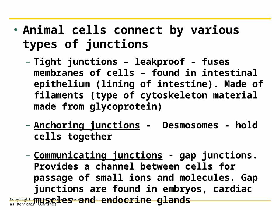
• Animal cells connect by various types of junctions
– Tight junctions – leakproof – fuses membranes of cells – found in intestinal epithelium (lining of intestine). Made of filaments (type of cytoskeleton material made from glycoprotein)
– Anchoring junctions - Desmosomes - hold cells together
– Communicating junctions - gap junctions. Provides a channel between cells for passage of small ions and molecules. Gap junctions are found in embryos, cardiac muscles and endocrine glands

Copyright © 2004 Pearson Education, Inc. publishing as Benjamin Cummings
Figure 4.22
Extracellular matrix
(a) Tight junctions
(b) Anchoring junctions
(c) Communicating junctions
Plasma membranesof adjacent cells
Extracellular matrix

Copyright © 2004 Pearson Education, Inc. publishing as Benjamin Cummings