cor triatriatum with pericardial agenesis -...
TRANSCRIPT
Thorax (1969), 24, 667.
Cor triatriatum with pericardial agenesisM. JIMItNEZ MARTINEZ', J. S. FRANCO VAZQUEZ,
R. GUTIERREZ BOSQUE, J. J. PI2REZ ALVAREZ, ANDR. ARGUERO SANCHEZ
From the Departments of Cardiovascular Surgery and Cardiology, Hospital de Pediatria,Centro Medico Nacional, Mexico City, Mexico
A case of cor triatriatum associated with pericardial agenesis, which was successfully treated, isreported. Details of the clinical picture and surgical treatment are given, with emphasis on thegood prognosis which results when the diagnosis is made before attempting surgical correction.
Cor triatriatum is a rather uncommon congenitalmalformation; it was first described by Churchin 1868. Since then 72 cases have been reported inthe literature, of which only 22 have been correctlydiagnosed pre-operatively or at operation (Ahn,Hosier, and Sirak, 1968; Enjalbert, Calazel,Bounhoure, Vadhat, Puel, and Meriel, 1966;Lasalle, Ethier, Stanley, and Davignon, 1963;McGuire, Nolan, Reeve, and Dammann, 1965;Neufeld, Pauzner, Gueron, Deutsch, and Cohen,1965; Soulie, Vernant, Corone, Galey, Bouchard,Poisson, and Guerin, 1965).The association of cor triatriatum with peri-
cardial agenesis has not been reported previously.In the present case the diagnosis of cor triatriatumwas made before attempting surgical repair.
CASE REPORT
R. H. J., a 10-year-old boy, was first seen in Septem-ber 1965 because of coughing episodes. A heartmurmur had been heard six months previously andthere was a history of repeated upper respiratoryinfections, exertional dyspnoea, and fatigue.On physical examination the child was poorly
developed, with no evidence of congestive heartfailure. The chest was deformed with a prominentprecordium. The maximum cardiac impulse wasvisible and palpable at the sixth left intercostal spaceover the anterior axillary line. A thrill was felt at theapex and along the sternal border. A grade 2 to 3/4systolic murmur was heard at the apex. The secondheart sound was split and the pulmonary componentaccentuated. Peripheral pulses were found to be nor-mal with a blood pressure of 120/60 mm. Hg.Routine laboratory tests were within normal limits.A chest radiograph showed cardiac enlargement and
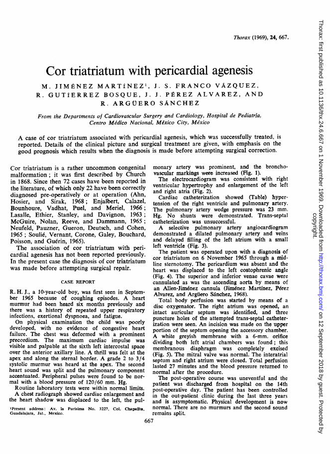
the heart shadow was displaced to the left, the pul-'Present address: Av. la Purisima No. 3227, Col. Chapalita,Guadalajara, Jal., Mexico.
monary artery was prominent, and the broncho-vascular markings were increased (Fig. 1).The electrocardiogram was consistent with right
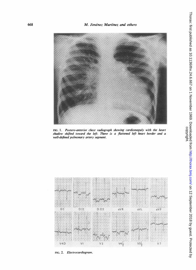
ventricular hypertrophy and enlargement of the leftand right atria (Fig. 2).
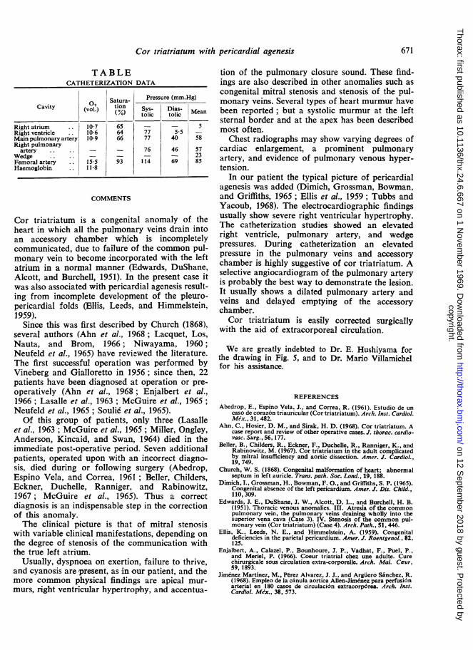
Cardiac catheterization showed (Table) hyper-tension of the right ventricle and pulmonary artery.The pulmonary artery wedge pressure was 23 mm.Hg. No shunts were demonstrated. Trans-septalcatheterization was unsuccessful.A selective pulmonary artery angiocardiogram
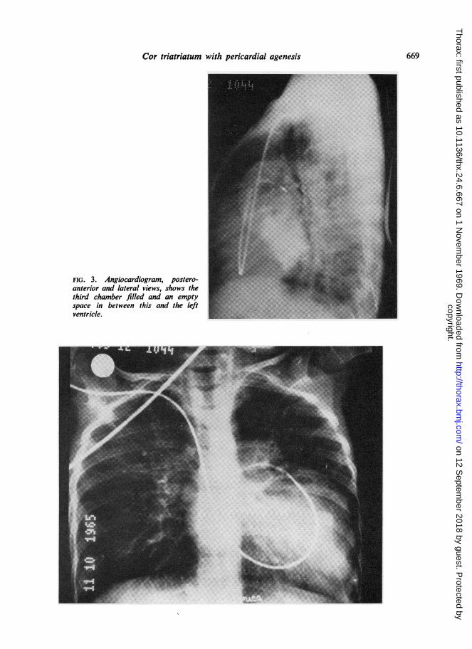
demonstrated a dilated pulmonary artery and veinsand delayed filling of the left atrium with a smallleft ventricle (Fig. 3).The patient was operated upon with a diagnosis of
cor triatriatum on 6 November 1965 through a mid-line sternotomy. The pericardium was absent and theheart was displaced to the left costophrenic angle(Fig. 4). The superior and inferior venae cavae werecannulated as was the ascending aorta by means ofan Allen-Jimenez cannula (Jim6nez Martinez, PdrezAlvarez, and Argilero Sdnchez, 1968).Total body perfusion was started by means of a
disc oxygenator. The right atrium was opened, anintact auricular septum was identified, and threepuncture holes of the attempted trans-septal catheter-izaition were seen. An incision was made on the upperportion of the septum opening the accessory chamber.A white greyish membrane with a 6-mm. orificedividing both left atrial chambers was found; thismembranous diaphragm was completely excised(Fig. 5). The mitral valve was normal. The interatrialseptum and right atrium were closed. Total perfusionlasted 27 minutes and the blood pressure returned tonormal after the procedure.The post-operative course was uneventful and the
patient was discharged from hospital on the 14thpost-operative day. The patient has been controlledin the out-patient clinic during the last three yearsand is asymptomatic. Physical development is nownormal. There are no murmurs and the second soundremains split.
667
copyright. on 12 S
eptember 2018 by guest. P
rotected byhttp://thorax.bm
j.com/
Thorax: first published as 10.1136/thx.24.6.667 on 1 N
ovember 1969. D
ownloaded from
M. Jime'nez Martinez and others
FIG. 1. Postero-anterior chest radiograph showing cardiomegaly with the heartshadow shifted toward the left. There is a flattened left heart border and awell-defined plmonary artery segment.
C, ^Av ,...
r%Jrtrt%~ .
Di D II
, !I o<
V4D
D ITDII!
e ....4 r
;
cVR
/; V 2 4 5R
FIG. 2. Electrocardiogram.
668
.L a,
a VL 2cVF
,
*tv Nr
-4-1. 1--,W. "'.% -.N;I
copyright. on 12 S
eptember 2018 by guest. P
rotected byhttp://thorax.bm
j.com/
Thorax: first published as 10.1136/thx.24.6.667 on 1 N
ovember 1969. D
ownloaded from
Cor triatriatum with pericardial agenesis
FIG. 3. Angiocardiogram, postero-anterior and lateral views, shows thethird chamber filled and an emptyspace in between this and the leftventricle.
669
copyright. on 12 S
eptember 2018 by guest. P
rotected byhttp://thorax.bm
j.com/
Thorax: first published as 10.1136/thx.24.6.667 on 1 N
ovember 1969. D
ownloaded from
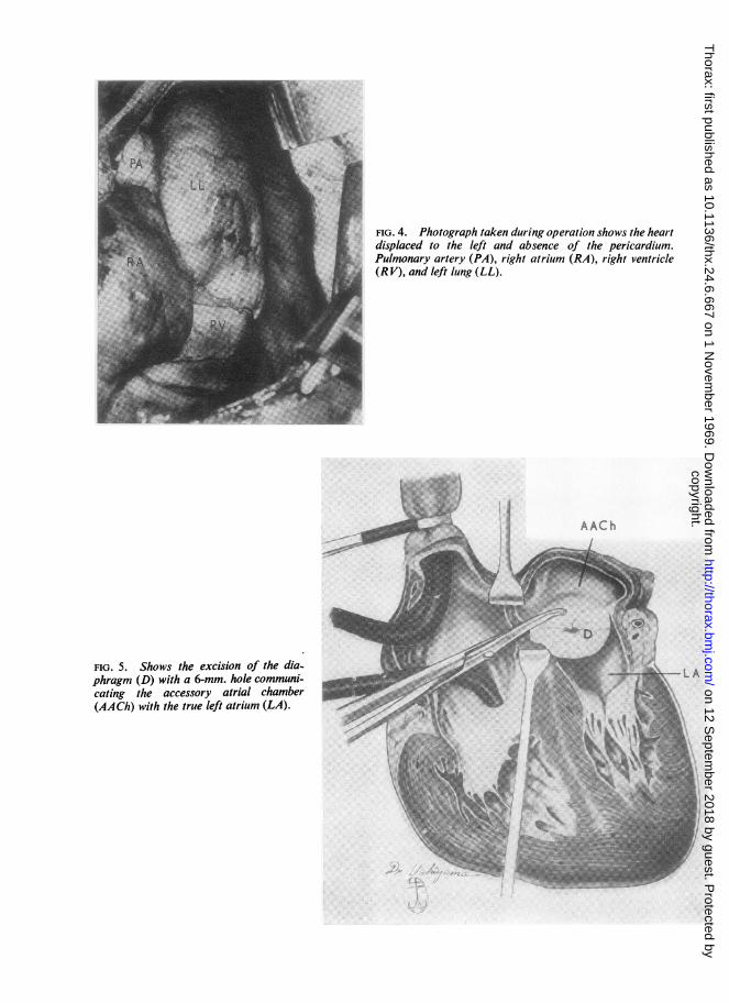
FIG. 4. Photograph taken during operation shows the heartdisplaced to the left and absence of the pericardium.Pulmonary artery (PA), right atrium (RA), right ventricle(RV), and left lung (LL).
_ u >.~~~~~~~~.AC h
...-°C
FIG. 5. Shows the excision of the dia-phragm (D) with a 6-mm. hole communi-cating the accessory atrial chamber(AACh) with the true left atrium (LA).
copyright. on 12 S
eptember 2018 by guest. P
rotected byhttp://thorax.bm
j.com/
Thorax: first published as 10.1136/thx.24.6.667 on 1 N
ovember 1969. D
ownloaded from
Cor triatriatum with pericardial agenesis
TABLECATHETERIZATION DATA
Oo Satura-Pressure (mm.Hg)
02 Satura
Cavity (vol.) tion Sys- Dias- MeanM tolic tolic Ma
Right atrium .. 10-7 65 - - 5Right ventricle .. 10-6 64 77 55 -Main pulmonary artery 10-9 66 77 40 58Right pulmonary
artery .. .. - _ 76 46 57Wedge .. .. _ - - - 23Femoral artery .. 15 5 93 114 69 85Haemoglobin .. 11*8
COMMENTS
Cor triatriatum is a congenital anomaly of theheart in which all the pulmonary veins drain intoan accessory chamber which is incompletelycommunicated, due to failure of the common pul-monary vein to become incorporated with the leftatrium in a normal manner (Edwards, DuShane,Alcott, and Burchell, 1951). In the present case itwas also associated with pericardial agenesis result-ing from incomplete development of the pleuro-pericardial folds (Ellis, Leeds, and Himmelstein,1959).
Since this was first described by Church (1868),several authors (Ahn et al., 1968; Lacquet, Los,Nauta, and Brom, 1966; Niwayama, 1960;Neufeld et al., 1965) have reviewed the literature.The first successful operation was performed byVineberg and Gialloretto in 1956; since then, 22patients have been diagnosed at operation or pre-operatively (Ahn et al., 1968; Enjalbert et al.,1966; Lasalle et al., 1963; McGuire et al., 1965;Neufeld et al., 1965; Soulid et al., 1965).Of this group of patients, only three (Lasalle
et al., 1963; McGuire et al., 1965; Miller, Ongley,Anderson, Kincaid, and Swan, 1964) died in theimmediate post-operative period. Seven additionalpatients, operated upon with an incorrect diagno-sis, died during or following surgery (Abedrop,Espino Vela, and Correa, 1961; Beller, Childers,Eckner, Duchelle, Ranniger, and Rabinowitz,1967; McGuire et al., 1965). Thus a correctdiagnosis is an indispensable step in the correctionof this anomaly.The clinical picture is that of mitral stenosis
with variable clinical manifestations, depending onthe degree of stenosis of the communication withthe true left atrium.
Usually, dyspnoea on exertion, failure to thrive,and cyanosis are present, as in our patient, and themore common physical findings are apical mur-murs, right ventricular hypertrophy, and accentua-
tion of the pulmonary closure sound. These find-ings are also described in other anomalies such ascongenital mitral stenosis and stenosis of the pul-monary veins. Several types of heart murmur havebeen reported; but a systolic murmur at the leftsternal border and at the apex has been describedmost often.
Chest radiographs may show varying degrees ofcardiac enlargement, a prominent pulmonaryartery, and evidence of pulmonary venous hyper-tension.
In our patient the typical picture of pericardialagenesis was added (Dimich, Grossman, Bowman,and Griffiths, 1965; Ellis et al., 1959; Tubbs andYacoub, 1968). The electrocardiographic findingsusually show severe right ventricular hypertrophy.The catheterization studies showed an elevatedright ventricle, pulimonary artery, and wedgepressures. During catheterization an elevatedpressure in the pulmonary veins and accessorychamber is highly suggestive of cor triatriatum. Aselective angiocardiogram of the pulmonary arteryis probably the best way to demonstrate the lesion.It usually shows a dilated pulmonary artery andveins and delayed emptying of the accessorychamber.Cor triatriatum is easily corrected surgically
with the aid of extracorporeal circulation.
We are greatly indebted to Dr. E. Hushiyama forthe drawing in Fig. 5, and to Dr. Mario Villamichelfor his assistance.
REFERENCESAbedrop, E., Espino Vela. J., and Correa, R. (1961). Estudio de un
caso de coraz6n triauricular (Cor triatriatum). Arch. Inst. Cardiol.Mex., 31, 482.
Ahn, C., Hosier, D. M., and Sirak, H. D. (1968). Cor triatriatum. Acase report and review of other operative cases. J. thorac. cardio-vasc. Surg., 56, 177.
Beller, B., Childers, R., Eckner, F., Duchelle, R., Ranniger, K., andRabinowitz, M. (1967). Cor triatriatum in the adult complicatedby mitral insufficiency and aortic dissection. Amer. J. Cardiol.,19, 749.
Church, W. S. (1868). Congenital malformation of heart; abnormalseptum in left auricle. Trans. path. Soc. Lond., 19, 188.
Dimich, I., Grossman, H.. Bowman, F. O., and Griffiths, S. P. (1965).Congenital absence of the left pericardium. Amer. J. Dis. Child.,110, 309.
Edwards, J. E., DuShane, J. W., Alcott, D. L., and Burchell, H. B.(1951). Thoracic venous anomalies. III. Atresia of the commonpulmonary vein, the pulmonary veins draining wholly into thesuperior vena cava (Case 3). IV. Stenosis of the common pul-monary vein (Cor triatriatum) (Case 4). Arch. Path., 51, 446.
Ellis, K., Leeds, N. E., and Himmelstein, A. (1959). Congenitaldeficiencies in the parietal pericardium. Amer. J. Roentgenol., 82,125.
Enjalbert, A., Calazel, P., Bounhoure, J. P., Vadhat, F., Puel, P.,and Meriel, P. (1966). Coeur triatrial chez une adulte. Curechirurgicale sous circulation extra-corporelle. Arch. Mal. Caur,59, 1893.
Jimenez Martinez, M., P6rez Alvarez, J. J., and Arguero Sainchez, R.(1968). Empleo de la canula aortica Allen-Jimenez para perfusionarterial en 180 casos de circulacion extracorp6rea. Arch. Inst.Cardiol. Mix., 38, 573.
671
copyright. on 12 S
eptember 2018 by guest. P
rotected byhttp://thorax.bm
j.com/
Thorax: first published as 10.1136/thx.24.6.667 on 1 N
ovember 1969. D
ownloaded from
672 M. Jimenez Martinez and others
Lacquet, L. K., Los, J. A., Nauta, J., and Brom, A. G. (1966). Cor Neufeld, H. N., Pauzner, Y., Gueron, M., Deutsch, V., and Cohen,triatriatum. Thorax, 21, 175. B. (1965). Cor triatriatum: report of a case successfully operated
with special reference to angiocardiographic diagnosis. Israel J.Lasalle, R., Ethier, M., Stanley, P., and Davignon, A. (1963). Cor med. Sci., 1, 71.
triatriatum: report of a case with emphasis on cineangiocardio- Niwayama, G. (1960). Cor triatriatum. Amer. Heart J., 59, 291.graphy. Canad. med. Ass. J., 89, 616. Souli6, P., Vernant, P., Corone, P., Galey, J. J., Bouchard, F., Poisson,
McGuire, L. B., Nolan, T. B., Reeve, R., and Dammann, J. F. M., and Guerin, F. (1965). Le coeur triatrial. Arch. Mal. Cavur,(1965). Cor triatriatum as a problem of adult heart disease. 5Circulation, 31, 263. Tubbs, 0. S., and Yacoub, M. H. (1968). Congenital pericardial
defects. Thorax, 23, 598.Miller, G. A. H., Ongley, P. A., Anderson, M. W., Kincaid, 0. W., Vineberg, A., and Gialloreto, 0. (1956). Report of a successful
and Swan, H. J. (1964). Cor triatriatum: hemodynamic and operation for stenosis of common pulmonary vein (cor triatria-angiocardiographic diagnosis. Amer. Heart J., 68, 298. tum). Canad. med. Ass. J., 74, 719.
copyright. on 12 S
eptember 2018 by guest. P
rotected byhttp://thorax.bm
j.com/
Thorax: first published as 10.1136/thx.24.6.667 on 1 N
ovember 1969. D
ownloaded from