core.ac.ukdepartamento de biotecnología y biología vegetal escuela tÉcnica superior de...
TRANSCRIPT

UNIVERSIDAD POLITÉCNICA DE MADRID
ESCUELA TÉCNICA SUPERIOR DE INGENIERÍA AGRONÓMICA,
ALIMENTARIA Y DE BIOSISTEMAS
(CENTRO DE BIOTECNOLOGÍA Y GENÓMICA DE PLANTAS)
Roles of C1A peptidases during barley leaf senescence mediated by abiotic stresses
TESIS DOCTORAL
BLANCA VELASCO ARROYO
Licenciada en Ciencias Ambientales
2017


Departamento de Biotecnología y Biología Vegetal
ESCUELA TÉCNICA SUPERIOR DE INGENIERÍA AGRONÓMICA, ALIMENTARIA
Y DE BIOSISTEMAS
CENTRO DE BIOTECNOLOGÍA Y GENÓMICA DE PLANTAS (CBGP, UPM-INIA)
UNIVERSIDAD POLITÉCNICA DE MADRID
Tesis Doctoral
Roles of C1A peptidases during barley leaf senescence mediated
by abiotic stresses
Autor:
Blanca Velasco Arroyo, Licenciada en Ciencias Ambientales
Directores:
Isabel Díaz Rodríguez, Catedrática de Universidad
Manuel Martínez Muñoz, Profesor Titular de Universidad
2017


UNIVERSIDAD POLITÉCNICA DE MADRID
Tribunal nombrado por el Magfco. y Excmo. Sr. Rector de la
Universidad Politécnica de Madrid, el día de de 20 .
Presidente:
Secretario:
Vocal:
Vocal:
Vocal:
Suplente:
Suplente:
Realizado el acto de defensa y lectura de Tesis el día de de
20 en el Centro de Biotecnología y Genómica de Plantas (CBGP,
UPM-INIA).
EL PRESIDENTE LOS VOCALES
EL SECRETARIO


VII
ACKNOWDLEDGMENTS
This Thesis has been performed in the Plant-Insect Interaction laboratory of “Centro de
Biotecnología y Genómica de Plantas (CBGP UPM-INIA)”.
This work has been supported by the Spanish “Ministerio de Economía y
Competitividad (MINECO)” through a grant “Formación del Personal Investigador (BES-
2012-051962)” associated to the project AGL2011-23650.
My stay in the Centre for Plant Sciences at the Faculty of Biology, Leeds University
(United Kingdom), was possible thanks to the fellowship for short stays granted by
MINECO (EEBB-I-15-09251).
My stay in the “Instituto de Fisiología Vegetal (INFIVE; UNLP-CONICET)” in La Plata,
(Argentina), was possible thanks to the fellowship for short stays granted by MINECO
(EEBB-I-16-11230) and to the “Premio a Jóvenes Excelentes 2015” granted by
“Fundación Caja Burgos”.
I greatly acknowledge Prof. Christine Foyer for bringing me the possibility to work in
the “Redox Homeostasis, Signalling and Responses to Stress” group at the Centre for
Plant Sciences in Leeds, learning new concepts and techniques and enjoying a
wonderful experience.
I greatly acknowledge Dr. Juan José Guiamet for bringing me the possibility to work in
the “Aspectos bioquímicos, moleculares y celulares del desarrollo de plantas” group at
INFIVE, in la Plata, learning new concepts and techniques and enjoying a wonderful
experience. Likewise, I greatly acknowledge Dra. Lorenza Costa who patientlly guided
me along this great stay, providing me with all information and methodologies.
I would like to especially acknowledge my thesis supervisors, Prof. Isabel Díaz and
Manuel Martínez, and all the Plant-Insect Interaction group members for training,
technical assistance and helpful discussion.


IX
AGRADECIMIENTOS Cuando hace ya varios años empecé en el laboratorio a veces me preguntaba a mí misma cuántos días duraría una experta en Agroalimentación y Medio Ambiente en un ámbito tan nuevo para mí en aquel entonces como el de la Biología Molecular. Y digamos que precisamente no empecé con buen pie teniendo en cuenta que en mi primera semana hice desaparecer el microondas del laboratorio y casi escaldo a medio personal…Pero mis ganas eran tremendas, muy superiores a mi torpeza inicial. La principal razón de que hoy en día siga en esto se debe, prácticamente en su totalidad, al apoyo incondicional de mis directores. Isabel, tú siempre has depositado confianza en mí y me has sabido guiar cuando realmente necesitaba un ligero toque de atención, lo que me ha ayudado enormemente y me ha hecho madurar, no sólo en lo profesional, sino en lo personal. Gracias por tus reconocimientos y ánimos, y por saber decir las cosas sin reñir, muy pocos líderes cuentan con esa aptitud. Manu, gracias por estar siempre dispuesto a escuchar y a aconsejar. A los dos, os agradezco vuestro apoyo como directores, pero sobre todo aprecio que seáis como sois. Qué suerte tuve al caer en el 121. Y esa suerte no se debió al azar. Lola, tú eres la persona responsable de que yo cayera aquí. Siempre te estaré profundamente agradecida por abrirme de par en par la puerta que me conduciría a desarrollar la oportunidad de mi vida.
A Jacinto, siempre tengo un recuerdo cariñoso y agradecido hacia ti. Me enseñaste a ser organizada y meticulosa en el laboratorio y ahora, siempre que enseño a alguien que se incorpora al 121, le digo: ¡esto me lo enseño mi Jacin! Me hiciste reír tremendamente en mis inicios, ¡conserva siempre esa gracia especial que te distingue!
A Estrella, tu fuerza, arrojo y energía son admirables. Nunca tienes una queja de cansancio, eres puro esfuerzo y dedicación, por eso vas a conseguir todo lo que te propongas. Calla oh!! Se me olvidaba decir que, por supuesto, gracias por tu ayuda en tantas ocasiones, y mil gracias por tus historias diarias, contadas con esa gracia asturianina, que tanto me han hecho reír.
A Ana, mi burgalesa del 121, cómo es el destino, ¿verdad? Nuestras enseñanzas han sido mutuas. Eres una trabajadora como pocas personas he conocido. El orden y limpieza, tan necesarios en una profesión como la nuestra, tienen tu firma con nombre y apellidos en el 121. Gracias Anita, te va a ir fenomenal. ¡¡Y qué Viva Burgos y su clima!!!
A Merche, gracias por transmitirme tus conocimientos sobre proteínas, por tu paciencia durante horas con los cientos de western, por interesarte sobre mi estado de inquietud e incertidumbre cada vez que salía de la cámara oscura…`Pues parece que aquí se intuye una banda, ¿no?´ Y gracias por tu ayuda y consejos en muchos otros momentos, te deseo toda la suerte del mundo con todos tus proyectos profesionales y personales.
Al resto de los miembros del 121: Pablo, gracias por tu paciencia en la sala de microscopía y por tus acertadas hipótesis sobre varios de mis resultados; Andrea, aunque nos conocemos hace poquito, te deseo lo mejor en esta tesis, le pones mucho esfuerzo e ilusión y te va a ir muy bien, todo mi ánimo y apoyo de cualquier índole; a los que hace tiempo os marchasteis, Inés, Isra, os recuerdo con cariño y os agradezco vuestra ayuda a lo largo de varios momentos iniciales de esta tesis. A Ana Laureano, ¡qué viva tu alegría!. Y a Miguelín, por favor no cambies nunca. Pocas personas tan transparentes como tú he conocido a lo largo de mi vida. Suerte a todos chicos. Y por supuesto, a Josico (José o José Domingo, como prefieras): mi compañero de “sentadillas” para hacer fotos de buena calidad en el módulo de transgénicos durante tu etapa en el 121, y, durante el día a día, mi compañero de comidas en el Centro de Empresas, ¡cómo nos gusta el buen comer y el paseíllo diario! Ana, nos abandonaste en este momento tan

X
necesario para recuperar energías para las largas tardes de poyata. Esperamos reincorporación próxima.
A la gente tan maja que he conocido en el CBGP, Laura Carrillo, Bea y Marta, Ana Castro, Marcela y Manuel, Jan. Y en especial a mi colombianita, “Pío”, la distancia no ha conseguido romper nuestra amistad. Y a muchos otros que me dejo en el tintero pero que también han conformado momentos de mi historia personal en este Centro, perdonad que no os nombre a todos.
A Teresa, Dani y Viky, por ayudarme tanto durante mi estancia en Leeds. Compañeros y amigos.
A Alicia, te convertiste en tres meses en una de mis mejores amigas, mi gran cariño hacia Argentina se debe a ti. Gracias, gracias y mil veces gracias. A Celes, Flavia y Lu, y al resto de los chicos del INFIVE, Santi, Pepe y los demás.
A todos mis burgaleses, presentes o esparcidos por el mundo, en especial a Emma, no cambies nunca tu manera de pensar y estar en la vida, te admiro. A Rosa, otra de las mejores personas que conozco, gracias por ser tan buena. A Nachete, serio y divertido como pocos. A los dos, ánimo con esas oposiciones, lo conseguiréis. A mis amigas de la infancia, siempre juntas en tantas aventuras por nuestra preciosa tierra: Sofi, Cris y Laura. A mis amigos de Agrícolas, en especial Pili, ánimo preciosa. A mis amigos de Ambientales y de mi etapa en Salamanca, en especial a mis niñas: Vir, Lu, Lau, Nazita y Paula. ¡Ay mi Vir!, qué abandonada te he tenido este año. Prometo visita con las chicas a Tenerife. Os quiero y sois de lo mejor que me ha pasado, por tantos momentos compartidos.
A mi prima y mejor amiga, María. Eres madura, buena y luchadora como poca gente de tu edad. Vas a conseguir lo que quieras, ya lo verás.
A mis hermanos, Lidia, Gonzalo y Quique. En parte soy como soy gracias a la niñez tan feliz que pasé al tener hermanos mayores. Lidia, además de mi hermana, mi confidente y amiga. Os quiero por igual a los tres.
A mi abuela, en muy poquito tiempo cumples un siglo, ¡qué afortunados de vivirlo a tu lado y qué bien te sienta el paso de los años Tita! Me siento afortunada por tener un libro vivo de historia, con tantas vivencias y tan bien explicado con tu potente chorro de voz.
A mis padres, me habéis transmitido la pasión por la cultura, la naturaleza, el viajar, el buen comer y el saber dialogar en cualquier contexto, primando la escucha a la alabanza propia. Vosotros me lo habéis consentido todo pero con la mesura, la rectitud y el cariño que me han permitido llegar a ser lo que soy. Gracias por dejarme siempre elegir, por aguantar mis fallos, por alabar todos mis logros y por enseñarme el valor del esfuerzo. Os quiero.
A Jose, mi compañero de ilusiones y de proyecto de vida. Sin ti esta tesis no sería lo que es. Sólo
tu sonrisa me da una inyección de optimismo y fuerza diarios. Qué orgullosa estoy de tu tesón y
fuerza de voluntad. Te quiero muchísimo.

XI
ABBREVIATIONS
% Percentage
ºC Celsius Degree
Ø Diameter
µg Microgram
µl Microliter
µm Micrometer
µM Micromolar
ABA Abscisic Acid
AMC Amido Methyl Coumarin
amiRNA Artificial Micro RNA
AP Autophagosome
ATG Autophagy- related gene
ATP Adenosine triphosphate
B-FVR B-Phe-Val-Arg-7-amido-4-methyl coumarin
BiFC Bimolecular Fluorescence Complementation
bp Base Pairs
BSA Bovine Serum Albumin
CCV Chloroplast Vesiculation-Containing Vesicles
cDNA Complementary DNA
CH Carbohydrates
CK Cytokinin
CV Chloroplast Vesiculation protein
cv Cultivar
CysProt Cysteine Proteases
DNA Deoxyribonucleic Acid
dNTP Deoxyribonucleotide Triphosphate
DTT Dithiothreitol
E-64 [1-[N-[(L-3-trans-carboxyoxirane-2-carbonyl)-L-leucyl]amino]-4-
guanidinobutane]
EDTA Ethylenediaminetetraacetic Acid
EL Electrolyte Leakage
ER Endoplasmic Reticulum
EST Expressed Sequence Tag
eQTL Expression Quantitative Trait Loci
FAO Food and Agriculture Organization
g/l Grams per Liter
GA Gibberellic Acid
Gb Gigabase
gdw Grams of Dry Weight

XII
GFP Green Fluorescent Protein
gfw Grams of Fresh Weight
gs Stomata conductance
GS Glutamine Synthetase
GS1 Cytosolic Glutamine Synthetase
GS2 Chloroplastic Glutamine Synthetase
ha Hectare
hai Hours After Imbibition
HSD Honestly Significant Difference test
IPCC International Panel on Climate Change
IPTG Isopropyl β-D-1-thiogalactopyranoside
JA Jasmonic Acid
KD Knock-Down
kDa Kilodalton
Ki Inhibition constant
LHCI Light Harvesting Complex I
LHCII Light Harvesting Complex II
LSD Leaf Senescence Database
M Molar
MAPK Mitogen-Activated Protein Kinase
mg Milligram
ml Milliliter
mM Millimolar
Mr Molar mass
mRNA Messenger RNA
NCC Non-Fluorescent Catabolite
nm Nanometers
nmol Nanomol
NMR Nuclear Magnetic Resonance
NO Nitric Oxide
NR Nitrate Reductase
NUE Nitrogen Use Efficiency
OD Optical Density
OE Overexpressing
PAO Pheophorbide a Oxygenase
PBS Sodium Perborate
PCD Programmed Cell Death
PCR Polymerase Chain Reaction
PDB Protein Database
pFCC Primary Fluorescent Chlorophyll-Derived Catabolites
PhyCys PhytoCystatins

XIII
PSI Photosystem I
PSII Photosystem II
PWC Aerial Plant Water Content
RCB RuBisCo Containing Body
RNA Ribonucleic Acid
RNAseq RNA sequencing
ROS Reactive Oxygen Species
rpm Revolutions Per Minute
RT-qPCR Real-Time Quantitative Polymerase Chain Reaction
RuBisCo Ribulose-1,5-Bisphosphate Carboxylase/Oxygenase
SA Salicylic Acid
SAG Senescence Associated Gene
SAV Senescence Associated Vacuole
SDG Senescence Down-regulated Gene
SDS Sodium Dodecyl Sulfate
SGR Stay Green
ssDNA Single-Stranded DNA
SWC Soil Water Content
SWD Soil Water Deficit
TBO Toluidine Blue O
TCA Tricarboxylic Acid
TEMED Tetramethylethylenediamine
TF Transcription Factor
ud Units
v/v Volume/Volume
VPE Vacuolar Processing Enzyme
w/v Weight/Volume
WT Wild Type
ZFR-AMC Z-Phe-Arg-7-amido-4-methyl coumarin
ZRR-AMC Z-Arg-Arg-7-amido-4-methyl coumarin

Index
XV
INDEX
Acknowdledgments…………………………………………………………………………………………………………………vii
Agradecimientos………………………………………………………………………………………………………………………ix
Abbreviations……………………………………………………………………………………………………………………….….xi
Index…………………………………………………………………………………………………………………….…………………xv
Abstract………………………………………………………………………………………………………………………………….21
Resumen………………………………………………………………………………………………………………………………..23
.......................................................................... 25
1.1. LEAF SENESCENCE: A NATURAL EVENT MODULATED BY STRESSES ............................. 27
1.1.1. A general leaf senescence overview ................................................................... 27
1.1.2. Leaf senescence, grain quality and yield ............................................................. 32
1.2. SENESCENCE AND ABIOTIC STRESS ............................................................................... 38
1.2.1. Climate change scenario ..................................................................................... 38
1.2.2. Stress concept ..................................................................................................... 41
1.2.3. Overlaps, similarities and divergences among developmental leaf senescence,
abiotic and biotic stresses ................................................................................................... 42
1.3. REGULATION OF SENESCENCE AND STRESS: HORMONES, TRANSCRIPTION FACTORS
AND REACTIVE OXYGEN SPECIES ............................................................................................ 46
1.3.1. Hormonal and transcription factors cross-talking ............................................... 47
1.3.2. Redox regulatory networks ................................................................................. 49
1.4. SENESCENCE RELIES ON PROTEOLYSIS .......................................................................... 50
1.4.1. General overview of degradation mechanisms in plants .................................... 50
1.4.2. Chloroplast dismantling ...................................................................................... 51
1.4.3. Plant Proteases and Protease Inhibitors ............................................................. 59
1.5. BARLEY AS A MODEL SPECIES FOR THE POACEAE FAMILY ............................................ 69
1.5.1. Barley as an economic, genetic and climate-change adaptable resource .......... 69
1.5.2. Proteases and cystatins involved in barley germination, senescence and stress 73
1.6. REFERENCES .................................................................................................................. 79
........................................................................................... 99
…………………………………………………………………………………………………….………………..103

Index
XVI
3.1. LEAF SENESCENCE AND PROTEIN BREAKDOWN ......................................................... 105
3.2. C1A CYSTEINE PROTEASES IN LEAF SENESCENCE ........................................................ 107
3.2.1. Cathepsin L-like CysProt .................................................................................... 108
3.2.2. Cathepsin H-like CysProt ................................................................................... 111
3.2.3. Cathepsin B-like CysProt.................................................................................... 111
3.2.4. Cathepsin F-like CysProt .................................................................................... 112
3.3. BARLEY C1A CYSTEINE PROTEASES IN LEAF SENESCENCE ........................................... 112
3.4. CYSTEINE PROTEASE-CYSTATIN INTERACTION IN LEAF SENESCENCE ......................... 115
3.5. CONCLUSIONS ............................................................................................................. 118
3.6. REFERENCES ................................................................................................................ 120
3.7. SUPPLEMENTAL DATA ................................................................................................. 127
.......................................................... 131
4.1. INTRODUCTION ........................................................................................................... 133
4.2. MATERIALS AND METHODS ........................................................................................ 136
4.2.1. Plant material and growth conditions ............................................................... 136
4.2.2. Photosynthetic pigment measurements ........................................................... 136
4.2.3. Protein quantification and protease activities .................................................. 137
4.2.4. Real-time RT-qPCR Analysis ............................................................................... 138
4.2.5. Immunoblot analysis ......................................................................................... 138
4.2.6. Specimen processing for microscopy ................................................................ 138
4.2.7. Structural analysis and immunofluorescence detection of HvPap-1, HvPap-16
and HvPap-19 .................................................................................................................... 139
4.2.8. Confocal imaging of HvPap-1, HvPap-19 and HvPap-16 ................................... 140
4.2.9. Starch quantification ......................................................................................... 140
4.2.10. Data analysis ...................................................................................................... 140
4.3. RESULTS ....................................................................................................................... 140
4.3.1. Structural and physiological changes in leaves under severe stresses ............. 140
4.3.2. C1A proteases and proteolytic patterns are modified in barley leaves under
induction of severe senescence ........................................................................................ 141
4.3.3. Transgenic barley lines over-expressing or silencing the HvPap-1 CysProt ...... 146
4.3.4. Transgenic barley HvPap-1 lines show alterations associated with stress
mediated by darkness ....................................................................................................... 147

Index
XVII
4.3.5. Physiological changes are associated with stress mediated by darkness in
HvPap-1 transgenic barley lines ........................................................................................ 150
4.4. DISCUSSION ................................................................................................................. 153
4.5. REFERENCES ................................................................................................................ 157
4.6. SUPPLEMENTAL DATA ................................................................................................. 162
............................................................................. 171
5.1. INTRODUCTION ........................................................................................................... 173
5.2. MATERIALS AND METHODS ........................................................................................ 175
5.2.1. Plant material .................................................................................................... 175
5.2.2. Analysis of the copy number in transgenic barley lines .................................... 176
5.2.3. Grain phenotype and starch analyses ............................................................... 177
5.2.4. Metabolomic analyses ....................................................................................... 177
5.2.5. Grain protein analysis ........................................................................................ 178
5.2.6. Fractionation and analysis of barley grain storage proteins ............................. 179
5.2.7. Germination assays ........................................................................................... 180
5.2.8. Enzymatic activity assays ................................................................................... 180
5.2.9. Immunoblot analyses ........................................................................................ 180
5.2.10. Real-time quantitative PCR analyses ................................................................. 181
5.2.11. Embryo structural analysis and immuno-fluorescence detection .................... 181
5.2.12. Data analysis ...................................................................................................... 183
5.3. RESULTS ....................................................................................................................... 183
5.3.1. Transgenic barley lines over-expressing or silencing HvPap-1 protease or
silencing Icy-2 cystatin ....................................................................................................... 183
5.3.2. Grain phenotype and starch accumulation are altered in barley transgenic
lines………………………………………………………………………………………………………………………………184
5.3.3. Grain protein content is modified in barley transgenic lines ............................ 185
5.3.4. Grain metabolomic analyses show changes in amino acid accumulation in barley
transgenic lines .................................................................................................................. 187
5.3.5. Germination is delayed in HvPap-1 transgenic lines ......................................... 190
5.3.6. Proteolytic activities are affected in dry and germinating grains ..................... 190
5.3.7. C1A CysProt patterns are altered in transgenic barley grains........................... 194

Index
XVIII
5.3.8. C1A CysProt are differentially located in embryos of transgenic and wild-type
barley lines ........................................................................................................................ 195
5.4. DISCUSSION ................................................................................................................. 196
5.5. REFERENCES ................................................................................................................ 200
5.6. SUPPLEMENTAL DATA ................................................................................................. 205
................................................................. 215
6.1. INTRODUCTION ........................................................................................................... 217
6.2. MATERIALS AND METHODS ........................................................................................ 220
6.2.1. Plant material and growth conditions ............................................................... 220
6.2.2. Phenotypical assessment .................................................................................. 220
6.2.3. Physiological and biochemical parameters ....................................................... 221
6.2.4. Real-time quantitative PCR analyses ................................................................. 221
6.2.5. Electrolyte leakage ............................................................................................ 221
6.2.6. Protein quantification and protease activities .................................................. 222
6.2.7. Metabolomics analyses ..................................................................................... 222
6.2.8. Statistical analyses ............................................................................................. 223
6.3. RESULTS ....................................................................................................................... 224
6.3.1. Soil water deficit alters plant physiological status and drought-associated
biomarkers ........................................................................................................................ 224
6.3.2. Barley cystatins Icy-2 and Icy-4 are induced by drought treatment ................. 225
6.3.3. KD Icy2 and KD Icy4 lines display opposite phenotypes during natural and
drought-induced senescence ............................................................................................ 226
6.3.4. Transgenic barley lines show slight variations related to physiological and
biochemical parameters after drought treatment ............................................................ 228
6.3.5. Protein content after drought treatment is related to protease activity and
membrane stability ........................................................................................................... 230
6.3.6. Molecular alterations within transgenic lines show striking compensation effects
at the transcriptional level ................................................................................................ 232
6.3.7. Slight changes in metabolites contents are detected in barley transgenic lines
subjected to drought ......................................................................................................... 234
6.4. DISCUSSION ................................................................................................................. 236
6.5. REFERENCES ................................................................................................................ 239

Index
XIX
6.6. SUPPLEMENTAL DATA ................................................................................................. 244
........................................................................... 255
7.1. GENERAL DISCUSSION ................................................................................................. 257
7.2. REFERENCES ................................................................................................................ 267
........................................................................ 271
List of Publications…………………………………………………….……………………………………………………...275


Abstract
21
Abstract
Protein breakdown and mobilization from old or stressed tissues, such as leaves, to
growing and sink organs, such as grains or tubers, are some of the metabolic features
associated with leaf senescence, essential for nutrient recycling. Senescence may be
naturally activated by endogenous signals and/or modified by the prevalence of
abiotic/biotic stresses, as a survival strategy. Protein breakdown in senescing leaves
involves many plastidial and nuclear proteases, regulators, different subcellular
locations and a dynamic protein traffic to ensure transformation of high molecular
weight proteins into transportable and useful hydrolyzed products. C1A cysteine
proteases are the most abundant key players responsible for the proteolytic activity
during leaf senescence. Besides, cystatins, as specific modulators of C1A protease
activities, exert a regulatory role along the process. In barley (Hordeum vulgare), the
whole gene family members of C1A cysteine proteases and cystatins have been
identified. Elucidating the role of barley C1A proteases in response to abiotic stresses is
crucial due to their impact on plant growth and grain yield and quality.
Darkness and nitrogen starvation treatments were used to induce leaf
senescence in barley. Both abiotic stresses strongly induced the expression of the
HvPap-1 gene encoding a cathepsin F-like protease. Morphological changes presuming
chloroplast dismantling designated darkness as an ideal stressor for inducing and
analyzing senescence. Differences in biochemical parameters and C1A gene expression
and protein accumulation among wild-type and transgenic barley plants over-
expressing or silencing this gene were detected under the stress. Besides, a lifespan-
delayed phenotype of HvPap-1 silenced lines was evidenced, indicating a functional
role for this protease along the senescence process.
Proteolysis is likewise essential throughout the mobilization of storage proteins
in barley grains during germination. Manipulation of the proteolytic machinery could
enhance grain yield and quality through alterations along these stages. Transgenic
barley plants silencing or over-expressing HvPap-1 showed differential accumulation of
starch, proteins, and free amino acids in the grain. The phenotype displayed by
silencing HvPap-1 lines, showing a drastic delay in germination, was particularly

Abstract
22
striking. Alterations in the proteolytic activities associated with changes in the
expression levels of several C1A proteases were also detected. Similarly, down-
regulating Icy-2, encoding one of the proteinaceous inhibitors of the studied cathepsin
F-like protease, also brought about important effects on grain filling.
The cooperative role of cystatins and their functional relationship with cysteine
proteases have been highlighted in the current study by the enhanced/reduced
tolerance of plants silencing phytocystatins towards drought. Two barley
phytocystatins, HvCPI-2 and HvCPI-4, were induced by this stress. Alterations in the
proteolytic patterns by silencing these cystatins were concomitant with modifications
in the expression of target proteases. As a result, accelerated or delayed leaf
senescence, depending on the silenced cystatin, was exhibited. Results support the
potential use of these plants to modulate plant responses facing abiotic stress and, at
the same time, to maintain or even increase crop yields under the evidenced climate
change framework
According to data reported in this thesis, manipulation of C1A proteases-
cystatins interactions in barley has the potential to modulate sensitivity towards
specific abiotic stresses through modifications over established developmental leaf
senescence programs. In addition, the in vivo implication of this proteolytic network
during remobilization of stored compounds along barley grain germination is
demonstrated. As a general remark, caution should be taken when designing related
biotechnological tools since the plant tries to compensate the genetic modifications by
modulating the expression of some other proteases or inhibitors.

Resumen
23
Resumen
La degradación y movilización de proteínas desde tejidos maduros o sometidos a
estrés, como las hojas, hasta los órganos en desarrollo o sumidero, como los granos de
los cereales, son procesos metabólicos inherentes a la senescencia foliar. Los
programas de senescencia se activan tanto en respuesta a señales endógenas como a
estreses abióticos y bióticos como estrategia de supervivencia. La proteólisis en hojas
senescentes implica multitud de proteasas de origen nuclear y plastidial, reguladores,
diversas localizaciones subcelulares, así como un tráfico dinámico cuyo fin es asegurar
la transformación de proteínas de alto peso molecular en productos hidrolizados que
puedan transportarse y reutilizarse. La familia C1A de cisteín-proteasas engloba un
buen número de enzimas responsables de la actividad proteolítica asociada a la
senescencia foliar. Además, las cistatinas, inhibidores específicos de dichas proteasas,
ejercen un papel regulador durante este proceso fisiológico. En cebada (Hordeum
vulgare), las familias completas de proteasas C1A y cistatinas han sido identificadas.
Dilucidar el papel funcional de las proteasas C1A de cebada en respuesta a estreses
abióticos es esencial, debido a su impacto sobre el crecimiento de las plantas y la
alteración del rendimiento y calidad del grano.
Los tratamientos de oscuridad y de carencia de nitrógeno se utilizaron para
inducir senescencia foliar en cebada. Ambos estreses indujeron claramente la
expresión del gen HvPap-1, que codifica una proteasa tipo catepsina F. Cuando se
compararon plantas control frente a líneas transgénicas de sobrexpresión y de
silenciamiento para este gen en oscuridad, se observaron alteraciones significativas en
parámetros bioquímicos, en patrones de expresión de genes de proteasas C1A, así
como en el contenido proteico. Por otro lado, el fenotipo “stay-green” de las líneas de
silenciamiento evidenció una vida útil más prolongada en estas plantas, demostrando
la implicación funcional de esta proteasa a lo largo del proceso de senescencia.
La proteólisis es asimismo esencial para la movilización de proteínas de reserva
del grano durante la germinación. La manipulación de la maquinaria proteolítica
durante este proceso fisiológico podría tener un efecto de mejora sobre la calidad del
grano y el rendimiento del cultivo. Las líneas transgénicas de sobreexpresión y

Resumen
24
silenciamiento del gen HvPap-1 mostraron una acumulación diferencial de almidón,
proteínas y amino ácidos en la semilla. El fenotipo de los granos de las líneas
silenciadas evidenció un claro retraso en el proceso germinativo. También se
observaron alteraciones en las actividades proteolíticas, asociadas a las variaciones en
los niveles de expresión de genes C1A. De forma paralela, al silenciarse el gen Icy-2 que
codifica uno de los inhibidores de la catepsina F estudiada, se observaron efectos en
relación con el llenado y calidad del grano.
La interacción y la implicación funcional de cisteín-proteasas y cistatinas en
cebada se ha constatado en este estudio, tal y como se infiere de la tolerancia alterada
frente a sequía en las líneas de silenciamiento de cistatinas. Dos fitocistatinas, HvCPI-2
y HvCPI-4, se indujeron específicamente por dicho estrés. Las alteraciones en los
patrones proteolíticos al silenciar estas cistatinas fueron paralelas a las variaciones en
la expresión de genes de sus proteasas diana. En función de la cistatina silenciada, se
apreció un retraso o una aceleración en la senescencia. Estos resultados apoyan el uso
de estas líneas con el objetivo de modular las respuestas a estreses diversos y
mantener, o incluso incrementar, los rendimientos en el marco evidente del cambio
climático.
De acuerdo con los resultados obtenidos, la manipulación de las interacciones
entre proteasas C1A y cistatinas en cebada permitiría modular la sensibilidad frente a
estreses abióticos concretos en base a modificaciones sobre los programas de
senescencia endógenos. Se confirma asimismo, la importancia in vivo de esta compleja
red proteolítica durante la germinación. Como observación general, cuando se diseñen
estrategias biotecnológicas basadas en estos mecanismos moleculares se han de
considerar los efectos de compensación derivados de la expresión de otros inhibidores
y/o proteasas de la planta.


Chapter 1. General Introduction
27
1.1. LEAF SENESCENCE: A NATURAL EVENT MODULATED BY STRESSES
1.1.1. A GENERAL LEAF SENESCENCE OVERVIEW
Senescence is a natural process which occurs in all plants when the maturity phase is
coming to its end, leading to the death or completion of a life cycle. Senescence-like
processes occur in angiosperm and non-angiosperm land plants, algae and
photosynthetic prokaryotes (Gan and Amasino, 1997; Lim et al., 2007; Thomas et al.,
2009). This developmental phase is illustrated by the striking changes in leaf color
observed during the autumn for trees and other perennial plants in temperate regions.
In annual crops such as cereals, something similar is observed when the green color
changes to golden as the grain ripens (Buchanan-Wollaston et al., 2003). It is
noteworthy that these color changes evidence chlorophyll degradation or/and de novo
synthesis of anthocyanins (Rapp et al., 2015), among other protective compounds.
In an overall view, the main goal of this complex physiological event can be
compared to the three R´s theory of the environment (reduce, reuse and recycle), in
this case with an extra R (remobilize). After `Reducing´ the photosynthetic rate in
response to the activation of a senescence program, a massive `Recycling´ of nutrients
that will be `Reutilized´ as scaffolds for new macromolecule biosynthesis and insurance
of the next generation survival begins. This implies an important `Remobilization´ of
nutrients through the phloem, from the source plant parts, such as senescent leaves,
towards sink organs such as emergent leaves, grains, tubers or fruits. This strictly
controlled event is integral to the flowering plant life-cycle and is determined by
endogenous developmental signals governed by the reproductive age (Ghanem et al.,
2012). In many monocarpic plants the developing reproductive structures often govern
the timing and onset of leaf senescence, thereby affecting all organs of a given plant
(Munné-Bosch, 2008).These intrinsic cues are continuously modulated by external
factors (abiotic environmental stresses, like drought or flooding, high irradiance or
darkness, extreme temperatures, salinity, wounding or accumulation of pollutants; and
biotic stresses, i.e., pathogens and pests) which modify, to some extent, the natural

Chapter 1. General Introduction
28
senescence programs of the plant (Fig.1.1). The degree of influence of such stresses
will determine if it causes an impact on the yield (Breeze et al., 2011).
Fig. 1.1. Schematic representation of source to sink nutrient recycling favored by leaf senescence.
All the dramatic changes undergoing along senescence are finely tuned and do
not constitute a mere chaotic event. Genetic and epigenetic mechanisms regulating
phase change from juvenility to maturity directly influence the capacity for responding
to senescence signals (Thomas, 2013). The endogenous signals and the environmental
stresses perceived by a plant are integrated into the natural senescence program and
subsequently transmitted, forming complex interactions of regulatory pathways
among plant hormonal routes, transcription factors (TF), signaling transduction
cascades of calcium, phosphatases, kinases and others, to control the onset and
progression of senescence. These sophisticated networks somehow channel the
impacts from the environment and determine multiple changes in gene expression
patterns during senescence (Schippers et al., 2007). The orderly and orchestrated
sequential changes in cellular physiology, biochemistry and metabolism are strongly
triggered by a rapid reprogramming in the expression of an important battery of

Chapter 1. General Introduction
29
Senescence Associated Genes (SAGs; He et al., 2001; Breeze et al., 2008, 2011). The
degree of effectiveness in the response of the plant after the detection of a stressor
factor will determine the degree of reversibility, delimiting a narrow border between
degenerative cell death and senescence as a recycling process.
The senescent phase is reversible in the green mesophyll cells until almost all
macromolecules have been recycled and exported to the rest of the plant (Thomas,
2013). Senescing leaves can, under certain conditions, re-green and regain their
photosynthetic capacity (Rapp et al., 2015). Cells within the same organ can be at
different stages in the progression from senescence to death (Thomas, 2013). Leaf
senescence is thus a type of Programmed Cell Death (PCD) but some key hallmarks
make it distinguishable from other PCD (van Doorn, 2004; Lim et al., 2007; Avila-
Ospina et al., 2014). It proceeds at the organ-level whereas other PCD occur in limited
tissues and cell types; it shows a slower rate than other PCD; and, regarding the
physiological goal, leaf senescence fulfills the essential role of recycling cellular
nutritional components for plant survival and productivity (Breeze et al., 2008).
The participation of hormones during the regulation of leaf senescence is
becoming evident through characterization of genetic mutants and global gene
expression analysis. In general, senescence is accelerated by brassinosteroids, abscisic
acid (ABA), ethylene, jasmonic acid (JA), and salicylic acid (SA), and slowed down by
auxin, cytokinins (CK) and gibberellic acid (GA; Podzimska-Sroka et al., 2015). One of
the signals during the onset of leaf senescence involves cell sugar status alterations as
a consequence of the initial dismantling of the photosynthetic apparatus. There are
some lines of research demonstrating that accumulation of sugars compromises the
photosynthetic capacity and accelerates leaf senescence (Lim et al., 2007). In addition,
the production and accumulation of reactive oxygen species (ROS), derived from
alterations in the cell machineries, has also been proposed as an important promoting
signal during natural and altered senescence. Albeit ROS production is known to have
harmful effects upon diverse biomolecules, it has been proven that a certain level is
required to trigger the activation of genetically programmed pathways of gene

Chapter 1. General Introduction
30
expression during leaf senescence (Khanna-Chopra, 2012; Zhang and Zhou, 2013;
Noctor et al., 2014, 2016).
Considering that within the leaf the main source of nitrogen-containing
molecules is located inside the chloroplasts, it is not surprising that the earliest
structural, biochemical and metabolic changes are observed inside these organelles. All
enzymes required for carbon fixation and nitrogen assimilation, such as ribulose
bisphosphate carboxylase/oxygenase (RuBisCo), as well as most of the proteins that
plants can use for nitrogen recycling and mobilization, are inside this organelle
(Masclaux-Daubresse and Krupinska, 2014; Havé et al., 2016). Leaf cells require a
certain energy status until late stages of senescence; thus, nucleus and mitochondria,
essential for gene expression and power generation, are the last organelles being
degraded (Yoshida, 2003; Lim et al., 2007). Furthermore, during senescence, most of
the fatty acids from membranes are oxidized to provide energy. An evident drop in the
nucleic acid content, especially total RNA, has also been documented (Lim et al., 2007).
A decrease in the overall protein anabolism is one of the best studied markers for the
leaf senescence progress (Lim et al., 2007; Díaz-Mendoza et al., 2014), besides the
decline in photosynthesis and chlorophyll content. As the amount of polysomes and
ribosomes has been observed to decrease fairly early, it clearly reflects a cessation in
protein synthesis (Lim et al., 2007). The bulk macromolecule degradation mainly relies
on proteolysis. Among proteases, serine, and mostly cysteine proteases (CysProt)
participate during important events related to senescence and stress (Roberts et al.,
2012; Kidric et al., 2014; Velasco-Arroyo et al., 2016).
During senescence and stress, many genes related to anabolism, mainly those
related to photosynthesis, are down-regulated. These are usually referred as
“senescence down-regulated genes” (SDG). On the opposite side, those genes that are
induced along this process belong to the SAGs group (Ay et al., 2014). These genes fall
into different categories according to their function as they may be participating in
protein, lipid and nucleic acid turnover, in transport of nutrients, amino acids, sugars,
and in defense mechanisms. However, not all SAGs are induced by external cues and
some stress-associated genes are not influenced by natural senescence (Buchanan-

Chapter 1. General Introduction
31
Wollaston et al., 2005), evincing a complex crosstalk between and among the routes
drawn by developmental- or stress-induced senescence (He et al., 2001).
Senescence has been intensively studied in the model plant Arabidopsis
(Quirino et al., 2000; Guo et al., 2004; Buchanan-Wollaston et al., 2005), with more
than 800 genes identified as SAG. This reflects the dramatic alteration in cellular
physiology that underlies this plant stage (Lim et al., 2007). Probably, the best well-
known SAG gene is SAG12 from Arabidopsis. SAG12 is a CysProt specifically induced
during the last stages of developmentally-controlled senescence (Gan and Amasino,
1997), being widely used as a leaf senescence-associated molecular marker. Another
well-known Arabidopsis SAG marker corresponds to the WRKY53 transcription factor
which, in contrast, is activated at the onset of the process (Zentgraf et al., 2010). In
crop plants, knowledge related to molecular mechanisms driving leaf senescence is not
sufficiently extensive. Some sporadic reports have broadened the information
concerning this field in maize, wheat or barley (Smart et al., 1995; Kleber-Janke and
Krupinska, 1997; Uauy et al., 2006). Interestingly, Jukanti et al. (2008) found a new
regulatory SAG in senescing primary barley leaves consisting on a transmembrane
protein kinase.
Many investigations in the field of plant senescence and stress can be
integrated into two different but complementary areas: research based upon
dilucidation of the molecular basis underlying this crucial event at different layers; and
translation of basic research to design tools through biotechnological approaches in
combination with conventional breeding to manipulate senescence for agronomic
advantages; i.e., translating laboratory bench findings to practical projects (Gan and
Hörtensteiner, 2013). Special emphasis is being undertaken in the maintenance or
improvement of acceptable yields in important crops for human feed such as cereals,
in a context of an evident climate change scenario. It is of pivotal importance to invest
efforts to interpret the processes behind the decrease in productivity under adverse
situations, which substantially relies on a deeper knowledge of chloroplast dismantling
mechanisms in both model and crop species. Accordingly, there exists a continuous
effort in updating the resources related with leaf senescence information, as

Chapter 1. General Introduction
32
evidenced the last releases of the Leaf Senescence Database (LSD; Liu et al., 2011; Li et
al., 2014). A growing senescence community continuously sheds light on some relevant
and particular aspects concerning senescence and stress, as it is evidenced by the
elevated number of reports and reviews related to this fascinating topic, mainly
focused in signaling and regulatory pathways, nutrient management and nitrogen use
efficiency (NUE), chlorophyll and chloroplast degradation mechanisms, with a key
participation of proteases and protease inhibitors (Masclaux-Daubresse and Krupinska,
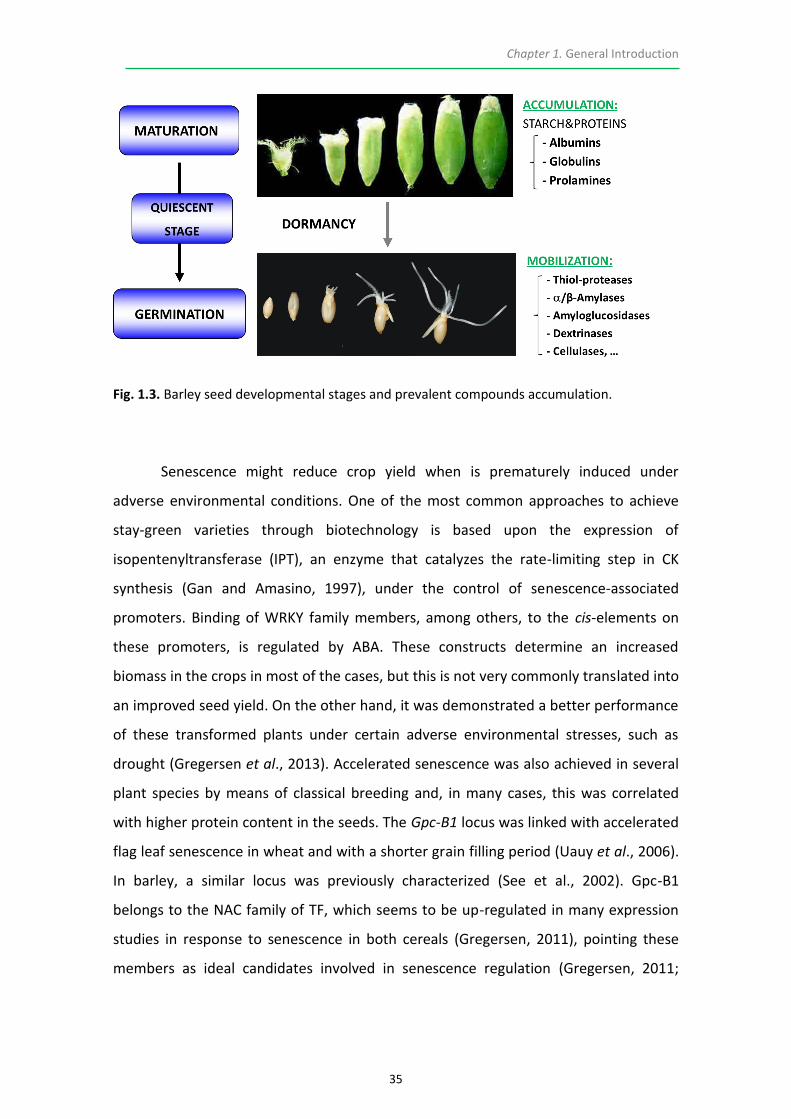
2014; Díaz-Mendoza et al., 2014, 2016b).
1.1.2. LEAF SENESCENCE, GRAIN QUALITY AND YIELD
1.1.2.1. Nitrogen economy in plants: The `Dilemma´ of Senescence and The Stay-
Green trait
The timing of the senescence process affects the length of the photosynthetic period,
thus influencing the grain filling in the case of cereals and therefore determining the
yield and/or the quality. In 2008, Gregersen et al. raised the concept of `Dilemma of
senescence´ questioning wether is better to delay or to accelerate senescence (Fig.
1.2). When late senescence occurs, looking at the grain, a higher carbohydrate (CH)
content and a lower protein accumulation are observed, whereas when senescence
arrives earlier the opposite trend is appreciated. It is paramount to understand the
molecular mechanisms behind senescence in order to improve these traits, depending
on the end-product usage. For instance, applied to barley, delayed senescence would
be desirable for malting purposes, since a higher amount of CH is necessary for
fermentation. Conversely, early senescence would be ideal for manufacturing meals
for animal feeding, since a high content in proteinaceous components is required.
Improvement of cereal cultivars requires a delicate balance among senescence
timing, grain nutrient content, NUE, and yield (Distelfeld et al., 2014). The physiological
stage at which a plant faces a given stress will largely influence upon the activation and
progression of the senescence programs, determining the efficiency in the
remobilization of nutrients as a strategy for survival.

Chapter 1. General Introduction
33
Fig. 1.2. Schematic hypothesis for the breeder´s dilemma. Delayed senescence leads to an extended photosynthetic period resulting in an increased biomass accumulation whereas accelerated senescence provokes a faster remobilization of nutrients translated into an increase in the grain number and size. Gregersen et al., (2014).
“Stay-green” crops have the potential for higher plant yields due to an
extended photosynthetic period. There exists a comprehensive classification for these
mutants: those which present the ability of extending the vegetative phase which
results in higher yields are designated as ‘‘functional stay-green’’ mutants. These
mainly include Type A and Type B. In the first case, senescence proceeds at a normal
rate but its initiation is retarded. In the Type B, the process begins on schedule but is
somehow slowed down, while the active photosynthetic stage is usually prolonged.
“Non-functional stay-green” mutants or cosmetic mutants (type C) are those that
remain green due to impaired chlorophyll catabolism, but lack photosynthetic
competence. These last ones were very helpful when elucidating the enzymes and
steps involved in the route of chlorophyll catabolism (Thomas and Howarth, 2000). In
these mutants, the stay-green character is heritable (Thomas and Ougham, 2014), and
it renders a greener phenotype due to a mutation in the Mendel’s green cotyledon
gene sgr, encoding a chloroplast protein required to initiate chlorophyll degradation
(Sato et al., 2007). Stay-green mutants have been used to understand the correlation
between senescence, yield and total nitrogen content of the grain in several crop
species (Kichey et al., 2007). A wheat stay-green variety presented a reduced harvest

Chapter 1. General Introduction
34
index even after a prolonged grain filling period. It was postulated that remobilization
of carbon was inefficient and that extra photoassimilates remained in the vegetative
parts instead of being translocated to the grain. Nitrogen concentration in the straw of
a stay-green line of wheat remained higher than in controls. Extended photosynthesis
did not mean an increase in grain yield as expected; instead, these plants necessitated
more nitrogen uptake to achieve a grain protein content comparable to that for wild-
type (Chen et al., 2011). Another wheat mutant, tasg1, showing delayed leaf
senescence, was identified as a functional stay-green (Hui et al., 2012). The
explanation for the last examples in which extended photosynthesis did not result in
higher harvest index relies on the fact that sink tissues may have a limitation in their
capacity, which is in term influencing a major trait, the growth and size of the seed
(Serrago and Miralles, 2014). In a detailed transcriptome study performed in barley,
Sreenivasulu et al. (2008) analyzed late seed maturation and initial germination stages.
They concluded that during maturation, the barley grain stores all required compounds
and regulators, among them many TF, meaning that plant seeds prepare for
germination already during seed maturation. This leads to conclude that maturation of
the grain is a crucial developmental stage, and apparently alterations in source/sink
communication influenced by modifications along senescence timing may have
negative effects upon the accumulation of valuable elements required for a later and
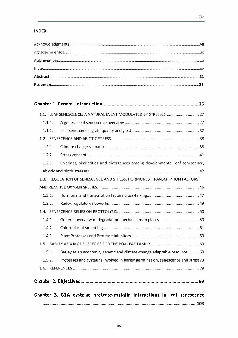
successful germination during next generation (Fig. 1.3).
Chapter 1. General Introduction
35
Fig. 1.3. Barley seed developmental stages and prevalent compounds accumulation.
Senescence might reduce crop yield when is prematurely induced under
adverse environmental conditions. One of the most common approaches to achieve
stay-green varieties through biotechnology is based upon the expression of
isopentenyltransferase (IPT), an enzyme that catalyzes the rate-limiting step in CK
synthesis (Gan and Amasino, 1997), under the control of senescence-associated
promoters. Binding of WRKY family members, among others, to the cis-elements on
these promoters, is regulated by ABA. These constructs determine an increased
biomass in the crops in most of the cases, but this is not very commonly translated into
an improved seed yield. On the other hand, it was demonstrated a better performance
of these transformed plants under certain adverse environmental stresses, such as
drought (Gregersen et al., 2013). Accelerated senescence was also achieved in several
plant species by means of classical breeding and, in many cases, this was correlated
with higher protein content in the seeds. The Gpc-B1 locus was linked with accelerated
flag leaf senescence in wheat and with a shorter grain filling period (Uauy et al., 2006).
In barley, a similar locus was previously characterized (See et al., 2002). Gpc-B1
belongs to the NAC family of TF, which seems to be up-regulated in many expression
studies in response to senescence in both cereals (Gregersen, 2011), pointing these
members as ideal candidates involved in senescence regulation (Gregersen, 2011;

Chapter 1. General Introduction
36
Distelfeld et al., 2014; Christiansen et al., 2016). In fact, it was proposed that NAC TF
might be associated with ABA signaling in plants (Jensen et al., 2007).
1.1.2.2. Coordinated Carbon and Nitrogen assimilation during remobilization events
In cereal species, senescence is predominantly controlled at the level of the individual
leaf, and remobilization usually begins in the older leaves towards the younger ones
and the flag leaf, this last making important contribution to the remobilization of the
major part of photoassimilates during seed filling (Wiedemuth et al., 2005). As
opposed to dicotyledonous species, cereal leaves have a basal meristem, and the leaf
tip consists on the oldest cells while the youngest are at the leaf base (Gregersen et al.,
2008). Therefore, cereals represent suitable models to study the progression of
senescence. Even though interactions between senescence associated-remobilization
and grain filling are complex and poorly understood (Thomas and Howarth, 2000;
Gregersen et al., 2013), a wealth of literature evidence supports the importance of the
remobilization during natural or induced senescence, making special emphasis on
nitrogen remobilization, since nitrogen starvation is a well-known trigger of
accelerated senescence in many crops (Havé et al., 2016). Thereby, several
transcriptomic, proteomic and ultra-structural reports, as well as large and properly
documented revision works, discuss about the topic, focusing on cereals (Gregersen et
al., 2013) and other crop species such as Brassica napus (Avice and Etienne, 2014).
Some authors propose the thorough study of tissue-specific structural modifications in
order to determine possible links with NUE and remobilization during a stress episode,
as observed in the changes of palisade and spongy parenchyma in oilseed rape leaves
during senescence (Sorin et al., 2014).
Phloem-specific metabolic compounds might signal high grain demands for N to
distantly located plant organs (Kohl et al., 2012). Schiltz et al. (2004) analyzed protein
variations during nitrogen mobilization from leaves to filling seeds in pea (Pisum
sativum), proving that a chloroplastic protease (FtsH) increased during N mobilization.
They proposed that a better understanding of the processes occurring during grain
filling from senescing leaves required an estimation of protein turnover by means of

Chapter 1. General Introduction
37
[35S] Met- or [35S] Cys-labeled proteins. Several CysProt and N transporter genes of
the AAT family appeared to play a role in remobilization and accumulation of nitrogen
as observed in a RNAseq analysis of flag leaves, glumes and developing grains from
barley (Kohl et al., 2012). In addition, transcriptomes of flag leaves from field
experiments subjected to variable levels of nitrogen supply were analyzed (Hollmann
et al., 2014). HvPAP-14 and HvPAP-20 encoding CysProt, and SCPL51 encoding a serine
protease, were differentially expressed. HvPAP-20 encodes a cathepsin-B-like CysProt
(Martinez and Diaz, 2008) also known to be upregulated during barley grain
germination (Sreenivasulu et al., 2008; Diaz-Mendoza et al., 2016a).
More than 75% of the potentially remobilizable reduced nitrogen in plants is
located inside the chloroplasts and mainly assembled into Rubisco (Hörtensteiner and
Feller, 2002) and other stromal components, such as glutamine synthetase (GS).
Chlorophyll-apoprotein complexes from thylakoids represent the second major
fraction. Likewise, it was estimated that around 70% of the nitrogen from senescing
vegetative organs is exported during seed development in most annual crop plants
(Peoples and Dalling, 1988). Although a part of ammonia is evaporated from leaves,
the bulk ammonium content is exported from the senescing leaf and utilized to build
new amino acids. An intense traffic of amino acids occurs along the phloem during
developing and maturation grain stages. The major phloem-exported amino acid in
barley and wheat is glutamate (Forde and Lea, 2007), followed by aspartate,
glutamine, threonine and serine (Kichey et al., 2007). Two forms of GS have been
identified in plants, the cytosolic GS1 and the chloroplastic/mitochondrial GS2
(Swarbreck et al., 2011). In non-senescing leaves, GS2 is the abundant isoform in the
mesophyll cells, where it assimilates ammonium originating from nitrate reduction and
photorespiration. During leaf senescence GS1 fulfills a key function in the assimilation
and recycling of ammonium generated from various catabolic processes (Masclaux-
Daubresse et al., 2010). This role is particularly important after anthesis and during
grain development and filling when nitrogen is remobilized to the reproductive sinks
(Kichey et al., 2007; Brauer et al., 2011). Schildhauer et al. (2008) followed the
expression patterns of two genes involved in nitrogen metabolism in barley during
reversal of senescence after supply with nitrogen: GS2 and lysine-ketoglutarate

Chapter 1. General Introduction
38
reductase/saccharopine dehydrogenase (LKR⁄SDH). LKR⁄SDH catalyzes the first two
steps in the degradation of the important amino acid lysine. In Arabidopsis, a higher
total amino acid content in shoots of plants grown under continuous N limitation was
observed in comparison to control conditions; authors explained that there was a
reduced utilization of amino acids for protein synthesis (Tschoep et al., 2009), possibly
as a consequence of a slowdown in the tricarboxylic acid (TCA) cycle, which
determined a general down-regulation of biosynthetic metabolism (Balazadeh et al.,
2014).
A rapid reversion in the cytosolic carbon to nitrogen (C⁄N) ratio is required to
revert leaf senescence. In both barley and Arabidopsis thaliana, senescence can be
completely reversed when additional nitrate is resupplied after a nitrogen starvation
period (Schildhauer et al., 2008). A situation of carbon feast (high CH levels)
undergoing in source senescing organs may act as a first signal to start remobilization
of nutrients; but a state of carbon starvation in the sink organs may also represent the
initial stimulus for beginning the maintenance of molecules (Parrott et al., 2005).
Importantly, a set of proteases were induced under these conditions.
Given the complexity and the lack of precise descriptions on the events taking
place during senescence, either developmental or stress-induced, there is a need to
discern which is the main mechanism involved. Since amino acid and nutrient
transport are usually the main hallmarks, it seems very likely that, in general,
proteolysis represents the ruling process.
1.2. SENESCENCE AND ABIOTIC STRESS
1.2.1. CLIMATE CHANGE SCENARIO
According to recent estimations, world population is increasing at an alarming rate and
is expected to reach about 9 billion by the end of 2050 (http://www.fao.org). These
striking predictions coincide with the pessimistic data concerning climate change.
Rainfall frequency and distribution patterns are expected to vary in most of the

Chapter 1. General Introduction
39
regions. Precipitations will come in the form of severe storms, with irregular trends
followed by scarcity periods, which will be translated into flooding and drought
episodes, closely linked to the loss of soil fertility and increased salinity. In addition,
extreme temperatures are also predicted to represent important threats for
agriculture (Fig. 1.4), either due to increased temperatures or chilling and freezing
events (Cutforth et al., 2007; Jury and Vaux, 2007; Manavalan et al., 2009; Simova-
Stoilova et al., 2010). Such trends in predicted weather patterns are seriously
threatening plant development and seed production in agricultural lands (United
Nations Convention to Combat Desertification, FAO, 2014), with drought representing
the most harmful stressor limiting crop yield, thus being one of the major constraints
to global food security (Godfray et al., 2010). Moreover, apart from abiotic
environmental cues, the occurrence of biotic stresses may be also triggered under
these conditions, stimulating the expansion of plant diseases and pests into new
geographical areas, wreaking havoc on crop performance (Fig. 1.5). For instance, the
invasive insect and acari species are expected to cause severe damage on plants under
climate change episodes (Atkinson and Urwin, 2012; Ximénez-Embún et al., 2016).
To meet the increasing nutritional demands for this sharp growing population
and avoid a food crisis, there is an urgent need to design strategies in order to reach
productions around 70% higher than the current ones by the year 2050 (Mahajan and
Tuteja, 2005). This ideal framework should be achieved without land surface
expansions and with a reduction in the inputs based on inorganic fertilizers, which
damage soil fertility (Comadira et al., 2015).
This background leads the scientific community to implement projects focused
in ameliorating resistance, tolerance or acclimation in relevant crops, with a special
focus on drought, temperature and/or salinity among other relevant constraints, by
means of plant biotechnology.

Chapter 1. General Introduction
40
Fig. 1.4. Predicted change in average surface temperature for the interval (A) 1986-2005 and (B)2081-2100; and predicted change in average precipitation for the interval (C) 1986-2005 and (D) 2081-2100 (IPPC, Synthesis Report for Climate Change, 2014). https://www.ipcc.ch/report/ar5/syr/
Fig. 1.5. Estimated risks for food production (% of yield projection, increased or decreased) posed by climate change for different year intervals (IPPC, Synthesis Report for Climate Change, 2014). https://www.ipcc.ch/report/ar5/syr/

Chapter 1. General Introduction
41
1.2.2. STRESS CONCEPT
Very recently, Gilbert and Medina (2016) published a useful document in which they
give proper definitions related to stress, adaptation and drought. The home message
indicates that it is essential to think about the precise mechanism on which the
research will be focused on when initially designing an experiment. This is sometimes
obviated and results from various investigations are somehow uncertain and
confusing. They define stress `as a negative change in the physiology of a plant away
from a reference state as a result of the action of an external stress factor or internal
stress´. They consider ‘stress factors’ as external and ‘stresses’ as physiological
responses. Albeit the manuscript is based on drought, we could make a general
assumption for all environmental factors. Hence, they describe four hypothetical cases
for drought: “SWD (Soil Water Deficit) avoidance” refers to a mechanism that, for
instance, includes plants that explore deeper soils or match phenology to the wet
season; “stress avoidance” requires more specialized mechanisms such as succulence;
those adaptations that allow plants to tolerate some negative external factor are
included in “damage avoidance” term, for example through changes in leaf orientation
or altered root to shoot ratios; lately, “damage tolerance” refers to the state at which
plants may tolerate damage through recovery mechanisms, for instance during the
night-time or by generating new conductive tissue. All those mechanisms rely on
physiological, metabolic and biochemical changes determined by alterations in gene
expression. For this reason, knowledge about adaptation mechanisms at the
macromolecular level is essential, but it needs to be intimately linked to a perfect
comprehension of molecular interplays. A rising number of research papers shows, in
most of the cases, adaptation mechanisms responding to the last definition listed
above: damage avoidance through recovery mechanisms (Simova-Stoilova et al., 2010;
Dinakar and Bartels, 2013).

Chapter 1. General Introduction
42
1.2.3. OVERLAPS, SIMILARITIES AND DIVERGENCES AMONG DEVELOPMENTAL LEAF
SENESCENCE, ABIOTIC AND BIOTIC STRESSES
Responses to abiotic stresses resemble, in many molecular and phenotypic aspects,
the plant senescence syndrome. According to recent studies, plant stress tolerance,
apart from crop yield and nutritional values, may be modified through manipulating
the timing of senescence (Gepstein and Glick, 2013). Much investment has been made
towards identification of stress-protective or adaptation-related genes activated
during abiotic stress (Bray et al., 2000). Overexpression of these genes could help
plants to increase tolerance. A wide spectrum of reports demonstrates the potential of
abiotic stresses to trigger leaf senescence by reprogramming specific subsets of SAGs
differentially expressed in distinct tissues and several species, including crops. For
instance, in order to detect senescence-associated physiological changes involving SAG
expression in wheat, detached leaves were subjected to several abiotic and hormonal
treatments. TaSAG3 and TaSAG5 were expressed in natural senescent leaves and
showed differences in expression patterns depending upon the treatment, although
both were upregulated immediately after leaf detachment (Zhao et al., 2012). In sweet
potato, the calmodulin gene SPCAM is NaCl-inducible and participates in salt stress-
mediated leaf senescence regulating the expression of specific SAGs (Chen et al.,
2012). Evident effects upon modulation of salt- and osmotic-induced leaf senescence
in Capsicum annuum L. were likewise observed when downregulating the CaCP gene in
this species (Xiao et al., 2014).
Light is essential for photosynthesis and acts as the main signal for natural
development and interactions with the environment. Its deprivation potentially leads
to sugar starvation, which is already known to be one of the signals promoting
senescence (Parrott et al., 2005). Dark-induced senescence results in chlorophyll loss,
slowdown of photosynthetic activity and dismantling of cellular constituents, in a
similar manner to that observed during age-dependent natural senescence (Fujiki et
al., 2001; Buchanan-Wollaston et al., 2005). Variations in the light intensity modulate
the timing of senescence and, under certain conditions, the senescence process may
be reversed (Humbeck and Krupinska, 2003). Although darkness cannot be considered

Chapter 1. General Introduction
43
as a true abiotic stress in nature, apart from those events regarding extreme shading of
the lower parts in dense canopies, it has been extensively used to analyze mechanisms
of leaf senescence in plants based on its immediate effects on the photosynthetic
machinery (Gan, 2007). Sometimes, the dark treatment was applied on entire and
intact plants (Buchanan-Wollaston et al., 2005), but in most cases it was used in
detached leaves (Fischer and Feller, 1994; Chiba et al., 2003; Thoenen et al., 2007;
Zhao et al., 2012), provoking rapid genome-wide alterations and metabolic responses,
which helped to elucidate specific gene functions. Dark-induced senescence has been
extensively used since several decades both in Arabidopsis (Keech et al., 2007; Niu and
Guo, 2012) and in crops, mostly in cereals (Kleber-Janke and Krupinska, 1997; Chrost et
al., 2004). These experiments have shed light about regulatory networks, as
demonstrates the darkness-induced transcription of AtWRKY22 that suggests its
participation in the signal transduction pathway mediated by this abiotic stress in
Arabidopsis (Zhang and Zhou, 2013). More recently, it was reported that Phytochrome-
Interacting Factors 4 and 5 from Arabidopsis promoted dark-induced and natural
senescence by directly activating the expression of typical SAG like ORESARA1 (ORE1)
and ETHYLENE INSENSITIVE3 (EIN3) (Piao et al., 2015). The implication of autophagy-
related pathways during senescence was recently demonstrated using darkness (Avila-
Ospina et al., 2016). Besides, in a broad range of darkness-based studies,
overexpressed SAGs corresponded to CysProt (Parrott et al., 2005; Thoenen et al.,
2007; Watanabe et al., 2009; Carrión et al., 2013).
Drought stress is produced when evapotranspiration rate exceeds the amount
of absorbed water through root tissues (Lawlor and Cornic, 2002). In other words, it
involves `a decrease in water inputs into an agro/ecosystem over time that is sufficient
to result in SWD´ (Kramer, 1983). At field capacity, this situation is associated with high
temperature episodes which results in a negative synergic effect causing severe
damage to crops and production losses up to 50% (Bray et al., 2000). The severity of
drought stress, besides its duration and intensity, will produce a greater or lesser
impact depending upon the plant developmental phase at which the stress is faced. If
this occurs along the vegetative growth phase, the stress is mostly transient and plant
growth slows down to be restored after a new rainfall period, as exemplified in pre-

Chapter 1. General Introduction
44
summer drought events. The plants may even start wilting and yield is normally
impacted since a lower number of ear-bearing tillers per plant are often quantified
(Sreenivasulu et al., 2008). Premature leaf senescence, causing acceleration of the
whole-plant maturation, is usually detected when drought episodes arise during the
generative plant development, i.e., around flowering (Gan, 2007). A considerable set of
research is focused on the roles of proteases and their inhibitors in relation with
drought-induced senescence, since enhanced expression of genes coding for proteases
is a common event both in senescence and under various environmental stresses
(Simova-Stoilova et al., 2010.) As reported in an expression profiling of genes in
juvenile barley, significant correlations exist within the group of genes involved in
drought stress and those acting in leaf senescence (Wehner et al., 2016). Neverthless,
in some cases the proteases expressed during drought episodes may substantially
differ from those expressed during senescence (Simova-Stoilova et al., 2010). Drought-
induced and natural senescence were monitored in the cowpea leaf, with a focus on
CysProt, concluding that the abiotic stress induces many forms of these proteases not
observed during developmental senescence (Khanna-Chopra et al., 1999). The
involvement of acidic proteases in soil drought response of winter wheat in three
cultivars differing in water stress tolerance was likewise studied. Results suggested
that lower proteolytic activity and decreased expression of certain protease genes
under water deficit during early developmental stages could be regarded as an
indicator for drought resistance of winter wheat cultivars (Simova-Stoilova et al.,
2010).
Plants respond to water stress generally by synthesis of ABA, inhibition of
photosynthesis and respiration, accumulation of osmotically active compounds,
synthesis of protective proteins, such as dehydrins and chaperones, by adjusting
sink/source allocation and by speeding up senescence (Simova-Stoilova et al., 2010).
Suppression of drought-induced leaf senescence in transgenic tobacco plants caused
by the accumulation of CK due to IPT overexpression has been linked to an increase of
dehydrins and heat shock proteins (Rivero et al., 2007). Production of CK also
contributed to an enhanced drought tolerance in transgenic cassava and peanut (Qin
et al., 2011). It has been illustrated that the application of exogenous ABA combined

Chapter 1. General Introduction
45
with salinity stress provokes the over-expression of SAGs and the acceleration of
senescence (Yang et al., 2003), suggesting the connection among leaf senescence, ABA
and abiotic stress signaling (Podzimska-Sroka et al., 2015). Besides, drought-induced
ABA was positively and significantly correlated with carbon remobilization from
senescing leaves to grains in wheat plants subjected to drought stress (Yang et al.,
2003). Potassium alterations also affect stress tolerance (Restrepo-Diaz et al., 2008).
Drought stimulated signal transduction chains involving ROS and Ca2+ signaling lead to
the induction of K+ transporters and channels in roots and guard cells (Cheong et al.,
2007). Barley genotypes with a higher K+ nutritional status in the flag leaf showed
superior drought tolerance by promoting ABA degradation and attenuation of starch
catabolism, which delays flag leaf senescence (Hosseini et al., 2016).
Few studies give precise information about interactions between biotic stresses
and leaf senescence and, within, those regarding pests are even less abundant than in
the case of diseases. The effects and interactions of biotic stress and senescence may
be interpreted in two ways: either the presence of a biotic factor promotes senescence
after surpassing plant defenses through modification of common SAGs implied in
primary metabolism; conversely, it could happen that senescence is already activated
in the plant, naturally or induced by some abiotic factor, thus making the plant more
prone for the establishment of pests and pathogens (Masclaux-Daubresse et al., 2010;
Fagard et al., 2014; Ximénez-Embún et al., 2016). The first assumption would be the
case, for instance, for an early senescence and premature cell death detected after
down-regulation of OsSAG12-1 in rice, after inoculation with Xanthomonas oryzae
(Singh et al., 2013). Likewise, green peach aphid infestation in Arabidopsis accelerated
senescence-like mechanisms as resembled in the elevated expression of several SAGs
(Pegadaraju et al., 2005; Louis et al., 2010). The mechanisms that allow acclimation
and adaptive responses to isolated biotic and abiotic stresses have been extensively
characterized in a wide range of studies. Nevertheless, regarding combined
biotic/abiotic stress responses, little information is still available. Deciphering the
signaling pathways participating in the common crosstalk drawn by biotic and abiotic
stresses will allow the identification of new targets for increasing environmental
resilience in crops (Foyer et al., 2016). `The exposure to one type of stress confers a

Chapter 1. General Introduction
46
general increase in resistance to a range of different stresses, a phenomenon called
cross-tolerance´. This phenomenon relies on the synergistic co-activation of non-
specific, stress-responsive pathways that cross biotic–abiotic stress boundaries, and
which are usually related with altered redox and phytohormone signaling (Foyer et al.,
2014). Cross-tolerance to different stresses triggered by an exposure to a single stress
is widespread in plants (Munné-Bosch et al., 2013). Mechanisms of adjustment to
water deficit may be associated to an enhanced cotton resistance to mites (Sadras et
al., 1998). As aforementioned, ABA is very important during the response to drought
episodes. Through stomatal closure, the ABA- induced signaling pathway intersects
with both abiotic and biotic stress factors (Lee and Luan, 2012). In maize, differences at
the proteome level were detected depending if two stresses, drought and the
presence of the two-spotted spider mite, were applied individually or combined
(Dworak et al., 2016).
1.3. REGULATION OF SENESCENCE AND STRESS: HORMONES,
TRANSCRIPTION FACTORS AND REACTIVE OXYGEN SPECIES
Phytohormones are key regulators of leaf senescence (Li et al., 2012). The increasing
knowledge on this regulatory field relies on several investigations about hormonal
responses during abiotic and/or biotic episodes. The signaling processes involving plant
hormones are modified under the presence of an external cue which triggers
alterations in stress-responsive genes that eventually interfere with developmental
senescence pathways (Zhang and Zhou, 2013). Several plant hormones, such as
ethylene, ABA, SA and JA induce or accelerate senescence, as well as small molecules,
such as oxygen. Conversely, CK, auxins, nitric oxide (NO) as well as molecules like Ca2+,
may retard leaf senescence (Li et al., 2012). Regarding developmental senescence, it
appears that its onset is mainly regulated by CK levels, which seem to decrease along
this event. Several groups induced the expression of this hormone through alterations
in its biosynthetic route in order to delay the process (Podzimska-Sroka et al., 2015).
Ethylene has a key role during fruit ripening and during the regulation of the onset of
senescence, apart from being an important molecule tuning germination and seedling
development (Podzimska-Sroka et al., 2015). Endogenous ABA levels increase during

Chapter 1. General Introduction
47
senescence (van der Graaff et al., 2006), although this phytohormone is considered
more an enhancer rather than a triggering factor. Furthermore, under drought, salinity
or extreme temperature episodes, the content of ABA usually rises, which is consistent
with its proved importance in the response towards abiotic stress (Guo and Gan,
2005). In plants defective in the SA pathway, developmental leaf senescence is
delayed, but this is not observed during dark-induced conditions, which illustrates the
relevance of this hormone specifically during natural senescence (Buchanan-Wollaston
et al., 2005). Strigolactones are a class of plant hormones that fulfill several functions
in plants, like the regulation of shoot branching and root architecture, as well as the
mediation during leaf senescence and stress. The production of strigolactones is
induced in response to nitrogen and phosphorous deficiency (Yamada and Umehara,
2015). Cystatins, widely known to be key regulators of CysProt activity in plants, are
being successfully utilized in several biotechnological approaches towards stress
tolerance. In a specific research in which the rice cystatin (oryzacystatin OCI) was
ectopically expressed in soybean and Arabidopsis, plants presented an enhanced
drought tolerance compared to WT presumably through effects on strigolactone
pathways (Quain et al., 2014).
1.3.1. HORMONAL AND TRANSCRIPTION FACTORS CROSS-TALKING
Various TF families, such as NAC and WRKY, seem to be potentially involved in the
regulation of senescence and stress response (Guo et al., 2004; Christiansen and
Gregersen, 2014; Christiansen et al., 2016). Some of them were discovered during
forward genetic screenings used to characterized Arabidopsis mutants with altered
leaf senescence (Breeze et al., 2008). Some of these mutants presented alterations
related to hormonal signaling (Woo et al., 2004; Kim, 2006). For instance, in a
subtractive hybridization experiment, the WRKY53 gene was identified and designated
as a potential positive regulator of senescence, as illustrated knockout mutants with
delayed senescence, whereas overexpression lines senesced prematurely (Miao et al.,
2004). In addition, WRKY53 seemed to be possibly involved in the interaction of SA and
JA signaling pathways along this process (Miao and Zentgraf, 2007).

Chapter 1. General Introduction
48
The NAC family of TF is plant-specific, contains 117 members in Arabidopsis,
151 in rice, and 152 in both soybean and tobacco. Transcriptome analyses have
associated approximately one third of Arabidopsis and many crop NAC genes with
senescence, pointing them out as crucial regulators (Podzimska-Sroka et al., 2015). To
date, ~50 NAC genes have been identified in barley (Christiansen et al., 2016).
HvNAC005 is associated with developmental senescence in this species, being
significantly up-regulated after ABA treatment, but not during dark-induced
senescence. Accordingly, RNA interference studies of wheat Gpc-B1, encoding the NAC
protein NAM-B1, presented a significant reduction in grain zinc, iron, and protein
content in parallel with a delayed leaf senescence phenotype (Uauy et al., 2006;
Waters et al., 2009). In adittion, a positive regulator of senescence in rice, OsNAP,
interfered during the grain-filling period affecting harvestable indexes (Liang et al.,
2014). Another example implies the family or the drought-responsive element binding
proteins (DREBs). DREB2 and DREB3 genes were overexpressed under the control of
constitutive duplicated CaMV35S promoter and drought-inducible ZmRAB17 promoter
from maize. Transgenic barley plants carrying these constructs were more tolerant to
drought without undesirable effects on plant growth and development (Mrízová et al.,
2014). Besides, Je et al. (2014) demonstrated that DREB2 acts as transcriptional
activator of the thermotolerance-related cystatin 4 gene from Arabidopsis, with a
concomitant and expected reduction in the associated protease activity.
In addition, the single stranded DNA binding protein (ssDNA) WHIRLY-1 seems
to be a promising target in order to mitigate several stress signals. WHIRLY-1 was
shown to bind to the promoter of the senescence-associated gene HvS40, a marker for
leaf senescence in barley, which is induced both under natural and stress-induced
senescence. WHIRLY-1 is expected to fulfill an important role during retrograde
signaling between plastid and nucleus (Krupinska et al., 2014b; Foyer et al., 2014),
which is in accordance with its first demonstrated function regarding nucleoid
compaction inside the chloroplasts (Krupinska et al., 2013). This hypothesis was made
over the fact that WHIRLY-1 is a thioredoxin target (Foyer et al., 2014), so it may act as
a sensor of ROS alterations provoked by stress signals, which are first sensed in the
chloroplasts. The WHIRLY-1 protein would change its structural conformation after

Chapter 1. General Introduction
49
these signals, leading to a monomerization process. WHIRLY-1 monomers would reach
the nucleus, where they may activate specific-stress and senescence-responsive genes,
as those related with photosynthesis, possibly enabling NUE improvement in barley
(Comadira et al., 2015).
1.3.2. REDOX REGULATORY NETWORKS
ROS conform one of the earliest responses of plant cells under abiotic and biotic
stresses, and senescence. Oxygen free radicals interplay with sugars and nitrogen
content, photoperiod signals, calcium cascades, and a big set of phytohormone signals
acting synergistic or antagonistically to trigger and regulate leaf senescence. As
chloroplasts are the main targets of ROS-linked damage during various environmental
stresses and natural senescence, it is noteworthy that a well suited arsenal of
antioxidant systems is contained inside this organelle (Khanna-Chopra, 2012). ROS
behavior could be interpreted in two senses: as degrading harmful molecules when an
excessive concentration is surpassed or as necessary signaling compounds. ROS
signaling is controlled by production and scavenging, in contrast to Ca2+ signaling,
which is based upon storage and release (Bieker et al., 2012). The primary targets of
ROS signals are amino acids such as cysteine. ROS accumulation is also intrinsically
linked to the production of NO. Redox gradient across the plasma membrane also
deserves special attention regarding signaling in response to stress, since it seems to
be directly involved in the regulation of specific protease-encoding genes that target
proteins with carbonyl groups for degradation. Cellular redox state alterations imply a
feed-forward re-amplification of the MAPK cascades, which function downstream of
sensors/receptors to transform extracellular stimuli into intracellular responses,
amplifying the transducing signal along the process (Munné-Bosch et al., 2013).
ROS are common elements in response to nutrient deficiencies. Interestingly, N
deficiency promotes callose accumulation in vascular bundles and restricts the grain-
filling rate in wheat coinciding with high ROS detection. A lower load of amino acids in
the phloem after N deprivation increases the oxidation state in the cells promoting
earlier senescence responses. As callose deposition at the neck region of

Chapter 1. General Introduction
50
plasmodesmata determines its permeability during grain filling events, alterations in
ROS metabolism will directly impact on macromolecular trafficking to the seed. The
described situation is deleterious for seed quality and yield; however, it is necessary
for the initiation of grain filling, since a minimum amount of ROS is required in order to
activate senescence-associated remobilization of nutrients (Taylor et al., 2012).
Drought is also considered to significantly increase the accumulation of ROS (Noctor et
al., 2014). Likewise, ROS mediated in biotic stress responses, interacting with hormonal
cascades (Kerchev et al., 2012). Interestingly, the progression of senescence along and
throughout the plant architecture in tobacco plants also reflected ROS-related
changes. Leaf interveinal and veinal regions senesced in a different manner in this
species, due to a spatial heterogeneity in the accumulation of H2O2 and O2- which
differentially regulated the expression of SDGs and SAGs. Authors speculated that
integrity and compartmentation of the leaf are maintained until very late stages of
senescence, enabling an efficient recycling (Niewiadomska et al., 2009).
1.4. SENESCENCE RELIES ON PROTEOLYSIS
1.4.1. GENERAL OVERVIEW OF DEGRADATION MECHANISMS IN PLANTS
Under stressful situations plants undergo changes related to their primary CH
metabolism, using three main alternative substrates to obtain energy: proteins,
chlorophylls and lipids (Hörtensteiner, 2013). Since plants are sessile organisms,
recycling is especially important in order to face stress-induced senescence and avoid
dramatic consequences over the offspring (Avila-Ospina et al., 2016). Plants use an
escape strategy based upon a rapid degradation of source tissues and an accelerated
development of sinks, therefore accelerating and ensuring seed production for next
generation. Proteolysis, being the main hallmark during senescence and stress, fulfills
the main goal of providing free peptides or amino acids to redistribute them within the
plant. Amino acid catabolism will support the TCA cycle and will supply the
mitochondrial electron transport chain (Araújo et al., 2011). Knowledge on which are
the natural substrates for proteases is very limited due to the difficulty of its
identification and the existence of functional redundancy (Avila-Ospina et al., 2014).

Chapter 1. General Introduction
51
Several research teams are focusing their efforts in order to elucidate which are the
key counterparts involved during senescence associated proteolysis, i.e., which are the
targets for proteases (Kidrič et al., 2014; Tsiatsiani et al., 2012).
1.4.2. CHLOROPLAST DISMANTLING
Chloroplast proteins represent the main source for nutrient remobilization, especially
nitrogen (Schiltz et al., 2004; Avila-Ospina et al., 2014). Most studies have been
performed in this organelle rather than in the rest of cell compartments, although it
was recently postulated that the mitochondria, the plasma membrane and the
apoplast exert key signaling-related functions regarding nutrient remobilization during
senescence (Martínez and Guiamet, 2014). Whilst chloroplast disorganizes, the
photosynthetic capacity of the leaf progressively decreases (Sato et al., 2007; Chen et
al., 2010). Plastid disorganization is reversible and the rest of cell organelles remain
practically intact until the end of senescence (Taylor et al., 2012). As it is shown in Fig.
1.6, different environmental stresses may lead to different energetic cell status and
this will determine the nature of degradation during senescence, presumably
prevailing chloroplast, cytosolic or vacuolar proteases depending on the required
mechanism (Carrión et al., 2014). Ubiquitin-related pathways could be only directed to
specific cytosolic but not chloroplastic proteins during senescence. It was proposed
that the ubiquitin-26S proteasome pathway mediates senescence-associated protein
degradation through the ubiquitin N-end rule pathway, removing proteins which are
mislocalized or unprocessed. At the onset of senescence, a negative regulator in the
cytosol may be degraded through this system promoting a response which would
trigger the dismantling of the chloroplast (Watanabe et al., 2009). In order to give
precise information, an appropriate distinction between stromal (mainly participating
in CO2 fixation) and thylakoidal components (proteins, pigments and cofactors
participating in light-harvesting processes within the photosystems and during the
electron transport chain), should be established.
Chapter 1. General Introduction
52
1.4.2.1. Stromal components
Rubisco and chloroplastic GS2 are the most studied stromal proteins recycled during
senescence (Feller et al., 2008a; Ishida et al., 2008). Other enzymes, as those related to
sulphur assimilation, have been poorly investigated. Most evidences established that
degradation of these stromal proteins generally accounts earlier than chlorophyll and
thylakoidal ones (proteins D1, LHCII of the PSII reaction center and PSII antenna;
Krupinska et al., 2012).
Fig. 1.6. Schematic representation of different cellular compartments: nucleus, Golgi apparatus, endoplasmic reticulum (ER), chloroplast, mitochondria, proteasome, cytosol, Rubisco containing bodies (RCB), Senescence Associated Vesicles (SAVs), Chloroplast Vesiculation-Containing Vesicles (CCVs), vacuole, apoplast and cell wall. The group of proteases identified in each compartment is shown: CysProt (CP), serine-proteases (SP), metallo-proteases (MP), threonine-proteases (TP) and aspartic-proteases (AP). From Diaz-Mendoza et al. (2016b).
Rubisco and Rubisco activase (the main regulator of Rubisco activity) appear to be the
main targets for CysProt during foliar senescence (Prins et al., 2008), but Rubisco

Chapter 1. General Introduction
53
activase is still poorly studied. In C4 plants, Rubisco, PEPC and pyruvate
orthophosphate dikinase are the soluble proteins which, together, represent the same
percentage as Rubisco alone in C3 plants (Feller et al., 2008b). For this reason,
understanding the mechanisms of Rubisco degradation has become a key purpose for
several research groups. Desimone et al. (1996) studied the nature of Rubisco
degradation under oxidative stress in isolated chloroplasts of barley. Likewise, Irving
and Robinson, (2006) investigated Rubisco degradation events in cereals. Through a
proteomics approach, Schiltz et al. (2004) confirmed that Rubisco was the main source
of nitrogen during seed filling in pea. Under different environmental conditions, the
kinetics of Rubisco degradation may vary while comparing to other stromal and
extraplastidial proteins (Feller et al., 2008a). Initiation of Rubisco degradation may
account both near the C-terminus or the N-terminus (Thoenen et al., 2007). How
proteolysis is promoted and initiated is being investigated. Several hypotheses stand
that ROS might be involved, in terms of oxidizing certain cysteine residues, thus
converting the protein in a more susceptible target for protease cleavage (Desimone et
al., 1996), or “tagging” the protein for further degradation, provoking its denaturation
through cross-linking of SH-groups (Garcia-Ferris and Moreno, 1994). It has been
proposed that Rubisco fragments show an increased affinity to bind to the chloroplast
external membranes (Irving and Robinson, 2006) facilitating its exit from this organelle.
One of the most abundant amino acids found in the phloem of several cereal species
undergoing leaf senescence is glutamine (Simpson and Dalling, 1981). The plastidial
form (GS2) is quite susceptible to proteolysis as shown in isolated tobacco chloroplasts
and is lost during early stages in cereal leaves, but the cytosolic (GS1), key enzyme for
ammonia assimilation and de novo synthesis of amino acids from proteolyzed
fragments, is maintained (Khanna-Chopra, 2012). Proteolysis of GS2 seems to be
initiated through oxidative carbonylation of histidine residues (Ishida et al., 2008),
although this ROS prompted degradation does not seem to be enough for complete
degradation (Desimone et al., 1996). Some in vitro approaches showed that GS2 is
degraded before other enzymes from carbon assimilation (e.g. Rubisco) (Thoenen et
al., 2007). Besides Rubisco and GS2, Fischer and Feller, (1994) examined another set of
plastidial enzymes by inmunoblotting in young winter leaves. Results proved that

Chapter 1. General Introduction
54
proteolysis within the same organelle may be selectively regulated, since they
observed different degradation rates.
1.4.2.2. Thylakoidal proteins
Degradation of proteins from PSII, which includes the reaction centers of
photosynthesis and antenna systems, represents the second largest pool of
remobilizable nitrogen from chloroplasts during leaf senescence, harboring 30% of the
total chloroplast protein (Matile, 1992; Feller et al., 2008a). While disorganization of
thylakoids proceeds and gerontoplasts develop, a proliferation of plastoglobuli is
observed (Sorin et al., 2014). These formations contain rests of thylakoid galactolipids,
probably hydrolyzed via the glyoxylate cycle, triggering signals for the rest of
components ´degradation (He et al., 2002). Apart from membrane disassembling, the
core photosynthetic proteins belonging both to PSI and PSII are also proteolyzed as
observed in ultrastructural studies (Ghosh et al., 2001; Krupinska et al., 2012). The loss
of cytochrome b6⁄f may precede degradation of photosystems I and II, and of ATP
synthase. At early phases of senescence, the yield of PSII remains quite constant,
meaning that this compartment seems to be dismantled later (Guiamet et al., 2002).
Although consensus concerning that a certain degree of damage in thylakoids before
chlorophyll degradation must exist, different theories regarding the sequence of
degradation events over thylakoids have emerged. Some authors reported that PSII
activity decreases earlier than that of PSI during senescence, whilst others sustained
the opposite (Ghosh et al., 2001). A faster declined in PSII versus PSI was detected
during heat-stress promoted leaf senescence in wheat (Hörtensteiner, 2013). Relative
levels of some key components of PSI and PSII, such as D1 (PsbA) or Lhcb1 apoprotein,
were analyzed in a high–yield variety of barley at late stages of senescence.
Immunoblot and ultrastructural results illustrated a preferential degradation of grana
over stromal proteins, resulting in an unexpected increase in the chlorophyll a/b ratio
indicating that chlorophyll b is degraded faster (Krupinska et al., 2012). Authors
speculated that this might be a means for avoiding the risk of photoinhibition by
overexcitation of PSII, which is favorable to maintain ATP machinery in stroma
thylakoids, to prevent overproduction of singlet oxygen and thus, to sustain higher

Chapter 1. General Introduction
55
yields (Apel and Hirt, 2004). Although in most species grana thylakoids show higher
stability compared to the stromal fraction, from these results it could be asserted that
there is a wide range of variability among and within species, varieties and even
different aged- chloroplasts.
A battery of studies has reported that the proteolytic machinery responsible for
degradation of thylakoid proteins during senescence is within the chloroplast. It
includes ATP-dependent proteases from Deg, Clp, FtsH and Lon families. FtsH
proteases are the best studied metalloproteases in plants (Roberts et al., 2012). Leaf
proteome analysis from genotypes with different NUE in oilseed rape reported that a
plastidial FtsH plays a significant role in the breakdown of thylakoid proteins (Avice and
Etienne, 2014). Nine members from the FtsH family in Arabidopsis contributed to
plastid differentiation and repairing of the core protein of PSII, D1. As this status was
also contemplated during senescence, it may be contributing to degrade this core
protein (Carrión et al., 2014). Zelisko and Jackowski (2004) demonstrated that a similar
metalloprotease was involved in the degradation of Lhcb3 in detached barley leaves
induced to senesce under darkness.
1.4.2.3. Chlorophylls
The main hallmark of senescence, leaf yellowing, responds to the preferential
degradation of chlorophyll over carotenoids (Matile, 1992). Chlorophyll degradation
pathway owes its name to the first involved enzyme: PAO “pheophorbide a
oxygenase”. This important catalytic route is divided into two steps: formation of
primary fluorescent chlorophyll-derived catabolites (pFCC), and modification followed
by isomerization to render non-fluorescent catabolites (NCCs). The first phase occurs
within the plastid and the second takes place in the cytosol and central vacuole
(Hörtensteiner, 2013). Although chlorophylls contain much N, the 4 moles of N
associated with each mole of chlorophyll are not exported from senescing leaves. It
was demonstrated that colorless chlorophyll catabolites are not exported outside the
cell but remains within the central vacuole in the form of linear tetrapyrrolic
catabolites to further provide required by-products. A fine-tuned mechanism avoiding

Chapter 1. General Introduction
56
accumulation of phototoxic pieces derived from chlorophyll catabolism was described
within plant cells (Christ and Hörtensteiner, 2014). First studies on chlorophyll
catabolism were performed with barley, and transformation of chlorophyll b into
chlorophyll a before degradation was detected (Matile et al., 1992). Some
experimental evidences have tried to correlate to some extent the involvement of
proteases during chlorophyll degradation. Arabidopsis expressing SPCP2, encoding a
sweet potato papain-like CysProt, showed that the decrease in chlorophyll in senescing
leaves did not correlate with the induction of the protease, concluding that SPCP2 is
not related to chlorophyll degradation initiation (Chen et al., 2010).
Few evidences support that SGR protein, lacking in stay-green mutants (sgr),
has the key role of binding and destabilizing LHII during the initial steps of senescence
(Thomas and Ougham, 2014). Various experimental approaches showed that SGR is
acting just at the onset of senescence and it may act destabilizing the protein-
chlorophyll complexes releasing apoproteins and pigments. It was postulated that SGR
protein may be involved in the general process of N remobilization through the
recruitment of specific proteases (Ghosh et al., 2001). After activation of proteins like
SGR during senescence, partial destabilization of pigment-protein complexes occur
inside the chloroplast. Then, some structural changes take place, which may allow the
correspondent protease/s to access to their target apoprotein/s (Schelbert et al.,
2009). In addition, Sato et al. (2007) suggested the implication of SGR in the activation
of chlorophyll-degrading mechanisms in senescing tissues through the characterization
of sgr mutants displaying higher stability of chlorophyll-protein complexes, but
decreased levels of Rubisco than WT.
1.4.2.4. Traffic of proteolytic products
Observed mechanisms for chloroplast degradation along leaf senescence point out the
existence of different proteolytic pathways, inside and outside the organelle, which
may indicate the existence of redundant routes for photosynthetic protein
degradation. Conversely, they may represent alternative ways of responding to
different stresses and to meet variable needs, which in turn determines the

Chapter 1. General Introduction
57
preferential degradation of one or another component in an spatial-temporal scale
(Guiamet et al., 2002; Xie et al., 2015).
Over the past years, biochemical, ultrastructural and molecular procedures
have been developed in order to gain knowledge about the mechanisms for trafficking
and degradation of compounds outside chloroplasts. Senescence Associated Vesicles
(SAVs), Rubisco Containing Bodies (RCBs) linked to autophagy, and more recently,
Chloroplast Vesiculation-Containing Vesicles (CCVs), have been proposed as the main
mechanisms (Fig. 1.7). As it was demonstrated in Arabidopsis, soybean and tobacco
plants, SAVs, small acidic organelles with a single membrane, only carried stromal
proteins and contained most of the CysProt activity in senescing cells (Otegui et al.,
2005; Prins et al., 2008; Martínez et al., 2008; Carrión et al., 2013). Thylakoid
components were not detected, although a small portion of chlorophyll a appeared
under certain conditions, which authors attributed to possible differences in pigment
disassembly (Martínez et al., 2008). Besides, SAVs seemed independent of autophagy
and they did not appear to reach the central vacuole, exhibiting an independent
proteolytic activity (Xie et al., 2015). Data concerning SAVs formation and how the
proteinaceous components enter and bind to the lumen still remain elusive. Likewise,
there are few evidences regarding autophagy and RCBs formation. In this case,
cytosolic components are engulfed by a specialized double-membrane structure
(autophagosome) to finally attach to the vacuole and deliver carried components for
recycling. Electron microscopy observations confirmed stromules protruding from
chloroplasts and RCBs, which again only contained the stromal fraction, lacking visible
proteolytic activity (Chiba et al., 2003; Ishida et al., 2008). Interestingly, authophagy-
mediated processes are involved in the delivery of starch granules derived from the
chloroplast to the vacuole during the night, a process not necessary linked to stress.
Importantly, it has been postulated that autophagy mediates in nitrogen
remobilization during grain filling in Arabidopsis, maize and rice. In barley, 24
autophagy-related (ATG) genes were identified from EST libraries (Avila-Ospina et al.,
2016; Masclaux-Daubresse, 2016), and the expression of one of them, HvATG5, was
much greater in the flag leaves than in seedlings, clearly indicating the key contribution
of the flag leaf during nitrogen remobilization to the seeds.

Chapter 1. General Introduction
58
Wang and Blumwald, (2014) demonstrated a third plastid degradation
pathway, independent from SAVs and RCBs. The Chloroplast Vesiculation (CV) protein,
firstly identified in rice (Oryza sativa) and with homologs in all sequenced plant
species, was induced during senescence and after abiotic stress. Subsequent work on
Arabidopsis showed how this protein induced the formation of CV-containing vesicles
(CCVs) which contributed to plastidial degradation, both of stromal and thylakoid
fractions. Authors also suggested that CCVs are formed from thylakoid membranes.
Interestingly, knock-down lines for CV presented a retarded degradation of
chloroplasts and an increased tolerance to stress (Wang and Blumwald, 2014).
Apparently, CV seems to be unique and specific for chloroplast degradation, exhibiting
more destructive effects than autophagy, which seems more related to general cellular
degradation and recycling inside the cell (Xie et al., 2015).
Fig. 1.7. Working model for the degradation pathways of chloroplast proteins displaying three different proteolytic routes. (A) SAVs; (B) RCBs; and (C) CCVs. What determines which degradation pathway will be activated, and whether these three pathways co-exist in the same cell at the same time still remain elusive. Dashed line indicate the breakdown of the chloroplast and the thylakoid membranes. AP, autophagosome. From Xie et al. (2015).

Chapter 1. General Introduction
59
1.4.3. PLANT PROTEASES AND PROTEASE INHIBITORS
As previously stated, recycling of nutrients is undoubtedly the main goal during
senescence. Thus, hydrolysis of a wide class of macromolecules from diverse
organelles, most precisely protein degradation, would represent the most important
process involved on it. Nearly 7% of around 2500 genes expressed in senescing leaves
code for various types of hydrolases, including proteases (Gepstein, 2004). The first
sort of classification for proteolytic enzymes was based on the catalytic mechanism
regarding the location of the peptide bonds they cleave. Those enzymes that cleave
peptide bonds within the polypeptide chain were named endopeptidases, whereas
exopeptidases exert their cleavage mechanism at the termini positions. Within the last
group, aminopeptidases and carboxypeptidases are further distinguished depending if
they excise the peptide bonds at the N-end or at the C-end location, respectively
(Kidrič et al., 2014). Additionally, based on the first classification proposed by Barrett,
(1986), peptidases can be subdivided depending on the amino acid residue present in
their catalytic site. Therefore, in accordance to the MEROPS peptidase database
(http://merops.sanger.ac.uk/), current nomenclature is as follows: aspartic peptidases
(A), cysteine peptidases (C), glutamic peptidases (G), metallo peptidases (M), serine
peptidases (S), threonine peptidases (T) and asparagine lyases (N). The term
‘peptidase’ is usually applied for any proteolytic enzyme, although a few of them are
not strictly hydrolases but instead lyases. In addition, those members of mixed
catalytic type (P) and peptidases of unknown catalytic function (U) are also included
into this classification. Finally, inhibitors for the peptidases are referred with the letter
`I´ (Rawlings et al., 2016).
MEROPS database is a very useful resource continuously updated with
information regarding proteolytic enzymes, inhibitors and, although less abundant,
putative substrates. Here, the peptidases are subdivided into families and clans
according to their evolutionary relationships. Families harbor homologous peptidases
based on amino acid sequences similarities. A clan conforms a set of families, for which
there is evidence of a common ancestry (Rawlings et al., 2016).

Chapter 1. General Introduction
60
1.4.3.1. Cysteine Proteases. C1A and C13 families
According to Van der Hoorn, (2008), serine proteases represent the most abundant
class in plants, followed by aspartic members. Regarding senescence, almost nearly all
groups of proteases seem to intervene (Roberts et al., 2012), but the most frequently
detected class among protease SAGs corresponds to the cysteine group (CysProt; Guo
et al., 2004; Díaz-Mendoza et al., 2014; Velasco-Arroyo et al., 2016). Among about 800
proteases encoded by plant genomes, approximately 140 correspond to CysProt
encompassing 15 families belonging to five clans. The papain-like (C1) from clan CA is
the most abundant and most reported plant CysProt family. Members of the papain-
like subfamily C1A are the most widely studied among plant CysProt, conforming the
main group that participates along leaf senescence (Díaz-Mendoza et al., 2014,
Velasco-Arroyo et al., 2016). Besides, legumains (family C13), metacaspases (family
C14), calpains (family C2) and proteases related to ubiquitin-dependent pathways
(families C12, C19 and C85) have also been identified as proteolytic enzymes with
putative roles during this process (Martinez and Diaz, 2008; Julián et al., 2013).
C1A proteases were classified based on their homology to cathepsins in
mammals (Martínez and Diaz, 2008; Martinez et al., 2012). They were grouped as
cathepsins L-, B-, H- and F-like according to their gene structures and phylogenetic
relationship. C1A proteases participate in protein degradation during senescence and
abscission processes, programmed cell death, and accumulation and mobilization of
storage proteins in seeds and tubers (van der Hoorn, 2008; Martinez et al., 2009; Díaz-
Mendoza et al., 2014; Díaz-Mendoza et al., 2016b). Aside, C1A CysProt corresponding
genes are strongly expressed in response to multiple stresses, such as darkness,
drought, nutrient starvation, extreme temperatures, salt, or pest and pathogen attacks
(Parrott et al., 2010; Guo and Gan, 2012; Diaz and Martinez, 2013; Kempema et al.,
2015; Velasco-Arroyo et al., 2016). Members of this family share a highly conserved
catalytic mechanism including the three amino acids Cys, His and Asn in the catalytic
triad, and a Gln residue which seems to be essential for maintaining an active enzyme
conformation (Fig. 1.8). Furthermore, they usually contain three disulfide bonds and
their chain is folded to form a globular protein with two interacting domains delimiting

Chapter 1. General Introduction
61
a cleft at the surface where substrates can be bound (Kidrič et al., 2014). This tertiary
structure is similar between C1A peptidases from animal and plant origin. Pre-proteins
comprise an N-terminal signal peptide with a few amino acids which acts as a tag to
determine the precise location for the protein into the secretory pathway; a
propeptide sequence of 130–150 amino acids; and the mature protein, which is about
200–300 residues long. In order to ensure an efficient proteolysis, both in temporal
and spatial scales, CysProt are synthesized as inactive precursors. To become active,
C1A CysProt need to be self-processed or hydrolyzed by other enzymes (Wiederanders
et al., 2003; Cambra et al., 2012b). Propeptides contain the non-contiguous ERFNIN
signature in cathepsins L- and H-like or the ERFNAQ variant in cathepsin F-like,
whereas cathepsin B-like proteases lack this motif. The propeptide blocks substrate
access to the active site of the enzyme, and binds in reverse orientation compared to
substrate binding.
Fig. 1.8. General schematic representation for C1A CysProt. SP, Signal peptide region. In the propeptide region, C1A CysProt contain a consensus motif GxNxFxD, apparently essential for the correct processing of the peptidase precursors, and the non-contiguous ERFNIN signature found in L- and H-like cathepsins, or the ERFNAQ variant in F-like cathepsin, both of unknown function. This signature lacks in B-like cathepsins. In the mature part of the protease, the active site residues Cys, His and Asn are crucial for the catalytic mechanism. The GCNGG like-motif, common to all CysProts, and several cysteine residues, presumably involved in the formation of disulphide bridges to maintain the three-dimensional structure of the protein, are also identified. KDEL indicates the ER retention signal found in some C1A members. In some cases, a Pro-rich domain and a granulin domain are identified (Cambra et al., 2012a,b).
Some C1A members are synthesized with the C-terminal ER-retention signal
KDEL that target these proteases to specialized lytic vesicles (Fig. 1.8). Moreover, a
subclass of proteases from the papain family presents a C-terminal extension with a
Pro-rich region and a granulin domain with homology to animal proteases of the

Chapter 1. General Introduction
62
epithelin/granulin family. This extension participates in the regulation of protease
solubility and in its self-activation (Yamada et al., 2001) .
C1A protease activity is regulated at the transcriptional and protein levels.
Protease expression is mainly controlled by TF. Importantly, protein activity is
regulated by binding to specific inhibitors (cystatins and propeptides), cofactors, and
by means of the previously described mechanism of zymogen activation, in which pH is
determinant. Complexity is additionally increased through post-transcriptional
alternative splicing and differential polyadenylation (Simova-Stoilova et al., 2010).
Lately, the C1A protease activity seems to be regulated at the post-translational level
by legumains, Asn-specific CysProt involved in polypeptide processing and protein
breakdown (Martínez et al., 2012).
Legumains are CysProt belonging to the C13 family and clan CD, with
increasingly recognized physiological significance in plants. Legumains are also named
asparaginyl endopeptidases or vacuolar processing enzymes (VPEs), since they are
vacuolar CysProt that intervene in the breakdown of peptidic bonds after asparagine
or aspartic amino acid residues (Álvarez-Fernández et al., 1999). These proteases use a
cysteine and a histidine to catalyze the enzymatic reaction. VPEs were shown to
mediate in cell death in response to a variety of stress inducers and during
development of different organs (Hara-Nishimura et al., 2005; Julián et al., 2013). In
plants, the activity of legumains is regulated by the members of the cystatin family of
peptidase inhibitors with an extension in the C-terminal part of the protein (Martinez
et al., 2007; Cambra et al., 2010).
1.4.3.2. Phytocystatins
Protease inhibitors are proteins, usually small, in a range from 2-20 kDa. They can be
classified according to their reaction mechanism (competitive, non-competitive,
uncompetitive, suicide protease inhibitors), or to their specificity, i.e., depending if
they are able to inhibit one or several classes of proteases, one specific family or even
solely one protease type (Kidrič et al., 2014). As with their counterparts, the MEROPS

Chapter 1. General Introduction
63
database offers a detailed and appropriate classification and is organized in a similar
way to that for proteases. Currently there are 81 protease inhibitors families in the
MEROPS database (release 10.0), including 22 of plant origin. Of the latter, 10 families
include those isolated exclusively from plants. One of the largest families of CysProt
inhibitors in plants corresponds to the plant cystatin or phytocystatin (PhyCys) family
(I25). Cystatins are tightly bound and reversible inhibitors of C1A CysProt and, some
members, also inhibit the C13 legumain family. Their inhibitory mechanism is
characterized by a tight and reversible interaction with their target, which involves a
tripartite wedge formed by the partially flexible N-terminus containing a Gly residue
and two hairpin loops carrying, respectively, a conserved Gln-X-Val-X-Gly motif
(QXVXG, where X is any amino acid) and a Trp residue (Martínez and Díaz, 2008;
Martínez et al., 2009). Some PhyCys have a molecular mass around 23 kDa, due to the
C-terminal extension with a conserved Ser-Asn-Ser-Leu motif (SNSL), responsible for
their capability for inhibiting the legumain family. The asparagine of the SNSL motif is
essential in this inhibition (Martínez et al., 2007). Additionally, PhyCys of 85-87 kDa,
designed as multicystatins, which contain several cystatin domains, have also been
characterized (Nissen et al., 2009). Besides, most PhyCys have a signal peptide
suggesting a non-cytosolic location (Martinez and Diaz, 2008).
Like their CysProt targets, endogenous PhyCys are regulated by internal and
environmental cues. PhyCys have been repeatedly implicated in the control of plant
development and defense due to their action over endogenous and heterologous
CysProt (Neuteboom et al., 2009). They have an important role by tuning endogenous
proteolytic activities during seed maturation and germination (Hwang et al., 2010;
Cambra et al., 2012b; Diaz-Mendoza et al., 2016a), PCD events (Belenghi et al., 2003),
fruit ripening (Neuteboom et al., 2009), and they also regulate plant defense by
controlling the activities of CysProt from pests and pathogens (Martinez et al., 2009;
Martínez et al., 2012, 2016). Lately, PhyCys involvement regarding abiotic stress
tolerance and/or crop quality amelioration represent research topics with a rising
attention (Je et al., 2014; Quain et al., 2014).

Chapter 1. General Introduction
64
1.4.3.3. C1A papain-like cysteine proteases and their phytocystatins counterparts in
senescence
Among all plant biological functions in which C1A papain-like CysProt and PhyCys
participate, those related to senescence, nutrient recycling and stress tolerance
towards abiotic stresses perhaps belong to the less studied area. Nonetheless, in the
last decades an increasing number of research groups are currently dealing with these
topics. Proteases are highly abundant during senescence, both under natural or
induced conditions. Roberts et al. (2012) proposed a classification based on the
expression profile along the time-course for leaf senescence. The authors drew a
subdivision taking into account the putative and variable biological roles: Class 1 of
senescence-associated proteases comprises those expressed both in green (non-
senescent) and senescent tissues, with barely invariable levels. They seem to be
involved in housekeeping functions in order to maintain cell viability. Class 2 includes
proteases which show low basal levels in green tissues and are rapidly induced soon
after senescence, probably required for bulk proteolysis. Class 3 clusters members
which are predicted to fulfill significant roles in the late stages of senescence and cell
death, being specifically synthesized de novo and exclusively in senescent tissues. Class
4 comprises proteases which seem to be involved in the early breakdown of
chloroplastic proteins, and which are transiently expressed during senescence. Finally,
proteases which are inversely associated with senescence can be grouped inside Class
5 (Fig. 1.9).
During natural and induced-senescence, CysProt members consistently
represent the most up-regulated group of proteolytic enzymes (Bhalerao et al., 2003,
Guo et al., 2004). For instance, Drake et al. (1996) already isolated and analyzed two
cDNAs encoding tomato CysProt (SENU2 and SENU3) expressed during the later stages
of visible leaf senescence, as well as during seed germination, indicating a possible role
in protein turnover. The sweet potato (Ipomoea batatas) senescence-associated
papain-like CysProt SPCP2 and SPCP3 presented an enhanced accumulation both under
natural and induced senescence, as demonstrated when ectopically expressed in
Arabidopsis, causing increased sensitivity to drought and salt stresses (Chen et al.,

Chapter 1. General Introduction
65
2013). Virus-induced gene-silencing of CaCP from pepper (Capsicum annuum L.)
provoked an enhanced tolerance to salt- and osmotic-induced stress, apart from its
participation during developmental senescence (Xiao et al., 2014). In contrast, in
tobacco NtCP1 seemed to be related to senescence, but NtCP2 was only relevant in
young leaves, with a possible role more linked to PCD, as it was proposed for similar
KDEL-tailed CysProt (Beyene et al., 2006). Another CysProt expressed in Arabidopsis
leaves under water-stress or/and senescence was RD21, which contains a C-terminal
granuline extension. After its accumulation in the vacuoles in an aggregated form, it
slowly renders a soluble protease by removing the granuline domain during leaf
senescence (Yamada et al., 2001).
Fig. 1.9. Schematic representation of the protease classes participating along leaf senescence. Subdivision takes into account the putative and variable biological roles and the time-course. Adapted from Roberts et al. (2012).
In wheat, the flag leaf starts to senesce during post-anthesis, when the grain
filling has already begun and nutrient recycling is required from source leaves
(Krupinska and Humbeck, 2004). D.E. Martinez et al. (2007) showed that vacuolar
CysProt of wheat were common in attached flag leaves senescing naturally during
post-anthesis and also after stress. Thoenen et al. (2007) demonstrated that a wheat
cysteine endopeptidase was involved in the degradation of the large subunit of
Rubisco although the results indicated that depending on the senescence-inducing
conditions, different proteolytic enzymes may be involved. Changes on leaf protein

Chapter 1. General Introduction
66
content and Rubisco turnover due to the expression of the rice cystatin OC-I in tobacco
were translated in decreased CysProt associated activities and higher Rubisco
contents, clearly involving CysProt in its degradation in leaves under optimal and stress
conditions (Prins et al., 2008). Besides, papain-like CysProt may regulate related
partners along senescence, as observed in the Arabidopsis Cathepsin B group. Triple
mutants for the three AtCATHB isoforms resulted in retarded senescence and a seven-
fold decrease in theaccumulation of the senescence marker gene SAG12, which
seemed to be downstream of AtCATHB members during this process (McLellan et al.,
2009). Knowledge about senescence-associated CysProt has also been applied looking
at postharvest amelioration of leafy vegetables. Thus, using the senescence-specific
SAG12 promoter from Arabidopsis, the expression of the ipt gene was monitored
during development and postharvest in lettuce, resulting in a delayed senescence in
mature heads concomitant with higher concentrations of CK and hexoses in the older
leaves (McCabe et al., 2001). Similarly, this construct was used to transform the
miniature potted rose (Rosa hybrida cv. Linda), where it resulted in a reduced
sensitivity to ethylene and a delayed senescence phenotype (Zakizadeh et al., 2013).
Members of the legumain family have also been reported to mediate in the activation
of downstream proteases involved in amino acids recycling during senescence (Rojo et
al., 2003). Likewise, papain-like CysProt are processed by legumains in order to
degrade reserve proteins in Vigna mungo and O. sativa (Kato et al., 2005).
A wealth of evidence also suggests that serine and aspartic proteases play key
roles during senescence and N remobilization. Serine proteases were detected as the
predominant type in wheat during monocarpic senescence both during heat stress and
under natural conditions (Chauhan et al., 2009). In addition, silencing of the
chloroplast aspartic protease CND41 from tobacco leaves resulted in delayed
senescence and accumulation of Rubisco in the oldest leaves, suggesting a failure in N
remobilization. Conversely, its overexpression led to accelerated senescence and
increased Rubisco degradation (Kato et al., 2005).
Biotechnological approaches based on PhyCys have been mostly directed to
hamper insect and acari feeding (Atkinson et al., 2004; Carrillo et al., 2011a) and to

Chapter 1. General Introduction
67
achieve improved antiphatogenic effects (Carrillo et al. 2011a,b; Martinez et al., 2016).
Besides, PhyCys-based strategies have been designed to reach better yields of bio-
engineered proteins such as vaccines and metabolic enzymes (Pillay et al., 2012). Apart
from the well-known defense function, the other key role of PhyCys as plant regulators
of endogenous protein turnover has also been documented for diverse plant
physiological processes (Belenghi et al., 2003; Benchabane et al., 2010; Diaz-Mendoza
et al., 2016a,b). In contrast, studies regarding acquisition of resistance or tolerance
against some particular negative abiotic stimulus, as well as research linked to seed
quality or yield, are more scarce (Quain et al., 2014).
In order to ameliorate resilience and quality in the face of climate change, it is
paramount to identify specific PhyCys members differentially expressed under various
abiotic stresses. In addition, it is crucial to understand how PhyCys and their targets
CysProt maintain an equilibrium which allows a recovery after a given stress, instead of
an excessive proteolysis which would result in irreversible PCD (Fig. 1.10; Kunert et al.,
2015).
Overexpression of several PhyCys genes has been the strategy to investigate
the mechanisms behind the increasing tolerance to cold, drought, oxidation, high
salinity, wounding or heat stress in several species (Van der Vyver et al., 2003; Hwang
et al., 2010). The up-regulation of transcript levels for several CysProt in response to
high temperatures led to hypothesize that PhyCys and CysProt may be counterparts in
the specific response to heat stress (Huang and Xu, 2008). To highlight, the study
performed by Je et al. (2014) demonstrated that the cystatin 4 gene from Arabidopsis
mediated in thermo-tolerance processes under the control of the DREB2C cascade.
Transformed tobacco plants expressing OCI showed more resistance towards the
negative impacts of chilling episodes (Van der Vyver et al., 2003). Its ectopic expression
in tobacco, soybean and Arabidopsis presumed a protective role towards the
photosynthetic machinery during both natural and stress-induced senescence (Prins et
al., 2008; Quain et al., 2014). Eason et al. (2014) characterized the role of a protease
inhibitor (BoCPI-1) in broccoli (Brassica oleracea var. italic) during the regulation of
protease activity along postharvest senescence. In wheat, varying effects on the

Chapter 1. General Introduction
68
transcript levels for several plant inhibitors and some CysProt were observed when
comparing roots and leaves of water deprived plants with different tolerance to
drought (Vaseva et al., 2014). All reported effects seem to be settled on the precise
inhibition mechanisms of the PhyCys against their specific targets, the CysProt.
From previously remarked data and based on formerly information (Martinez
and Diaz, 2008), it cannot be elusive that an important coevolution of plant proteases,
specifically C1A CysProt, and PhyCys, appears critical for many plant physiological
events. Despite current evidences indicating that upon abiotic stresses plant cells
perform dramatic restructuring with parallel and striking alterations on CysProt and
PhyCys levels, the precise interactions and regulatory networks underpinning these
crucial modifications are still poorly understood.
Fig. 1.10. Outline representing a balance which indicates the prevalence of PhyCys or CysProt expression after stress perception, leading to recovery if these inhibitors are restricting proteolysis or, conversely, inducing senescence and ultimately cell death if characteristic degradation and recycling are activated. From Kunert et al. (2015).
The Protein Data Bank (PDB, rcsb.org; Berman et al., 2000) contains only a few
examples from these interactions due to the difficulties of isolating and solving
complex structures. The structure of the tarocystatin–papain complex from the
tropical species taro (Colocasia esculenta) was resolved by Chu et al. (2011). Results

Chapter 1. General Introduction
69
indicated a similar binding mode to inhibit CysProt activity as that observed for the
human cystatins. Conversely, many individual structures for both proteases and
cystatins have been released (Diaz and Martinez, 2013). Since 1968, date in which the
three-dimensional structure of papain was determined by X-ray diffraction at 2.8 ˚A
resolution (Drenth et al., 1986), the crystal structure of several plant C1A CysProt has
been resolved (e.g. barley EPB2; Bethune et al., 2006). In the case of cystatins, the
nuclear magnetic resonance (NMR) structure of OC-I was the first available (Nagata et
al., 2000), and more recently, a crystal structure from potato multicystatin 2 (PMC2)
was resolved (Nissen et al., 2009). As these physical in vitro interactions are usually
difficult to achieve, in silico data may facilitate the elucidation of putative functions
underlying proteolysis-related members. Homology modeling has been extensively
used as the approach for predicting protein complex structures. For instance, the
interaction between the cathepsin B (HvPap-19) and the cystatin 6 (HvCPI-6) from
barley was suggested through this docking technique. Besides, this protein–protein
interaction was further evidenced in vivo through a Bimolecular Fluorescence
Complementation (BiFC) assay (Martinez et al., 2009). The formation of a CysProt-
cystatin complex was reported in senescent spinach leaves using several molecular and
biochemical assays (Tajima et al., 2011). More recently, Vorster et al. (2013) reviewed
the involvement of the CysProt-cystatin system in another key process, Rhizobia-
mediated N2 fixation, during nodule development and senescence in legumes.
1.5. BARLEY AS A MODEL SPECIES FOR THE POACEAE FAMILY
1.5.1. BARLEY AS AN ECONOMIC, GENETIC AND CLIMATE-CHANGE ADAPTABLE
RESOURCE
Domesticated barley (Hordeum vulgare L.) belongs to the monocotyledonous family of
Poaceae which embraces a great number of agriculturally important species such as
maize (Zea mays L.), wheat (Triticum aestivum L.), and rice (Oryza sativa L.). It derives
from its wild antecessor H. vulgare spp spontaneum, being among the world’s earliest
domesticated crop plants. Archaeological evidence indicates that barley and wheat
were domesticated 10,000 years ago in the Fertile Crescent (Schulte et al., 2009;

Chapter 1. General Introduction
70
Mrízová et al., 2014). Cultivated barley represents the fourth most important cereal
grain in both area and tonnage harvested (FAO statistics; http://faostat.fao.org, 2015),
ranking after maize, rice and wheat. Barley is an inbreeding crop showing a wide
genetic variability which has rendered cultivars particularly tolerant to cold, salinity,
drought and alkaline soils. Hence, it represents an economically important species that
can be cultivated in diverse environments ranging from boreal to equatorial regions,
and evenly in artificially irrigated fields in Sub-Saharan Africa (Mrízová et al., 2014).
Due to its early maturation stage feature and its paramount importance for the
malting and brewing industry, the barley grain is one of the best-studied systems
within cereal crops and it can be regarded as a general model for Poaceae seed
development and germination processes (Sreenivasulu et al., 2008; Schulte et al.,
2009). Approximately three-quarters of the barley global production are destined to
animal forage, around 20% is malted for use in alcoholic and non-alcoholic beverages,
and the remaining 5% is used for human feed. Although the human diet is not a
primary use, barley grain is particularly enriched in soluble dietary fiber, offering a
wealth of potential health benefits, and still representing the major calorie source in
several parts of the world (Schulte et al., 2009). In addition, barley grain has been
successfully used as a bioreactor for the production of therapeutic proteins for the
past few years (Mrízová et al., 2014). Finally, it should be considered the potential
exploitation of barley straw for the production of second and third generation biofuels,
especially bioethanol, due to its cell-wall polysaccharides enrichment (Li et al., 2011).
As aforementioned, barley is an excellent model species for genetic research
within the Poaceae family. Golden Promise is a two-rowed spring barley malting
cultivar that is routinely used in biotechnological applications as it is amenable to
transformation, currently representing the most responsive genotype in tissue culture
(Møller et al., 2011). Improvements in transformation efficiency, reduction of time
required for preparation of stable transgenic lines, extensive barley germplasm and
mutant collections and, most importantly, availability of its sequenced genome
(International Barley Genome Sequencing Consortium, 2012), support this relevance.
Barley is a diploid species with a large haploid genome of 5.1 gigabases (Gb)
distributed within 7 chromosomes (2n = 2x = 14; Mayer et al., 2012). Knowledge of the

Chapter 1. General Introduction
71
full genome sequence is essential for understanding natural genetic variation and
development of modern strategies for breeding programs. Although its functional
annotation is still incomplete, the gene space provides a new molecular and cellular
insight into the biology of the species, providing a platform to advance gene discovery
and genome-assisted crop improvement in association with comparative sequence and
transcriptome data (Mayer et al., 2012). A systematic synteny analysis with model
species from the Poaceae family with already annotated genomes (rice, maize,
sorghum and Brachypodium) confirmed the existence of over 30,000 barley genes,
mainly located to specific chromosomal loci. It is assumed that full annotation of the
barley genome will appear soon, as more than 90% of predicted gene structures are
currently available on public databases (http:// webblast.ipk-
gatersleben.de/barley/index.php). Since 2003, Affymetrix Inc. provides a GeneChip
Barley Genome Array containing more than 22,000 unique probes designed on the
basis of EST libraries (Close et al., 2004). The technology so far allowed over sixty
whole-transcriptome comparative studies focused on malting properties, pest and
disease control, abiotic stress tolerance, nutritional characteristics, and reproductive
development (http://www.plexdb.org). Abundant alternative splicing, premature
termination codons and novel transcriptionally active regions suggest that post-
transcriptional processing represents an important regulatory layer (Mayer et al.,
2012).
Given the mentioned importance of chloroplast organelles in cereals, the
existence of detailed information concerning its genome would be very informative. An
important study carried out by Zhelyazkova et al. (2012) released key data about
primary transcriptome of the barley chloroplast genome, exhibiting many more
promoters than genes and unveiling numerous noncoding RNA candidates. Very
interestingly, a barley whole exome capture was also performed (Mascher et al.,
2013). Authors proposed that targeted sequencing of the mRNA-coding exome reduces
barley genomic complexity more than 50-fold. They reported the implementation and
evaluation of a whole exome capture assay for cultivated barley, demonstrating its
applicability to genome-wide variant discovery in the genus Hordeum and beyond.

Chapter 1. General Introduction
72
Barley is an excellent model for understanding agricultural responses to climate
change (Dawson et al., 2015). For example, under elevated CO2, the carbon and energy
supplies are usually higher, which could facilitate the energetically expensive salt
tolerance mechanisms present in some crops. In this context, H. vulgare seedlings
adapt to hypersalinity in soil by actively increasing solute concentration and cell wall
elasticity. Both processes improved plant water uptake and leaf turgor maintenance
leading barley species to succeed in salinized areas in which growth is not currently
possible (Pérez-López et al., 2010). In addition, elevated CO2 also reduced negative
drought effects on nitrogen metabolism in barley plants, as stated during drought and
recovery experiments (Robredo et al., 2011). In this case, high levels of CO2 would
enhance nitrate reduction, and this enhancement would be mediated by the
stimulation of both synthesis and activity of Nitrate Reductase (NR). Likewise, GS
activity raised in the study, leading to an increase in the amount of ammonia
assimilated and in the protein content. In conclusion, elevated CO2 seems to palliate
drought effects on nitrogen metabolism by improvement of photosynthesis and
mitigation of water deficit, although other factors could be involved.
Large collections of barley germplasm containing geographically diverse elite
varieties, landraces and wild accessions contain alleles that could ameliorate the
effects of climate change (Mayer et al., 2012). In a review assessing barley’s resilience
as a crop, Newton et al. (2011) described many of the genes that may be involved in
responding to important abiotic and biotic stresses in a climate change framework.
Recently initiated international projects such as WHEALBI (www.whealbi.eu; 2014) are
using exome capture tools to characterize carefully chosen panels of barley and wheat
seeking for climate-related adaptive traits. In the transnational project CLIMBAR
(http://plen.ku.dk; 2014), additional questions linking climate change and epigenetic
memory are addressed. It is known, for example, that epigenetic modifications are
responsive to drought and act in barley seeds (Kapazoglou et al., 2013). The particular
goal for this project consists on determining if epigenetic information modulated
within precise climate scenarios for different parts of Europe by 2070 may be
transmitted to the next generation in barley plants.

Chapter 1. General Introduction
73
1.5.2. PROTEASES AND CYSTATINS INVOLVED IN BARLEY GERMINATION,
SENESCENCE AND STRESS
In barley, 41 papain-like CysProt (C1A), 8 legumain-like CysProt (C13) and 13 cystatins
have been described (Martinez and Diaz, 2008; Julián et al., 2013; Díaz-Mendoza et al.,
2014). The functional characterization of an important battery of these members has
been performed in order to expand knowledge on different physiological processes
(Martinez et al., 2009; Cambra et al., 2012; Julián et al., 2013; Díaz-Mendoza et al.,
2014; Diaz-Mendoza et al., 2016a; Velasco-Arroyo et al., 2016). The classification for
the whole C1A CysProt family in this species, based on an evolutionary comparative
genomic analysis, rendered four groups according to gene structures and phylogenetic
relationships: 34 cathepsin L-, 3 cathepsin B-, 3 cathepsin F- and 1 cathepsin H- like
C1A proteases. The largest group, composed by cathepsin L-like members, is further
subdivided into five subgroups (Martinez and Diaz, 2008). On the other hand, the
entire cystatin gene family from barley, which contains 13 non redundant genes, was
identified and characterized along with their target enzymes, the barley cathepsin L-
like proteases (Martinez et al., 2009). The structural and functional diversity within the
cystatin family in barley was likewise previously studied in depth by Abraham et al.
(2006).
Cereal grains, crucial on human and livestock nutrition, largely accumulate
starch, proteins and lipids. These compounds are used during the germination process,
a period that is crucial for the survival of the seedling until photosynthesis is fully
established. During grain development and maturation, proteins involved in
germination are stored in the endosperm together with starch and lipids. Storage,
structural, metabolic and protective proteins are present in the grains. Storage
proteins represent nearly 80% of these components and fall into three different
fractions: albumins, globulins and prolamins, these last being the most abundant, and
named hordeins in barley (Shewry and Halford, 2002). There are three groups of
hordeins: B (sulfur-rich), C (sulfur-poor), and D (high Mr), with several subgroups
within the B-group (Shewry et al., 1995). Hordeins are coordinately expressed during
endosperm development and their expression is tightly regulated (Diaz et al., 2005;

Chapter 1. General Introduction
74
Rubio-Somoza et al., 2006; Moreno-Risueño et al., 2007). Hydrolysis of these reserves
is consequently crucial, being accomplished by several enzymes, such as amylases and
proteases, which are either stored during grain maturation or newly synthesized
during early germination. The products of this degradation are absorbed by the
scutellum to nurture the developing embryo and favour seedling establishment.
Limited proteolysis mediated by peptidases is essential for initiation of storage protein
breakdown (Müntz, 1996). C1A CysProt conform one of the most abundant group of
proteases participating along these processes. Their role during germination has been
reported in a wide range of both monocot and dicot species (Grudkowska and
Zagdanska, 2004; Tan-Wilson and Wilson, 2012). Some of the barley C1A proteases
expressed in grain tissues have been characterized. Jacobsen et al. (1970) performed
the initial research relating C1A CysProt and germination processes in barley. Zhang
and Jones, (1995) reported that 27 C1A CysProt are among the 42 proteases involved
in the germination of barley grain. In this species, several cathepsin L-like proteases of
the scutellar epithelium and the aleurone layer secreted to the endosperm upon
germination in response to GA were identified (Mikkonen et al., 1996; Martinez et al.,
2009). A cathepsin H-like protease isolated from GA-induced aleurone cells was
targeted to vacuoles, and a cathepsin B-like protein was expressed in this tissue also
after treatment with GA (Holwerda and Rogers, 1992; Martínez et al., 2003).
Interestingly, Sreenivasulu et al. (2008) developed a detailed transcriptomic analysis
regarding barley grain maturation, desiccation and germination, in two distinct tissue
fractions (starchy endosperm/aleurone and embryo/scutellum). In this study, several
C1A CysProt were highly expressed. Among them, a protein (probe Contig2402_s_at)
belonging to the cathepsin F-like group, which had not been previously characterized
in the barley grain, and that corresponded to the HvPap-1 protein identified in barley
by Martinez and Diaz, (2008). This F-like peptidase was modulated by its own
propeptide and its inhibitors, the cystatins, was able to in vitro degrade different
substrates, including barley endosperm proteins (hordeins, albumins, and globulins),
was localized in protein bodies and vesicles of the embryo, and was induced by GA in
aleurone cells (Cambra et al., 2012a,b). Based on these results and considering recent
data (Diaz-Mendoza et al., 2016a), it can be cocluded that this protease fulfills a key
role in the mobilization of storage proteins, mainly hordeins, during germination,

Chapter 1. General Introduction
75
probably together with other cysteine and serine proteases. Functional interactions
between cystatins and cathepsin L-like proteases were inferred from their common
implications as counterparts during mobilization of storage proteins upon barley seed
germination (Martinez et al., 2009; Cambra et al., 2012b) Cystatins have been
extensively related to the regulation of physiological processes in seeds (Benchabane
et al., 2010). The barley cystatin HvCPI-2 encoded by the Icy-2 gene was a good
inhibitor of different barley cathepsin L- and F-like CysProt. It was strongly expressed in
the germinating embryo and repressed by GA in aleurone layers, suggesting a key role
for HvCPI-2 in the regulation of the CysProt activity in the barley grain (Martinez et al.,
2009; Cambra et al., 2012b).
Related to legumains, an in-depth molecular and functional characterization of
the whole family from barley was performed by Julián et al. (2013). The study revealed
a multifunctional role within the group, since there was a wide variability in expression
depending upon the biotic/abiotic stimulus applied. Strikingly, results evidenced that
these proteases presented caspase-like activity, and that one specific member, HvLeg-
2, was able to degrade storaged seed globulins. Besides, the ability of the cystatin
HvCPI-4, the only member with a long C-terminal extension conferring additional
capacity to inhibit both CysProt from C1A and C13 families, was tested. Results
indicated that HvCPI-4 modulated the proteolytic action of the HvLeg-2 in vitro.
Regarding senescence and barley, several investigations have shed light about
particular features on this topic, some of them through multiple–omics technologies.
In 2005, Parrott et al. analyzed the activities and transcript levels for several
proteolytic enzymes from putative plastidial, cytosolic and vacuolar origin, detecting a
clear induction after the exposure to elevated carbon levels in stem girdled leaves. In
2010, the same group performed a similar experiment inducing high leaf C/N, which is
known to accelerate senescence, in order to analyze the specific behavior of the family
C1A, resulting in a clear overexpression of several members in the treated samples.
Other reports contain information about individual members. This is the case for the
papain-like HvCP3, which was highly induced in green leaves of barley, but it seemed
not to be involved during recycling as it drastically decreased along natural senescence

Chapter 1. General Introduction
76
(Watanabe et al., 2009). Authors postulated a possible maintainer role in cytosol for
this protease.
Senescence of barley leaves has been studied under field or altered conditions,
and many experiments have been developed using the flag leaf, known as a key source
of nutrients during proteolysis leading to seed filling. A cDNA library screening
obtained from naturally and dark-induced senescent barley leaves, permitted the
isolation of three cDNA clones. HvSF6 and HvSF42 transcripts accumulated in both
experimental settings, while the transcripts of HvSF2 did it exclusively during natural
senescence (Scharrenberg et al., 2003). The sequence of the cDNA clone HvSF42
corresponds to the papain-like CysProt HvPap-1 (Martinez and Diaz, 2008). The
transcriptomes of barley flag leaves collected from field plots supplied with two N
regimes (standard and high), were analyzed with the purpose of identifying genes
specifically associated with nitrogen remobilization during leaf senescence (Hollmann
et al., 2014). The strongly induced levels of expression observed for a NAC TF, a serine
protease and various autophagy-related genes in those samples collected from
standard N regimes, led to hypothesize that the corresponding proteins fulfill
important roles during nitrogen remobilization under natural conditions. In
comparison, those genes upregulated in both regimes would possibly be implicated in
general senescence processes associated with late leaf development. Among them, the
papain-like CysProt HvPAP-14 and HvPAP-20 were detected, as well as the serine
protease SCPL51. qRT-PCR confirmed a higher upregulation of HvPAP-20 under high
nitrogen supply, in contrast to the pattern observed for SCPL51. The serine protease
was previously shown to be upregulated in germinating barley seeds (Druka et al.,
2006; Sreenivasulu et al., 2008), and after anthesis in leaves of a high-grain-protein
variety compared with the leaves of a low- grain-protein variety (Jukanti et al., 2008).
Results are in accordance with the idea that SCPL51 is involved in nitrogen
remobilization, an event likewise essential for two intimately related processes,
senescence and germination (Schaller, 2004). In addition, HvPAP-20 encodes a
cathepsin B-like CysProt (Martínez and Diaz, 2008), also known to be upregulated
during barley germination (Druka et al., 2006; Sreenivasulu et al., 2008).

Chapter 1. General Introduction
77
Nitrogen remobilization during senescence, especially in cereals like barley and
wheat, has captured the attention of numerous multidisciplinary groups (Gregersen et
al., 2008; Distelfeld et al., 2014). Apart from those reports researching over specific
genes, such as those encoding proteases and cystatins, other papers use a wide omics
approach to identify abiotic stress-responsive genes (reviewed in Gürel et al., 2016). As
an example, the responses of barley root and shoot proteomes to long-term nitrogen
deficiency and short-term nitrogen and ammonium starvation were investigated to
provide insights into mechanisms of N uptake and assimilation (Møller et al., 2011). A
report giving the first characterization of 24 HvATG genes from barley was recently
released, providing molecular data to further understand regulation of the autophagy
machinery during natural and dark-induced leaf senescence and in response to
nitrogen deficiency (Avila-Ospina et al., 2016). Interestingly, the expression level for
one specific HvATG gene was much more perceptible in the flag leaf than in the
seedlings, highlighting again the importance of this organ during recycling and grain
filling along senescence. An important set of studies are likewise focused on
transcription factors related to senescence and stress events in barley. WHIRLY1 (Foyer
et al., 2014) functions in the control of responses to nitrogen deficiency but not to
aphid (Myzus persicae) infestation (Comadira et al., 2015). This protein is known to
bind to the promoter of the senescence-associated gene HvS40, marker for natural and
stress-induced senescence (Krupinska et al., 2014a). Furthermore, in a recent study
focused on the barley transcription factor HvNAC005, Christiansen et al. (2016)
demonstrated that it is associated with developmental but not with dark-induced
senescence, as do another barley NAC members and a papain-like CysProt. HvNAC005
is significantly up-regulated after ABA treatment, as supported by ABA-responsive
elements in its promoter. Data provided support that HvNAC005 represents a strong
positive regulator of senescence in barley, working in the complex crosstalk with
hormonal and specific signaling pathways and representing another possible target for
optimizing nutrient remobilization in cereals.
There exists a wide diversity of cultivars among cereals species, as a
consequence of the great effort made though classical breeding in order to obtain
varieties with enhanced yields. According with this statement, several research groups

Chapter 1. General Introduction
78
have developed comparative studies focused on structural properties of the
photosynthetic machineries, i.e., chloroplasts, through detailed microscopy
observations. Very interestingly, in the high-yield barley variety Lomerit, a preferential
degradation of grana thylakoids was detected (Krupinska et al., 2012), in contrast to
the usual pattern observed in other cultivars (Scharrenberg et al., 2003). This is a
favorable alternative strategy of chloroplast dismantling which would be indeed
favorable for the plant’s photosynthetic performance to maintain stroma thylakoids
with the ATP synthesis machinery, leading to increased and stable harvest indexes
(Krupinska et al., 2012).
There is an increasing prevalence of studies focused on drought/senescence
effects using this model cereal, mainly relying on metabolomics and proteomics data.
Drought episodes during juvenile stages of crop development led to premature leaf
senescence, negatively impacting on biomass production. Thus, the expression
profiling of genes involved in drought and leaf senescence in juvenile barley was
investigated in a genome wide association study through an expression quantitative
trait loci (eQTL) approach for an important set of barley cultivars (Wehner et al., 2016).
Significant correlations were detected within the group of genes involved in drought
stress and those intervening in leaf senescence. Released data may help to develop
markers for future barley breeding programs in order to ameliorate tolerance to
drought in this and related crops. In other research study, proteomic and metabolomic
changes in leaves and roots of two barley genotypes, with contrasting drought
tolerance, were evaluated. Several of the identified proteins and metabolites whose
accumulation levels increased under drought in the susceptible variety presented an
elevated constitutive accumulation in the drought-resistant line (Chmielewska et al.,
2016). A whole transcriptome response, both for aerial and root tissues, was deeply
analyzed under severe drought and subsequent re-watering in WT barley plants during
stem elongation (Kokáš et al., 2016). In the companion paper (Vojta et al., 2016),
information was expanded and interesting details arose. Through an RNA-seq study
authors inspected all processes related with CK metabolism. The introduction of one
CK-degrading enzyme into the barley genome under the control of a mild promoter
resulted in a CK-insensitive phenotype, which enabled plants to regenerate better after

Chapter 1. General Introduction
79
a water deficit episode. Leaf revitalization was accompanied by up-regulation of
photosynthetic genes, mainly those encoded by the chloroplast genome, translated
into a greater regeneration of transgenic plants and higher biomass accumulation.
Interestingly, up-regulation of four aquaporin genes was likewise detected in all
transgenic genotypes, perhaps allowing the faster recovery in comparison to WT plants
(Vojta et al., 2016).
Suming up, the broad number of genetic tools and the great amount of
knowledge gathered on barley physiology make this crop a model for dissecting the
role of proteases and their inhibitors under natural and stress conditions in order to
design biotechnological strategies leading to improve stress tolerance in crops.
1.6. REFERENCES
Abraham Z, Martinez M, Carbonero P, Diaz I (2006) Structural and functional diversity within the cystatin gene family of Hordeum vulgare. J Exp Bot 57: 4245–4255
Alvarez-Fernandez M, Barrett AJ, Gerhartz B, Dando PM, Ni J, Abrahamson M (1999) Inhibition of mammalian legumain by some cystatins is due to a novel second reactive site. J Biol Chem 274: 19195–19203
Apel K, Hirt H (2004) Reactive oxygen species: metabolism, oxidative stress, and signal transduction. Ann Rev Plant Biol 55: 373–399.
Araújo WL, Tohge T, Ishizaki K, Leaver CJ, Fernie AR (2011) Protein degradation - an alternative respiratory substrate for stressed plants. Trends Plant Sci 16: 489–498
Atkinson HJ, Grimwood S, JohnstonK, Green J (2004) Prototype demonstration of transgenic resistance to the nematode Rodopholus simies conferred on banana by a cystatin. Transgenic Res 13: 135–14
Atkinson NJ, Urwin PE (2012) The interaction of plant biotic and abiotic stresses: from genes to the field. J Exp Bot 63: 3523–3543
Avice J-C, Etienne P (2014) Leaf senescence and nitrogen remobilization efficiency in oilseed rape (Brassica napus L.). J Exp Bot 65: 3813–3824
Avila-Ospina L, Marmagne A, Soulay F, Masclaux-Daubresse C (2016) Identification of barley (Hordeum vulgare L.) autophagy genes and their expression levels during leaf senescence, chronic nitrogen limitation and in response to dark exposure. Agronomy 6 (doi: 10.3390)
Avila-Ospina L, Moison M, Yoshimoto K, Masclaux-Daubresse C (2014) Autophagy, plant senescence, and nutrient recycling. J Exp Bot 65: 3799–3811
Ay N, Janack B, Humbeck K (2014) Epigenetic control of plant senescence and linked

Chapter 1. General Introduction
80
processes. J Exp Bot 65: 3875–3887
Balazadeh S, Schildhauer J, Araújo WL, Munné-Bosch S, Fernie AR, Proost S, Humbeck K, Mueller-Roeber B (2014) Reversal of senescence by N resupply to N-starved Arabidopsis thaliana: Transcriptomic and metabolomic consequences. J Exp Bot 65: 3975–3992
Barret AJ (1986) The classes of proteolytic enzymes. IN: Plant Proteolytic Enzymes, Dalling MJ Editor, pp. 1–16. CRC Press, Boca Raton, FL (USA)
Beers EP, Jones AM, Dickerman AW (2004) The S8 serine, C1A cysteine and A1 aspartic protease families in Arabidopsis. Phytochemistry 65: 43–58
Belenghi B, Acconcia F, Trovato M, Perazzolli M, Bocedi A, Polticelli F, Ascenzi P, Delledonne M (2003) AtCYS1, a cystatin from Arabidopsis thaliana, suppresses hypersensitive cell death. Eur J Biochem 270: 2593–2604
Benchabane M, Schluter U, Vorster J, Goulet MC, Michaud D (2010) Plant cystatins. Biochimie 92: 1657–1666
Berman HM, Westbrook J, Feng Z, Gilliland G, Bhat TN, Weissig H (2000) The protein data bank. Nucleic Acids Res 28: 235–242
Bethune MT, Strop P, Tang Y, Sollid LM, Khosla C (2006) Heterologous expression, purification, refolding, and structural-functional characterization of EP-B2, a self-activating barley cysteine endoprotease. Chem Biol 13: 637–647
Beyene G, Foyer CH, Kunert KJ (2006) Two new cysteine proteinases with specific expression patterns in mature and senescent tobacco (Nicotiana tabacum L.) leaves. J Exp Bot 57: 1431–1443
Bhalerao R, Keskitalo J, Sterky F, Erlandsson R, Bjorkbacka H, Birve SJ, Karlsson J, Gardestrom P, Gustafsson P, Lundeberg J, Jansson S (2003) Gene expression in autumn leaves. Plant Physiol 131: 430–442
Bieker S, Riester L, Stahl M, Franzaring J, Zentgraf U (2012) Senescence-specific alteration of hydrogen peroxide levels in Arabidopsis thaliana and oilseed rape spring variety Brassica napus L. cv. Mozart. J Integr Plant Biol 54: 540–554
Brauer EK, Rochon A, Bi YM, Bozzo GG, Rothstein SJ, Shelp BJ (2011) Reappraisal of nitrogen use efficiency in rice overexpressing glutamine synthetase. Physiol Plant 141: 361–372
Bray EA, Bailey-Serres J, Weretilnyk E (2000) Responses to abiotic stresses. IN: Biochemistry and molecular biology of plants. Gruissem W, Buchannan B, Jones R, Editors, pp. 1158–1249. American Society of Plant Physiology, Rockville, MD (USA)
Breeze E, Harrison E, McHattie S, Hughes L, Hickman R, Hill C, Kiddle S, Kim YS, Penfold C a, Jenkins D, et al. (2011) High-resolution temporal profiling of transcripts during Arabidopsis leaf senescence reveals a distinct chronology of processes and regulation. Plant Cell 23: 873–894
Breeze E, Harrison E, Page T, Warner N, Shen C, Zhang C, Buchanan-Wollaston V (2008) Transcriptional regulation of plant senescence: From functional genomics to systems biology. Plant Biol 10: 99–109

Chapter 1. General Introduction
81
Buchanan-Wollaston V, Page T, Harrison E, Breeze E, Pyung OL, Hong GN, Lin JF, Wu SH, Swidzinski J, Ishizaki K, et al. (2005) Comparative transcriptome analysis reveals significant differences in gene expression and signaling pathways between developmental and dark/starvation-induced senescence in Arabidopsis. Plant J 42: 567–585
Buchanan-Wollaston V, Earl S, Harrison E, Mathas E, Navabpour S, Page T, Pink D (2003) The molecular analysis of leaf senescence—A genomics approach. Plant Biotechnol 1: 3–22
Cambra I, Garcia FJ, Martinez M (2010) Clan CD of cysteine peptidases as an example of evolutionary divergences in related protein families across plant clades. Gene 449: 59–69
Cambra I, Hernández D, Diaz I, Martinez M (2012a) Structural basis for specificity of propeptide-enzyme interaction in barley C1A cysteine peptidases. PLoS One 7: 1–7
Cambra I, Martinez M, Dáder B, González-Melendi P, Gandullo J, Santamaría ME, Diaz I (2012b) A cathepsin F-like peptidase involved in barley grain protein mobilization, HvPap-1, is modulated by its own propeptide and by cystatins. J Exp Bot 63: 4615–4629
Carrillo L, Martinez M, Alvarez-Alfageme F, Castañera P, Smagghe G, Diaz I, Ortego F (2011a) A barley cysteine-proteinase inhibitor reduces the performance of two aphid species in artificial diets and transgenic Arabidopsis plants. Transgenic Res 20: 305–319
Carrillo L, Martinez M, Ramessar K, Cambra I, Castañera P, Ortego F, Diaz I (2011b) Expression of a barley cystatin gene in maize enhances resistance against phytophagous mites by altering their cysteine-proteases. Plant Cell Rep 30: 101–112
Carrión CA, Costa ML, Martínez DE, Mohr C, Humbeck K, Guiamet JJ (2013) In vivo inhibition of cysteine proteases provides evidence for the involvement of “senescence-associated vacuoles” in chloroplast protein degradation during dark-induced senescence of tobacco leaves. J Exp Bot 64: 4967–4980
Carrión C, Martínez DE, Costa M, Guiamet J (2014) Senescence-Associated Vacuoles, a specific lytic compartment for degradation of chloroplast proteins? Plants 3: 498–512
Chauhan S, Srivalli S, Nautiyal AR, Khanna-Chopra R (2009) Wheat cultivars differing in heat tolerance show a differential response to monocarpic senescence under high temperature stress and the involvement of serine proteases. Photosynthetica 47: 536–547
Chen CC, Han GQ, He HQ, Westcott M (2011) Yield, protein, and remobilization of water soluble carbohydrate and nitrogen of three spring wheat cultivars as influenced by nitrogen input. Agron J 103: 786–795
Chen HJ, Lin ZW, Huang GJ, Lin YH (2012) Sweet potato calmodulin SPCAM is involved in salt stress-mediated leaf senescence, H2O2 elevation and senescence-associated gene expression. J Plant Physiol 169: 1892–1902

Chapter 1. General Introduction
82
Chen HJ, Su CT, Lin CH, Huang GJ, Lin YH (2010) Expression of sweet potato cysteine protease SPCP2 altered developmental characteristics and stress responses in transgenic Arabidopsis plants. J Plant Physiol 167: 838–847
Chen LJ, Wuriyanghan H, Zhang YQ, Duan KX, Chen HW, Li QT, Lu X, He SJ, Ma B, Zhang WK, et al. (2013) An S-domain receptor-like kinase, OsSIK2, confers abiotic stress tolerance and delays dark-induced leaf senescence in rice. Plant Physiol 163: 1752–1765
Cheong YH, Pandey GK, Grant JJ, Batistic O, Li L, Kim BG, et al. (2007) Two calcineurin B-like calcium sensors, interacting with protein kinase CIPK23, regulate leaf transpiration and root potassium uptake in Arabidopsis. Plant J 52: 223–239.
Chiba A, Ishida H, Nishizawa NK, Makino A, Mae T (2003) Exclusion of Ribulose-1,5-bisphosphate Carboxylase/oxygenase from chloroplasts by specific bodies in naturally senescing leaves of wheat. Plant Cell Physiol 44: 914–921
Chmielewska K, Rodziewicz P, Swarcewicz B, Sawikowska A, Krajewski P, Marczak Ł, Ciesiołka D, Kuczyńska A, Mikołajczak K, Ogrodowicz P, et al. (2016) Analysis of drought-induced proteomic and metabolomic changes in barley (Hordeum vulgare L.) leaves and roots unravels some aspects of biochemical mechanisms involved in drought tolerance. Front Plant Sci 7: 1–14
Christ B, Hörtensteiner S (2014) Mechanism and significance of chlorophyll breakdown. J Plant Growth Regul 33: 4–20
Christiansen MW, Gregersen PL (2014) Members of the barley NAC transcription factor gene family show differential co-regulation with senescence-associated genes during senescence of flag leaves. J Exp Bot 65: 4009–4022
Christiansen MW, Matthewman C, Podzimska-Sroka D, O’Shea C, Lindemose S, Møllegaard NE, Holme IB, Hebelstrup K, Skriver K, Gregersen PL (2016) Barley plants over-expressing the NAC transcription factor gene HvNAC005 show stunting and delay in development combined with early senescence. J Exp Bot 67: 5259–5273
Chrost B, Daniel A, Krupinska K (2004) Regulation of alpha-galactosidase gene expression in primary foliage leaves of barley (Hordeum vulgare L.) during dark-induced senescence. Planta 218: 886–889
Chu MH, Liu KL, Wu HY, Yeh KW, Cheng YS (2011) Crystal structure of tarocystatin-papain complex: implications for the inhibition property of group-2 phytocystatins. Planta 234: 243–254
Close TJ, Wanamaker SI, Caldo RA, Turner SM, Ashlock DA, Dickerson JA, Wing RA, Muehlbauer GJ, Kleinhofs A, Wise RP (2004) A new resource for cereal genomics: 22K barley GeneChip comes of age. Plant Physiol 134: 960–968
Comadira G, Rasool B, Kaprinska B, Garcia BM, Morris J, Verrall SR, Bayer M, Hedley PE, Hancock RD, Foyer CH (2015) WHIRLY1 functions in the control of responses to nitrogen deficiency but not aphid infestation in barley. Plant Physiol 168: 1140–1151

Chapter 1. General Introduction
83
Cutforth HW, McGinn SM, McPhee KE, and Miller PR (2007) Adaptation of pulse crops to the changing climate of the Northern Great Plains. Agron J 99: 1684–1699
Dawson IK, Russell J, Powell W, Steffenson B, Thomas WT, Waugh R (2015) Barley: a translational model for adaptation to climate change. New Phytol 206: 913–931
Desimone M, Henke A, Wagner E (1996) Oxidative stress induces partial degradation of ribulose-1,5-bisphosphate carboxylase/oxygenase in isolated chloroplasts of barley. Plant Physiol 111: 789–796
Diaz I, Martinez M, Isabel-LaMoneda I, Rubio-Somoza I, Carbonero P (2005) The DOF protein, SAD, interacts with GAMYB in plant nuclei and activates transcription of endosperm-specific genes during barley seed development. Plant J 42: 652–662
Diaz I, Martinez M (2013) Plant C1A cysteine peptidases in germination and senescence. IN: Handbook of proteolytic enzymes. Rawlings ND, Salvesen G, Editors, pp. 1853–1858. Elsevier Academic Press, Amsterdam (The Netherlands)
Díaz-Mendoza M, Velasco-Arroyo B, González-Melendi P, Martínez M, Díaz I (2014) C1A cysteine protease-cystatin interactions in leaf senescence. J Exp Bot 65: 3825–3833
Diaz-Mendoza M, Dominguez-Figueroa JD, Velasco-Arroyo B, Cambra I, Gonzalez-Melendi P, Lopez-Gonzalvez A, Garcia A, Hensel G, Kumlehn J, Diaz I, et al. (2016a) HvPap-1 C1A protease and HvCPI-2 cystatin contribute to barley grain filling and germination. Plant Physiol 170: 2511–2524
Diaz-Mendoza M, Velasco-Arroyo B, Santamaria ME, González-Melendi P, Martinez M, Diaz I (2016b) Plant senescence and proteolysis: two processes with one destiny. Genet Mol Biology 39: 329–338
Dinakar C, Bartels D (2013) Desiccation tolerance in resurrection plants: new insights from transcriptome, proteome and metabolome analysis. Front Plant Sci 4: 482
Distelfeld A, Avni R, Fischer AM (2014) Senescence, nutrient remobilization, and yield in wheat and barley. J Exp Bot 65: 3783–3798
Drake R, John I, Farrell A, Cooper W, Schuch W, and Grierson D (1996) Isolation and analysis of cDNAs encoding tomato cysteine proteases expressed during leaf senescence. Plant Mol Biol 30: 755–767
Drenth J, Jansonius JN, Koekoek R, Swen HM, Wolthers BG (1968) Structure of papain. Nature 218: 929–32
Druka A, Muehlbauer G, Druka I, Caldo R, Baumann U, Rostoks N, Schreiber A, Wise R, Close T, Kleinhofs A, et al. (2006) An atlas of gene expression from seed to seed through barley development. Funct Integr Genomics 6: 202–211
Dworak A, Nykiel M, Walczak B, Miazek A, Szworst-Łupina D, Zagdańska B, Kiełkiewicz M (2016) Maize proteomic responses to separate or overlapping soil drought and two-spotted spider mite stresses. Planta 244: 939–60
Eason JR, West PJ, Brummell D a., Watson LM, Somerfield SD, McLachlan ARG (2014) Overexpression of the protease inhibitor BoCPI-1 in broccoli delays chlorophyll loss after harvest and causes down-regulation of cysteine protease gene expression. Postharvest Biol Technol 97: 23–31

Chapter 1. General Introduction
84
Fagard M, Launay A, Clement G, Courtial J, Dellagi A, Farjad M, Krapp A, Soulie MC, Masclaux-Daubresse C (2014) Nitrogen metabolism meets phytopathology. J Exp Bot 65: 5643–5656
Feller U, Anders I, Demirevska K (2008a) Degradation of Rubisco and other chloroplast proteins under abioticsStress. Plant Physiol 34: 5–18
Feller U, Anders I, Mae T (2008b) Rubiscolytics: Fate of Rubisco after its enzymatic function in a cell is terminated. J Exp Bot 59: 1615–1624
Fischer A, Feller U (1994) Senescence and protein degradation in leaf segments of young winter wheat- influence of lead age. J Exp Bot 45: 103–109
Forde BG, Lea PJ (2007) Glutamate in plants: metabolism, regulation, and signalling. J Exp Bot 58: 2339–2358
Foyer CH, Karpinska B, Krupinska K (2014) The functions of WHIRLY1 and REDOX-RESPONSIVE TRANSCRIPTION FACTOR 1 in cross tolerance responses in plants: a hypothesis. Philos Trans R Soc Lond B Biol Sci 369: 20130226
Foyer CH, Rasool B, Davey JW, Hancock RD (2016) Cross-tolerance to biotic and abiotic stresses in plants: a focus on resistance to aphid infestation. J Exp Bot 67: 2025–2037
Fujiki Y, Ito M, Nishida I, Watanabe A (2001) Leucine and its keto acid enhance the coordinated expression of genes for branched-chain amino acid catabolism in Arabidopsis under sugar starvation. FEBS Lett 499: 161–165
Gan S, Amasino RM (1997) Making sense of senescence (molecular genetics regulation and manipulation of leaf senescence). Plant Physiol 113: 313–319
Gan S (2007) Senescence processes in plants. IN: Annual plant reviews. Gan S, Editor, volume 26. Blackwell, Oxford (UK)
Gan SS, Hörtensteiner S (2013) Frontiers in plant senescence research: From bench to bank. Plant Mol Biol 82: 503–504
Garcia-Ferris C, Moreno J (1994) Oxidative modification and breakdown of Ribulose-1,5-bisphosphate carboxylase oxygenase induced in Euglena gracilis by nitrogen starvation. Planta 193: 208–215
Gepstein S, Glick BR (2013) Strategies to ameliorate abiotic stress-induced plant senescence. Plant Mol Biol 82: 623–633
Gepstein S (2004) Leaf senescence: not just a wear and tear phenomenon. Genome Biol 5: 212
Ghanem ME, Ghars MA, Frettinger P, Perez-Alfocea F, Lutts S, Wathelet JP, du Jardin P, Fauconnier ML (2012) Organ- dependent oxylipin signature in leaves and roots of salinized tomato plants (Solanum lycopersicum). J Plant Physiol 169: 1090–1101
Ghosh S, Mahoney SR, Penterman JN, Peirson D, Dumbroff EB (2001) Ultrastructural and biochemical changes in chloroplasts during Brassica napus senescence. Plant Physiol Biochem 39: 777–784
Gilbert ME, Medina V (2016) Drought adaptation mechanisms should guide experimental design. Trends Plant Sci 21: 639–647
Chapter 1. General Introduction
85
Godfray HCJ, Beddington JR, Crute IR, Haddad L, Lawrence D, Muir JF, Pretty J, Robinson S, Thomas SM and Toulmin C (2010) Food security: the challenge of feeding 9 billion people. Science 327: 812–818
Gregersen PL, Culetic A, Boschian L, Krupinska K (2013) Plant senescence and crop productivity. Plant Mol Biol 82: 603–622
Gregersen PL, Holm PB, Krupinska K (2008) Leaf senescence and nutrient remobilisation in barley and wheat. Plant Biol 10: 37–49
Gregersen PL (2011) Senescence and nutrient remobilization in crop plants. IN: The molecular and physiological basis of nutrient use efficiency in crops. Hawkesford MJ, Barraclough P, Editors, pp. 83–102. Wiley-Blackwell, Oxford (UK)
Gregersen PL, Foyer CH, Krupinska K (2014) Photosynthesis and leaf senescence as determinants of plant productivity. IN: Biotechnological approaches to barley improvement, Berlin, Germany: Springer-Heidelberg, 113–138
Grudkowska M, Zagdańska B (2004) Multifunctional role of plant cysteine proteinases. Acta Biochim Pol 51: 609–624
Guiamet JJ, Tyystjärvi E, Tyystjärvi T, John I, Kairavuo M, Pichersky P, Noodén LD (2002) Photoinhibition and loss of photosystem II reaction center proteins during senescence of soybean leaves. Enhancement of photoinhibition by the “stay- green” mutation cytG. Physiol Plant 115: 468–478
Guo Y, Cai Z, Gan S (2004) Transcriptome of Arabidopsis leaf senescence. Plant Cell Environ 27: 521–549
Guo Y, Gan S (2012) Convergence and divergence in gene expression profiles induced by leaf senescence and 27 senescence-promoting hormonal, pathological and environmental stress treatments. Plant Cell Environ 35: 644–655
Guo Y, Gan S (2005) Leaf senescence: Signals, execution, and regulation. Current Top Dev Biol 71: 83–112
Gürel F, Öztürk ZN, Uçarlı C, Rosellini D (2016) Barley genes as tools to confer abiotic stress tolerance in crops. Front Plant Sci 7: 1137
Hara-Nishimura I, Hatsugai N, Kuroyanagi M, Nakaune S, Nishimura M (2005) Vacuolar processing enzyme: An executor of plant cell death. Curr Opini Plant Biol 8: 404–408
Havé M, Marmagne A, Chardon F, Masclaux-Daubresse C (2016) Nitrogen remobilisation during leaf senescence: lessons from Arabidopsis to crops. J Exp Bot (doi: 10.1093/jxb/erw365)
He Y, Tang W, Swain JD, Green AL, Jack TP, Gan S (2001) Networking senescence-regulating pathways by using Arabidopsis enhancer trap lines. Plant Physiol 126: 707–716
He Y, Fukushige H, Hildebrand DF, Gan S (2002) Evidence supporting a role of jasmonic acid in Arabidopsis leaf senescence. Plant Physiol 128: 876–884

Chapter 1. General Introduction
86
Holwerda BC, Rogers JC (1992) Purification and characterization of aleurain: a plant thiol protease functionally homologous to Mammalian cathepsin H. Plant Physiol 99: 848–855
Hollmann J, Gregersen PL, Krupinska K (2014) Identification of predominant genes involved in regulation and execution of senescence-associated nitrogen remobilization in flag leaves of field grown barley. J Exp Bot 65: 3963–3974
Hörtensteiner S (2013) Update on the biochemistry of chlorophyll breakdown. Plant Mol Biol 82: 505–517
Hörtensteiner S, Feller U (2002) Nitrogen metabolism and remobilization during senescence. J Exp Bot 53: 927–937
Hosseini SA, Hajirezaei MR, Seiler C, Sreenivasulu N, von Wirén N (2016) A potential role of flag leaf potassium in conferring tolerance to drought-induced leaf senescence in barley. Front Plant Sci 7: 206
Huang B, Xu C (2008) Identification and characterization of proteins associated with plant tolerance to heat stress. J Integr Plant Biol 50: 1230–1237
Hui Z, Tian FX, Wang GK, Wang GP, Wang W (2012) The antioxidative defense system is involved in the delayed senescence in a wheat mutant tasg1. Plant Cell Rep 31: 1073–1084
Humbeck K, Krupinska K (2003) The abundance of minor chlorophyll a/b binding proteins CP29 and LHCI of barley (Hordeum vulgare L.) during leaf senescence is controlled by light. J Exp Bot 54: 375–383
Hwang JE, Hong JK, Lim CJ, Chen H, Je J, Yang KA, Kim DY, Choi YJ, Lee SY, Lim CO (2010) Distinct expression patterns of two Arabidopsis phytocystatin genes, AtCYS1 and AtCYS2, during development and abiotic stresses. Plant Cell Rep 29: 905–915
Irving LJ, Robinson D (2006) A dynamic model of Rubisco turnover in cereal leaves. New Phytol 169: 493–504
Ishida H, Yoshimoto K (2008) Chloroplasts are partially mobilized to the vacuole by autophagy. Autophagy 4: 961–962
Ishida H, Yoshimoto K, Izumi M, Reisen D, Yano Y, Makino A, Ohsumi Y, Hanson MR, Mae T (2008) Mobilization of rubisco and stroma-localized fluorescent proteins of chloroplasts to the vacuole by an ATG gene-dependent autophagic process. Plant Physiol 148: 142–155
Jacobsen JV, Scandalios JG, Varner J (1970) Multiple forms of amylase induced by gibberellic acid in isolated barley aleurone layers. Plant Physiol 45: 367–371
Je J, Song C, Hwang JE, Chung WS, Lim CO (2014) DREB2C acts as a transcriptional activator of the thermo tolerance-related phytocystatin 4 (AtCYS4) gene. Transgenic Res 23: 109–123
Jensen M, Rung J, Gregersen P, Gjetting T, Fuglsang A, Hansen M, Joehnk N, Lynkjaer M, Collinge D (2007) The HvNAC6 transcription factor: a positive regulator of penetration resistance in barley and Arabidopsis. Plant Mol Biol 65: 137–150

Chapter 1. General Introduction
87
Jibran R, Hunter A, Dijkwel P (2013) Hormonal regulation of leaf senescence through integration of developmental and stress signals. Plant Mol Biol 82: 547–561
Jukanti AK, Heidlebaugh NM, Parrott DL, Fischer IA, McInnerney K, Fischer AM (2008) Comparative transcriptome profiling of near-isogenic barley (Hordeum vulgare) lines differing in the allelic state of a major grain protein content locus identifies genes with possible roles in leaf senescence and nitrogen reallocation. New Phytol 177: 333–349
Julián I, Gandullo J, Santos-Silva LK, Diaz I, Martinez M (2013) Phylogenetically distant barley legumains have a role in both seed and vegetative tissues. J Exp Bot 64: 2929–2941
Jury WA, Vaux HJ (2007) The emerging global water crisis: managing scarcity and conflict between water users. Adv Agron 95: 1–76
Kapazoglou A, Drosou V, Argiriou A, Tsaftaris AS (2013) The study of a barley epigenetic regulator, HvDME, in seed development and under drought. BMC Plant Biol 13: 172
Kato Y, Yamamoto Y, Murakami S, Sato F (2005) Post-translational regulation of CND41 protease activity in senescent tobacco leaves. Planta 222: 643–651
Keech O, Pesquet E, Ahad A, Asne A, Nordvall D, Vodnala SM, Tuominen H, Hurry V, Dizengremel P, Gardestrom P (2007) The different fates of mitochondrial and chloroplasts during dark-induced senescence in Arabidopsis leaves. Plant Cell Environ 30: 1523–1534
Kempema LA, Cui X, Holzer FM and Walling LL (2015) Arabidopsis transcriptome changes in response to phloem- feeding silver leaf whitefly nymphs. Similarities and distinctions in responses to aphids. Plant Physiol 143: 849–865
Kerchev PI, Fenton B, Foyer CH, Hancock RD (2012) Infestation of potato (Solanum tuberosum L.) by the peach-potato aphid (Myzus persicae Sulzer) alters cellular redox status and is influenced by ascorbate. Plant Cell Environ 35: 430–440
Khanna-Chopra R (2012) Leaf senescence and abiotic stresses share reactive oxygen species-mediated chloroplast degradation. Protoplasma 249: 469–481
Khanna-Chopra R, Srivalli B, Ahlawat YS (1999) Drought induces many forms of cysteine proteases not observed during natural senescence. Biochem Biophys Res Commun 255: 324–327
Kichey T, Hirel B, Heumez E, Dubois F, Le Gouis J (2007) In winter wheat (Triticum aestivum L.), post-anthesis nitrogen uptake and remobilisation to the grain correlates with agronomic traits and nitrogen physiological markers. Field Crop Res 102: 22–32
Kidrič M, Kos J, Sabotič J (2014) Proteases and their endogenous inhibitors in the plant response to abiotic stress. Bot Serbica 38: 139–158
Kim SY (2006) The role of ABF family bZIP class transcription factors in stress response. Physiol Plant 126: 519–527

Chapter 1. General Introduction
88
Kleber-Janke T, Krupinska K (1997) Isolation of cDNA clones for genes showing enhanced expression in barley leaves during dark-induced senescence as well as during senescence under field conditions. Planta 203: 332–340
Kohl S, Hollmann J, Blattner FR, Radchuk V, Andersch F, Steuernagel B, Schmutzer T, Scholz U, Krupinska K, Weber H, et al. (2012) A putative role for amino acid permeases in sink-source communication of barley tissues uncovered by RNA-seq. BMC Plant Biol 12: 154
Kokáš F, Vojta P, Galuszka P (2016) Dataset for transcriptional response of barley (Hordeum vulgare) exposed to drought and subsequent re-watering. Data Br 8: 334–341
Kramer PJ (1983) Problems in water relations of plants and cells. Int Rev Cytol 85: 253–286
Krupinska K, Dähnhardt D, Fischer-Kilbienski I, Kucharewicz W, Scharrenberg C, Trösch M, Buck F (2014a) Identification of WHIRLY1 as a factor binding to the promoter of the stress- and senescence-associated gene HvS40. J Plant Growth Regul 33: 91–105
Krupinska K, Humbeck K (2004) Photosynthesis and chloroplast breakdown. IN: Cell death in plants. Nooden LD, Editor, pp. 169–188. Elsevier Academic Press, San Diego (USA)
Krupinska K, Biswal U, Biswal B (2013) The dynamic role of chloroplasts in integrating plant growth and development. IN: Plastid development in leaves during growth and senescence. Biswal B, Krupinska K, Biswal UC, Editors, pp. 3–16. Springer, Dordrecht (The Netherlands)
Krupinska K, Mulisch M, Hollmann J, Tokarz K, Zschiesche W, Kage H, Humbeck K, Bilger W (2012) An alternative strategy of dismantling of the chloroplasts during leaf senescence observed in a high-yield variety of barley. Physiol Plant 144: 189–200
Krupinska K, Oetke S, Desel C, Mulisch M, Schäfer A, Hollmann J, Kumlehn J, Hensel G (2014b) WHIRLY1 is a major organizer of chloroplast nucleoids. Front Plant Sci 5: 1–11
Kunert KJ, Van Wyk SG, Cullis CA, Vorster BJ, Foyer CH (2015) Potential use of phytocystatins in crop improvement, with a particular focus on legumes. J Exp Bot 66: 3559–3570
Lawlor DW, Cornic G (2002) Photosynthetic carbon assimilation and associated metabolism in relation to water deficits in higher plants. Plant Cell Environ 25: 275–294
Lee SC, Luan S (2012) ABA signal transduction at the crossroad of biotic and abiotic stress responses. Plant Cell Environ 35: 53–60
Li Z, Peng J, Wen X, Guo H (2012) Gene network analysis and functional studies of senescence-associated genes reveal novel regulators of Arabidopsis leaf senescence. J Integr Plant Biol 54: 526–539
Li Z, Zhao Y, Liu X, Peng J, Guo H, Luo J (2014) LSD 2.0: An update of the leaf

Chapter 1. General Introduction
89
senescence database. Nucleic Acids Res 42: 1200–1205
Li Z, Liu Y, Liao W, Chen S, Zemetra RS (2011) Bioethanol production using genetically modified and mutant wheat and barley straws. Biomass Bioenergy 35: 542-548
Liang C, Wang Y, Zhu Y, et al. (2014) OsNAP connects abscisic acid and leaf senescence by fine-tuning abscisic acid biosynthesis and directly targeting senescence-associated genes in rice. Proc Natl Acad Sci USA 111: 10013–10018
Lim PO, Kim HJ, Nam HG (2007) Leaf senescence. Ann Rev Plant Biol 58: 115–136
Liu X, Li Z, Jiang Z, Zhao Y, Peng J, Jin J, Guo H, Luo J (2011) LSD: A leaf senescence database. Nucleic Acids Res 39: 1–5
Louis J, Lorenc-Kukula K, Singh V, Reese J, Jander G, Shah J (2010) Antibiosis against the green peach aphid requires the Arabidopsis thaliana MYZUS PERSICAE-INDUCED LIPASE1 gene. Plant J 64: 800–811
Machado-Assefh CR, Lucatti AF, Alvarez AE (2014) Induced senescence promotes the feeding activities and nymph development of Myzus persicae (Hemiptera: Aphididae) on Potato Plants. J Insect Sci 14: 155–155
Mahajan S, Tuteja N (2005) Cold, salinity and drought stresses: an overview. Arch Biochem Biophys 444: 139–158
Manavalan LP, Guttikonda SK, Tran LSP, Nguyen HT (2009) Physiological and molecular approaches to improve drought resistance in soybean. Plant Cell Physiol 50: 1260–1276
Martínez DE, Bartoli CG, Grbic V, Guiamet JJ (2007) Vacuolar cysteine proteases of wheat (Triticum aestivum L.) are common to leaf senescence induced by different factors. J Exp Bot 58: 1099–1107
Martínez DE, Costa ML, Gomez FM, Otegui MS, Guiamet JJ (2008) “Senescence-associated vacuoles” are involved in the degradation of chloroplast proteins in tobacco leaves. Plant J 56: 196–206
Martinez M, Cambra I, Carrillo L, Diaz-Mendoza M, Diaz I (2009) Characterization of the entire cystatin gene family in barley and their target cathepsin L-like cysteine-proteases, partners in the hordein mobilization during seed germination. Plant Physiol 151: 1531–1545
Martínez M, Cambra I, González-Melendi P, Santamaría ME, Díaz I (2012) C1A cysteine-proteases and their inhibitors in plants. Physiol Plant 145: 85–94
Martinez M, Diaz I (2008) The origin and evolution of plant cystatins and their target cysteine proteinases indicate a complex functional relationship. BMC Evol Biol 8: 198
Martinez M, Diaz-Mendoza M, Carrillo L, Diaz I (2007) Carboxy terminal extended phytocystatins are bifunctional inhibitors of papain and legumain cysteine proteinases. FEBS Lett 581: 2914–2918
Martínez M, Rubio-Somoza I, Carbonero P, Díaz I (2003) A cathepsin B-like cysteine protease gene from Hordeum vulgare (gene CatB) induced by GA in aleurone cells is under circadian control in leaves. J Exp Bot 54: 951–959

Chapter 1. General Introduction
90
Martinez M, Santamaria ME, Diaz-Mendoza M, Arnaiz A, Carrillo L, Ortego F, Diaz I (2016) Phytocystatins: defense proteins against phytophagous insects and acari. Int J Mol Sci 17: 1747
Martínez DE, Guiamet JJ (2014) Senescence-related changes in the leaf apoplast. J Plant Growth Regul 33: 44–55
Mascher M, Richmond TA, Gerhardt DJ, Himmelbach A, Clissold L, Sampath D, Ayling S, Steuernagel B, Pfeifer M, D’Ascenzo M, et al. (2013) Barley whole exome capture: A tool for genomic research in the genus Hordeum and beyond. Plant J 76: 494–505
Masclaux-Daubresse C (2016) Authophagy controls carbon, nitrogen, and redox homeostasis in plants. Autophagy 12: 896–897
Masclaux-Daubresse C, Daniel-Vedele F, Dechorgnat J, Chardon F, Gaufichon L, Suzuki A (2010) Nitrogen uptake, assimilation and remobilization in plants: challenges for sustainable and productive agriculture. Ann Bot 105: 1141–1157
Masclaux-Daubresse C, Krupinska K (2014) Preface. J Exp Bot 65: 3781–3782
Matile P (1992) Chloroplast senescence. IN: Crop photosynthesis: spatial and temporal determinants. Baker NR, Thomas H, Editors, pp. 413–440. Elsevier, Amsterdam (The Netherlands)
Mayer KF, Waugh R, Brown JW, Schulman A, Langridge P, Platzer M, Fincher GB, Muehlbauer GJ, Sato K, Close TJ, et al. (2012) A physical, genetic and functional sequence assembly of the barley genome. International Barley Genome Sequencing Consortium. Nature 491: 711–71
McCabe MS, Garratt LC, Schepers F, Jordi WJRM, Stoopen GM, Davelaar E, Rhijn JHA, Power JB, Davey MR (2001) Effects of PSAG12-IPT gene expression on development and senescence in transgenic lettuce. Plant Physiol 127: 505–516
McLellan H, Gilroy EM, Yun BW, Birch PRJ, Loake GJ (2009) Functional redundancy in the Arabidopsis Cathepsin B gene family contributes to basal defense, the hypersensitive response and senescence. New Phytol 183: 408–418
Miao Y, Zentgraf U (2007) The antagonist function of Arabidopsis WRKY53 and ESR⁄ESP in leaf senescence is modulated by the jasmonate and salicylic acid equilibrium. Plant Cell 19: 819–830
Miao Y, Laun T, Zimmermann P, Zentgraf U (2004) Targets of the WRKY53 transcription factor and its role during leaf senescence in Arabidopsis. Plant Mol Biol 55: 853–867
Mikkonen A, Porali I, Cercos M, Ho TH (1996) A major cysteine proteinase, EPB, in germinating barley seeds: structure of two intronless genes and regulation of expression. Plant Mol Biol 31: 239–254
Møller ALB, Pedas P, Andersen B, Svensson B, Schjoerring JK, Finnie C (2011) Responses of barley root and shoot proteomes to long-term nitrogen deficiency, short-term nitrogen starvation and ammonium. Plant Cell Environ 34: 2024–2037

Chapter 1. General Introduction
91
Moreno-Risueño MA, Diaz I, Carrillo L, Fuentes R, Carbonero P (2007) The HvDOF19 transcription factor mediates the abscisic acid-dependent repression of hydrolase genes in germinating barley aleurone. Plant J 51: 352–365
Mrízová K, Holasková E, Öz MT, Jiskrová E, Frébort I, Galuszka P (2014) Transgenic barley: A prospective tool for biotechnology and agriculture. Biotechnol Adv 32: 137–157
Munné-Bosch S (2008) Do perennials really senesce? Trends Plant Sci 13: 216–220
Munné-Bosch S, Queval G, Foyer CH (2013) The impact of global change factors on redox signaling underpinning stress tolerance. Plant Physiol 161: 5–19
Nagata K, Kudo N, Abe K, Arai S, Tanokura M (2000) Three dimensional solution of oryzacystatin-I, a cysteine proteinase inhibitor of rice, Oryza sativa L. japonica. Biochemistry 39: 14753–14760
Neuteboom LW, Matsumoto KO, Christopher DA (2009) An extended AE-rich N-terminal trunk in secreted pineapple cystatin enhances inhibition of fruit bromelain and is posttranslationally removed during ripening. Plant Physiol 151: 515–527
Newton AC, Flavell AJ, George TS, Leat P, Mullholland B, Ramsay L, Revoredo- Giha C, Russell J, Steffenson BJ, Swanston JS et al. (2011) Crops that feed the world 4. Barley: a resilient crop? Strengths and weaknesses in the context of food security. Food Sec 3: 141
Niewiadomska E, Polzien L, Desel C, Rozpadek P, Miszalski Z, Krupinska K (2009) Spatial patterns of senescence and development-dependent distribution of reactive oxygen species in tobacco (Nicotiana tabacum) leaves. J Plant Physiol 166: 1057–1068
Nissen MS, Kumar GNM, Youn B, Knowles DB, Lam Ks, Ballinger WJ, Knowles NR, Kang CH (2009) Characterization of Solamun tuberosum multicystatin and its structural comparison with other cystatins. Plant Cell 21: 861–875
Niu YH, Guo FQ (2012) Nitric oxide regulates dark‐induced leaf senescence through EIN2 in Arabidopsis. J Integr Plant Biol 54: 516–525
Noctor G, Mhamdi A, Foyer CH (2014) The roles of reactive oxygen metabolism in drought: not so cut and dried. Plant Physiol 164: 1636–1648
Noctor G, Mhamdi A, Foyer CH (2016) Oxidative stress and antioxidative systems: Recipes for successful data collection and interpretation. Plant Cell Environ 39: 1140–1160
Otegui MS, Noh YS, Martínez DE, Vila Petroff MG, Staehelin LA, Amasino RM, Guiamet JJ (2005) Senescence-associated vacuoles with intense proteolytic activity develop in leaves of Arabidopsis and soybean. Plant J 41: 831–844
Parrott DL, Martin JM, Fischer AM (2010) Analysis of barley (Hordeum vulgare) leaf senescence and protease gene expression: A family C1A cysteine protease is specifically induced under conditions characterized by high carbohydrate, but low to moderate nitrogen levels. New Phytol 187: 313–331
Parrott DL, Yang L, Shama L, Fischer AM (2005) Senescence is accelerated, and several proteases are induced by carbon “feast” conditions in barley (Hordeum vulgare L.)

Chapter 1. General Introduction
92
leaves. Planta 222: 989–1000
Pegadaraju V, Knepper C, Reese J and Shah J (2005) Premature leaf senescence modulated by the Arabidopsis PHYTOALEXIN DEFICIENT4 gene is associated with defense against the phloem-feeding green peach aphid. Plant Physiol 139: 1927–1934
Peoples MB, Dalling MJ (1988) The interplay between proteolysis and amino acid metabolism during senescence and nitrogen realocation. IN: Senescence and aging in plants. Nooden LD, Leopold AC, Editors, pp. 181-217. Elsevier Academic Press, New York (USA)
Pérez-López U, Robredo A, Lacuesta M, Muñoz-Rueda A, Mena-Petite A (2010) Atmospheric CO2 concentration influences the contributions of osmolyte accumulation and cell wall elasticity to salt tolerance in barley cultivars. J Plant Physiol 167: 15–22
Piao W, Kim EY, Han S-H, Sakuraba Y, Paek NC (2015) Rice Phytochrome B (OsPhyB) negatively regulates dark- and starvation- induced leaf senescence. Plants 4: 644–663
Pillay P, Kibido T, de Plessis M, Vyver C, Beyene G, Vorster BJ, Kunert KJ, Schlüter U (2012) Use of transformed oryzacystatin-I-expressing plants enhances recombinant protein production. Appl Biochem Biotechnol 168: 1608–1620
Podzimska-Sroka D, O ’shea C, Gregersen PL, Skriver K (2015) NAC transcription factors in senescence: from molecular structure to function in crops. Plants 4: 412–448
Prins A, van Heerden PDR, Olmos E, Kunert KJ, Foyer CH (2008) Cysteine proteinases regulate chloroplast protein content and composition in tobacco leaves: a model for dynamic interactions with ribulose-1,5-bisphosphate carboxylase/oxygenase (Rubisco) vesicular bodies. J Exp Bot 59: 1935–1950
Qin H, Gu Q, Zhang J, Sun L, Kuppu S, Zhang Y, Burow M, Payton P, Blumwald E, Zhang H (2011) Regulated expression of an isopentenyltransferase gene (IPT) in peanut significantly improves drought tolerance and increases yield under field conditions. Plant Cell Physiol 52: 1904–1914
Quain MD, Makgopa ME, Márquez-García B, Comadira G, Fernandez-Garcia N, Olmos E, Schnaubelt D, Kunert KJ, Foyer CH (2014) Ectopic phytocystatin expression leads to enhanced drought stress tolerance in soybean (Glycine max) and Arabidopsis thaliana through effects on strigolactone pathways and can also result in improved seed traits. Plant Biotechnol J 12: 903–913
Quirino BF, Noh Y-S, Himelblau E and Amasino RM (2000) Molecular aspects of leaf senescence. Trends Plant Sci 5: 278–282
Rapp Y, Ransbotyn V, Grafi G (2015) Senescence meets dedifferentiation. Plants 4: 356–368
Rawlings ND, Barrett AJ, Finn R (2016) Twenty years of the MEROPS database of proteolytic enzymes, their substrates and inhibitors. Nucleic Acids Res 44: 343–350

Chapter 1. General Introduction
93
Restrepo-Diaz H, Benlloch M, Fernández-Escobar R (2008) Plant water stress and K+ starvation reduce absorption of foliar applied K+ by olive leaves. Sci Hortic 116: 409–413
Rivero RM, Kojima M, Gepstein A, Sakakibara H, Mittler R, Gepstein S, Blumwald E (2007) Delayed leaf senescence induces extreme drought tolerance in a flowering plant. Proc Natl Acad Sci USA 104: 19631–19636
Roberts IN, Caputo C, Criado MV, Funk C (2012) Senescence-associated proteases in plants. Physiol Plant 145: 130–139
Robredo A, Pérez-López U, Miranda-Apodaca J, Lacuesta M, Mena-Petite A, Muñoz-Rueda A (2011) Elevated CO2 reduces the drought effect on nitrogen metabolism in barley plants during drought and subsequent recovery. Environ Exp Bot 71: 399–408
Rojo E, Zouhar J, Carter C, Kovaleva V, Raikhel NV (2003) A unique mechanism for protein processing and degradation in Arabidopsis thaliana. Proc Natl Acad Sci USA 100: 7389-7394
Rubio-Somoza I, Martinez M, Diaz I, Carbonero P (2006) HvMCB1, a R1MYB transcription factor from barley with antagonistic regulatory functions during seed development and germination. Plant J 45: 17–30
Sadras V, Wilson L, Lally D (1998) Water deficit enhanced cotton resistance to spider mite herbivory. Ann Bot 81: 273–286
Sato Y, Morita R, Nishimura M, Yamaguchi H, Kusaba M (2007) Mendel’s green cotyledon gene encodes a positive regulator of the chlorophyll-degrading pathway. Proc Natl Acad Sci USA 104: 14169–14174
Schaller A (2004) A cut above the rest: the regulatory function of plant proteases. Planta 220: 183–197
Scharrenberg C, Falk J, Quast S, Haussuhl K, Humbeck K, Krupinska K (2003) Isolation of senescence-related cDNAs from flag leaves of field grown barley plants. Physiol Plant 118: 278–288
Schelbert S, Aubry S, Burla B, Agne B, Kessler F, Krupinska K, Hörtensteiner S (2009) Pheophytin pheophorbide hydrolase (pheophytinase) is involved in chlorophyll breakdown during leaf senescence in Arabidopsis. Plant Cell 21: 767–785
Schildhauer J, Wiedemuth K, Humbeck K (2008) Supply of nitrogen can reverse senescence processes and affect expression of genes coding for plastidic glutamine synthetase and lysine-ketoglutarate reductase/saccharopine dehydrogenase. Plant Biol 10: 76–84
Schiltz S, Gallardo K, Huart M, Negroni L, Sommerer N, Burstin J (2004) Proteome reference maps of vegetative tissues in pea. An investigation of nitrogen mobilization from leaves during seed filling. Plant Physiol 135: 2241–2260
Schippers JHM, Jing HC, Hille J, Dijkwel PP (2007) Developmental and hormonal control of leaf senescence. IN: Senescence Processes in Plants. Gan S, Editor, pp. 145–170. Blackwell, Oxford (UK)
Schulte D, Close TJ, Graner A, Langridge P, Matsumoto T, Muehlbauer G, Sato K, Schulman AH, Waugh R, Wise RP, et al. (2009) The international barley sequencing

Chapter 1. General Introduction
94
consortium–At the threshold of efficient access to the barley genome. Plant Physiol 149: 142–147
See D, Kanazin V, Kephart K, Blake T (2002) Mapping genes controlling variation in barley grain protein concentration. Crop Sci 42: 680–685
Serrago RA, Miralles DJ (2014) Source limitations due to leaf rust (caused by Puccinia triticina) during grain filling in wheat. Crop Pasture Sci 65: 185–193
Shewry PR, Halford NG (2002) Cereal seed storage proteins: structures, properties and role in grain utilization. J Exp Bot 53: 947–958
Shewry PR, Napier JA, Tatham AS (1995) Seed storage proteins: structures and biosynthesis. Plant Cell 7: 945–956
Simova-Stoilova L, Vaseva I, Grigorova B, Demirevska K, Feller U (2010) Proteolytic activity and cysteine protease expression in wheat leaves under severe soil drought and recovery. Plant Physiol Biochem 48: 200–206
Simpson RJ, Dalling MJ (1981) Nitrogen redistribution during grain growth in wheat (Triticum aestivum L.), III: enzymology and transport of amino acids from senescing flag leaves. Planta 151: 447–456
Singh S, Giri MK, Singh PK, Siddiqui A and Nandi AK (2013) Down-regulation of OsSAG12-1 results in enhanced senescence and pathogen-induced cell death in transgenic rice plants. J Biosci 38: 583–592
Smart CM, Hosken SE, Thomas H, Greaves JA, Blair BJ, Schuch W (1995) The timing of maize leaf senescence and characterization of senescence-related cDNAs. Physiol Plant 93: 673–682
Sorin C, Musse M, Mariette F, Bouchereau A, Leport L (2014) Assessment of nutrient remobilization through structural changes of palisade and spongy parenchyma in oilseed rape leaves during senescence. Planta 241: 333–346
Sreenivasulu N, Usadel B,Winter A, Radchuk V, Scholz U, Stein N, WeschkeW, Strickert M, Close TJ, Stitt M, Graner A, Wobus U (2008) Barley grain maturation and germination: metabolic pathway and regulatory network commonalities and differences highlighted by new mapMan/PageMan profiling tools. Plant Physiol 146: 1738–1758
Swarbreck SM, Defoin-Platel M, Hindle M, Saqi M, Habash DZ (2011) New perspectives on glutamine synthetase in grasses. J Exp Bot 62: 1511–1522
Tajima T, Yamaguchi A, Matsushima S, Satoh M, Hayasaka S, Yoshimatsu K, Shioi Y (2011) Biochemical and molecular characterization of senescence-related cysteine protease-cystatin complex from spinach leaf. Physiol Plant 141: 97–116
Tan-Wilson AL, Wilson KA (2012) Mobilization of seed protein reserves. Physiol Plant 145: 140–153
Taylor P, Kong L, Wang F, Wang Z, Si J, Feng B, Zhang B, Li S (2012) Increasing in ROS levels and callose deposition in peduncle vascular bundles of wheat (Triticum aestivum L .) grown under nitrogen deficiency. J Plant Interact 8: 37–41

Chapter 1. General Introduction
95
Taylor J, Williams ME, Zhong Y (2012) The End: Senescence and Cell Death. Teaching Tools in Plant Biology: Lecture Notes. The Plant Cell (online), doi/10.1105/tpc.112.tt0112
Thoenen M, Herrmann B, Feller U (2007) Senescence in wheat leaves: Is a cysteine endopeptidase involved in the degradation of the large subunit of Rubisco? Acta Physiol Plant 29: 339–350
Thomas H (2013) Senescence, ageing and death of the whole plant. New Phytol 197: 696–711
Thomas H, Howarth CJ (2000) Five ways to stay green. J Exp Bot 51: 329–337
Thomas H, Huang L, Young M, Ougham H (2009) Evolution of plant senescence. BMC Evol Biol 9: 163
Thomas H, Ougham H (2014) The stay-green trait. J Exp Bot 65: 3889–3900
Tschoep H, Gibon Y, Carillo P, Armengaud P, Szecowka M, Nunes-Nesi A, Fernie AR, Koehl K, Stitt M (2009) Adjustment of growth and central metabolism to a mild but sustained nitrogen-limitation in Arabidopsis. Plant Cell Environ 32: 300–318
Tsiatsiani L, Gevaert K, Van Breusegem F (2012) Natural substrates of plant proteases: how can protease degradomics extend our knowledge? Physiol Plant 145: 28–40
United Nations to Combat Desertification (2014) ISBN: 978-92-95043-93-0. UN Campus, Platz der Vereinten Nationen 1, 53113 Bonn (Germany)
Uauy C, Distelfeld A, Fahima T, Blechl A, Dubcovsky J (2006) A NAC gene regulating senescence improves grain protein, zinc, and iron content in wheat. Science 314: 1298–1301
van der Graaff E, Schwacke R, Schneider A, Desimone M, Flügge UI, Kunze R (2006) Transcription analysis of Arabidopsis membrane transporters and hormone pathways during developmental and induced leaf senescence. Plant Physiol 141: 776–792
van der Hoorn RAL (2008) Plant proteases: from phenotypes to molecular mechanism. Ann Rev Plant Biol 59: 191–223
Van der Vyver C, Schneidereit J, Driscoll S, Turner J, Kunert K, and Foyer CH (2003) Oryzacystatin I expression in transformed tobacco produces a conditional growth phenotype and enhances chilling tolerance. Plant Biotechnol J 1: 101–112
van Doorn WG (2004) Is petal senescence due to sugar starvation? Plant Physiol 134: 35–42
Vaseva I, Zehirov G, Stoychev V, Kirova1 E, Simova-Stoilova L, Sabotic J, Sustar-Vozlic J, Meglič V, Kidric M (2014) Semi-quantitative RT-PCR analysis of selected protease inhibitors in drought-stressed Triticum aestivum. Genet Plant Physiol 4: 57–67
Velasco-Arroyo B, Diaz-Mendoza M, Gandullo J, Gonzalez-Melendi P, Santamaria ME, Dominguez-Figueroa JD, Hensel G, Martinez M, Kumlehn J, Diaz I (2016) HvPap-1 C1A protease actively participates in barley proteolysis mediated by abiotic stresses. J Exp Bot 67: 4297–4310

Chapter 1. General Introduction
96
Vojta P, Kokáš F, Husičková A, Grúz J, Bergougnoux V, Marchetti CF, Jiskrová E, Ježilová E, Mik V, Ikeda Y, et al. (2016) Whole transcriptome analysis of transgenic barley with altered cytokinin homeostasis and increased tolerance to drought stress. N Biotechnol 33: 676–691
Vorster B, Schlüter U, du Plessis M, van Wyk S, Makgopa M, Ncube I, Quain M, Kunert K, Foyer C (2013) The cysteine protease–cysteine protease inhibitor system explored in soybean nodule development. Agronomy 3: 550–570
Wang S, Blumwald E (2014) Stress-induced chloroplast degradation in Arabidopsis is regulated via a process independent of autophagy and senescence-associated vacuoles. Plant Cell 26: 4875–4888
Watanabe Y, Matsushima S, Yamaguchi A, Shioi Y (2009) Characterization and cloning of cysteine protease that is induced in green leaves of barley. Plant Sci 176: 264–271
Waters MT, Wang P, Korkaric M, Capper RG, Saunders NJ, Langdale JA (2009) GLK transcription factors coordinate expression of the photosynthetic apparatus in Arabidopsis. Plant Cell 21: 1109–1128
Wehner G, Balko C, Humbeck K, Zyprian E, Ordon F (2016) Expression profiling of genes involved in drought stress and leaf senescence in juvenile barley. BMC Plant Biol 16: 1
Wiedemuth K, Müller J, Kahlau A, Amme S, Mock H, Grzam A, Hell R, Egle K, Beschow H, Humbeck K (2005) Successive maturation and senescence of individual leaves during barley whole plant ontogeny reveals temporal and spatial regulation of photosynthetic function in conjunction with C and N metabolism. J Plant Physiol 162: 1226–1236
Wiederanders B, Kaulmann G, Schilling K (2003) Functions of propeptide parts in cysteine proteases. Current Prot Pep Sci 4: 309–326
Woo HR, Kim JH, Nam HG, Lim PO (2004) The delayed leaf senescence mutants of Arabidopsis, ore1, ore3, and ore9 are tolerant to oxidative stress. Plant Cell Physiol 45: 923–932
Xiao HJ, Yin YX, Chai WG, Gong ZH (2014) Silencing of the CaCP gene delays salt- and osmotic-induced leaf senescence in Capsicum annuum L. Int J Mol Sci 15: 8316–8334
Xie Q, Michaeli S, Peled-Zehavi H, Galili G (2015) Chloroplast degradation: One organelle, multiple degradation pathways. Trends Plant Sci 20: 264–265
Ximénez-Embún MG, Ortego F, Castañera P (2016) Drought-stressed tomato plants trigger bottom–up effects on the invasive Tetranychus evansi. PLoS One 11: e0145275
Yamada K, Matsushima R, Nishimura M, Hara-Nishimura I (2001) A slow maturation of a cysteine protease with a granulin domain in the vacuoles of senescing Arabidopsis leaves. Plant Physiol 127: 1626–1634
Yamada Y, Umehara M (2015) Possible roles of strigolactones during leaf senescence. Plants 4: 664–677

Chapter 1. General Introduction
97
Yang JC, Zhang JH, Wang ZQ, Zhu QS, Liu LJ (2003) Involvement of abscisic acid and cytokinins in the senescence and remobilization of carbon reserves in wheat subjected to water stress during grain filling. Plant Cell Environ 26: 1621–1631
Yoshida S (2003) Molecular regulation of leaf senescence. Curr Opin Plant Biol 6: 79–84
Zakizadeh H, Lütken H, Sriskandarajah S, Serek M, Müller R (2013) Transformation of miniature potted rose (Rosa hybrida cv. Linda) with PSAG12-ipt gene delays leaf senescence and enhances resistance to exogenous ethylene. Plant Cell Rep 32: 195–205
Zelisko A, Jackowski G (2004) Senescence-dependent degradation of Lhcb3 is mediated by a thylakoid membrane-bound protease. J Plant Physiol 161: 1157–1170
Zentgraf U, Laun T, Miao Y (2010) The complex regulation of WRKY53 during leaf senescence of Arabidopsis thaliana. Eur J Cell Biol 89: 133–137
Zhang N, Jones BL (1995) Characterization of germinated barley endoproteolytic enzymes by two dimensional gel electrophoresis. J Cereal Sci 21: 145–153
Zhang H, Zhou C (2013) Signal transduction in leaf senescence. Plant Mol Biol 82: 539–545
Zhao L, Zhang H, Zhang B, Bai X, Zhou C (2012) Physiological and molecular changes of detached wheat leaves in responding to various treatments. J Integr Plant Biol 54: 567–576
Zhelyazkova P, Sharma CM, Förstner KU, Liere K, Vogel J, Börner T (2012) The primary transcriptome of barley chloroplasts: numerous noncoding RNAs and the dominating role of the plastid-encoded RNA polymerase. Plant Cell 24: 123–136


Chapter 2. Objectives
101
Reports on proteases, cystatins and leaf senescence, especially those related with
abiotic stresses, still leave many uncovered gaps. Thereupon, the current thesis has
tried to perform, on one side, an exhaustive research about the state of the art.
Besides, the current PhD dissertation tries to expand knowdlege through experimental
evidences, using barley as a model species, and trying to undertake the following
specific objectives:
2.1. To examine the response of the whole barley C1A cysteine protease and
cystatin families at the transcriptional level, after the induction of leaf senescence by a
specific abiotic stress, i.e., darkness and drought. These studies aimed to select
relevant members with significant expression to further investigate their possible
functional roles during abiotic-induced senescence.
2.2. To characterize previously generated homozygous transgenic barley plants with
altered expression for those individual members, either corresponding to C1A
proteases (HvPap-1, overexpressing and silencing lines), or cystatins (Icy-2 and Icy-4,
silencing lines).
2.3. To examine how alterations in the levels of an individual member, HvPap-1,
could affect leaf senescence progression. Accordingly, senescence was induced by
darkness and several proteolysis-related parameters were in-depth studied. The aim
was to conclude if this particular protease may significantly contribute to protein
degradation and mobilization associated to leaf senescence.
2.4. To examine and compare how alterations in the levels of HvPap-1, together
with alterations in the levels of a cystatin, HvCPI-2, could affect the hydrolysis of
storage proteins upon barley grain germination. The aim was to in vivo corroborate
formerly information about the key role of the protease. Furthermore, analyses with
the cystatin lines could provide relevant information about the interplay and balance
between cysteine proteases and cystatins along germination.

Chapter 2. Objectives
102
2.5. To dissect the role of two protease inhibitors, the cystatins HvCPI-2 and HvCPI-
4, during drought-induced senescence, through molecular, biochemical, physiological
and phenotypical analyses. These studies aimed to provide new information on how
cystatins could be related with tolerance or sensitivity towards particular
environmental cues, through alterations in the plant´s lifespan.
2.6. To integrate all information and try to correlate these intimately linked
processes, leaf senescence and germination, with the aim to provide a theoretical and
empirical basis for future designing biotechnological strategies by manipulation of
plant senescence.


Chapter 3. C1A-Cystatins in leaf senescence
105
3.1. LEAF SENESCENCE AND PROTEIN BREAKDOWN
Leaf senescence is a physiological process that leads to a massive degradation of
macromolecules to mobilize nutrients from leaves to sink tissues in order to sustain
further growth and development. Senescence is controlled by intrinsic and
environmental factors that trigger a coordinated sequence of events such as loss of
chlorophyll with the subsequent reduction of photosynthesis, degradation of
macromolecules, relocation of nutrients, dismantling of cellular components and
finally, cell death (Krupinska, 2007; Lim et al., 2007; Jing and Nam, 2012; Krupinska et
al., 2012). These chemical, structural and metabolic changes involve modification in
the expression of thousands of genes, down- or up-regulated during the senescence
time course. Each senescence-promoting factor up-regulates a subset of Senescence-
Associated Genes, known as SAGs, sequentially involved in perception, signal
transduction pathways and final responses, with all of them subjected to complex
regulatory crosstalk (Breeze et al., 2011; Guo, 2013).
Protein breakdown is one of the most important hydrolytic processes in the
senescent leaf with a crucial role in nutrient recycling, especially nitrogen. Many of the
SAGs encoding proteases are synthesized de novo or induced during senescence.
Among the 800 proteases encoded by plant genomes, serine- and cysteine-proteases
(CysProt) are the most abundant enzymes associated with leaf senescence described in
different plant species (Bhalerao et al., 2003; Roberts et al., 2012; Diaz and Martinez,
2013). Members of aspartic- and threonine-proteases have also been described as
participants in leaf senescence whereas few reports have shown a role for metallo-
proteases in this process (Graham et al., 1991; Roberts et al., 2012). Expression studies
have evidenced changes in the temporal patterns of proteases along senescence,
which is consistent with increases in proteolytic activities and reductions, mainly of
chloroplastic proteins (Breeze et al., 2011; Roberts et al., 2012). Particularly, Rubisco
(D-Ribulose-1,5-bisphosphate carboxylase/oxygenase) which represents the major
nitrogen investment in crops and the first source of transportable nitrogen is the main
target of proteases (Guo et al., 2004; Masclaux-Daubresse et al., 2007; 2010; Feller et
al., 2008).

Chapter 3. C1A-Cystatins in leaf senescence
106
The degradation of most of the chloroplastic proteins is probably initiated
within the organelle, mediated by its own proteases, and it is followed outside through
the action of other proteases. Intra-plastid proteolysis involves multiple isomeric forms
of FtsH-, Clp- and Lon-like ATP-dependent proteases and DegP ATP-independent proteases,
both derived from bacterial ancestors (Olinares et al., 2011). However, the targets and
the functional significance of many of these proteases are still not characterised. It is
known that members of the DegP and Clp serine-protease and FtsH metallo-protease
classes are up-regulated in senescing leaves and participate in the degradation of
plastid proteins such as those of photosystem II (Roberts et al., 2012). Moreover, Kato
et al. (2004) described the proteolytic action of the chloroplast CND41 aspartic
protease on Rubisco break down during senescence as well as its implication in
nitrogen translocation. The overexpression of CND41 reduced Rubisco in senescing
tobacco leaves whereas CND41 silenced lines delayed senescence and maintained
higher levels of Rubisco in older leaves (Kato et al., 2004; 2005). In addition, the non-
chloroplastic proteolysis is produced by the action of vacuolar proteases and by the
independent Senescent-Associated Vacuoles (SAVs) enriched in CysProt activities
(Otegui et al., 2005, Martinez et al., 2008; Carrion et al., 2013). The lytic central
vacuoles also contain C1A CysProt among other enzymes (Thoenen et al., 2007; Ishida
et al., 2008; van Doorn et al., 2011). This extraplastidial pathway of degradation is
dependent on ATG genes and implies a complex trafficking of proteins from the
chloroplast to the central vacuole, not yet studied in depth. The detection of Rubisco-
containing bodies (RCBs) carrying stromal proteins to the central vacuole in senescing
leaves of several plant species corroborate this dynamic protein traffic (Chiba et al.,
2003; Ishida et al., 2008; Prins et al., 2008; Carrion et al., 2013). Interestingly, Prins et
al. (2008) found larger increases in the amount of immunogold-labelled Rubisco in
RCBs as well as in the chloroplast of tobacco plants overexpressing the rice CysProt
inhibitor, cystatin OC-1, in comparison to the non-transformed controls. Besides, OC-1
protein was immuno-located in the cytosol, vacuole and chloroplasts of these
transformed plants. It was even detected the presence of cytosolic inclusion bodies
containing crystalline structures. These bodies strongly suggested interactions of the
OC-1 cystatin with CysProt based on their similarity to inclusions bodies found in
tomato transgenic plants over-expressing wound- and jasmonate-inducible tomato

Chapter 3. C1A-Cystatins in leaf senescence
107
cystatin (Madureira et al., 2006). The subcellular location of these proteins evidence
the link between the protein degradation machinery in the chloroplasts, cytosol and
vacuoles.
All these data confirm the importance of C1A CysProt as the main proteases
located in different cell compartments during the leaf senescence process, as well as
the important regulatory effect of cystatins in the leaf senescence physiology.
However, there are still many gaps to be clarified indicating that more studies have to
be done.
3.2. C1A CYSTEINE PROTEASES IN LEAF SENESCENCE
The MEROPS database is dedicated to the analyses of proteolytic enzymes (Rawlings et
al., 2013). In this database proteases are classified into clans based on structural
similarity or sequence features, and clans are divided into families based on common
ancestry. Among the approximately 140 CysProt that belong to 15 families in 5 clans,
papain-like family C1A CysProt (family C1, clan CA) is one of the most abundant groups
of plant proteases responsible for protein degradation in many physiological
processes, including leaf senescence (Martinez et al., 2012; Roberts et al., 2012; Diaz
and Martinez, 2013). We focus our attention in this complex C1A peptidase family
although other CysProt such as legumains or vacuolar processing enzymes (family
C13), metacaspases (family C14), calpains (family C2) and proteases related to
ubiquitin-dependent pathways (family C12, C19 and C85), have been also identified as
enzymes with putative roles in plant senescence (Diaz and Martinez, 2013; Rawlings et
al., 2013). As all CysProt, C1A proteases have a nucleophilic cysteine thiol in their
catalytic triad (Cys, His and Asn) and are synthesized as inactive or weakly active
precursors to prevent unspecific proteolysis and to guarantee that the mature enzyme
is formed in the right place and/or at the right time. To become active, the C1A
peptidases need to be processed either by self-processing or with the aid of processing
enzymes. Protease precursor activation depends on the pH, the action of other
proteases and protease inhibitors, and the cellular or extra-cellular environment (Diaz
and Martinez, 2013). Additionally, some cathepsin L-like members also contain a C-

Chapter 3. C1A-Cystatins in leaf senescence
108
terminal extension sequence, which includes a Pro-rich region and a granulin domain
with a high homology to animal proteases of the epithelin/granulin family (Yamada et
al., 2001).
Based on their similarity to cathepsins, a family of lysosomal proteolytic
enzymes of mammalian cells, all the C1A proteases from plants can be grouped as L-,
B-, H-, or F-like cathepsins (Martinez and Diaz, 2008). L-like cathepsins are distributed
in five independent groups (A to E) whereas H-, F-, and B-like cathepsins are not sub-
classified. Transcriptomic and proteomic data have consistently assigned a major role
to members of the four cathepsin L-, B-, H- and F-like classes in leaf senescence in
different plant species (Guo et al., 2004; Gregersen et al., 2008; Ruuska et al., 2008;
Martinez et al., 2012). Figure 3.1 represents a phylogenetic tree constructed according
to Martinez and Diaz (2008). The tree includes plant C1A proteases previously
described as up-regulated enzymes during leaf senescence (Table S 3.1), as well as
barley C1A CysProt (see below the section on barley C1A CysProt in leaf senescence).
Examples of C1A proteases involved in leaf senescence are described in the next
subsections.
3.2.1. CATHEPSIN L-LIKE CYSPROT
SAG12, originally identified in Arabidopsis, is probably the best studied senescence-
induced cathepsin L-like CysProt. It shows a strictly senescence-associated pattern of
expression in leaves and is currently used as a senescence marker (Hensel et al., 1993;
Lohman et al., 1994). Otegui et al. (2005) demonstrated that the AtSAG12 fused to the
green fluorescent protein driven by the AtSAG12 promoter colocalized together with
Rubisco and other stromal proteins in SAVs in Arabidopsis. More recently, the same
research group has shown in a very elegant way that active CysProt are responsible for
the degradation of some plastid proteins in SAVs during dark-induced senescence of
tobacco leaves (Carrion et al., 2013). Studies with potential orthologs of Arabidopsis
SAG12 gene in other dicot species, such as BnSAG12-1 and BnSAG12-2 in Brassica
napus (Noh and Amasino, 1999; Desclos et al., 2008; Gombert et al., 2006), SPG31 in

Chapter 3. C1A-Cystatins in leaf senescence
109
Figure 3.1. Phylogram of the C1A CysProt from barley and the senescence associated C1A
cysteine proteases from other plant species (in red). The amino acid sequences were aligned
by MUSCLE and analyzed with the Maximum Likelihood method. aLRT values from the main
phylogenetic clades are indicated. The senescence induced barley CysProt are highlighted in
bold. The barley CysProt that are not expressed in leaves are marked in grey.

Chapter 3. C1A-Cystatins in leaf senescence
110
sweet potato (Chen et al., 2002) and NtCP1 and NtSAG12 in tobacco (Beyene et al.,
2006; Carrion et al., 2013) corroborated this role of SAG12. In rice, the SAG39 gene
expressed at late senescence stages is the putative ortholog of Arabidopsis SAG12.
When the isopentenyltransferase gene (ipt), that is involved in cytokinin synthesis, was
expressed under the SAG39 promoter in rice plants, the chlorophyll level of the flag
leaf used to monitor senescence confirmed the stay-green phenotype vs wild-type
plants. Changes in the cytokinin content led to early flowering and greater number of
emerged panicles in the transgenic rice lines. Besides, measurements of sugar and
nitrogen contents in flag leaves demonstrated a transition in the source-sink
relationship in transgenic plants triggered at the onset of leaf senescence with the
nitrogen content decreasing more slowly. In contrast, sugars were removed more
rapidly than in wild-type plants (Liu et al., 2010). XCP1 and XCP2 are other cathepsin L-
like CysProt involved in Arabidopsis senescence. XCP1 is xylem-specific and its ectopic
expression produces early leaf senescence and a rapid loss of chlorophyll (Funk et al.,
2002). Recently, the Tr-cp14 protease, with high homology to XCP1 and XCP2 has been
identified in white clover (Trifolium repens). This protease is localized in the
endoplasmic reticulum and was associated with the development of tracheary
elements in senescent leaves (Mulisch et al., 2013). Immunocytochemical analysis
showed that chloroplasts of senescing French bean leaves were translocated into the
central vacuole where VmSH-EP protease is located, suggesting a possible involvement
of this protease in the degradation of Rubisco (Minamikawa et al., 2001).
The granulin domain C1A CysProt has also been associated with degradation in
senescence in different plant species. An example is RD21A, which causes a major
dominant protease activity in Arabidopsis leaf extracts and it is responsible for induced
proteome degradation in the vacuoles of senescing leaves (Yamada et al., 2001; Gu et
al., 2012). RD21A is synthesized as a 57-kDa precursor and then is slowly processed to
the 33-kDa mature protein via a 38-kDa intermediate. These intermediates are
accumulated in the vacuoles as aggregates and after maturation a soluble protease is
formed by removing the granulin domain during leaf senescence (Yamada et al., 2001).
Phylogenetic analysis showed that RD21A displayed a close relationship with SPCP3, a
granulin CysProt from sweet potato, significantly induced upon natural and dark-

Chapter 3. C1A-Cystatins in leaf senescence
111
treatment senescence (Chen et al., 2006). Similarly, the 41-kDa CysProt with the
granulin domain SoCP is induced in senescent leaves of Spinacia oleraceae (Tajima et
al., 2011). Another example of granulin domain CysProt is the tomato 65-kDa protease
TDI-65, related to drought-induced senescence as well as programed cell death (Harrak
et al., 2001).
3.2.2. CATHEPSIN H-LIKE CYSPROT
AtSAG2 is a cathepsin H-like CysProt that participates in leaf senescence in Arabidopsis
(Hensel et al., 1993). NtCP-23 from tobacco shows also significant similarities in its
amino acid sequence to plant senescence-associated AtSAG2. Its expression is induced
by amino acid remobilization in naturally senescing leaves (Ueda et al., 2000). SENU2
and SENU3 genes from tomato, with a significant homology to cathepsin H-like
CysProt, are highly expressed in advanced stages of senescent leaves. Transgenic lines
deficient in ethylene biosynthesis, in which leaf senescence was delayed, showed that
the accumulation of SENU2 and SENU3 was slowed down but not prevented (Drake et
al., 1996). Other examples of senescence-associated cathepsin H-like CysProt are:
PhP21, first identified in callus of petunia grown under low cytokinin concentrations,
that showed the highest expression levels in senescing petunia leaves (Tournaire et al.,
1996); LmSee1, identified in leaves of ryegrass (Lolium multiflorum) that is also induced
during senescence (Li et al., 2000; 2004); and two maize senescence related genes
See1 and See2 that are highly expressed in leaves from earlier senescent maize
varieties than in stay-green varieties (He et al., 2005).
3.2.3. CATHEPSIN B-LIKE CYSPROT
As far as we know, the only report of cathepsin B-like CysProt associated with leaf
senescence is the case of AtCathB genes of Arabidopsis described by McLellan et al.
(2009). Three CathB homologues (AtCathB1, AtCathB2 and AtCathb3) were identified
in Arabidopsis and showed significant increases in expression after 2 days of dark-
induced senescence. This increased gene expression was most marked for the
AtCathB3 gene. Single, double and triple knockout mutants presented a senescence-
Chapter 3. C1A-Cystatins in leaf senescence
112
related phenotype but only the Atcathb triple mutant evidenced a significant delay in
dark-induced senescence compared to the wild-type. A RT-qPCR analysis revealed a
significant decrease of the SAG12 marker accumulation in the triple mutants at 4 days
post dark-treatment compared with the wild type SAG12 expression levels. This was
also accompanied by significant greater chlorophyll content in the triple mutant than
in the control (McLellan et al., 2009).
3.2.4. CATHEPSIN F-LIKE CYSPROT
Following the previous classification, SPCP2 is a cathepsin F-like CysProt isolated from
senescing leaves of sweet potato, which is enhanced in natural and induced senescent
leaves and weakly detected in mature green tissues (Chen et al., 2010). Its
overexpression caused altered developmental characteristics and stress responses in
transgenic Arabidopsis plants. GMCP3 of Glycine max is another cathepsin F-like
CysProt induced by senescence and diverse stresses in non-seed tissues (Esteban-
Garcia et al., 2010).
Besides the CysProt from different classes commented above, there are other
studies regarding the role of CysProt in leaf senescence. However, many of them
correspond to partial gene/protein sequences or there is no information about which
CysProt class they are. These CysProt have been found in Solanaceae, legume, cereal
and woody species (Khanna-Chopra et al., 1999; Xu and Chye, 1999; Bhalerao et al.,
2003; Sillanpää et al., 2005; Thoenen et al., 2007). The results from these reports
suggest that differences in protease classes, activity timing, compartmentalization and
regulatory factors depend on the plant species and the senescence- inducing
conditions (Buchanan-Wollaston et al., 2003; 2005; Thoenen et al., 2007).
3.3. BARLEY C1A CYSTEINE PROTEASES IN LEAF SENESCENCE
Most studies performed so far on CysProt and leaf senescence involved individual
members from multiple plant species. Determining the functions of all gene family
members within a unique species and identifying their targets would be very

Chapter 3. C1A-Cystatins in leaf senescence
113
informative. Barley could be assumed as a model for leaf senescence in monocots
according to the numerous research analyses focused on this cereal species. Barley
natural leaf senescence has been studied under field conditions to identify up-
regulated genes (Scharrenberg et al., 2003). A cDNA library prepared from mRNA of
senescent flag leaves was differentially screened and three cDNA clones were isolated.
Their expression patterns were studied either in leaves at different developmental
stages or in darkness-induced senescence. cDNA clone HvSF42 (HvPap-1) was found to
be accumulated during senescence of flag leaves as well as during dark-induced
senescence of attached primary foliage leaves (Scharrenberg et al., 2003).
High levels of carbohydrates were previously shown to promote the onset of
senescence (Parrott et al., 2007). Carbohydrate accumulation in barley plants can be
experimentally induced by steam-girdling at the leaf base by occluding the phloem.
Gene regulation under these conditions was investigated using the 22K Affymetrix
Barley GeneChip array and quantitative real-time reverse transcriptase PCR (RT-qPCR)
(Parrott et al., 2007). Several C1A CysProt, among other senescence-specific genes,
were up-regulated under these conditions, including the cathepsin L-like sequences
Contig10941_at (renamed HvSAG12 or HvPap-13), Contig9006_at (HvPap-8) and
Contig5626_s_at (HvPap-14) and the cathepsin B-like sequences Contig2680_at
(HvPap-19) and Contig2683_s_at (HvPap-20). Analysis of barley leaf senescence and
protease gene expression showed that whereas HvPap-13 is only specifically induced
under conditions of high carbohydrate content, HvPap-8 is up-regulated under high
carbohydrate levels and low to moderate nitrogen levels. This implies that HvPap-8
most likely participates in bulk protein degradation during barley leaf senescence
(Parrott et al., 2010).
Massive analyses of gene expression have facilitated the unveiling of the
molecular events and pathways associated to leaf senescence. Particularly, microarray
barley experiments highlight the importance of transcriptomic approaches to find out
the members of a particular protein family involved in a specific process (Parrott et al.,
2007; Gregersen et al., 2008). However, the results of these microarray analyses
should be validated by RT-qPCR to establish the accuracy of the analyses and to avoid

Chapter 3. C1A-Cystatins in leaf senescence
114
the putative problem of gene specificity. The genome of barley has been recently
published and annotated (International Barley genome Sequencing Consortium, 2012)
and can be accessed to determine the extent of the C1A family in this species.
Searches in the annotation of the barley genome (webblast.ipk-gatersleben.de/barley)
using BlastP as well as the nucleotide sequence using tBlastN, together with searches
in the Gene Index expressed sequences collection (compbio.dfci.harward.edu/tgi), has
allowed us to identify the whole C1A family in barley (Table S 3.2). It is made up by 41
members (HvPap-1 to HvPap-42, being HvPap-10 and HvPap-11 different allelic
variants of the same gene).
The participation of all members of the C1A peptidase family in barley leaf
senescence was assessed by RT-qPCR analysis (see Supplementary data S3). C1A
protease gene expressions were compared in control vs senescing barley leaves (Figure
3.2). Ten out of the 34 barley L-like cathepsins were expressed in leaves in control
and/or senescing conditions. Seven of them (HvPap-4, -7, -8, -13, -14, -15 and -22)
were induced when plants were grown under darkness, especially for the HvPap-4 and
HvPap-22 genes (Figure 3.2A). The expression of B-, F- and H-like cathepsins was
higher than L-like cathepsins in control leaves (Figure 3.2B). Cathepsin H-like (HvPap-
12), two out of the three cathepsins F-like (HvPap-1 and HvPap-2) and two out of the
three cathepsins B-like (HvPap-19 and HvPap-20) were expressed. The most expressed
members of these C1A sub-families (HvPap-1, 12 and 19) were induced in response to
darkness, with higher relative expression levels than that of L-like enhanced
cathepsins. These results suggest a wide representation of members of the barley C1A
family, including representatives of all the main groups of C1A CysProt. Barley genes
from all the groups represented in the phylogenetic tree (Figure 3.1) were induced by
leaf senescence, with the exception of group L-like A, in which HvPap-17 was
expressed in leaves but was not induced after darkness treatment. C1A proteases from
other plants were strongly represented in the cathepsins H-like group and absent in
the group L-like E. Surprisingly, AtSAG12, the widely used CysProt marker for
senescence, belongs to the same group (L-like A) that the non-induced HvPap-17 gene
and its previously described orthologous HvPap-13 is found in a different subgroup (L-

Chapter 3. C1A-Cystatins in leaf senescence
115
like E). The relatively short distance between these two sub-groups suggests that their
members could share similar physiological function.
Figure 3.2. Expression of barley C1A CysProt genes in leaves after seven days of senescence
treatment (darkness) or seven days of 16:8 h light/dark photoperiod (control), as determined
by real time quantitative PCR. Values are expressed as relative mRNA levels of C1A CysProt
genes normalized to barley cyclophilin mRNA content. A. Expression of L-like cathepsins. B.
Expression of H-, F-, and B-like cathepsins.
3.4. CYSTEINE PROTEASE-CYSTATIN INTERACTION IN LEAF SENESCENCE
Plant cystatins (phytocystatins) are natural specific inhibitors of cysteine proteases of
the papain C1A family, although some of them with a carboxy-terminal extension are
also able to inhibit CysProt of the legumain C13 family (Martinez et al., 2007). The
structure of the tarocystatin-papain complex has been recently resolved showing that
the phytocystatin inhibitory mechanism is produced by a tight and reversible
interaction with its target C1A enzymes (Chu et al., 2011). This interaction involves a
tripartite wedge formed by the partially flexible N-terminus containing a glycine
residue and two hairpin loops carrying a conserved Gln-Xaa-Val-Xaa-Gly motif in the
central region of the polypeptide (where Xaa is any amino acid) and a Pro-Trp (or Leu-
Trp) in the C-terminal region. Indirect inhibitory protease assays using commercial
protease-degradable fluorescence substrates and measuring the capacity of inhibition
of cystatins on proteases, support this physical interaction (Martinez et al., 2009).

Chapter 3. C1A-Cystatins in leaf senescence
116
Likewise, interplay between C1A CysProt and their inhibitors have been tested by
experiments using plant tissues. Plant protease-cystatin complexes have been purified
by immunoaffinity or hydrophobic chromatography from leaves of maize and spinach
(Yamada et al., 1998; Tajima et al., 2011). Cystatins and cathepsin L-like proteases,
tagged to fluorescence proteins, have been localized to the endomembrane system in
plant cells and interaction between them has been detected by bimolecular
fluorescence complementation assays (Martinez et al., 2009).
Phytocystatins have a dual function, as defense proteins and as endogenous
regulators of protein turn-over. They participate in physiological processes, such as
plant growth and development, programmed cell death, accumulation and
mobilization of storage protein in seeds and tubers (Solomon et al., 1999; Martinez et
al., 2009; Weeda et al., 2009; Cambra et al., 2012), plant defense (Carrillo et al., 2011;
Santamaria et al., 2012) and senescence (Kleber-Janke and Krupinska, 1997; Sugawara
et al., 2002; Prins et al., 2008; Neuteboom et al., 2009, Tajima et al., 2011). Sugawara
et al. (2002) described the Dc-CP1 CysProt inhibitor Dc-CPIn involved in the regulation
of petal wilting in senescing carnation (Dianthus caryophyllus) flowers. The
recombinant Dc-CPIn protein completely inhibits the activity of Dc-CP1 extracted from
carnation petals. Northern blot analysis showed that the mRNA for Dc-CP1
accumulated in large amounts, whereas that for Dc-CPIn disappeared, corresponding
to the onset of petal wilting in flowers undergoing natural senescence and exogenous
ethylene-induced senescence. Based on these findings, a role of Dc-CPIn in the
regulation of petal wilting was suggested, acting as a suppressor of petal wilting
inhibiting Dc-CP1 CysProt.
Neuteboom et al. (2009) characterized AcCYS1, a pineapple cystatin, with a 63
residues AE-rich N-terminal trunk (NTT) that enhances inhibition (>95%) of fruit
bromelain CysProt and is post-translationally removed during fruit ripening. AcCYS1
mRNA was present in roots and leaves but was most abundant in fruit. Ripe fruit
extracts proteolytically removed the NTT of 27-kDa AcCYS1 in vitro to produce a 15-
kDa species that poorly inhibits bromelain, which implies an increase in tissue
proteolysis, softening, and degradation during fruit ripening (Neuteboom et al., 2009).
Chapter 3. C1A-Cystatins in leaf senescence
117
The most detailed analysis of the participation of a cystatin in leaf senescence is
that of the biochemical and molecular characterization of a senescence-related
cysteine protease-cystatin complex from spinach leaf (Tajima et al., 2011). This
complex was composed of the 41-kDa CysProt SoCP and a 14-kDa cystatin CPI. Purified
recombinant CPI had a strong inhibitory activity against SoCP and the release of the
cystatin from the SoCP-CPI complexes implied a concomitant activation of the enzyme
activity. The coordinated expression of the mRNAs of CPI and SoCP in senescent leaves
suggested that this protease was involved in leaf senescence. A second analysis that
supports a role of phytocystatins in leaf plant senescence was reported by Prins et al.
(2008) on the effect of transgenic tobacco plants expressing the rice cystatin OC-I
(oryzacystatin I) on leaf protein accumulation. These plants grew more slowly than the
controls and showed changes in leaf protein content with an increased abundance,
among others, of two Rubisco activase isoforms, together with delayed leaf
senescence. Western-blot analysis of 14-weeks old plants revealed considerable higher
levels in the amount of Rubisco protein in the overexpressing OC-I leaves compared to
controls. These results demonstrated that C1A CysProt and phytocystatins were
involved in Rubisco turnover in leaves undergoing senescence, and confirmed the
importance of the protease-inhibitor interaction in leaf senescence. In barley, the
isolation of a cDNA clone that includes a complete open reading frame with homology
to the sequence of a cystatin in leaves during dark-induced senescence as well as
during natural senescence under field conditions, suggests a role of this protease
inhibitor in leaf senescence (Kleber-Janke and Krupinska, 1997). The whole family of
barley cystatins was identified and biochemically and molecularly characterized by
Martinez et al. (2009). Thirteen proteins were described (HvCPI-1 to -13), which were
assigned to three different phylogenetic clades (Group A, HvCPI-1 to -4; Group B,
HvCPI-5 and -9; Group C, HvCPI-6 to -8 and HvCPI-10 to -13). Blast searches in the
barley genome have not found any additional barley cystatins. Their expression
patterns in different tissues, sub-cellular location, as well as inhibitory capacity against
barley CysProt were analyzed. The participation of all barley cystatin family members
in leaf senescence was assessed by RT-qPCR (Supplemental data S3). Figure 3.3 shows
that six barley cystatins (genes Icy3- to -6, -8 and -9; encoding HvCPI-3 to -6, -8 and -9
proteins) were induced by darkness treatment, one cystatin expressed in leaves was

Chapter 3. C1A-Cystatins in leaf senescence
118
not induced (gene Icy-2; HvCPI-2 protein), and the rest of cystatins are not expressed
in leaves under control or senescence conditions. The co-induction of C1A CysProt and
cystatins by darkness senescence treatment supports a tight regulation of this
physiological process, in which protease inhibitors have a role modulating the
degradative activity of endogenous induced proteases. Therefore, the balance of
CysProt and cystatins accumulation levels is crucial for the regulation of the
senescence process induced by darkness. This co-regulation could be extended to the
senescence induced by some other stimuli, and to the physiologically different natural
senescence.
Figure 3.3. Expression of barley cystatin genes in leaves after seven days of senescence
treatment (darkness) or seven days of 16:8 h light/dark photoperiod (control), as determined
by real time quantitative PCR. Values are expressed as relative mRNA levels of cystatin genes
encoding HvCPI proteins, normalized to barley cyclophilin mRNA content.
3.5. CONCLUSIONS
The senescence-associated proteolysis includes different subcellular compartments,
several types of proteases and regulators and a complex trafficking of proteins that
leads to a massive protein turn-over with a crucial role in nutrient recycling. CysProt
and cystatins have appeared as active partners on the proteolytic events during leaf
senescence. The current knowledge of this process allows the creation of a

Chapter 3. C1A-Cystatins in leaf senescence
119
hypothetical model of degradation pathways involving the chloroplast and
extraplastidial compartments where CysProt and cystatins develop their actions
(Figure 3.4). The degradation of proteins induced by senescence treatments might take
place within the plastid itself mediated by CysProt, among other proteases. Rubisco,
other stromal proteins and thylakoid proteins are probably bound to the chloroplast
envelope membrane to promote association between chloroplasts and other
organelles (Prins et al., 2008). Outside the chloroplast, plastid proteins including
Rubisco, occur in the vesicular transport system mediated by SAVs where proteolysis
may continue due to the presence of active CysProt (Carrion et al., 2013). Besides,
RCBs derived from chloroplast and carrying stromal proteins or their hydrolytic
products, are redirected to the central vacuole. Thereafter, protein degradation
products appear in the vacuoles either to be broken down in smaller molecules or to
be transiently storage as amino acids until they are re-localize to other plant tissues.
Cystatins, localized in cytosol, chloroplasts and vacuoles may participate regulating the
CysProt activities (Prins et al., 2008; Martinez et al., 2009), as is indicated in Figure 3.4.
These CysProt inhibitors must be subjected to a complex regulatory crosstalk in
response to specific factors operating during senescence. Further studies on senescent
leaves are needed to clarify the unknown steps on protein transport and degradation
as part of the dismantling of cellular components during leaf senescence.

Chapter 3. C1A-Cystatins in leaf senescence
120
Figure 3.4. Scheme of potential pathways of degradation for chloroplast proteins during leaf
senescence, mediated by CysProt among other proteases, and regulated by cystatins. SAV:
Senescecne Associated Vacuole; RCB: Rubisco-Containing Body; R: Rubisco (yellow colour); SP:
stromal protein (red colour); Tp: thylakoid protein (blue colour). Insert: scheme of CysProt
(green colour) and cystatin (white colour) interaction regulating the protein degradation
process.
3.6. REFERENCES
Beyene G, Foyer CH, Kunert KJ (2006) Two new cysteine proteinases with specific expression patterns in mature and senescent tobacco (Nicotiana tabacum L.) leaves. J Exp Bot 57: 1431–1443
Bhalerao R, Keskitalo J, Sterky F, Erlandsson R, Björkbacka H, Birve SJ, Karlsson J, Gardeström P, Gustafsson P, Lundeberg J, Jansson S (2003) Gene expression in autumn leaves. Plant Physiol 131: 430–442
Breeze E, Harrison E, McHattie S, Hughes L, Hickman R, Hill C, Kiddle S, Kim YS, Penfold CA, Jenkins D et al. (2011) High-resolution temporal profiling of transcripts during Arabidopsis leaf senescence reveals a distinct chronology of processes and regulation. Plant Cell 23: 873–894
Buchanan-Wollaston V, Earl S, Harrison E, Mathias E, Navadpour S, Page T, Pink D (2003) The molecular analysis of senescence-a genomic approach. Plant Biotechnol J 1: 3–22

Chapter 3. C1A-Cystatins in leaf senescence
121
Buchanan-Wollaston V, Page T, Harrison E, Breeze E, Lim PO, Nam HG, Lin J.F, Wu S-H, Swidzinski J, Ishizaki K, Leavwer C (2005) Comparative transcriptomic analysis reveals significant differences in gene expression and signalling pathways between developmental and dark/starvation-induced senescence in Arabidopsis. Plant J 42: 567–585
Cambra I, Martinez M, Dáder B, González-Melendi P, Gandullo J, Santamaría ME, Diaz I (2012) A cathepsin F-like peptidase involved in barley grain protein mobilization, HvPap-1, is modulated by its own propeptide and by cystatins. J Exp Bot 63: 4615–4629
Carrillo L, Martinez M, Ramessar K, Cambra I, Castañera P, Ortego F, Diaz I (2012) Expression of a barley cystatin gene in maize enhances resistance against phytophagous mites by altering their cysteine-proteases. Plant Cell Rep 30: 101–112
Carrion CA, Costa ML, Martinez DE, Mohr C, Humbeck K, Guiamet JJ (2013) In vivo inhibition of cysteine proteases provides evidence for the involvement of “senescence-associated vacuoles” in chloroplast protein degradation during dark-induced senescence of tobacco leaves. J Exp Bot 64: 4967–4980
Chen GH, Huang LT, Yap MN, Lee RH, Huang YJ, Cheng MC, Chen SC (2002) Molecular characterization of a senescence-associated gene encoding cysteine proteinase and its gene expression during leaf senescence in sweet potato. Plant Cell Physiol 43: 984–991
Chen HJ, Huang DJ, Hou WC, Liu JS, Lin YH (2006) Molecular cloning and characterization of a granulin-containing cysteine protease SPCP3 from sweet potato (Ipomoea batatas) senescent leaves. J Plant Physiol 163: 863–876
Chen HJ, Su CT, Lin CH, Huang GJ, Lin YH (2010) Expression of sweet potato cysteine protease SPCP2 altered developmental characteristics and stress responses in transgenic Arabidopsis plants. J Plant Physiol 167: 838–847
Chiba A, Ishida H, Nishizawa NK, Makino A, Mae T (2003) Exclusion of ribulose-1,5-bisphosphate carboxylase/oxygenase from chloroplasts by specific bodies in naturally senescing leaves of wheat. Plant Cell Physiol 44: 914–921
Chu MH, Liu KL, Wu HY, Yeh KW, Cheng YS (2011) Crystal structure of tarocystatin-papain complex: implications for the inhibition property of group-2 phytocystatins. Planta 234: 243–254
Desclos M, Etienne P, Coquet L, Jouenne T, Bonnefoy J, Segura R, Reze S, Ourry A, Avice JC (2009) A combined 15N tracing/proteomics study in Brassica napus reveals the chronology of proteomics events associated with N remobilisation during leaf senescence induced by nitrate limitation or starvation. Proteomics 9: 3580–608
Diaz I, Martinez M (2013) Plant C1A Cysteine peptidases in germination and senescence. IN: Handbook of proteolytic enzymes. Rawlings ND, Salvesen G, Editors, pp. 1853–1858. Elsevier Academic Press, Amsterdam (The Netherlands)
Drake R, John I, Farrell A, Cooper W, Schuch W, Grierson D (1996) Isolation and analysis of cDNAs encoding tomato cysteine proteases expressed during leaf senescence. Plant Mol Biol 30: 755–767

Chapter 3. C1A-Cystatins in leaf senescence
122
Esteban-García B, Garrido-Cárdenas JA, Alonso DL, García-Maroto F (2010) A distinct subfamily of papain-like cysteine proteinases regulated by senescence and stresses in Glycine max. J Plant Physiol 167: 1101–1108
Feller U, Anders I, Mae T (2008) Rubiscolytics: fate of Rubisco after its enzymatic function in a cell is terminated. J Exp Bot 59: 1615–1624
Funk V, Kositsup B, Zhao C, Beers EP (2002) The Arabidopsis xylem peptidase XCP1 is a tracheary element vacuolar protein that may be a papain ortholog. Plant Physiol 128: 84–94
Gombert G, Etienne P, Ourry A, Le Dily F (2006) The expression patterns of SAG12/Cab genes reveal the spatial and temporal progression of leaf senescence in Brassica napus L. with sensitivity to the environment. J Exp Bot 57: 1949–1956
Graham JS, Xiong J, Gillikin JW (1991) Purification and development analysis of a metalloendoproteinase from the leaves of Glycine max. Plant Physiol 97: 786–792
Gregersen PL, Holm PB, Krupinska K (2008) Leaf senescence and nutrient remobilization in barley and wheat. Plant Biol 10: 37–49
Gu C, Shabab M, Strasser R, Wolters PJ, Shindo T, Niemer M, Kaschani F, Mach L, van der Hoorn RA (2012) Post-translational regulation and trafficking of the granulin-containing protease RD21 of Arabidopsis thaliana. PLoS One 7: e32422
Guo Y (2013) Towards systems biological understanding of leaf senescence. Plant Mol Biol 82: 519–528
Guo Y, Cai Z, Gan S (2004) Transcriptome of Arabidopsis leaf senescence. Plant Cell Environ 27: 521–549
Harrak H, Azelmat S, Baker EN, Tabaeizadeh Z (2001) Isolation and characterization of a gene encoding a drought-induced cysteine protease in tomato (Lycopersicum esculentum). Genome 44: 368–374
He P, Osaki M, Takebe M, Shinano T, Wasaki J (2005) Endogenous hormones and expression of senescence-related genes in different senescent types of maize. J Exp Bot 56: 1117–1128
Hensel LL, Grbić V, Baumgarten DA, Bleecker AB (1993) Developmental and age-related processes that influence the longevity and senescence of photosynthetic tissues in Arabidopsis. Plant Cell 5: 553–564
International Barley Genome Sequencing Consortium (2012) A physical, genetic and
functional sequence assembly of the barley genome. Nature 491: 711–716
Ishida H, Yoshimoto K, Izumi M, Reisen D, Yano Y, Makino A, Ohsumi Y, Hanson MR, Mae T (2008) Mobilization of Rubisco and stroma-localized fluorescent proteins of chloroplast to the vacuole by ATG gene-dependent autophagic process. Plant Physiol 148: 142–155
Jing C-H, Nam HG (2012) Leaf senescence in plants: from model plants to crops, still so many unknowns. J Integr Plant Biol 54: 514–515
Kato Y, Murakami S, Yamamoto Y, Chantani H, Kondo Y, Nakano T, Yokota A, Sato F (2004) The DNA-binding protease, CND41, and the degradation of ribulose-1,5-

Chapter 3. C1A-Cystatins in leaf senescence
123
bisphosphate carboxylase/oxygenase in senescent leaves of tobacco. Planta 220: 97–104
Kato Y, Yamamoto Y, Murakami S, Sato F (2005) Post-translational regulation of CND41 protease activity in senescent tobacco leaves. Planta 222: 643–651
Khanna-Chopra R, Srivalli B, Ahlawat YS (1999) Drought induces many forms of cysteine proteases not observed during natural senescence. Biochem Biophys Res Commun 255: 324–327
Kleber-Janke T, Krupinska K (1997) Isolation of cDNA clones for genes showing enhanced expression in barley leaves during dark-induced senescence as well as during senescence under field conditions. Planta 203: 332–340
Krupinska K (2007) Fate and activities of plastids during leaf senescence. IN: The structure and function of plastids. Wise RR, Hoober JK, Editors, pp. 433–449. Springer, Dordrecht (The Netherlands)
Krupinska K, Mulisch M, Hollmann J, Tokarz K, Zschiesche W, Kage H, Humbeck K, Biler W (2012) An alternative strategy of dismantling of the chloroplast during leaf senescence observed in a high-yield variety of barley. Physiol Plant 144: 189–200
Li Q, Bettany AJ, Donnison I, Griffiths CM, Thomas H, Scott IM (2000) Characterization of a cysteine protease cDNA from Lolium multiflorum leaves and its expression during senescence and cytokinin treatment. Biochim Biophys Acta 1492: 233–236
Li Q, Robson PR, Bettany AJ, Donnison IS, Thomas H, Scott IM (2004) Modification of senescence in ryegrass transformed with IPT under the control of a monocot senescence-enhanced promoter. Plant Cell Rep 22: 816–821
Lim PO, Kim Hj, Nam HG (2007) Leaf senescence. Annu Rev Plant Biol 58: 115–136
Liu L, Zhou Y, Szczerba MW, Li X, Lin Y (2010) Identification and application of a rice senescence-associated promoter. Plant Physiol 153: 1239–1249
Lohman KN, Gan S, John MC, Amasino RM (1994) Molecular analysis of natural leaf senescence in Arabidopsis thaliana. Physiol Plant 92: 322–328
Madureira HC, Da Cunh M, Jacinto T (2006) Immunolocalization of a defense-related 87 kDa cystatin in leaf blade of tomato plants. Environ Exp Bot 55: 201–208
Martinez DE, Costa ML, Guiamet JJ (2008) Senescence-associated degradation of chloroplast proteins inside and outside the organelle. Plant Biol 10: Suppl 1, 15–22
Martinez M, Cambra I, Carrillo L, Diaz-Mendoza M, Diaz I (2009) Characterisation of the entire cystatin gene family in barley and their target cathepsin L-like cysteine-proteases, partners in the hordein mobilization during seed germination. Plant Physiol 151: 1531-1545
Martinez M, Cambra I, Gonzalez-Melendi P, Santamaria ME, Diaz I (2012) C1A cysteine-proteases and their inhibitors in plants. Physiol Plant 145: 85–94
Martinez M, Diaz I (2008) The origin and evolution of plant cystatins and their target cysteine proteinases indicate a complex functional relationship. BMC Evol Biol 8: 198-210

Chapter 3. C1A-Cystatins in leaf senescence
124
Martinez M, Diaz-Mendoza M, Carrillo L, Diaz I (2007) Carboxy terminal extended phytocystatins are bifunctional inhibitors of papain and legumain cysteine proteinases. FEBS Lett 581: 2914–2918
Masclaux-Daubresse C, Daniel-Vedele F, Dechorgnat J, Chardon F, Gaufichon L, Suzuki A (2010) Nitrogen uptake, assimilation and remobilization in plants: challenges for sustainable and productive agriculture. Ann Bot 105: 1141–1157
Masclaux-Daubresse C, Reisdorf-Cren M, Orsel M (2007) Leaf nitrogen remobilization for plant development and grain filling. Plant Biol 10: Suppl 1, 23–36
McLellan H, Gilroy EM, Yun B-W, Birch PRJ, Loake GJ (2009) Functional redundancy in the Arabidopsis cathepsin B gene family contributes to basal defence, the hypersensitive response and senescence. New Phytol 183: 408–418
Minamikawa T, Toyooka K, Okamoto T, Hara-Nishimura I, Nishimura M (2001) Degradation of ribulose-bisphosphate carboxylase by vacuolar enzymes of senescing French bean leaves: immunocytochemical and ultrastructural observations. Protoplasma 218: 144–153
Mulisch M, Asp T, Krupinska K, Hollmann J, Holm PB (2013) The Tr-cp 14 cysteine protease in white clover (Trifolium repens) is localized to the endoplasmic reticulum and is associated with programmed cell death during development of tracheary elements. Protoplasma 250: 623–629
Müntz K (1996) Proteases and proteolytic cleavage of storage proteins in developing and germinating dicotyledoneous seeds. J Exp Bot 47: 605–622
Neuteboom LW, Matsumoto KO, Christopher DA (2009) An extended AE-rich N-terminal trunk in secreted pineapple cystatin enhances inhibition of fruit bromelain and is posttranslationally removed during ripening. Plant Physiol 151: 515-527
Noh YS, Amasino RM (1999) Regulation of developmental senescence is conserved between Arabidopsis and Brassica napus. Plant Mol Biol 41: 195–206
Olinares PDB, Kim J, Wijk KJ (2011) The Clp protease system; a central component of the chloroplast protease network. Biochim Biophys Acta 1807: 999–1011
Otegui MS, Noh Y-S, Martinez DE, Petroff AGV, Staehelin LA, Amasino RM, Guiamet JJ (2005) Senescence-associated vacuoles with intense proteolytic activity develop in leaves of Arabidopsis and soybean. Plant J 41: 831–844
Parrott DL, McInnerney K, Feller U, Fischer AM (2007) Steam-girdling of barley (Hordeum vulgare) leaves leads to carbohydrate accumulation and accelerated leaf senescence, facilitating transcriptomic analysis of senescence-associated genes. New Phytol 176: 56–69
Parrott DL, Martin JM, Fischer AM (2010) Analysis of barley (Hordeum vulgare) leaf senescence and protease gene expression: a family C1A cysteine protease is specifically induced under conditions characterized by high carbohydrate, but low to moderate nitrogen levels. New Phytol 187: 313–331
Prins A, van Heerden PDR, Olmos E, Kunert KJ, Foyer C (2008) Cysteine proteinases regulate chloroplast protein content and composition in tobacco leases: a model for

Chapter 3. C1A-Cystatins in leaf senescence
125
dynamic interactions with ribulose-1,5-bisphosphate carboxylase/oxygenase (Rubisco) vesicular bodies. J Exp Bot 59: 1935–1950
Roberts IN, Caputo C, Criado MV, Funk C (2012) Senescence-associated proteases in plants. Physiol Plant 145: 130–139
Rawlings ND, Waller M, Barrett AJ, Bateman A (2012) MEROPS: the database of
proteolytic enzymes, their substrates and inhibitors. Nucleic Acids Res 40: 343–350
Ruuska SA, Lewis DC, Kennedy G, Furbank RT, Jenkins CL, Tabe LM (2008) Large scale
transcriptome analysis of the effects of nitrogen nutrition on accumulation of stem
carbohydrate reserves in reproductive stage wheat. Plant Mol Biol 66: 15–32
Santamaria ME, Cambra I, Martinez M, Pozancos C, Gonzalez-Melendi P, Grbic V, Castañera P, Ortego F, Diaz I (2012) Gene pyramiding of peptidase inhibitors enhances plant resistance to the spider mite Tetranychus urticae. PLoS One 7: e43011
Scharrenberg C, Falk J, Quast S, Haussühl K, Humbeck K, Krupinska K (2003) Isolation of senescence-related cDNAs from flag leaves of field grown barley plants. Physiol Plant 118: 278–288
Sillanpää M, Kontunen-Soppela S, Luomala EM, Sutinen S, Kangasjärvi J, Häggman H, Vapaavuori E (2005) Expression of senescence-associated genes in the leaves of silver birch (Betula pendula). Tree Physiol 25: 1161–1172
Solomon M, Belenghi B, Delledonne M, Levine A (1999) The involvement of cysteine proteases and protease inhibitor genes in programmed cell death in plants. Plant Cell 11: 431–444
Sugawara H, Shibuya k, Yoshioka T, Hashiba T, Satoh S (2002) Is a cysteine protease inhibitor involved in the regulation of petal wilting in senescing carnation (Dianthus caryophyllus L.) flowers? JExp Bot 53: 407–413
Tajima T, Yamaguchi A, Matsushima S, Satoh M, Hayasaka S, Yoshimatsu K, Shioi Y (2011) Biochemical and molecular characterization of senescence-related cysteine protease-cystatin complex from spinach leaf. Physiol Plant 141: 97–116
Thoenen M, Herrmann B, Feller U (2007) Senescence in wheat leaves: is a cysteine endopeptidase involved in the degradation of the large subunit of Rubisco? Acta Physiol Plant 29 : 339–350
Tournaire C, Kushnir S, Bauw G, Inzé D, Teyssendier de la Serve B, Renaudin JP (1996) A thiol protease and an anionic peroxidase are induced by lowering cytokinins during callus growth in Petunia. Plant Physiol 111: 159–168
Ueda T, Seo S, Ohashi Y, Hashimoto J (2000) Circadian and senescence-enhanced expression of a tobacco cysteine protease gene. Plant Mol Biol 44: 649–657
Xu FX, Chye ML (1999) Expression of cysteine proteinase during developmental events associated with programmed cell death in brinjal. Plant J 17: 321–327
van der Hoorn RAL (2008) Plant proteases: from phenotypes to molecular mechanism. Annu Rev Plant Biol 59: 191–223

Chapter 3. C1A-Cystatins in leaf senescence
126
van Doorn WG, Beers EP, Dangl JL, Franklin-Tong VE, Gallois P, Hara-Nishimura I, Jones AM, Kawai-Yamada M, Lam M, Mundy J et al. (2011) Morphological classification of plant cell deaths. Cell Death Differ 18: 1241–1246
Weeda AM, Kumar GNM, Knowles NR (2009) Developmentally linked changes in proteases and protease inhibitors suggest a role for potato multicystatin in regulating protein content of potato tubers. Planta 230: 73–84
Yamada K, Mtsushima R, Nishimura M, Hara-Nishimura I (2001) A slow maturation of cysteine protease with a granulin domain in the vacuoles of senescing Arabidopsis leaves. Plant Physiol 127: 1626–1634
Yamada T, Ohta H, Masuda T, Ikeda M, Tomita N, Ozawa A, Shioi Y, Takamiya K (1998) Purification of a novel type of SDS-dependent protease in maize using a monoclonal antibody. Plant Cell Physiol 39: 106–114

Chapter 3. C1A-Cystatins in leaf senescence
127
3.7. SUPPLEMENTAL DATA
Supplemental File S 3.1. RNA extraction, cDNA synthesis and Real-time quantitative PCR
analyses.
For real-time quantitative PCR (qRT-PCR) studies, seven day-old plants of barley (Hordeum
vulgare) cv. Bomi grown in a 16/8 h light/dark photoperiod were incubated either in darkness
or kept them in the same photoperiod during seven additional days. Leaf samples from these
plants were collected, frozen into liquid N2 and stored at -80 ºC until being used for
RNA/protein extraction.
Total RNA was extracted from frozen samples by the phenol/chloroform method,
followed by precipitation with 3 M LiCl and digestion with DNAse. cDNAs were synthesized
from 1 μg RNA using the High Reverse Transcription kit (Applied Biosystems) following
manufacturer’s instructions. qRT-PCR analyses were performed for duplicated samples by
means of a CFX96 Real-time system (BioRad) using a SYBR Green detection system.
Quantification was normalized to barley cyclophilin mRNA levels. The primers used for PCR
amplification are described in Table S 3.2.

Chapter 3. C1A-Cystatins in leaf senescence
128
Table S 3.1. C1A CysProt genes induced in leaf senescence in different plant species.
CysProt Gene Accession numbera Species Reference
AtSAG12 At5G45890 Arabidopsis thaliana Lohman et al. 1994 Otegui et al., 2005
NtSAG12 ADV41672.1 Nicotiana tabacum Carrion et al., 2013
BnSAG12-1 AAD53011.1 Brassica napus Noh and Amasino, 1999
BnSAG12-2 AAD53012.1 Brassica napus Noh and Amasino, 1999
IbSPG31 AAL14199.1 Ipomoea batatas Chen et al., 2002
NtCP1 AY881011.2 Nicotiana tabacum Beyene et al., 2006
OsSAG39 CAD40026.2 Oryza sativa Liu et al., 2010
AtXCP1 At4g35350 Arabidopsis thaliana Funk et al., 2002
AtXCP2 At1g20850 Arabidopsis thaliana Funk et al., 2002
Tr-cp14 AAP32192.1 Trifolium repens Mulisch et al., 2013
VmSH-EP P12412.1 Vigna mugo Minamikawa et al., 2001
AtRD21A At1g47128 Arabidopsis thaliana Yamada et al., 2001 Gu et al., 2012
IbSPCP3 AAK48495.1 Ipomoea batatas Chen et al., 2006
SoCP AB377534.1 Spinacia oleracea Tajima et al., 2011
SlTDI-65 AAD48496.1 Solanum lycopersicum Harrak et al., 2001
AtSAG2 At5g60360 Arabidopsis thaliana Hensel et al., 1993
NtCP-23 BAA96501.1 Nicotiana tabacum Ueda et al., 2000
SlSENU3 CAA88629.1 Solanum lycopersicum Drake et al., 1996
PhP21 AAC49361.1 Petunia x hybrida Tournaire et al., 1996
LmSee1 CAB71032.1 Lolium multiflorum Li et al., 2000
ZmSee1 X99936.1 Zea mays Li et al., 2004 He et al., 2005
ZmSee2 NP001105479.1 Zea mays He et al., 2005
AtCathB At4g01620 Arabidopsis thaliana Guo et al., 2004 Parrott et al., 2007 McLellan et al., 2009
IbSPCP2 AAK27969.1 Ipomoea batatas Chen et al., 2010
GMCP3 NP001236888.1 Glycine max Esteban-Garcia et al., 2010
a NCBI or TAIR (for Arabidopsis thaliana genes) accession numbers

Chapter 3. C1A-Cystatins in leaf senescence
129
Table S 3.2. Gene specific primer pairs and accession numbers for barley C1A CysProt genes.
Barley C1A CysProt Gene Barley Genome Accession Numbera Oligonucleotide sequences (5’ to 3’)
HvPap-1 MLOC_79498.2 HvPap-1F TCCTGGAGTCGATCTTTGGTTTC
HvPap-1R CAAGCATACTGTTGCGGCTTC
HvPap-2 MLOC_67471.3 HvPap-2F ATGGCTCGCCTCCGCCTCCGC
HvPap-2R CTATTCCTTCTTAGAGGTATG
HvPap-3 MLOC_61862.1 HvPap-3F ATGGCCGCCCGGCTCGTCTCC
HvPap-3R TCACGGGGCGACGGCGACGG
HvPap-4 MLOC_66677.2 HvPap-4F CCTTGAGAGTCCTTGTTCCCGA
HvPap-4R CCATGTTCGTCGTTTTAACCGA
HvPap-5 MLOC_65684.2 HvPap-5F TGCTGTTCTTCTCCTCTGTGTC
HvPap-5R AGCCACTTCTCAAACAGCTC
HvPap-6 MLOC_81876.1 HvPap-6F TGCAATTGACGGCAAGAAGA
HvPap-6R TGGATCACCAGGTGATCATTTG
HvPap-7 AK371876 HvPap-7F AAAAACGCGAAGGTGGTGAG
HvPap-7R TTCTGCAGCGATTTCTCGTC
HvPap-8 AK358908 HvPap-8F AGTTTGCTGCAGCGATAACC
HvPap-8R GACCTTGGAGAAAGCTTGCTTC
HvPap-9 MLOC_73076.2 HvPap-9F ACTGCGACAACGTCAACAAC
HvPap-9R TCTTCTGGATGAACTGGAAGGC
HvPap-10 MLOC_76470.1 HvPap-10F TCGATCCATGTGCTTATCCGA
HvPap-10R AACACACGCCTAATCCTTGGC
HvPap-11 MLOC_76470.1 HvPap-11F TCGATCCATGTGCTTATCCGA
HvPap-11R AACACACGCCTAATCCTTGGC
HvPap-12 MLOC_34809.1 HvPap-12F ATGTGCGCTATTGCTACCTGC
HvPap-12R CACCTTATTCATGTCTGGCGAA
HvPap-13 AK365745 HvPap-13F AGTTGGACCATGGTTGCAAC
HvPap-13R TCACAAGTGTCGTCCTTTGC
HvPap-14 AK368788 HvPap-14F TCGCACTTCCAGTTCTACTCG
HvPap-14R CCCCATGAGTTTTTCACGATCC
HvPap-15 MLOC_74627.1 HvPap-15F TGATGAACGCTGTGGCAAAC
HvPap-15R TACATGGCCCGTTGTAGATTCC
HvPap-16 MLOC_23006.1 HvPap-16F CTGGATCGGTAAGAACTCGTGG
HvPap-16R TGATGGAGGTGCCATCATATGA
HvPap-17 AK364080 HvPap-17F AGCTGCGTGTGCATTTATCATG
HvPap-17R GCGGTGAAATATGCAACCCA
HvPap-18 AK251286.1 HvPap-18F TGCGGTTCCAAGTCTTCAAG
HvPap-18R TCGCCGAACCTGTTCATACTC
HvPap-19 AK364590 HvPap-19F TCGATGAAGAACATCGCCG
HvPap-19R TGCCCGCTTAATTTGACAGG
HvPap-20 AK368127 HvPap-20F GGAGGTCACGCTGTCAAGTT
HvPap-20R GTATCCGTCATCACCCCATC
HvPap-21 MLOC_47161.1 HvPap-21F AAGAACTCGTTTGGCCAGTC
HvPap-21R TACGAGTTGATGCTGCACAG
HvPap-22 AK354649 HvPap-22F AGAACGAGTTCACCGACCTTAC
HvPap-22R ATCTCCGGCCAAAGTAGTGATG
HvPap-23 BI948364b
HvPap-23F TGGAGCTGTTAGTCGAGTCAAG
HvPap-23R TTCCACTGCTGCAATTGCTG
HvPap-24 MLOC_70665.1 HvPap-24F ACAGCAACGAAAGGGCTTTG
HvPap-24R TGTGGGTGGTGGTTTTGTTG
HvPap-25 CAJW010136629c
HvPap-25F AGTACTGGCTGGTGAAGAACTC
HvPap-25R TTTCGCCATCTTCGGGTAGAC
HvPap-26 MLOC_4941.2 HvPap-26F TTCTTGCTGCTAGGCTTTGC
HvPap-26R ACCAGGAGCTGAAGAGATCATG
HvPap-27 MLOC_3692.1 HvPap-27F TTCACCGGGTTCAACAACAC
HvPap-27R TGAAACTTGATGCCGGTGAC
HvPap-28
MLOC_25346.1 HvPap-28F TCACAGGTTCCAACAACACC
HvPap-28R ACAAGCAAGTGCCCTGAAAC

Chapter 3. C1A-Cystatins in leaf senescence
130
HvPap-29 MLOC_68042.1 HvPap-29F ACAAGCTGTTGTTCGACCAC
HvPap-29R TAGAACTGGAACTCCCACGTG
HvPap-30 AK368614 HvPap-30F GCTGCTCGTCGTCCTCTC
HvPap-30R AGTTTGCGAGGTATGGGTTG
HvPap-31 AK371580 HvPap-31F AAACAACAACGCCGTTACCC
HvPap-31R ATGGCGTGGAAACCTTCAAC
HvPap-32 AK370699 HvPap-32F ATGCCGGCTTCAACAAGAAC
HvPap-32R TTTTTGACACCGCTGACAGC
HvPap-33 MLOC_67087.1 HvPap-33F AGTCAAGCACCAAGGTCACTG
HvPap-33R CTGCTGCTCTGACATGGAAATG
HvPap-34 MLOC_54413.1 HvPap-34F AATCCACGGGTGACAAGTACTG
HvPap-34R AAGGATTTTGCGCTGCATGC
HvPap-35 MLOC_70133.1 HvPap-35F TACTTCATCAAGACGGGGAAGC
HvPap-35R TCTCCATGATCCACTGGAAAGC
HvPap-36 MLOC_25316.1 HvPap-36F AAGGGAAATGCGGATGTTGC
HvPap-36R TTCTTCCACCATTGCAACCG
HvPap-37 MLOC_11887.1 HvPap-37F AAGGTCAGTGCAAAGGGAAC
HvPap-37R ACACTTCGCTGTTGCTCTTG
HvPap-38 MLOC_55561.1 HvPap-38F TTTCGATCGCGTCAAACAGC
HvPap-38R TGCATGTGTCAGCTTAGTGG
HvPap-39 AK371477 HvPap-39F AACCGGCAAACTCGTTTCAC
HvPap-39R TTAAACCACCGTTGCAACCC
HvPap-40 MLOC_54873.1 HvPap-40F ATCCTTGTGGGCGTTGTATG
HvPap-40R TGCGTGCATTCTCCTTGAAC
HvPap-41 MLOC_69737.1 HvPap-41F ACAAGCGTGAAGACACAAGG
HvPap-41R ATTGTTGCGCAGACAACGTC
HvPap-42 AK248416.1 HvPap-42F TTCGCCGGCTCCCGCGTCAA
HvPap-42R TGGCGCCAGTCCACGGACCG
a Barley genome, annotated genes (http://www.public.iastate.edu/~imagefpc/IBSC Webpage/IBSC Template-seq
resources.html) b Barley Gene Index reference number (http://compbio.dfci.harvard.edu/tgi/cgi-bin/tgi/gimain.pl?gudb=barley)
c Morex genomic reference number (http://www.public.iastate.edu/~imagefpc/IBSC Webpage/IBSC Template-seq
resources.html)


Chapter 4. HvPap-1, darkness, senescence
133
4.1. INTRODUCTION
Plants respond to different environmental stresses by reprogramming the expression
of subsets of genes depending on each stress-promoting feature. Once these
molecular events are initiated, a complex physiological network is activated, triggering
metabolic pathways that finally impact on the plant physiology (Suzuki et al., 2014;
Zmienko et al., 2015). Generally, to overcome abiotic/biotic stresses, plants develop
alternative strategies by inducing premature senescence and early flowering. Plant
proteolysis associated with these physiological processes is essential for plant survival
by promoting recycling of nutrients from stressed tissues to growing or sink tissues. As
an outcome, a massive degradation of macromolecules, dismantling of cellular
structures, mainly chloroplasts, and the subsequent mobilization of mineral nutrients
and N-containing molecules to sustain further growth and development are observed
(Hörtensteiner, 2007; Diaz-Mendoza et al., 2014). Up-regulation of proteases, from
plastidial and nuclear genomes, are needed for protein breakdown throughout stress
responses, implying a complex traffic of proteins, peptides and amino acids among
subcellular compartments (Roberts et al., 2012; Carrion et al., 2013; Diaz-Mendoza et
al., 2014). In particular, Cysteine Proteases (CysProt) of the C1A papain family are the
predominantly up-regulated plant proteases (Roberts et al., 2012; Diaz and Martinez,
2013; Diaz-Mendoza et al., 2014). C1A CysProt genes are strongly expressed in
response to multiple stresses, such as darkness, drought, nutrient starvation, extreme
temperatures, salt and pest and pathogen attack (Rabbani et al., 2003; Parrott et al.,
2010; Guo and Gan, 2012; Diaz-Mendoza et al., 2014; Kempema et al., 2015).
However, changes in protease gene expression do not necessarily lead to changes in
protease activity, probably due to parallel alterations in the expression of genes
encoding protease inhibitors (Diaz-Mendoza et al., 2014; Kidric et al., 2014).
The role of C1A proteases in response to abiotic stresses is clearly shown in
Arabidopsis plants constitutively expressing CysProt genes from wheat (TaCP) and
sweet potato (SPCP2). Over-expressing plants showed enhanced tolerance to drought
and/or salt stress and higher enzymatic activity than the wild-type (WT) plants (Chen et
al., 2010; Zang et al., 2010). In contrast, the sweet potato SPCP3 CysProt ectopically

Chapter 4. HvPap-1, darkness, senescence
134
expressed in Arabidopsis plants caused altered phenotypic traits and increased
sensitivity to drought stress (Chen et al., 2013). Furthermore, suppression of the CaCP
protease in pepper plants retarded salt- and osmotic-induced leaf stress responses
(Xiao et al., 2014), and expression of an antisense construct of the cathepsin H-like
protease gene BoCP5 delayed floret senescence in broccoli (Eason et al., 2005).
Altogether, these data suggest that different CysProt may play opposite roles in the
same or similar physiological processes. In this context, the interplay between CysProt
and their specific inhibitors, cystatins, is very relevant and remains subjected to a
complex regulatory crosstalk dependent on the specific treatment to induce plant
stress. In the last decades, transgenic expression of phytocystatins has been used to
improve plant behaviour under biotic and abiotic stresses although little has been
published about the pathways involved (Martinez et al., 2009; 2012). Recently,
transgenic soybean and Arabidopsis plants over-expressing a rice cystatin displayed
enhanced drought tolerance by altering strigolactone pathways (Quain et al., 2014).
Most abiotic stresses are closely related to plant senescence and activate the
down-regulation of genes involved in the photosynthetic process in parallel to the up-
regulation of genes responsible for chlorophyll breakdown in chloroplasts (Gregersen
et al., 2008; Krupinska et al., 2012). Chloroplasts regulate the onset of plant
senescence by generating reactive oxygen species (ROS) and modifying the oxidative
state of electron transporters (Keech et al., 2007; Queval and Foyer, 2012, Baxter et
al., 2014). In addition, genes associated with mitochondrial electron transport, -
oxydation of fatty acids, glutamine and asparagine synthesis and nucleic acid
fragmentation are also up-regulated in senescent leaves (Buchanan-Wollaston, 1997;
Yang and Ohlrogge, 2009; Avila-Ospina et al., 2014; Christiansen and Gregersen, 2014;
Orsel et al., 2014; Sakamoto and Takami, 2014).
In cereals, abiotic stresses induce degradation of leaf nitrogen-containing
macromolecules (mainly proteins) to free amino acids (glutamate, glutamine and
asparagine, among others) prior to be phloem loaded and transported to developing
grains. This process provides most of the nitrogen needed for grain filling. Particularly,
chloroplast proteins including Rubisco (D-Ribulose-1,5-bisphosphate

Chapter 4. HvPap-1, darkness, senescence
135
carboxylase/oxygenase), Calvin cycle enzymes and proteins involved in photosynthetic
light reactions are the main targets for proteases and represent the first source of
transportable nitrogen (Masclaux-Daubresse et al., 2007; 2010; Feller et al., 2008).
Variations in the stress type and timing may disturb the active photosynthetic period
leading to alterations in efficient N and micronutrient mobilization and, therefore,
affecting crop quality and yield (Gregersen et al., 2013; Distelfeld et al., 2014). Small-
grain cereals such as barley, wheat and rice, mobilize up to 90% of the nitrogen from
vegetative tissues to the grains (Gregersen et al., 2008). Understanding these
proteolytic pathways, in particular chloroplastic protein degradation, may extend the
photosynthetic period, as occurs in “stay green” phenotypes, and modify the total
nitrogen content of the grain. Thus, the manipulation of key factors involved in protein
remobilization such as C1A proteases, seems a promising strategy to improve the
outcome of senescence.
Barley is an excellent model for studying proteolysis induced by stress within
monocots according to the numerous research analyses focused on this species and
the comprehensive knowledge of the whole C1A CysProt family (Martinez et al., 2009;
Diaz-Mendoza et al., 2014). Besides, barley has been recently considered a
translational model for adaptation to climate change (Perez-Lopez et al., 2012; Dawson
et al., 2015).
In this work, we demonstrate the contribution of barley C1A proteases in the
proteolytic processes induced in leaves by darkness and nitrogen starvation abiotic
treatments. We have generated barIey transgenic plants over-expressing and silencing
the stress-induced HvPap-1 cathepsin F-like protease to characterize the stress-
response features triggered by the biotechnological modification of proteolytic
pathways. A senescence-delayed phenotype observed in HvPap-1 silenced lines during
natural and induced-senescence entails the big potential to this genetically engineered
plants.

Chapter 4. HvPap-1, darkness, senescence
136
4.2. MATERIALS AND METHODS
4.2.1. PLANT MATERIAL AND GROWTH CONDITIONS
Grains of barley (Hordeum vulgare L. cv “Golden Promise”) were germinated in trays
with vermiculite (to induce severe stress conditions) or soil (to induce moderate stress
conditions), daily watered and incubated at 22ºC under 16 h light/8 h dark
photoperiod for 7 d in Sanyo MLR-350-H chambers. 7 d-old plants were incubated in
continuous darkness at 22ºC or with 16 h light/8 h dark photoperiod (control plants).
In parallel, 7 d-old plants were grown in pots filled with vermiculite and daily watered
with complete Hoagland nutrient solution (Hoagland, 1920) for control plants, or with
Hoagland nutrient solution without any N source to induce severe senescence, at 22ºC
and 16 h/8 h light/dark photoperiod. Whole plant leaves were harvested after 3 and 7
d of treatment (darkness, nitrogen starvation and control), frozen into liquid nitrogen
and stored at -80ºC for further analysis.
Barley transgenic lines over-expressing or silencing the barley HvPap-1 gene
(OE Pap1 and KD Pap1, respectively) were generated in collaboration with the IPK-
Gatersleben, Plant Reproductive Biology Group and molecularly analyzed as described
by Diaz-Mendoza et al. 2016. Transgenic plants were grown in soil under the same
temperature and photoperiod conditions described above. Moderate stress mediated
by darkness was induced in 7 d-old transgenic and control plants grown in soil as
described above. Whole plant leaves were harvested after 7, 14 and 21 d of treatment
(darkness/control), frozen into liquid nitrogen and stored at -80ºC for further analysis.
4.2.2. PHOTOSYNTHETIC PIGMENT MEASUREMENTS
Chlorophyll a and b, total chorophyll and carotenoids (xanthophylls and carotenes)
were quantified in WT and transgenic lines incubated under abiotic stresses and
control conditions. 100 mg of leaves were ground in a mortar with liquid nitrogen and
suspended in 15 ml of 80% (v/v) acetone in photo-protected tubes. After
centrifugation at 13 000 g for 2 min, the absorbance of 1 ml of the supernatant was

Chapter 4. HvPap-1, darkness, senescence
137
measured at 470, 663 and 646 nm, for carotenoids, chlorophyll a and chlorophyll b,
respectively, using a UV-vis spectrophotometer (UltroSpecTM 3300pro, Amersham
Bioscience). Pigment content was calculated using the extinction coefficients and
equations determined by Lichtenthaler (1987).
4.2.3. PROTEIN QUANTIFICATION AND PROTEASE ACTIVITIES
Total soluble proteins were extracted from treated and control leaves by grinding plant
tissues into liquid nitrogen before the addition of 500 µl of extraction buffer (150 mM
NaCl, 50 mM sodium phosphate, pH 6 and 2 mM EDTA). After centrifugation at 16 300
g for 10 min at 4ºC, the supernatant was used for protein quantification according to
the method of Bradford (1976), with bovine serum albumin as standard.
Protease activities were assayed by measuring the hydrolysis of substrates
containing the AMC (7-amino-4-methyl coumarin) fluorophore carried out in microtiter
plate format. The standard assay volume was 100 µl, using 5 µg of barley protein
extract and the corresponding substrate added to a final concentration of 0.25 mM.
Cathepsin B-like and L-/F-like activities were assayed using Z-RR-AMC (N-
carbobenzoxy-Arg-Arg-AMC) and Z-FR-AMC (N-carbobenzoxy-Phe-Arg-AMC)
substrates, respectively. For these CysProt a buffer containing 0.1 M of sodium
phosphate pH 6.5, and 10 mM cysteine, 10 mM EDTA and 0.1% (v/v) Brij 35 was used.
Legumain-like activity was determined using the substrate Z-AAN-AMC (N-
carbobenzoxyloxy-Ala-Ala-Asn-7-AMC) at 100 µM concentration, in 50 mM HEPES
buffer (pH 7.5) containing 2.5 mMdithiothreitol (DTT). Trypsin-like activity was
analyzed using Z-R-AMC (N-carbobenzoxy-Arg-AMC) and elastase-like activity using
MeOSAAPV-AMC (MeOSuc-Ala-Ala-Pro-Val-AMC) in buffer Tris-HCl 0.1 M, pH 7.5. The
reaction was incubated at 30ºC for 1 h and emitted fluorescence was measured with a
365 nm excitation wavelength filter and a 465 nm emission wavelength filter. All
assays were carried out in triplicate. Blanks were used to account for spontaneous
breakdown of substrates and results were expressed as nmol of hydrolysed substrate
per mg of protein per min (nmol mg-1 min-1). The system was calibrated with known
amounts of AMC in a standard reaction mixture.

Chapter 4. HvPap-1, darkness, senescence
138
4.2.4. REAL-TIME RT-QPCR ANALYSIS
Total RNA was extracted from frozen barley leaves by the phenol/chloroform method,
followed by precipitation with 8 M LiCl (Oñate-Sanchez and Carbajosa, 2008) and
digestion with DNase. cDNAs were synthesized from 2 µg of RNA using High Reverse
Transcription kit (Applied Biosystems) following the manufacturer’s instructions. RT-
qPCR analyses were performed by triplicate samples by means of a CFX96 Real-time
system (BioRad) using the SYBR Green detection system. Quantification was
standardized to barley cyclophilin (HvCycl) mRNA levels following Diaz-Mendoza et al.
(2014). The primers used are shown in Supporting Information Table S 4.1.
4.2.5. IMMUNOBLOT ANALYSIS
Protein extracts were prepared from frozen leaf samples as indicated above. After
separation on SDS-polyacrylamide gels (12%, w/v) according to Laemmli (1970),
proteins were electro-transferred onto nitrocellulose membrane (GE Healthcare) and
blocked in PBS (phosphate buffered saline) antisera buffer containing 5% (w/v)
powdered skim milk, for 3 h. Immunoblotting was performed with anti-peptide
polyclonal antibodies specifically selected against each protease. Supporting
Information Table S 4.2 shows the peptide sequences used to get the antibodies. All
protease antibodies were produced in rabbits by Pineda Antibody Services. Polyclonal
antibody against the Large Subunit of Rubisco (anti-LSR) was supplied by Agrisera.
Optimal dilutions of primary antibodies were adapted to each protease. Peroxidase-
conjugated anti-rabbit IgG (Sigma) diluted at 1:10,000 (v/v) was used as secondary
antibody for detection with ECL Plus (GE Healthcare).
4.2.6. SPECIMEN PROCESSING FOR MICROSCOPY
Thin strips of leaves cut from control and senescence-induced barley plants were fixed
in a freshly prepared solution of 4% (w/v) formaldehyde in PBS first at room
temperature under vacuum until the specimens sank, then overnight at 4ºC. Samples
were washed in PBS and dehydrated in a series of increasing concentrations of

Chapter 4. HvPap-1, darkness, senescence
139
methanol in water as follows: methanol 30%, 50% and 70% (v/v) for 30 min each and
methanol 100% for 90 min (with 3 changes) at 4ºC. Specimens were progressively
infiltrated in LRwhite resin (Agar Scientific) in series of mixtures methanol:LRwhite
(v/v) with increasing concentrations of the resin (2:1; 1:1; 1:2) for 1 h each at 4ºC, then
let in pure resin with 0.5% (v/v) benzoin- methyl- ether as a catalyst, overnight at 4ºC.
Polymerisation in capsules was performed under UV light at -20ºC for 2 days and at
22ºC for 1 day. 1-2 μm thin sections were cut from the polymerised blocks in a Leica
EM UC6 ultramicrotome.
4.2.7. STRUCTURAL ANALYSIS AND IMMUNOFLUORESCENCE DETECTION OF HVPAP-
1, HVPAP-16 AND HVPAP-19
Thin sections of 1-2 μm were carefully collected on water drops on 10-well Teflon-
printed slides (Fisher Scientific Inc.), let dry down and stored at room temperature
until further use. To assess any possible structural rearrangements at the subcellular
level in the stressed samples vs the controls, the sections were stained with 0.05%
(w/v) toluidine blue O (TBO, Panreac), rinsed in distilled water, mounted and observed
on a Zeiss Axiophot microscope under bright field. Photographs were taken with a
Leica DFC300 FX CCD camera using the Leica Application Suite 2.8.1 build 1554
acquisition software.
For immunofluorescence the sections on the 10-well slides were hydrated with
PBS for 5 min and unspecific binding sites were blocked by 10 min incubation with 5%
(w/v) BSA (bovine serum albumin) in PBS. Then, they were incubated with 20 µl
drops/well of either a rabbit-raised antibody to the CysProt (HvPap-1, HvPap-16,
HvPap-19) applied 1:50 (v/v) in PBS, for 1 h at room temperature in a humid chamber.
After two washes of 15 min in PBS, an Alexa Fluor 488 anti-rabbit antibody (Molecular
Probes) was applied in a 1:25 solution in 2.5% (w/v) BSA in PBS, for 45 min at room
temperature in a humid chamber and darkness. Subsequent to another two washes of
PBS for 15 min each, the slides were mounted in a 50:50 (v/v) solution of glycerol/PBS.

Chapter 4. HvPap-1, darkness, senescence
140
4.2.8. CONFOCAL IMAGING OF HVPAP-1, HVPAP-19 AND HVPAP-16
Serial sections were collected on a Leica SP8 confocal microscope using the laser
excitation lines of 488 nm (to detect the proteases) and 633 nm (to detect the red
auto-fluorescence from the chlorophyll). All series were captured under the same
conditions (pinhole size, gain, offset, magnification). The management of the series
was performed with either the LAS-AF-Lite 3.1.0_8587 or Fiji software. To composite
the corresponding figure in Adobe Photoshop CS3, the maximum projections of the
green and red channels were overlaid for each treatment shown. Only the automatic
levels were adjusted.
4.2.9. STARCH QUANTIFICATION
Thirty mg of fresh leaves from transgenic and WT barley lines darkness-treated were
used for total starch quantification with STA20 Kit (Sigma) following the
manufacturer´s recommendations. Dilutions were carried out as necessary to fit into
linearity of the glucose standard curve. Measurements were performed six times for
each sample. After calculations, starch content was expressed as grams of transformed
starch per 100 grams of initial fresh weight.
4.2.10. DATA ANALYSIS
Statistical differences among treatments and/or lines were analyzed by one-way
ANOVA followed by Tukey´s (HSD) multiple comparison test performed using the soft R
Project (v.3.1.2) package.
4.3. RESULTS
4.3.1. STRUCTURAL AND PHYSIOLOGICAL CHANGES IN LEAVES UNDER SEVERE
STRESSES
For comparison purposes two stress treatments were firstly used to analyze barley leaf
responses. 7 d-old plants were grown in vermiculite under continuous darkness or in

Chapter 4. HvPap-1, darkness, senescence
141
hydroponic cultures without any N source, for 3 and 7 d. Chlorophyll, carotenoids and
protein contents were analyzed as standard parameters of abiotic stress. A significant
decrease in the chlorophyll level was observed in darkness- and N-depleted leaves, at
both time points. This effect was particularly striking in plants grown under darkness
conditions in comparison to controls (Fig. 4.1A,B). Similarly, a significant reduction in
the chlorophyll/carotenoids ratio was observed after 7 d of treatment, which was
particularly conspicuous in leaves grown under N starvation. The level of total soluble
proteins was also reduced in the treated plants vs controls (Fig. 4.1A,B). An early
yellowing phenotype found in darkness- and N-starved leaves was mainly observed in
the leaf apex (Supplementary Fig. S 4.1), paralleled strong subcellular rearrangements
in the mesophyll, as seen on toluidine blue O (TBO)-stained sections (Fig. 4.1C). After 7
d under darkness, the chloroplasts looked smaller and round-shaped losing their
typical peripheral location within the cell and forming aggregates. Under N starvation,
small chloroplasts kept their peripheral position although assumed a rather spherical
shape with spaces among them (Fig. 4.1C). All these phenotypic, cell structure and
physiological parameters confirm strong alterations associated with severe senescence
occurring under darkness and N starvation treatments.
4.3.2. C1A PROTEASES AND PROTEOLYTIC PATTERNS ARE MODIFIED IN BARLEY
LEAVES UNDER INDUCTION OF SEVERE SENESCENCE
The activity of two main protease groups, cysteine- and serine-proteases, was analyzed
using specific substrates for cathepsin L-/F- and B-like and legumain (CysProt) or
trypsin and elastase (serine-proteases), respectively. Members of both protease
groups, in particular trypsin, cathepsin L-/F- and B- like participated in the degradation
of leaf proteins. Only cathepsin L-/F- and B- like activities were significantly increased
in senescent leaves (up to 2.5- and 3.4-fold under 7 d of darkness and N starvation
treatment, respectively) compared to the non-treated controls (Fig. 4.2).

Chapter 4. HvPap-1, darkness, senescence
142
Figure 4.1. Biochemical and cell structural changes in barley leaves after severe stress
treatments. (A) Chlorophyll content, chlorophyll/carotenoids ratio and total protein content of
barley leaves grown in vermiculite under continuous darkness or with 16 h/8 h photoperiod for
3 and 7 days. Data are means ± standard error of six measurements. (B) Chlorophyll content,
chlorophyll/carotenoids ratio and total protein levels of barley leaves grown in Hoagland
nutrient solution with or without N source for 3 and 7 days. Data are means ± standard error
of six measurements. Significant differences between control and treated plants are indicated
with capital letters (3 d) and small letters (7 d) (P < 0.05, HSD). (C) Structural cell changes of
barley leaves grown under continuous darkness, N starvation or control conditions for 7 d.
Leaves were stained with 0.05% (w/v) toluidine blue O and observed on a Zeiss Axiophot
microscope under bright field.
The expression profile of the 41 C1A CysProt barley genes and the 13 genes
encoding cystatins in 7 d of darkness-induced leaf senescence was previously assessed
by quantitative Real Time-PCR (RT-qPCR) by our group (Diaz-Mendoza et al., 2014).

Chapter 4. HvPap-1, darkness, senescence
143
Based on these data, three cathepsin L-like (HvPap-4, -6 and -16 genes), one cathepsin
F-like (HvPap-1 gene), one cathepsin H-like (HvPap-12 gene) and one cathepsin B-like
(HvPap-19 gene) were selected to study their expression under stresses induced by
continuous darkness and by the absence of N at 3 and 7 d. The results, expressed as
mRNA levels normalized to the constitutively active barley cyclophilin gene, revealed
that the cathepsin F-, B- and H-like genes were highly induced in stressed leaves vs
controls. This induction was dependent on the treatment with some differences on the
expression time course. Small alterations on the expression of HvPap-4, HvPap-6 and
HvPap-16 cathepsin L-like were also detected after darkness and N deprivation
(Fig.4.3A,B).
Figure 4.2. Proteolytic patterns of barley leaf after stress treatments. Proteolytic activities of
barley leaves grown in vermiculite under continuous darkness or with 16 h/8 h photoperiod, or
grown in Hoagland nutrient solution with or without N source for 3 and 7 d. Specific substrates
to be degraded by cathepsin L-/F-like (Cat L/F) and B-like (Cat B), legumain (Leg), trypsin (Tryp)
and elastase (Elast) were used. Data are means ± standard error of six measurements.
Significant differences between control and treated plants are indicated with different letters
(P < 0.05, HSD).

Chapter 4. HvPap-1, darkness, senescence
144
Immunoblot assays using antibodies against peptides of the HvPap-1, HvPap-16 and
HvPap-19 CysProt were performed after checking peptide specificity to avoid cross-
reactivity (Diaz-Mendoza et al., 2016). Results from immunoblots pointed out a link
between transcript and protein accumulation patterns in most of the samples (Fig.
4.3). As previously reported by Cambra et al. (2012), the protein profile of HvPap-1
showed two bands of different size corresponding to the immature protein (40 kDa),
the same size of the inactive recombinant protein expressed in Escherichia coli, and
the mature processed form (26 kDa). Both bands increased their signal in senescing
leaf samples at 7 d of treatment. A similar induction pattern was observed for the
active form of the HvPap-19 cathepsin B-like protein while no differences were
detected for HvPap-16. The two treatments (darkness and N starvation) also altered
Rubisco levels in barley leaves. A clear depletion of the Rubisco enzyme was found by a
specific antibody against its large subunit (LS), more strikingly in leaves grown under 7
d of continuous darkness (Fig. 4.3C).
To analyze the subcellular localization of these C1A CysProt in stressed leaves,
immunofluorescence was performed with the same specific antibodies used for
immunoblot experiments. The maximum projection of confocal Z series is shown in Fig.
4.4. Green fluorescence signal from the CysProt HvPap-1, HvPap-19 and HvPap-16 was
mainly localized to the epidermis in control leaves (Fig. 4.4A-C). After 7 d without any
nitrogen source the labelling of HvPap-1 and HvPap-19 was observed in a high number
of small vesicles within mesophyll cells (Fig. 4.4D,E). As seen on TBO-stained samples
(Fig. 4.1C), the chloroplasts looked more spherical and smaller than those from the
controls. Besides, their distribution was different, with large spaces between them at
the periphery of the cell. A more striking phenotype was observed after 7 d in
darkness. HvPap-1 and HvPap-19 CysProt localized to larger patches and the
chloroplasts clustered together (Fig. 4.4G,H). The red auto-fluorescence from the
chlorophyll was weaker in the senescence-induced specimens than in the controls. The
localization pattern of HvPap-16 was restricted to epidermal layers in both treatments
(Fig. 4.4F,I), as in the controls.

Chapter 4. HvPap-1, darkness, senescence
145
Figure 4.3. Transcripts and protein levels of C1A CysProt (cathepsin F-, H-, B- and L-like) in
barley leaves after severe stress treatments. (A) Expression of CysProt genes (HvPap-1, -4, -6, -
12, -16 and -19) in leaves grown in vermiculite under continuous darkness or with 16 h/8 h
photoperiod, for 3 and 7 d. (B) Expression of CysProt genes (HvPap-1, -4, -6, -12, -16 and -19) in
leaves grown in Hoagland nutrient solution with or without N source, for 3 and 7 d. Data were
determined by RT-qPCR and expressed as relative mRNA levels of C1A CysProt genes
normalized to barley cyclophilin mRNA content. (C) Protein accumulation pattern of CysProt in
barley leaves after severe stress treatments using immunoblot assays. Recombinant CysProt
proteins purified from E. coli cultures (rC) were used as control size. Molecular bands
corresponding to inactive and active forms of CysProt are indicated by arrows. Rubisco protein
pattern was analyzed using a specific antibody against its Large Subunit (LS Rubisco).

Chapter 4. HvPap-1, darkness, senescence
146
Figure 4.4. Overlay of maximum projections of confocal series during the immunofluorescence
localization of CysProt (green) and the detection of chlorophyll auto-fluorescence (red) in old
barley leaves after 7 d of darkness, N starvation or control treatments. CysProt HvPap-1 (A, D,
G), HvPap-19 (B, E, H) and HvPap-16 (C, F, I). Epidermal cells (ep); mesophyll cells (ms). Bar: 50
μm.
4.3.3. TRANSGENIC BARLEY LINES OVER-EXPRESSING OR SILENCING THE HVPAP-1
CYSPROT
To investigate the in vivo behaviour of C1A CysProt, the HvPap-1 gene was selected
based on its high expression levels induced by the described stress treatments and
because it is a member of the barley cathepsin F-like family, which has been thus far
poorly studied in plants. For that, transgenic plants overexpressing (OE Pap1 lines) or
silencing (KD Pap1 lines) the HvPap-1 gene previously generated were analyzed (Diaz-

Chapter 4. HvPap-1, darkness, senescence
147
Mendoza et al., 2016). The phenotypes of the selected transgenic lines revealed
differences throughout the plant growth cycle in comparison to WT plants since the
first stages of development (Supplementary Fig. S 4.2). OE Pap1 lines presented early
yellowish symptoms in the apex of the oldest leaves at 3 wk of growth while evidences
of stress were not observed either in KD Pap1 lines or in the WT plants at this time
point (Supplementary Fig. S 4.2). After 5 wk, OE Pap1 lines showed lower numbers of
green leaves than the silencing or WT ones and this feature became more prominent
in the following wk. At 9 wk, when the spikes had appeared, a clear delay in natural
senescence in the KD Pap1 lines respect to the over-expressing and non-transformed
plants was observed. This delayed-senescence phenotype of HvPap-1 silenced lines
was remarkable at 10-11 wk, when KD Pap1 1175 and 1130 lines still maintained green
leaves whereas OE Pap1 lines and WT plants turned yellow and completely dried-out
(Fig. 4.5).
4.3.4. TRANSGENIC BARLEY HVPAP-1 LINES SHOW ALTERATIONS ASSOCIATED WITH
STRESS MEDIATED BY DARKNESS
The implication of the studied CysProt in response to darkness was analyzed by
comparing transgenic and non-transgenic lines. To mimic natural conditions,
experimental plant growth parameters were slightly modified with respect to the
severe treatment previously used. 7 d-old plants were grown in soil and then subjected
to continuous darkness for 3, 7, 14 and 21 d. As expected, leaves from dark treated
plants were shorter than those grown under photoperiod conditions. After 21 d of
darkness, the leaves were much smaller than in the controls and presented severe
damages (Supplementary Fig. S 4.3). These phenotypic observations and the
determination of standard parameters associated with stress (chlorophyll, carotenoids
and proteins) led us to select the 14 d-time point for further molecular, biochemical

Chapter 4. HvPap-1, darkness, senescence
148
Figure 4.5. Natural senescence phenotypes of 10 wk-old HvPap-1 over-expressing (OE Pap1:
919 and 937 lines), silencing (KD Pap1: 1130 and 1175 lines) and wild-type (WT) barley plants.
and physiological studies. According to Keech et al. (2007) and Zmienko et al. (2015),
long term treatment of continuous darkness (2 wk) are suited to study protease impact
on chloroplast degradation.
The phenotype of transgenic and control lines after 14 d of continuous
darkness presented slower growth and shorter size than the same genotypes grown
under dark/light photoperiod (Fig. 4.6). However, the most interesting observation was
the absence of brownish symptoms in the oldest leaf of whole darkened KD Pap1 lines.
In contrast, strong stressed phenotypes were detected in the oldest leaf apex of OE
Pap1 lines as well as in the WT plants after 14 d of darkness (Fig. 4.6). The expression
patterns of some CysProt in darkness-treated and control leaves from selected over-
expressing and silencing lines were investigated by RT-qPCR assays. As shown in
Supporting Information Fig. S 4.4, HvPap-1 transcripts increased in plants grown under
darkness independently of the transgene insertion, although messenger levels were
lower in knock down lines than in over-expressing or WT lines. The mRNA profile for
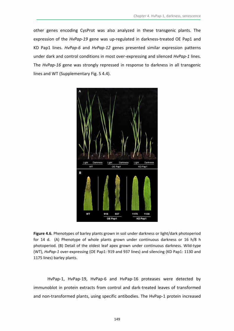
Chapter 4. HvPap-1, darkness, senescence
149
other genes encoding CysProt was also analyzed in these transgenic plants. The
expression of the HvPap-19 gene was up-regulated in darkness-treated OE Pap1 and
KD Pap1 lines. HvPap-6 and HvPap-12 genes presented similar expression patterns
under dark and control conditions in most over-expressing and silenced HvPap-1 lines.
The HvPap-16 gene was strongly repressed in response to darkness in all transgenic
lines and WT (Supplementary Fig. S 4.4).
Figure 4.6. Phenotypes of barley plants grown in soil under darkness or light/dark photoperiod
for 14 d. (A) Phenotype of whole plants grown under continuous darkness or 16 h/8 h
photoperiod. (B) Detail of the oldest leaf apex grown under continuous darkness. Wild-type
(WT), HvPap-1 over-expressing (OE Pap1: 919 and 937 lines) and silencing (KD Pap1: 1130 and
1175 lines) barley plants.
HvPap-1, HvPap-19, HvPap-6 and HvPap-16 proteases were detected by
immunoblot in protein extracts from control and dark-treated leaves of transformed
and non-transformed plants, using specific antibodies. The HvPap-1 protein increased

Chapter 4. HvPap-1, darkness, senescence
150
not only in the over-expressing OE Pap1 lines in comparison with the WT, but also after
the darkness treatment. In contrast, HvPap-1 diminished in all KD Pap1 lines (Fig. 4.7).
A slight increase of HvPap-6 and HvPap-19 proteins was also observed in leaves grown
under darkness. No alterations in the HvPap-16 protein levels were detected in OE
Pap1 lines compared to WT plants. Nevertheless, a clear increase of the inactive form
of the HvPap-16 protease (upper band) was detected in light-grown KD lines while this
inactive form disappeared when plants were subjected to darkness. Additionally,
Rubisco was slightly reduced in stressed leaves, being more pronounced in the over-
expressing HvPap-1 lines (Fig. 4.7).
Figure 4.7. Protein patterns of C1A CysProt in transgenic and wild-type (WT) barley lines grown
under darkness (D) or 16 h/8 h photoperiod (L) for 14 d and assayed by immunoblot. Proteins
were extracted from leaves of WT, HvPap-1 over-expressing (OE Pap1: 919, 920, 932, 937
lines) and silencing (KD Pap1: 1128, 1130, 1175, 1178 lines). Rubisco protein content was
assayed using a specific antibody against its Large Subunit (LS Rubisco).
4.3.5. PHYSIOLOGICAL CHANGES ARE ASSOCIATED WITH STRESS MEDIATED BY
DARKNESS IN HVPAP-1 TRANSGENIC BARLEY LINES
The total amount of soluble proteins was quantified in darkness-treated and non-
treated transgenic lines as well as in non-transgenic controls. OE Pap1 lines did not
show significant differences in protein content as compared to non-transgenic when
grown under photoperiod but a slight protein reduction was appreciated in some

Chapter 4. HvPap-1, darkness, senescence
151
transgenic lines grown in darkness (Supplementary Fig. S 4.5A). By contrast, most KD
Pap1 lines presented increased levels of protein in darkness-treated and non-treated
leaves in comparison with their corresponding WT (Supplementary Fig. S 4.5B).
Additionally, the proteolytic activity pattern (cathepsin L-/F- and B-like CysProt) of OE,
KD and WT plants grown in the darkness or under control conditions was determined
using specific substrates. No significant differences on the cathepsin L-/F-like activity
were detected between transgenic and WT lines when plants were grown under light
conditions. Under darkness, the cathepsin L-/F-like activity mostly decreased under
darkness into KD lines (Supplementary Fig. S 4.6A). Similar data resulted from the
measurements of cathepsin B-like activity (Supplementary Fig. S 4.6B).
Photosynthetic pigments were also determined in whole aerial biomass of the
plants after 14 d of darkness vs control conditions. Differences in the chlorophyll a
levels were nearly undetectable among OE, KD and WT when these plants were grown
under photoperiod conditions. A decrease of chlorophyll a was observed in all
darkness-treated leaves compared to the non-treated ones, particularly in the WT
(Supplementary Fig. S 4.7A). Similar results were found for the quantification of
carotenoids in most lines (Supplementary Fig. S 4.7C). In contrast, the amount of
chlorophyll b was drastically reduced in treated and non-treated HvPap1 amiRNA
leaves in comparison with the WT and presented an undefined pattern in OE Pap1
lines (Supplementary Fig. S 4.7B).
Additionally, the total chlorophyll content of the oldest leaf was observed after
14 d of stress treatment, by detecting its auto-fluorescence under the confocal
microscope. Since the apex of the leaf is older than the medium/basal part (segments
1 and 2, respectively, in Fig. 4.8), the highest chlorophyll fluorescence was generally
found in the non-stressed segment 2 (Fig. 4.8). A lower fluorescence emission was
detected in OE Pap1 lines than in WT. In contrast, auto-fluorescence levels were
increased in the KD Pap1 lines. Similar patterns, although less remarkable, were found
in leaf tissues grown under darkness which correlated with alterations in the tissue
structures observed under bright field (Fig. 4.8). Besides, Table 4.1 shows that the total
amount of starch in OE Pap1 and KD Pap1 transgenic leaves grown under photoperiod

Chapter 4. HvPap-1, darkness, senescence
152
was approximately half of the starch amount in WT leaves. As expected, the starch
accumulation was strongly reduced in 14 d dark-treated leaves but no remarkable
differences were detected between transgenic and WT samples.
Figure 4.8. Chlorophyll detection in the oldest leaf of transgenic and wild-type barley lines
grown under darkness or 16 h/8 h photoperiod for 14 d. Leaf fragments from HvPap-1 over-
expressing (OE Pap1: 919 line), silencing (KD Pap1: 1175 line) and wild-type (WT) plants were
collected and observed under a Leica SP8 confocal microscope using the laser excitation lines
633 nm to detect the red auto-fluorescence from the chlorophyll. Same images were taken
under light field conditions. Leaves were cut in two fragments (segments 1 and 2,
corresponding to apical and medium-basal section of the leaf, respectively) as is indicated in
the upper part of the figure. Scale bars: 200 μm.

Chapter 4. HvPap-1, darkness, senescence
153
CONSTRUCT LINE STARCH CONTENT* (g/100 gFW)
Co
ntr
ol
Wild-type WT 8.38 ± 0.30A
OE Pap1 919 3.23 ± 0.15B
937 3.58 ± 0.15B
KD Pap1 1130 4.30 ± 0.10B
1175 4.20 ± 0.46B
Dar
knes
s 1
4d
Wild-type WT 0.84 ± 0.17a
OE Pap1 919 0.83 ± 0.11a
937 0.79 ± 0.07a
KD Pap1 1130 0.90 ± 0.10a
1175 0.73 ± 0.09a
*Data, referred as grams of transformed starch per 100 grams of fresh weight, are means ±
standard error of duplicate measurements of six independent replicates for each sample.
Different letters indicate significant differences between lines. (P < 0.05, one-way ANOVA
followed by HSD test).
Table 4.1. Starch content of the oldest leaf in the HvPap-1 over-expressing (OE Pap1: 919, 937
lines), silencing (KD Pap1: 1130 and 1175 lines) and wild-type (WT) plants after 14 d of
darkness or 16 h/8 h photoperiod.
4.4. DISCUSSION
Protein breakdown and mobilization are some of the major metabolic features
associated with abiotic stresses, essential for nutrient recycling. Rubisco, the most
abundant protein in plants, is likely the major target for proteases when proteolytic
processes are activated (van der Hoorn, 2008; Theonen et al., 2007; Krupinska et al.,
2012; Martinez et al., 2012). The identification of these proteases is crucial to
understand the physiological mechanisms behind the process in order to bioengineer
plants with altered timing of senescence, which is closely related to grain quality and
yield. The barley-C1A CysProt system is a promising model to analyze the role of
proteases in plants subjected to abiotic treatments. C1A CysProt are strongly up-
regulated in response to multiple stresses, barley is a model crop whose genome has
been sequenced and transgenic technology is well established. Besides, the whole
family of C1A CysProt and their inhibitors (cystatins) has been identified in this species
(Martinez et al., 2009; Diaz-Mendoza et al., 2014).

Chapter 4. HvPap-1, darkness, senescence
154
Slow growth rates and yellowish leaf apex were the first phenotypes displayed
by barley plants under severe darkness and N starvation treatments (Supplementary
Fig. S 4.1). Reduction in chlorophyll and carotenoid content was observed parallel to
the dismantling of the cell structure (Fig. 4.1), probably due to microtubule
rearrangements (Keech et al., 2010). These parameters are considered reliable
indicators of stress and senescence. In particular, chlorophyll abundance is a useful
indicator of the chloroplast status because it tends to remain constant in
photosynthetically active leaves (Sorin et al., 2015). Major subcellular rearrangements
in mesophyll cells undergoing severe stresses involved changes in chloroplast size and
distribution within the cell. Chloroplasts lost their typical lenticular size and became
more spherical and smaller. Under N starvation they still occupied the cell periphery
with spaces among them. After the darkness treatment they formed aggregates (Fig.
4.1C). These results are in accordance with previous publications, and demonstrate
that chlorophyll degradation is a common early event in stressed leaves and leaf
senescence, closely related to plastid disassembly (Krupinska et al., 2012; Carrion et
al., 2014; Hollmann et al., 2014). Keech et al. (2007) reported that metabolism in
whole darkened Arabidopsis leaves entered in a “stand-by mode” with low
mitochondria activity to preserve active photosynthetic machinery, while in individual
darkened leaves the high mitochondrial activity provided energy and carbon skeletons
for a rapid degradation of cellular components. In this study, stress parameters
measured in whole darkened barley plants or in N-depletion treated plants indicate
that senescence was actually induced, since chlorophyll was degraded and chloroplasts
were altered.
Another important parameter associated with abiotic stresses is the reduction
of total protein content. The significant decrease in protein levels in darkness- and N-
starved leaves in comparison to the controls (Fig. 4.1B) suggested either the inhibition
of protein synthesis or/and the activation of protease activities associated to nutrient
recycling. In this way, darkness and N starvation clearly induced cathepsin L-/F- and B-
like activities in leaves (Fig. 4.2). These data were supported by the up-regulation of
genes encoding barley C1A CysProt, particularly HvPap-1, HvPap-12 and HvPap-19 and
the detection of these proteases by immunoblot assays under the assayed

Chapter 4. HvPap-1, darkness, senescence
155
experimental conditions (Fig. 4.3). The implication of different classes of CysProt
cathepsin F, B-, L- and H-like, suggested a functional redundancy of these proteases in
protein turnover upon treatments. So far, transcriptomic and proteomic data from
different authors have consistently assigned a major role to members of all C1A
CysProt groups during abiotic stresses induced in several plants species (Gregersen et
al., 2008; Martinez et al., 2012; Diaz-Mendoza et al., 2014; Hollmann et al., 2014).
Additionally, the subcellular localization of C1A CysProt revealed a dynamic trafficking
of proteins to be degraded from the chloroplasts to the central vacuole. Senescence-
Associated Vacuoles (SAVs) and the central lytic vacuole, both containing chloroplastic
proteins and peptides, are enriched in CysProt activities during leaf senescence (Otegui
et al., 2005; Ishida et al., 2008; Carrion et al., 2013; 2014). The immunofluorescence
signal of HvPap-1 and HvPap-19 CysProt was detected in small vesicles, probably SAVs,
within parenchyma cells of leaves undergoing senescence (Fig. 4.4), which highly
supports their involvement in the degradation of chloroplastic proteins. In contrast,
HvPap-1 and HvPap-19 were less abundant and mainly localized in epidermal cells of
control leaves.
HvPap-1 is a cathepsin F-like CysProt previously characterized with an
important role in grain filling and germination. It actively participates in the hydrolysis
and mobilization of storage proteins, mainly hordeins, controlling the grain amino acid
composition (Cambra et al., 2012; Diaz-Mendoza et al., 2016). Besides, HvPap-1 gene is
up-regulated in response to severe abiotic treatments. The functional relevance of this
CysProt in response to darkness can be inferred from both forward and reverse genetic
approaches. The alteration of its expression in over-expressing or silencing barley
plants could disturb stress progress, and thereby nutrient mobilization. Transgenic
over-expressing HvPap-1 barley lines did not only exhibit high levels of mRNA and
protein in control leaves but also in response to darkness-induced treatment, while
opposite effects were observed in knock-down lines (Supplementary Fig. S 4.6 and Fig.
4.7). The accumulation of the HvPap-1 protease did not result in an increase in the
proteolytic activity. This might be due to the lack of a specific substrate to be
exclusively degraded by cathepsin F-like enzymes, since the one used in these
proteolytic assays simultaneously targeted cathepsin F- and L-like activities, and to

Chapter 4. HvPap-1, darkness, senescence
156
compensating effects among protease activities. In senescing leaves of knock-down
lines a clear decrease on protease activities, both cathepsin L-/F- and B-like classes
(Supplementary Fig. S 4.8) also suggests that accumulation of other proteases (Fig. 4.7)
and/or alternatively protease inhibitors is altered.
From a physiological point of view, the reduction of total protein paralleled to
that of Rubisco was observed in most of the darkness-treated OE Pap-1 lines vs
control. These results together with the reduced autofluorescence emission from the
chlorophyll (Fig. 4.8) and the small amount of starch detected in the over-expressing
leaves (Table 4.1) indicate that leaf senescence was sped up. In contrast, the high
protein content and retarded loss of chlorophyll in the amiRNA leaves, mostly detected
in the apex of the oldest leaf, indicated a delay in the senescence process. The
carbohydrate content of barley leaves, mainly represented by low concentrations of
sucrose, starch, fructans and hexoses (Sicher et al., 1984), is completely remobilized in
response to darkness-induced treatment and indicates that the photosynthetic
partitioning was similar in transgenic and WT leaves. As shown in Fig. 4.5 and
Supplementary information Fig. S 4.4, a clear delayed-senescence phenotype of
HvPap-1 amiRNA lines was observed both in barley plants grown either under
light/dark photoperiod, corresponding to the natural lifespan, or under continuous
darkness for 2 wk (Fig. 4.6). These phenotypes are presumably due to chloroplasts
protection from degradation. Based on these results, it can be concluded that HvPap-1
is a functional stress-associated gene and alterations in its expression bring about
changes in barley abiotic stress responses. Previous reports have shown similar effects
in plant behaviour by down-regulating the expression of senescence-related CysProt,
in particular BoCP5 and CaCP genes from broccoli and pepper, respectively (Eason et
al., 2005; Xiao et al., 2014). In addition, the over-expression of the broccoli cystatin
BoCPI-1 significantly contributed to a delay in the yellowing of broccoli florets than did
the suppression of BoCP5 (Eason et al., 2014). Many cystatins are strongly expressed
upon exposure to abiotic stresses and the implication of the C1A CysProt-cystatin
system has been reviewed (Martinez and Diaz, 2008; Martinez et al., 2012; Diaz-
Mendoza et al., 2014; Kidric et al., 2014). Very recently, the potential use of

Chapter 4. HvPap-1, darkness, senescence
157
phytocystatins in crop improvement to mitigate the negative impacts of climate
change has been considered (Kunert et al., 2015).
A positive correlation between chlorophyll content, cereal grain yield and total
nitrogen content of the grain has been reported (Distelfeld et al., 2014). Delay in leaf
senescence, so-called ‘stay green’ phenotypes have a longer period of active
photosynthesis and in consequence potentially higher plant productivity
(Hörtensteiner 2007; Gregersen et al., 2013). It has also been described that delayed
leaf senescence leads to a high grain yield, inefficient nitrogen remobilization and a
lower harvest index, whereas acceleration of senescence confers efficient nitrogen
remobilization and high protein content, but also a lower total grain yield (Gregersen
et al., 2008). In this context, protein accumulation in barley grains is important
depending on the end-product use of the harvested crop. Breweries do not require
barley grains enriched in proteins while high levels of proteins are a particularly
valuable character for food and animal feed. Further research is aimed to determine
HvPap-1 CysProt responses to abiotic/biotic stresses able to induce leaf senescence
and to characterize the functional role for other barley CysProt and cystatins.
In conclusion, our results clearly support that CysProt are associated with
tolerance or sensitivity to environmental cues and consequently linked to leaf
senescence, providing important basis for the improvement of small grain cereals,
among other crops.
4.5. REFERENCES
Avila-Ospina L, Moison M, Yoshimoto K, Masclaux-Daubresse C (2014) Autophagy, plant senescence, and nutrient recycling. J Exp Bot 65: 3799–3811
Baxter A, Mittler R, Suzuki N (2014) ROS as key players in plant stress signaling. J Exp Bot 65: 1229–1240
Bradford MM (1976) A rapid and sensitive method for the quantification of microgram quantities of protein utilizing the principle of protein-dye binding. Anal Biochem 72: 248–254
Buchanan-Wollaston V (1997) The molecular biology of leaf senescence. J Exp Bot 48: 181–199

Chapter 4. HvPap-1, darkness, senescence
158
Cambra I, Martinez M, Dader B, González-Melendi P, Gandullo J, Santamaría ME, Diaz I (2012) A cathepsin F-like peptidase involved in barley grain protein mobilization, HvPap-1, is modulated by its own propeptide and by cystatins. J Exp Bot 63: 4615–4629
Carrion CA, Costa ML, Martinez DE, Mohr C, Humbeck K, Guiamet JJ (2013) In vivo inhibition of cysteine proteases provides evidence for the involvement of ‘senescence-associated vacuoles’ in chloroplast protein degradation during dark-induced senescence of tobacco leaves. J Exp Bot 64: 4967–4980
Carrion CA, Martinez DE, Costa ML, Guiamet JJ (2014) Senescence-associated vacuoles, a specific lytic compartment for degradation of chloroplastic proteins? Plants 3: 498-512
Chen HJ, Su CT, Lin CH, Huang GJ, Lin YH (2010) Expression of sweet potato cysteine protease SPCP2 altered developmental characteristics and stress responses in transgenic Arabidopsis plants. J Plant Physiol 167: 838–847
Chen HJ, Tsai YJ, Shen CY, Tsai TN, Huang GJ, Lin YH (2013) Ectopic expression of sweet potato cysteine protease SPCP3 alters phenotypic traits and enhances drought stress sensitivity in transgenic Arabidopsis plants. J Plant Growth Regul 32: 108–121
Christiansen MW, Gregersen PL (2014) Members of barley NAC transcription factor gene family show differential co-regulation with senescence-associated genes during senescence of flag leaves. J Exp Bot 65: 4009–4022
Dawson IK, Russell J, Powell W, Steffenson B, Thomas WTB, Waugh R (2015) Barley, a translational model for adaptation to climate change. New Phytol 206: 913–931
Diaz I, Martinez M (2013) Plant C1A cysteine peptidases in germination and senescence. IN: Handbook of proteolytic enzymes. Rawlings ND, Salvesen G, Editors, pp. 1853–1858. Elsevier Academic Press, Amsterdam (The Netherlands)
Diaz-Mendoza M, Velasco-Arroyo B, Gonzalez-Melendi P, Martinez M, Diaz I (2014)
C1A cysteine protease-cystatin interactions in leaf senescence. J Exp Bot 65: 3825–
3833
Diaz-Mendoza M, Dominguez-Figueroa JD, Velasco-Arroyo B, Cambra I, Gonzalez-Melendi P, Lopez-Gonzalvez A, Garcia A, Hensel G, Kumlehn J, Diaz I, Martinez M (2016) HvPap-1 C1A protease and HvCPI-2 cystatin contribute to barley grain filling and germination. Plant Physiol 170: 2511–2524
Distelfeld A, Avni R, Fischer AM (2014) Senescence, nutrient remobilization, and yield
in wheat and barley. J Exp Bot 65: 37833–3798
Eason JR, Ryan DJ, Watson LM, Hedderley D, Christey MC, Braun RH, Coupe SA (2005)
Suppression of the cysteine protease, aleurain, delays floret senescence in Brassica
oleracea. Plant Mol Biol 57: 645–657
Eason JR, West PJ, Brummell DA, Watson LM, Somerfield SD, McLachlan ARG (2014)
Overexpression of the protease inhibitor BoCPI-1 in broccoli delays chlorophyll loss

Chapter 4. HvPap-1, darkness, senescence
159
after harvest and causes down-regulation of cysteine protease gene expression.
Postharvest Biol Technol 97: 23–31
Feller U, Anders I, Mae T (2008) Rubiscolytics, fate of Rubisco after its enzymatic function in a cell is terminated. J Exp Bot 59: 1615–1624
Gregersen PL, Culetic A, Boschian L, Krupinska K (2013) Plant senescence and crop productivity. Plant Mol Biol 82: 603–622
Gregersen PL, Holm PB, Krupinska K (2008) Leaf senescence and nutrient remobilization in barley and wheat. Plant Biol 10: 37–49
Guo Y, Gan SH (2012) Convergence and divergence in gene expression profiles induced by leaf senescence and 27 senescence-promoting hormonal, pathological and environmental stress treatments. Plant Cell Environ 35: 644–655
Hoagland DR (1920) Optimum nutrient solution for plants. Science 52: 562–564
Hollmann J, Gregersen PL, Krupinska K (2014) Identification of predominant genes involved in regulation and execution of senescence-associated nitrogen remobilization in flag leaves of field grown barley. J Exp Bot 65: 3963–3973
Hörtensteiner S (2007) Chlorophyll degradation during senescence. Annu Rev Plant Biol 57: 55–77
Ishida H, Yoshimoto K, Izumi M, Reisen D, Yano Y, Makino A, Ohsumi Y, Hanson MR, Mae T (2008) Mobilization of Rubisco and stroma-localized fluorescent proteins of chloroplast to the vacuole by ATG gene-dependent autophagic process. Plant Physiol 148: 142–155
Keech O, Pesquet E, Ahad A, Asne A, Nordvall D, Vodnala SM, Tuominen H, Hurry V, Dizengremel P, Gardestrom P (2007) The different fates of mitochondrial and chloroplasts during dark-induced senescence in Arabidopsis leaves. Plant Cell Environ 30: 1523–1534
Keech O, Pesquet E, Gutierrez L, Ahad A, Bellini C, Smith SM, Gardeström P (2010) Leaf senescence is accompanied by an early disruption of the microtubule network in Arabidopsis. Plant Physiol 154: 1710–1720
Kempema LA, Cui X, Holzer FM, Walling LL (2015) Arabidopsis transcriptome changes in response to phloem-feeding silverleaf whitefly nymphs. Similarities and distinctions in responses to aphids. Plant Physiol 143: 849–865
Kidric M, Kos J, Sabotic J (2014) Protease and their endogenous inhibitors in the plant response to abiotic stress. Bot Serb 38: 139–158
Krupinska K, Mulisch M, Hollmann J, Tokarz K, Zschiesche W, Kage H, Humbeck K, Biler W (2012) An alternative strategy of dismantling of the chloroplast during leaf senescence observed in a high-yield variety of barley. Physiol Plant 144: 189–200
Kunert KJ, van Wyk SG, Cullis CA, Vorster BJ, Foyer CH (2015) Potential use of phytocystatins in crop improvement, with a particular focus on legumes. J Exp Bot 66: 3559–3570
Laemmli UK (1970) Cleavage of structural proteins during assembly of the head of bacteriophage T4. Nature 277: 680–685

Chapter 4. HvPap-1, darkness, senescence
160
Lichtenthaler HK (1987) Chlorophylls and carotenoids, Pigments of photosynthetic biomembranes. Methods Enzymol 148: 350–382
Martinez M, Diaz I (2008) The origin and evolution of plant cystatins and their target
cysteine proteinases indicate a complex functional relationship. BMC Evol Biol 8: 198
Martinez M, Cambra I, Carrillo L, Diaz-Mendoza M, Diaz I (2009) Characterization of the entire cystatin gene family in barley and their target cathepsin L-Like cysteine-proteases, partners in the hordein mobilization during seed germination. Plant Physiol 151: 153–1545
Martinez M, Cambra I, Gonzalez-Melendi P, Santamaria ME, Diaz I (2012) C1A cysteine-proteases and their inhibitors in plants. Physiol Plant 145: 85–94
Masclaux-Daubresse C, Daniel-Vedele F, Dechorgnat J, Chardon F, Gaufichon L, Suzuki A (2010) Nitrogen uptake, assimilation and remobilization in plants, challenges for sustainable and productive agriculture. Ann Bot 105: 1141–1157
Masclaux-Daubresse C, Reisdorf-Cren M, Orsel M (2007) Leaf nitrogen remobilization for plant development and grain filling. Plant Biol 10: Suppl1, 23–36
Oñate-Sánchez L, Vicente-Carbajosa J (2008) DNA-free RNA isolation protocols for Arabidopsis thaliana, including seeds and siliques. BMC Res Notes 1: 93
Orsel M, Moison M, Clouet V, Thomas J, Leprince F, Canoy AS, Just J, Chalhoub B, Masclaux-Daubresse (2014) Sixteen cytosolic glutamine synthetase genes identified in Brassica napus L. genome are differentially regulated depending on nitrogen regimes and leaf senescence. J Exp Bot 65: 3927–3947
Otegui MS, Noh Y-S, Martinez DE, Petroff AGV, Staehelin LA, Amasino RM, Guiamet JJ (2005) Senescence-associated vacuoles with intense proteolytic activity develop in leaves of Arabidopsis and soybean. Plant J 41: 831–844
Parrott DL, Martin JM, Fischer AM (2010) Analysis of barley (Hordeum vulgare) leaf senescence and protease gene expression, a family C1A cysteine protease is specifically induced under conditions characterized by high carbohydrate, but not low to moderate nitrogen levels. New Phytol 187: 313–331
Pérez-López U, Robredo A, Lacuesta M, Mena-Petite A, Muñoz-Rueda A (2012) Elevated CO2 reduces stomatal and metabolic limitations on photosynthesis caused by salinity in Hordeum vulgare. Photosynth Res 111: 269–83
Quain MD, Makgopa ME, Marquez-Garcia B, Comadira G, Fernandez-Garcia N, Olmos E, Schnaubelt D, Kunert KJ, Foyer CH (2014) Ectopic phytocystatin expression leads to enhances drought stress tolerance in soybean (Glycine max) and Arabidopsis thaliana through effects on strigolactone pathways and can result in improved seed traits. Plant Biotechnol J 12: 903–913
Queval G, Foyer CH (2012) Redox regulation of photosynthetic gene expression. Philos Trans R Soc Lond B Biol Sci 367: 3475–3485
Rabbani MS, Maruyama K, Abe H, Khan MA, Katsura K, Ito Y, Yoshiwara K, Seki M, Shinozaki K, Yamaguchi-Shinozaki K (2003) Monitoring expression profiles of rice genes under cold, drought, and high salinity stresses and abscisic acid application using cDNA microarray and RNA gel-blot analyses. Plant Physiol 133: 1755–1767

Chapter 4. HvPap-1, darkness, senescence
161
Roberts IN, Caputo C, Criado MV, Funk C (2012) Senescence-associated proteases in plants. Physiol Plant 145: 130–139
Sakamoto W, Takami T (2014) Nucleases and higher plants and their possible involvement in DNA degradation during senescence. J Exp Bot 65: 3835–3843
Sicher RC, Kremer DF, Harris WG (1984) Diurnal carbohydrate metabolism of barley primary leaves. Plant Physiol 76: 165–169
Sorin C, Musse M, Mariette F, Bouchereau A, Leport L (2015) Assessment of nutrient remobilization through structural changes of palisade and spongy parenchyma in oilseed rap leaves during senescence. Planta 241: 333–346
Suzuki N, Rivero RM, Shulaev V, Blumwald E, Mittler R (2014) Abiotic and biotic stress combinations. New Phytol 203: 32–43
Theonen M, Herrmann B, Feller U (2007) Senescence in wheat leaves, is a cysteine endopeptidase involved in the degradation of the large subunit of Rubisco? Acta Physiol Plant 29: 339–350
Van der Hoorn R (2008) Plant proteases, from phenotypes to molecular mechanisms. Annu Rev Plant Biol 59: 191-223
Xiao HJ, Yin YX, Chai WG, Gong ZH (2014) Silencing of the CaCP gene delays salt- and osmotic-induced leaf senescence in Capsicum annuum L. Int J Mol Sci 15: 8316–8334
Yang Zl, Ohlrogge JB (2009) Turnover of fatty acids during natural senescence of Arabidopsis, Brachypodium, and switchgrass and in Arabidopsis beta-oxidation mutants. Plant Physiol 150: 1981–1989
Zang QW, Wang CX, Li XY, Gui ZA, Jing RL, Zhao J, Chang XP (2010) Isolation and characterization of a gene encoding a polyethylene glycol-induced cysteine protease in common wheat. J Bioscience 35: 379–388
Zmienko A, Goralski M, Samelak-Czajka A, Sobieszczuk-Nowicka E, Figlerowicz M (2015) Time course transcriptional profiling of senescing barley leaves. Genom Data 4: 78–81

Chapter 4. HvPap-1, darkness, senescence
162
4.6. SUPPLEMENTAL DATA
Figure S 4.1. Phenotypes of wild-type (WT) barley plants grown in vermiculite under
continuous darkness or 16 h/8 h photoperiod, or in pots filled with Hoagland nutrient solution
with or without N source for 3 and 7 d.

Chapter 4. HvPap-1, darkness, senescence
163
Figure S 4.2. Phenotype of barley plants at 3 (A), 5 (B), 7 (C) and 9 (D) wk of development.
Wild-type (WT), HvPap-1 over-expressing (OE Pap1: 919 and 937 lines) and silencing (KD Pap1:
1130 and 1175 lines) plants.

Chapter 4. HvPap-1, darkness, senescence
164
Figure S 4.3. Phenotype of wild-type (WT) barley plants grown in soil under continuous
darkness or 16 h/8 h photoperiod for 3, 7, 14 and 21 d.

Chapter 4. HvPap-1, darkness, senescence
165
Figure S 4.4. Messenger expression levels of C1A CysProt genes in transgenic and wild-type
(WT) barley lines grown under darkness or control treatment and assayed by RT-qPCR. Total
RNA was extracted from leaves of WT, HvPap-1 over-expressing (OE Pap1: 919, 920, 932 and
937 lines) and silencing (KD Pap1: 1175, 1176, 1128, 1130 lines) barley plants grown in soil
under continuous darkness or with 16 h/8 h photoperiod for 14 d. Data were determined by
RT-qPCR and expressed as relative mRNA levels of C1A CysProt genes normalized to barley
cyclophilin mRNA content.

Chapter 4. HvPap-1, darkness, senescence
166
Figure S 4.5. Total protein content from HvPap-1 transgenic and wild-type lines. (A) HvPap-1
over-expressing (OE Pap1: 919, 920, 932 and 937 lines) and wild-type (WT) barley plants. (B)
HvPap-1 silencing (KD Pap1: 1175, 1176, 1128, 1130 lines) and wild-type (WT) barley plants.
Plants were grown in soil under continuous darkness or 16 h/8 h photoperiod for 14 d. All
parameters are given in mg of total soluble protein per gram of initial fresh weight. Data are
means ± standard error of at least three independent analyses. Different letters indicate
statistical differences between lines (P < 0.05, HSD).

Chapter 4. HvPap-1, darkness, senescence
167
Figure S 4.6. Proteolytic activities of leaf extracts from HvPap-1 transgenic and wild-type (WT)
barley plants. (A) HvPap-1 over-expressing (OE Pap1: 919, 920, 932 and 937 lines) and wild-
type (WT) barley plants. (B) HvPap-1 silencing (KD Pap1: 1175, 1176, 1128, 1130 lines) and
wild-type (WT) barley plants. Plants were grown in soil without light or 16 h/8 h photoperiod
for 14 d. Specific substrates to be degraded by cathepsin L-/F-like (a) and B-like (b) were used.
Data are means ± standard error of at least three independent analyses. Different letters
indicate statistical differences between lines (P < 0.05, HSD).

Chapter 4. HvPap-1, darkness, senescence
168
Figure S 4.7. Determination of photosynthetic pigments in whole plant leaves from HvPap-1
over-expressing (OE Pap1: 919, 920, 932 and 937 lines), silencing (KD Pap1: 1175, 1176, 1128,
1130 lines) and wild-type (WT) barley plants grown in soil under darkness or 16 h/8 h
photoperiod for 14 d. Chlorophyll a (A), chlorophyll b (B) and carotenoids (C) content. All
parameters are referred in mg of pigment per gram of initial fresh weight. Data are means ±
standard error of at least three independent analyses. Different letters indicate statistical
differences between lines (P < 0.05, HSD).

Chapter 4. HvPap-1, darkness, senescence
169
Table S 4.1 Primer sequences used for the amplification of barley genes by RT-qPCR assays.
HvPap-4, HvPap-6 and HvPap-16 genes (cathepsin L-like proteases), HvPap-1 gene (cathepsin
F-like protease), HvPap-19 gene (cathepsin B-like protease) and HvCycl (cyclophilin).
Barley genes Primers
HvCycl forward: 5’-TCCACCGGAGAGGAAGTACAGT-3’
reverse: 5’-AATGTGCTCAGAGATGCAAGGA-3’
HvPap-1 forward: 5´-TCCTGGAGTCGATCTTTGGTTTC-3’
reverse: 5´-CAAGCATACTGTTGCGGCTTC-3’
HvPap-4 forward: 5´-CCTTGAGAGTCCTTGTTCCCGA-3´
reverse: 5´-CCATGTTCGTCGTTTTAACCGA-3´
HvPap-6 forward: 5´-TGCAATTGACGGCAAGAAGA-3´
reverse: 5´-TGGATCACCAGGTGATCATTTG-3´
HvPap-12 forward: 5´-ATGTGCGCTATTGCTACCTGC-3´
reverse: 5´-CACCTTATTCATGTCTGGCGAA-3´
HvPap-16 forward: 5´-CTGGATCGGTAAGAACTCGTGG-3´
reverse: 5´-TGATGGAGGTGCCATCATATGA-3´
HvPap-19 forward: 5´-CACCTTATTCATGTCTGGCGAA-3´
reverse: 5´-TGCCCGCTTAATTTGACAGG-3´

Chapter 4. HvPap-1, darkness, senescence
170
Table S 4.2. CysProt amino acid sequences used for specific antibody production.
Barley proteases Peptides
HvPap-1 SGFAPSRFKEKPYWIIKN
HvPap-6 IDSEEDYPYKERDNRC
HvPap-16 RLRSYKKVTPPGNEAGLKE
HvPap-19 CQEKKHFSIDAYQVNSDPHD


Chapter 5. HvPap-1 and HvCPI-2, germination
173
5.1. INTRODUCTION
Barley (Hordeum vulgare L.) is an annual, monocotyledonous plant of the Poaceae
family, one of the world’s earliest domesticated and most important crop plants.
Nowadays it represents the fourth most abundant cereal in both surface and tonnage
harvested. According to FAO the barley cultivated area in 2013 was 49,148,479 ha with
a yield of more than 2,929 Kg/ha, meaning average world production of approximately
144 Mt (FAOSTAT, http://faostat3.fao.org/download/Q/QC/E). The high production of
this crop is mainly due to its importance in the malting industry and breweries as well
as to the nutritional intake for humans and livestock. Barley is considered as a model
species for cereal research since its genome is fully sequenced (Mayer et al., 2012).
Furthermore, it constitutes one of the best options to genetically improve cultivars to
tackle climate change and guaranteeing cereal production (Nevo et al., 2012; Dawson
et al., 2015).
During grain development and maturation proteins involved in germination are
stored in the endosperm together with starch and lipids. The cereal grain protein
concentration is about 10-12%, which is relatively low as compared to legume seeds.
Nevertheless, cereals impact on humans and livestock nutrition is about three times
higher than the effect of the most protein-rich legume seeds (Shewry and Halford,
2002). Storage, structural, metabolic and protective proteins are present in the grains.
The major part corresponds to storage proteins, which represent near 80% of total
proteins, falling into three different fractions based on the extraction method and their
solubility: albumins (water soluble), globulins (soluble in salt solutions) and prolamins
(soluble in alcohol/water mixtures) (Shewry et al., 1995). With the exceptions of oat
and rice, the major endosperm storage proteins in all cereal grains are prolamins
(Shewry and Halford, 2002), named hordeins in barley. Hordeins are highly
hydrophobic molecules, rich in proline and glutamine and with a low content of
charged amino acids, particularly the essential amino acid lysine. There are three
broad groups of hordeins: B (sulphur-rich), C (sulphur-poor) and D (high molecular
weight: HMW), with several subgroups within the B-group (Shewry et al., 1995). All of
them are co-ordinately expressed during endosperm development (Sorensen et al.,

Chapter 5. HvPap-1 and HvCPI-2, germination
174
1989) and their expression is tightly regulated (Diaz et al., 2005).
Limited proteolysis mediated by peptidases is essential for the initiation of
storage protein breakdown to nurture the developing embryo (Müntz, 1996).
Peptidases were formerly classified as cysteine- (Cys), serine- (Ser), aspartic- (Asp) and
metallo-proteases (metallic ion), according to the residue present in the active site of
the enzyme. Papain-like cysteine proteases (CysProt), known as C1A (family C1, clan
CA) according to the MEROPS database (Rawlings et al., 2014), constitute one of the
most abundant groups of proteases responsible for the degradation and mobilization
of storage proteins in seeds. Their role during germination has been reported in a wide
range of both monocot and dicot plants (Grudkowska and Zagdańska, 2004; Tan-
Wilson and Wilson, 2012). This group of proteases has a high number of members in
angiosperms, ranking from 32 in Arabidopsis to 45 in rice, which are classified into
cathepsins L-, B-, H- and F- like (Martinez and Diaz, 2008); in barley, 41 members have
been identified (Diaz-Mendoza et al., 2014). A previous analysis reported that 27 C1A
CysProt are among the 42 proteases involved in the germination of barley grain (Zhang
and Jones, 1995).
A complete transcriptome analysis in two tissue fractions (starchy endosperm/
aleurone and embryo/scutellum) has shown the induction of a high number of CysProt
genes during germination, most of them mediated by gibberellins (GA) (Sreenivasulu et
al., 2008). Some of the barley C1A proteases expressed in grain tissues have been
characterized. Among them, several cathepsin L-like proteases of the scutellar
epithelium and the aleurone layer were secreted to the endosperm upon germination
in response to GA (Koehler and Ho, 1990; Mikkonen et al., 1996; Martinez et al., 2009).
A cathepsin H-like protease isolated from GA-induced aleurone cells was targeted to
vacuoles and a cathepsin B-like protein was expressed in the aleurone and induced by
GA treatment (Holwerda and Rogers, 1992; Martinez et al., 2003). Recently, the
HvPap-1, a cathepsin F-like peptidase, has been described to be involved in barley
grain protein mobilization and to be modulated by its own propeptide and its
inhibitors, the cystatins (Cambra et al., 2012). This protein is able to in vitro degrade
different substrates, including barley endosperm proteins (hordeins, albumins and

Chapter 5. HvPap-1 and HvCPI-2, germination
175
globulins). It has been localized in protein bodies and vesicles of the embryo and it is
induced by GA in aleurone cells (Cambra et al., 2012).
Cystatins are the endogenous proteinaceous inhibitors of C1A CysProt. They
have several roles in plants and have been extensively related to the regulation of
physiological processes in seeds (Benchabane et al., 2010). In barley, the whole
cystatin family has been identified and characterized (Martinez et al., 2009). The
functional relationship between barley cystatins and cathepsin L- and F-like proteases
has been inferred from their common implication as counterparts during hordein
storage protein mobilization upon barley grain germination (Martinez et al., 2009;
Cambra et al., 2012). The barley cystatin HvCPI-2 encoded by the Icy2 gene is a good
inhibitor of different barley cathepsin L- and F-like CysProt, such as HvPap-1, -4, -6, -10,
and -16. It is strongly expressed in the germinating embryo and repressed by
gibberellic acid in aleurone layers. These results suggest a key role for HvCPI-2 in the
regulation of the CysProt activity in barley grain (Martinez et al., 2009; Cambra et al.,
2012).
Previous research indicates that a complex regulatory network including C1A
CysProt and their inhibitors is involved in the regulation of the barley grain
germination process. This work demonstrates how biotechnological modifications of
the proteolytic machinery may affect grain composition and, consequently,
germination in barley. For this purpose, in planta participation of the cathepsin F-like
HvPap-1 and the cystatin HvCPI-2 proteins during grain filling and mobilization of
stored proteins was analyzed in barley transgenic lines over-expressing the HvPap-1
gene or knocking-down the expression of either the HvPap-1 or Icy-2 genes.
5.2. MATERIALS AND METHODS
5.2.1. PLANT MATERIAL
The samples used in this work were obtained from barley plants of Hordeum vulgare
spring type cv. Golden Promise, grown at 22ºC under 16 h light/8 h dark photoperiod

Chapter 5. HvPap-1 and HvCPI-2, germination
176
in soil (Sanyo/Panasonic MLR-350-H). Barley transgenic lines over-expressing or
silencing the barley HvPap-1 gene (OE Pap1 and KD Pap1, respectively) and silencing
the barley Icy-2 gene (KD Icy2) were generated in collaboration with the IPK
Gatersleben, Plant Reproductive Biology Group (Hensel et al., 2009). To generate over-
expression lines, the HvPap-1 gene was transferred into the intermediate vector pUbi-
AB (DNA-Cloning-Service, Hamburg, Germany) and cloned using the SfiI restriction sites
into the p6U binary vector (DNA-Cloning-Service, Hamburg, Germany). This binary
vector included the HPT (HYGROMYCIN PHOSPHOTRANSFERASE) selectable marker
gene driven by the Zea mays UBIQUITIN-1 promoter with first intron. The HvPap-1
gene was driven by the same promoter. KD Pap1 and KD Icy2 knock-down lines were
produced by artificial microRNA (amiRNA) technology developed in the MicroRNA
Designer Web platform (WMD3, http://wmd3.weigelworld.org). The amiRNA
constructs were engineered from pNW55 vector replacing the 21 bases of the natural
Osa-MIR528 miRNA by 21 bases to silence specifically the HvPap-1 or Icy-2 genes (5’
TTATGCGGCATTGATACCGGT 3’ and 5’ TAAATTATTGTGTGGGGACTC 3’, respectively).
The final products of 554 bp were cloned into the p6d35S binary vector (DNA-Cloning-
Service, Hamburg, Germany) using the pUbi-AB vector as intermediary. This binary
vector included the HPT (HYGROMYCIN PHOSPHOTRANSFERASE) selectable marker
gene driven by the doubled enhanced CaM35S-promoter. Immature embryos of barley
cv “Golden Promise” were transformed with the binary vectors using the
Agrobacterium tumefaciens strain AGL1 as described (Hensel et al., 2008).
Homozygous transgenic plants were obtained by double haploid technology (Marthe et
al., 2015). The presence of the antibiotic resistance marker was confirmed by PCR
using specific primer pairs (Hensel et al., 2008). Homozygous transgenic lines for
inserted gene constructions were characterized and used for grain analyses.
5.2.2. ANALYSIS OF THE COPY NUMBER IN TRANSGENIC BARLEY LINES
Total DNA was isolated from leaves of barley transgenic and non-transformed control
lines. The RT-qPCR conditions were 40 cycles with 15 s at 95ºC, 1 min at 60ºC and 5 s
at 65ºC. FastStart Universal SYBR Green Master (Rox, Roche) was used in a total
volume of 20 μL. PCR amplification was performed in multiplates (Bio-Rad MLL9601)

Chapter 5. HvPap-1 and HvCPI-2, germination
177
using PCR sealersTM (Bio-Rad MSB1001). The reactions were carried out in a C1000TM
thermal cycler with CFX96TM optical reaction module and results were analyzed with
the CFX Manager Software 2.0 (Bio-Rad). Copy number was calculated by the 2 -∆∆Ct
method to study gene expression (Livak and Schmittgen, 2001) adapted to estimate
copy number (Ingham et al., 2001; Li et al., 2008). Cyclophilin and 4-hydroxyphenyl-
pyruvate dioxygenase (HvCycl and Hv4Hppd genes) were used as endogenous and
calibrator genes, respectively (Falk et al., 2002; Burton et al., 2004). The primers used
are shown in Supplemental Table S 5.2. Analysis of dissociation curves was performed
to check gene-specific amplification and reactions were performed in triplicate.
5.2.3. GRAIN PHENOTYPE AND STARCH ANALYSES
Grains from wild-type and transgenic barley plants were harvested and imbibed in
double-distilled water. After 24 hours, grains were longitudinally dissected and starch
was stained with Lugol's iodine staining reagent (Sigma). Stained and non-stained
grains were visualized and photographed with a Leica MZ10 F stereo microscope and a
Leica DFC420C CCD camera. For total starch quantification, six dry de-embryonated
grains from transgenic and control lines were ground and 10 mg were assayed with
STA20 Kit (Sigma) following the manufacturer recommendations. Wheat starch,
included in the commercial kit, was used as control. Dilutions were carried out as
necessary to fit into linearity of the glucose standard curve. Measurements were
performed three times for each sample. After calculations, starch content was
expressed as grams of transformed starch per 100 grams of initial kernel weight. Three
independent biological replicates were used.
5.2.4. METABOLOMIC ANALYSES
Samples were obtained from dry de-embryonated grains. 50 mg of sample were re-
suspended in 500 µL MeOH and disrupted by 3 cycles of frost/defrosting and
processed with TissueLyser LT (Qiagen). After centrifugation at 19,300 x g, 20 min, the
supernatant was transferred to a new tube and evaporated to dryness in a SpeedVac.
The metabolite extracts were re-suspended in 0.1 mmol L−1 formic acid containing 0.2
mmol L−1 methionine sulfone (internal standard) by 1 min vortex mixing and then

Chapter 5. HvPap-1 and HvCPI-2, germination
178
centrifuged (19,300 x g, 15 min). Clear solution was analyzed by capillary
electrophoresis coupled to a mass spectrometer detector (CE-ESI-MS-TOF). 19 nitrogen
metabolites, mainly amino acids and other related compounds: Ala, sarcosine, Ser, Pro,
Val, betaine, Ile, Leu, Asn, Asp, Gln, Lys, Glu, Met, His, Phe, Arg, Tyr and Trp, were
analyzed as previously described (Moraes et al., 2011) with slight modifications. A CE
System (7100 Agilent) coupled to a time-of-flight mass spectrometry system (Agilent
6224) was used. The separation occurred in a fused-silica capillary (Agilent) (total
length, 100 cm; i.d., 50 μm). Separation was under normal polarity with a background
electrolyte containing 1.0 mol L-1 formic acid in 10% (v/v) methanol at 20°C. Sheath
liquid (6 µL min-1) was methanol/water (1/1, v/v) containing 1.0 mmol L-1 formic acid
with two reference masses to allow correction and higher accurate mass in the MS.
Samples were hydrodynamically injected at 50 mbar for 35 s and stacked by injecting
background electrolyte at 100 mbar for 10 s. The optimized MS parameters were:
fragmentor 150 V, Skimmer 65 V, octopole 750 V, nebulizer pressure 10 psi, drying gas
temperature at 200ºC and flow rate 10.0 L min-1. The capillary voltage was 3,500 V.
Data were acquired in positive ESI mode with a full scan from m/z 87 to 1,000 at a rate
of 1.41 scan/s. The resulting CE-MS data files were cleaned of background noise and
unrelated ions by the Targeted Feature Extraction tool with Profinder software
(B.06.00, Agilent). Data were extracted using data mining algorithm based on the
software. This software contains a list of standards used with their exact monoisotopic
mass, migration time and molecular formula. Metabolites were previously identified in
the wild-type barley samples by comparison of their migration time and spectra with
pure standards. After their quantitation with standard solutions differences for
individual metabolites were evaluated by a t-test comparison of every case group vs
wild-type (where p values < 0.05 were considered significant). SIMCA-P+ 12.0.1
(Umetrics) and MetaboAnalyst v. 3.0 (Xia et al., 2012) were used for PCA plotting and
Heatmap, respectively.
5.2.5. GRAIN PROTEIN ANALYSIS
Plant protein extracts were obtained from 20 dry de-embryonated grains and from 20
de-embryonated grains after 24 and 72 h of seed imbibition. Three independent

Chapter 5. HvPap-1 and HvCPI-2, germination
179
biological replicates were used. Samples were ground and around 100 mg were
resuspended in 500 μl of protein extraction buffer (50 mM sodium phosphate pH 6.0,
15 mM NaCl and 2 mM EDTA pH 8.0). The suspension was centrifuged at 15,600 x g for
15 min at 4ºC. The protein content of the supernatants were quantified (Bradford,
1976) with bovine serum albumin (BSA) as a standard. Protein content was analyzed
using SDS-PAGE technique. 20 µg of protein extracts were denatured using dissociation
buffer (12.5 mM Tris-HCl pH 6.8, 0.25% (w/v) SDS, 2.5% (v/v) β-mercaptoethanol,
0.01% (w/v) bromophenol blue and 3.75% (v/v) glycerol) for 5 min at 90ºC.
Electrophoretic detection of proteins was performed using denaturing polyacrylamide
gels at 15% (w/v). The electrophoresis was carried out using a BioRad Mini Protein
Electrophoresis Cell system for 90 min at 130 V and prestained SDS-PAGE standard
broad range (BioRad) was used as a weight molecular pattern. Gels were submerged
into a staining solution 0.3% (w/v) Coomassie Blue G-250 in 40% (v/v) methanol and
10% (v/v) acetic acid to stain the proteins, and were distained in 25% (v/v) methanol
and 10% (v/v) acetic acid.
5.2.6. FRACTIONATION AND ANALYSIS OF BARLEY GRAIN STORAGE PROTEINS
Albumins and globulins were sequentially extracted from 20 dry barley de-
embryonated grains (Shi and Xu, 2009) and from 20 de-embryonated grains after 24
and 72 h of water imbibition. De-embryonated grains were completely crushed in a
mortar, resuspended in distilled water, and continuously stirred for 12 h at 4ºC.
Soluble and insoluble fractions were separated by centrifugation at 5,900 x g for 30
min at 4ºC. The supernatant, enriched in albumins, was isolated. Globulins were
extracted from the pellet by adding 5% (w/v) NaCl in distilled water at 4ºC. After a
second centrifugation step at 5,900 x g for 30 min, the recovered soluble fraction was
enriched in globulins. Hordeins were extracted from a similar set of de-embryonated
grains after incubation in a buffer containing 55% (v/v) 2-propanol and 1% (v/v) 2-
mercaptoethanol, for 1 h at 60ºC and centrifugation for 10 min at 13,300 x g (Martinez
et al., 2009). Protein concentrations and their electrophoretic bands patterns were
analyzed as above described. Three independent biological replicates were used.

Chapter 5. HvPap-1 and HvCPI-2, germination
180
5.2.7. GERMINATION ASSAYS
Twenty barley grains from each transgenic line and from the wild-type cultivar were
placed into a 150 mm petri dish with moistened, sterile filter paper. The germination
process was followed by counting the number of germinated grains at 24 and 72 hours
after imbibition (hai), considering as germinated grains those that had broken the grain
coat or testa. Photographs were taken with an OLYMPUS VR-320 digital camera. The
length of the emerging roots and coleoptiles was quantified using a measuring ruler.
Three independent biological replicates were used.
5.2.8. ENZYMATIC ACTIVITY ASSAYS
The enzymatic activity of the same de-embryonated grain protein extracts used for
grain protein quantification was determined using the fluorogenic substrates Z-FR-
AMC (N-carbobenzoxy-Phe-Arg-AMC) and Z-RR-AMC (N-carbobenzoxy-Arg-Arg-AMC),
susceptible to degradation by cathepsin L-/F- and B-like proteases, respectively.
Mixtures of proteases and substrates were incubated in 100 mM sodium phosphate pH
6.0 buffer containing 10 mM cysteine, 1 mM EDTA, and 0.01% (v/v) Brij35 at 30ºC.
Trypsin-like activity was also analyzed using the fluorogenic substrate Z-R-AMC (N-
carbobenzoxy-Arg-AMC), assays were incubated in the buffer Tris-HCl 0.1 M, pH 7.5 at
30ºC. Emitted fluorescence was measured with a 365 nm excitation and a 465 nm
emission wavelength filter. Triplicate assays were performed for determination of each
value and the average was calculated. Blanks were used to account for spontaneous
breakdown of substrates and results were expressed as nmol of hydrolyzed substrate
by mg of protein by min (nmol/mg*min). The system was calibrated with known
amounts of AMC (7-amido-4-methylcoumarin) hydrolysis product in a standard
reaction mixture.
5.2.9. IMMUNOBLOT ANALYSES
The same de-embryonated grain protein extracts obtained for total protein
quantification were used. After separation on an SDS–polyacrylamide gel, proteins
were electrotransferred onto a nitrocellulose membrane (GE Healthcare).

Chapter 5. HvPap-1 and HvCPI-2, germination
181
Immunoblotting was performed with specific crude anti-HvPap-1, -6 and -19 peptide
polyclonal antibodies produced in rabbits by Pineda Antibody Services. The sequences
used to produce them were: HvPap-1-IgG, NH2-CSGFAPSRFKEKPYWIIKN-CONH2;
HvPap-6-IgG, NH2-CGWSPVKDVNDPHVQEL-CONH2; and HvPap-19-IgG, NH2-
CQEKKHFSIDAYQVNSDPHD-CONH2. Optimal dilution of the antibody was incubated in
PBS (137 mM NaCl, 27 mM KCl, 10 mM Na2HPO4, 2 mM KH2PO4 pH 7.4) with 5% (w/v)
milk. Peroxidase-conjugated anti-rabbit IgG (Sigma) diluted at 1:10,000 was used as
secondary antibody for detection with ECL Plus (GE Healthcare). The specificity of
these antibodies was confirmed by immunoblotting using the purified recombinant
proteases HvPap-1, -6, and -19 (Supplemental Fig. S 5.7).
5.2.10. REAL-TIME QUANTITATIVE PCR ANALYSES
For real-time quantitative PCR (RT-qPCR) studies, isolated embryos from 24 h
germinated grains were used. Twenty grains and three independent biological
replicates per line were used. All samples were frozen in liquid nitrogen and stored at -
80ºC until used for RNA extraction. Samples were ground using a mortar and a pestle.
Total RNA was extracted by the phenol/chloroform method, followed by precipitation
with 3 M LiCl (Oñate-Sánchez and Vicente-Carbajosa, 2008) and digestion with DNase.
cDNAs were obtained from 2 μg of RNA using the RevertAid H Minus First Strand cDNA
Synthesis Kit (Thermo Scientific) following the manufacturer´s instructions. RT-qPCR
analyses were performed for duplicated samples by means of a CFX96 Real-time
system (BioRad) using SYBR Green as a detection system. Relative expression values
normalized to Cyclophilin gene (Burton et al., 2004) and expressed as 2-∆Ct. The primers
used for PCR amplification are described in Supplemental Table S 5.3.
5.2.11. EMBRYO STRUCTURAL ANALYSIS AND IMMUNO-FLUORESCENCE DETECTION
Embryos from wild-type and transgenic plants were fixed in a freshly prepared solution
of 4% (w/v) formaldehyde in PBS (phosphate buffered saline) first at room
temperature under vacuum until the specimens sank, then overnight at 4ºC. Next day
the samples were washed twice in PBS for 15 min. Dehydration in a series of increasing
concentrations of methanol, resin infiltration, embedding and polymerization were

Chapter 5. HvPap-1 and HvCPI-2, germination
182
performed by Progressive Lowering of Temperature (PLT) in a Leica AFS. From
methanol 30% to 70% (v/v) the temperature dropped from 0 to -20ºC at a speed of -
70ºC/h, with changes every 30 min. Subsequent steps were performed at -20ºC.
Further dehydration was achieved in 100% methanol for 90 min, with three changes.
Resin infiltration was done in a series of mixtures methanol:LRwhite (Agar Scientific)
with increasing concentrations of the resin (2:1; 1:1; 1:2, v:v), for 1 h each, and finally
pure resin with 0.5% (w/v) bezoin-methyl-ether as a catalyst overnight. Polymerisation
in plastic capsules was accomplished under UV light at -20ºC for 2 days and at 22ºC for
1 day. 1-2 μm thin sections were cut from the polymerized blocks in a Leica EM UC6
ultramicrotome, carefully collected on water drops on 10-well Teflon-printed slides
(Fisher Scientific Inc.) and let dry down and stored at room temperature until further
use.
To assess any possible structural rearrangements at the sub-cellular level in the
transgenic samples vs the wild-type, the sections were stained with 0.3% (w/v)
coomassie brilliant blue (Coomassie G-250, BioRad) or 20% (v/v) lugol (Lugol's iodine
staining reagent, Sigma), rinsed in distilled water, mounted and observed on a Zeiss
Axiophot microscope under bright field. Photographs were taken with a Leica DFC300
FX CCD camera using the Leica Application Suite 2.8.1 build 1554 acquisition software.
For immuno-fluorescence the sections on the 10-well slides were hydrated with
PBS for 5 min and unspecific binding sites were blocked by 10 min incubation with 5%
(w/v) BSA in PBS. Then, they were incubated with 20 µl drops/well of either a rabbit-
raised antibody to the CysProt HvPap-1, HvPap-6 or HvPap-19, applied 1/50 in PBS, for
1 h at room temperature in a humid chamber. After two washes of 15 min in PBS, an
Alexa Fluor 488 anti-rabbit antibody (Molecular Probes) was applied in a 1/25 solution
in 2.5% (v/v) BSA in PBS, for 45 min at room temperature in a humid chamber and
darkness. Subsequent to another two washes of PBS for 15 min each, the slides were
mounted in a 50:50 solution of glycerol:PBS. Serial sections across the specimens were
collected on a Leica SP8 confocal microscope using the laser excitation line of 488 nm
to detect the proteases. All series were captured under the same conditions (pinhole
size, gain, offset, magnification). The management of the series was performed with

Chapter 5. HvPap-1 and HvCPI-2, germination
183
either the LAS-AF-Lite 3.1.0_8587 or Fiji software. To composite the corresponding
figure in Adobe Photoshop CS3, the maximum projections of the green channel were
overlaid for each treatment shown. Only the automatic levels were adjusted.
5.2.12. DATA ANALYSIS
Statistical differences among treatments or lines were analyzed by one-way ANOVA
followed by Tukey´s (HSD) multiple comparison test performed using the software R
Project (v.3.1.2) package.
5.3. RESULTS
5.3.1. TRANSGENIC BARLEY LINES OVER-EXPRESSING OR SILENCING HVPAP-1
PROTEASE OR SILENCING ICY-2 CYSTATIN
Transgenic barley plants were obtained from immature embryos after Agrobacterium
co-culture and selection on hygromycin-containing media. Transgenic barley plants
ubiquitously over-expressing the HvPap-1 gene were generated using p6U and p6d35s
binary vectors. Silencing of the HvPap-1 and Icy-2 genes was generated using the
artificial microRNA (amiRNA) technology. For each construct, 30 independent primary
plants were generated. Afterwards, four to six T1 events were preliminary used for
molecular characterization. Homozygous material was generated via embryogenic
pollen cultures (Coronado et al., 2005) and obtained homozygous plants were analyzed
in depth. Two HvPap-1 over-expressing lines (OE Pap1: 919 and 937), two HvPap-1
silencing lines (KD Pap1: 1130 and 1175) and two Icy-2 silencing lines (KD Icy2: 1318
and 1399) were selected based on transgene copy number, transcript and protein
content for further studies (Supplemental Fig. S 5.1A and Supplemental Fig. S 5.2A).
Following these criteria, the over-expressing lines showed two copies of HvPap-1 gene,
the endogenous and the transgene, estimated by RT-qPCR assay and the 2-∆∆Ct method
(Supplemental Fig. S 5.1B) and presented higher accumulation of mRNAs and protein
than the wild-type (Supplemental Fig. S 5.1C,D). The amiRNA lines contained a unique
transgene insertion and the expression levels of their messengers and accumulation of

Chapter 5. HvPap-1 and HvCPI-2, germination
184
proteins were reduced in comparison with the wild-type (Supplemental Fig. S 5.1B-D
and Supplemental Fig. S 5.2B-D). However, neither mRNA accumulation nor protein
content was completely knocked out in the amiRNA plants.
5.3.2. GRAIN PHENOTYPE AND STARCH ACCUMULATION ARE ALTERED IN BARLEY
TRANSGENIC LINES
Kernels from transgenic and control plants were obtained and their grains
phenotypically compared 24 hours after imbibition (hai). The OE Pap1 and KD Pap1
grains were of similar size but were elongated and presented darker endosperms than
the control grains. The KD Icy2 lines also showed grains with a slightly darker
endosperm than wild-type ones (Fig. 5.1A). These phenotypic differences could be
related to a different grain composition. The amount of starch can be inferred by the
intensity of the dark-blue/black colour after Lugol staining. OE Pap1 and KD Icy2 grains
presented a weaker colour than wild-type grains, indicating lower amount of starch. In
contrast, the staining of KD Pap1 grains was stronger than the signal observed in wild-
type grains (Fig. 5.1B). No differences in the balance of amylose/amylopectin could be
detected since the typical amylopectin redish colouration was not observed (Fig. 5.1B).
Quantification of the starch content in grains corroborated that the KD Pap1 silencing
lines contained significantly higher quantities of starch than the control line
(Supplemental Fig. S 5.3).

Chapter 5. HvPap-1 and HvCPI-2, germination
185
Figure 5.1. Phenotype and starch staining of barley grains. A, Structure of longitudinal
dissected grains 24 hours after imbibition from wild-type and transgenic plants. B, Lugol's
iodine staining of transgenic and wild-type barley grains.
5.3.3. GRAIN PROTEIN CONTENT IS MODIFIED IN BARLEY TRANSGENIC LINES
The protein quantity of de-embryonated grains was also quantified (Fig. 5.2A). Dry
grains from all transgenic lines (OE Pap1, KD Pap1 and KD Icy2) presented similar
quantities of protein between them, but significantly higher protein amounts per grain
than the non-transformed line (Fig. 5.2A). These proteins are degraded as germination
proceeds. At 24 and 72 hours after imbibition (hai), the amount of protein decreased
in all transgenic and non-transgenic lines. However, the rate of remaining protein was
different. About 40% of the grain protein was degraded at 72 hai for all lines with the
exception of KD Pap1 lines, in which about 70% of the grain protein was broken down
at 72 hai (Fig. 5.2A). These variations in the speed of protein degradation were

Chapter 5. HvPap-1 and HvCPI-2, germination
186
confirmed by electrophoretic band patterns, which were very similar for wild-type, OE
Pap1 and KD Icy2 barley grains but different for KD Pap1 ones. KD Pap1 lines showed a
fainter pattern for medium-high MW (molecular weight) protein bands (Supplemental
Fig. S 5.4A).
Variations in the quantity of grain proteins could be related to alterations in the
type of storage proteins in each transgenic line. Hordeins, albumins and globulins are
the main storage proteins in barley and fractions enriched in each of these compounds
were isolated. Quantification of these fractions revealed differences related to the
storage and processing of these proteins (Fig. 5.2B-D). The total amount of hordeins
was higher in OE Pap1 dry grains than in the rest of grains analyzed, but was more
quickly degraded in these lines, reaching similar values at 72 hai than in the other
transgenic and non-transgenic grains analyzed (Fig. 5.2B). These specific degradative
patterns are shown by the appearance of intermediated MW bands in the
electrophoretic gels for OE Pap1 (Supplemental Fig. S 5.4B). Globulins showed a
striking pattern, with a significant higher accumulation in dry grain, in both OE Pap1
and KD Pap1 transgenic lines than in the wild-type or KD Icy2 lines (Fig. 5.2C).
However, the rate of degradation was different. In the wild-type, OE Pap1 and KD Icy2
lines around 50% of stored globulins were broken down at 72 hai, whereas in the KD
Pap1 lines approximately 75% of the stored albumins were degraded at the same time
point. Electrophoretic gels showed this rapid degradation of most globulin bands in the
KD Pap1 lines (Supplemental Fig. S 5.4C). Finally, albumins were also accumulated
differentially in the dry grains from transgenic and non-transgenic lines, with a
significantly higher quantity in the KD Icy2 lines and in the 919 OE Pap1 line (Fig. 5.2D).
However, variations on protein degradation patterns among the different lines were
not clearly detected (Fig. 5.2D and Supplemental Fig. S 5.4D).

Chapter 5. HvPap-1 and HvCPI-2, germination
187
Figure 5.2. Quantification of the protein content per grain in wild-type and transgenic lines at
different hours after imbibition (hai). A, Grain proteins. B, Hordeins. C, Globulins. D, Albumins.
Different letters indicate significant differences between lines for each time point (P < 0.05,
HSD). Capital, small and Greek letters are used for dry grains, 24 hai grains and 72 hai grains,
respectively.
5.3.4. GRAIN METABOLOMIC ANALYSES SHOW CHANGES IN AMINO ACID
ACCUMULATION IN BARLEY TRANSGENIC LINES
Differential protein accumulation in grains could be related to differences in the free
amino acids composition in de-embryonated grains. To test that, a metabolomic
analysis was performed comparing the free amino acids in transgenic and non-
transgenic mature grains. PCA (principal component analysis) is an orthogonal linear
transformation of possibly correlated variables into a smaller number of uncorrelated
variables called principal components, where the greatest variance within the data is
explained on the first coordinate. Samples that group together represent a specific

Chapter 5. HvPap-1 and HvCPI-2, germination
188
phenotype. According to the PCA scores plot, it can be observed that biotechnological
proteolytic modifications induced specific amino acid perturbations. Samples
originating from different lines clustered into different areas in the plots (Fig. 5.3A).
Samples from KD Pap1 1130, and KD Icy2 1399 lines clustered closely and far from
wild-type grains. OE Pap1 937 samples clustered together and separately from the rest
of the lines. As expected, the plot showed that variation among the different groups
was more pronounced on the component 1 which accounted for the highest variation
in the models. Specific differences in the amino acid composition from grain extracts
are illustrated in the heatmap (Fig. 5.3B). The heatmap readily shows changes in the
concentrations of amino acids in the grains from the different transgenic and non-
transgenic lines. The quantification and significance of these changes is reported in
Supplemental Table S 5.1. In the samples coming from both KD lines, most amino acid
molecules were significantly reduced, with the exception of proline in both lines and
glutamine in the KD Icy2 line. On the contrary, in the OE Pap1 samples, the most
remarkable difference in comparison to wild-type samples is a pronounced increase in
the levels of two amino acids, proline and glutamine.

Chapter 5. HvPap-1 and HvCPI-2, germination
189
Figure 5.3. Principal component analysis (PCA) models and metabolite heatmap of amino acid
composition of dry de-embryonated grains. A, PCA scores plot of all samples included in the
study (red triangles, WT; green dots, KD Pap1 1130; blue squares, KD Icy2 1399; turquoise
diamonds, OE Pap1 937). Cumulative R2 and Q2 were 0.892 and 0.803, respectively. B,
Heatmap showing the relative contributions of each amino acid in the different type of
samples. The colour code shows the abundance level.

Chapter 5. HvPap-1 and HvCPI-2, germination
190
5.3.5. GERMINATION IS DELAYED IN HVPAP-1 TRANSGENIC LINES
To analyze whether differences in grain composition affect germination ability, twenty
barley grains per line were imbibed in watered paper on Petri dishes and germination
was followed for 72 hours. A striking delay was observed for KD Pap1 lines (Fig. 5.4).
The number of germinated grains was counted at 24 and 72 hai (Fig. 5.5). Whereas no
differences were observed in the percentage of germinated grains between wild-type,
OE Pap1 and KD Icy2 lines, a lower germination rate was detected in KD Pap1 lines,
especially at 24 hai (Fig. 5.5A and B). Differences in development were also observed
among germinated grains. All transgenic lines had shorter roots and coleoptiles than
wild-type control line at 24 hai (Fig. 5.5C and E). However, a higher variability was
observed for the transgenic lines at 72 hai (Fig. 5.5D and F). OE Pap1 grains had shorter
coleoptiles but the length of their roots was similar to the wild-type. KD Pap1 roots and
coleoptiles remained shorter than in the wild-type. Finally, KD Icy2 lines grew rapidly,
with similar coleoptile lengths and longer roots than wild-type plants.
5.3.6. PROTEOLYTIC ACTIVITIES ARE AFFECTED IN DRY AND GERMINATING GRAINS
Differences in germination between wild-type and transgenic lines may be due to
variations in grain composition but also to a distinct mobilization of the storage
compounds. Thus, the main proteolytic activities in barley de-embryonated grains
(cathepsin L-/F- and B-like CysProt and trypsin-like serine protease) were analyzed at
different germination times. Cathepsin L-/F-like activity was detected in all transgenic
and non-transgenic lines in the dry grain, being significantly higher in the lines over-
expressing the HvPap-1 gene (Fig. 5.6A). At 24 hai this activity did not increase and was
significantly lower in all transgenic lines than in the wild-type. However, a sharp
increase of cathepsin L-/F-like activity was shown in all lines at 72 hai, reaching similar
levels of this peptidase activity. Dynamics of cathepsin B-like activities was similar to
that of cathepsin L-/F-like (Fig. 5.6B), appearing in all dry grains at similar levels,
without a significant increase in all transgenic lines at 24 hai, and showing a strong
increase in all lines at 72 hai.

Chapter 5. HvPap-1 and HvCPI-2, germination
191
Figure 5.4. Photographs of the germination process in wild-type and transgenic lines. A-D, I-L,
General appearance of the germination process of grains 24 and 72 hours after imbibition
(hai), respectively. E-H, M-P, Detailed appearance of two grains per line at 24 and 72 hai,
respectively.
However, the activity levels reached at this time point were distinct among different
lines. All transgenic lines showed a significantly lower activity than the wild-type,
especially KD Pap1 lines. Finally, trypsin-like activity behaved differentially (Fig. 5.6C). It
increased progressively during the germination of wild-type grains, although maximum
levels were lower than CysProt activities at 72 hai. This pattern was similar for OE Pap1
and KD Icy2 grains, but the activity in these lines did not increase at 24 hai and the
activity at 72 hai was significantly lower than in the wild-type. In contrast, KD Pap1
lines reached more trypsin-like activity in the dry grain than wild-type; this activity

Chapter 5. HvPap-1 and HvCPI-2, germination
192
peaked up at 24 hai at levels similar to that observed in wild-type grains at 72 hai, and
then the activity decreased.
Figure 5.5. Quantification of the germination process for wild-type and transgenic grains at 24
and 72 hours after imbibition (hai). A-B, Percentage of germinated grains. C-D, Root length. E-
F, Coleoptile length. Different letters indicate significant differences between lines (P < 0.05,
HSD).

Chapter 5. HvPap-1 and HvCPI-2, germination
193
Figure 5.6. Proteolytic activities in de-embryonated grains of wild-type and transgenic lines. A,
Cathepsin L-/F-like CysProt activity. B, Cathepsin B-like CysProt activity. C, Trypsin-like serine
protease activity. Different letters indicate significant differences between lines for each time
point (P < 0.05, HSD). Capital, small and Greek letters are used for dry grains, 24 hours after
imbibition (hai) grains and 72 hai grains, respectively.

Chapter 5. HvPap-1 and HvCPI-2, germination
194
5.3.7. C1A CYSPROT PATTERNS ARE ALTERED IN TRANSGENIC BARLEY GRAINS
The protein profiles of CysProt HvPap-1, HvPap-6 and HvPap-19 were analyzed by
immunoblot assays using total protein purified from de-embryonated grains (Fig. 5.7).
The active (mature protein) and inactive (including the inhibitory propeptide) forms of
these CysProt were observed. HvPap-1 active and inactive forms increased
progressively during germination in all analyzed lines, although different levels for this
protein were observed. As expected, OE Pap1 plants showed a higher quantity of this
protein than wild-type whereas this protein was clearly reduced in KD Pap1 lines. The
HvPap-19 active form was mainly present in wild-type dry grains. Its protein quantity
increased during germination, reaching elevated levels at 72 hai in all lines. In KD Pap1
samples, the accumulation of HvPap-19 CysProt was lower. The active form of the
HvPap-6 CysProt increased in all transgenic lines at 72 hai but in wild-type grains the
inactive form was predominant at the same time point.
In addition, grain embryos at 24 hai were used to analyze the expression of
several proteases of the C1A family by RT-qPCR. De-embryonated grains were not used
since the quantity and quality of the RNA obtained from this tissue was insufficient for
an accurate analysis. Ten proteases were analyzed: two cathepsin F-like (HvPap-1 and -
2), five cathepsin L-like (HvPap-4, -6, -9, -10 and -17), one cathepsin H-like (HvPap-12)
and two cathepsin B-like (HvPap-19 and -20). The results indicated a great variability in
the expression levels for these genes in embryos from wild-type and transformed
plants (Supplemental Fig. S 5.5). Whereas several genes, such as HvPap-1, -4, -10 and -
19, were highly expressed, others, such as HvPap-2, -9, were poorly expressed.
Comparing transgenic with wild-type embryos, several genes were repressed in all
transgenic lines (HvPap-4, -9 and -10); HvPap-6 and -12 were induced in KD Icy2 lines;
HvPap-17 was repressed in OE Pap1 and KD Icy2 lines; and the two cathepsin B-like
genes HvPap-19 and -20 were repressed in KD Pap1 lines. As expected, HvPap-1 was
induced in OE Pap1 lines and repressed in KD Pap1 lines.

Chapter 5. HvPap-1 and HvCPI-2, germination
195
Figure 5.7. Immunoblot analyses in de-embryonated grains of wild-type and transgenic lines.
The accumulation of three different C1A CysProt, HvPap-1, -6, and -19 was determined at
different times during grain germination (0, 24 and 72 hours after imbibition).
5.3.8. C1A CYSPROT ARE DIFFERENTIALLY LOCATED IN EMBRYOS OF TRANSGENIC
AND WILD-TYPE BARLEY LINES
Embryos were histochemically characterized at 24 hai (Supplemental Fig. S 5.6). De-
embryonated grains were not used since the features of this tissue prevent the
obtaining of thin sections for an accurate analysis. Higher protein content was revealed
by Coomassie blue staining in OE Pap1 and KD Icy2 embryos (Supplemental Fig. S 5.6A-
D). Starch was almost absent in OE Pap1 embryos after Lugol staining (Supplemental
Fig. S 5.6E-H).
C1A CysProt HvPap-1, HvPap-19 and HvPap-6 were localized by
immunofluorescence in wild-type and transgenic embryos at 24 hai (Fig. 5.8). A
punctate or ring-shaped pattern of labelling was observed, the latter probably
corresponding to the location of the proteases at the periphery of the protein bodies
identified by the histochemical study. HvPap-1 was strongly detected in OE Pap1 and
KD Icy2 lines. HvPap-19 was localized to all four specimens analyzed but it was
particularly strong in KD Icy2 embryos. HvPap-6 showed a weak punctate labelling in
amiRNA events, KD Pap1 and KD Icy2 lines.

Chapter 5. HvPap-1 and HvCPI-2, germination
196
Figure 5.8. Maximum projections of confocal series of the immuno-fluorescence localization of
barley CysProt HvPap-1 (A, E, I, M), HvPap-19 (B, F, J, N) and HvPap-6 (C, G, K, O) in embryos
from transgenic and non-transgenic grains at 24 hours after imbibition.
5.4. DISCUSSION
Barley germination involves the activity of several proteases and amylases that
hydrolyze and mobilize storage compounds. To date, the mechanisms regulating the

Chapter 5. HvPap-1 and HvCPI-2, germination
197
action of these hydrolases are poorly understood. Various studies have been focused
at the transcriptional level, comprising a complex network of regulatory pathways (An
and Lin, 2011). The participation of several barley C1A CysProt during germination has
been predicted since members of the C1A subgroups L-, B-, H, and F-cathepsins, such
as HvPap-6, HvPap-19, HvPap-12 and particularly HvPap-1 are induced by GA
treatment in barley grain (Holwerda and Rogers, 1992; Martinez et al., 2003; Martinez
et al., 2009; Cambra et al., 2012). The cathepsin F-like protease HvPap-1 was firstly
identified in barley grains during germination by a transcriptomic analysis
(Sreenivasulu et al., 2008). HvPap-1 was expressed in grain tissues during germination
and it was able to efficiently degrade stored hordeins in vitro (Cambra et al., 2012).
To demonstrate the in vivo involvement of this peptidase during germination
we have generated over-expression and knock-down barley transgenic plants for the
HvPap-1 gene. If HvPap-1 is one of the responsible enzymes to in vivo degrade stored
proteins, a delay in the germination process should be expected for the silencing lines
as well as an acceleration of the event should occur in the over-expressing ones.
Several experiments were carried out to identify alterations during this process (Fig.
5.4 and Fig. 5.5). A decrease in the number of germinated grains over time was found
in HvPap-1 knock-down plants, especially at 24 hai. The germination percentage was
similar at 72 hai compared to the wild-type, although the knock-down grains were
notably in an earlier stage of development. Although we could expect an increase in
the germination rate in over-expressing plants, the number of germinated grains was
similar to the wild-type and, in terms of developmental stage; they even had a slight
delay as compared to wild-type. There are two possible explanations for these
observations: i) over-expression or silencing of the HvPap-1 gene leads to significant
modifications in the grain composition that subsequently affect the germination
process; ii) over-expression or silencing of the HvPap-1 gene alter the expression of
some other hydrolytic activities crucial in the mobilization of stored compounds.
The capacity to store different molecules in the grain is related to these two
hypotheses. An increased capacity to uptake sucrose was previously related to a higher
content of storage proteins in wheat (Weichert et al., 2010), and inactivation of

Chapter 5. HvPap-1 and HvCPI-2, germination
198
cytosolic ADP-Glc pyrophosphorylase resulted in decreased starch and storage proteins
in barley endosperms (Faix et al., 2012). Modifications of grain composition have been
found for both plants with altered HvPap-1 expression, OE Pap1 and KD Pap1, in terms
of a dissimilar accumulation of starch and free amino acids and a higher quantity of
protein in the dry grains (Fig. 5.1, Fig. 5.2 and Fig. 5.3). These differences and
additional variations in some other stored molecules (Bowerman et al., 2015), not
tested in this work, imply a differential specificity in the source of nutrients that the
embryo can use to develop in a new plant.
Likewise, alteration in the genetic content of HvPap-1 implies changes in the
expression of some other genes. Transcriptomic analyses of several C1A CysProt in
embryos at 24 hai revealed that alterations in the expression of the HvPap-1 gene
were associated with variations in the expression of some other C1A CysProt
(Supplemental Fig. S 5.5). This result is corroborated by immunolocation analyses
performed in OE Pap1 and KD Pap1 embryos, in which differences on protein
accumulation for HvPap-1, -6 and -19 have been found (Fig. 5.8). Besides, in de-
embryonated grains, immunoblot analyses show that the accumulation of the same
HvPap-1, -6 and -19 C1A CysProt varies between the different lines (Fig. 5.7).
Alterations in the expression for these proteolytic enzymes should be
correlated to variations in the enzymatic activity showed by the de-embryonated grain
during germination. Enzymatic activities measurements confirmed this circumstance
(Fig. 5.6) and highlight the fact that enzymatic compensations would be involved in the
response to alterations in the proteolytic mechanisms of the plant. However, an
increase in the activity of the proteolytic machinery was not translated into a higher
germination rate. KD Pap1 lines had a lower cathepsin L-/F-like and B-like enzymatic
activity at 24 hai than wild-type plants but, in contrast, they had a higher trypsin
activity. Although C1A CysProt is the main activity related to degradation of stored
proteins during late germination, trypsin activities are important during early
germination events (Wrobel and Jones, 1992). Thus, a quick and effective degradation
of stored proteins could be correlated to a rapid germination in the KD Pap1 lines,
which has not been shown. In fact, several other hydrolytic enzymes are necessary for

Chapter 5. HvPap-1 and HvCPI-2, germination
199
a proper degradation of the stored compounds, and genes encoding enzymes involved
in the degradation of cell wall, lipids, starch and nucleic acids are also transcribed
during this process (Sreenivasulu et al., 2008). Modifications in the levels of these
enzymes will also affect the onset and speed of barley grain germination.
Furthermore, the proteolytic activity in the grain should be correlated to the
degradation of different fractions of stored proteins. Several peptidases with cathepsin
L-/F-like proteolytic activity had the capacity to degrade stored hordeins in vitro
(Martinez et al., 2009; Cambra et al., 2012), and they are probably involved in the
mobilization of the amino acid content of albumins and globulins. Different C1A
CysProt could have specific capacities to degrade storage proteins. HvPap-1 has the
ability to mainly degrade hordeins (Cambra et al., 2012), and a higher rate of
degradation for this protein fraction was observed in OE Pap1 lines (Fig. 5.2). However,
as mentioned above, a direct relationship between gene over-expression and
physiological activity cannot be concluded since perturbations in the expression of
other proteolytic enzymes may change the expected effect on protein degradation.
Thus, the changes observed in the speed of the germination process in both OE
Pap1 and KD Pap1 lines should be globally considered as a consequence of both grain
composition and the machinery necessary to mobilize the stored compounds.
Cystatins are key members in the regulation of C1A CysProt during barley grain
germination (Martinez et al., 2009; Cambra et al., 2012). According to this, silencing of
a cystatin in the grain could lead to an acceleration of the germination process, since
inhibition of the C1A CysProt would be minor. The results obtained in KD Icy2 lines
reinforce the importance of the complex network modulating mobilization of stored
proteins. Compensating effects implying proteases such as HvPap-6 and HvPap-19 and
probably some other enzymes (Fig. 5.7 and Fig. 5.8, Supplementary Fig. S 5.5) led to
altered proteolytic activities (Fig. 5.6) and modified grain composition in this line (Fig.
5.1, Fig. 5.2 and Fig. 5.3). These perturbations would explain the distinct germination
process observed in KD Icy2 grains (Fig. 5.4 and Fig. 5.5).

Chapter 5. HvPap-1 and HvCPI-2, germination
200
Free amino acids are major determinants related to grain processing, quality
and food safety (Halford et al., 2015). Although most of the nitrogen in the grain is
incorporated into proteins, free amino acids are crucial during germination. Genetic
modifications leading to variations in the accumulation of storage proteins may alter
the amino acid composition in the grain. For example, antisense C-hordein barley
grains had metabolic changes leading to alterations in amino acid biosynthesis
(Schmidt et al., 2015). Extensive changes have been detected in the free amino acid
composition in all the different transgenic barley grains (Fig. 5.3). The lowest
accumulation of most amino acids in both the KD Pap1 and KD Icy2 lines could be also
related with the slow start of germination showed in both lines, which was most
remarkable in KD Pap1 line. Besides, OE Pap1 line strongly accumulates the amino
acids proline and glutamine. Hordeins are enriched in these two amino acids. Their
abundance in the dry grain could be used as a source to get a higher accumulation of
hordeins in OE Pap1 lines. However, a higher concentration of this storage protein
does not correlate to an accelerated germination process.
In conclusion, the importance of HvPap-1 and HvCPI-2 proteins during grain
germination has been demonstrated. Delayed germination phenotype observed in
silencing HvPap-1 plants agrees with a role for this protease in degrading grain stored
proteins. However, caution should be taken when plants are modified over-expressing
or silencing a peptidase or an inhibitor, since the plant tries to compensate the
modified proteolytic effect by modulating the expression of some other peptidases or
their inhibitors. The non-expected phenotypes during grain germination for
overexpressing HvPap-1 and silencing HvCPI-2 plants are examples of this proteolytic
reprogramming. Future work will aim to carry out similar analysis on other C1A
proteases and their inhibitors in order to demonstrate the existence of a regulatory
network during the germination process.
5.5. REFERENCES
An YQ, Lin L (2011) Transcriptional regulatory programs underlying barley germination and regulatory functions of Gibberellin and abscisic acid. BMC Plant Biol 11: 105

Chapter 5. HvPap-1 and HvCPI-2, germination
201
Benchabane M, Schlüter U, Vorster J, Goulet MC, Michaud D (2010) Plant cystatins. Biochimie 92: 1657–1666
Bowerman AF, Newberry M, Dielen AS, Whan A, Larroque O, Pritchard J, Gubler F, Howitt CA, Pogson BJ, Morell MK, Ral JP (2015) Suppression of glucan, water dikinase in the endosperm alters wheat grain properties, germination and coleoptile growth. Plant Biotechnol J 14: 398–408
Bradford MM (1976) A rapid and sensitive method for the quantitation of microgram quantities of protein utilizing the principle of protein-dye binding. Anal Biochem 72: 248–254
Burton RA, Shirley NJ, King BJ, Harvey AJ, Fincher GB (2004) The CesA gene family of barley. Quantitative analysis of transcripts reveals two groups of co-expressed genes. Plant Physiol 134: 224–236
Cambra I, Martinez M, Dader B, Gonzalez-Melendi P, Gandullo J, Santamaria ME, Diaz I (2012) A cathepsin F-like peptidase involved in barley grain protein mobilization, HvPap-1, is modulated by its own propeptide and by cystatins. J Exp Bot 63: 4615–4629
Coronado M-J, Hensel G, Broeders S, Otto I, Kumlehn J (2005) Immature pollen-derived doubled haploid formation in barley cv. Golden Promise as a tool for transgene recombination. Acta Physiol Plantarum 27: 591
Dawson IK, Russell J, Powell W, Steffenson B, Thomas WT, Waugh R (2015) Barley: a translational model for adaptation to climate change. New Phytol 206: 913–931
Diaz I, Martinez M, Isabel-LaMoneda I, Rubio-Somoza I, Carbonero P (2005) The DOF protein, SAD, interacts with GAMYB in plant nuclei and activates transcription of endosperm-specific genes during barley seed development. Plant J 42: 652-662
Diaz-Mendoza M, Velasco-Arroyo B, Gonzalez-Melendi P, Martinez M, Diaz I (2014) C1A cysteine protease-cystatin interactions in leaf senescence. J Exp Bot 65: 3825–3833
Faix B, Radchuk V, Nerlich A, Hümmer C, Radchuk R, Emery RJ, Keller H, Götz KP, Weschke W, Geigenberger P, Weber H (2012) Barley grains, deficient in cytosolic small subunit of ADP-glucose pyrophosphorylase, reveal coordinate adjustment of C:N metabolism mediated by an overlapping metabolic-hormonal control. Plant J 69: 1077–1093
Falk Jon, Krauß N, Dähnhardt D, Krupinska K (2002) The senescence associated gene of barley encoding 4-hydroxyphenylpyruvate dioxygenase is expressed during oxidative stress. J Plant Physiol 159: 1245–1253
Grudkowska M, Zagdańska B (2004) Multifunctional role of plant cysteine proteinases. Acta Biochim Pol 51: 609–624

Chapter 5. HvPap-1 and HvCPI-2, germination
202
Halford NG, Curtis TY, Chen Z, Huang J (2015) Effects of abiotic stress and crop management on cereal grain composition: implications for food quality and safety. J Exp Bot 66: 1145–1156
Hensel G, Kastner C, Oleszczuk S, Riechen J, Kumlehn J (2009) Agrobacterium-mediated gene transfer to cereal crop plants: current protocols for barley, wheat, triticale, and maize. Int J Plant Genomics 2009: 835608
Hensel G, Valkov V, Middlefell-Williams J, Kumlehn J (2008) Efficient generation of transgenic barley: the way forward to modulate plant-microbe interactions. J Plant Physiol 165: 71–82
Holwerda BC, Rogers JC (1992) Purification and characterization of aleurain : a plant thiol protease functionally homologous to Mammalian cathepsin H. Plant Physiol 99: 848–855
Ingham DJ, Beer S, Money S, Hansen G (2001) Quantitative real-time PCR assay for determining transgene copy number in transformed plants. Biotechniques 31: 132-134, 136–140
Koehler SM, Ho TH (1990) Hormonal regulation, processing, and secretion of cysteine proteinases in barley aleurone layers. Plant Cell 2: 769–783
Li J, Brunner AM, Shevchenko O, Meilan R, Ma C, Skinner JS, Strauss SH (2008) Efficient and stable transgene suppression via RNAi in field-grown poplars. Transgenic Res 17: 679–694
Livak KJ, Schmittgen TD (2001) Analysis of relative gene expression data using real-time quantitative PCR and the 2(-Delta Delta C(T)) Method. Methods 25: 402–408
Marthe C, Kumlehn J, Hensel G (2015) Barley (Hordeum vulgare L.) transformation using immature embryos. Methods Mol Biol 1223: 71–83
Martinez M, Cambra I, Carrillo L, Diaz-Mendoza M, Diaz I (2009) Characterization of the entire cystatin gene family in barley and their target cathepsin L-like cysteine-proteases, partners in the hordein mobilization during seed germination. Plant Physiol 151: 1531–1545
Martinez M, Diaz I (2008) The origin and evolution of plant cystatins and their target cysteine proteinases indicate a complex functional relationship. BMC Evol Biol 8: 198
Martinez M, Rubio-Somoza I, Carbonero P, Diaz I (2003) A cathepsin B-like cysteine protease gene from Hordeum vulgare (gene CatB) induced by GA in aleurone cells is under circadian control in leaves. J Exp Bot 54: 951–959
Mayer KF, Waugh R, Brown JW, Schulman A, Langridge P, Platzer M, Fincher GB, Muehlbauer GJ, Sato K, Close TJ, Wise RP, Stein N, Consortium IBGS (2012) A

Chapter 5. HvPap-1 and HvCPI-2, germination
203
physical, genetic and functional sequence assembly of the barley genome. Nature 491: 711–716
Mikkonen A, Porali I, Cercos M, Ho TH (1996) A major cysteine proteinase, EPB, in germinating barley seeds: structure of two intronless genes and regulation of expression. Plant Mol Biol 31: 239–254
Moraes EP, Rupérez FJ, Plaza M, Herrero M, Barbas C (2011) Metabolomic assessment with CE-MS of the nutraceutical effect of Cystoseira spp extracts in an animal model. Electrophoresis 32: 2055–2062
Müntz K (1996) Proteases and proteolytic cleavage of storage proteins in developing and germinating dicotyledoneous seeds. J Exp Bot 47: 605–622
Nevo E, Fu YB, Pavlicek T, Khalifa S, Tavasi M, Beiles A (2012) Evolution of wild cereals during 28 years of global warming in Israel. Proc Natl Acad Sci USA 109: 3412–3415
Oñate-Sánchez L, Vicente-Carbajosa J (2008) DNA-free RNA isolation protocols for Arabidopsis thaliana, including seeds and siliques. BMC Res Notes 1: 93
Rawlings ND, Waller M, Barrett AJ, Bateman A (2014) MEROPS: the database of proteolytic enzymes, their substrates and inhibitors. Nucleic Acids Res 42: 503–509
Schmidt D, Rizzi V, Gaziola SA, Medici LO, Vincze E, Kozak M, Lea PJ, Azevedo RA (2015) Lysine metabolism in antisense C-hordein barley grains. Plant Physiol Biochem 87: 73–83
Shewry PR, Halford NG (2002) Cereal seed storage proteins: structures, properties and role in grain utilization. J Exp Bot 53: 947–958
Shewry PR, Napier JA, Tatham AS (1995) Seed storage proteins: structures and biosynthesis. Plant Cell 7: 945–956
Shi C, Xu LL (2009) Characters of cysteine endopeptidases in wheat endosperm during seed germination and subsequent seedling growth. J Integr Plant Biol 51: 52–57
Sorensen MB, Cameron-Mills V, Brandt A (1989) Transcriptional and post-transcriptional regulation of gene expression in developing barley endosperm. Mol Gen Genet 217: 195–201
Sreenivasulu N, Usadel B, Winter A, Radchuk V, Scholz U, Stein N, Weschke W, Strickert M, Close TJ, Stitt M, Graner A, Wobus U (2008) Barley grain maturation and germination: metabolic pathway and regulatory network commonalities and differences highlighted by new MapMan/PageMan profiling tools. Plant Physiol 146: 1738–1758
Tan-Wilson AL, Wilson KA (2012) Mobilization of seed protein reserves. Physiol Plant 145: 140–153

Chapter 5. HvPap-1 and HvCPI-2, germination
204
Weichert N, Saalbach I, Weichert H, Kohl S, Erban A, Kopka J, Hause B, Varshney A, Sreenivasulu N, Strickert M, Kumlehn J, Weschke W, Weber H (2010) Increasing sucrose uptake capacity of wheat grains stimulates storage protein synthesis. Plant Physiol 152: 698–710
Wrobel R, Jones BL (1992) Appearance of endoproteolytic enzymes during the germination of barley. Plant Physiol 100: 1508–1516
Xia J, Mandal R, Sinelnikov IV, Broadhurst D, Wishart DS (2012) MetaboAnalyst 2.0–a comprehensive server for metabolomic data analysis. Nucleic Acids Res 40: 127–133
Zhang N, Jones BL (1995) Characterization of germinated barley endoproteolytic enzymes by two dimensional gel electrophoresis. J Cereal Sci 21: 145–153

Chapter 5. HvPap-1 and HvCPI-2, germination
205
5.6. SUPPLEMENTAL DATA
Figure S 5.1. Selection of HvPap-1 transgenic homozygous lines of barley generated by double
haploid technology. Over-expressing (OE Pap1) and knock-down (KD Pap1) plants were
selected following a double criteria, single transgene integration and high mRNA and protein
content. A, Number of independent homozygous lines per transformation event, number of
events per construct and final selected lines used for molecular characterization. B, Estimation
of transgene copy number by RT-qPCR assays coupled to the 2-∆∆Ct method. Values are
expressed as the average ± standard error of triplicate measurements. Hv4hppd and HvCycl
genes were used as references for single copy and endogenous calibrators, respectively. CN:
copy number for each group. C, Expression levels of the HvPap-1 gene in transgenic barley
lines by RT-qPCR technology, referred as relative mRNA levels of C1A CysProt genes
normalized to barley cyclophilin mRNA content. D, Expression of HvPap-1 proteins in
transgenic barley lines by western-blot assays.

Chapter 5. HvPap-1 and HvCPI-2, germination
206
Figure S 5.2. Selection of Icy-2 transgenic homozygous lines of barley generated by double
haploid technology. Knock-down (KD Icy2) plants were selected following a double criteria,
single transgene integration and low mRNA and protein content. A, Number of independent
homozygous lines per transformation event, number of events per construct and final selected
lines used for molecular characterization. B, Estimation of transgene copy number by RT-qPCR
assays coupled to the 2-∆∆Ct method. Values are expressed as the average ± standard error of
triplicate measurements. Hv4hppd and HvCycl genes were used as references for single copy
and endogenous calibrators, respectively. CN: copy number for each group. C, Expression
levels of the HvIcy-2 gene in transgenic barley lines by RT-qPCR technology, referred as relative
mRNA levels normalized to barley cyclophilin mRNA content. D, Expression of HvCPI-2 protein
in transgenic barley lines by immunoblot assays.

Chapter 5. HvPap-1 and HvCPI-2, germination
207
Figure S 5.3. Starch content of de-embryonated dry grain of OE Pap1: 919, 937 lines, KD Pap1:
1130 and 1175 lines, KD Icy2: 1318 and 1399 lines, and wild-type (WT) plants. Data, referred as
grams of transformed starch per 100 grams of fresh weight, are means ± standard error of
three independent replicates. Significant differences between wild-type and transgenic lines
are indicated with capital letters (P < 0.05, HSD).

Chapter 5. HvPap-1 and HvCPI-2, germination
208
Figure S 5.4. Protein patterns of de-embryonated grains at different germination times (0, 24
and 72 hours after imbibition) of wild-type and transgenic OE Pap1, KD Pap1 and KD Icy2 lines
carried out by SDS-PAGE and stained with Coomassie Brilliant Blue G-250. Twenty µg of each
grain protein extract and of each hordein, globulin and albumin enriched fraction were used.
A, Grain protein. B, Hordein fraction. C, Globulin fraction. D, Albumin fraction.

Chapter 5. HvPap-1 and HvCPI-2, germination
209
Figure S 5.5. RT-qPCR analyses of the mRNA accumulation in embryos of wild-type and
transgenic lines at 24 hours after imbibition. Ten different C1A CysProt were analyzed (HvPap-
1, -2, -4, -6, -9, -10, -12, -17, -19, -20). Relative expression was normalized to barley cyclophilin
mRNA content.

Chapter 5. HvPap-1 and HvCPI-2, germination
210
Figure S 5.6. Structural characterization of wild-type, OE Pap1, KD Pap1 and KD Icy2 embryos
at 24 hours after imbibition. A-D, Protein structures stained with Coomassie Brilliant Blue G-
250. E-H, Starch accumulation stained with Lugol. Images were observed on a Zeiss Axiophot
microscope under bright field.

Chapter 5. HvPap-1 and HvCPI-2, germination
211
Figure S 5.7. Immuno-blot of recombinant barley CysProt (HvPap-19, 1, and 6) purified from E.
coli cultures used to check peptide specificity. Only protein bands corresponding to the
inactive form of each CysProt were observed. Protein detection was analyzed by using specific
antibodies.

Chapter 5. HvPap-1 and HvCPI-2, germination
212
Table S 5.1. Identified compounds with statistical significance and their variation tendency for each of the comparisons. RMT (relative migration time versus
IS). % change (average concentration in the case group-average concentration in the WT group *100/ average concentration in the WT group).
Compound Formula Mass RMT
KD Pap1 1130 vs WT
KD Icy2 1399 vs WT OE Pap1 937 vs WT
% Change p-Value % Change p-Value % Change p-Value
Alanine C3 H7 N O2 89.0477 0.67 -84.4 5.42E-04 -73.0 1.05E-03
Sarcosine C3 H7 N O2 89.0477 0.79 -77.0 1.39E-03 -36.4 1.64E-02
Serine C3 H7 N O3 105.0426 0.86 -86.4 1.55E-03 -73.2 3.03E-03 134.5 9.22E-03
Proline C5 H9 N O2 115.0633 0.92 144.3 4.31E-02 810.2 3.21E-07
Valine C5 H11 N O2 117.0790 0.86 -80.8 4.67E-04 -65.2 1.22E-03
Betaine C5 H11 N O2 117.0790 0.96 -77.7 6.65E-06 -36.7 2.36E-04 -66.2 1.47E-05
Leucine + Isoleucine C6 H13 N O2 131.0946 0.88 -84.5 3.30E-04 -72.0 6.82E-04
Asparagine C4 H8 N2 O3 132.0535 0.90 -59.0 1.20E-03 -34.7 9.44E-03
Aspartic acid C4 H7 N O4 133.0375 0.97 -83.3 1.85E-04 -84.1 1.74E-04
Glutamine C5 H10 N2 O3 146.0692 0.92 -89.6 1.94E-02 1758.9 6.43E-04
Lysine C6 H14 N2 O2 146.1055 0.63 -88.7 5.09E-04 -76.5 9.88E-04
Glutamic acid C5 H9 N O4 147.0532 0.93 -82.2 8.85E-04 -66.5 2.10E-03 -37.3 1.39E-02
Methionine C5 H11 N O2 S 149.0514 0.91 -91.0 5.25E-04 -83.8 7.44E-04
Histidine C6 H9 N3 O2 155.0695 0.67 -92.6 3.87E-03 -91.4 4.07E-03 23.2 6.84E-01
Phenylalanine C9 H11 N O2 165.0795 0.94 -86.1 1.30E-04 -78.4 2.06E-04 175.2 1.29E-02
Arginine C6 H14 N4 O2 174.1117 0.65 -80.4 6.49E-04 -61.8 1.91E-03
Tyrosine C9 H11 N O3 181.0739 0.96 -84.9 3.80E-04 -77.6 5.79E-04
Tryptophan C11 H12 N2 O2 204.0899 0.94 -96.3 4.35E-05 -95.6 4.54E-05 -79.5 9.16E-05

Chapter 5. HvPap-1 and HvCPI-2, germination
213
Table S 5.2. Primer sequences used for RT-qPCR amplification to analyze the copy number in
transgenic barley plants. HvCycl gene (cyclophilin), Hv4Hppd gene (4-hydroxyphenyl-pyruvate
dioxygenase), HvPap-1 gene (cathepsin F-like protease), Icy-2 gene (cystatin) and miR (Osa-
MIR528 miRNA gene).
Barley genes Primers
HvCycl forward: 5´-CCTGTCGTGTCGTCGGTCTAAA-3´ reverse: 5´-ACGCAGATCCAGCAGCCTAAAG-3´
Hv4Hppd forward: 5´-GCTCCAAATCTTCACCAAGC-3´ reverse: 5´-CTCTTCCCCTCTCTCGTCCT-3´
HvPap-1 forward: 5´-TCCTGGAGTCGATCTTGGTTTC-3´ reverse: 5´- CAAGCATACTGTTGCGGCTTC-3´
Icy-2 forward: 5´-TCCTGGAGTCGATCTTGGTTTC-3´ reverse: 5´-CAAGCATACTGTTGCGGCTTC-3´
miR forward: 5´-AGTTATGCGGCATTGATACCGGTCAGGAGATTCAGTTTGA-3´ reverse: 5´-AATTATGCGGCATAGATTCCGGTAGAGAGGCAAAAGTGAA-3´

Chapter 5. HvPap-1 and HvCPI-2, germination
214
Table S 5.3. Primer sequences used for the amplification of barley genes in RT-qPCR assays.
HvPap-4, HvPap-6, HvPap-9, HvPap-10 and HvPap-17 genes (cathepsin L-like protease), HvPap-
1 and HvPap-2 genes (cathepsin F-like protease), HvPap-12 gene (cathepsin H-like protease)
HvPap-19 and HvPap-20 genes (cathepsin B-like protease) and HvCycl (cyclophilin).
Barley genes Primers
HvCycl forward: 5’-TCCACCGGAGAGGAAGTACAGT-3’ reverse: 5’-AATGTGCTCAGAGATGCAAGGA-3’
HvPap-1 forward: 5´-TCCTGGAGTCGATCTTTGGTTTC-3’ reverse: 5´-CAAGCATACTGTTGCGGCTTC-3’
HvPap-2 forward: 5’-ATGGCTCGCCTCCGCCTCCGC-3’ reverse: 5’-CTATTCCTTCTTAGAGGTATG-3’
HvPap-4 forward: 5´-CCTTGAGAGTCCTTGTTCCCGA-3´ reverse: 5´-CCATGTTCGTCGTTTTAACCGA-3´
HvPap-6 forward: 5´-TGCAATTGACGGCAAGAAGA-3´ reverse: 5´-TGGATCACCAGGTGATCATTTG-3´
HvPap-9 forward: 5’-ACTGCGACAACGTCAACAAC-3’ reverse: 5’-TCTTCTGGATGAACTGGAAGGC-3’
HvPap-10 forward: 5’-TCGATCCATGTGCTTATCCGA-3’ reverse: 5’-AACACACGCCTAATCCTTGGC-3’
HvPap-12 forward: 5´-ATGTGCGCTATTGCTACCTGC-3´ reverse: 5´-CACCTTATTCATGTCTGGCGAA-3´
HvPap-16 forward: 5´-CTGGATCGGTAAGAACTCGTGG-3´ reverse: 5´-TGATGGAGGTGCCATCATATGA-3´
HvPap-17 forward: 5’-AGCTGCGTGTGCATTTATCATG-3’ reverse: 5’-GCGGTGAAATATGCAACCCA-3’
HvPap-19 forward: 5´-CACCTTATTCATGTCTGGCGAA-3´ reverse: 5´-TGCCCGCTTAATTTGACAGG-3´
HvPap-20 forward: 5’-GGAGGTCACGCTGTCAAGTT-3’ reverse: 5’-GTATCCGTCATCACCCCATC-3’


Chapter 6. HvCPI-2 and HvCPI-4, drought
217
6.1. INTRODUCTION
Drought is expected to represent the leading environmental stress in agriculture
threatening world´s food security by directly reducing yields of major crops in the
current climate change framework. Detrimental effects may be even amplified due to
synergistic events during the prevalence of biotic factors (Gupta et al., 2016; Valim et
al., 2016). Plants respond to water deprivation by activating specific molecular and
physiological changes to minimize damage. Generally, stomata are closed to prevent
transpiration, photosynthesis and respiration are inhibited, osmotically active
compounds and protective proteins are accumulated, sink/source allocation is
adjusted, senescence is accelerated and changes in the expression of phytohormones,
mainly abscisic acid as regulator of the global process, are synthesized (Golldack et al.,
2011; Thomas, 2013). A significant relationship within genes involved in leaf
senescence and those participating in drought response has been described (Wehner
et al., 2016). Accelerated leaf senescence when a stress is perceived usually leads to a
decreased in the canopy as an initial strategy, but normally this determines a reduced
yield in annual crops. In most drought-conducted research works, authors hypothesize
that it is possible to enhance drought tolerance without reducing harvest indexes by
delaying leaf senescence during drought episodes (Rivero et al., 2007). In several
crops, drought stress resistance coincides with a stay-green trait (Gregersen et al.,
2013). Among the transgenic approaches to achieve stay-green phenotypes,
expression of the isopentenyltransferase (IPT), an enzyme that catalyses the rate-
limiting step in cytokinin synthesis under the control of senescence-associated
promoters, has been the most successful (Gan and Amasino, 1995). This approach
resulted in enhanced drought tolerance through a delay in leaf senescence,
presumably through enhanced expression of dehydrins and heat shock proteins
(Rivero et al., 2007; Zhang et al., 2010; Qin et al., 2011). In most crops transformed
with such constructs, the stay-green character has led to increased biomass, but this is
not always translated into improved yields (Borrell et al., 2014).
Enhanced expression of genes coding for proteases and their inhibitors is a
common related event necessary for the reorganization of plant metabolism and

Chapter 6. HvCPI-2 and HvCPI-4, drought
218
nutrient recycling (D.E. Martinez et al., 2007; Diaz-Mendoza et al., 2016a,b; Velasco-
Arroyo et al., 2016). Although proteases involved in drought may differ from those
specifically expressed in senescence (Khanna-Chopra et al., 1999; Beyene et al., 2006),
experimental evidences suggest that drought-sensitive plants have higher proteolytic
activity compared to resistant ones (Simova-Stoilova et al., 2010). In this scenario, it is
essential a fine-tuning regulation of protease activities mediated by specific inhibitors,
cofactors and the activation of zymogens in response to specific environmental
conditions (Cambra et al., 2012a; Martinez et al., 2012). Protease inhibitory proteins
are the main regulators of protein degradation caused by proteases. Among them,
phytocystatins (PhyCys) are members of the plant subgroup to the ubiquitous family of
cystatins (MEROPS identifier I25). PhyCys are proteins that tightly and reversible bond
to the cysteine proteases (CysProt) from the C1A papain-like family. Some members
contain a carboxy-terminal extension involved in the inhibition of a second family, the
C13 legumain-like CysProt (M. Martinez et al., 2007; Martinez and Diaz, 2008). The
structural complexity of cystatin complements in plant genomes typically encoding
multiple inhibitory variants with distinct expression patterns and complementary
affinity spectra for CysProt, suggests a diversity of functions (Benchabane et al., 2010;
Martinez et al., 2016). Several findings support that PhyCys are involved in plant
response to abiotic stresses. A multicystatin, VuC1, was induced by drought-stress in
cowpea (Vigna unguiculata) leaves (Diop et al., 2004). Christova et al. (2006) reported
the induction of a cystatin in winter wheat (Triticum aestivum L.). Drought repressed
the expression of protease inhibitors (cystatins and serpins) in roots, while variable
levels were detected in leaves when comparing two winter wheat varieties with
differing drought sensitivity (Vaseva et al., 2014). Ectopic cystatin expression suggested
that this response implies a protective function for cystatins against abiotic stresses.
Transformed tobacco plants expressing the rice cystatin oryzacystatin-I (OCI) were
more resistant to the negative impacts of chilling stress on photosynthesis (Van der
Vyver et al., 2003), showing delayed senescence and higher protein contents than WT
at late stages. The OCI-dependent inhibition of CysProt also enhanced drought
tolerance in soybean and Arabidopsis through strigolactones-mediated signaling
(Quain et al., 2014). Moreover, over-expression of two cystatins in Arabidopsis, AtCYSa
and AtCYSb, increased resistance to drought, salt, cold and oxidative stress (Zhang et

Chapter 6. HvCPI-2 and HvCPI-4, drought
219
al., 2008). Also, cystatin AtCYS4 seemed to be involved in thermotolerance under the
control of the DREB2C cascade, leading to a decline of the endogenous CysProt activity
(Je et al., 2014). All described effects are a consequence of the direct inhibition of
protease targets. Thus, manipulation of CysProt activities by altered PhyCys
expression might improve resilience and quality towards the obtaining of crops with
the capacity to adapt and/or tolerate restricted water regimes while rendering
appropriate yields under these stressful conditions (Kunert et al., 2015). However,
pleiotropic effects of recombinant protease inhibitors in planta demonstrate that the
current knowledge of plant proteolytic processes is still incomplete.
Barley represents a perfect model to study leaf stress, senescence and cereal
improvement in the climate change scenario, specifically during drought events. It is
paramount to generate stress-resistant varieties without neglecting quality (Perez-
Lopez et al., 2010; Dawson et al., 2015). In barley, 41 C1A and 8 legumain CysProt have
been identified, as well as 13 cystatins (Martinez et al., 2009; Julian et al., 2013; Diaz-
Mendoza et al., 2014). The relationship of barley CysProt with senescence associated
to abiotic stimuli has been reported. Several C1A and C13 CysProt differentially
respond to abiotic stresses such as drought, low nitrogen, darkness or abscisic acid
treatments (Parrot et al., 2007; Julian et al., 2013; Diaz-Mendoza et al., 2014; Velasco-
Arroyo et al., 2016). Among PhyCys, HvCPI-2 and HvCPI-4 present the highest
expression in leaves. While the expression of HvCPI-4 and HvCPI-3 was induced by
darkness treatment, the expression of HvCPI-2 was not altered (Diaz-Mendoza et al.,
2014). All these cystatins are able to inhibit C1A CysProt, but their efficiency depends
on the specific CysProt involved (Martinez et al., 2009). HvCPI-4 is the only barley
PhyCys that presents a long C-terminal extension which is also able to inhibit legumain-
like proteases (M. Martinez et al., 2007). The functional relationship between barley
cystatins and C1A CysProt is reinforced from their common implication as counterparts
during hordein storage protein mobilization upon barley grain germination (Cambra et
al., 2012b; Diaz-Mendoza et al., 2016a).
The current work analyzes the implications of PhyCys under drought conditions,
assessing the impact of silencing the barley´s Icy-2 and lcy-4 genes encoding cystatins

Chapter 6. HvCPI-2 and HvCPI-4, drought
220
HvCPI-2 and HvCPI-4, respectively. The ultimate aim of this study is to decipher their
putative in vivo roles and to deepen on the existing knowledge about molecular events
underlying proteolysis-induced senescence.
6.2. MATERIALS AND METHODS
6.2.1. PLANT MATERIAL AND GROWTH CONDITIONS
Transgenic barley lines (Hordeum vulgare L. cv. Golden Promise) silencing Icy-2 and Icy-
4 genes (KD Icy2 and KD Icy4 hereafter) were generated by amiRNA technology in
collaboration with the IPK Gatersleben, Plant Reproductive Biology Group, according to
Hensel et al. (2009). The partial molecular characterization for the homozygous KD Icy2
lines was recently reported in Diaz-Mendoza et al. (2016a) and it has been completed
in the current research. Genetically fixed KD Icy4 transgenic plants were obtained and
evaluated (gene copy number, mRNA expression levels), as described in Diaz-Mendoza
et al. (2016a). The amiRNA construct for KD Icy4 lines was likewise engineered from
pNW55 vector replacing the 21 bases of the natural Osa-MIR528 miRNA to specifically
silence the Icy-4 gene (5’-TCAAACTACATTACTGTGGGC-3’). Grains of transgenic and
non-transformed (WT) barley lines were germinated in pots of 8x8x8cm size with a
mixture of soil and vermiculite (3:1), at 22°C under a 16 h light/8 h darkness
photoperiod, for 7 days in Sanyo MLR-350-H chambers. At this point, they were
subjected to drought stress imposed by continuous water deprivation for 7, 10, 14 or
21 days, watering in alternate days in the case of controls. Pots were placed over
plastic plates to individualize watering. For characterization and selection experiments,
7 day-old plants were used. Whole plant leaves were harvested, frozen into liquid
nitrogen and stored at −80 ºC until further analysis. Each experiment was set up three
times (independent biological replicates).
6.2.2. PHENOTYPICAL ASSESSMENT
A monitoring of senescence symptoms was performed with drought experiments,
comparing control vs treated WT and transgenic lines. In addition, the natural

Chapter 6. HvCPI-2 and HvCPI-4, drought
221
senescence progression for all lines growing under controlled greenhouse conditions in
individual pots (15cm Ø) was weekly documented. Likewise, the oldest leaf of each
sample was examined under a Leica MZ10 F stereomicroscope (Leica, Wetzlar,
Germany) and serial sections were collected on a Leica SP8 confocal microscope (Leica,
Wetzlar, Germany) to detect the red auto-fluorescence from the chlorophyll (laser
excitation line 633 nm).
6.2.3. PHYSIOLOGICAL AND BIOCHEMICAL PARAMETERS
Drought-induced senescence progression was assessed by determining several
physiological parameters. Aerial plant biomass was measured by weighting (Precisa XB
2200 C) at the end of each treatment (fresh weight), and after drying in a stove at 70ºC
(dry weight). Aerial plant water content (PWC) was obtained from these
measurements (fresh - dry weight). Soil water content (SWC) was analyzed with a soil
moisture sensor (SM150 Delta-T-Devices, Cambridge, UK). Stomatal conductance (gs)
was determined with a leaf porometer (SC-1 Decagon-T, Pullman, USA).
Photosynthetic pigments, total chlorophyll and carotenoids contents were
quantified in treated and non-treated WT and transgenic lines following Velasco-
Arroyo et al. (2016). Results were expressed as milligrams per gram of dry weight.
6.2.4. REAL-TIME QUANTITATIVE PCR ANALYSES
Leaves from WT and transgenic plants were used for RT-qPCR analyses. RNA
extraction, cDNA synthesis and RT-qPCR conditions were performed as described in
Velasco-Arroyo et al. (2016). Primers are specified in Supplementary Tables S 6.1 and S
6.2.
6.2.5. ELECTROLYTE LEAKAGE
Electrolyte leakage (EL) was determined in WT and transgenic barley lines under 14
days of drought vs control conditions following Rolny et al. (2011). Four freshly cut

Chapter 6. HvCPI-2 and HvCPI-4, drought
222
apical old leaves (3 cm length) were floated on 15 mL of deionized water with
continuous shaking. EL content in the solution was measured immediately (C0) and
after 3 h (C3) of incubation at room temperature using a conductimeter (EC-Metro
BASIC 30, CRISON). Total electrolyte content was determined in the same way after
boiling for 10 min (TC). Results were expressed as percentage of EL: % EL= 100 * (C3-
C0)/TC.
6.2.6. PROTEIN QUANTIFICATION AND PROTEASE ACTIVITIES
Total soluble protein was extracted from barley WT and transgenic lines at different
time points, following the procedures detailed in Velasco-Arroyo et al. (2016). Protein
quantification was carried out on a microplate reader (Varioskan Flash, Thermo
Scientific) according to Bradford (1976), with bovine serum albumin as standard.
Results were expressed as milligrams of protein per gram of dry weight.
In vitro enzymatic assays with the obtained crude protein extracts were
performed to test cathepsin L-/F-, B- and legumain-like activities. Fluorogenic
substrates, buffers and assays were as in Velasco-Arroyo et al. (2016). Results were
expressed as nanomoles of substrate per milligram of protein hydrolysed in a minute.
Blanks were used to account for spontaneous breakdown of the commercial
substrates.
6.2.7. METABOLOMICS ANALYSES
For metabolomic assays, all samples were obtained from dried grinded barley leaves
after 14 days of drought or watering treatments. Sugars were analyzed at the Gas
Chromatography Service at CIB (CSIC, Madrid, Spain), as previously described by
Bernabe et al. (2011) with slight modifications. After extraction in methanol,
polysaccharides were hydrolysed with 3 M trifluoroacetic acid (TFA). The resulting
metabolites were identified and quantified by gas-liquid chromatography (GLC), after
derivatization of the monosaccharides released to their corresponding TMS-oxymes.
The absolute configuration of the sugars was determined by gas-liquid

Chapter 6. HvCPI-2 and HvCPI-4, drought
223
chromatography-mass spectrometry (GC-MS). A list of standards with their exact
monoisotopic mass, migration time, and molecular formula was used.
Amino acids were analyzed at the Protein Chemistry Service at CIB (CSIC,
Madrid, Spain), following the procedure of Spackman et al. (1958), with slight
modifications. After MeOH resuspension and disruption, samples were centrifuged and
the supernatant was evaporated to dryness in a SpeedVac. The metabolite extracts
were resuspended in 6 N hydrochloric acid containing a known concentration of
norleucina (used as internal standard), vortexed and then centrifuged. Finally, samples
were injected on a Biochrom 30 Amino Acid Analyser (Biochrom, USA) and amino acids
were post-column derivatised with ninhydrin reagent and detected by absorbance at
440 (proline) or 570 (rest of amino acids) nm. A mixture of amino acids at known
concentrations (Sigma) was used as standard. After quantitation, differences for
individual metabolites were evaluated, values were transformed to a log2 scale and
used for Heat map representation using the software R Project (v.3.1.2) package.
6.2.8. STATISTICAL ANALYSES
A t-Student test was used to assess differences between control and drought for each
tested barley Icy-gene in the correspondent RT-qPCR experiments. Data related to
characterization experiments were analyzed by One-Way ANOVA followed by a
Student-Newman-Keuls (SNK) test. Two-Way ANOVA analysis was used to evaluate the
effects of drought during timing-based experiments with WT, and among treatment,
lines and their interaction on all dependent variables when using transgenic material.
Means were compared using Tukey’s (HSD) test. P-values less or equal to 0.05 were
considered statistically significant.
A linear trend line was drawn through EL (%) and total protein content in the
different plant lines. The R2 value indicates how well data fits the line. To test the
statistical significance of the correlation between EL (%) and total protein in each line,
a Pearson Product Moment Correlation test was performed. A negative correlation

Chapter 6. HvCPI-2 and HvCPI-4, drought
224
coefficient (r) and a p value lower than 0.05 described a negative correlation. The
software GraphPad Prism 6 was used for all statistical analyses.
6.3. RESULTS
6.3.1. SOIL WATER DEFICIT ALTERS PLANT PHYSIOLOGICAL STATUS AND DROUGHT-
ASSOCIATED BIOMARKERS
To establish a time point to further analyze the effects of drought, seven day-old
barley plants were subjected to water deprivation and results at 7, 10, 14 and 21 days
of treatment were analyzed. Phenotypic observations showed a progressive decrease
on aerial biomass, leaf number and turgor leading to the appearance of senescence
symptoms at 10 days of treatment (Suppl. Fig. S 6.1a). After 14 days, an incipient
senescence-like status was displayed as indicated by the yellowish of the oldest leaf.
Besides, 21 days of drought induced an acute phenotype indicating cell death. As
expected, plant and soil water contents (PWC and SWC, respectively) decreased over
time in stressed samples and limited moisture was detected in soil after 14 days of
water deprivation (Suppl. Fig. S 6.1b,c). A significant reduction in other drought
markers, such as stomatal conductance, chlorophyll and carotenoids content, and total
soluble protein, were also detected when drought-stressed and watered plants were
compared after 7 days of treatment. These parameters progressively decreased over
time and were hardly measurable in 21 days-treated plants (Suppl. Fig. S 6.1d-g).
Finally, control and stressed plants showed similar levels of cathepsin L-/F- and B-like
enzymatic activities after 7 days of treatment but a continuous reduction of these
activities was observed in treated plants (Suppl. Fig. S 6.1h,i). Altogether, these results
suggest that 14 days of water deprivation was the time point to perform drought
experiments and analyze its effects. At this time of water deprivation, the stress was
evident and there was limited water in the soil, while severe symptoms, as those
observed after 21 days of treatment, were not detected yet.

Chapter 6. HvCPI-2 and HvCPI-4, drought
225
6.3.2. BARLEY CYSTATINS HVCPI-2 AND HVCPI-4 ARE INDUCED BY DROUGHT
Since proteolysis is a key process associated to senescence induced by abiotic stresses,
the expression of protease inhibitors should be regulated to tightly control protease
activity. A RT-qPCR analysis of the whole barley cystatin family was conducted at 14
days of drought. Only two (Icy-2 and Icy-4) out of the 13 members were significantly
induced in stressed vs control plants (Fig. 6.1). To obtain further insights on the role of
these two PhyCys, we decided to use silencing transgenic plants for the mentioned
cystatins, using soil water deprivation as the stressor factor.
Transgenic Icy-2 knock-down barley plants (KD Icy2) were previously obtained
by artificial microRNA (amiRNA) technology (Diaz-Mendoza et al., 2016a).
Homozygous transgenic barley plants for Icy-4 gene (KD Icy4) were generated using the
same amiRNA approach via Agrobacterium-mediated transformation. Two additional
KD Icy2 silencing lines and four KD Icy4 silencing lines belonging to separated
transformation events were selected for further characterization (Suppl. Fig. S 6.2a).
The selection was based on transgene copy number, transcript abundance and CysProt
activities. All transgenic lines showed a unique transgene insertion, estimated by RT-
qPCR assays and the 2-ΔΔCt method (Suppl. Fig. S 6.2b). Their transcript levels were
reduced in comparison to WT, although mRNA accumulation was not completely
knocked out (Suppl. Fig. S 6.2c). The proteolytic activity pattern assessed in KD Icy4
lines revealed variations between transgenic and WT lines, as display the results on
cathepsin L-/F-, B- and legumain-like proteolytic activities (Suppl. Fig. S 6.3).

Chapter 6. HvCPI-2 and HvCPI-4, drought
226
Fig. 6.1. Expression of barley cystatin genes in leaves after 14 days of water-deprivation
(drought) and under optimal watering regime (control) treatments. Data were determined by
quantitative real-time reverse-transcription PCR (RT-qPCR) and are expressed as relative mRNA
levels of cystatin genes (Icy), normalized to barley cyclophilin mRNA content. Data represent
the mean ± SE of at least 6 technical replicates from three independent experiments. Asterisks
(*) indicate significant differences between control and drought (t-student, p<0.05).
6.3.3. KD ICY2 AND KD ICY4 LINES DISPLAY OPPOSITE PHENOTYPES DURING
NATURAL AND DROUGHT-INDUCED SENESCENCE
The in vivo behaviour of cystatin knock-down plants was firstly analyzed by controlling
their phenotypes during the whole plant life cycle (natural senescence) and at the
initial growing stages under controlled water deprivation. Natural growth revealed a
greener phenotype and higher biomass for KD Icy2 lines in comparison to WT during all
monitored stages (Suppl. Fig. S 6.4a-d). In contrast, KD Icy4 lines showed an earlier
lighter green coloration, and a general smaller size and thinner stems (Suppl. Fig. S
6.4e-h). In addition, a striking phenotype was observed in the last natural senescence

Chapter 6. HvCPI-2 and HvCPI-4, drought
227
stage (12 weeks). KD Icy2 lines presented a stay-green-like phenotype in which
senescence was initiated on schedule but the photosynthetically active phase was
expanded (Fig. 6.2). KD Icy4 plants showed a quite similar senescence related
phenotype at the plant life end compared to WT, aside from differences in size and
tiller abundance (Fig. 6.2).
Fig. 6.2. Natural senescence phenotypes of 12 week-old wild-type (WT), KD Icy2 (1318 and
1399) and KD Icy4 (1453 and 1599) silencing barley plants.
Phenotypes were likewise recorded after drought experiments. KD Icy2 lines
presented a similar size than WT plants but exhibited a healthier aspect concerning
leaf turgor than WT and KD Icy4 plants during drought treatments. Conversely, KD Icy4
plants always displayed shorter leaves after drought than WT (Fig. 6.3a). Apical
senescence in the oldest leaf was clearly drawn in drought-stressed WT while no
apparent symptoms were detected over KD Icy2 samples. In contrast, an advanced
stage of brownish and yellowish symptoms was detected in the oldest leaf apex of the
KD Icy4 stressed plants, even greater than in WT (Fig. 6.3b). Chlorophyll auto-
fluorescence in drought-treated samples was assessed in the medium part of the
oldest leaf, and observed under the confocal microscope (Fig. 6.3c). Whereas the
lowest fluorescence signal was recorded in KD Icy4 lines, KD Icy2 ones emitted a
strikingly higher signal than WT. Concomitantly, observed chlorophyll alterations were

Chapter 6. HvCPI-2 and HvCPI-4, drought
228
coincident with the monitored status in the tissue structures under bright field (Fig.
6.3c).
6.3.4. TRANSGENIC BARLEY LINES SHOW SLIGHT VARIATIONS RELATED TO
PHYSIOLOGICAL AND BIOCHEMICAL PARAMETERS AFTER DROUGHT
TREATMENT
To test how genetically modified plants responded to soil water deficit from a
physiological aspect, various parameters were measured. Regarding stomatal
conductance and fresh weight of the aerial part, small differences were observed
among lines. However, when comparing treated vs control conditions, all plants
evinced a significant decrease in both parameters (Suppl. Fig. S 6.5a,b). In contrast, KD
Icy2 plants retained significantly more water in the plant than WT or KD Icy4 plants
after drought (Suppl. Fig. S 6.5c). Likewise, whereas limited moisture was recorded in
the soil of WT and KD Icy4 plants after the stress, some water was retained in the
substrate of KD Icy2 plants (Suppl. Fig. S 6.5d).
The chlorophyll content of KD Icy2 and KD Icy4 plants was significantly higher or
lower, respectively, in well-watered plants in comparison to WT (Suppl. Fig. S 6.5e). A
significant reduction in the total amount of chlorophyll was observed in treated plants.
Interestingly, KD Icy2 lines maintained significantly higher chlorophyll levels than KD
Icy4 ones (Suppl. Fig. S 6.5e), which corroborates chlorophyll auto-fluorescence
observations (Fig. 6.3c). Carotenoids levels showed a great variability independently of
the transgene insertion in both treated and non-treated lines, and they were only
significantly reduced in WT plants after drought treatment (Suppl. Fig. S 6.5f).

Chapter 6. HvCPI-2 and HvCPI-4, drought
229
Fig. 6.3. Phenotypes of wild-type (WT), KD Icy2 (1318, 1322, 1390 and 1399) and KD Icy4
(1453, 1509, 1558 and 1599) silencing barley plants after 14 days of water deprivation. (a)
Whole plant phenotypes; (b) detail of the oldest leaf apex; (c) chlorophyll detection in the
oldest leaf of transgenic and WT barley lines. Plants were collected and observed under a Leica
SP8 confocal microscope using the laser excitation lines 633 nm to detect the red
autofluorescence from the chlorophyll. The same images were taken under light field
conditions, and the fluorescence signal overlap is likewise documented. Scale bar, 200 μm.

Chapter 6. HvCPI-2 and HvCPI-4, drought
230
6.3.5. PROTEIN CONTENT AFTER DROUGHT TREATMENT IS RELATED TO PROTEASE
ACTIVITY AND MEMBRANE STABILITY
To test how PhyCys-silenced barley plants modify their protein content status, the
total amount of soluble protein was quantified in drought-treated and non-treated
transgenic and WT lines. Small differences were found in the protein content of non-
treated plants (Fig. 6.4a). After drought, all plants, except KD Icy2 1399 line, showed a
significant reduction in the protein content (Fig. 6.4a). However, soluble protein
concentration was significantly higher in KD Icy2 plants than in WT or KD Icy4 plants. In
order to assess the concomitant protein degradation associated to leaf senescence,
CysProt activities were measured using specific substrates for cathepsin L-/F-, B- and
legumain-like proteases (Fig. 6.4b-d). Some differences on these activities were
detected between transgenic and WT lines when plants were grown under optimal
conditions. After applying a drought treatment, protease activities were significantly
lower in all lines with the exception of the L-/F- and B-like activities in KD Icy4 plants.
These plants rendered a similar proteolytic activity to that observed in well-watered
plants, whose proteolytic activity was significantly higher than that observed in
drought-stressed WT and KD Icy2 plants.
Protein content may be related to membrane stability. Thus, leaf electrolyte
leakage and its correlation with protein content were analyzed in control and drought-
stressed WT and transgenic lines (Fig. 6.5). The electrolyte leakage inversely correlated
with the total protein content in the WT and KD Icy4 plants. Samples from control and
drought treatments grouped in two quite separated clusters, with control plants
showing lower values of electrical conductivity and higher values of total protein
content whereas drought samples presented the opposite pattern. Conversely,
regardless of the treatment and the protein content, KD Icy2 lines showed low
electrolyte leakage which ended with a loss of the correlation between both
parameters in treated/non treated samples.

Chapter 6. HvCPI-2 and HvCPI-4, drought
231
Fig. 6.4. Total soluble protein content and proteolytic activities of leaf extracts from wild-type
(WT), KD Icy2 (1318 and 1399) and KD Icy4 (1453 and 1599) silencing barley plants at 14 days
of water deprivation (black bars) or under optimal watering regime (gray bars). (a) Total
protein content in milligrams per gram of dry weight (gdw). Proteolytic patterns using specific
substrates to be degraded by (b) L-/F- cathepsin-like, (c) B- cathepsin-like and (d) legumain-
like, expressed as nanomoles per milligram of protein and minute. Data represent the mean ±
SE of at least 6 technical replicates from three independent experiments. Different letters
indicate significant differences between plant lines and treatment, as determined by a Two-
way ANOVA test (Tukey´s, HSD, at p<0.05).

Chapter 6. HvCPI-2 and HvCPI-4, drought
232
Fig. 6.5. Correlations between electrolyte leakage (EL) and total protein. Dispersion graphs
showing the linear trend between the two parameters represented per line, the
correspondent correlation coefficient (R2) and the statistical result (r; P). Data for different
lines are indicated in different colours and shapes. Wild-type (WT; black rhombus), 1318 (blue
square), 1399 (green triangle), 1453 (red circle) and 1599 (orange square). Drought and control
conditions are indicated by empty/filled shapes respectively.
6.3.6. MOLECULAR ALTERATIONS WITHIN TRANSGENIC LINES SHOW STRIKING
COMPENSATION EFFECTS AT THE TRANSCRIPTIONAL LEVEL
Since Icy-2 and Icy-4 transcripts were upregulated after drought stress in non-
transformed plants, their expression patterns were likewise studied by RT-qPCR under
the tested conditions within all transgenic lines. A significant increase in Icy-2
transcripts was observed in KD Icy4 lines in comparison to WT and KD Icy2 drought-
treated samples (Fig. 6.6a). Interestingly, a mutual pattern was repeated in KD Icy2
background, in which Icy-4 transcripts significantly increased during drought in
comparison to WT and KD Icy4 lines (Fig. 6.6b). Although both genes were induced
after drought-stress in their correspondent knock-down backgrounds, as expected,
their expression levels were always below those observed in the WT plants.

Chapter 6. HvCPI-2 and HvCPI-4, drought
233
Fig. 6.6. Expression of barley cystatin genes Icy-2 and Icy-4 in wild-type (WT), KD Icy2 (1318
and 1399) and KD Icy4 (1453 and 1599) silencing barley plant leaves after 14 days of water
deprivation (drought, black bars) or under optimal watering regime (control, grey bars)
treatments. Data were determined by RT-qPCR and are expressed as relative mRNA levels of
cystatin genes (Icy-2 and Icy-4), normalized to barley cyclophilin mRNA content. Data represent
the mean ± SE of at least 6 technical replicates from three independent experiments. Different
letters indicate significant differences between plant lines and treatment, as determined by a
Two-way ANOVA test (Tukey´s, HSD, at p<0.05).
Variations in the transcript content of PhyCys may have an effect on the
expression of their targets. Thus, the expression patterns for several CysProt belonging
to different C1A subgroups (F-, L-, H-, and B-like cathepsins) were analyzed by RT-qPCR
under the same conditions (Suppl. Fig. S 6.6). Transcripts of HvPap-1, HvPap-12 and
HvPap-19 significantly increased in stressed plants independently of the transgene
insertion. To highlight, HvPap-1 levels were significantly higher in WT and KD Icy4 than
in KD Icy2 plants after drought. On the othe hand, HvPap-4 and HvPap-16 showed a

Chapter 6. HvCPI-2 and HvCPI-4, drought
234
general repression pattern after drought. Based on these results, the observed great
variability among and within genotypes and treatments may underlie compensation
effects at the transcriptional level.
6.3.7. SLIGHT CHANGES IN METABOLITES CONTENTS ARE DETECTED IN BARLEY
TRANSGENIC LINES SUBJECTED TO DROUGHT
Differences on protein content in the leaves for the examined lines may be reflecting
changes in specific amino acids’ metabolism. To test this hypothesis, metabolomics
analyses were performed comparing transgenic and control treated and untreated leaf
samples (Fig. 6.7a, Suppl. Table S 6.3). The most remarkable result is that the drastic
reduction in total amino acids observed in drought-stressed WT in comparison to non-
treated WT plants was not reproducible in any of the transgenic lines (Fig. 6.7a).
However, the results revealed a differential global pattern among lines with minor
variations when comparing the up/down individual trends of all the amino acids for a
specific line. KD Icy4 lines (mainly KD Icy4 1599) had higher amino acid content than
WT and KD Icy2 lines in control conditions. However, the response of each genotype to
drought largely varied. All amino acids, except the sulfur-containing residues cysteine
and methionine, decreased their content in WT plants. KD Icy2 plants (mainly KD Icy2
1318) increased their amino acid content, which was significantly higher than in WT
drought-stressed plants for all amino acids except cysteine and arginine. On the
contrary, the amino acids content of KD Icy4 plants was not altered after drought
treatment and the quantity of every amino acid, except cysteine, was higher than in
WT stressed plants.
Recycling along leaf senescence may alter reserve molecules other than
proteins. Accordingly, several monosaccharides (glucose, galactose, arabinose and
xylose) were quantified in a separated metabolomics assay (Fig. 6.7b, Suppl. Table S
6.3). Small differences were found in their content among all tested lines in control or
drought conditions and no significant differences for both lines of a specific genotype
could be detected within control and/or drought conditions when comparing all tested
samples.

Chapter 6. HvCPI-2 and HvCPI-4, drought
235
Fig. 6.7. Heat maps showing drought-induced changes in the accumulation levels of
metabolites identified in wild-type (WT), KD Icy2 (1318 and 1399) and KD Icy4 (1453 and 1599)
silencing barley leaves after 14 days of water deprivation (drought) or under optimal watering
regime (control) treatments. (a) Amino acids, (b) carbohydrates and others components.
Results of quantitative analyses for identified barley metabolites are normalized to an internal
standard, averaged over three technical replicates from two independent experiments and
transformed to a log2 scale. Data were presented in red (higher abundance) and blue (lower
abundance), with the scale below the heat map. Quantitative results and statistical significance
are shown in supplementary Table S 6.1.

Chapter 6. HvCPI-2 and HvCPI-4, drought
236
Interestingly, the last metabolomics released data revealed the possible
implication of one compound that participates in the Kelvin cycle, the butanedioic or
succinic acid. This compound largely accumulates in both KD transgenic genotypes in
comparison to WT, either under watering or drought conditions, but with a clear
decrease during dehydration in all treated samples.
6.4. DISCUSSION
Altered phytocystatin expression has been postulated as a way to improve crops in the
face of climate change (Kunert et al., 2015). Since proteolysis is a crucial event involved
in the response to abiotic stresses, such as drought, the modification of PhyCys
expression patterns should be correlated to a modulation of CysProt activity.
Genetically modified plants have been used to understand how the
overexpression of a PhyCys affects the response of the plant to a specific abiotic stress.
For example, enhanced tolerance to drought has been achieved by overexpressing
PhyCys in soybean and Arabidopsis (Prins et al., 2008; Zhang et al., 2008; Quain et al.,
2014). However, there is only one report that analyzes the response to an abiotic
stress in a PhyCys silenced transgenic plant. Knock-down rice plants for the
bifunctional OcXII inhibitor exhibited slower growth under alkali stress (Christoff et al.,
2016).
The phenotype of the genetically modified plant depends on the strength and
the specificity of the inhibition of the endogenous proteases. For example, most of the
13 barley PhyCys are able to inhibit the same cathepsin L-like proteases, such as
papain and the endogenous HvPap-4, -6, -10 and -16 C1A CysProt, but with different Ki
values (Martinez et al., 2009). This inhibitory redundancy could be translated into a
stronger phenotype in overexpressing transgenic plants, since most PhyCys are able to
inhibit several endogenous CysProt. Conversely, in knock-down transgenic plants,
other PhyCys could overcome the loss of the inhibitory capacity of a single PhyCys. The
exception would be the specific inhibition of a CysProt by a PhyCys. In this case, a
strong phenotype would be found. Then, whereas the overexpression of a PhyCys is

Chapter 6. HvCPI-2 and HvCPI-4, drought
237
the way to obtain broader responses against stresses, the silencing of a PhyCys is the
way to evaluate the actual role of a PhyCys against a specific stress.
The final response to an abiotic stress is a consequence of the length and
severity of the treatment. To detect a strong response, we selected 14 days of
continuous water deprivation as the time point, since plants had an obvious
deleterious phenotype but they were healthy enough to allow the extraction of
proteins and nucleic acids. The importance of a gene in the plant´s reaction to a stress
or stimulus usually relies on variations in its expression. The increase in mRNA levels of
a gene is often related to an acquisition of the correct cellular state to ensure the best
physiological condition. When we tested the changes in the expression levels of the
thirteen barley PhyCys after the drought treatment, a strong induction for two of
them, Icy-2 and Icy-4, was detected, which implies a putative role of these PhyCys in
the control of the specific proteolytic mechanisms triggered by drought stress.
Interestingly, the phenotypes of barley knock-down plants for Icy-2 and Icy-4
were different. The natural phenotype of KD Icy2 plants was characterized by a higher
biomass than WT plants and a delayed senescence, leading to a stay-green stage.
Conversely, KD Icy4 plants presented lesser biomass than WT ones and exhibited an
earlier appearance of a pale green coloration. In agreement with these features,
whereas KD Icy2 kept more chlorophyll than WT plants, KD Icy4 lines lost chlorophyll
quickly along drought. Since C1A CysProt have been largely associated to the cellular
dismantling occurring during natural or stress- induced senescence processes (Diaz-
Mendoza et al., 2014, 2016b), the phenotypes observed for KD Icy2 and KD Icy4 plants
should be related to the differential inhibition of drought-associated C1A CysProt.
Three out of the five C1A proteases analyzed, a cathepsin F-like, HvPap-1, a cathepsin
H-like, HvPap-12, and a cathepsin B-like, HvPap-19, were up-regulated after drought
treatment. Interestingly, whereas these three proteases and the cathepsin L-like
protease HvPap-4 were induced after darkness and low nitrogen treatments (Velasco-
Arroyo et al., 2016), HvPap-4 was repressed after drought. Differential
induction/repression of C1A CysProt and their inhibitors may be the responsible for
the global cathepsin activities, which were increased after girdling, darkness and low

Chapter 6. HvCPI-2 and HvCPI-4, drought
238
nitrogen treatments (Parrott et al., 2010; Velasco-Arroyo et al., 2016), but decreased
after drought. Therefore, the distinguishing element between KD Icy2 and KD Icy4
could rely on the inhibitory properties of the silenced inhibitors. HvCPI-2 is able to
efficiently inhibit HvPap-1, as well as several cathepsin L-like barley proteases
(Martinez et al., 2009; Cambra et al., 2012b). HvCPI-4 is a worse inhibitor of these
proteases, but it is the only barley PhyCys able to inhibit C13 legumain proteases
because of its C-terminal extra domain (Martinez et al., 2007; Julian et al., 2013). In the
physiological context, HvCPI-4 showed stronger inhibitory features than HvCPI-2
against the proteolytic activity of barley leaf extracts. HvCPI-4 is a better inhibitor of
cathepsin L-/F- like activity and, in contrast to HvCPI-2, it is also able to efficiently
inhibit cathepsin H-like activity (Martinez et al., 2009).
At this point it is important to address the consequences of the induction of
PhyCys in the context of a process whose success relies on the accuracy of specific
sequential protein degradation. PhyCys induction by water-deficit has been extensively
reported suggesting a key role for these inhibitors in the regulation of the plant
response (Diop et al., 2004; Christova et al., 2006; Valdes-Rodriguez et al., 2007; Zhang
et al., 2008; Megdiche et al., 2009; Tan et al., 2014; Chojnacka et al., 2015; Wang et al.,
2015). Therefore, the induction of Icy-2 and Icy-4 genes should be linked to the control
of the CysProt activities during the recycling process, avoiding sudden protein
degradation by inhibiting some/all CysProt expressed along the drought treatment.
When Icy-2 gene is silenced, the barley plant responds by reducing the
expression of HvPap-1 CysProt and by increasing the expression of the broad range
inhibitor Icy-4 gene after drought treatment. These alterations, probably together with
additional modifications in the expression of other proteases and/or inhibitors lead to
a decrease in the protease activity, a lower protein degradation, a conservation of
amino acid levels and a lower alteration of membrane permeability as measured by
electrolytic leakage. As a consequence, the senescence process is delayed, KD Icy2
plants remain greener after the treatment, and the soil is able to retain some water as
the plants reduce water losses. On the contrary, when Icy-4 gene is silenced, a higher
induction of Icy-2 and HvPap-12 than in WT plants is triggered by drought. The

Chapter 6. HvCPI-2 and HvCPI-4, drought
239
combination of these alterations with presumable modifications in the expression of
other proteases and/or inhibitors differentially affects the response of KD Icy4 plants.
The cathepsin activities do not decrease and proteins are degraded reaching levels as
in WT plants. This steady proteolytic activity does not lead to an even stronger
senescence phenotype in KD Icy4 plants, probably because the high basal
accumulation of amino acids in these plants allows a quick replacement of degraded
proteins. Metabolite accumulation could also be related to the known role of some
amino acids (such as proline) or sugars (such as glucose) as osmolytes to maintain cell
turgor (Jorge et al., 2016). Thus, the differential levels of metabolites, mainly amino
acids, showed by WT and transgenic plants could be linked to their own capacity to
maintain cell turgor and viability.
Altogether, these observations lead to hypothesize a cooperative role behind
the induction of Icy2 and Icy4 genes. The broad range of targets for HvCPI-4, that
includes cathepsins F-, L-, and H- like and legumains, along with the high efficiency of
HvCPI-2 in the inhibition of some drought-induced proteases, would permit a tight
modulation of the protein degradation in response to water deprivation. Changes in
the expression of other proteases/inhibitors and pleiotropic effects associated to the
silencing of any of these cystatins leads to a quicker/slower response to the abiotic
treatment. In many cases, drought tolerance, stay-green phenotype and yield are
closely related (Gregersen et al., 2013). Whether differential responses to silencing
PhyCys plants have consequences in grain yield and composition is a question that
remains to be elucidated and that would shed light on the suitability of these plants to
be used as biotechnological tools to face stressful environmental conditions.
6.5. REFERENCES
Benchabane M, Schluter U, Vorster J, Goulet MC, Michaud D (2010) Plant cystatins. Biochimie 92: 1657–1666
Bernabe M, Salvachua D, Jimenez-Barbero J, Leal J A, Prieto A (2011) Structures of wall heterogalactomannans isolated from three genera of entomopathogenic fungi. Fungal Biol 115: 862–870
Beyene G, Foyer CH, Kunert KJ (2006) Two new cysteine proteinases with specific

Chapter 6. HvCPI-2 and HvCPI-4, drought
240
expression patterns in mature and senescent tobacco (Nicotiana tabacum L.) leaves. J Exp Bot 57: 1431–1443
Borrell AK, Mullet JE, George-Jaeggli B, van Oosterom EJ, Hammer GL, Klein PE, Jordan DR (2014) Drought adaptation of stay-green sorghum is associated with canopy development, leaf anatomy, root growth, and water uptake. J Exp Bot 65: 6251–6263
Bradford MM (1976) A rapid and sensitive method for the quantification of microgram quantities of protein utilizing the principle of protein-dye binding. Anal Biochem 72: 248–254
Cambra I, Hernandez D, Diaz I, Martinez M (2012a) Structural basis for specificity of propeptide-enzyme interaction in barley C1A cysteine pepetidases. PLoS One 7: e37234
Cambra I, Martinez M, Dader B, Gonzalez-Melendi P, Gandullo J, Santamaria ME, Diaz I (2012b) A cathepsin F-like peptidase involved in barley grain protein mobilization, HvPap-1, is modulated by its own propeptide and by cystatins. J Exp Bot 63: 4615–4629
Chojnacka M, Szewińska J, Mielecki M, Nykiel M, Imai R, Bielawski W, Orzechowski S (2015) A triticale water-deficit-inducible phytocystatin inhibits endogenous cysteine proteinases in vitro. J Plant Physiol 174: 161–165
Christoff AP, Passaia G, Salvati C, Alves-Ferreira M, Margis-Pinheiro M, Margis R (2016) Rice bifunctional phytocystatin is a dual modulator of legumain and papain-like proteases. Plant Mol Biol 92: 193–207
Christova PK, Christov NK, Imai R (2006) A cold inducible multidomain cystatin from winter wheat inhibits growth of the snow mold fungus, Microdochium nivale. Planta 223: 1207–1218
Dawson IK, Russell J, Powell W, Steffenson B, Thomas WTB, Waugh R (2015) Tansley review Barley : a translational model for adaptation to climate change. New Phytol 206: 913–931
Diaz-Mendoza M, Dominguez-Figueroa JD, Velasco-Arroyo B, Cambra I, Gonzalez-Melendi P, Lopez-Gonzalvez A, Garcia A, Hensel G, Kumlehn J, Diaz I, et al. (2016a) HvPap-1 C1A protease and HvCPI-2 cystatin contribute to barley grain filling and germination. Plant Physiol 170: 2511–2524
Díaz-Mendoza M, Velasco-Arroyo B, González-Melendi P, Martínez M, Díaz I (2014) C1A cysteine protease-cystatin interactions in leaf senescence. J Exp Bot 65: 3825–
3833
Diaz-Mendoza M, Velasco-Arroyo B, Santamaria ME, González-Melendi P, Martinez M, Diaz I (2016b) Plant senescence and proteolysis: two processes with one destiny. Genet Mol Biol 39: 329–338
Diop NN, Kidric M, Repellin A, Gareil M, d’Arcy-Lameta A, Pham Thi AT, Zuily- Fodil Y (2004) A multicystatin is induced by drought-stress in cowpea (Vigna unguiculata (L.) Walp.) leaves. FEBS Lett 577: 545–550

Chapter 6. HvCPI-2 and HvCPI-4, drought
241
Gan S, Amasino RM (1995) Inhibition of leaf senescence by autoregulated production of cytokinin. Science 270: 1986–88
Golldack D, Luking I, Yang O (2011) Plant tolerance to drought and salinity: stress regulating transcription factors and their functional significance in the cellular transcriptional network. Plant Cell Rep 30: 1383–1391
Gregersen PL, Culetic A, Boschian L, Krupinska K (2013) Plant senescence and crop productivity. Plant Mol Biol 82: 603–622
Gupta A, Sarkar AK, Senthil-Kumar M (2016) Global transcription analysis reveals unique and sahred responses in Arabidopsis thaliana exposed to combined drought and pathigen stress. Front Plant Sci 7: 686
Hensel G, Kastner C, Oleszczuk S, Riechen J, Kumlehn J (2009) Agrobacterium- mediated gene transfer to cereal crop plants: current protocols for barley, wheat, triticale and maize. Int J Plant Genomics 2009: 835608
Je J, Song C, Hwang JE, Chung WS, Lim CO (2014) DREB2C acts as a transcriptional activator of the thermo tolerance-related phytocystatin 4 (AtCYS4) gene. Transgenic Res 23: 109–123
Jorge TF, Rodrigues JA, Caldana C, Schmidt R, van Dongen JT, Thomas-Oates J, António C (2016) Mass spectrometry-based plant metabolomics: Metabolite responses to abiotic stress. Mass Spect Review 35: 620–649
Julian I, Gandullo J, Santos-Silva LK, Diaz I, Martinez M (2013) Phylogenetically distant barley legumains have a role in both seed and vegetative tissues. J Exp Bot 64: 2929–2941
Khanna-Chopra R, Srivalli B, Ahlawat YS (1999) Drought induces many forms of cysteine proteases not observed during natural senescence. Biochem Biophys Res Commun 255: 324–327
Kunert KJ, Van Wyk SG, Cullis CA, Vorster BJ, Foyer CH (2015) Potential use of phytocystatins in crop improvement, with a particular focus on legumes. J Exp Bot 66: 3559–3570
Martinez DE, Bartoli CG, Grbic V, Guiamet JJ (2007) Vacuolar cysteine proteases of wheat (Triticum aestivum L.) are common to leaf senescence induced by different factors. J Exp Bot 58: 1099–1107
Martinez M, Cambra I, Carrillo L, Diaz-Mendoza M, Diaz I (2009) Characterization of the entire cystatin gene family in barley and their target cathepsin L-Like cysteine-proteases, partners in the hordein mobilization during seed germination. Plant Physiol 151: 1531–1545
Martinez M, Cambra I, Gonzalez-Melendi P, Santamaria ME, Diaz I (2012) C1A cysteine-proteases and their inhibitors in plants. Physiol Plant 145: 85–94
Martinez M, Diaz-Mendoza M, Carrillo L, Diaz I (2007) Carboxy terminal extended phytocystatins are bifunctional inhibitors of papain and legumain cysteine proteinases. FEBS Lett 581: 2914–2918

Chapter 6. HvCPI-2 and HvCPI-4, drought
242
Martinez M, Diaz I (2008) The origin and evolution of plant cystatins and their target cysteine proteinases indicate a complex functional relationship. BMC Evol Biol 8: 198
Parrott DL, Martin JM, Fischer AM (2010) Analysis of barley (Hordeum vulgare) leaf senescence and protease gene expression: a family C1A cysteine protease is specifically induced under conditions characterized by high carbohydrate, but low to moderate nitrogen levels. New Phytol 187: 313–331
Perez-Lopez U, Robredo A, Lacuesta M, Muñoz-Rueda A, Mena-Petite A (2010) Atmospheric CO2 concentration influences the contributions of osmolyte accumulation and cell wall elasticity to salt tolerance in barley cultivars. J Plant Physiol 167: 15–22
Prins A, van Heerden PD, Olmos E, Kunert KJ, Foyer CH (2008) Cysteine proteinases regulate chloroplast protein content and composition in tobacco leaves: a model for dynamic interactions with ribulose-1,5-bisphosphate carboxylase/ oxygenase (Rubisco) vesicular bodies. J Exp Bot 59: 1935–1950
Qin H, Gu Q, Zhang J, Sun L, Kuppu S, Zhang Y, Burow M, Payton P, Blumwald E, Zhang H (2011) Regulated expression of an isopentenyl transferase gene (IPT) in peanut significantly improves drought tolerance and increases yield under field conditions. Plant Cell Physiol 52: 1904–1914
Quain MD, Makgopa ME, Márquez-García B, Comadira G, Fernandez-Garcia N, Olmos E, Schnaubelt D, Kunert KJ, Foyer CH (2014) Ectopic phytocystatin expression leads to enhanced drought stress tolerance in soybean (Glycine max) and Arabidopsis thaliana through effects on strigolactone pathways and can also result in improved seed traits. Plant Biotechnol J 12: 903–913
Rivero RM, Kojima M, Gepstein A, Sakakibara H, Mittler R, Gepstein S, Blumwald E (2007) Delayed leaf senescence induces extreme drought tolerance in a flowering plant. Proc Natl Acad Sci USA 104: 19631–19636
Rolny N, Costa L, Carrión C, Giamet JJ (2011) Is the electrolyte leakage assay an unequivocal test of membrane deterioration during leaf senescence? Plant Physiol Biochem 49: 1220–1227
Simova-Stoilova L, Vaseva I, Grigorova B, Demirevska K, Feller U (2010) Proteolytic activity and cysteine protease expression in wheat leaves under severe soil drought and recovery. Plant Physiol Biochem 48: 200–206
Spackman DH, Stein WH, Moore S (1958) Automatic recording apparatus for use in the chromatography of amino acids. Anal Chem 30: 1190–1206
Tan Y, Wang S, Liang D, Li M, Ma F (2014) Genome-wide identification and expression profiling of the cystatin gene family in apple (Malus × domestica Borkh.). Plant Physiol Biochem 79: 88–97
Thomas H (2013) Senescence, ageing and death of the whole plant. New Phytol 197: 696–711
Valdes-Rodriguez S, Guerrero-Rangel A, Melgoza-Villagomez C, Chagolla-Lopez A, Delgado-Vargas F, Martinez-Gallardo N, Sanchez-Hernandez C, Delano-Frier J

Chapter 6. HvCPI-2 and HvCPI-4, drought
243
(2007) Cloning of a cDNA encoding a cystatin from grain amaranth (Amaranthus hypochondriacus) showing a tissue-specific expression that is modified by germination and abiotic stress. Plant Physiol Biochem 45: 790–198
Valim JO, Teixeira NC, Santos NA, Oliveira MGA, Campos WG (2016) Drought-induced acclimatization of a fast-growing plant decreases insect performance in leaf-chewing and sap-sucking guilds. Arthropod-Plant Inter 10: 351–363
Van der Vyver C, Schneidereit J, Driscoll S, Turner J, Kunert K, Foyer CH (2003) Oryzacystatin I expression in transformed tobacco produces a conditional growth phenotype and enhances chilling tolerance. Plant Biotechnol J 1: 101–112
Vaseva I, GZ, Stoychev V, Kirova1 E, Simova-Stoilova L, Sabotic J, Sustar-Vozlic VM, Kidric M (2014) Semi-quantitative RT-PCR analysis of selected protease inhibitors in drought-stressed Triticum aestivum. Genet Plant Physiol 4: 57–67
Velasco-Arroyo B, Diaz-Mendoza M, Gandullo J, Gonzalez-Melendi P, Santamaria ME, Dominguez-Figueroa JD, Hensel G, Martinez M, Kumlehn J, Diaz I (2016) HvPap-1 C1A protease actively participates in barley proteolysis mediated by abiotic stresses. J Exp Bot 14: 4297–4310
Wang W, Zhao P, Zhou XM, Xiong HX, Sun MX (2015) Genome-wide identification and characterization of cystatin family genes in rice (Oryza sativa L.). Plant Cell Rep 34: 1579–1592
Wehner G, Balko C, Humbeck K, Zyprian E, Ordon F (2016) Expression profiling of genes involved in drought stress and leaf senescence in juvenile barley. BMC Plant Biol 16: 3
Zhang X, Liu S, Takano T (2008) Two cysteine proteinase inhibitors from Arabidopsis thaliana, AtCYSa and AtCYSb, increasing the salt, drought, oxidation and cold tolerance. Plant Mol Biol 68: 131–143
Zhang A, Lu Q, Yin Y, Ding S, Wen X, Lu C (2010) Comparative proteomic analysis provides new insights into the regulation of carbon metabolism during leaf senescence of rice grown under field conditions. J Plant Physiol 167: 1380–1389

Chapter 6. HvCPI-2 and HvCPI-4, drought
244
6.6. SUPPLEMENTAL DATA
Figure S 6.1. Phenotypical, physiological and biochemical parameters of wild-type barley plants
at 7, 10, 14 and 21 days (D) of water-deprivation (drought, black line) or under optimal
watering regime (control, grey line). (a) Phenotype, (b) plant water content (PWC, %), (c) soil
water content (SWC, %) and (d) stomatal conductance (gs). (e) Total soluble protein content,
(f) chlorophyll and (g) carotenoid content in milligrams per gram of dry weight (gdw). (h) L-/F-
cathepsin-like and (i) B-cathepsin-like activities. Data represent the mean ± SE of at least 6
technical replicates from three independent experiments. An asterisk (*) indicates significant
differences between control and drought within each time (Two-way ANOVA test, Tukey´s,
HSD, at p<0.05).

Chapter 6. HvCPI-2 and HvCPI-4, drought
245
Figure S 6.2. Selection of Knock-down Icy-2 (KD Icy2) and Icy-4 (KD Icy4) transgenic
homozygous barley lines generated by artificial microRNA and double haploid technology.
Knock-down plants were selected following a double criteria, single transgene integration and
low mRNA content. (a) Construct, number of events per construct, number of independent
homozygous lines analyzed per transformation event and final selected lines used for
molecular characterization. (b) Estimation of transgene copy number by RT-qPCR assays
coupled to the 2-ΔΔCt method. CN: copy number for each group. (c) Expression levels for Icy-2 or
Icy-4 genes in wild-type (WT), KD Icy2 and KD Icy4 transgenic barley lines by RT-qPCR
technology, referred as relative mRNA levels normalized to barley cyclophilin mRNA content.
Data represent the mean ± SE of triplicate measurements from three independent
experiments. Different letters indicate significant differences (One-way ANOVA, Newman-
Keuls test, p<0.05).

Chapter 6. HvCPI-2 and HvCPI-4, drought
246
Figure S 6.3. Proteolytic patterns of barley transgenic lines (KD lcy4) and non-transformed
wild-type (WT) from 7 day-old barley plants expressed as nanomoles per milligram of protein
and minute, using specific substrates to be degraded by (a) L-/F-cathepsin-like, (b) B-cathepsin-
like and (c) legumain-like. Data are means ± SM of triplicate measurements from three
independent experiments. Different letters indicate significant differences (One-way ANOVA,
Newman-Keuls test, p<0.05).

Chapter 6. HvCPI-2 and HvCPI-4, drought
247
Figure S 6.4. Phenotype of barley plants at (a, e) 4, (b, f) 6, (c, g) 8 and (d, h) 10 weeks of
development. Wild-type (WT), KD Icy2 (1318, 1322, 1390 and 1399 lines) and KD Icy4 (1453,
1509, 1558 and 1599 lines) silencing plants.

Chapter 6. HvCPI-2 and HvCPI-4, drought
248
Figure S 6.5. Physiological and biochemical parameters for wild-type (WT), KD Icy2 (1318 and
1399 lines) and KD Icy4 (1453 and 1599 lines) silencing barley plants at 14 days of water
deprivation (drought, black bars) or under optimal watering regime (control, grey bars)
treatments. (a) Stomatal conductance (gs); (b) plant biomass in grams of fresh weight (gfw); (c)
plant water content (PWC%) and (d) soil water content (SWC%). (e) Chlorophylls and (f)
carotenoids contents in milligrams per grams of dry weight (gdw). Data represent the mean
±SE of at least 6 technical replicates from three independent experiments. Different letters
indicate significant differences between plant lines and treatment, as determined by a Two-
way ANOVA test (Tukey´s, HSD, at p<0.05).

Chapter 6. HvCPI-2 and HvCPI-4, drought
249
Figure S 6.6. Expression of barley CysProt-coding genes HvPap-1, -4, -12, -16 and -19, in wild-
type (WT), KD Icy2 (1318 and 1399) and KD Icy4 (1453 and 1599) silencing barley leaves after
14 days of water deprivation (drought, black bars) or under optimal watering regime (control,
grey bars) treatments. Transcript levels of (a) cathepsin F-, (b, d) L-, (c) H- and (e) B-like
members. Data were determined by RT-qPCR and are expressed as relative mRNA levels of
cysteine proteases genes (HvPap-1, -4, -12, -16 and -19), normalized to barley cyclophilin
mRNA content. Data represent the mean ±SE of at least 6 technical replicates from three
independent experiments. Different letters indicate significant differences between plant lines
and treatment, as determined by a Two-way ANOVA test (Tukey´s, HSD, at p<0.05).

Chapter 6. HvCPI-2 and HvCPI-4, drought
250
Table S 6.1. Primer sequences used for RT-qPCR amplification to analyze the copy number in
transgenic barley plants. HvCycl gene (cyclophilin), Hv4Hppd gene (4-hydroxyphenyl-pyruvate
dioxygenase), Icy-2 gene and Icy-4 gene (cystatins), and miR (Osa-MIR528 miRNA gene).
Barley genes Primers
HvCycl forward: 5’- CCTGTCGTGTCGTCGGTCTAAA -3’
reverse: 5’- ACGCAGATCCAGCAGCCTAAAG -3’
Hv4Hppd forward: 5´- GCTCCAAATCTTCACCAAGC -3’
reverse: 5´- CTCTTCCCCTCTCTCGTCCT -3’
miR forward: 5´- AGTTATGCGGCATTGATACCGGTCAGGAGATTCAGTTTGA -3´
reverse: 5´- AATTATGCGGCATAGATTCCGGTAGAGAGGCAAAAGTGAA -3´
Icy-2 forward: 5´- TCCTGGAGTCGATCTTGGTTTC -3´
reverse: 5´- CAAGCATACTGTTGCGGCTTC -3´
Icy-4 forward: 5´- TTGTTGAATGCGGCACGAT -3´
reverse: 5´- GCAGCCAACAGTACCCTGAGTT -3´

Chapter 6. HvCPI-2 and HvCPI-4, drought
251
Table S 6.2. Primer sequences used for RT-qPCR assays. HvPap-4 and HvPap-16 genes
(cathepsin L-like proteases), HvPap-1 gene (cathepsin F-like protease), HvPap-19 gene
(cathepsin B-like protease) and Icy-1 to Icy-13 genes (cystatins).
Barley genes Primers
HvPap-1 forward: 5’- TCCTGGAGTCGATCTTTGGTTTC -3’
reverse: 5’- CAAGCATACTGTTGCGGCTTC -3’
HvPap-4 forward: 5´- CCTTGAGAGTCCTTGTTCCCGA -3’
reverse: 5´- CCATGTTCGTCGTTTTAACCGA -3’
HvPap-12 forward: 5´- ATGTGCGCTATTGCTACCTGC -3´
reverse: 5´- CACCTTATTCATGTCTGGCGAA -3´
HvPap-16 forward: 5´- CTGGATCGGTAAGAACTCGTGG -3´
reverse: 5´- TGATGGAGGTGCCATCATATGA -3´
HvPap-19 forward: 5´- CACCTTATTCATGTCTGGCGAA -3´
reverse: 5´- TGCCCGCTTAATTTGACAGG -3´
Icy-1 forward: 5’- CGGCCGCCTAATGAAAAGA -3’
reverse: 5’- CATATTACGCCAGTATCCAGCG -3’
Icy-3 forward: 5´- ATGGCACCCAAGATGCAC -3’
reverse: 5´- TTCAGAACCTCATGTTCCGACC -3’
Icy-5 forward: 5´- CTGCGTGTCTTTTTTGCGC -3´
reverse: 5´- CCTGCCTCATATCGTACAGCCT -3´
Icy-6 forward: 5´- TGCCGGTTAAATTCATCTGCTC -3´
reverse: 5´- CAGGAACTGAATGCCTGACGA -3´
Icy-7 forward: 5´- TTGGTGTCAAGGTTGGCATGT -3´
reverse: 5´- CCAAATCGATCGCTTGGTTG -3´
Icy-8 forward: 5’- CTACGTCGCCGAGGACTACGAG -3’
reverse: 5’- TCAAAGGGGCGAGCGGACGAG -3’
Icy-9 forward: 5´- CGTCTCCCAAATAATCGACAGG -3’
reverse: 5´- CGAGAAAAAGAGTGCAACCAGG -3’
Icy-10+13 forward: 5´- AACCAGTTGTGGAGCACAACA -3´
reverse: 5´- TGAACTCCAACACTGCCCAA -3´
Icy-11 forward: 5´- TATCAGCTCGTCATTGACGCC -3´
reverse: 5´- TGAACTTTCGTGTCTTCGCG -3´
Icy-12 forward: 5´- TCTGTGTCATCGTTGTTGCGA -3´
reverse: 5´- CCCCTGGATATGTTGGTCGTT -3´
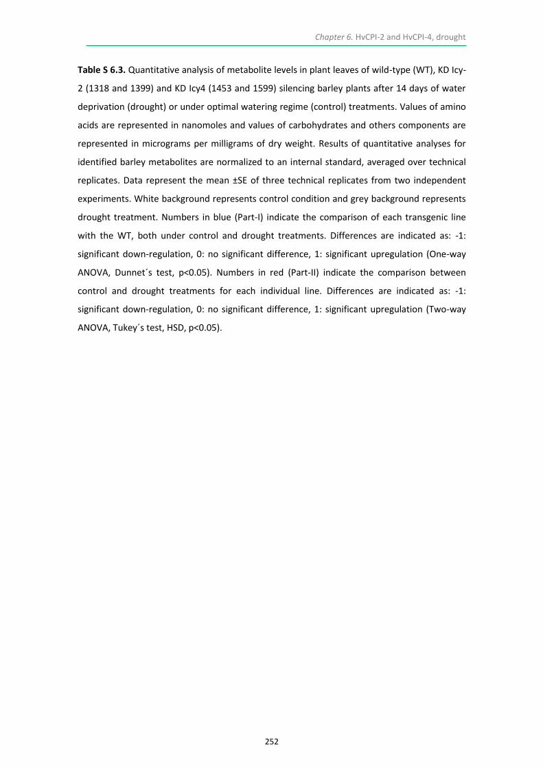
Chapter 6. HvCPI-2 and HvCPI-4, drought
252
Table S 6.3. Quantitative analysis of metabolite levels in plant leaves of wild-type (WT), KD Icy-
2 (1318 and 1399) and KD Icy4 (1453 and 1599) silencing barley plants after 14 days of water
deprivation (drought) or under optimal watering regime (control) treatments. Values of amino
acids are represented in nanomoles and values of carbohydrates and others components are
represented in micrograms per milligrams of dry weight. Results of quantitative analyses for
identified barley metabolites are normalized to an internal standard, averaged over technical
replicates. Data represent the mean ±SE of three technical replicates from two independent
experiments. White background represents control condition and grey background represents
drought treatment. Numbers in blue (Part-I) indicate the comparison of each transgenic line
with the WT, both under control and drought treatments. Differences are indicated as: ‐1:
significant down‐regulation, 0: no significant difference, 1: significant upregulation (One-way
ANOVA, Dunnet´s test, p<0.05). Numbers in red (Part-II) indicate the comparison between
control and drought treatments for each individual line. Differences are indicated as: ‐1:
significant down‐regulation, 0: no significant difference, 1: significant upregulation (Two-way
ANOVA, Tukey´s test, HSD, p<0.05).

Chapter 6. HvCPI-2 and HvCPI-4, drought
253
Table S 6.3. Part-I

Chapter 6. HvCPI-2 and HvCPI-4, drought
254
Table S 6.3. Part-II


Chapter 7. General Discussion
257
7.1. GENERAL DISCUSSION
Previous research on CysProt and PhyCys shed light about the implications of these
counterparts during a key physiological process in barley, grain germination (Martinez
et al., 2009; Cambra et al., 2012). Specifically, it was demonstrated that the barley
HvPap-1 cathepsin F-like peptidase participated as an active component in the
mobilization of grain stored proteins, mainly hordeins, during last stages of grain
germination. Its strictly controlled regulation was exerted, at least in part, by the
action of endogenous cystatins. On the other hand, HvPap-1 was also expressed in
vegetative tissues such as leaves and roots, presuming its participation in additional
physiological roles. The molecular and functional characterization of the entire
cystatin family along with their potential CysProt targets through in vitro and in vivo
approaches in distinct barley tissues also demonstrated the importance of this
complex proteolytic network (Martinez et al., 2009). Interestingly, one barley cystatin,
HvCPI-4, was reported as the only member able to inhibit both C1A and C13 CysProt
families (Martinez et al., 2007). Formerly information encouraged us to complete data
on germination through in vivo analyses and, importantly, to initiate the research on a
key proteolytic-based process, leaf senescence specifically induced by abiotic stresses.
Although developmental and stress-induced senescence share commonalities
and diverge in certain molecular mechanisms, specific information is still quite
fragmentary. Regarding the main goal of the process, what is clear is that any type of
senescence implies intense proteolysis, different subcellular compartments, several
types of proteases and regulators as well as a complex traffic of proteins, aminoacids
and nutrients that ultimately impact grain filling, thus influencing quality, yield and
compromising offspring success. In the last stages of a leaf´s lifespan, organ-level
senescence and, eventually cell death, represent the ruling processes (Woo et al.,
2016). Intriguingly, senescing leaves show more coordinated temporal changes in
transcriptomes than do growing leaves, with sophisticated regulatory networks
comprising distinct interorganellar coordination of various transcriptomes (Kim et al.,
2016). So far, transcriptomic and proteomic data have consistently assigned a major
role to members from the C1A CysProt family in several plants species facing abiotic

Chapter 7. General Discussion
258
stress (Parrott et al., 2007; Gregersen et al., 2008; Martinez et al., 2012; Hollmann et
al., 2014; Diaz-Mendoza et al., 2014; Velasco-Arroyo et al., 2016). Cystatins play a key
role in leaves by controlling the action of CysProt during protein recycling along
senescence and through the activation of protective metabolic cascades under abiotic
stress (Prins et al., 2008; Quain et al., 2014; Je et al., 2014). Massive analyses of gene
expression have facilitated the unveiling of the molecular events and pathways
associated to leaf senescence. Particularly, microarray barley experiments highlight
the importance of transcriptomic approaches to find out the members of a particular
protein family (Parrott et al., 2007; Gregersen et al., 2008).
The whole C1A family from barley, which comprises 41 members, as well as the
whole cystatin family, made up by 13 genes, were analyzed in the current study under
different abiotic stresses able to trigger leaf senescence. Several C1A members
belonging to different subgroups as well as various cystatin genes were upregulated at
the transcriptional level upon the exposure to darkness and drought. Interestingly,
several C1A-encoding genes exhibited significant increased relative expression under
both stress conditions, such as HvPap 1, HvPap-12, HvPap-19 and HvPap-22, whereas
the expression of HvPap-4 was induced by darkness and repressed by drought. In the
case of the inhibitors, while Icy-4 was upregulated under both tested stresses, other
cystatins were altered after a specific treatment or did not respond to it. These results
suggest certain degree of specificity depending upon the activated senescence-related
proteolytic mechanism triggered by a particular stress, although several proteases and
inhibitors may be acting coordinately along the course of a particular, or various
environmental cues.
We initially tested two different stressors in whole 7-day-old WT barley plants
grown under severe conditions: nitrogen starvation, since it represents an essential
nutrient that in small-grain cereals such as barley, wheat and rice, is greatly (up to
90%) mobilized from vegetative tissues to grains (Gregersen et al., 2008), apart from
being a major constituent of Calvin cycle enzymes, like Rubisco; and darkness, which
apparently does not pose a threat in nature, but it represents a simple way to directly
impact on the photosynthetic machinery and to rapidly alter associated metabolism,

Chapter 7. General Discussion
259
since the prolonged lack of light obviously implies a cellular collapse. In fact, when we
studied all biochemical and morphological parameters, darkness appeared to cause
the greatest effects. Chlorophyll abundance, useful indicator of the chloroplast status,
tends to remain constant in photosynthetically active leaves (Sorin et al., 2015).
Chlorophyll reduction was one of the first symptoms perceived in stressed plants, and
carotenoids accumulation seemed to increase in all cases since the ratio
chlorophyll/carotenoids diminished, consistently with its presumed protective role
towards specific abiotic stresses. After darkness, chloroplasts formed aggregates and
cells were close to collapse. All these data clearly indicated that, under these
conditions, whole barley stressed plants had already activated their intrinsic
senescence programs. Consequently, we further analyzed other molecular and
biochemical parameters. The significant decrease in protein levels in darkness and N-
starved leaves in comparison to the controls suggested either the inhibition of protein
synthesis or/and the activation of protease activities associated to nutrient recycling.
Both stresses induced cathepsin L-/F- and B-like activities, suggesting a functional
redundancy in protein turnover. Data were supported by the up-regulation of genes
encoding barley C1A CysProt, particularly HvPap-1, HvPap-12 and HvPap-19, later
confirmed by protein accumulation as shown on immunoblot assays. Rubisco, the
most abundant protein in plants, is likely the major target for proteases when
proteolytic processes are activated (van der Hoorn, 2008; Theonen et al., 2007;
Krupinska et al., 2012; Martinez et al., 2012) and its accumulation showed the greatest
degradation after darkness in this study. The subcellular location of HvPap-1 and
HvPap-19 CysProts revealed a dynamic trafficking, probably involving specific
organelles developed under senescence like SAVs or similar vesicles, which contain
proteins derived from chloroplasts and which are enriched on CysProts (Otegui et al.,
2005; Ishida et al., 2008; Carrion et al., 2013, 2014). Interestingly, HvPap-16, another
C1A member which accordingly to previous studies was only expressed in leaves
(Martinez et al., 2009), exhibited a steady pattern in the epidermal cells both under
control and stress conditions, suggesting that this particular member may not be
intervening on plastidial degradation.

Chapter 7. General Discussion
260
HvPap-1 is required during grain filling and germination (Cambra et al., 2012;
Diaz-Mendoza et al., 2016a) and is strongly induced by darkness and N-starvation.
Thus, we decided to investigate how alterations on its expression could disturb
senescence progress, and thereby nutrient translocation. We used darkness based on
previously explained reasoning, under a mild stress, replacing vermiculite by organic
soil in order to abolish nutrient limitations. Time required to detect senescence
symptoms was obviously longer than in severe experiments. Keech et al. (2007)
reported a “stand-by mode” in whole darkened Arabidopsis leaves showing low
mitochondria activity to preserve active photosynthetic machinery, while individual
darkened leaves presented higher mitochondrial activity to deliver energy and speed
up degradation. Anyhow, at that time senescence had already started and therefore
we fixed 14 days of darkness as the point to perform subsequent experiments with
overexpressing and silencing lines (OE Pap1 and KD Pap1, respectively). Under these
conditions, barley WT dark-treated plants showed a lower increase in the proteolytic
activity as that observed under severe conditions, suggesting a stand-by mode
induced by darkness perception. HvPap-1 levels increased, as expected, at mRNA and
protein levels in OE Pap1, but this did not result in an increase in the proteolytic
activity, probably due to compensating effects among protease activities. In knock-
down treated lines both cathepsin L-/F- and B-like protease activities exhibited a clear
reduction.
A reduced total protein content in the leaves, which paralleled to that of
Rubisco in most of the darkness-treated OE Pap1 lines, together with the reduced
autofluorescence emission from the chlorophyll and the low amount of starch in this
genotype, led us to assume that leaf senescence was sped up. In contrast, all analyses
indicated a delay in the senescence process in KD lines. Interestingly, KD treated plants
showed a striking reduction on chlorophyll b. The light-harvesting chlorophyll a/b
binding protein complex of LHCII is the main antenna for this photosystem. Plants
change their LHCII content depending on the light environment and chlorophyll b is
indispensable for the accumulation of LHCII. Thus, its degradation induces the
catabolism of the complex (Sato et al., 2015). Based on these assumptions, it could be

Chapter 7. General Discussion
261
happening that chlorophyll b was completely transformed into chlorophyll a under a
stand-by mode in order to improve and/or protect photosynthesis.
A clear delayed-senescence phenotype of KD Pap1 lines was observed both in
barley plants grown either under control or darkness conditions, probably indicating
chloroplasts protection from degradation. Another hypothesis assumes a slower
growth for these plants, which could be a consequence of alterations from the
germination events. Manipulation of the proteolytic machinery is a potential way to
enhance grain yield and quality, which lately will influence the mobilization of storage
compounds along germination. Barley germination involves the activity of several
proteases and amylases that hydrolyze and mobilize storage compounds. The
participation of several barley C1A CysProt during germination has been predicted
since members of the C1A subgroups L-, B-, H, and F-cathepsins were induced by GA in
the barley grain (Holwerda and Rogers, 1992; Martinez et al., 2003; Martinez et al.,
2009; Cambra et al., 2012). HvPap-1 was firstly identified in barley grains during
germination (Sreenivasulu et al., 2008), it was expressed in grain tissues during this
stage and it efficiently degraded stored hordeins in vitro (Cambra et al., 2012). To
demonstrate its in vivo involvement along this process, OE Pap1 and KD Pap1 barley
transgenic grains were exhaustively analyzed. If HvPap-1 was one of the responsible
enzymes to in vivo degrade stored proteins, a delay in the germination process should
be expected for the KD Pap1 lines as well as an acceleration for overexpressing ones,
due to a more efficient hydrolysis over accumulated reserves. Indeed, a decrease in
the number of germinated grains over time was observed in KD Pap1 plants.
Nevertheless, although OE Pap1 lines consistently exhibited a higher degradationrate
for hordeins, the number of germinated grains in these lines was quite similar to that
in the WT. HvPap-1 alterations could lead to significant modifications in the grain
composition that subsequently affect the normal progression of the germination
process, or alternatively the expression of some other hydrolytic enzymes might be
enhanced as a compensating effect in these genotypes. Although most of the nitrogen
in the grain is incorporated into proteins, free amino acids are crucial during
germination, and they are major determinants related to grain processing, quality and
food safety (Halford et al., 2015). Genetic modifications leading to variations in the

Chapter 7. General Discussion
262
accumulation of storage proteins may alter the amino acid composition in the grain.
Differential specificity in the source of nutrients that the embryo can use to develop in
a new plant arises from the great variability of results accounting for amino acids,
starch and protein contents among compared phenotypes. The lowest accumulation
of most amino acids detected in KD Pap1 lines could be related with the slow start of
germination exhibited on those lines. OE Pap1 lines strongly accumulated proline and
glutamine, in which hordeins are enriched, but this enrichment did not involve an
accelerated germination. On the other hand, changes in the in the genetic content of
HvPap-1 provoked modifications in the expression of some other related genes, as
evidenced transcriptomic data, which were likewise supported by immunoblots.
Immunolocation analyses performed in embryos for proteases HvPap-1, HvPap-6 and
HvPap-19 revealed an interesting dotted pattern coincident with protein-enriched
bodies. Alterations in the expression for these proteolytic enzymes should be
correlated to variations in the enzymatic activity, but results were not as expected,
again probably due to enzymatic compensations and, most likely, to the intervention
of cystatins. This could explain why in the cases where an increase in the activity of
the proteolytic machinery was measured, it was not translated into a higher
germination rate, as occurred in OE Pap1 lines.
Cystatins are key members in the regulation of C1A CysProt during barley grain
germination (Martinez et al., 2009; Cambra et al., 2012). According to this, silencing of
a cystatin in the grain could lead to an acceleration of the germination process, since
inhibition of the target C1A CysProts would be minor. The functional relationship
between barley cystatins and cathepsins L- and F-like was inferred from their common
implication as counterparts during hordein mobilization upon germination. HvCPI-2 is
a good inhibitor of different barley cathepsin L- and F-like CysProts (Martinez et al.,
2009; Cambra et al., 2012) and it also displays a role along this process (Cambra et al.,
2012; Diaz-Mendoza et al., 2016a). Results obtained for KD Icy2 lines reinforce the
importance of the complex network modulating mobilization of stored proteins, since
the expected acceleration in the germination process was not observed in these lines.
Likewise, in OE Pap1 lines, the higher expression of HvPap-1, target for HvCPI-2, may
be affecting the expression and late accumulation of this and/or related cystatin

Chapter 7. General Discussion
263
members. These inhibitors would be assuming the goal of finely shelter specific
protein fractions from degradation thus regulating germination timing. Again,
compensating effects implying proteases and inhibitors that led to perturbed
proteolytic patterns would explain the unexpected germination processes observed in
KD Icy2 grains.
Importantly, a positive correlation between length of the active photosynthetic
period and cereal grain yield and quality has been reported (Distelfeld et al., 2014).
Accordingly, a delay in leaf senescence, leading to ‘stay green’ phenotypes
(Hörtensteiner, 2007; Gregersen et al., 2013), would determine a higher grain yield
mainly due to increased CH accumulation, as observed in the grains of KD Pap1 lines,
but inefficient nitrogen remobilization and lower protein content. Conversely,
acceleration of senescence confers efficient nitrogen remobilization and higher
protein content, such as is the case of OE Pap1 grains, but instead renders a lower
total grain yield (Gregersen et al., 2008) since starch and related CH are less abundant.
In this context, protein accumulation patterns in the barley grains are important
depending on the end-product use of the harvested crops. For instance, the
production of alcoholic beverages is based upon fermentation and thus enrichment on
CH would be ideal, as well as a faster germination rate. In contrast, to produce feed
for animals, higher protein content would be an added value. Interestingly, the total
amount of hordeins was higher in OE Pap1 dry grains than in the rest of analyzed
seeds. This particular result perfectly links senescence and germination: hordeins, the
main storage proteins in the barley grain that accumulate along barley maturation,
were more abundant in OE Pap1 lines, which exhibit an accelerated leaf senescence.
Furthermore, given that KD Pap1 lines senesced and germinated later, we could also
speculate that the senescence timing is determined by the germination speed.
However, OE Pap1 plants did not germinate faster but did senesce earlier, therefore
indicating that this physiological function was effectively based upon alterations over
HvPap-1 levels. From these data, it can be concluded that HvPap-1 is a functional
stress-associated gene and alterations in its expression bring about changes in barley
abiotic stress responses through modulation of senescence related pathways. Previous
reports have shown similar effects in plant behavior by down-regulating the

Chapter 7. General Discussion
264
expression of senescence-related CysProt, in particular BoCP5 and CaCP genes from
broccoli and pepper, respectively (Eason et al., 2005; Xiao et al., 2014).
Back to abiotic stress, genetically modified plants silencing those cystatin-
encoding genes which were induced under drought treatment, Icy-2 and Icy-4, were
further characterized, presuming a putative role for these PhyCys in the control of
specific senescence-related proteolytic mechanisms. Fourteen days of soil water
deficit was chosen by measuring specific physiological parameters indicators of the
plant and environmental (soil) status, guiding us towards the precise point at which
drought had induced leaf senescence but plants were still healthy enough to perform
all required analyses. Altered phytocystatin expression has been postulated as a way
to palliate negative effects facing climate change (Kunert et al., 2015), under which
severe drought episodes are expected to increase. Proteolysis is a crucial event
involved in the response to drought, and modifications of PhyCys expression patterns
should be correlated to a modulation of CysProt activity. Under drought, plants
reprogram their metabolism in order to overcome this negative episode and
importantly, senescence pathways that fulfill degradation of compounds for recycling
and survival are activated during this, in principle, reversible stage.
Surprisingly, the phenotypes of barley knock-down plants for Icy-2 and Icy-4
(KD Icy2 and KD Icy4, respectively) were different. The natural phenotype of KD Icy2
plants was characterized by a higher biomass when comparing to WT and a delayed
senescence, resembling a kind of functional stay-green, in which the entire
senescence syndrome was delayed, slowed down, or both (Thomas and Howarth,
2000). Conversely, KD Icy4 plants had lesser biomass than WT and exhibited an earlier
chlorophyll disappearance, aside from differences in size and tiller number. In several
crops, drought stress resistance coincides with a stay-green trait (Gregersen et al.,
2013). Under stress, previously observed phenotypes were indeed confirmed.
Furthermore, KD Icy2 plants exhibited increased drought tolerance, apparently
through a delay in leaf senescence, an hypothesis previously reported (Rivero et al.,
2007).

Chapter 7. General Discussion
265
Since C1A CysProt have been largely associated to the cellular dismantling
occurring during natural or stress-induced senescence processes (Diaz-Mendoza et al.,
2014, 2016b), phenotypes observed for KD Icy2 and KD Icy4 plants should be related
to the differential inhibition of drought-associated C1A CysProt. Three out of the five
C1A proteases analyzed (intriguingly HvPap-1, confirmed as a functional senescence-
associated protease) were up-regulated after drought either in WT or modified
genotypes. Interestingly, whereas these three members and the cathepsin L-like
protease HvPap-4 were induced after darkness and low nitrogen treatments (Velasco-
Arroyo et al., 2016), HvPap-4 was repressed after drought. Differential
induction/repression of C1A CysProt and their inhibitors may be subjacent to the
general variability on cathepsin activities, which decreased with drought in WT and KD
Icy2 lines, whereas increased in KD Icy4, consistently with the higher protein
degradation observed for these lines. Therefore, the distinguishing element between
KD Icy2 and KD Icy4 could rely on the inhibitory properties of the silenced inhibitors.
HvCPI-2 is able to efficiently inhibit HvPap-1, as well as several cathepsin L-like barley
proteases (Martinez et al., 2009; Cambra et al., 2012b). HvCPI-4, although a worst
inhibitor of these proteases, represents the only barley PhyCys able to additionally
inhibit C13 legumain proteases (Martinez et al., 2007; Julian et al., 2013). Two barley
legumains were detected in both vegetative and germinative tissues and responded to
biotic and abiotic stimuli, and HvCPI-4 was able to in vitro inhibit them and in vivo
interact with HvLeg-2 (Julián et al., 2013) With these premises, an increase in
concomitant CysProt activity was expected when downregulating Icy-4 in response to
stress, data confirmed in the current research.
Conversely, when Icy-2 gene is silenced, an unexpected decrease in the
protease activity was observed. The barley plant responds by reducing the expression
of HvPap-1 and by increasing the expression of the broad range inhibitor Icy-4 gene
after drought treatment. These alterations, probably together with additional
modifications in the expression of other proteases and/or inhibitors provokes a lower
protein degradation, a conservation of amino acid levels and a lower alteration of the
membrane integrity. The senescence process is delayed, KD Icy2 plants remain
greener after the treatment, and the soil is able to retain some water as the plants

Chapter 7. General Discussion
266
reduce water losses, probably due to a modulation of stomata closure at that time.
These results confirm an active protective role for some cystatins in planta, as
previously reported (Quain et al., 2014; Kunert et al., 2015). On the contrary, the
reduced stomata conductance displayed by KD Icy4 plants might be a consequence of
activated senescence programs in response to drought, reflecting an already initiated
degradation instead of a leaf protection function.
The increased in specific proteolytic activities concomitant with higher protein
degradation rates observed in treated KD Icy4 plants did not lead to stronger
senescence phenotypes when comparing to WT, probably because the high basal
accumulation of amino acids in these plants allowed a quick replacement of degraded
proteins. Differential levels of metabolites, mainly amino acids, could be linked to the
capacity to maintain cell turgor and viability. On the other hand, the weaker
phenotypes detected on these plants (fewer and thinner tillers and leaves) could be
the result of an altered germination. In line with this assumption, when KD Icy2 barley
germination was assessed, rates were slower as expected, with an initial growth
parallel to WT. Hereafter, senescence is delayed and photosynthesis is extended
under natural conditions. Paradoxically, KD Icy2 grains contained more protein.
However, the regular amount of starch, either detected with lugol staining or
biochemically quantified, in combination with the reduced hordein fraction allowed us
to hypothesize that this extended photosynthetic period drives the increased
accumulation of other proteins fractions other than hordeins, globulins and albumins.
Likewise, the lowest accumulation of most amino acids, especially proline and
glutamine, in the grains of KD Icy2 lines could be related with the lower amount of
storaged hordeins. These last data suggest that pleiotropic effects associated to the
silencing of any of these inhibitors lead to a quicker/slower response to the abiotic
treatment. Whether differential responses to silencing PhyCys indeed have
consequences over grain yield and composition is a question that remains to be
elucidated.
Results from this thesis support previous literature at the same time that
broadening information related to C1A-PhyCys complex alterations in barley. This

Chapter 7. General Discussion
267
manipulation has the potential to modulate sensitivity towards specific abiotic
stresses, through modifications over established developmental leaf senescence
programs. According to presented data, proteolytic reprogramming should be
considered since the plant tries to compensate the genetic modifications by
modulating the expression of some other peptidases or inhibitors, as occurred for OE
Pap1 and KD Icy2 lines. On the other hand, it is corroborated the in vivo relevance of
this proteolytic network during barley grain remobilization upon germination, in a
manner that might be economically important for agriculture. Ongoing experiments
related to other specific members, such as HvPap-16 or HvPap-19, are being
undertaken through analyses on specific transgenic lines and the obtaining of double
mutants. Besides, according to presented results, a research related to traffic and
characterization involving specialized vesicles, such as SAVs, is going to be developed.
Several RNAseq analyses over selected transgenic lines, in some cases subjected to a
combination of abiotic and biotic stresses, have also been performed to find out
underlying molecular events which could help to elucidate interconnected pathways
between leaf senescence, stress and CysProt-Cystatin complexes. Studies on whole-
plant senescence using multi-omics approaches will greatly broaden the information
regarding senescence at the whole plant level, to further understand how it is
systematically achieved and differentially regulated in response to internal and
external factors (Kim et al., 2016). Data from agronomic valuable crops would further
help to improve productivity.
7.2. REFERENCES
Cambra I, Martinez M, Dáder B, González-Melendi P, Gandullo J, Santamaría ME, Diaz I (2012) A cathepsin F-like peptidase involved in barley grain protein mobilization, HvPap-1, is modulated by its own propeptide and by cystatins. J Exp Bot 63: 4615–4629
Carrion CA, Costa ML, Martinez DE, Mohr C, Humbeck K, Guiamet JJ (2013) In vivo inhibition of cysteine proteases provides evidence for the involvement of “senescence-associated vacuoles” in chloroplast protein degradation during dark-induced senescence of tobacco leaves. J Exp Bot 64: 4967–4980
Carrion CA, Martinez DE, Costa ML, Guiamet JJ (2014) Senescence-associated vacuoles, a specific lytic compartment for degradation of chloroplastic proteins? Plants 3: 498–512

Chapter 7. General Discussion
268
Sato R, Ito H, Tanaka A (2015) Chlorophyll b degradation by chlorophyll b reductase under high-light conditions. Photosynth Res 126: 249–259
Christoff AP, Passaia G, Salvati C, Alves-Ferreira M, Margis-Pinheiro M, Margis R (2016) Rice bifunctional phytocystatin is a dual modulator of legumain and papain-like proteases. Plant Mol Biol 92: 193–207
Diaz-Mendoza M, Velasco-Arroyo B, Gonzalez-Melendi P, Martinez M, Diaz I (2014)
C1A cysteine protease-cystatin interactions in leaf senescence. J Exp Bot 65: 3825-
3833
Diaz-Mendoza M, Dominguez-Figueroa JD, Velasco-Arroyo B, Cambra I, Gonzalez-Melendi P, Lopez-Gonzalvez A, Garcia A, Hensel G, Kumlehn J, Diaz I, Martinez M (2016a) HvPap-1 C1A protease and HvCPI-2 cystatin contribute to barley grain filling and germination. Plant Physiol 170: 2511–2524
Diaz-Mendoza M, Velasco-Arroyo B, Santamaria ME, González-Melendi P, Martinez M, Diaz I (2016b) Plant senescence and proteolysis: two processes with one destiny. Genet Mol Biol 39: 329–338
Distelfeld A, Avni R, Fischer AM (2014) Senescence, nutrient remobilization, and yield in wheat and barley. J Exp Bot 65: 3783–3798
Eason JR, West PJ, Brummell D a., Watson LM, Somerfield SD, McLachlan ARG (2014) Overexpression of the protease inhibitor BoCPI-1 in broccoli delays chlorophyll loss after harvest and causes down-regulation of cysteine protease gene expression. Postharvest Biol Technol 97: 23–31
Gregersen PL, Culetic A, Boschian L, Krupinska K (2013) Plant senescence and crop productivity. Plant Mol Biol 82: 603–622
Gregersen PL, Holm PB, Krupinska K (2008) Leaf senescence and nutrient remobilisation in barley and wheat. Plant Biol 10: 37–49
Halford NG, Curtis TY, Chen Z, Huang J (2015) Effects of abiotic stress and crop management on cereal grain composition: implications for food quality and safety. J Exp Bot 66: 1145–1156
Hollmann J, Gregersen PL, Krupinska K (2014) Identification of predominant genes involved in regulation and execution of senescence-associated nitrogen remobilization in flag leaves of field grown barley. J Exp Bot 65: 3963–3974
Holwerda BC, Rogers JC (1992) Purification and characterization of aleurain: a plant thiol protease functionally homologous to Mammalian cathepsin H. Plant Physiol 99: 848–855
Hörtensteiner S (2007) Chlorophyll degradation during senescence. Annu Rev Plant Biol 57: 55–77
Ishida H, Yoshimoto K, Izumi M, Reisen D, Yano Y, Makino A, Ohsumi Y, Hanson MR, Mae T (2008) Mobilization of Rubisco and stroma-localized fluorescent proteins of chloroplast to the vacuole by ATG gene-dependent autophagic process. Plant Physiol 148: 142–155

Chapter 7. General Discussion
269
Je J, Song C, Hwang JE, Chung WS, Lim CO (2014) DREB2C acts as a transcriptional activator of the thermo tolerance-related phytocystatin 4 (AtCYS4) gene. Transgenic Res 23: 109–123
Julián I, Gandullo J, Santos-Silva LK, Diaz I, Martinez M (2013) Phylogenetically distant barley legumains have a role in both seed and vegetative tissues. J Exp Bot 64: 2929–2941
Kim J, Woo HRR, Nam HGG (2016) Toward systems understanding of leaf senescence: an integrated multi-omics perspective on leaf senescence research. Mol Plant 9: 813–825
Krupinska K, Mulisch M, Hollmann J, Tokarz K, Zschiesche W, Kage H, Humbeck K, Biler W (2012) An alternative strategy of dismantling of the chloroplast during leaf senescence observed in a high-yield variety of barley. Physiol Plant 144: 189–200
Kunert KJ, Van Wyk SG, Cullis CA, Vorster BJ, Foyer CH (2015) Potential use of phytocystatins in crop improvement, with a particular focus on legumes. J Exp Bot 66: 3559–3570
Martinez M, Cambra I, Carrillo L, Diaz-Mendoza M, Diaz I (2009) Characterization of the entire cystatin gene family in barley and their target cathepsin L-like cysteine-proteases, partners in the hordein mobilization during seed germination. Plant Physiol 151: 1531–1545
Martinez M, Cambra I, Gonzalez-Melendi P, Santamaria ME, Diaz I (2012) C1A cysteine-proteases and their inhibitors in plants. Physiol Plant 145: 85–94
Martinez M, Diaz-Mendoza M, Carrillo L, Diaz I (2007) Carboxy terminal extended phytocystatins are bifunctional inhibitors of papain and legumain cysteine proteinases. FEBS Lett 581: 2914–2918
Martinez M, Rubio-Somoza I, Carbonero P, Diaz I (2003) A cathepsin B-like cysteine protease gene from Hordeum vulgare (gene CatB) induced by GA in aleurone cells is under circadian control in leaves. J Exp Bot 54: 951–959
Otegui MS, Noh YS, Martínez DE, Vila Petroff MG, Staehelin LA, Amasino RM, Guiamet JJ (2005) Senescence-associated vacuoles with intense proteolytic activity develop in leaves of Arabidopsis and soybean. Plant J 41: 831–844
Parrott DL, McInnerney K, Feller U, Fischer AM (2007) Steam-girdling of barley (Hordeum vulgare) leaves leads to carbohydrate accumulation and accelerated leaf senescence, facilitating transcriptomic analysis of senescence-associated genes. New Phytol 176: 56–69
Prins A, van Heerden PDR, Olmos E, Kunert KJ, Foyer CH (2008) Cysteine proteinases regulate chloroplast protein content and composition in tobacco leaves: a model for dynamic interactions with ribulose-1,5-bisphosphate carboxylase/oxygenase (Rubisco) vesicular bodies. J Exp Bot 59: 1935–1950
Quain MD, Makgopa ME, Márquez-García B, Comadira G, Fernandez-Garcia N, Olmos E, Schnaubelt D, Kunert KJ, Foyer CH (2014) Ectopic phytocystatin expression leads to enhanced drought stress tolerance in soybean (Glycine max)

Chapter 7. General Discussion
270
and Arabidopsis thaliana through effects on strigolactone pathways and can also result in improved seed traits. Plant Biotechnol J 12: 903–913
Rivero RM, Kojima M, Gepstein A, Sakakibara H, Mittler R, Gepstein S, Blumwald E (2007) Delayed leaf senescence induces extreme drought tolerance in a flowering plant. Proc Natl Acad Sci USA 104: 19631–19636
Sorin C, Musse M, Mariette F, Bouchereau A, Leport L (2015) Assessment of nutrient remobilization through structural changes of palisade and spongy parenchyma in oilseed rap leaves during senescence. Planta 241: 333–346
Sreenivasulu N, Usadel B, Winter A, Radchuk V, Scholz U, Stein N, Weschke W, Strickert M, Close TJ, Stitt M, Graner A, Wobus U (2008) Barley grain maturation and germination: metabolic pathway and regulatory network commonalities and differences highlighted by new MapMan/PageMan profiling tools. Plant Physiol 146: 1738–1758
Theonen M, Herrmann B, Feller U (2007) Senescence in wheat leaves, is a cysteine endopeptidase involved in the degradation of the large subunit of Rubisco? Acta Physiol Plant 29: 339–350
Thomas H, Howarth CJ (2000) Five ways to stay green. J Exp Bot 51: 329–337
van der Hoorn RAL (2008) Plant proteases: from phenotypes to molecular mechanism. Ann Rev Plant Biol 59: 191–223
Velasco-Arroyo B, Diaz-Mendoza M, Gandullo J, Gonzalez-Melendi P, Santamaria ME, Dominguez Figueroa JD, Hensel G, Martinez M, Kumlehn J, Diaz I (2016) HvPap-1 C1A protease actively participates in barley proteolysis mediated by abiotic stresses. J Exp Bot 67: 4297–4310
Woo HR, Koo HJ, Kim J, Jeong H, Yang JO, Lee IH, Jun JH, Choi SH, Park SJ, Kang B et al. (2016) Programming of plant leaf senescence with temporal and inter-organellar coordination of transcriptome in Arabidopsis. Plant Physiol 171: 452–467
Xiao HJ, Yin YX, Chai WG, Gong ZH (2014) Silencing of the CaCP Gene Delays Salt- and Osmotic-Induced Leaf Senescence in Capsicum annuum L. Int J Mol Sci 15: 8316–8334


Chapter 8. General Conclusions
273
The results of this thesis permit the extraction of several conclusions alluding to the
role of barley C1A cysteine proteases and their inhibitors throughout leaf senescence
and grain germination:
1. In barley, members from all C1A CysProt groups are expressed in response to
abiotic stresses able to induce leaf senescence, such as darkness, nitrogen
starvation and drought. A certain degree of specificity is likely to rule the process.
Some members respond to various environmental cues, such as HvPap-1, HvPap-
12 and HvPap-19, while others display opposite patterns, such as HvPap-4 and
HvPap-6.
2. The delayed-senescence phenotype displayed by knock-down HvPap-1 plants,
resembling a stay-green phenotype, and the opposite trend exhibited by
overexpressing HvPap-1 lines, either under natural and darkness-induced
senescence, designates HvPap-1 as a functional stress-associated gene. Alterations
in its expression bring about changes at the biochemical and molecular levels,
indicating a modulation in the responses through alterations over established
developmental leaf senescence pathways.
3. The delay in germination observed in silencing HvPap-1 lines, together with
alterations in the grain composition, confirms a role for this protease during
degradation of stored proteins. Overexpressing HvPap-1 barley grains accumulate
the highest amount of hordeins and show increased levels of proline and
glutamine, in which hordeins are enriched on. However, this observation is not
accompanied by an accelerated germination rate.
4. Among cystatins, Icy-4 appeared as the main upregulated gene both under
darkness and drought, while Icy-2 was specifically altered under drought.
5. Under drought, knock-down Icy-2 plants exhibited an increased tolerance,
apparently through a delay in leaf senescence. Conversely, knock-down Icy-4

Chapter 8. General Conclusions
274
plants display opposite trends, similar to those observed for overexpressing
HvPap-1 lines under darkness.
6. When a drought-induced cystatin is silenced, the other is overexpressed,
suggesting a cooperative role for both members in response to the stress. The
broad range of targets for HvCPI-4, along with the high efficiency of HvCPI-2 in the
inhibition of particular drought-induced proteases, would allow a tight modulation
during protein degradation in response to water deprivation.
7. Either in leaves undergoing senescence or in germinating barley grains, the plant
tries to compensate the genetic modifications by modulating the expression of
some other peptidases and inhibitors. Therefore, proteolytic reprogramming
should be considered when designing biotechnological strategies based on the
manipulation of mechanisms involving CysProt-PhyCys.
8. From the data reported on this thesis it can be concluded that senescence timing
is influencing grain filling and composition, therefore impacting on germination
events. Accordingly, manipulation of lifespan on cereals through biotechnological
approaches based on proteolytic mechanisms seems a promising strategy to
produce grains with enhanced properties for specific uses.
List of Publications
275
- PhD publications:
Díaz-Mendoza M, Velasco-Arroyo B, González-Melendi P, Martínez M, Díaz I (2014)
C1A cysteine protease-cystatin interactions in leaf senescence. J Exp Bot 65: 3825–3833
Velasco-Arroyo B, Diaz-Mendoza M, Gandullo J, Gonzalez-Melendi P, Santamaria ME,
Dominguez-Figueroa JD, Hensel G, Martinez M, Kumlehn J, Diaz I (2016) HvPap-1 C1A
protease actively participates in barley proteolysis mediated by abiotic stresses. J Exp Bot
67: 4297-4310
Diaz-Mendoza M, Dominguez-Figueroa JD, Velasco-Arroyo B, Cambra I, Gonzalez-
Melendi P, Lopez-Gonzalvez A, Garcia A, Hensel G, Kumlehn J, Diaz I, et al. (2016) HvPap-1
C1A protease and HvCPI-2 cystatin contribute to barley grain filling and germination. Plant
Physiol 170: 2511–2524
Velasco-Arroyo B, Diaz-Mendoza M, Gomez-Sanchez A, Moreno-Garcia B, Santamaria
ME, Torija-Bonilla M, Hensel G, Kumlehn J, Martinez M, Diaz I (2016) Silencing barley
cystatins HvCPI-2 and HvCPI-4 specifically modifies plant responses to drought (submitted)
- Other publications:
Diaz-Mendoza M, Velasco-Arroyo B, Santamaria ME, González-Melendi P, Martinez M,
Diaz I (2016) Plant senescence and proteolysis: two processes with one destiny. Genet Mol
Biol 39: 329-338
- Book Chapters:
Diaz-Mendoza M, Velasco-Arroyo B, Dominguez-Figueroa JD, Santamaria ME,
González-Melendi P, Torija-Bonilla M, Hensel G, Kumlehn J, Martinez M, Diaz I (2015) C1A
peptidases actively participate in the barley leaf senescence events driven by abiotic
stresses. Curr Res Plant Physiol pp. 252. Fenoll C, Escobar C, Martin M, Mena M, Editors.
Toledo, Spain. ISBN: 978-84-606-8883-9
Velasco-Arroyo B, Diaz-Mendoza M, Santamaria ME, Gonzalez-Melendi P, Gomez-
Sanchez A, Arnaiz A, Martinez M, Diaz I (2016) Senescence-associated genes in response to
abiotic/biotic stresses. Progress in Botany. Springer (submitted)
