corolla is a novel protein that contributes to the ... · corolla: a novel sc protein collins et...
TRANSCRIPT

Collins et al.
1
Corolla is a novel protein that contributes to the architecture of the synaptonemal complex of Drosophila
Kimberly A. Collins*,1, Jay R. Unruh*, Brian D. Slaughter*, Zulin Yu*, Cathleen M. Lake*, Rachel J. Nielsen*, Kimberly S. Box*,2, Danny E. Miller*,‡, Justin P. Blumenstiel§, Anoja G. Perera*, Kathryn E. Malanowski*, and R. Scott Hawley*,‡
* Stowers Institute for Medical Research, Kansas City, MO, United States of America
§ Department of Ecology and Evolutionary Biology, University of Kansas, Lawrence, KS,
United States of America
‡ Department of Molecular and Integrative Physiology, University of Kansas Medical Center,
Kansas City, KS, United States of America
1 Current address: Department of Comparative Medicine, University of Washington, Seattle,
WA, United States of America
2 Current address: Department of Molecular Biology, Princeton University, Princeton, NJ,
United States of America
Short title: Corolla: a novel SC protein
Key words: meiosis, chromosome segregation, synaptonemal complex, structured illumination
microscopy, next-generation sequencing
To whom correspondence should be addressed:
R. Scott Hawley
Stowers Institute for Medical Research
Kansas City, MO 64110
Phone (816) 926-4427
Fax (816) 926-2060
Genetics: Early Online, published on June 9, 2014 as 10.1534/genetics.114.165290
Copyright 2014.
Corolla: a novel SC protein
Collins et al.
2
ABSTRACT In most organisms the synaptonemal complex (SC) connects paired homologs along their entire length during much of meiotic prophase. To better understand the structure of the SC, we aim to identify its components and to determine how each of these components contributes to SC function. Here, we report the identification of a novel SC component in Drosophila melanogaster female oocytes, which we have named Corolla. Using structured illumination microscopy, we demonstrate that Corolla is a component of the central region of the SC. Consistent with its localization, we show by yeast two-hybrid analysis that Corolla strongly interacts with Cona, a central element protein, demonstrating the first direct interaction between two inner-synaptonemal complex proteins in Drosophila. These observations help provide a more complete model of SC structure and function in Drosophila females. INTRODUCTION The Laws of Mendelian Inheritance are little more than a statistical restatement of the events that underlie the proper segregation of homologous chromosomes at the first meiotic division. Homologous segregation requires a number of distinct processes, such as pairing and recombination, but our focus here is on the formation of a complex, proteinaceous structure known as the synaptonemal complex (SC). The SC forms between homologous chromosomes during early meiotic prophase and is essential for such functions as the maintenance of homolog pairing, the conversion of programmed double-strand breaks (DSBs) into crossovers (PAGE and HAWLEY 2004), and the facilitation of both homologous and heterologous centromeric associations (TAKEO et al. 2011; TANNETI et al. 2011). In terms of its overall structure and dimensions, the SC is highly conserved (CARPENTER 1975; ZICKLER and KLECKNER 1999; SCHILD-PRÜFERT et al. 2011; FRAUNE et al. 2012a), consisting of two lateral elements (LEs) and a central region (CR). The CR is comprised of both transverse filament proteins (TFs) and central element (CE) proteins. LE proteins run the length of each homolog and function to connect the
sister chromatids and to compact the chromosome axes with a complex composed of cohesins as well as SC-specific components (LAMMERS et al. 1994; ANDERSON et al. 2005; KHETANI and BICKEL 2007; ALSHEIMER et al. 2010; WATTS and HOFFMANN 2011). In Drosophila melanogaster, the SC-specific components of the LE are Ord, Solo and C(2)M (MANHEIM and MCKIM 2003; WEBBER et al. 2004; ANDERSON et al. 2005; KHETANI and BICKEL 2007; YAN and MCKEE 2013). The CE is located at the very center of the SC. In Drosophila, the Cona protein is thought to be a component of the CE. Cona appears to be required for “zippering” together the TF proteins that both span the width of the SC and overlap in an interlocking manner at the center of the SC (PAGE et al. 2008)). Many organisms appear to have a single TF protein, however, C. elegans has multiple TF proteins that act together to bridge the width of the SC (MACQUEEN et al. 2002; COLAIÁCOVO et al. 2003; SMOLIKOV et al. 2007; SMOLIKOV et al. 2009; SCHILD-PRÜFERT et al. 2011). Although it has long been thought that Drosophila also possesses a single TF protein, known as C(3)G (PAGE and HAWLEY 2001), we will present evidence

Corolla: a novel SC protein
Collins et al.
3
below that the novel protein Corolla is a TF protein. Corolla is encoded by a novel Drosophila gene, CG8316, and corolla mutants are defective in SC assembly and/or maintenance. Using structured illumination microscopy (SIM), we show that Corolla localizes to the CR of the Drosophila SC. Consistent with Corolla’s localization to the CR by SIM, we show that Corolla physically interacts with Cona, demonstrating the first direct interaction between two inner-SC proteins in Drosophila. We will propose that Corolla, which possesses several predicted coiled-coil domains, functions both by directly binding to Cona and by interacting with C(3)G, perhaps via their coiled-coil domains, to stabilize the ability of C(3)G to link the LEs to the CE and thereby establishing the SC. MATERIALS AND METHODS Drosophila Genetics All stocks were maintained on standard medium containing yeast, cornmeal, corn syrup, malt extract, and agar at 25o. Stocks used in this study include:, y w cv v corolla1 FRT19A/C(1)DX, y f/Y (COLLINS et al. 2012), y w corolla1 FRT19A/C(1)DX, y f /y+Y; (COLLINS et al. 2012), w1118
P{XP]CG8316d01774/C(1)DX, y f /y+Y (created from stock Bloomington 19165), Df(1)BSC643 (Bloomington 25733), w1118/Binsinscy and Df(1)BSC583, w1118/Binsinscy (Bloomington 25417) were used in mapping corolla, w; pCa4-attB-genomic-Corolla (this study), okraAA cn bw/CyO (GHABRIAL et al. 1998), okraRU cn bw/CyO (GHABRIAL et al. 1998), ru1 h1 th1 st1 cu1 sr1 es ca1 (Bloomington 576), ru1 h1 th1 st1 cu1 sr1 es Pr1 ca/TM6B, Bri1, Tb1 (Bloomington 1711), ru1 h1 th1 st1 cu1 sr1 es ca1/TM3 Sb were used in recombination assays, nanos-Gal4; conaf04903/TM3, y w
eyFLP; FRT82B y+ TPN1, Tb, Ser (PAGE et al. 2008), yw eyFLP;FRT82B conaA12/TM6B y+ TPN1, Tb (PAGE et al. 2007), y w FRT19A/hs-hidY (used as wild-type stock) (COLLINS et al. 2012), y w nanos-Gal4/FM7a; c(3)G68e ca/TM3 Ser (JEFFRESS et al. 2007), y w ; P{UASP-c(3)G-Cdel4-Flag ; c(3)G[68] e/TM3, Sb Ser e; pol/+ (JEFFRESS et al. 2007); and w/BsY; c(3)G68e/TM3, Ser. The following genotypes were used throughout the manuscript and represent the null genotypes of corolla, cona, c(3)G, and okra: y w cv v corolla1 FRT19A/y w corolla1 FRT19A, conaf04903/conaA12, c(3)G68 e/c(3)G68 e ca and okraAA cn bw/okraRU cn bw respectively.
To assay chromosome nondisjunction, tester female virgins were crossed to X^Y, In(1)EN,v f B; C(4)RM,ci eyR males. Calculations were performed as previously described (ZITRON and HAWLEY 1989; HAWLEY et al. 1992). To assay recombination on the third chromosome, single tester female virgins (either y w FRT19A; ru h th st cu sr es Pr ca/+ or y w corolla FRT19A; ru h th st cu sr es Pr ca/+) were crossed to ru h th st cu sr es ca males in vials and all single and double recombinants in the female progeny between th and ca were scored through day 18. Mapping of the corolla mutants to CG8316 Next-generation sequencing (NGS) was used to identify the causative lesion in the novel complementation group previously reported (COLLINS et al. 2012), which contains mutants corolla1, corolla129 and corolla166. A multi-allele whole genome sequencing approach was done for two of these alleles (corolla1 and corolla129) and compared to the parental stock used in the

Corolla: a novel SC protein
Collins et al.
4
screen, y w FRT19A, according to (BLUMENSTIEL et al. 2009) with the following modifications. For each NGS sample, 1-3 µg of genomic DNA was sheared to less than 700-base pair (bp) fragments using a Bioruptor sonicator (Diagenode). Following manufacturer’s directions, short fragment libraries were made using the Illumina TruSeq DNA LT Sample Prep Kit v2-set B (Illumina, Cat. No. FC-121-2002). The resulting libraries were quantified using a Bioanalyzer (Agilent Technologies) and a Qubit Fluorometer (Life Technologies). All libraries were pooled, quantified and run as 100-bp paired-end reads on an Illumina HiSeq 2000 instrument using HiSeq Control Software 1.4.8. Following sequencing, Illumina Primary Analysis version RTA 1.12.4.2 and Secondary Analysis version CASAVA-1.8.2 were run to demultiplex reads and generate FASTQ files. Reads were trimmed to 90 bp by the FastX toolkit (http://hannonlab.cshl.edu/fastx_toolkit/index.html). Reads were aligned to the UCSC dm3 reference genome using BWA, version 0.5.9 (LI and DURBIN 2009). Duplicate reads potentially caused by amplification were removed using samtools rmdup. Samtools and Picard were used to index and sort the reads to prepare for variant analysis. Indel and SNP detection was performed using the GATK pipeline (MCKENNA et al.
2010; DEPRISTO et al. 2011). This tool was used to perform local realignment around indels and then call SNPs and indels for the region of interest. Predicted variant effects were found using the snpEff tool (version 2.0.5d) (CINGOLANI et al. 2012). Post processing, filtering, and analysis of variants was performed using custom scripts.
Three SNPs were detected within the CG8316 gene region in the parental stock compared with the reference sequence. The “extended gene region” from Flybase of CG8316 was downloaded and the first nucleotide is equivalent to position 1 in the following SNP designations. The three SNPs correspond to a synonymous mutation G2299A in exon 2, G3776T in exon 4 which results in an Asp to Glu amino acid change, and a deletion of 4067A after the stop codon in exon 4. corolla1 has an A2579T mutation in CG8316, resulting in a single stop codon at amino acid 173; corolla129 has a 22-bp deletion located at position 2814–2836 (just 1 bp following amino acid 190), which results in a frame shift leading to a series of stop codons with the first beginning at amino acid 199; and corolla166 has a C2804T mutation, which creates a single stop codon at amino acid 188 (Figure 1A). CG8316 was the only gene on the X chromosome that we identified which had a single nucleotide polymorphism or a deletion in the two mutant chromosomes tested when compared to the reference parental chromosome. Transgene rescue For transgene rescue of corolla, a genomic rescue construct was made by performing PCR on the y w FRT19A stock that was used for the mutagenesis in which corolla
was isolated. The transgene included 1,019 bp upstream of the start codon of CG8316 and 227 bp of downstream sequence, which includes the 3’ untranslated region. Naturally occurring SpeI and BamHI sites were utilized for cloning this genomic fragment. The entire gene region was sequenced after cloning into pCasPeR4-attB (gift from Perrimon lab), at SpeI and BamHI sites in the multi-cloning region, for

Corolla: a novel SC protein
Collins et al.
5
site-specific integration at attP40 (by Genetic Services, MA) (MARKSTEIN et al. 2008). Yeast two-hybrid The Matchmaker Gold Yeast Two-Hybrid System User Manual (Clontech Protocol No. PT4084-1, Version No. PR033493) was followed for yeast transformation, testing for autoactivation and for yeast two-hybrid assays. AH109 yeast were used in place of Y2Hgold. AH109 genotype is as follows: MATa, trp1-901, leu2-3, 112, ura3-52, his3-200, gal4Δ, gal80Δ,LYS2 : : GAL1UAS-GAL1TATA-HIS3, GAL2UAS-GAL2TATA-ADE2, URA3 : : MEL1UAS-MEL1 TATA-lacZ (JAMES et al. 1996). Y187 genotype is as follows: MATα, ura3-52, his3-200, ade2-101, trp1-901, leu2-3, 112, gal4Δ, met–, gal80Δ, URA3 : : GAL1UAS-GAL1TATA-lacZ (WADE
HARPER et al. 1993). BD-Corolla autoactivated the HIS3 reporter but did not autoactivate the ADE2 reporter and only weakly autoactivated the MEL1 reporter (data not shown). Cytology Germarium preparations for whole mount immunofluorescence were as according to Page and Hawley (PAGE and HAWLEY 2001) with minor exceptions. 0- to 1-day-old females were collected and yeasted overnight in the presence of males. Ovaries were dissected in PBS for no longer than 20 min prior to fixing. In all incubation steps, ovaries were incubated at room temperature while nutating unless otherwise specified. Ovaries were then washed three times for 10 min in PBS with 0.1% Tween (PBST). Ovaries were cut at approximately stage 4–5 and the tips containing the germarium were dissected apart with forceps. Germarium tips were then blocked in PBST with 1% BSA for 1 hr, and the primary antibody diluted in PBST was incubated with germarium tips
overnight at 4o while nutating. After washing three times for 15 min in PBST the secondary antibodies were applied for 4 hr followed by the addition of 4’6-diamididino-2-phenylindole (DAPI) at a concentration of 1 µg/ml for the final 10 min of incubation. After washing as before, the samples were mounted in ProLong Gold (Invitrogen) and allowed to cure for 24 hours. Chromosome spreads were prepared as described in Khetani and Bickel with the two following minor exceptions (KHETANI and BICKEL 2007). 16% formaldehyde (Ted Pella) was diluted to 1% in water and pH was not adjusted. Normal goat serum was substituted for normal donkey serum in the blocking solution. Mouse anti-C(3)G 1A8-1G2 was used at 1:500 (ANDERSON et al. 2005). Rabbit anti-Corolla (animal 210) was used at 1:1000 or 1:1500 in all experiments with indistinguishable results (this study). Guinea pig anti-Cona was used at 1:500 (PAGE et al. 2008). Rat anti-CID (gift of Sunkel Laboratory) was used at 1:1000 (MARTINS et al. 2009). Guinea pig anti-SMC1 was used at 1:500 (KHETANI and BICKEL 2007). Mouse anti-Orb antibodies 4H8 and 6H4 (Developmental Studies Hybridoma Bank, Iowa) were used at 1:50 each. Anti-Flag M2 antibody (Sigma) was used at 1:500. Mouse anti-γ-H2AV (LAKE et al. 2013) supernatant was used at 1:500. In experiments where both mouse anti-γ-H2AV (isotype IgG2b-kappa) and mouse anti-Orb were used together, only anti-Orb 4H8 (isotype IgG1) was used. Secondary goat anti-mouse, rabbit, guinea pig or rat Alexa-488, Alexa-555 and Alexa-647 IgG H&L chain or isotype specific (IgG1 and IgG2b) conjugated antibodies were used at 1:500 (Molecular Probes). With the exception of Figures 2 and S2, all images were acquired with a DeltaVision

Corolla: a novel SC protein
Collins et al.
6
microscopy system (GE Healthcare) consisting of a 1x70 inverted microscope with a high-resolution CCD camera. Images were deconvolved using SoftWoRx (Applied Precision/GE Healthcare) software and maximum intensity projections were made unless otherwise noted. SIM images were acquired on an Applied Precision OMX Blaze microscope (Issaquah, WA, USA) equipped with a PCO Edge sCMOS camera (Kelheim, Germany). An Olympus (Center Valley, PA, USA) 60x 1.42NA Plan Apo N oil objective was used. SIM reconstruction was performed with the Applied Precision Softworx software package (Life Technologies, Carlsbad, CA, USA) following the Applied Precision protocols. All analysis was performed using ImageJ and custom plugins written for ImageJ available at http://research.stowers.org/imagejplugins. Prior to analysis, the SIM reconstructed image was scaled 2 by 2 with bilinear interpolation. Alignment between different colored channels was performed on single slices or projections using the TurboReg plugin for ImageJ (THEVENAZ et al. 1998). Well-formed regions of the SC that ran approximately parallel to the xy plane were manually selected and intensity profiles were averaged over a 3-pixel-wide stripe perpendicular to the SC. For C(3)G distance measurements, these intensity profiles were then fit to double Gaussian functions with a background component. Sanger sequencing DNA was isolated from a single aged male according to (GLOOR et al. 1993). Sequencing primers for CG8316/corolla are available upon request. Centromere clustering analysis Analysis was performed as previously described (TAKEO et al. 2011). We analyzed
oocyte nuclei in region 3 of the germarium in both wild-type and SC-defective lines. Region 3 oocytes were identified by co-staining with the cytoplasmic marker Orb. Generation of polyclonal anti-Corolla antibodies The cDNA of CG8316 (Berkeley Drosophila Genome Project, Drosophila Gene Collection #LD15362) was amplified with primers containing Nde1 and BamH1 sites (primer sequence available upon request), and subcloned into pET19b at NdeI and BamHI sites within the multi-cloning region. The clone was sequence-verified and transformed into BL21 DE 3 cells (New England Biolabs). Protein expression was done following the QiaExpressionist Handbook for purification under denaturing conditions, and the 6xHis protein was purified using ProBond Nickel Chelating Resin (Invitrogen, CA). Antibodies were made in rabbits using 6xHis-Corolla protein as antigen (Cocalico Biologicals, Inc). Pre-immune rabbit (animal 210) serum did not stain Drosophila ovaries (data not shown). Quantification of γ-H2AV foci
To score the number of γ-H2AV foci, we
first identified the nuclei of the oocyte in region 3 cysts that had concentrated cytoplasmic Orb staining. okra mutants also display a delay in selection of the oocyte (JOYCE and MCKIM 2009). Therefore, in those region 3 cysts that had two oocytes, both oocytes were score if each had concentrated Orb staining and robust SC staining. We then identified the z-stacks pertaining to the oocyte nuclei. Using Imaris 7.0.0 software (Bitplane, Zurich, Switzerland), we performed a 3D crop of the selected nuclei and displayed the z-sections using the gallery function. Only clearly defined foci where counted in the corresponding z-series.
Corolla: a novel SC protein
Collins et al.
7
RESULTS The corolla gene was first identified by the recovery of three allelic mutants, originally named mei-391, mei-39129 and mei-39166, in a screen for X-linked meiotic mutants in Drosophila (COLLINS et al. 2012). Based on the cytological phenotype we will describe below, we have named this gene corolla. We also have renamed the mei-391, mei-39129 and mei-39166 mutants corolla1, corolla129 and corolla166. As described in Materials and Methods, these mutants were mapped by whole genome sequencing to a transcription unit denoted CG8316 (Figure 1A). The meiotic phenotype exhibited by these corolla alleles was confirmed to be the result of mutations in CG8316 by showing both that two overlapping deficiencies that uncover CG8316 and a P-element insertion mutation, P(XP)CG8316d01774, fail to complement the corolla1 allele as assayed by measuring meiotic nondisjunction (data not shown). We further showed by a transgene rescue assay that expression of CG8316 fully suppresses the meiotic nondisjunction phenotype of the corolla1 mutant (Figure 1B). The corolla gene is predicted to encode a 554-amino acid protein with three internal coiled-coil domains (FlyBase and Coils) (Figure 1C and Figure S1) (LUPAS et al. 1991). All four known alleles of corolla (shown in Figure 1A) exhibit DNA sequence changes in the third exon of this gene. corolla1, corolla129 and corolla166 are the result of mutations that would be predicted to truncate the Corolla protein at amino acid 173, 199 and 188 (see Materials and Methods), respectively, while P(XP)CG8316d01774 is a P-element insertion mutant in exon three. The three EMS-induced alleles behave similarly in all
assays tested, and therefore we will discuss corolla1 exclusively in this manuscript. In addition, based on both studies we will show below and the fact that the meiotic phenotype of corolla1 homozygotes was similar to corolla1/Df(corolla) heterozygotes (data not shown), we will propose that corolla1 is a null allele. Corolla encodes a SC component and is required for proper SC formation Given that corolla appears to encode a protein with coiled-coil domains—a known conserved domain within components of the CR—and that mutations in corolla cause high levels of meiosis I nondisjunction (COLLINS et al. 2012), we speculated that Corolla might be a component of the SC. We generated antibodies to Corolla and performed immunofluorescence assays on ovaries from wild-type females. As shown in Figure 1D, Corolla localizes to the SC in a pattern that closely mimics that of the transverse filament protein C(3)G and the CE protein Cona. In the absence of corolla, SC formation is abolished (Figure 1D). This defect in SC formation can be rescued by a transgene bearing a genomic copy of corolla (data not shown), confirming that Corolla is essential for SC formation. corolla mutants exhibit phenotypes consistent with a role in SC formation and function Proteins involved in forming the structure of the SC tend to display many of the same phenotypes. In general terms, mutations in these genes cause: 1) a failure both to cluster centromeres in early prophase and to maintain the pairing of homologous centromeres (TAKEO et al. 2011; TANNETI et al. 2011), 2) a reduction in the frequency of programmed DSBs (MEHROTRA and MCKIM 2006), and 3) greatly reduced or abolished meiotic recombination (PAGE and HAWLEY

Corolla: a novel SC protein
Collins et al.
8
2001; MANHEIM and MCKIM 2003; PAGE et al. 2007). As described in detail below, corolla mutants also affect each of these processes, consistent with the hypothesis that Corolla functions as an integral component of the SC. We begin by demonstrating that corolla mutants exhibit a severe defect in meiotic centromere clustering. Drosophila females have four pairs of homologous chromosomes, which cluster in early prophase within the germarium. The number of centromere clusters is obtained by counting the number of foci observed using an antibody against the Drosophila CID protein (CENP-A homolog) (BLOWER and KARPEN 2001). Typically one or two CID clusters are seen in wild-type oocytes (TAKEO et al. 2011; TANNETI et al. 2011). However, in mutants that are defective in SC formation/maintenance, centromere clustering is abrogated, increasing the number of CID foci to three or four, and often homologous centromeres become unpaired, resulting in the observation of more than four CID foci (TAKEO et al. 2011). We measured the extent of both centromere clustering and centromere pairing in corolla by determining the percentage of a given genotype that displayed either a decrease in oocytes with one or two CID foci (a centromere clustering defect) or displayed more than four CID foci (a centromere pairing defect). Since corolla is defective in SC formation, it is difficult to distinguish oocyte nuclei from nurse cell nuclei in early stages of pachytene. Therefore, we chose to analyze oocytes in region 3 of the germarium where the oocyte nucleus is easily identifiable by co-staining with Orb, which concentrates in the cytoplasm of the specified oocyte. As shown in Figure 1E and Table S1, the
number of oocytes with one or two CID foci decreased from 76.3% observed in wild type to 0% in corolla, indicative of a strong defect in centromere clustering. Additionally, although oocytes with more than four foci were never observed in wild-type oocytes, in corolla oocytes we found that 21.9% of nuclei scored had more than four CID foci, suggesting that corolla mutants are also strongly defective in centromere pairing. These defects in centromere clustering and centromere pairing are comparable to those exhibited by other SC mutants. For example, a c(3)G mutant, showed a significant reduction in one or two CID foci [80.7% in wild type to 6.3% in c(3)G] and 28.1% of region 3 oocyte nuclei had greater than four CID foci (TAKEO et al. 2011). Previous studies have also shown that SC disruption can lead to reduced DSB formation (MEHROTRA and MCKIM 2006). To assay the number of DSBs, we used a monoclonal antibody recognizing the phosphorylated form of H2AV, called γ-H2AV (LAKE et al. 2013), and used the cytoplasmic marker Orb to mark the early-mid-pachytene oocyte nuclei in region 3 (Figure 1F). Because the formation and repair of DSBs is a dynamic process, and in order to determine more accurately the number of DSBs generated in early pachytene, we utilized a DSB repair-defective background (okra) to assay the total number of DSBs made (GHABRIAL et al. 1998; JANG et al. 2003; MEHROTRA and MCKIM 2006). As anticipated, we found that corolla; okra double mutants exhibited a reduced number of γ-H2AV foci (average of 8.3 foci per oocyte, n=15 oocytes), displaying only 37% of the DSBs observed in the okra control genotype (average of 22.3 foci per oocyte, n=10 oocytes) (set to 100%) (Table S2). This is similar to the range of reduction seen by Mehrotra and McKim in the LE mutant c(2)M (44.8% of okra control) and the TF mutant c(3)G (15%

Corolla: a novel SC protein
Collins et al.
9
to 21.3% of okra control) in the absence of repair (MEHROTRA and MCKIM 2006). Thus, although Corolla is not essential for DSB formation, like other SC proteins it is required for normal levels of DSB formation. Moreover, the following analysis of recombination shows that Corolla is required to process DSBs into mature crossovers. The high levels of nondisjunction seen in corolla mutants are easily explained by the reduction in meiotic recombination (Figure 1G). While the wild-type control exhibited levels of recombination on the third chromosome similar to previous studies (MANHEIM and MCKIM 2003), corolla exhibited no recombination throughout most of the euchromatin and exhibited a reduced frequency of recombination in the centromere-proximal interval. These reduced levels of recombination are fully consistent with the high levels of nondisjunction observed in corolla mutants. Although the same number of females were tested, fewer progeny were scored in corolla (n=168) than in wild type (n=1271) due to a fertility defect of corolla mutants (Table 1), which may well be the consequence of chromosome missegregation. Taken together, these data suggest that, at least as assayed genetically, Corolla acts as an integral member of the SC, such that corolla mutants show the same cluster of meiotic defects exhibited by mutants in genes encoding other known SC components.
Corolla localizes to the central region of the synaptonemal complex To determine the precise location of Corolla within the SC, we performed studies using SIM. First, to ascertain if we could resolve the lateral edge of the SC from the central
region, we analyzed by SIM the colocalization of C(3)G and Cona. Using an antibody to the C-terminal globular domain of C(3)G that has been shown by immuno-EM to localize at the lateral edges of the TF, abutting the LE (ANDERSON et al. 2005), we were able to resolve two parallel tracks of C(3)G localization. As predicted from preliminary immuno-EM studies used to analyze the location of Cona::venus overexpressed in wild-type ovaries (LAKE and HAWLEY 2012), Cona localized between the two tracks of the lateral edges of the TF (Figure S2). We determined the distance between the two C(3)G tracks to be 137 ± 3 nm (Figure S3A-B). Our data for SC width is slightly larger than the number (109 ± 8 nm) obtained by Carpenter’s (CARPENTER 1975) EM analysis of the Drosophila SC. One possible explanation for this discrepancy may be that the C-terminal epitope of C(3)G is buried within the LE. Indeed, the immuno-EM done by Anderson and colleagues using this antibody shows C(3)G localization that appears within the electron dense LE tracks (ANDERSON et al. 2005). As we were able to resolve the lateral sides of the SC from the central region using SIM, we next determined the localization of Corolla within the SC by co-staining wild-type oocytes with antibodies to Corolla and the C-terminal end of C(3)G (Figure 2A, Figure S2B and File S1–S2). The immunofluorescence signal for Corolla was similar to that for Cona, in that the Corolla signal ran between the two tracks of C(3)G. When a line profile was drawn perpendicular to a parallel track of C(3)G, two peaks of C(3)G signal are clearly visible and only one peak of Corolla is present in between the two C(3)G peaks (Figure 2B). While in the vast majority of cases (33/34) Corolla signal appeared as a single peak in

Corolla: a novel SC protein
Collins et al.
10
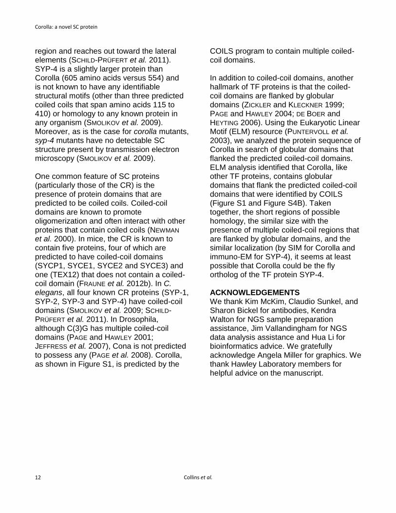
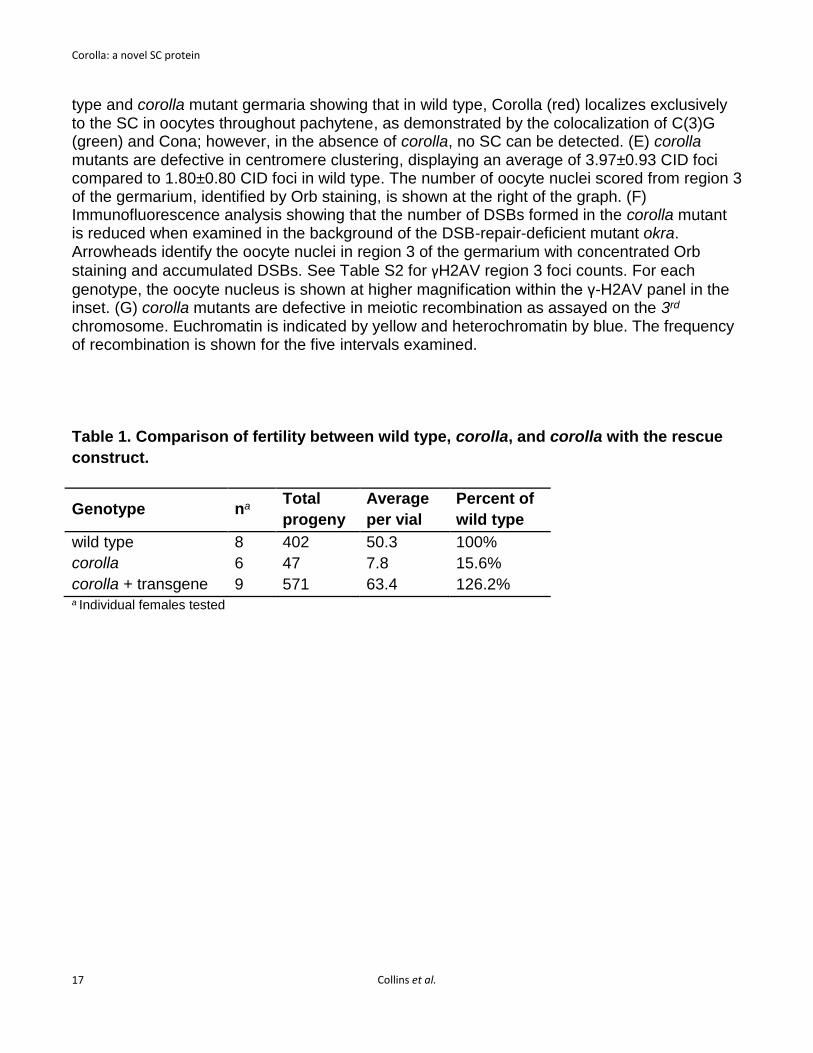
a line profile, Corolla appeared as a double peak in 1/34 cases. The significance of this one unusual profile, if any, is currently not understood. In conclusion, SIM technology enabled us to localize Corolla within the SC; specifically, Corolla localizes to the CR of the SC. Corolla interacts with Cona by yeast two-hybrid Since SIM analysis virtually always identified Corolla as a single track running between the lateral sides of the SC—a pattern very similar to Cona—we wanted to determine whether Corolla and Cona physically interact. To test this, we performed yeast two-hybrid analysis (Figure 3). We assayed BD-Corolla/AD-Cona interaction on the ADE2 reporter, as well as in the MEL1 assay. Diploids containing BD-Corolla and AD-Cona were able to grow on media lacking adenine and exhibited a deep blue color indistinguishable from the diploid positive control in the presence of X-alpha-gal, thus illustrating that Cona-Corolla strongly interact. This provides the first evidence of a physical linkage between any two Drosophila inner-SC proteins. Corolla was named for this interaction with Cona, whose full name is Corona, as Corolla is the Latin diminutive of Corona. Corolla may self-associate, but does not appear to localize to the chromosome axes in the absence of C(3)G Since Corolla contains coiled-coil domains and physically interacts with Cona, we next addressed whether Corolla could associate with the chromosomes in the absence of the TF protein, C(3)G. Whole mount immunofluorescence imaging using standard deconvolution microscopy demonstrated that Corolla signal persisted in early pachytene nuclei (region 2A) in the absence of the TF protein C(3)G, although
the signal appeared more diffuse and less ribbon-like than the wild type (Figure 4). The Corolla staining observed in Figure 4B was unanticipated since previous EM analysis of c(3)G mutants did not reveal any SC structure (SMITH and KING 1968). In an attempt to resolve this issue, we performed immunofluorescence analysis on chromosome spreads of wild-type and c(3)G mutant oocytes immunostained with Corolla and the cohesin component SMC1 to visualize the lateral elements of the SC (Figure 4C-D). Previous studies have shown that loading of cohesin components SMC1 and SMC3 is not affected in the absence of c(3)G (KHETANI and BICKEL 2007). Chromosome spread preparation of ovaries, a procedure in which soluble components not attached to chromatin are removed (KHETANI and BICKEL 2007), enabled us to determine whether Corolla localizes to meiotic chromosomes in the absence of c(3)G. In 40 partially intact germaria, we did not detect any chromatin-associated Corolla (one example shown in Figure 4D), suggesting that the structure of Corolla seen in whole mount c(3)G oocytes does not appear to associate with chromosomes. We speculate that Corolla may be self-assembling, via the coiled-coil domains, however, these complexes do not associate with the LEs. Thus, based on these observations and those described above, Corolla and C(3)G appear mutually dependent with regard to localization to meiotic chromosomes. In addition, we also analyzed Corolla’s localization in oocytes expressing a mutation of c(3)G which lacks the C-terminal globular domain of the C(3)G protein (JEFFRESS et al. 2007). In this mutant, C(3)G cannot attach to the LEs, and therefore instead of assembling along the chromosomes the proteins comprising

Corolla: a novel SC protein
Collins et al.
11
the central region of the SC are assembled into aggregates of SC-like material called polycomplexes. In the absence of C(3)G association with the LEs, we find that Corolla, like Cona, assembles into polycomplexes (Figure 4E). This result further supports the findings that Corolla is a CR component of the SC and does not associate with the LEs in the absence of C(3)G. DISCUSSION This paper both describes the utility of SIM for the analysis of SC in wild-type and mutant Drosophila oocytes and reports the identification of Corolla, a new component of the central region of the Drosophila SC. We have further shown that Corolla physically interacts with another central region protein, Cona. We have previously presented preliminary immuno-EM data suggesting that Cona localizes to the edges of the CE (PAGE et al. 2008; LAKE and HAWLEY 2012). The discovery of Corolla raises the number of central region proteins identified in Drosophila to three [Corolla, Cona, and C(3)G], each of which is dependent on the other two for establishing the SC. We propose that while the binding of these three proteins to the paired homologs is mediated by the attachment of the C terminus of C(3)G to the LE, both Cona and Corolla are required to stabilize the interactions between oppositely oriented C(3)G proteins (transverse filament proteins), which are required to form nascent complexes that can be attached to the LE. In the absence of the C terminus of C(3)G, the three known central region proteins (and presumably others) can form SC-like polycomplexes that fail to attach to the chromosomes.
Perhaps because these three proteins are so functionally interdependent, the phenotypes of corolla mutants are virtually identical to those of cona or c(3)G mutants (PAGE and HAWLEY 2001; MEHROTRA and MCKIM 2006; PAGE et al. 2008; TAKEO et al. 2011). These phenotypes include a reduction in the frequency of meiotic DSBs, a near elimination of meiotic recombination (and a concomitant increase in meiotic nondisjunction), and a loss of centromere clustering. We imagine that the similarity of meiotic defects exhibited by all three mutants reflects the fact that these act as part of a functional identity, such that each component is left functionless without the other two. Anderson et al. (ANDERSON et al. 2005) used immuno-EM to show that C(3)G spanned the distance between the LEs and the middle of the CE, and Öllinger et al. (ÖLLINGER et al. 2005) has shown that expression of the rat TF protein, SCP1, alone in cultured cells can facilitate the assembly of polycomplex-like structures. Why then does the Drosophila oocyte require additional proteins such as Cona and Corolla to build an SC, and what specifically is the function of Corolla? Perhaps the answer lies in 1) Cona’s position in the central region, where it could act to stabilize the anti-parallel interactions between C(3)G homodimers emanating from opposite LEs; 2) Corolla’s ability to physically interact with Cona; and 3) a hypothetical interaction between the coiled-coil domains of Corolla and C(3)G. Although corolla encodes what appears to be a novel protein, we have identified three short regions of amino acid homology between Corolla and the C. elegans SYP-4 protein (Supplemental Figure 4). SYP-4 is one of the C. elegans TF proteins, which, like Corolla, localizes within the central
Corolla: a novel SC protein
Collins et al.
12
region and reaches out toward the lateral elements (SCHILD-PRÜFERT et al. 2011). SYP-4 is a slightly larger protein than Corolla (605 amino acids versus 554) and is not known to have any identifiable structural motifs (other than three predicted coiled coils that span amino acids 115 to 410) or homology to any known protein in any organism (SMOLIKOV et al. 2009). Moreover, as is the case for corolla mutants, syp-4 mutants have no detectable SC structure present by transmission electron microscopy (SMOLIKOV et al. 2009). One common feature of SC proteins (particularly those of the CR) is the presence of protein domains that are predicted to be coiled coils. Coiled-coil domains are known to promote oligomerization and often interact with other proteins that contain coiled coils (NEWMAN et al. 2000). In mice, the CR is known to contain five proteins, four of which are predicted to have coiled-coil domains (SYCP1, SYCE1, SYCE2 and SYCE3) and one (TEX12) that does not contain a coiled-coil domain (FRAUNE et al. 2012b). In C. elegans, all four known CR proteins (SYP-1, SYP-2, SYP-3 and SYP-4) have coiled-coil domains (SMOLIKOV et al. 2009; SCHILD-PRÜFERT et al. 2011). In Drosophila, although C(3)G has multiple coiled-coil domains (PAGE and HAWLEY 2001; JEFFRESS et al. 2007), Cona is not predicted to possess any (PAGE et al. 2008). Corolla, as shown in Figure S1, is predicted by the
COILS program to contain multiple coiled-coil domains. In addition to coiled-coil domains, another hallmark of TF proteins is that the coiled-coil domains are flanked by globular domains (ZICKLER and KLECKNER 1999; PAGE and HAWLEY 2004; DE BOER and HEYTING 2006). Using the Eukaryotic Linear Motif (ELM) resource (PUNTERVOLL et al. 2003), we analyzed the protein sequence of Corolla in search of globular domains that flanked the predicted coiled-coil domains. ELM analysis identified that Corolla, like other TF proteins, contains globular domains that flank the predicted coiled-coil domains that were identified by COILS (Figure S1 and Figure S4B). Taken together, the short regions of possible homology, the similar size with the presence of multiple coiled-coil regions that are flanked by globular domains, and the similar localization (by SIM for Corolla and immuno-EM for SYP-4), it seems at least possible that Corolla could be the fly ortholog of the TF protein SYP-4.
ACKNOWLEDGEMENTS We thank Kim McKim, Claudio Sunkel, and Sharon Bickel for antibodies, Kendra Walton for NGS sample preparation assistance, Jim Vallandingham for NGS data analysis assistance and Hua Li for bioinformatics advice. We gratefully acknowledge Angela Miller for graphics. We thank Hawley Laboratory members for helpful advice on the manuscript.

Corolla: a novel SC protein
Collins et al.
13
REFERENCES
ALSHEIMER, M., A. BAIER, S. SCHRAMM, W.
SCHÜTZ and R. BENAVENTE, 2010
Synaptonemal Complex Protein SYCP3 Exists
in Two Isoforms Showing Different
Conservation in Mammalian Evolution.
Cytogenetic and Genome Research 128: 162-
168.
ANDERSON, L. K., S. M. ROYER, S. L. PAGE, K. S.
MCKIM, A. LAI et al., 2005 Juxtaposition of
C(2)M and the transverse filament protein
C(3)G within the central region of Drosophila
synaptonemal complex. Proc Natl Acad Sci U S
A 102: 4482-4487.
BLOWER, M. D., and G. KARPEN, 2001 The role of
Drosophila CID in kinetochore formation, cell-
cycle progression and heterochromatin
interactions. Nat Cell Biol 3: 730-739.
BLUMENSTIEL, J. P., A. C. NOLL, J. A. GRIFFITHS, A.
G. PERERA, K. N. WALTON et al., 2009
Identification of EMS-Induced Mutations in
Drosophila melanogaster by Whole-Genome
Sequencing. Genetics 182: 25-32.
CARPENTER, A. T., 1975 Electron microscopy of
meiosis in Drosophila melanogaster females: II.
The recombination nodule--a recombination-
associated structure at pachytene? Proc Natl
Acad Sci U S A 72: 3186-3189.
CINGOLANI, P., A. PLATTS, L. L. WANG, M. COON,
T. NGUYEN et al., 2012 A program for
annotating and predicting the effects of single
nucleotide polymorphisms, SnpEff: SNPs in the
genome of Drosophila melanogaster strain
w1118; iso-2;iso-3. Fly 6: 80-92.
COLAIÁCOVO, M. P., A. J. MACQUEEN, E.
MARTINEZ-PEREZ, K. MCDONALD, A. ADAMO
et al., 2003 Synaptonemal Complex Assembly
in C. elegans Is Dispensable for Loading Strand-
Exchange Proteins but Critical for Proper
Completion of Recombination. Developmental
Cell 5: 463-474.
COLLINS, K. A., J. G. CALLICOAT, C. M. LAKE, C. M.
MCCLURKEN, K. P. KOHL et al., 2012 A
Germline Clone Screen on the X Chromosome
Reveals Novel Meiotic Mutants in Drosophila
melanogaster. G3: Genes|Genomes|Genetics 2:
1369-1377.
DE BOER, E., and C. HEYTING, 2006 The diverse
roles of transverse filaments of synaptonemal
complexes in meiosis. Chromosoma 115: 220-
234.
DEPRISTO, M. A., E. BANKS, R. POPLIN, K. V.
GARIMELLA, J. R. MAGUIRE et al., 2011 A
framework for variation discovery and
genotyping using next-generation DNA
sequencing data. Nat Genet 43: 491-498.
FRAUNE, J., M. ALSHEIMER, J.-N. VOLFF, K. BUSCH,
S. FRAUNE et al., 2012a Hydra meiosis reveals
unexpected conservation of structural
synaptonemal complex proteins across
metazoans. Proceedings of the National
Academy of Sciences 109: 16588-16593.
FRAUNE, J., S. SCHRAMM, M. ALSHEIMER and R.
BENAVENTE, 2012b The mammalian
synaptonemal complex: Protein components,
assembly and role in meiotic recombination.
Experimental Cell Research 318: 1340-1346.
GHABRIAL, A., R. P. RAY and T. SCHÜPBACH, 1998
okra and spindle-B encode components of the
RAD52 DNA repair pathway and affect meiosis
and patterning in Drosophila oogenesis. Genes
& Development 12: 2711-2723.
GLOOR, G. B., C. R. PRESTON, D. M. JOHNSON-
SCHLITZ, N. A. NASSIF, R. W. PHILLIS et al.,
1993 Type I repressors of P element mobility.
Genetics 135: 81-95.
HAWLEY, R. S., H. IRICK, A. E. ZITRON, D. A.
HADDOX, A. LOHE et al., 1992 There are two
mechanisms of achiasmate segregation in
Drosophila females, one of which requires
heterochromatic homology. Dev Genet 13: 440-
467.
JAMES, P., J. HALLADAY and E. A. CRAIG, 1996
Genomic Libraries and a Host Strain Designed
for Highly Efficient Two-Hybrid Selection in
Yeast. Genetics 144: 1425-1436.
JANG, J. K., D. E. SHERIZEN, R. BHAGAT, E. A.
MANHEIM and K. S. MCKIM, 2003 Relationship
of DNA double-strand breaks to synapsis in
Drosophila. Journal of Cell Science 116: 3069-
3077.
JEFFRESS, J. K., S. L. PAGE, S. M. ROYER, E. D.
BELDEN, J. P. BLUMENSTIEL et al., 2007 The
Formation of the Central Element of the
Synaptonemal Complex May Occur by Multiple
Mechanisms: The Roles of the N- and C-
Terminal Domains of the Drosophila C(3)G

Corolla: a novel SC protein
Collins et al.
14
Protein in Mediating Synapsis and
Recombination. Genetics 177: 2445-2456.
JOYCE, E. F., and K. S. MCKIM, 2009 Drosophila
PCH2 Is Required for a Pachytene Checkpoint
That Monitors Double-Strand-Break-
Independent Events Leading to Meiotic
Crossover Formation. Genetics 181: 39-51.
KHETANI, R. S., and S. E. BICKEL, 2007 Regulation
of meiotic cohesion and chromosome core
morphogenesis during pachytene in Drosophila
oocytes. Journal of Cell Science 120: 3123-3137.
LAKE, C. M., and R. S. HAWLEY, 2012 The
Molecular Control of Meiotic Chromosomal
Behavior: Events in Early Meiotic Prophase in
Drosophila Oocytes. Annual Review of
Physiology 74: 425-451.
LAKE, C. M., J. K. HOLSCLAW, S. P. BELLENDIR, J.
SEKELSKY and R. S. HAWLEY, 2013 The
Development of a Monoclonal Antibody
Recognizing the Drosophila melanogaster
Phosphorylated Histone H2A Variant (γ-H2AV).
G3: Genes|Genomes|Genetics 3: 1539-1543.
LAMMERS, J. H., H. H. OFFENBERG, M. VAN
AALDEREN, A. C. VINK, A. J. DIETRICH et al.,
1994 The gene encoding a major component of
the lateral elements of synaptonemal complexes
of the rat is related to X-linked lymphocyte-
regulated genes. Molecular and Cellular Biology
14: 1137-1146.
LI, H., and R. DURBIN, 2009 Fast and accurate short
read alignment with Burrows–Wheeler
transform. Bioinformatics 25: 1754-1760.
LUPAS, A., M. VAN DYKE and J. STOCK, 1991
Predicting coiled coils from protein sequences.
Science 252: 1162-1164.
MACQUEEN, A. J., M. P. COLAIÁCOVO, K.
MCDONALD and A. M. VILLENEUVE, 2002
Synapsis-dependent and -independent
mechanisms stabilize homolog pairing during
meiotic prophase in C. elegans. Genes &
Development 16: 2428-2442.
MANHEIM, E. A., and K. S. MCKIM, 2003 The
Synaptonemal Complex Component C(2)M
Regulates Meiotic Crossing over in Drosophila.
Current Biology 13: 276-285.
MARKSTEIN, M., C. PITSOULI, C. VILLALTA, S. E.
CELNIKER and N. PERRIMON, 2008 Exploiting
position effects and the gypsy retrovirus
insulator to engineer precisely expressed
transgenes. Nat Genet 40: 476-483.
MARTINS, T., A. F. MAIA, S. STEFFENSEN and C. E.
SUNKEL, 2009 Sgt1, a co-chaperone of Hsp90
stabilizes Polo and is required for centrosome
organization. EMBO J 28: 234-247.
MCKENNA, A., M. HANNA, E. BANKS, A.
SIVACHENKO, K. CIBULSKIS et al., 2010 The
Genome Analysis Toolkit: A MapReduce
framework for analyzing next-generation DNA
sequencing data. Genome Research 20: 1297-
1303.
MEHROTRA, S., and K. S. MCKIM, 2006 Temporal
Analysis of Meiotic DNA Double-Strand Break
Formation and Repair in Drosophila Females.
PLoS Genet 2: e200.
NEWMAN, J. R. S., E. WOLF and P. S. KIM, 2000 A
computationally directed screen identifying
interacting coiled coils from Saccharomyces
cerevisiae. Proceedings of the National
Academy of Sciences 97: 13203-13208.
ÖLLINGER, R., M. ALSHEIMER and R. BENAVENTE,
2005 Mammalian Protein SCP1 Forms
Synaptonemal Complex-like Structures in the
Absence of Meiotic Chromosomes. Molecular
Biology of the Cell 16: 212-217.
PAGE, S., R. J. NIELSEN, K. TEETER, C. M. LAKE, S.
ONG et al., 2007 A Germline Clone Screen for
Meiotic Mutants in Drosophila melanogaster.
Fly 1: 172-181.
PAGE, S. L., and R. S. HAWLEY, 2001 c(3)G encodes
a Drosophila synaptonemal complex protein.
Genes & Development 15: 3130-3143.
PAGE, S. L., and R. S. HAWLEY, 2004 THE
GENETICS AND MOLECULAR BIOLOGY
OF THE SYNAPTONEMAL COMPLEX.
Annual Review of Cell and Developmental
Biology 20: 525-558.
PAGE, S. L., R. S. KHETANI, C. M. LAKE, R. J.
NIELSEN, J. K. JEFFRESS et al., 2008 corona Is
Required for Higher-Order Assembly of
Transverse Filaments into Full-Length
Synaptonemal Complex in Drosophila Oocytes.
PLoS Genet 4: e1000194.
PUNTERVOLL, P., R. LINDING, C. GEMÜND, S.
CHABANIS-DAVIDSON, M. MATTINGSDAL et al.,
2003 ELM server: a new resource for
investigating short functional sites in modular
eukaryotic proteins. Nucleic Acids Research 31:
3625-3630.
SCHILD-PRÜFERT, K., T. T. SAITO, S. SMOLIKOV, Y.
GU, M. HINCAPIE et al., 2011 Organization of

Corolla: a novel SC protein
Collins et al.
15
the Synaptonemal Complex During Meiosis in
Caenorhabditis elegans. Genetics 189: 411-421.
SMITH, P. A., and R. C. KING, 1968 GENETIC
CONTROL OF SYNAPTONEMAL
COMPLEXES IN DROSOPHILA
MELANOGASTER. Genetics 60: 335-351.
SMOLIKOV, S., A. EIZINGER, K. SCHILD-PRUFERT, A.
HURLBURT, K. MCDONALD et al., 2007 SYP-3
Restricts Synaptonemal Complex Assembly to
Bridge Paired Chromosome Axes During
Meiosis in Caenorhabditis elegans. Genetics
176: 2015-2025.
SMOLIKOV, S., K. SCHILD-PRÜFERT and M. P.
COLAIÁCOVO, 2009 A Yeast Two-Hybrid
Screen for SYP-3 Interactors Identifies SYP-4, a
Component Required for Synaptonemal
Complex Assembly and Chiasma Formation in
Caenorhabditis elegans Meiosis. PLoS Genet 5:
e1000669.
TAKEO, S., CATHLEEN M. LAKE, E. MORAIS-DE-SÁ,
CLÁUDIO E. SUNKEL and R. S. HAWLEY, 2011
Synaptonemal Complex-Dependent Centromeric
Clustering and the Initiation of Synapsis in
Drosophila Oocytes. Current Biology 21: 1845-
1851.
TANNETI, NIKHILA S., K. LANDY, ERIC F. JOYCE
and KIM S. MCKIM, 2011 A Pathway for
Synapsis Initiation during Zygotene in
Drosophila Oocytes. Current Biology 21: 1852-
1857.
THEVENAZ, P., U. E. RUTTIMANN and M. UNSER,
1998 A pyramid approach to subpixel
registration based on intensity. Image Processing,
IEEE Transactions on 7: 27-41.
WADE HARPER, J., G. R. ADAMI, N. WEI, K.
KEYOMARSI and S. J. ELLEDGE, 1993 The p21
Cdk-interacting protein Cip1 is a potent inhibitor
of G1 cyclin-dependent kinases. Cell 75: 805-
816.
WATTS, F. Z., and E. HOFFMANN, 2011 SUMO
meets meiosis: An encounter at the
synaptonemal complex. BioEssays 33: 529-537.
WEBBER, H. A., L. HOWARD and S. E. BICKEL, 2004
The cohesion protein ORD is required for
homologue bias during meiotic recombination.
The Journal of Cell Biology 164: 819-829.
YAN, R., and B. D. MCKEE, 2013 The Cohesion
Protein SOLO Associates with SMC1 and Is
Required for Synapsis, Recombination,
Homolog Bias and Cohesion and Pairing of
Centromeres in Drosophila Meiosis. PLoS Genet
9: e1003637.
ZICKLER, D., and N. KLECKNER, 1999 MEIOTIC
CHROMOSOMES: Integrating Structure and
Function. Annual Review of Genetics 33: 603-
754.
ZITRON, A. E., and R. S. HAWLEY, 1989 The genetic
analysis of distributive segregation in
Drosophila melanogaster. I. Isolation and
characterization of Aberrant X segregation (Axs),
a mutation defective in chromosome partner
choice. Genetics 122: 801-821.

Corolla: a novel SC protein
Collins et al.
16
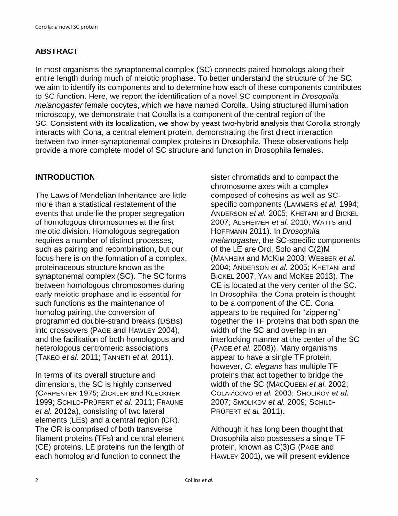
Figure 1. Corolla is an essential component of the Drosophila SC. (A) A schematic of the CG8316 gene region shows the relative location of the corolla alleles, with coding sequence in blue and the 5’ and 3’ untranslated regions in gray. Note that all alleles are located in exon 3. Mutations in corolla1, corolla129 and corolla166 are predicted to truncate the protein at amino acid 173, 199 and 188, respectively (see Materials and Methods). (B) corolla exhibits elevated levels of X and 4th chromosome nondisjunction, which can be fully suppressed by expression of one copy of corolla (pCa4-genomic-Corolla-attB). (C) A schematic of Corolla showing the location of the internal coiled-coil domains as predicted by Coils (http://embnet.vital-it.ch/software/COILS_form.html). (D) Immunofluorescence of wild-
Corolla: a novel SC protein
Collins et al.
17
type and corolla mutant germaria showing that in wild type, Corolla (red) localizes exclusively to the SC in oocytes throughout pachytene, as demonstrated by the colocalization of C(3)G (green) and Cona; however, in the absence of corolla, no SC can be detected. (E) corolla mutants are defective in centromere clustering, displaying an average of 3.97±0.93 CID foci compared to 1.80±0.80 CID foci in wild type. The number of oocyte nuclei scored from region 3 of the germarium, identified by Orb staining, is shown at the right of the graph. (F) Immunofluorescence analysis showing that the number of DSBs formed in the corolla mutant is reduced when examined in the background of the DSB-repair-deficient mutant okra. Arrowheads identify the oocyte nuclei in region 3 of the germarium with concentrated Orb
staining and accumulated DSBs. See Table S2 for γH2AV region 3 foci counts. For each
genotype, the oocyte nucleus is shown at higher magnification within the γ-H2AV panel in the inset. (G) corolla mutants are defective in meiotic recombination as assayed on the 3rd chromosome. Euchromatin is indicated by yellow and heterochromatin by blue. The frequency of recombination is shown for the five intervals examined. Table 1. Comparison of fertility between wild type, corolla, and corolla with the rescue
construct.
Genotype na Total
progeny
Average
per vial
Percent of
wild type
wild type 8 402 50.3 100%
corolla 6 47 7.8 15.6%
corolla + transgene 9 571 63.4 126.2% a Individual females tested

Corolla: a novel SC protein
Collins et al.
18
Figure 2. Corolla localizes to the central region of the SC. (A) DeltaVision OMX microscopy of C(3)G (C-terminal domain) (magenta, AF488) and Corolla (green, AF555). Maximum-intensity projections are shown of a few Z slices. See Figure S2B for a representative image of a maximum-intensity projection through an entire pachytene pro-oocyte. (B) Representative line profiles plot the normalized intensity for Corolla (green) and C(3)G (magenta). The line profile in (B) is from panel (A). Two peaks of C(3)G intensity represent the parallel tracks of C(3)G, and the Corolla peak is positioned clearly between the two C(3)G peaks.
Figure 3. Corolla strongly interacts with Cona by yeast two-hybrid. All diploid strains grow equally well under selection for both the AD and BD plasmids (–trp–leu). AD-Cona and BD-Corolla strongly interact on the ADE2 reporter assay (–trp–leu–ade). No interaction was detected with AD-empty and a BD-Corolla construct or between AD-Cona and an empty BD construct. AD-Cona and BD-Corolla strongly interact on the MEL1 reporter assay (of –trp–leu + X-alpha Gal). It should be noted that BD-Corolla weakly autoactivates the MEL1 reporter. AD-Cona and BD-Corolla strongly interact under the most stringent selection of –trp–leu–ade + X-alpha Gal.
C(3)GCorollaMerge
A
1µm
No
rmalized
in
ten
sit
y
0 100 200 3000.00
0.25
0.50
0.75
1.00
Width (nm)
B Line profile for (A)
CorollaC(3)G
-trp -leu -trp -leu -ade -trp -leu + alpha Gal -trp -leu -ade + alpha Gal
control diploids
AD-Cona + BD-Corolla
AD-empty + BD-Corolla
AD-Cona + BD-Empty
A B C D

Corolla: a novel SC protein
Collins et al.
19
Figure 4. Corolla localization in c(3)G mutants. (A–B) Pachytene pro-oocyte nuclei from wild type and a c(3)G mutant showing the localization of Corolla (green) and C(3)G (magenta) in whole mount tissue. (A) C(3)G and Corolla appear as clear ribbons of SC by standard deconvolution imaging in wild-type oocytes. (B) Corolla signal persists in early pachytene nuclei of c(3)G mutants as a diffuse ribbon-like structure. (C-D) Pachytene nuclei from wild type and a c(3)G mutant showing localization of Corolla (green) and SMC1 (magenta) in chromosome spread preparations. (C) Corolla colocalizes with SMC1 in wild-type oocytes. (D) Corolla staining is not preserved in chromosome spreads of a c(3)G mutant. (E) DeltaVision microscopy of C(3)G (green) identified by anti-Flag antibody, Corolla (red) and Cona (blue) in early-mid pachytene oocytes from nanos-GAL4::VP16/yw; UASp-c(3)GCdel-Flag/+; c(3)G68. Corolla localizes to polycomplexes when C(3)G is unable to associate with the LEs. Maximum-intensity projections are shown of a few Z slices in (A-D), and a single Z slice is shown in (E).
C(3)GCorollaMerge
1µm
wild
typ
ec(3
)G1µm
SMC1CorollaMerge
wild
typ
ec(3
)Gc(3
)GC
de
l ; c(3
)G
A
B
C
D
1µm
1µm
Merge ConaCorollaC(3)G
1µm
E