coronary artery disease the diagnostic and prognostic ... · and 120 kv in bmi >26 kg/m2. an...
TRANSCRIPT

The diagnostic and prognostic valueof coronary CT angiographyin asymptomatic high-risk patients:a cohort study
Fabian Plank,1 Guy Friedrich,2 Wolfgang Dichtl,2 Andrea Klauser,1
Werner Jaschke,1 Wolfgang-Michael Franz,2 Gudrun Feuchtner1
To cite: Plank F, Friedrich G,Dichtl W, et al. Thediagnostic and prognosticvalue of coronary CTangiography in asymptomatichigh-risk patients: a cohortstudy. Open Heart 2014;1:e000096. doi:10.1136/openhrt-2014-000096
Received 26 February 2014Revised 23 May 2014Accepted 15 July 2014
1Department of Radiology,Innsbruck Medical University,Innsbruck, Austria2Department of Cardiology,Innsbruck Medical University,Innsbruck, Austria
Correspondence toDr Gudrun Feuchtner;[email protected]
ABSTRACTObjective: To prospectively assess the value ofcoronary CT angiography (CTA) in asymptomaticpatients with high ‘a priori’ risk of coronary arterydisease (CAD).Methods: 711 consecutive asymptomatic patients(61.8 years; 40.1% female) with high ‘a priori’ risk ofCAD were prospectively examined with a coronarycalcium score (CCS) and CTA. Coronary arteries wereevaluated for atherosclerotic plaque (non-calcified andcalcified) and stenosis (mild <50%, intermediate 50–70%or high-grade >70%). Coronary Segment InvolvementScore (SIS, total number of segments with plaque) andnc (non-calcified) SIS were calculated. Primary endpoints were major adverse cardiac events (ST-elevationMI, non-ST-elevation MI and cardiac death); secondaryend points were coronary revascularisation and >50%stenosis by invasive angiography.Results: Of 711 patients, 28.3% were negative for CADand 71.7% positive (CAD+) by CTA (15.6% had plaqueswithout stenosis, 23.9% mild, 10.7% intermediate and21.5% high-grade stenosis). CCS zero prevalence was306 (43%), out of those 100 (32.7%) had non-calcifiedplaque only. Mean follow-up period was 2.65 years.MACE rate was 0% in CAD negative and higher (1.2%) inCAD positive by CTA. Coronary revascularisation rate was5.5%. Patients with SIS ≥5 had an HR of 6.5 (95% CI1.6 to 25.8, p<0.013) for MACE, patients with ncSIS ≥1had an HR of 2.4 (95% CI 1.2 to 4.6, p<0.01) forsecondary end point. The sensitivity of CTA for stenosis>50% compared with invasive angiography was 92.9%(95% CI 83.0% to 98.1%). Negative predictive value ofCTA was 99.4% (95% CI 98.3% to 99.8%) for combinedend points.Conclusions: CAD prevalence by CTA in asymptomatichigh-risk patients is high. CCS zero does not excludeCAD. CTA is highly accurate to exclude CAD. Totalcoronary plaque burden and nc plaques, even if only onesegment is involved, are associated with an increasedrisk of adverse outcome.
INTRODUCTIONMyocardial infarction (MI) in previouslyasymptomatic patients with subclinical
coronary artery disease (CAD), who remainundetected with conventional screeningmethods, is linked with high morbidity, mor-tality and socioeconomic burden.Further, asymptomatic ‘silent’ myocardial
ischaemia increases the likelihood of future cor-onary events, particularly in healthy males.1–3
Coronary calcium scoring (CCS) using CTis recommended in asymptomatic patientswith low or intermediate risk of CAD fordetection of calcifying plaque.4 5 Screeningof asymptomatic patients with intermediate-to-high risk of CAD by coronary CT angiog-raphy (CTA) is currently under debate andwas graded as an uncertain but potentiallyuseful clinical application according to 2010ACCF/SCCT/ACR guidelines.5
KEY MESSAGES
What is already known about this subject?▸ Asymptomatic patients without chest pain symp-
toms but a high coronary risk profile carry ahigher risk of myocardial infarct. Currently, CTCalcium Scoring (CCS) is recommended for riskstratification in this cohort.
What does this study add?▸ Coronary CT-Angiography adds values over CCS
in terms of providing quantification of total andnon-calcifying coronary plaque burden (SIS andncSIS score) and stenosis graduation (mild,intermediate and severe). Our study shows thatbeyond coronary calcium, non-calcifying plaqueburden as well predicts outcome.
How might this impact on clinical practice?▸ Coronary CT-Angiography should be considered
in asymptomatic patients with high life-time riskof CAD as more accurate screening tool for CADdue to its ability to detect non-calcifying plaquesand to quantify plaque burden (SIS and ncSISscore), as well as coronary stenosis severitygraduation.
Plank F, Friedrich G, Dichtl W, et al. Open Heart 2014;1:e000096. doi:10.1136/openhrt-2014-000096 1
Coronary artery disease
on 1 March 2019 by guest. P
rotected by copyright.http://openheart.bm
j.com/
Open H
eart: first published as 10.1136/openhrt-2014-000096 on 12 August 2014. D
ownloaded from

CTA has the unique advantage over CCS of detectingnon-calcifying plaques in addition to calcifying lesions,6
thus allowing for direct visualisation of early atheroscler-osis stages such as lipid and fibrous atheroma, which arerisk factors for future coronary events.7 Long-termstudies report of an increased risk of adverse outcomeassociated with vulnerable fibroatheroma, whereas calci-fying lesions tend to remain rather stable.8
Studies investigating the accuracy, outcome and, thus,the diagnostic benefit of coronary CTA (CCTA) inasymptomatic patients are scarce.9–12
Higher radiation exposure of 12 mSv using 64-sliceCTA13 was an issue of concern for using CTA as a wide-spread screening tool in asymptomatic patients. A varietyof recently introduced new CT-technologies such as high-pitch CTA, prospective ECG-triggering and iterativereconstruction reduce the radiation dose to 1 mSv oreven less, thus equalising the radiation exposure of CCSand CTA.14 15
Therefore, the purpose of this study was to assess thevalue of CCTA in an asymptomatic patient cohort withhigh ‘a priori’ risk of CAD based on risk profile and con-ventional tests and to identify risk factors for adverseoutcome (major adverse cardiac events (MACE) andcoronary revascularisation).
METHODSStudy designSeven hundred and eleven asymptomatic, consecutivepatients referred to CCTA between 2005 and 2012 wereincluded into this prospective. IRB approved study.Inclusion criteria were: asymptomatic patients with
low, intermediate or high Framingham risk profile basedon conventional risk factors. Blood tests were performedwithin a maximum of 14 days prior to CT and definitionswere made according to the most recent ESC guidelines:arterial hypertension (systolic blood pressure (SBP)>140 bpm or diastolic BP >90 bpm), dyslipidaemia (totalcholesterol >200 mg/dL or high-density lipoprotein<40 mg/dL), family history (MI or sudden cardiac deathin an immediate male relative <55 years or female<65years), smoker (current smoker or those who quit in thepast 6 months) and diabetes.16 17
A preceding non-invasive ECG-treadmill stress test wasscheduled. The patients were included if the test wasinconclusive (eg, low physical performance), not feasibledue to comorbidity or incompliance, not specific patho-logical (eg, premature ventricular contraction, arterialblood pressure increase, arrhythmia, etc) or borderlineor mild positive for myocardial ischaemia (ST-segmentdepression of 1–2 mV).18
Patients were finally included if they had a high ‘apriori’ risk of CAD defined as: either a (1) high-‘lifetime’ cardiovascular risk estimated by Berry et al19
including patients with diabetes or (2) low lifetime risk19
but a borderline or non-specific mild pathologicalECG-treadmill stress test result (see criteria above)
After a minimum of 1 year up to 8 years, follow-up wasperformed via phone call and examining the cardiolo-gist’s or hospitalisation chart results of the patient. Ourcentre is the only invasive coronary angiography (CAG)and cardiac surgery unit in a large geographic area(200 km). Outcome data, including MACE, death(cardiac vs non-cardiac), invasive angiography (IA)results (stenosis >50%) and coronary revascularisationprocedures (either via percutaneous coronary interven-tion (PCI) or coronary artery bypass grafting (CABG))were collected.Exclusion criteria were: renal dysfunction (serum GFR
<60 mL/min/1.73 m2), hyperthyroidism, iodine allergy,pregnancy, known CAD, previous PCI or CABG or previ-ous myocardial infarction/acute coronary syndrome.
Multislice CTFrom December 2005 until December 2009, a 64-sliceCT (Somatom Sensation 64, Siemens, detector collima-tion 64×0.75 mm, rotation time 0.33 s) was utilised.From December 2009, CTA exams were performedusing a 128-slice dual source CT (DSCT) (SomatomDefinition Flash, Siemens).First, a non-contrast-enhanced CCS standardised CT
scan (detector collimation 64×1.5 mm, 120 kV,ECG-gating, slice thickness 3 mm and heart viewmedium smooth kernel B 35 f) was performed and theAgatston Score was calculated.20
Second, CCTA was performed. Scan parameters for128-DSCT (Somatom Definition Flash, Siemens) and64-slice CT (Somatom Sensation 64, Siemens) were:detector collimation 2×64×0.6 mm with a z-flying spotand 64×0.6 mm, rotation time 0.28 and 0.33 s, respect-ively. Patients were scanned in supine position in mid-inspiration breath-hold while an ECG was recorded.Based on the patient’s individual heart rate (HR) and
body mass index (BMI), different scan protocols wereused. For 128-DSCT, prospective ECG-triggering wasapplied for regular HR <65 bpm (either high-pitchFLASH-mode (pitch 3.4) if HR was <57 bpm and BMI<24 or sequential mode in the remaining) and retro-spective ECG-gating was used for patients with HR>65 bpm or irregular HR. For 64-slice CT, retrospectiveECG-gating was applied.Tube voltage was 100 kV in patients with BMI <26 kg/m³
and 120 kV in BMI >26 kg/m2.An iodine contrast agent (CA) with 370 mg/mL
iodine concentration (Iopromide, Ultravist 370, BayerSchering Pharma, Berlin, Germany) was triggered intoarterial phase, applying bolus tracking technique, whileinitiating the CT scan at a threshold of 100 Hounsfieldunits (HU). The CA volume ranged between 65 and120 cc depending on the individual patient’s bodyweight, iodine delivery rate and scan time using a stan-dardised scheme. The CA was injected intravenously at aflow of 4–6 mL/s followed by a 40 cc saline solutionbolus using an automated injector.
2 Plank F, Friedrich G, Dichtl W, et al. Open Heart 2014;1:e000096. doi:10.1136/openhrt-2014-000096
Open Heart
on 1 March 2019 by guest. P
rotected by copyright.http://openheart.bm
j.com/
Open H
eart: first published as 10.1136/openhrt-2014-000096 on 12 August 2014. D
ownloaded from

Axial images were reconstructed with 0.75 mm slicewidth (increment 0.4), a medium-smooth reconstruction(B 26 f) and evaluated using multiplanar reformationfor presence of stenosis less than 50%, 50–70% stenosisor greater than 70%. The presence and quality of coron-ary plaques (non-calcified, mixed or calcified) wereassessed for each coronary segment by one experiencedobserver (10 years of training, equivalent to ACCF/AHAlevel 3 accreditation) on a per segment basis (AHAmodified 16-segment classification).21
Coronary atherosclerosis: per-segment-based plaqueinvolvementThe segment involvement score (SIS) was used as aquantifying measure of coronary plaque. Each segmentwas scored individually as 0 or 1, based on the presenceof plaques, irrespective of the degree of stenosis. Thesum of all involved segments (ranging from 0 to 15) wascalculated for each patient.22
Similarly, a non-calcified segment involvement score(ncSIS) was calculated. Only non-calcified hypodenseplaques23 without any calcification (HU <130) wereincluded, and all involved segments summed.
Outcome analysisPrimary end point was any MACE including ST-elevationMI (STEMI), non-ST-elevation MI (NSTEMI) acute cor-onary syndrome or cardiac death. Non-cardiac deathswere not defined as MACE.Secondary end points were defined as (1) coronary
revascularisation rate (PCI or CABG) and (2) coronarystenosis >50% by IA.
Statistical analysisStatistical analysis was performed using SPSS software(V.17.0, SPSS Inc, Chicago, USA) and MedCalc (V.12.5,MedCalc Software bvba, Belgium). Quantitative variablesare expressed as means±SD and categorical variables arepresented as absolute values and percentages. A p valueof less than 0.05 was considered statistically significant.Group comparisons were performed using χ2 test orFisher’s exact test for categorical variables.Kaplan–Meier log-ranked survival probability analysis
was applied for the different CTA groups (stenosis sever-ity) for primary and secondary end points. Receiveroperating curve (ROC) analysis (C-index) was per-formed for CCS and CTA compared with stenosis >50%in IA. ROC pairwise comparison using DeLong’smethod was applied to test for differences among thepredictive value of CCS and CTA stenosis >50%.The risk for adverse outcome was calculated for SIS
and ncSIS scores using Cox proportional hazard riskmodel (DeLong’s approach), and thresholds were testedstepwise with increments of 1 until a statistically signifi-cant level was reached.
Figure 1 Patient recruitment. Out of all 990 asymptomatic
patients referred to coronary CT angiography, those with high
‘a priori’ risk were identified. Out of those, 711 could be
followed up for a minimum of 1 year (up to 8 years).
Table 1 Study population (n=711)
CT negative
n=201
CT CAD+
(no stenosis)
n=111
CT CAD+
<50%
n=170
CT CAD+
50–70%
n=76
CT CAD+
>70%
n=153
Age (years) 54.6 62.8 64.5 65.2 63.5
Gender (m/f) 121 (60) 41 (37) 68 (40) 26 (34) 33 (22)
BMI (kg/m2) 24.5 25.9 26.5 26.6 27.2
Risk factors (%)
Smoking 55 (34) 34 (35) 36 (26) 16 (27) 56 (42)
HT 70 (42) 47 (48) 82 (58) 40 (66) 93 (69)
FH 74 (46) 36 (40) 70 (51) 26 (43) 51 (39)
Dyslipidaemia 76 (48) 53 (60) 73 (53) 36 (62) 89 (70)
DM 9 (6) 10 (13) 16 (13) 6 (12) 21 (19)
Framingham Score 6.1% 11.3% 11.3% 12.1% 14.5%
The patient profile is shown for each CT group.Data expressed in numbers and percentage.BMI, body mass index; CAD, coronary artery disease; DM, diabetes mellitus; FH, family history, HT, arterial hypertension.
Plank F, Friedrich G, Dichtl W, et al. Open Heart 2014;1:e000096. doi:10.1136/openhrt-2014-000096 3
Coronary artery disease
on 1 March 2019 by guest. P
rotected by copyright.http://openheart.bm
j.com/
Open H
eart: first published as 10.1136/openhrt-2014-000096 on 12 August 2014. D
ownloaded from

RESULTSOf 880 asymptomatic patients referred to coronary CTwho met the inclusion criteria, 711 (80.8%) could befollowed up (F/U) and were finally enrolled (figure 1).F/U period was mean 2.65 years±1.5 (maximum8 years). Table 1 shows our study cohort characteristics.Figure 2 A illustrates CCS severity distribution. CCS
zero prevalence was 306 (43%). Out of those, non-calcified plaques were found in 98 (32%; total preva-lence of non-calcifying plaques and CCS zero, 7.6%).While the majority of CCS zero patients (figure 2B)
had no coronary stenosis (81%), 37 (12.1%) had <50%,14 (4.6%) had intermediate, and 7 (2.3%) had highgrade stenosis by CTA.Overall, 29 (19%) of 153 high-grade stenoses were
caused by non-calcified plaques. Non-calcified plaquescaused high-grade coronary stenosis in 15/46 (32.6%)patients with low CCS of ≤100 Agatston Units.
The distribution of total and non-calcifying plaqueSIS severity stratified in groups is demonstrated in figure2B, C. Atherosclerotic plaques were calcified (61.7%),mixed (20.4%) or non-calcified (17.9%).Figure 3 shows CTA findings and the corresponding
assignment of patients into stenosis severity groups:71.7% had signs of CAD+ and 28.3% were CAD−(15.6% plaques without stenosis, 23.9% mild, 10.7%intermediate and 21.5% high-grade stenosis).
Primary end point: MACETotal MACE rate was 0.8% (6/711, 4 STEMI and 2NSTEMI; including 2 cardiac deaths after STEMI).Figure 3 shows the MACE rates among the CTA stenosisseverity groups. There was no MACE in the negativeCTA (0%) group. Of six events in the CAD+ group,three patients had high-grade stenosis, two had
Figure 2 CT angiography results (vertical axis, N=count of patients): (A) total Coronary Calcium Score (CCS) (y-axis, Agatston
Score), (B) coronary stenosis severity degree (%; y-axis) in CCS zero patients (S=stenosis). (C) coronary artery plaque segment
involvement score (SIS) (y-axis) and (D) non-calcified plaque SIS (ncSIS) (y-axis) shows a declining % of patients with
increasing scores.
Figure 3 MACE rate among
CAD severity groups (total 0.8%;
6/711) based on CTA findings
(stenosis severity). MACE
occurred only in CAD-positive
patients but in none (0%) of CAD
negatives by CTA CAD, coronary
artery disease; CTA, CT
angiography; MACE, major
adverse cardiac events;
S, stenosis.
4 Plank F, Friedrich G, Dichtl W, et al. Open Heart 2014;1:e000096. doi:10.1136/openhrt-2014-000096
Open Heart
on 1 March 2019 by guest. P
rotected by copyright.http://openheart.bm
j.com/
Open H
eart: first published as 10.1136/openhrt-2014-000096 on 12 August 2014. D
ownloaded from
intermediate and one had mild stenosis by CTA (four ofsix patients had non-calcifying plaque).MACE rate in the CAD+ group was slightly higher
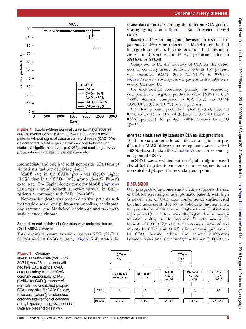
(1.2%) than in the CAD− (0%) group (p=0.27; Fisher’sexact test). The Kaplan-Meier curve for MACE (figure 4)illustrates a trend towards superior survival in CAD−patients as compared with CAD+ (p=0.063).Non-cardiac death was observed in five patients with
metastatic disease: one pulmonary embolism/carcinoma,one sarcoma, one Merkel-cell-carcinoma and two meta-static adenocarcinoma.
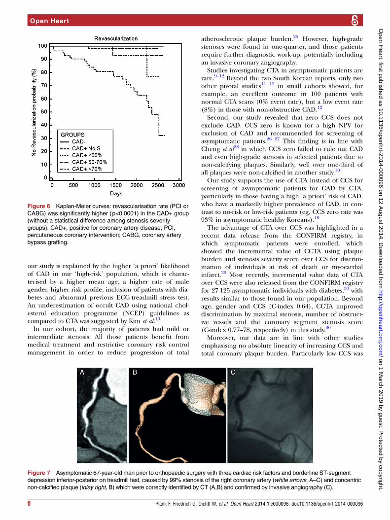
Secondary end points: (1) Coronary revascularisation and(2) IA >50% stenosisTotal coronary revascularisation rate was 5.5% (39/711,29 PCI and 10 CABG surgery). Figure 5 illustrates the
revascularisation rates among the different CTA stenosisseverity groups, and figure 6 Kaplan–Meier survivalcurve.Based on CTA findings and downstream testing, 161
patients (22.6%) were referred to IA. Of those, 93 hadhigh-grade stenosis by CT, the remaining had intermedi-ate or mild stenosis, or IA was performed due toNSTEMI or STEMI.Compared to IA, the accuracy of CTA for the detec-
tion of coronary artery stenosis >50% in 165 patientswas: sensitivity 92.5% (95% CI 81.8% to 97.9%).Figure 7 shows an asymptomatic patient with a 99% sten-osis by CTA and IA.For exclusion of combined primary and secondary
end points, the negative predictive value (NPV) of CTA(>50% stenosis) compared to ICA >50% was 99.3%(95% CI 98.3% to 99.7%) in 711 patients.CCS had a lower predictive value (c=0.64; 95% CI
0.558 to 0.711) as CTA >50% (c=0.71; 95% CI 0.632 to0.777; p<0.001) to predict >50% stenosis by CAG(p=0.11).
Atherosclerosis severity scores by CTA for risk predictionTotal coronary atherosclerosis SIS was a significant pre-dictor for MACE if five or more segments were involved(SIS≥5, hazard risk, HR 6.5; table 2) and for secondaryend point if SIS≥1.ncSIS≥1 was associated with a significantly increased
HR of 2.4 in patients with one or more segments withnon-calcified plaques for secondary end point.
DISCUSSIONOur prospective outcome study clearly supports the useof CTA for screening of asymptomatic patients with high‘a priori’ risk of CAD after conventional cardiologicalbaseline assessment, due to the following findings: First,the prevalence of CAD in our high-risk study cohort washigh with 71%, which is markedly higher than in asymp-tomatic healthy South Koreans9 10 with no-risk orlow-risk of CAD (22% rate for coronary stenosis of anyseverity by CTA9 and 11.4% atherosclerosis prevalenceby CTA). Beyond ethnic and genetic differencesbetween Asian and Caucasians,24 a higher CAD rate in
Figure 4 Kaplan–Meier survival curve for major adverse
cardiac events (MACE): a trend towards superior survival in
patients without signs of coronary artery disease (CAD; 0%)
as compared to CAD+ groups; with a close-to-borderline
statistical significance level (p=0.063), and declining survival
probability with increasing stenosis severity.
Figure 5 Coronary
revascularisation rate (total 5.5%;
39/711) was 0% in patients with
negative CAD findings. CAD,
coronary artery disease; CAG,
coronary angiography; CTA+,
positive for CAD (presence of
non-calcified or calcified plaque);
CTA–, negative for CAD; Revasc,
revascularisation (percutaneous
coronary intervention or coronary
artery bypass grafting); S, stenosis;
Data are presented as n (%).
Plank F, Friedrich G, Dichtl W, et al. Open Heart 2014;1:e000096. doi:10.1136/openhrt-2014-000096 5
Coronary artery disease
on 1 March 2019 by guest. P
rotected by copyright.http://openheart.bm
j.com/
Open H
eart: first published as 10.1136/openhrt-2014-000096 on 12 August 2014. D
ownloaded from
our study is explained by the higher ‘a priori’ likelihoodof CAD in our ‘high-risk’ population, which is charac-terised by a higher mean age, a higher rate of malegender, higher risk profile, inclusion of patients with dia-betes and abnormal previous ECG-treadmill stress test.An underestimation of occult CAD using national chol-esterol education programme (NCEP) guidelines ascompared to CTA was suggested by Kim et al.10
In our cohort, the majority of patients had mild orintermediate stenosis. All those patients benefit frommedical treatment and restrictive coronary risk controlmanagement in order to reduce progression of total
atherosclerotic plaque burden.25 However, high-gradestenoses were found in one-quarter, and those patientsrequire further diagnostic work-up, potentially includingan invasive coronary angiography.Studies investigating CTA in asymptomatic patients are
rare.9–12 Beyond the two South Korean reports, only twoother pivotal studies11 12 in small cohorts showed, forexample, an excellent outcome in 100 patients withnormal CTA scans (0% event rate), but a low event rate(8%) in those with non-obstructive CAD.12
Second, our study revealed that zero CCS does notexclude CAD. CCS zero is known for a high NPV forexclusion of CAD and recommended for screening ofasymptomatic patients.26 27 This finding is in line withCheng et al28 in which CCS zero failed to rule out CADand even high-grade stenosis in selected patients due tonon-calcifying plaques. Similarly, well over one-third ofall plaques were non-calcified in another study.10
Our study supports the use of CTA instead of CCS forscreening of asymptomatic patients for CAD by CTA,particularly in those having a high ‘a priori’ risk of CAD,who have a markedly higher prevalence of CAD, in con-trast to no-risk or low-risk patients (eg, CCS zero rate was93% in asymptomatic healthy Koreans).10
The advantage of CTA over CCS was highlighted in arecent data release from the CONFIRM registry, inwhich symptomatic patients were enrolled, whichshowed the incremental value of CCTA using plaqueburden and stenosis severity score over CCS for discrim-ination of individuals at risk of death or myocardialinfarct.29 Most recently, incremental value data of CTAover CCS were also released from the CONFIRM registryfor 27 125 asymptomatic individuals with diabetes,30 withresults similar to those found in our population. Beyondage, gender and CCS (C-index 0.64), CCTA improveddiscrimination by maximal stenosis, number of obstruct-ive vessels and the coronary segment stenosis score(C-index 0.77–78, respectively) in this study.30
Moreover, our data are in line with other studiesemphasising no absolute linearity of increasing CCS andtotal coronary plaque burden. Particularly low CCS was
Figure 6 Kaplan-Meier curves: revascularisation rate (PCI or
CABG) was significantly higher (p<0.0001) in the CAD+ group
(without a statistical difference among stenosis severity
groups). CAD+, positive for coronary artery disease; PCI,
percutaneous coronary intervention; CABG, coronary artery
bypass grafting.
Figure 7 Asymptomatic 67-year-old man prior to orthopaedic surgery with three cardiac risk factors and borderline ST-segment
depression inferior-posterior on treadmill test, caused by 99% stenosis of the right coronary artery (white arrows, A–C) and concentric
non-calcified plaque (inlay right, B) which were correctly identified by CT (A,B) and confirmed by invasive angiography (C).
6 Plank F, Friedrich G, Dichtl W, et al. Open Heart 2014;1:e000096. doi:10.1136/openhrt-2014-000096
Open Heart
on 1 March 2019 by guest. P
rotected by copyright.http://openheart.bm
j.com/
Open H
eart: first published as 10.1136/openhrt-2014-000096 on 12 August 2014. D
ownloaded from

significantly less reliable in predicting total plaqueburden due to their association with higher overallprevalence of non-calcified plaques. This has beenshown in other studies with a significant stenosis rate ofnearly 10%27 and high prevalence of non-calcifyingplaques (83.3%) in those with low CCS (≤100).10
Overall, the total rate of non-calcifying plaques andCalcium Score zero in our study cohort was 7.6%, similarto patients with atypical chest pain and an intermediateFramingham risk profile.6 Non-calcifying plaques maycause chest pain, for example, due to endothelial dys-function, which occurs at early stages of atherosclerosis,due to lack of regular flow-mediated vasodilation or evenvasoconstriction at a lesion with mild stenosis.31
Third, our study revealed that the total and non-calcified plaque burdens (SIS and ncSIS) predictadverse outcome in terms of MACE and revascularisa-tion procedures such as PCI or CABG, respectively,which is in line with results from the CONFIRM trial.32
In contrast, the CONFIRM trial mainly enrolled symp-tomatic patients.As a study novelty, we calculated a dedicated non-
calcifying plaque score (the ‘ncSIS’), which showed anincreased risk of adverse outcome for secondary endpoint, even if only one (or more) segment was involved.An increasing total coronary plaque burden by CTA
was associated with a significantly increased risk ofMACE and revascularisation rate, going in line withresults from multicentre-registry CONFIRM, in whichwere identified as risk factors. The total plaque burden,stenosis severity32 or number of proximal segments withmixed or calcified plaques by CTA.33
Finally, our study provides strong evidence that CTA isa highly reliable and accurate tool to exclude significantCAD and MACE events over a midterm follow-up periodof mean 2.65 years (up to 8 years). No cardiac eventsoccurred in patients with negative CTA findings.In summary, our data support screening of asymptom-
atic high-risk patients with CTA. Compared to CTCalcium Score, most recently introduced low-dose CTtechniques such as prospective ECG-gating allow foralmost equal or just slightly higher radiation exposure of≥1 mSv, however, while adding iodine CA.Compared to CCS, CTA provides the advantage of
detecting non-calcifying plaque (representing early stagesof atherosclerosis), coronary plaque load assessment andstenosis quantification for risk stratification. Patients withstenosis >50% by CTA yielded a markedly higher risk ofadverse outcome than increasing CCS values.Whether follow-up of high-risk patients by CCTA
should be performed is currently an open item for dis-cussion. There are no scientific data on CTA for moni-toring of CAD progression. Follow-up intervals of aminimum of 5–7 years may be reasonable for evaluationof CAD progression. In those with intermediate stenosis(50–70%), a myocardial perfusion stress test (eg,SPECT) should be appended in order to define thehaemodynamic significance of a coronary lesion.
STUDY LIMITATIONSFirst, we acknowledge a low total MACE rate. Onereason is the short-term follow-up period of 2.65 years,
Table 2 Cox proportional hazard risk model for primary end point (MACE) and secondary end point (revascularisation rate)
MACE
HR (95% CI) p Value
Coronary revascularisation
HR (95% CI) p Value
Smoker 1.41 (0.29 to 6.92) 0.67 1.31 (0.66 to 2.62) 0.45
HT 2.91 (0.35 to 24.70) 0.33 1.91 (0.86 to 4.23) 0.11
+FH – – 0.81 (0.39 to 1.69) 0.57
Cholesterol 1.02 (0.66 to 1.40) 0.94 1.26 (0.54 to 2.94) 0.60
DM 1.79 (0.29 to 11.17) 0.54 1.46 (0.61 to 3.50) 0.40
CCS
≥10 5.79 (0.70 to 47.59) 0.10 5.05 (2.12 to 12.05) <0.001
≥100 3.93 (0.89 to 17.42) 0.08 6.01 (3.03 to 11.92) <0.0001
≥400 5.57 (1.26 to 24.70) <0.05* 3.90 (1.99 to 7.65) <0.0001
CT
<50% 2.87 (0.69 to 11.93) 0.15 0.25 (0.06 to 1.03) 0.06
50–70% – – 0.32 (0.04 to 2.32) 0.26
>70% 3.40 (0.85 to 13.51) 0.08 27.92 (9.94 to 78.43) <0.0001*
SIS
≥1 1.33 (0.29 to 6.15) p=0.72 3.65 (1.9 to 7.0) p<0.0001*
≥5 6.50 (1.19 to 16.02) p<0.05* 7.13 (2.2 to 23.1) p<0.0001*
ncSIS
≥1 1.27 (0.23 to 5.05) p=0.74 2.36 (1.20 to 4.63) p<0.01*
≥5 5.75 (0.69 to 47.94) p=0.11 1.82 (0.43 to 747) p=0.42
*Statistical significance.+FH, positive family history; CAD, coronary artery disease; CCS, coronary calcium score; DM, diabetes mellitus; HT, arterial hypertension;ncSIS, non-calcified plaque segment involvement score; SIS, segment involvement score (reflecting total plaque burden).
Plank F, Friedrich G, Dichtl W, et al. Open Heart 2014;1:e000096. doi:10.1136/openhrt-2014-000096 7
Coronary artery disease
on 1 March 2019 by guest. P
rotected by copyright.http://openheart.bm
j.com/
Open H
eart: first published as 10.1136/openhrt-2014-000096 on 12 August 2014. D
ownloaded from
beyond patient population selection criteria with a lowrate of diabetes (12.4%).Second, our inclusion criteria for definition of our
cohorts ‘high-risk’ of CAD involved ECG-treadmill stresstesting (beyond coronary risk factors). TheECG-treadmill test has a low accuracy for detection ofmyocardial ischaemia.Third, the patient’s current medication (aspirin,
statins) was not recorded.
ConclusionIn summary, our study recommends the use of CCTA inasymptomatic patients with a high ‘a priori’ risk of CADin order to detect subclinical atherosclerosis andobstructive CAD. Non-calcified plaques were found in ahigh amount of asymptomatic patients, even in CCSzero patients. Non-calcified plaques pose a risk foradverse outcome, even if only small numbers of coron-ary segments are affected.CTA allows for coronary risk estimation based on
increasing total and non-calcifying plaque burden. CTAis a safe, accurate and reliable modality for exclusionof CADGiven low radiation exposure of CTA below <1 mSv
using the newest low-dose prospective ECG-gated CTtechniques such as high-pitch mode or iterative recon-struction, which is equivalent to CCS scanning, CTAshould be favoured over CCS in asymptomatic patientswith a high ‘a priori’ risk of CAD.
Contributors FP: data acquisition, manuscript drafting. GF: patient selection,manuscript drafting. WD: statistical analysis, manuscript drafting. AK:statistical analysis, manuscript drafting. WJ: manuscript drafting. WMF:manuscript drafting. GF: data acquisition, patient selection, manuscriptdrafting.
Funding This research received no specific grant from any funding agency inthe public, commercial or not-for-profit sectors.
Competing interests None.
Ethics approval Local IRB.
Provenance and peer review Not commissioned; externally peer reviewed.
Data sharing statement No additional data are available.
Open Access This is an Open Access article distributed in accordance withthe Creative Commons Attribution Non Commercial (CC BY-NC 3.0) license,which permits others to distribute, remix, adapt, build upon this work non-commercially, and license their derivative works on different terms, providedthe original work is properly cited and the use is non-commercial. See: http://creativecommons.org/licenses/by-nc/3.0/
REFERENCES1. Fleg JL, Gerstenblith G, Zonderman AB, et al. Prevalence and
prognostic significance of exercise-induced silent myocardialischemia detected by thallium scintigraphy and electrocardiographyin asymptomatic volunteers. Circulation 1990;81:428–36.
2. Deedwania PC. Silent ischemia predicts poor outcome in high-riskhealthy men. J Am Coll Cardiol 2001;38:80–3.
3. He ZX, Hedrick TD, Pratt CM. Severity of coronary arterycalcification by electron beam computed tomography predicts silentmyocardial ischemia. Circulation 2000;101:244–51.
4. Greenland P, Bonow RO, Brundage BH, et al. ACCF/AHA 2007clinical expert consensus document on coronary artery calciumscoring by computed tomography in global cardiovascular risk
assessment and in evaluation of patients with chest pain: a report ofthe American College of Cardiology Foundation Clinical ExpertConsensus Task Force (ACCF/AHAWriting Committee to Updatethe 2000 Expert Consensus Document on Electron Beam ComputedTomography) developed in collaboration with the Society ofAtherosclerosis Imaging and Prevention and the Society ofCardiovascular Computed Tomography. J Am Coll Cardiol2007;49:378–402.
5. Taylor AJ, Cerqueira M, Hodgson JM, et al. ACCF/SCCT/ACR/AHA/ASE/ASNC/ NASCI/SCAI/SCMR 2010 ACCF/SCCT/ACR/AHA/ASE/ASNC/NASCI/SCAI/SCMR 2010 Appropriate Use Criteria forCardiac Computed Tomography. A Report of the American Collegeof Cardiology Foundation Appropriate Use Criteria Task Force, theSociety of Cardiovascular Computed Tomography, the AmericanCollege of Radiology, the American Heart Association, the AmericanSociety of Echocardiography, the American Society of NuclearCardiology, the North American Society for Cardiovascular Imaging,the Society for Cardiovascular Angiography and Interventions, andthe Society for Cardiovascular Magnetic Resonance. Circulation2010;122:e525–55.
6. Hausleiter J, Meyer T, Hadamitzky M, et al. Prevalence ofnoncalcified coronary plaques by 64-slice computed tomography inpatients with an intermediate risk for significant coronary arterydisease. J Am Coll Cardiol 2006;48:312–18.
7. Hoffmann U, Moselewski F, Nieman K, et al. Noninvasiveassessment of plaque morphology and composition in culprit andstable lesions in acute coronary syndrome and stable lesions instable angina by multidetector computed tomography. J Am CollCardiol 2006;47:1655–62.
8. Dohi T, Mintz GS, McPherson JA. Non-fibroatheroma lesionphenotype and long-term clinical outcomes: a substudy analysisfrom the PROSPECT Study. JACC Cardiovasc Imaging2013;6:908–16.
9. Choi EK, Choi SI, Rivera JJ, et al. Coronary computed tomographyangiography as screening tool for the detection of occult coronaryartery disease in asymptomatic individuals. J Am Coll Cardiol2008;52:357–65.
10. Kim KJ, Choi SI, Lee MS, et al. The prevalence and characteristicsof coronary atherosclerosis in asymptomatic subjects classified aslow risk based on traditional risk stratification algorithm: assessmentwith coronary CT angiography. Heart 2013;99:1113–17.
11. Romeo F, Leo R, Clementi F, et al. Multislice computed tomographyin an asymptomatic high-risk population. Am J Cardiol2007;99:325–8.
12. Pundziute G, Schuijf JD, Jukema JW, et al. Prognostic value ofmultislice computed tomography coronary angiography in patientswith known or suspected coronary artery disease. J Am Coll Cardiol2007;49:62–70.
13. Hausleiter J, Meyer T, Hermann F, et al. Estimated radiationdose associated with cardiac CT angiography. JAMA2009;301:500–7.
14. Achenbach S, Marwan M, Ropers D, et al. Coronary computedtomography angiography with a consistent dose below 1 mSv usingprospectively electrocardiogram- triggered high-pitch spiralacquisition. Eur Heart J 2010;31:340–6.
15. Schuhbaeck A, Achenbach S, Layritz C, et al. Image quality of ultra-low radiation exposure coronary CT angiography with an effectivedose <0.1 mSv using high-pitch spiral acquisition and raw data-based iterative reconstruction. Eur Radiol 2013;23:597–606.
16. Reiner Z, Catapano AL, De Backer G, et al. ESC/EAS Guidelines forthe management of dyslipidaemias: the Task Force for themanagement of dyslipidaemias of the European Society ofCardiology (ESC) and the European Atherosclerosis Society (EAS).Eur Heart J 2011;32:1769–818.
17. Mancia G, Fagard R, Narkiewicz K, et al. 2013 ESH/ESC guidelinesfor the management of arterial hypertension: the Task Force for theManagement of Arterial Hypertension of the European Society ofHypertension (ESH) and of the European Society of Cardiology(ESC). Eur Heart J 2013;34:2159–219.
18. Gibbons RJ, Balady GJ, Bricker JT, et al. ACC/AHA 2002guideline update for exercise testing: summary article. a reportof the American College of Cardiology/American HeartAssociation Task Force on Practice Guidelines. Circulation2002;106:1883–92.
19. Berry JD, Dyer A, Cai X, et al. Lifetime risks of cardiovasculardisease. N Engl J Med 2012;366:321–9.
20. Agatston AS, Janowitz WR, Hildner FJ, et al. Quantification ofcoronary artery calcium using ultrafast computed tomography. J AmColl Cardiol 1990;15:827–32.
21. Austen WG, Edwards JE, Frye RL, et al. A reporting system onpatients evaluated for coronary artery disease: Report of the Ad Hoc
8 Plank F, Friedrich G, Dichtl W, et al. Open Heart 2014;1:e000096. doi:10.1136/openhrt-2014-000096
Open Heart
on 1 March 2019 by guest. P
rotected by copyright.http://openheart.bm
j.com/
Open H
eart: first published as 10.1136/openhrt-2014-000096 on 12 August 2014. D
ownloaded from

Committee for Grading of Coronary Artery Disease, Council onCardiovascular Surgery. Circulation 1975;51:5–40.
22. Øvrehus KA, Marwan M, Bøtker HE, et al. Reproducibility ofcoronary plaque detection and characterization using low radiationdose coronary computed tomographic angiography in patients withintermediate likelihood of coronary artery disease (ReSCAN study).Int J Cardiovasc Imaging 2012;28:889–99.
23. Leber AW, Knez A, von Ziegler F, et al. Quantification of obstructiveand nonobstructive coronary lesions by 64-slice computedtomography: a comparative study with quantitative coronaryangiography and intravascular ultrasound. J Am Coll Cardiol2005;46:147–54.
24. Hulten E, Villines TC, Cheezum MK, et al. CONFIRM Investigators.Usefulness of coronary computed tomography angiography topredict mortality and myocardial infarction among Caucasian, Africanand East Asian ethnicities (from the CONFIRM [Coronary CTAngiography Evaluation for Clinical Outcomes: An InternationalMulticenter] Registry). Am J Cardiol 2013;111:479–85.
25. Nissen SE, Nicholls SJ, Sipahi I, et al.; ASTEROID Investigators.Effect of very high-intensity statin therapy on regression of coronaryatherosclerosis: the ASTEROID trial. JAMA 2006;295:1556–6.
26. Shareghi S, Ahmadi N, Young E, et al. Prognostic significance ofzero coronary calcium scores on cardiac computed tomography.J Cardiovasc Comput Tomogr 2007;1:155–9.
27. Greenland P, LaBree L, Azen SP, et al. Coronary artery calciumscore combined with Framingham score for risk prediction inasymptomatic individuals. JAMA 2004;291:210–15.
28. Cheng VY, Lepor NE, Madyoon H, et al. Presence and severity ofnoncalcified coronary plaque on 64-slice computed tomographiccoronary angiography in patients with zero and low coronary arterycalcium. Am J Cardiol 2007;99:1183–6.
29. Al-Mallah MH, Qureshi W, Lin FY, et al. Does coronary CTangiography improve risk stratification over coronary calcium scoringin symptomatic patients with suspected coronary artery disease?Results from the prospective multicenter international CONFIRMregistry. Eur Heart J Cardiovasc Imaging 2014;15:267–74.
30. Min JK, Labounty TM, Gomez MJ, et al. Incremental prognosticvalue of coronary computed tomographic angiography over coronaryartery calcium score for risk prediction of major adverse cardiacevents in asymptomatic diabetic individuals. Atherosclerosis2014;232:298–304.
31. Heusch G, Baumgart D, Camici P, et al. Alpha-adrenergic coronaryvasoconstriction and myocardial ischemia in humans. Circulation2002;101:689–94.
32. Otaki Y, Arsanjani R, Gransar H, et al. What have we learned fromCONFIRM? Prognostic implications from a prospective multicenterinternational observational cohort study of consecutive patientsundergoing coronary computed tomographic angiography. J NuclCardiol 2012;19:787–95.
33. Hadamitzky M, Achenbach S, Al-Mallah M, et al.; CONFIRMInvestigators. Optimized prognostic score for coronary computedtomographic angiography: results from the CONFIRM registry(COronary CTAngiography EvaluatioN For Clinical Outcomes: anInteRnational Multicenter Registry). J Am Coll Cardiol 2013;62:468–76.
Plank F, Friedrich G, Dichtl W, et al. Open Heart 2014;1:e000096. doi:10.1136/openhrt-2014-000096 9
Coronary artery disease
on 1 March 2019 by guest. P
rotected by copyright.http://openheart.bm
j.com/
Open H
eart: first published as 10.1136/openhrt-2014-000096 on 12 August 2014. D
ownloaded from