coronary microvascular dysfunction is related to...
TRANSCRIPT

J A C C : H E A R T F A I L U R E V O L . 2 , N O . 4 , 2 0 1 4
ª 2 0 1 4 B Y T H E AM E R I C A N C O L L E G E O F C A R D I O L O G Y F O U N D A T I O N I S S N 2 2 1 3 - 1 7 7 9 / $ 3 6 . 0 0
P U B L I S H E D B Y E L S E V I E R I N C . h t t p : / / d x . d o i . o r g / 1 0 . 1 0 1 6 / j . j c h f . 2 0 1 4 . 0 3 . 0 0 9
CLINICAL RESEARCH
Coronary Microvascular DysfunctionIs Related to Abnormalities inMyocardial Structure and Functionin Cardiac Amyloidosis
Sharmila Dorbala, MD,*yz Divya Vangala, MA,y John Bruyere JR, MA,y Christina Quarta, MD,z Jenna Kruger, BS,zRobert Padera, MD,x Courtney Foster, MSC, CNMT,y Michael Hanley, MD,* Marcelo F. Di Carli, MD,*yzRodney Falk, MDzABSTRACT
Fro
(Ca
Mo
zCMe
Sch
Fo
De
OBJECTIVES The purpose of this study was to test the hypothesis that coronary microvascular function is impaired in
subjects with cardiac amyloidosis.
BACKGROUND Effort angina is common in subjects with cardiac amyloidosis, even in the absence of epicardial coro-
nary artery disease (CAD).
METHODS Thirty-one subjects were prospectively enrolled in this study, including 21 subjects with definite cardiac
amyloidosis without epicardial CAD and 10 subjects with hypertensive left ventricular hypertrophy (LVH). All subjects
underwent rest and vasodilator stress N-13 ammonia positron emission tomography and 2-dimensional echocardiogra-
phy. Global left ventricular myocardial blood flow (MBF) was quantified at rest and during peak hyperemia, and coronary
flow reserve (CFR) was computed (peak stress MBF/rest MBF) adjusting for rest rate pressure product.
RESULTS Compared with the LVH group, the amyloid group showed lower rest MBF (0.59 � 0.15 ml/g/min vs.
0.88 � 0.23 ml/g/min; p ¼ 0.004), stress MBF (0.85 � 0.29 ml/g/min vs. 1.85 � 0.45 ml/g/min; p < 0.0001), and
CFR (1.19� 0.38 vs. 2.23�0.88; p<0.0001) and higher minimal coronary vascular resistance (111� 40ml/g/min/mmHg
vs. 70 � 19 ml/g/min/mm Hg; p ¼ 0.004). Of note, almost all subjects with amyloidosis (>95%) had significantly
reduced peak stress MBF (<1.3 ml/g/min). In multivariable linear regression analyses, a diagnosis of amyloidosis,
increased left ventricular mass, and age were the only independent predictors of impaired coronary vasodilator
function.
CONCLUSIONS Coronary microvascular dysfunction is highly prevalent in subjects with cardiac amyloidosis, even
in the absence of epicardial CAD, and may explain their anginal symptoms. Further study is required to understand
whether specific therapy directed at amyloidosis may improve coronary vasomotion in amyloidosis. (J Am Coll Cardiol HF
2014;2:358–67) © 2014 by the American College of Cardiology Foundation.
m the *Noninvasive Cardiovascular Imaging Program, Heart and Vascular Center, Departments of Radiology and Medicine
rdiology), Brigham and Women’s Hospital, Harvard Medical School, Boston, Massachusetts; yDivision of Nuclear Medicine and
lecular Imaging, Department of Radiology, Brigham and Women’s Hospital, Harvard Medical School, Boston, Massachusetts;
ardiovascular Division and Cardiac Amyloidosis Program, Department of Medicine, Brigham and Women’s Hospital, Harvard
dical School, Boston, Massachusetts; and the xDepartment of Pathology, Brigham and Women’s Hospital, Harvard Medical
ool, Boston, Massachusetts. This study was supported by the Amyloid Foundation, American Society of Nuclear Cardiology
undation, National Institutes of Health (National Heart, Lung, and Blood Institute grant K23HL092299), and in part by the
marest Lloyd, Jr. Foundation (Dr. Falk). Dr. Dorbala has received a research grant from Astellas Global Pharma Development.
ABB R E V I A T I O N S
AND ACRONYMS
AL = amyloid light chain
CAD = coronary artery disease
CFR = coronary flow reserve
CT = computed tomography
LV = left ventricular
LVH = left ventricular
hypertrophy
MBF = myocardial blood flow
PET = positron emission
tomography
2D = 2-dimensional
J A C C : H E A R T F A I L U R E V O L . 2 , N O . 4 , 2 0 1 4 Dorbala et al.A U G U S T 2 0 1 4 : 3 5 8 – 6 7 Microvascular Dysfunction in Cardiac Amyloidosis
359
A myloidosis is a rare systemic disorder charac-terized by the extracellular deposition ofmisfolded protein in various organ systems,
including the heart (1,2). Among the several types ofamyloid fibrils, the light chain and transthyretin am-yloid proteins most commonly affect the heart. Car-diac amyloid deposits result in increased ventricularwall thickness and produce a restrictive cardiomyop-athy presenting primarily as biventricular congestiveheart failure. Anginal symptoms and signs ofischemia have been reported in some patients withcardiac amyloidosis without obstructive epicardialcoronary artery disease (CAD) (3–6). Autopsy studieshave shown amyloid deposits around and betweencardiac myocytes in the interstitium (7), the perivas-cular regions (8), and the media of intramyocardialcoronary vessels (9,10). Amyloidosis is thus a primeexample of a disorder with the potential to cause cor-onary microvascular dysfunction via 3 major mecha-nisms: structural (amyloid deposition in the vesselwall causing wall thickening and luminal stenosis),extravascular (extrinsic compression of the microvas-culature from perivascular and interstitial amyloiddeposits and decreased diastolic perfusion), andfunctional (autonomic and endothelial dysfunction).Accordingly, we sought to test the hypothesis thatcoronary flow reserve (CFR), a measure of microvas-cular function, is reduced in subjects with cardiacamyloidosis without evidence of epicardial CAD.Next, we sought to explore the hypothesis thatreduced CFR is a function of increased myocardialmass and increased left ventricular (LV) filling pres-sures and is associated with subclinical abnormalitiesin LV systolic dysfunction (strain). Therefore, our pri-mary aim was to study coronary microvascular func-tion in subjects with cardiac amyloidosis comparedwith subjects with hypertensive left ventricular hy-pertrophy (LVH). Our secondary aim was to studythe morphological and functional correlates of coro-nary microvascular dysfunction in subjects with car-diac amyloidosis.
METHODS
PATIENT COHORT. We prospectively enrolled 31subjects into 2 study groups. The amyloid groupconsisted of 21 subjects with confirmed light chain(n ¼ 15) or transthyretin (n ¼ 6) amyloidosis usingpre-defined inclusion and exclusion criteria (Online
Dr. Di Carli has received a research grant from Gilead Sciences. All other au
relevant to the contents of this paper to disclose. Ms. Vangala and Mr. Bruy
Manuscript received January 14, 2014; revised manuscript received Februar
Table 1). Ten subjects with hypertensiveLVH on 2-dimensional (2D) echocardiography(LV wall thickness >11 mm) served as con-trols. Hypertensive subjects with LVH did nothave documented kidney disease, peripheralvascular disease, cerebrovascular disease, orCAD (no history of chest pain, myocardialinfarction, angiographic CAD, or coronaryrevascularization). Amyloidosis was diag-nosed by endomyocardial biopsy (n ¼ 10) orby a positive extracardiac biopsy specimenwith typical features of cardiac involvementon 2D transthoracic echocardiography (n ¼ 11)(e.g., wall thickness measurements >11 mm,
bright echogenic myocardium, and echocardiographicevidence of diastolic dysfunction). All biopsy speci-mens stained positive for amyloid with eithersulfated alcian blue or congo red stain, and amyloidtyping was determined by a battery of stains,including immunoperoxidase stain for transthyretinand immunofluorescence stain for immunoglobulinsG, M, A, kappa, lambda, protein A, and transthyretin.In equivocal cases, biopsy specimens underwentproteomics evaluation.This study was approved by the PartnersHuman Research Committee. All study subjects wereprospectively enrolled, provided written informedconsent, and underwent evaluation of coronarymicrovascular function by a research test and vasodi-lator stress N-13 ammonia positron emission tomog-raphy/computed tomography (PET/CT) (except for 3subjects with LVH who underwent clinical N-13ammonia PET/CT). Obstructive epicardial CAD wascarefully excluded in all subjects with amyloidosis bycoronary angiography, as described in the followingtext. All subjects also underwent 2D transthoracicechocardiography with strain analysis to study cardiacmorphology and function. Detailed characterization ofthe amyloid subtype was available for all subjects withamyloidosis, including staining of biopsy specimens.
PET. Rest and vasodilator stress N-13 ammonia PET/CT was performed by using standard protocols andstandard preparation (see the Online Appendix).Imaging of all subjects was performed with a wholebody PET/CT scanner (Discovery Lightspeed VCT 64;GE Healthcare, Milwaukee, Wisconsin) after an over-night fast. Rest N-13 ammonia images were obtainedfor 20 min in 2D list mode after intravenous injectionof N-13 ammonia (w20 mCi). One hour after rest
thors have reported that they have no relationships
ere contributed equally to this work.
y 24, 2014, accepted March 7, 2014.
TABLE 1 Baseline Da
Age, yrs
Female
Body mass index, kg/m
Hypertension
Dyslipidemia
Diabetes
Symptoms
Chest pain
Dyspnea
Jaw claudication
Buttock claudication
NYHA functional clas
Medications
Beta-blockers
ACE inhibitors
Diuretics
Aspirin
Cholesterol medicati
ECG
Limb-lead voltage, m
Chest-lead voltage,
Atrial fibrillation/flut
Hemodynamics
Rest heart rate, beat
Rest systolic BP, mm
Rest RPP, beats/min
Rest diastolic BP, mm
Stress heart rate, be
Stress systolic BP, m
Stress RPP, beats/mi
Stress diastolic BP, m
Delta heart rate, bea
Delta systolic BP, mm
Delta RPP, beats/min
Values are mean � SD or %
ACE ¼ angiotensin-convtricular hypertrophy; NYHA
Dorbala et al. J A C C : H E A R T F A I L U R E V O L . 2 , N O . 4 , 2 0 1 4
Microvascular Dysfunction in Cardiac Amyloidosis A U G U S T 2 0 1 4 : 3 5 8 – 6 7
360
perfusion imaging, vasodilator stress was performedby using a standard infusion of adenosine (n ¼ 9),dipyridamole (n ¼ 18), or regadenoson (n ¼ 4). At peakhyperemia, a second dose of N-13 ammonia (w20 mCi)was given intravenously and stress images wererecorded in the same manner. The estimated wholebody effective radiation dose for the rest and stress N-13 ammonia PET/CT study was 5.4 mSv. Images wereinterpreted semiquantitatively and independently by2 experienced observers (interobserver reliabilitykappa: 0.95) (11) by using a standard 17-segmentmodeland a 5-point (0–4) scoring system. The global sum-med stress score, summed rest score, and summeddifference score (the difference between the summedstress score and summed rest score) were computed.A summed stress score >0 was considered abnormal.
ta on Patient Characteristics and Hemodynamics
LVH Group (n ¼ 10) Amyloid Group (n ¼ 21) p Value
62.6 � 12.3 61.7 � 9.5 0.8
60 38 0.32 33.5 � 6.4 25.4 � 3.1 <0.0001
100 43 0.002
60 33 0.2
30 5 0.05
20 24 0.8
30 62 0.09
0 15 0.6
0 5 0.7
s $II 0 45 <0.0001
40 19 0.2
50 10 0.01
60 62 0.9
90 19 <0.0001
ons 60 24 0.05
m 9.0 � 2.5 5.7 � 1.9 <0.0001
mm 12.6 � 5.0 9.4 � 2.9 0.03
ter 14 26
s/min 69 � 16 72 � 10 0.5
Hg 149 � 22 114 � 15 <0.0001
$ mm Hg 10,298 � 3,173 8,169 � 1,358 0.01
Hg 74 � 15 67 � 9 0.1
ats/min 81 � 17 77 � 15 0.5
m Hg 148 � 22 101 � 18 <0.0001
n $ mm Hg 12,094 � 3,627 7,790 � 2,302 <0.0001
m Hg 75 � 15 58 � 11 0.001
ts/min 12 � 12 4 � 9.7 0.06
Hg �0.6 � 12 �13 � 16 0.04
$ mm Hg 1,795 � 1,761 �379 � 14,840 0.001
.
erting enzyme; BP ¼ blood pressure; ECG ¼ electrocardiography; LVH ¼ left ven-¼ New York Heart Association; RPP ¼ rate pressure product.
Global LV myocardial blood flow (MBF) (ml/g/min)was quantified at rest and during peak hyperemiaby using a previously validated one-compartmentmodel (12) and commercially available software(FlowQuant, University of Ottawa Heart Institute,Ottawa, Canada). CFR was computed as the ratio ofstress MBF to rest MBF and is referred to as CFR*throughout this report. CFR was adjusted for rest ratepressure product and computed as the ratio of stressMBF to normalized rest MBF (rest MBF/rest ratepressure product) , 10,000, and is referred to as CFRthroughout this report. Coronary vascular resistance,the ratio of mean arterial pressure to MBF at rest(maximal coronary vascular resistance) and peak hy-peremia (minimal coronary vascular resistance) wasalso calculated. Reduced peak stress MBF, reducedCFR, and increased minimal coronary vascular resis-tance were considered to represent coronary micro-vascular dysfunction. Stress MBF images from onesubject in the amyloid group were uninterpretabledue to poor counts.
CORONARY ANGIOGRAPHY. Epicardial obstructiveCAD was excluded in all subjects with amyloidosis byclinical invasive coronary angiography or research CTcoronary angiography. Clinically performed invasivecoronary angiography (a median of 164 days beforethe PET study) reports were reviewed, and only sub-jects with <70% CAD in all coronary arteries wereinvited to participate in the study. All except onesubject with amyloidosis underwent coronary angi-ography to exclude CAD within the 2-year window(1 patient underwent coronary angiography within3 years). In 9 subjects with amyloidosis, research CTcoronary angiography was performed within a me-dian of �1 day of the PET study with standard pro-tocols (Online Appendix).
ECHOCARDIOGRAPHY. All subjects underwent 2Dtransthoracic echocardiography within a median of�1 day (interquartile range: �31 to þ24 days) of thePET study. Digitally acquired echocardiographyimages in DICOM format with acceptable imagequality (n ¼ 27) were uploaded and processed usingvendor-independent offline 2D Cardiac PerformanceAnalysis software (TomTec Imaging System, Munich,Germany) to compute peak LV longitudinal, radial,and circumferential strain values (Online Appendix).Throughout this report, we use the term “strain” torepresent LV strain.
PRIMARY OUTCOME MEASURES. The primary out-come measures of this study were peak stress MBF,CFR, and minimal coronary vascular resistance.
STATISTICAL ANALYSIS. The characteristics of thesubjects are described as mean � SD compared with

FIGURE 1 Rest and Vasodilator Stress N-13 Ammonia PET Images From a Subject With
Familial Transthyretin Amyloidosis
(A) Mildly abnormal N-13 ammonia myocardial perfusion images from a subject with
transthyretin amyloidosis. (B) CT coronary angiogram and histopathology images. The
images are shown in short axis, horizontal long axis, and vertical long axis projections and
demonstrate a reversible perfusion defect in the mid and basal septum despite normal
epicardial coronary arteries on CT coronary angiography. The coronary flow reserve was 1.3
(significantly impaired). The low-power photomicrograph of the myocardial biopsy spec-
imen (H&E stain) shows near complete loss of myocytes with extensive amyloid deposition.
The second and third high-power photomicrographs (H&E and sulfated alcian blue stains,
respectively) show a small vessel in which the lumen has been obliterated by vascular
amyloid deposition. ANT ¼ anterior; CT ¼ computed tomography; H&E ¼ hematoxylin and
eosin; HLA ¼ horizontal long axis; INF ¼ inferior; LAD ¼ left anterior descending coronary
artery; LAT ¼ lateral; LCX ¼ left circumflex coronary artery; OM ¼ obtuse marginal cor-
onary artery; PET ¼ positron emission tomography; RCA ¼ right coronary artery; SA ¼short axis; SAB ¼ sulfated Alcian Blue; SEP ¼ septum; VLA ¼ vertical long axis.
J A C C : H E A R T F A I L U R E V O L . 2 , N O . 4 , 2 0 1 4 Dorbala et al.A U G U S T 2 0 1 4 : 3 5 8 – 6 7 Microvascular Dysfunction in Cardiac Amyloidosis
361
the Student t test. Nonparametric variables arelisted as medians and compared with the Mann-Whitney U test. Discrete variables are describedas proportions and compared with the chi-squaretest. Correlations were performed using Pearson Ror nonparametric methods (Spearman rho) as indi-cated. Multivariable linear regression analyses wereperformed to study the independent contributions ofvarious parameters on stress MBF, CFR, and minimalcoronary vascular resistance. A parsimonious modelwith stepwise forward selection (probability of F forentry of 0.05 and for removal of 0.10) was performedto minimize model overfitting.
RESULTS
Baseline data on patient characteristics and hemo-dynamics are listed in Table 1. Notably, the amyloidgroup had lower body mass than the LVH group and38% were women. Approximately one-half of theamyloid group had a history of New York Heart As-sociation functional class $II heart failure. The amy-loid group had ischemic symptoms of chest pain(24%), shortness of breath (62%), and jaw or buttockclaudication (20%) with clinical evidence of auto-nomic (19%) or peripheral neuropathy (19%), pro-teinuria suggesting renal involvement (14%), oramyloid deposition in the liver (5%). Nine of the 15subjects with amyloid light chain (AL) amyloidosisreceived specific chemotherapy for amyloidosisbefore this study. As expected, the mean limb leadand chest lead electrocardiographic voltage was lowerin the amyloid group compared with the LVH group.
REGIONAL MYOCARDIAL PERFUSION. A variety ofperfusion patterns (no ischemia to severe ischemia)and high-risk scan findings (transient cavity dilationand right ventricular tracer uptake) were observed inthe amyloid group (Fig. 1, Online Fig. 1). In the amy-loid group, despite no epicardial CAD, 57% of thesubjects (12 of 21) had ischemic scans; 3 subjectshad severe ischemia. High-risk scan features, such asincreased right ventricular tracer uptake (62%; 13of 21 subjects) and transient cavity dilation of theleft ventricle on the post-stress images (76%; 16 of21 subjects), were frequently seen. The mean tran-sient cavity dilation ratio was significantly higherin the amyloid group than in the LVH group (1.18 �0.12 vs. 1.04 � 0.18; p ¼ 0.03). None of thesubjects with LVH had perfusion defects on the N-13ammonia study.
CORONARY VASOMOTOR FUNCTION. The mean restMBF, stress MBF, CFR, and CFR* were significantlylower in the amyloid group compared with the LVH
group (rest MBF: 0.59 � 0.15 ml/g/min vs. 0.88 �0.23 ml/g/min [p ¼ 0.004]; stress MBF: 0.85 � 0.29ml/g/min vs. 1.85 � 0.45 ml/g/min [p < 0.0001];CFR: 1.19 � 0.38 vs. 2.23 � 0.88 [p < 0.0001]; CFR*:1.44 � 0.36 vs. 2.20 � 0.67 [p < 0.0001]) (Table 2).Because the LVH group had significantly lower LVmass than the amyloid group, we normalized therest MBF, stress MBF, and CFR to LV mass as fol-lows: (MBF or CFR/LV mass) , 100. The values for
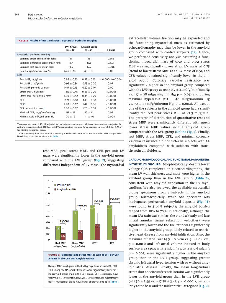
TABLE 2 Results of Rest and Stress Myocardial Perfusion Imaging
LVH Group(n ¼ 10)
Amyloid Group(n ¼ 21) p Value
Myocardial perfusion imaging
Summed stress score, mean rank 11 18 0.018
Summed difference score, mean rank 12.7 17.6 0.173
Summed rest score, mean rank 13.5 17.2 0.306
Rest LV ejection fraction, % 62.7 � 20 49 � 8 0.01
MBF
Rest MBF, ml/g/min 0.88 � 0.23 0.59 � 0.15 <0.0001 to 0.004
Rest MBF*, ml/g/min 0.92 � 0.34 0.73 � 0.20 0.07
Rest MBF per unit LV mass 0.47 � 0.19 0.22 � 0.16 0.001
Stress MBF, ml/g/min 1.85 � 0.45 0.85 � 0.29 <0.0001
Stress MBF per unit LV mass 1.00 � 0.42 0.34 � 0.29 <0.0001
CFR 2.23 � 0.88 1.19 � 0.38 <0.0001
CFR* 2.20 � 0.67 1.44 � 0.36 <0.0001
CFR per unit LV mass† 2.20 � 0.67 1.20 � 0.38 <0.0001
Maximal CVR, ml/g/min/mm Hg 117 � 28 147 � 41 0.05
Minimal CVR, ml/g/min/mm Hg 70 � 19 111 � 40 0.004
Values are n or mean � SD. *Unadjusted for rest rate pressure product; all stress values are also unadjusted forrest rate pressure product. †CFR per unit LV mass remained the same for an assumed LV mass of 0.5 or 0.75 offunctioning myocardial tissue.
CFR ¼ coronary flow reserve; CVR ¼ coronary vascular resistance; LV ¼ left ventricular; MBF ¼ myocardialblood flow; other abbreviations as in Table 1.
Dorbala et al. J A C C : H E A R T F A I L U R E V O L . 2 , N O . 4 , 2 0 1 4
Microvascular Dysfunction in Cardiac Amyloidosis A U G U S T 2 0 1 4 : 3 5 8 – 6 7
362
rest MBF, peak stress MBF, and CFR per unit LVmass were significantly lower in the amyloid groupcompared with the LVH group (Fig. 2), suggestingdifferences independent of LV mass. The myocardial
FIGURE 2 Mean Rest and Stress MBF as Well as CFR per Unit
LV Mass in the LVH and Amyloid Groups
The rest MBF was higher in the LVH group. Peak stress MBF, CFR
(CFR unadjusted)*, and CFR values were significantly lower in
the amyloid group than in the LVH group. CFR ¼ coronary flow
reserve; LV¼ left ventricular; LVH¼ left ventricular hypertrophy;
MBF ¼ myocardial blood flow; other abbreviations as in Table 1.
extracellular volume fraction may be expanded andthe functioning myocardial mass as estimated byechocardiography may thus be lower in the amyloidgroup compared with control subjects (22). Hence,we performed sensitivity analysis assuming a func-tioning myocardial mass of 0.50 and 0.75; stressMBF was significantly lower at an LV mass of 0.75(trend to lower stress MBF at an LV mass of 0.5), andCFR values remained significantly lower in the am-yloid group. Coronary vascular resistance wassignificantly higher in the amyloid group comparedwith the LVH group at rest (147 � 41 ml/g/min/mm Hgvs. 117 � 28 ml/g/min/mm Hg; p ¼ 0.05) and duringmaximal hyperemia (111 � 40 ml/g/min/mm Hgvs. 70 � 19 ml/g/min/mm Hg; p ¼ 0.004). All exceptone of the subjects in the amyloid group had a signif-icantly reduced peak stress MBF of <1.3 ml/g/min.The patterns of distribution of quantitative rest andstress MBF were significantly different with muchlower stress MBF values in the amyloid groupcompared with the LVH group (Online Fig. 2). Finally,rest MBF, stress MBF, CFR, and minimal coronaryvascular resistance did not differ in subjects with ALamyloidosis compared with subjects with trans-thyretin amyloidosis.
CARDIACMORPHOLOGICALANDFUNCTIONALPARAMETERS
IN THE STUDY GROUPS. Morphologically, despite lowervoltage QRS complexes on electrocardiography, themean LV wall thickness and mass were higher in theamyloid group than in the LVH group (Table 3),consistent with amyloid deposition in the LV myo-cardium. We also reviewed the available myocardialbiopsy specimens from 8 subjects in the amyloidgroup. Microscopically, while one specimen wasinadequate, perivascular amyloid deposits (Fig. 1B)were found in 5 of 8 subjects; the amyloid burdenranged from 10% to 70%. Functionally, although themean E/A ratio was similar, the e0 and a0 (early and latemitral annular tissue relaxation velocities) weresignificantly lower and the E/e0 ratio was significantlyhigher in the amyloid group, likely related to restric-tive heart disease from amyloid infiltration. Also, themaximal left atrial size (4.5 � 0.6 cm vs. 3.8 � 0.6 cm;p ¼ 0.003) and left atrial volume indexed to bodysurface area (40.5 � 13.4 ml/m2 vs. 23.2 � 9.6 ml/m2;p ¼ 0.002) were significantly higher in the amyloidgroup than in the LVH group, suggesting greaterchronic left atrial hypertension with or without amy-loid atrial disease. Finally, the mean longitudinalstrain (but not circumferential strain) was significantlylower in the amyloid group than in the LVH group(�11.50 � 2.99 vs. �17.78 � 3.41; p < 0.0001), particu-larly at the base and themidventricular regions (Fig. 3),

TABLE 3 Cardiac Morphological Features in the Study Groups
LVH Group(n ¼ 10)
Amyloid Group(n ¼ 20) p Value
Wall thickness, cm 1.33 � 0.13 1.80 � 0.36 <0.0001
LV mass, g 204 � 64 335 � 123 0.004
LV end-diastolic diameter, cm 4.1 � 1.0 4.1 � 0.7 0.89
LV end-systolic diameter, cm 2.8 � 0.9 2.9 � 0.6 0.48
LV ejection fraction, % 59.5 � 7.0 53.9 � 12.3 0.19
Peak E velocity, m/s 0.9 � 0.4 0.8 � 0.2 0.52
Peak A velocity, m/s 0.9 � 0.4 2.8 � 8.2 0.46
Peak E/A ratio 1.4 � 1.6 1.6 � 0.8 0.69
Average e0 velocity, cm/s 0.08 � 0.03 0.05 � 0.01 0.001
Average a0 velocity, cm/s 0.1 � 0.02 0.05 � 0.02 <0.0001
E/e0 ratio 11.99 � 5.2 17.98 � 8.06 0.045
Left atrial size, cm 3.79 � 0.55 4.52 � 0.57 0.003
Left atrial volume index, ml/m2 23.21 � 9.62 40.48 � 13.44 0.002
Longitudinal strain, global �17.78 � 3.41 �11.50 � 2.99 <0.0001
Circumferential strain �23.09 � 0.5.66 �25.38 � 7.17 0.49
Values are mean � SD. Echocardiogram was unevaluable for 1 subject in the amyloid group.
Abbreviations as in Tables 1 and 2.
FIGURE 3 Longitudinal Strain at the Base, Midventricle,
and Apex
Mean longitudinal strain at the base, midventricle, and apex
in the LVH group compared with the amyloid group. Mean
longitudinal strain was significantly reduced in the amyloid
group compared with the LVH group only in the base and
midventricular regions. Abbreviations as in Figure 2.
J A C C : H E A R T F A I L U R E V O L . 2 , N O . 4 , 2 0 1 4 Dorbala et al.A U G U S T 2 0 1 4 : 3 5 8 – 6 7 Microvascular Dysfunction in Cardiac Amyloidosis
363
consistent with previous reports of greater mid andbasal contractile impairment.
ASSOCIATIONS BETWEEN LV STRUCTURE AND
CORONARY VASCULAR FUNCTION. We found thatparameters of coronary microvascular function wereinversely correlated to increased LV mass (Fig. 4),increased diastolic filling pressures, and subclinicalsystolic dysfunction. Notably, in the few subjects withan LVmass of<300 g, for any given degree of LV mass,stress MBF and CFR were lower in the amyloid groupthan in the LVH group (Fig. 4). The mean e0, a0, and E/e0
ratio were inversely related to stress MBF, CFR, andminimal coronary vascular resistance (Table 4). Sub-clinical systolic dysfunction (mean LV longitudinalstrain) was linearly related to rest MBF, stress MBF,CFR, minimal coronary vascular resistance, and LVmass (Table 4, Fig. 5). In stepwise forward multiplelinear regression models (R ¼ 0.87; p < 0.0001)including rest MBF, stress MBF, LVmass, and presenceof amyloid, only LV mass (beta ¼ 0.21; p < 0.0001) andamyloid (beta ¼ 2.3; p ¼ 0.04) were significant inde-pendent predictors of impaired longitudinal strain.MULTIVARIABLE CORRELATES OF MBF, CFR, AND
CORONARY VASCULAR RESISTANCE. Cardiac amy-loidosis was associated with worse coronary micro-vascular function independent of LV mass, age, andsubclinical myocardial dysfunction. On separatestepwise forward multiple linear regression analysesfor rest MBF, stress MBF, CFR, and minimal coronaryvascular resistance, we adjusted for known con-founders including age, LV mass, and mean longitu-dinal strain. In thesemodels (restMBFmodel: R¼0.65;p< 0.001; stress MBFmodel: R¼ 0.89; p< 0.0001; CFRmodel: R ¼ 0.75; p < 0.0001 and minimal coronaryvascular resistance: R ¼ 0.50; p ¼ 0.009), cardiacamyloidosis was independently associated with lowerrest MBF (beta ¼ �0.645; p < 0.0001), stress MBF(beta ¼ �0.801; p < 0.0001), and CFR (beta ¼ �0.665;p < 0.0001) and higher minimal coronary vascularresistance (beta ¼ 0.5; p ¼ 0.009). Older age was anindependent predictor of lower CFR (beta ¼ �0.386;p ¼ 0.01), and higher LV mass was an independentpredictor of lower stressMBF (beta¼�0.001; p¼0.03).
DISCUSSION
We prospectively studied coronary microvascularfunction in the absence of epicardial CAD in subjectswith documented cardiac amyloidosis. The findingsof our study provide novel insights into themorphological correlates of coronary microvasculardysfunction and underscore the role of microvas-cular dysfunction as a probable mechanism foranginal symptoms in these subjects. Of note,
impaired coronary microvascular flow in our subjectswas almost universal and similar regardless of theunderlying type of amyloid deposits (light chain ortransthyretin). Minimal coronary vascular resistancewas markedly increased in the amyloid group; withstress, substantial reductions in stress MBF andCFR were found when compared with the hyperten-sive LVH group. Coronary microvascular dysfunctionwas associated with several classic imaging featuresof cardiac amyloidosis such as increased LV massand myocardial relaxation abnormalities such as
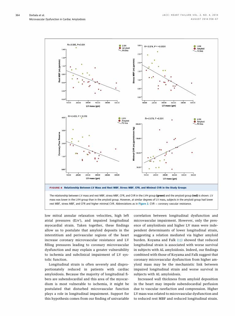
FIGURE 4 Relationship Between LV Mass and Rest MBF, Stress MBF, CFR, and Minimal CVR in the Study Groups
The relationship between LV mass and rest MBF, stress MBF, CFR, and CVR in the LVH group (green) and the amyloid group (red) is shown. LV
mass was lower in the LVH group than in the amyloid group. However, at similar degrees of LV mass, subjects in the amyloid group had lower
rest MBF, stress MBF, and CFR and higher minimal CVR. Abbreviations as in Figure 2. CVR ¼ coronary vascular resistance.
Dorbala et al. J A C C : H E A R T F A I L U R E V O L . 2 , N O . 4 , 2 0 1 4
Microvascular Dysfunction in Cardiac Amyloidosis A U G U S T 2 0 1 4 : 3 5 8 – 6 7
364
low mitral annular relaxation velocities, high leftatrial pressures (E/e0), and impaired longitudinalmyocardial strain. Taken together, these findingsallow us to postulate that amyloid deposits in theinterstitium and perivascular regions of the heartincrease coronary microvascular resistance and LVfilling pressures leading to coronary microvasculardysfunction and may explain a greater vulnerabilityto ischemia and subclinical impairment of LV sys-tolic function.
Longitudinal strain is often severely and dispro-portionately reduced in patients with cardiacamyloidosis. Because the majority of longitudinal fi-bers are subendocardial and this area of the myocar-dium is most vulnerable to ischemia, it might bepostulated that disturbed microvascular functionplays a role in longitudinal impairment. Support forthis hypothesis comes from our finding of univariable
correlation between longitudinal dysfunction andmicrovascular impairment. However, only the pres-ence of amyloidosis and higher LV mass were inde-pendent determinants of lower longitudinal strain,suggesting a relation mediated via higher amyloidburden. Koyama and Falk (13) showed that reducedlongitudinal strain is associated with worse survivalin subjects with AL amyloidosis. Indeed, our findingscombined with those of Koyama and Falk suggest thatcoronary microvascular dysfunction from higher am-yloid mass may be the mechanistic link betweenimpaired longitudinal strain and worse survival insubjects with AL amyloidosis.
Increased wall thickness from amyloid depositionin the heart may impede subendocardial perfusiondue to vascular rarefaction and compression. HigherLV mass was related to microvascular dysfunction andto reduced rest MBF and reduced longitudinal strain.

TABLE 4 Univariable Correlations Between Cardiac Morphological Features and Myocardial Blood Flow Parameters in the Study Groups
Variable
Rest MBF Stress MBF CFR CVR
R Value p Value R Value p Value R Value p Value R Value p Value
LV mass, g �0.49 0.006 �0.60 0.001 �0.41 0.03 0.44 0.02
LVED diameter, cm 0.03 0.8 0.005 0.9 0.0004 0.9 �0.05 0.8
LVES diameter, cm �0.21 0.3 �0.20 0.3 �0.09 0.6 0.12 0.5
LV wall thickness, cm �0.59 0.001 �0.66 <0.0001 �0.47 0.009 0.54 0.003
LV ejection fraction, % 0.25 0.2 0.22 0.3 0.06 0.8 �0.05 0.8
Peak E velocity, m/s 0.23 0.2 �0.07 0.7 �0.09 0.6 �0.007 0.9
Peak A velocity, m/s �0.07 0.8 �0.031 0.9 0.03 0.9 �0.19 0.4
E/A ratio �0.23 0.3 �0.19 0.4 �0.07 0.7 0.24 0.3
Mean e0 velocity, cm/s 0.29 0.1 0.50 0.006 0.49 0.008 �0.41 0.02
Mean a0 velocity, cm/s 0.56 0.002 0.67 <0.0001 0.49 0.01 �0.55 0.004
E/e0 ratio �0.13 0.5 �0.47 0.01 �0.47 0.01 0.42 0.03
Indexed left atrial volume, ml/m2 �0.32 0.11 �0.48 0.01 �0.31 0.13 0.36 0.07
Longitudinal strain, global �0.60 0.001 �0.67 <0.0001 �0.44 0.03 0.48 0.01
Circumferential strain �0.07 0.7 0.03 0.9 0.12 0.6 �0.11 0.6
ECG: limb lead voltage, mm 0.43 0.02 0.48 0.009 0.51 0.004 0.39 0.04
ECG: chest lead voltage, mm 0.27 0.2 0.23 0.2 0.33 0.07 �0.08 0.7
ED ¼ end diastolic; ES ¼ end systolic; other abbreviations as in Tables 1 and 2.
FIGURE 5 Relation Between Left Ventricular Longitudinal Strain and Rest, Stress Myocardial Blood Flow, Coronary Flow Reserve
and Minimal Coronary Vascular Resistance in the Study Groups
The relationship between mean longitudinal LV strain and rest MBF, stress MBF, CFR, minimal CVR, and LV mass in the LVH group (green)
and the amyloid group (red) is shown. Abbreviations as in Figures 2 and 4.
J A C C : H E A R T F A I L U R E V O L . 2 , N O . 4 , 2 0 1 4 Dorbala et al.A U G U S T 2 0 1 4 : 3 5 8 – 6 7 Microvascular Dysfunction in Cardiac Amyloidosis
365
Dorbala et al. J A C C : H E A R T F A I L U R E V O L . 2 , N O . 4 , 2 0 1 4
Microvascular Dysfunction in Cardiac Amyloidosis A U G U S T 2 0 1 4 : 3 5 8 – 6 7
366
Although these findings reinforce the notion thathigher LV mass contributes to coronary microvasculardysfunction, rest MBF, stress MBF, and CFR normal-ized to LV mass were also significantly lower in theamyloid group compared with the LVH group.This finding, along with the high frequency of vascularamyloid deposition in a small sample of subjects in ourstudy, argues for additional mechanism(s), such as therole of vascular amyloid deposition (Fig. 1B), presum-ably resulting in mechanical impairment of micro-vascular vasodilation and angina.
Additionally, autonomic dysfunction (either covertor overt) is prevalent in transthyretin and inAL amyloidosis manifesting clinically with abnormalvascular autonomic (sympathetic) modulation andimpaired baroreflex function (14). Autonomic dener-vation limits stress MBF and CFR, primarily vianorepinephrine-mediated mechanisms and also bychanges in metabolic autoregulation and endothe-lial dysfunction in diabetic autonomic dysfunction(15–17). Although not specifically evaluated in thisstudy, autonomic dysfunction may also contribute tomicrovascular dysfunction in amyloidosis.
Coronary microvascular dysfunction has beendescribed in hypertrophic and infiltrative heart dis-eases, including hypertensive heart disease, aorticstenosis, hypertrophic cardiomyopathy, and Fabrydisease (18). Microvascular dysfunction in these dis-eases may be mechanistically related to coronarymicrovascular remodeling, rarefaction and interstitialfibrosis (hypertensive heart disease), small-vesseldisease, relatively reduced capillary density, in-creased LV end-diastolic pressures and systoliccompression of the septal coronary arteries (19), orincreased LV mass (20). Some or all of these mecha-nisms may explain the coronary vasomotor dysfunc-tion in cardiac amyloidosis. The magnitude of themicrovascular dysfunction in our patients withamyloidosis is not only more severe than those seenin hypertensive disease but is also more severe thanpreviously reported data in dilated cardiomyopathy(21) and Fabry disease (20,22).STUDY LIMITATIONS. To the best of our knowledge,this is the first prospective study to characterizecoronary microvascular function noninvasively incarefully selected subjects with cardiac amyloidosisand no obstructive epicardial CAD. In this study,detailed characterization of epicardial coronary anat-omy and microvascular function was performed todistinguish microvascular dysfunction from flow-limiting epicardial CAD. The study size was modestbecause of our stringent inclusion and exclusioncriteria and the relative rarity of cardiac amyloidosisand may have limited the multivariable models. Also,
the p values presented were not corrected for multipletesting. Further, because of excellent blood pressurecontrol in the current era, hypertensive LVH withoutother end-organ damage is rare, limiting our enroll-ment of patients with hypertensive cardiomyopathyto subjects without severe LVH. Therefore, we studiedCFR values normalized to LVmass (including assumedfunctioning LV mass of 0.5 and 0.75), and they weresignificantly lower in the amyloid group. Some of thesubjects in the amyloid group received specific ther-apy for amyloidosis before study enrollment, poten-tially attenuating the effects of amyloid on MBF.However, significant differences in MBF wereobserved between the study groups, suggesting a largeeffect size and strengthening the study findings.Although the precise clinical implications of all ourfindings are not known, we believe that they mayexplain some of the functional limitations and poorprognosis seen in patients with cardiac amyloidosis.
CONCLUSIONS
Coronary vasodilation and minimal coronary vascularresistance are significantly impaired in subjects withcardiac amyloidosis even in the absence of epicardialCAD. Our findings suggest that increased myocardialamyloid burden (mass) correlates with microvasculardysfunction. Because increased amyloid mass is as-sociated with more widespread cardiac disease, it islikely that vascular amyloid deposits also play a role.An additional role of autonomic dysfunction in coro-nary microvascular dysfunction remains speculativeand needs to be further explored. Coronary micro-vascular dysfunction in amyloidosis also correlateswith diastolic dysfunction and subclinical systolicdysfunction. Successful hematologic treatment of ALamyloidosis is associated with a decrease in cardiacbiomarkers before a change in standard echocardio-graphic features. Thus, further study is required todetermine if coronary microvascular function may im-prove after specific therapy for cardiac amyloidosis.
ACKNOWLEDGMENT S The authors thank thesubjects who participated in this study and theircolleagues at the Brigham and Women’s HospitalCardiac Amyloidosis Program and the Boston Uni-versity Amyloidosis Program.
REPRINT REQUESTS AND CORRESPONDENCE: Dr.Sharmila Dorbala, Noninvasive Cardiovascular Imag-ing Program, Heart and Vascular Center, Departmentsof Radiology and Medicine (Cardiology), Brighamand Women’s Hospital, 70 Francis Street, Shapiro 5thFloor, Room 128, Boston, Massachusetts 02115.E-mail: [email protected].
J A C C : H E A R T F A I L U R E V O L . 2 , N O . 4 , 2 0 1 4 Dorbala et al.A U G U S T 2 0 1 4 : 3 5 8 – 6 7 Microvascular Dysfunction in Cardiac Amyloidosis
367
RE F E RENCE S
1. Westermark P, Benson MD, Buxbaum JN, et al.Amyloid: toward terminology clarification. Reportfrom the Nomenclature Committee of the Inter-national Society of Amyloidosis. Amyloid 2005;12:1–4.
2. Sharma PP, Payvar S, Litovsky SH. Histo-morphometric analysis of intramyocardial vessels inprimary and senile amyloidosis: epicardium versusendocardium. Cardiovasc Pathol 2008;17:65–71.
3. Tsai SB, Seldin DC, Wu H, O’Hara C, Ruberg FL,Sanchorawala V. Myocardial infarction with “cleancoronaries” caused by amyloid light-chain ALamyloidosis: a case report and literature review.Amyloid 2011;18:160–4.
4. Al Suwaidi J, Velianou JL, Gertz MA, et al.Systemic amyloidosis presenting with anginapectoris. Ann Intern Med 1999;131:838–41.
5. Ogawa H, Mizuno Y, Ohkawara S, et al. Cardiacamyloidosis presenting as microvascular angina—acase report. Angiology 2001;52:273–8.
6. Whitaker DC, Tungekar MF, Dussek JE. Anginawith a normal coronary angiogram caused byamyloidosis. Heart 2004;90:e54.
7. Buja LM, Khoi NB, Roberts WC. Clinically signi-ficant cardiac amyloidosis. Clinicopathologic find-ings in 15 patients. Am J Cardiol 1970;26:394–405.
8. Modesto KM, Dispenzieri A, Gertz M, et al.Vascular abnormalities in primary amyloidosis. EurHeart J 2007;28:1019–24.
9. Hongo M, Yamamoto H, Kohda T, et al. Com-parison of electrocardiographic findings in patientswith AL (primary) amyloidosis and in familialamyloid polyneuropathy and anginal pain andtheir relation to histopathologic findings. Am JCardiol 2000;85:849–53.
10. Smith RR, Hutchins GM. Ischemic heart diseasesecondary to amyloidosis of intramyocardial ar-teries. Am J Cardiol 1979;44:413–7.
11. Dorbala S, Vangala D, Sampson U, Limaye A,Kwong R, Di Carli MF. Value of vasodilator leftventricular ejection fraction reserve in evalu-ating the magnitude of myocardium at risk andthe extent of angiographic coronary artery dis-ease: a 82Rb PET/CT study. J Nucl Med 2007;48:349–58.
12. DeGrado TR, Hanson MW, Turkington TG, et al.Estimation of myocardial blood flow for longitu-dinal studies with 13N-labeled ammonia andpositron emission tomography. J Nucl Cardiol1996;3:494–507.
13. Koyama J, Falk RH. Prognostic significance ofstrain Doppler imaging in light-chain amyloidosis.J Am Coll Cardiol Cardiovasc Img 2010;3:333–42.
14. Bernardi L, Passino C, Porta C, Anesi E,Palladini G, Merlini G. Widespread cardiovascularautonomic dysfunction in primary amyloidosis:does spontaneous hyperventilation have acompensatory role against postural hypotension?Heart 2002;88:615–21.
15. Vinik AI, Ziegler D. Diabetic cardiovascularautonomic neuropathy. Circulation 2007;115:387–97.
16. Di Carli MF, Bianco-Batlles D, Landa ME, et al.Effects of autonomic neuropathy on coronaryblood flow in patients with diabetes mellitus.Circulation 1999;100:813–9.
17. Stevens MJ, Dayanikli F, Raffel DM, et al.Scintigraphic assessment of regionalized defectsin myocardial sympathetic innervation andblood flow regulation in diabetic patients with
autonomic neuropathy. J Am Coll Cardiol 1998;31:1575–84.
18. Camici PG, Crea F. Coronary microvasculardysfunction. N Engl J Med 2007;356:830–40.
19. Kofflard MJ, Michels M, Krams R, et al. Coro-nary flow reserve in hypertrophic cardiomyopathy:relation with microvascular dysfunction andpathophysiological characteristics. Neth Heart J2007;15:209–15.
20. Elliott PM, Kindler H, Shah JS, et al. Coronarymicrovascular dysfunction in male patients withAnderson-Fabry disease and the effect of treat-ment with alpha galactosidase A. Heart 2006;92:357–60.
21. Neglia D, Parodi O, Gallopin M, et al.Myocardial blood flow response to pacingtachycardia and to dipyridamole infusion in pa-tients with dilated cardiomyopathy without overtheart failure. A quantitative assessment by posi-tron emission tomography. Circulation 1995;92:796–804.
22. Tomberli B, Cecchi F, Sciagra R, et al. Coronarymicrovascular dysfunction is an early feature ofcardiac involvement in patients with Anderson-Fabry disease. Eur J Heart Fail 2013;15:1363–73.
KEY WORDS amyloidosis, coronarymicrovascular function, myocardial bloodflow, PET, strain
APPENDIX For an expanded Materials sec-tion, as well as a supplemental figure andtables, please see the online version of thispaper.