coronaviridaechapter 3 coronaviridae james a. robb and clifford w. bond* department of pathology...
TRANSCRIPT

CHAPTER 3
Coronaviridae James A. Robb and Clifford W. Bond*
Department of Pathology University of California, San Diego
La Jol/a, California 92093
1. INTRODUCTION
1.1. Summary
Corona viruses are widespread in nature, but relatively little attention has been given to them. They are pathogenic, enveloped RNA viruses whose large, single-stranded, nonsegmented genome is of a positive polarity. They cause a broad spectrum of disease in their natural hosts, including man, primarily by a cytocidal virus-cell interaction. The structure of the virion, the molecular mechanisms of their multiplication strategy, and their pathogenesis are just beginning to receive the attention they deserve.
Our goals in this chapter are three: (1) to define the criteria for membership and the present members of the coronavirus family; (2) to define the structure of the virion and the multiplication strategy for this family; and (3) to describe the spectrum and pathogenesis of disease produced by this family of important pathogenic viruses in their natural hosts. The preprints and unpublished data supplied by many of our colleagues are appreciated. Our emphasis will be, whenever possible, on the molecular description of the multiplication strategy of these viruses
* Present addresses: J. A. R., Department of Pathology, The Green Hospital of Scripps Clinic, La Jolla, California 92037. C. W. B., Department of Microbiology, Montana State University, Bozeman, Montana 59717.
193
H. Fraenkel-Conrat et al. (eds.), Comprehensive Virology© Plenum Press, New York 1979

194 Chapter 3
and on the molecular mechanisms involved in the pathogenesis of the disease produced by these viruses. There are at present large gaps in the description of the molecular virology of this family. In an attempt to bridge some of these gaps and make the review as up to date as possible, prudent speculation will be used and identified as such.
1.2 Definition and Members of the Coronavirus Family
1.2.1. Criteria for Membership and Tentative Members
Until recently, enveloped, RNA-containing viruses that budded solely from the endoplasmic reticular membranes, that had a spherical shape with a 60-220 nm diameter, and that had a "corona" of widely spaced, bulbous peplomers 12-24 nm in length were defined as coronaviruses (McIntosh, 1974). Acceptable biochemical criteria were not available. Such biochemical criteria are now available using avian infectious bronchitis virus (IBV) as the prototype virus for this family (Schochetman et af., 1977; Lomniczi, 1977; Lomniczi and Kennedy, 1977). The virion genome of enveloped viruses of the above morphology must be a large (6-8 X 10⁶ daltons) single piece of single-stranded infectious RNA of messenger (positive) polarity. In addition to IBV, the virions of mouse hepatitis virus (MHV), human coronavirus (HeV), and porcine transmissible gastroenteritis virus (TG EV) probably contain a genome of positive polarity as described in Section 2.2.2. In addition, several other viruses have satisfied the electron microscopic criteria for membership in this family and are listed in Table 1. Additional biochemical criteria will soon be forthcoming because the investigation of the molecular virology of the coronavirus family is rapidly gaining momentum.
1.2.2. Serological Relatedness of Members
The intraspecies and inter species serological relatedness of the various viruses are complex and not yet fully understood (Table 1). The production of useful vaccines against coronavirus diseases, many of which are economically important in agriculture and probably in the human economy as well, will require a more detailed understanding of the serological relatedness of these viruses than is currently available. The major problem in acquiring these data, as well as in understanding

Coronaviridae 195
TABLE 1
Tentative Members of the Coronavirus Family and Their Serological Relatedness
Virus"
Avian infectious bronchitis (IBV) Canine coronavirus (CCV) Feline coronavirus (FCV)
(infectious peritonitis) Huinan coronavirus (HCV) Human enteric corona virus (HECV) Murine hepatitis virus (MHV) Neonatral calf diarrhea corona-
virus· (NCDCV) Porcine transmissible gastro-
enteritis virus (TGEV) Porcine hemagglutinating
encephalitis virus (HEV) Rat coronavirus (RCV) Rat sialodacryoadenitis
virus (SDA V) Runde tick coronavirus (RTCV)
Turkey bluecomb disease virus (TCDV)
Natural host
Chicken Dog Cat
Man Man Mouse Bovine
Pig
Pig
Rat Rat
? Tick ? Sea bird Turkey
SerotypesC
Many' ? One' ? One'
Several"···· ? One Many' Several·
One"'"
? One'
? Ones ? Ones
? One'·
? One'
Serologically related to'
? TGEV' TGEV',' TGEV, but not HEV'
HEV, MHV··7 TGEV' RCV, SDA V, HCV, ?HEV··7
CCV., FCV', ?HEV', ?IBV'
MHV' MHV'
Not to IBV or MHV'·
" These names and abbreviations conform to those suggested by Tyrrell et al. (personal communication) in a revision to the previous recommendations (Tyrrell et al., 1975).
• Mucosal disease virus (border disease, Plant et al., 1976) of sheep may belong in this group (Snowdon et al., 1975).
C References: 1, Cowen and Hitchner (1975); 2, McIntosh (1974); 3, Kapikian (1975); 4, Binn et al. (1974); 5, Reynolds et al. (1977); 6, Monto (1974); 7, Kaye et al. (1977); 8, Hafez et al. (1976); 9, Kemeny (1976); 10, Traavik et al. (1977).
the molecular virology, is the relative difficulty in isolating and growing these viruses in cell culture. This problem is discussed in Section 3.
2. VIRIONS
2.1. Morphology
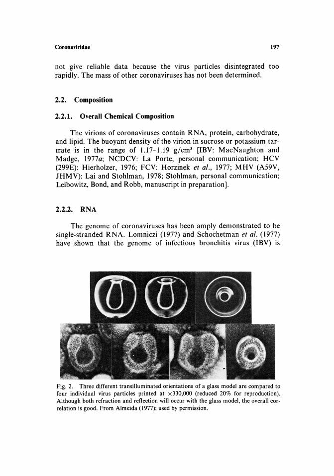
Coronavirus virions are pleomorphic spherical particles of 80-160 nm diameter with characteristic large, widely spaced, 12- to 24-nm-Iong spikes or peplomers that form a corona around the particle and provide the name for this virus family. Figures 1 and 2 show the novel mor-

196 Chapter 3
Fig. 1. A group of IBV virions treated with formaldehyde before staining with phosphotungstic acid. Almost all reveal the internal component. The majority display the tongue or flask orientation, but others show a circular structure or even two concentric rings. Figure 2 shows that all of these patterns are compatible with an internal membranous sac continuous with the outer membrane. Magnification x 135,000 (reduced 20% for reproduction). From Bingham and Almeida (1977); used by permission.
phology of the infectious bronchitis virion (Bingham and Almeida, 1977). Whether this is the only internal morphology available to a coronavirus virion remains to be determined. The topology of the ribonucleocapsid within a virion is not yet known. The nucleocapsid is composed of a 9-nm-wide ribonucleoprotein that forms a helical structure (Kennedy and Johnson-Lussenburg, 1975/1976; Pocock and Garwes, 1977) and probably corresponds to the flask like structure within the virion (Fig. 2). Using transmission electron microscopy, we have observed in mouse hepatitis virus (JHMV and A59V) infected 17CL-16 cells and BALB/c brains that the apparent nucleocapsids in cytoplasmic factories and in early stages of budding have horseshoe or flask like configurations (Robb et af., 1979a,b). Purification of nucleocapsids from virions and infected cells will be necessary to clarify the internal structure of the coronavirus virion.
The mass of a human coronavirus (OC43) is 390 ± 5 X 10⁶ daltons as determined by analytical ultracentrifugation (Hierholzer et al., 1972). Similar experiments with another human coronavirus (229E) did
Coronaviridae 197
not give reliable data because the virus particles disintegrated too rapidly. The mass of other coronaviruses has not been determined.
2.2. Composition
2.2.1. Overall Chemical Composition
The virions of coronaviruses contain RNA, protein, carbohydrate, and lipid. The buoyant density of the virion in sucrose or potassium tartrate is in the range of 1.17-1.19 g/cm3 [IBV: MacNaughton and Madge, 1977a; NCDCV: La Porte, personal communication; HCV (299E): Hierholzer, 1976; FCV: Horzinek et al., 1977; MHV (A59V, lHMV): Lai and Stohlman, 1978; Stohlman, personal communication; Leibowitz, Bond, and Robb, manuscript in preparation].
2.2.2. RNA
The genome of coronaviruses has been amply demonstrated to be single-stranded RNA. Lomniczi (1977) and Schochetman et al. (1977) have shown that the genome of infectious bronchitis virus (lBV) is
Fig. 2. Three different transilluminated orientations of a glass model are compared to four individual virus particles printed at x330,000 (reduced 20% for reproduction). Although both refraction and reflection will occur with the glass model, the overall correlation is good. From Almeida (1977); used by permission.

198 Chapter 3
infectious, and Lomniczi (personal communication) has shown that the genome of transmissible gastroenteritis virus (TGEV) is infectious.
The molecular size of the virion RNA of IBV has been variously reported to be 0.5-3.0 million daltons (4-40 S) by Tannock (1973), 9.0 million daltons (50 S) by Watkins et al. (1975), 8 million daltons (64 S) by Lomniczi and Kennedy (1977), 8 million daltons (58 S) by MacNaughton and Madge (1977b), and 5.5-5.7 million daltons (48 S) by Schochetman et al., (1977).
These variable data reflect the fact that the genome of coronaviruses is the largest viral RNA genome known and, as a result, no standards exist to size the RNA by traditional techniques. Lomniczi and Kennedy (1977) used the elegant method of RNase T1 oligonucleotide fingerprinting to confirm the molecular size of 8.1 ± 0.2 million daltons which had been determined on methyl mercury gels and isokinetic sucrose gradients.
Tannock and Hierholzer (1977) have shown that the RNA of a human coronavirus (HCV-OC43) is 6.1 million daltons (70 S). The genome RNAs of two mouse hepatitis viruses (MHV-A59V and JHMV) were found to be identical and to have a molecular size of about 5.4 million daltons (60 S) (Lai and Stohlman, 1978). The genome RNAs of coronavirus have covalently attached poly(A) (lBV: Lomniczi, 1977; Schochetman et al., 1977; MacNaughton and Madge, 1977b; MHV: (A59V and JHMV), Lai and Stohlman, 1978; Leibowitz, Chun-Akana, Bond, and Robb, unpublished observations; Y ogo et al., 1977; HCV (OC43): Tannock and Hierholzer, 1978). Most of the reports indicate that only a portion (25-50%) of labeled RNA was bound by oligo( dT) cellulose, suggesting (1) that the length of the poly(A) tract may be short, (2) that the RNA in the purified preparations may be degraded, or (3) that a percentage of the molecules have their poly(A) tract in a conformation that prevents binding to the oligo(dT) cellulose. However, Leibowitz, Chun-Akana, Bond, and Robb (unpublished observation) have demonstrated that nearly 100% of MHV RNA (A59V) from purified virions binds to poly(U) sepharose, which binds poly(A) tracts of less than 40-60 nucleotides. These data suggest that the poly(A) sequence may be less than 40 nucleotides in length (Hunter and Garrells, 1977). The poly(A) tract is presumably 3' terminal, but this must be demonstrated.
Treatment of the genomic RNA with heat, chaotropic agent, or high salt does not significantly alter the size or migration in gels or sucrose gradients of several coronavirus RNAs [IBV: Lomniczi and Kennedy, 1977; Schochetman et al., 1977; MacNaughton and Madge,

Coronaviridae 199
1977b; MHV (A59V and JHMV); Lai and Stohlman, 1978; Leibowitz, Bond, and Robb, unpublished observation]. In the cases where heat or chaotropic agents do alter the size or migration of the RNA (TGEVjHEV: Garwes et al., 1975; HCV-OC43: Tannock and Hierholzer, 1977), the reason may be contamination by the RNA of an activated retrovirus or the presence of an internal ribonuclease (Schochetman et al., 1977; Lai and Stohlman, 1978), or the nicking of the large RNA during isolation.
A RNA-dependent RNA polymerase has not been detected in the virions of IBV (Schochetman et al., 1977) or HCV (OC43) (Tannock and Hierholzer, 1978). A RNA-dependent DNA polymerase (reverse transcriptase) was not detected in the virion of IBV (H. Temin, personal communication). These findings support the contention that the coronavirus genome is of positive polarity and separate coronaviruses from retroviruses which are also enveloped RNA viruses containing a genome of positive polarity.
2.2.3. Proteins
The structural proteins that compose the Vlflon of several coronaviruses have been analyzed in detail by several laboratories (Table 2). The variety in the number and size of the structural proteins indicates some degree of complexity in the structure of the coronavirus virion. Much of the variability in the data can be accounted for by the different methods used in virion purification and detection of separated proteins on polyacrylamide gels. It is difficult to synthesize the data in Table 2 into a general description of the protein content of corona virus virions. The number of different proteins in a virion, however, appears to be about four to six, of which two or more are glycoslyated. All of the reports indicate the presence of a major nonglycosylated protein in the range of 45,000-65,000 daltons, probably the nucleocapsid protein. A glycolipoprotein has also been identified in the virions of human coronaviruses (OC43: Hierholzer et al., 1972; 229E: Hierholzer, 1976).
2.2.4. Lipids
The lipid content of transmissible gastroenteritis virions grown in either primary pig kidney cells or secondary adult pig thyroid cells has been compared to the lipid content of whole cells (Pike and Garwes,

N
TA
BL
E 2
<=
<=
Stru
ctur
al P
rote
ins
of C
oron
avir
uses
IBV
M
HV
(A59
V)
MH
V(J
HM
V)
HC
V(2
29E
) H
CV
(O
C43
) T
GE
V
HE
V
NC
DC
V
(1)·
(2
) (3
) (4
) (5
) (5
) (6
) (7
) (8
) (9
) (1
0)
130·
(G
P)"
52 (
GP
) 18
0 (G
P)
180
(GP
) 15
0 (G
P)
150
(GP
) 19
6 (G
P)
191
(GP
) 20
0 (G
P)
180
(GP
) 12
5 10
5 (G
P)
45
120
90
89
100
165
(GP
) 10
4 (G
P)
50
125
(GP
) 65
97
34
10
6 50
60
63
10
5 (G
P)
60 (
GP
) 30
(G
P)
100
(GP
) 50
81
(G
P)
32 (
GP
) 93
(G
P)
23 (
GP
) 20
18
66
(G
P)
47
28 (
GP
) 56
45
74
(G
P)
+1
0 m
inor
70
47
30
+
2 m
inor
26
(G
P)
36
spec
ies
spec
ies
51
58
31 (
GP
) 15
(G
P)
34
33
53
17
28
49
43
40
37 (
GP
) 32
29
24
16
14
(G
P)
Sta
in a
nd
Sta
in"
Sta
in
Sta
in
Isot
ope
Isot
ope
Isot
ope
Isot
ope
Sta
in
Isot
ope
Isot
ope
Sta
in
• R
efer
ence
s: 1
, M
acN
augh
ton
and
Mad
ge (
l977
a);
2, C
ollin
s et
al.
(197
6);
3, B
ingh
am (
1975
); 4,
Stu
rman
(197
7);
5, L
eibo
witz
, B
ond,
and
Rob
b (u
npub
lishe
d ob
serv
atio
ns);
6,
Hie
rhol
zer
(197
6);
7, H
ierh
olze
r et
al.
(197
2);
8, G
arw
es a
nd P
ococ
k (1
975)
; 9,
Poc
ock
and
Gar
wes
(19
77);
10,
La
Por
te (
pers
onal
com
-m
unic
atio
n).
n :r
• M
olec
ular
wei
ght
of p
rote
in X
10-
3•
., "1:1
C (G
P)
indi
cate
s id
enti
fica
tion
of t
he p
rote
in a
s a
glyc
opro
tein
. ....
" M
etho
d of
det
ecti
on o
f pro
tein
. !II ~

Coronaviridae 201
1977). The lipid content of the TG EV virion reflected the lipid content of the host cell in which it was grown, suggesting that the lipids of the virion are derived from the lipids of the host cell. As noted above, the virions of human coronaviruses (OC43 and 229E) contain lipoglycoprotein.
2.2.5. Carbohydrates
Glycoproteins are important in the structure of the coronavirus virion, especially in the formation of peplomers. Sturman and Holmes (1977) isotopically labeled cells infected by mouse hepatitis virus (A59V) with glucosamine and fucose and found that the GP180 and GP90 proteins were highly labeled with both sugars, while GP23 was labeled only with glucosamine. The analogous intracellular GP150 and GP90 proteins of mouse hepatitis virus (JHMV), however, are highly labeled with both glucosamine and fucose (Robb, unpublished observation). Differential labeling of carbohydrates in the virions of other corona viruses has not been reported.
2.3. Fine Structure and Arrangement of Virion Components
2.3.1. Peplomers
The peplomers, or spikes, or corona virus virions contain a major large glycoprotein. They are readily removed by treatment with bromelin or other proteases, a treatment that reduces the density of the virion. For example, the virion of human coronavirus (OC43) has a density of 1.18 g/cm3, and the "despiked" particle has a density of 1.15 g/ cm3 (Hierholzer et al., 1972). The results of treating the virions of six different coronaviruses with bromelin are shown in Table 3. Again, the results are variable but indicate that there are only one or two proteins in coronavirus peplomers.
The hemagglutinating activity in the coronaviruses that possess this property probably resides in the peplomers for the following reasons. Treatment of the neonatal calf diarrhea coronavirus virion with bromelin removes the peplomers and abolIshes hemagglutinating activity (Laporte, personal communication). Concanavalin A does not bind to bromelin-treated hemagglutinating encephalomyelitis virions (HEV) that have lost their peplomers (Greig and Bouillant, 1977). The binding of concanavalin A to untreated HEV virions with intact

TA
BL
E 3
Loc
aliz
atio
n of
Str
uctu
ral
Pro
tein
s in
the
Vir
ions
of C
oron
avir
us
IBV
M
HV
(A
59V
) H
CV
(22
9E)
HC
V (
OC
43)
TG
EV
H
EV
N
CD
CV
(I)a
(2
) (3
,4)
(5)
(6)
(7,8
) (9
) (1
0)
Spi
kes
(Pep
lo-
180.
(GP)
C
130
(GP
) 18
0 (G
P)
105
(GP
) 10
4 (G
P)
200
(GP
) 18
0 (G
P)
125
mer
s)
130
105
(GP
) 90
(G
P)
17 (
GP
) 15
(G
P)
125
(GP
) 65
10
6 74
10
0 (G
P)
83 (
GP
) 70
Env
elop
e 97
23
(G
P)
30 (
GP
) 26
(G
P)
81 (
GP
) 28
(G
P)
33
Nuc
leoc
apsi
d 51
50
50
56
Det
ecti
on
Sta
in
Sta
in
Isot
ope
Sta
in a
nd
Sta
in
Isot
ope
Isot
ope
Sta
in
met
hod
isot
ope
a R
efer
ence
s: 1
, B
ingh
am (
1975
); 2
, M
acN
augh
ton
et a
l. (1
977a
); 3
, S
turm
an (
1977
); 4,
Stu
rman
and
Hol
mes
(19
77);
5,
Hie
rhol
zer
(197
6);
6, H
ierh
olze
r et
al.
(197
2);
7, G
arw
es e
t al.
(197
6);
8, G
arw
es a
nd P
ococ
k (1
975)
; 9,
Poc
ock
and
Gar
wes
(19
77);
10,
La
Por
te (
pers
onal
com
mun
icat
ion)
. •
Mol
ecul
ar w
eigh
t of
pro
tein
x 1
0-3
•
C (G
P)
indi
cate
s id
enti
fica
tion
of t
he p
rote
in a
s a
glyc
opro
tein
. d
Met
hod
of d
etec
tion
of p
rote
in.
N = N g i ~

Coronaviridae 203
peplomers produces a loss in the hemagglutinating activity of the virions.
2.3.2. Envelope
The coronavirus virion envelope probably contains two to three proteins, at least one of which is glycosylated. Treatment of corona virus virions with the detergent NP40 results in the release of a ribonucleoprotein complex from the virion (Kennedy and JohnsonLussenburg, 1975/1976; MacNaughton et ai., 1977). Through the use of this method and bromelin digestion, MacNaughton et ai. (1977) suggested that the proteins VP97, GP81, and VP33 are located in the viral envelope of infectious bronchitis virus. In like manner, GP30 and GP28 are probably components of the envelope of transmissible gastroenteritis virus (Garwes and Pocock, 1975; Garwes et ai., 1976). When virions of mouse hepatitis virus (A59V) are digested with pronase or bromelin, GP23 is digested to a nonglycosylated protein, P*18, suggesting that GP23 is on the surface of the envelope with a tail of about 18,000 daltons embedded in the envelope (Sturman, 1977; Sturman and Holmes, 1977).
2.3.3. Nucleocapsid
2.3.3a. Structure
The ribonucleoprotein that forms the coronavirus nucleocapsid is probably a 9-nm-wide, helical structure that has a buoyant density of 1.27-1.30 g/cm3 and can be released from the virion by treatment with the detergent NP40 (Kennedy and Johnson-Lussenburg, 1975/1976; Garwes et ai., 1976; MacNaughton et ai., 1977). The topography of the helical nucleocapsid within the virion remains to be defined but may be the flask-shaped structure shown in Figs. 1 and 2.
2.3.3b. Protein Subunit
The 50,000-63,000 dalton nonglycosylated, arginine-rich Vlflon protein is present in the nucleocapsid of mouse hepatitis virus (A59V: Sturman, 1977), transmissible gastroenteritis virus (TGEV: Garwes et ai., 1976) and infectious bronchitis virus (MacNaughton and Madge,

204 Chapter 3
1977a). In support of this suggestion is the finding that the internal RNA-associated protein of retroviruses is also rich in arginine (Fleissner, 1971). A minor glycoprotein(s) may also be associated with the nucleocapsid of TGEV (Garwes et aI., 1976) and hemagglutinating encephalomyelitis virus (Pocock and Garwes, 1977).
3. GROWTH PROPERTIES IN CELL AND ORGAN CULTURE
3.1. Infectivity Assays
The major impediment to investigating both the pathogenesis and molecular virology of coronaviruses has been the difficulty in isolating and growing to sufficient titer many of the coronaviruses (McIntosh, 1974). The following in vitro systems have been used for coronavirus isolation, growth, and adaption. Infectious bronchitis virus: embryonated chicken eggs, chicken tracheal ring organ culture, and primary chicken kidney cells (Darbyshire et al., 1975, 1976; Dutta, 1974; Cook et al., 1976; Egan and Tannock, 1978). Canine coronavirus: primary dog kidney cells (Takeuchi et al., 1976). Feline corona virus: primary kitten peritoneal cells (Pedersen, 1976). Human coronavirus: secondary human embryonic kidney cells, WI38 cells, HeLa cells, human embryonic trachael organ culture, L132 cells, and human embryonic lung cells (McIntosh, 1974; Kennedy and Johnson-Lussenberg, 1975/1976; Hierholzer, 1976). Mouse hepatitis virus: primary mouse macrophage cells, mouse DBT cells, and mouse 17CL-l cells (McIntosh, 1974; Hirano et al., 1974, 1976; Takayama and Kirn, 1976; Sturman and Takemoto, 1972; Robb et al., 1979a). Mouse NCTC-1469 liver cells are heavily infected with a murine retrovirus (M-::Intosh, 1974), and we have found this line to be unacceptable for the 5rowth of MHV because of very poor experimental reproducibility (Robb et al., 1979a). Neonatal calf diarrhea coronavirus: rhesus mtmkey kidney cells and fetal bovine kidney cells (King and Harkness, 1975; Inaba et al., 1976; Sharpee et al., 1976; Matsuno et al., 19'77). Transmissible gastroenteritis virus/hemagglutinating encephalomyelitis virus: primary pig kidney, spleen, and thyroid cells and pig testis cells (McIntosh, 1974; Kemeny et al., 1974; Stark et al., 1974; Thomas and Dulac, 1976; Werdin et al., 1976). Rat coronavirus: primary rat kidney cells (McIntosh, 1974).
Plaque, cytopathic effect, and virus-specific immunofluorescence assays can be used in most of these systems. We have developed cyto-

Coronaviridae 205
pathic effect and immunofluorescence microassays for mouse hepatitis virus that should be applicable to many other corona virus systems (Robb et al., 1979a). These microassays are comparable to plaque assays, but are more rapid, precise, and economical. See Section 7.3.4 for additional references.
3.2. Growth Curves and Multiplication Kinetics
In general, most coronavirus infections have a 2- to 4-hr eclipse phase and a maximum progeny yield at 12-16 hr after infection at 37°C (McIntosh, 1974). The multiplication of infectious bronchitis virus (lBV) in chicken tracheal ring organ culture, embryonated chicken eggs, or primary chicken kidney cells is dependent on the IBV isolate (Cook et al., 1976). We have found that the maximum yield of progeny mouse hepatitis virus (JHMV and A59V) in mouse 17CL-16 cells, a subclone of BALB/3T3 cells, occurs by 11 hr after infection at 38.5°C and is independent of the multiplicity of infection between 0.01 and 10 (Robb et al., 1978a). The maximum yield of progeny JHMV in the brain of 4-week-old BALB/c mice after intracerebral inoculation is also independent of the multiplicity of infection between 1 and 10' IV / animal (Robb et al., 1979b). Furthermore, the production of progeny JHMV and A59V in 17CL-16 cells and mouse brain is independent of intercellular fusion and formation of syncytia (Fig. 3) (Robb et al., 1979a). These interesting properties have yet to be explained, but may represent general properties of coronavirus infection in vitro and in vivo. The only data we found concerning the number of coronavirus particles required to initiate an infection are our own for mouse hepatitis virus: JHMV and A59V have one-hit kinetics in 17CL-16 cells (Robb et al., 1979a).
Cytoplasmic coronavirus-specific antigen begins to appear at the end of the 2- to 4-hr eclipse phase. In addition, we have found MHVspecific (JHMV and A59V) granular intranuclear (Fig. 4) and patchy plasma membrane-associated antigens (Fig. 5) in infected 17CL-16 cells (Robb et al., 1979a). The intranuclear antigen appears before the cytoplasmic and surface antigens (Fig. 6), while the appearance of the surface antigen coincides with the appearance of the cytoplasmic antigen. All cells that contain the intranuclear antigen during the low-multiplicity infection (less than 0.5 IV/cell) go on to make cytoplasmic and surface antigens. In addition, some clones of 17CL-l cells persistently infected with JHMV or A59V contain intranuclear antigen in the

206 Chapter 3

Coronaviridae 207
absence of cytoplasmic or surface antigen (Leibowitz, Bond, and Robb, manuscript in preparation). No other report of coronavirus-specific intranuclear or surface antigen was found in our search of the literature.
3.3. Modification of Infectivity
Any agent that disrupts plasma membranes will disrupt the envelope of corona virus virions and decrease or abolish infectivity (Mcintosh, 1974). The infectivity of most corona viruses is stable to repeated freeze-thaw, heating, and acid environment (McIntosh, 1974; Robb et al., 1979a), but each isolate has to be studied for optimum storage conditions. Any alteration of the peplomer structure may decrease virion infectivity (L. Sturman, personal communication).
4. MULTIPLICATION OF VIRUS
4.1. Adsorption
Little is presently known about the molecular events in coronavirus adsorption. Their very narrow host range and organ tropism strongly suggest the presence of specific cell surface receptors. As described in Section 2.3, the peplomers of the virion are probably glycoprotein(s) and represent the most likely site for adsorptive specificity. Adsorption of canine coronavirus and neonatal calf diarrhea corona virus takes place on the plasma membrane of microvilli, crypts, and invaginations of intestinal absorptive epithelial cells or enterocytes (Doughri et al., 1976; Takeuchi et al., 1976).
(
Fig. 3. Cytopathic effect of mouse hepatitis virus (JHMV and A59V) infection in 17CL-16 mouse cells. A: Fibroblastoid morphology of the mock-infected 17CL-16 subclone. B,C: Characteristic corona viral syncytial formation of JHMV and A59V, respectively. The nuclei in JHMV -induced syncytia are usually two- to threefold more numerous than those produced by A59V. A syncytium will stay attached to the substrate for about 2 hr at 38.5°C and 3-4 hr at 33°C after its initial development. The unattached syncytia are apparently "dead," because they are freely permeable to trypan blue. Infected single cells can continue mitosis through telophase. See Robb et al. (1979a) for details. Scale bar in C is 30 11m.

208 Chapter 3
4.2. Penetration and Uncoating
Little is presently known about the penetration and uncoating phase of coronavirus replication. Viropexis and envelope-membrane fusion have both been described as mechanisms of corona virus penetration. One mechanism may predominate for one virus isolate or stock, while both mechanisms may be used by other isolates. Fusion of virion envelope to plasma membrane has been described for the penetration

Coronaviridae 209
mechanism of neonatal calf diarrhea coronavirus into enterocytes of the bovine small intestine (Doughri et al., 1976). Viropexis, on the other hand, appears to be the predominant penetration mechanism of infectious bronchitis virus (IBV) into chicken chorioallantoic membrane and primary chicken kidney cells (Chasey and Alexander, 1976; Patterson and Bingham, 1976). IBV attachment, but not viropexis, occurs at 4°C (Chasey and Alexander, 1976). The following finding suggests that the penetration of IBV is fairly rapid. The addition of neutralizing antiserum at 90 min after IBV infection at 4°C decreases the number of attached plaque-forming virions, while treatment after 90 min at 37°C does not decrease the number of attached plaque-forming virions (Chasey and Alexander, 1976). The mechanism of uncoating of any coronavirus is not known. The IBV -containing cytoplasmic vesicles formed by viropexis apparently do not fuse with lysosomes as a mechanism of uncoating (Chasey and Alexander, 1976).
4.3. Biosynthesis of Viral Macromolecules
4.3.1. Proof of Messenger Function of RNA
The genomic RNA of coronaviruses is of posItIve polarity. Lomniczi (1977), Schochetman et al. (1977) and Norman et al. (1968) have shown that purified infectious virion RNA from bronchitis (IBV) and transmissible gastroenteritis virus, respectively, is infectious. Lomniczi (personal communication) has demonstrated in preliminary experiments that the genomic RNA of IBV can be translated into IBV-
(
Fig. 4. Intranuclear and cytoplasmic mouse hepatitis virus (JHMV and A59V) specific antigens in 17CL-16 mouse cells. Cells were infected with a MOl of 0.1 and incubated at 38.5°C for 4 hr. They were either fixed with methanol (A, B) or formaldehyde-triton (C) before staining with polyspecific mouse anti-JHMV or anti-A59V. The photographs are of JHMV-infected cells. The immunofluorescent staining patterns are similar for both JHMV and A59V using virus-specific antiserum and in reciprocally stained preparations. The pattern is the same at 33°C. The sequence of syncytial formation is cell-cell contact (A), intercellular fusion (B), and continued recruitment of uninfected cells (C). The intranuclear antigen appears to be predominantly in the perinucleolar regions. The intranuclear and ground-glass cytoplasmic antigens are diminished after formaldehyde-triton treatment. The fluorescence remaining after formaldehyde-triton treatment appears to be in the Golgi apparatus and endoplasmic reticulum, the site of virus budding. Golgi apparatuses appear to coalesce during syncytial formation (C). See Robb et al. (l979a) for details. The scale bar in C is 10 ~m.

210 Chapter 3
B
c
Fig. 5. Presence of JHMV-specific antigen on the surface of living infected 17CL-16 ceIls. CeIls were infected with a MOl of 0.1 and incubated at 38.5°C for 4 hr. A: Patchy distribution of the JHMV-specific surface antigen on attached living ceIls. B: Patchy distribution of the JHMV-specific surface antigen at 3 hours after infection. Viral ribonucleoprotein has not been identified by transmission electron microscopy anywhere beneath the plasma membrane, including the regions containing surface antigen C: Specificity of the anti-JHMV serum as ferritin-labeled antibody attaches only to the infecting JHMV particles and not to adjacent plasma membrane. Scale bar in A is 10 Ilm and in Band Cis 100 nm. Similar results are obtained using mouse antiA59V serum and A59V-infected ceIls. JHMV- and A59V-infected ceIls give similar results with reciprocal antisera. Immune electron microscopy kindly performed by R. Garrett. See Robb et af. (1979a) for details.

Coronaviridae 211
specific proteins in a cell-free protein synthesis system. The genomic RNA also contains a covalently linked tract of polyadenylic acid, presumably at the 3' terminus (IBV: Lomniczi, 1977; Schochetman et al., 1977; MacNaughton and Madge, 1977b; mouse hepatitis virus: Lai and Stohlman, 1978; Leibowitz, Chun-Akana, Bond, and Robb, unpublished observations; human coronavirus: Tannock and Hierholzer, 1978).
4.3.2. Species of Corona virus RNAs in Infected Cells
Although very little information is available, there are probably four major groups of corona virus-specific RNA species in infected cells: (1) genomic RNA, (2) double-stranded replicative intermediates, (3) mutliple discrete species of mRNA, and (4) defective or deleted RNA species. The synthesis of all these groups of intracellular viral RNA is resistent to treatment with actinomycin D. Mishra and Ryan (1973) demonstrated coronavirus-specific intracellular RNA in pig kidney cells
'i 12
I • • 10
I ~ VI ....I ....I W U
8
o 6 w I-U W IL 4 z
IZ w 2 u a: w 0.
2 3 4 5
HOURS AFTER INFECTION
100
80
60
40
20
6
z ~ c _ () Z
r- " m m >() ;c .....
m > 0 z() :: m (;)rmrz III
o~ :-, ..... o ::t 90 : z 8 !<
Figure 6. Temporal appearance of mouse hepatitis virus (JHMV and A59V) infected l7CL-16 cells at 38.5°C as detected by virus-specific immunofluorescence. Cells were infected with a MOl of 0.10 in 32-mm Petri dishes and fixed at the indicated times with methanol before immunofluorescent staining with mouse anti-JHMV or anti-A59V. JHMV (0) and A59V (0) infected cells have only intranuclear fluorescence; total number of JHMV (e) or A59V (_) infected cells, i.e., both nuclear-positive and cytoplasmic/nuclear-positive cells, were counted.

212 Chapter 3
treated with actinomycin D and infected with transmissible gastroenteritis virus. During a 1-hr pulse, [3H]uridine was incorporated primarily into 18 Sand 4 S RNA, and to a lesser extent into 28 S RNA. During a 10-min pulse, label was similarly incorporated into 28 S, 18 S, and 4 S regions. Ribonuclease-resistant RNA in the 18-28 S size range was synthesized during the 10-min pulse.
Robb et al. (1979a) have demonstrated major species of 18 S to 28 Sand 50 S RNAs in 17CL-16 cells treated with actinomycin D and infected with mouse hepatitis virus (JHMV and A59V). Virus-specific cytoplasmic ribonucleoproteins were analyzed by sucrose gradients in the presence or absence of EDT A. The species of virus-specific ribonucleoprotein that shifted from higher to lower S values in the presence of EDTA were presumed to contain mRNA species (Figs. 7 and 8). Several discrete species of mRNA ranging in size from 4 S to 28 S, and possibly 50 S, were detected by agarose gel electrophoresis. A species of ribonucleoprotein that sedimented at 200 S (A 59 V) or 230 S (JHMV) did not shift its S value in the presence of EDT A (Fig. 7). This large ribonucleoprotein species contains 50 S RNA, and is likely to be an intermediate in progeny virus formation, probably the nucleocapsid. No data are available on the mechanism of replication or poly adenyl ation of the viral RNA species found in infected cells. Both the single-
5 5
4 'yo. 8f' j ' 4f'
• 4 2005 I
~ HE AV Y' LIGHt 1\
~ 3 .. c .~
2 .. 0
o 10 20 30 FRACTION NUMBER
Fig. 7. Analysis of A59V -specific intracellular provirion (nucleocapsid) and messenger ribonucleoprotein (RNP). Cytoplasmic ribonucleoprotein (RNP) synthesized and labeled with [3H]uridine in the presence of 1 JLg/ml actinomycin D was extracted with 1% NP40 from mock- and A59V-infected 17CL-16 cells. This extracted RNP was analyzed on 10-30% equal mass sterile sucrose-RSB gradient. Half of the extracted RNP was treated with 10 mM EDTA (e--e) before centrifugation. See Robb et al. (1979a) for details.

.., '0
r"
)(
W
I ::J
Z :I:
a:
w
Q.
(/)
I Z
::J o (
)
A
15
HE
AV
Y
PO
OL
41'
T
'8' 1
-O
MS
O
10
1\,
I \:~
>._.
I \,. ""
-/'.
. 5 ~ •
""
\ .
• "'"
'.
/ \
'....
'.
",'
t
' E
DT
" .....
...
. __
•• /
-:~:~:::::
::::::;.
/ +
-'-
"'-
'-'-
'-':'-'/
-E
OT
A
o c 1
0
+D
MS
O
5
Iv
20
3
0
'r-T
'f'
a-r-
":~'
t "
.\ .
~ ....
.... .
"-0
, /
"'-\
-ED
TA
" .
-. t
••
~ /
". '.-
. /"
.... o~
o,
'._
...
r +
EC
rA
~"
'a..
..
D~a"
" ,r
l '0
-0
"
10
2
0
30
15
10
5 10
5
.. '0
r"
)(
W
I ::J
Z :I:
a:
w
Q.
(/)
I Z
::J o (
)
4 3 2 o
4 3 2
FRA
CT
ION
N
UM
BE
R
B
LIG
HT
P
OO
L
-O
MS
O
10
D
+ D
MS
O
10
't' 2._
j
~\+.
o ..
IJ\~_·
OTA '\
,
.~
.~.4
\..
/ /
., t .
//'
--,
/;
-.-.-.,,
~. '.
2
0
30
'j'
r 1\"0,,
1\'"'"
I, .\ '.
.
\ '
,I
\,
I /o
7~
"'S ... e
...
.•. ~"'"
20
3
0
4 3 2 4 3 2
Fig
. 8.
A
naly
sis
of
A59
V-s
peci
fic
intr
acel
lula
r nu
cleo
caps
id a
nd m
esse
nger
RN
A. T
he "
heav
y" (
A)
and
"lig
ht"
(8)
pool
s de
rive
d fr
om t
he g
radi
ents
sho
wn
in F
ig.
7 w
ere
depr
otei
nize
d by
pro
tein
ase
K-c
old
phen
ol e
xtra
ctio
n an
d an
alyz
ed o
n 10
-30%
equ
al m
ass
sucr
ose
0.1 %
SD
S g
radi
ents
. H
alf
of e
ach
sam
ple
of R
NA
was
den
atur
ed w
ith D
MS
O (
C,D
) be
fore
cen
trif
ugat
ion.
See
Rob
b et
al.
(197
9a)
for
deta
ils.
r'l
Q .. Q = ~ ... :;' ~ ~ N
... W

214 Chapter 3
stranded RNA species (mRNA and deleted defective RNA) and the double-stranded RNA species (replicative intermediates) need to be purified by suitable gel electrophoresis, fingerprinted, and mapped onto the genomic RNA. The putative mRNA species have to be analyzed for the presence of 3'-terminal poly(A) tracts and for their ability to direct the cell-free synthesis of coronaviral proteins. These putative coronaviral proteins will have to be rigorously identified by radioimmune precipitation, high-resolution two-dimensional gel electrophoresis, and peptide analysis.
As noted above, actinomycin D is a useful drug for inhibiting cellular, but not coronaviral, RNA synthesis during infection. Although coronaviral RNA synthesis is not affected by the drug, the multiplication of progeny virus is inhibited during human coronavirus infection (229E, C. M. Johnson-Lussenburg, personal communication). The multiplication of mouse hepatitis virus (JHMV) is also inhibited by the drug, whereas the multiplication of MHV -A59V is not inhibited (S. Stohlman, personal communication; Leibowitz, Bond, and Robb, unpublished observations). Although the mechanism of the inhibition of JHMV multiplication is not yet known, it may involve the intranuclear antigen and putative nuclear phase of this virus (see Section 4.5). Other corona viruses need to be investigated for the sensitivity of their replication and multiplication to actinomycin D.
4.3.3. Proteins
Bond and Robb (1978) have demonstrated nine viral-specific proteins in cells infected by mouse hepatitis virus (JHMV and A59V). The molecular weights of the viral-specific proteins synthesized in cells infected by JHMV are slightly different from those in cells infected by A59V and are in the range of 18,000-150,000. The viral-specific intracellular proteins are first detectable at 4 hr after infection at 38.5°C and at 7 hr after infection at 33°C. The functions of the proteins, other than some being structural virion proteins (Table 2), are not known. In all probability, one or more of the proteins is a RNAdependent RNA polymerase responsible for messenger and genomic RNA synthesis, because such an enzyme is not present in the virion. No other data concerning the synthesis of intracellular coronaviral proteins have been reported.
Twelve to 16 proteins of 50,000 daltons each can be encoded in the coronaviral genomic RNA, depending on its size. For example, the mouse hepatitis viral genome is about 6 X 10⁶ daltons, encoding about

Coronaviridae 215
600,000 daltons of protein, while the infectious bronchitis viral genome is about 8 X 10⁶ daltons, encoding about 800,000 daltons of protein. The virion proteins of both viruses account for about 250,000-350,000 daltons, leaving about half to two-thirds of the genome available for regulatory activity and encoding of nonstructural proteins. It is difficult to determine how many independent polypeptides are encoded in the coronaviral genome because we have detected the posttranslational processing of at least two of the nine intracellular mouse hepatitis virus proteins. The identification of the protein(s) synthesized by each of the separate mRNA species and the genomic RNA in cell-free translation systems is essential for a complete definition of the coding capacity of the corona viral genome.
4.4. Assembly
The following sequence of events is probably the most likely mode of coronavirus assembly (McIntosh, 1974). Nucleocapsids are assembled in cytoplasmic viral factories and associate with the cisternal membranes of the endoplasmic reticulum and Golgi apparatus. As the nucleocapsid begins envelopment or budding, peplomers are added to some emerging particles. Not all budding particles receive peplomers, a fact that may account for the low burst size of about 10-100 IU / cell for coronaviruses (Patterson and Bingham, 1976; Robb et al., 1979a). Release of virus particles occurs by lysis of the plasma and/or endoreticular membranes and occasionally by fusion of virus-containing cytoplasmic vacuoles with the plasma membrane (Doughri et al., 1976). Both of these mechanisms may be mediated by the membraneassociated coronavirus protein (ehun-Akana, Bond, and Robb, manuscript in preparation).
4.5. General Comments
The following "most likely" scenario is based on the above facts and the known multiplication strategies of two other single-strand RNA viruses whose genomes are of positive polarity and whose virions do not contain a RNA-dependent RNA polymerase: the Picornaviridae and Togaviridae. Picornaviruses utilize their genomic RNA as a polycistronic mRNA to translate a very large polypeptide that is specifically cleaved to produce the terminal proteins. No other mRNA species is found in picornavirus-infected cells. Togaviruses, on the other hand, have two species of mRNA. Their genomic RNA is polycistronic

216 Chapter 3
and encodes the nonstructural viral proteins, while a smaller mRNA species is also polycistronic and encodes the virion structural proteins. Corona viruses are different from both of these families in their multiplication strategy because they use several species of mRNA, one or more of which may be polycistronic. Only additional data will improve the accuracy of the following corona virus multiplication sequence.
A coronavirus virion adsorbs to a susceptible cell through the interaction of an envelope glycoprotein (peplomer) with a specific glycoprotein receptor on the plasma membrane. Penetration requires energy and a fluid membrane and is accomplished by viropexis and/or envelope-membrane fusion. Uncoating occurs within the cytoplasm, and cytoplasmic "factories" are established for the synthesis of coronavirus RNA and protein. A nuclear function is probably involved in this phase of replication because of the presence of the early intranuclear antigen found in mouse hepatitis virus infection (Robb et al., 1979a). The infecting genomic RNA is of a positive polarity (mRNA) and, after being uncoated and activated, directs the synthesis of a virusspecific RNA polymerase (replicase) which replicates a complementary negative RNA strand. Multiple discrete mRNA species are then transcribed from the negative-strand RNA by a transcriptase, a function possibly residing in the replicase molecule. Viral protein synthesis occurs on both membrane-associated polysomes (glycoproteins) and on non-membrane-associated polysomes (nonglycosylated proteins). The nucleocapsids assemble in the cytoplasmic factories and mature by budding through the membrane of the cisternae of the endoplasmic reticulum and Golgi apparatus. Peplomers are added at the time of budding. Nucleocapsids do not migrate to positions beneath the plasma membrane, and budding does not occur from the plasma membrane, although a virus-specific plasma membrane-associated antigen is present. Both infectious (containing peplomers) and noninfectious (without peplomers) virus are released by cell lysis. Cell death may be mediated by altered membrane function induced by the presence of virus protein in the cellular membrane. The formation of syncytia (intercellular fusion) is not necessary for the production of progeny virions, nor does it amplify the yield of progeny virions (Robb et al., 1979a).
5. ALTERATION IN HOST CELL METABOLISM
Almost no work has been performed on the alteration of host cell metabolism during coronavirus infection. Information concerning the

Coronaviridae 217
regulation of nucleic acid, protein, carbohydrate, and lipid synthesis during infection is lacking. We have observed that general cellular protein synthesis is not affected until very late in the infectious cycle for mouse hepatitis virus. JHMV and A59V (Bond and Robb, 1978). We have observed, however, that synthesis of specific cellular proteins can be stimulated or abolished during JHMV and A59V infection in 17CL-16 cells when radiolabeled proteins are analyzed by high-resolution, two-dimensional gel electrophoresis (Bond and Robb, 1978). As described below, the investigation of cells persistently infected by corona viruses may be helpful in understanding the regulation of specific cellular protein synthesis during infection (Leibowitz, Bond, and Robb, manuscript in preparation).
6. DEFECTIVE VIRUS AND VIRAL INTERFERENCE
6.1. Defective Virus
Evidence of defective VIruS has been obtained in 17CL-16 cells infected at a multiplicity of 0.1 IU / cell by mouse hepatitis virus, JHMV and A59V (Bond, Leibowitz, and Robb, unpublished observations). The defective virus has a density of 1.16 g/cm3 , as compared to a density of 1.18 g/cm3 for intact virions. These defective particles have the same structural proteins as do virions. The RNA in the defective virus, however, is only 18 S in size.
6.2. Interference by Defective Virus
The only available data regarding the production of defective coronavirus particles are those briefly described for mouse hepatitis virus (JHMV and A59V) in Section 6.1. Whether these defective particles have any interfering potential in homologous or heterologous infections in vitro and in vivo remains to be investigated. These defective particles may not have an interference potential because we have serially passaged both JHMV and A59V 20 times in 17CL-16 cells using multiplicities of infection of 10-\ 0.1, and 20-100 (undiluted) IU / cell. No decrease in the yield of progeny virions occurred. An additional 20 serial undiluted passages of both JHMV and A59V had no effect on the yield of progeny virions.

218 Chapter 3
6.3. Other Forms of Interference
We have found that the dose-response curve for mouse hepatitis virus (JHMV and A59V) is linear for multiplicities between 0.01 and 0.5 IV jcell in 17CL-16 cells (Robb et al., 1979a) when the percentage of infected cells is scored by immunofluorescence. The experimental percentages of infected cells are similar to those predicted by the Poisson distribution. Above 0.5 IV j cell, the percentage of infected cells falls significantly below the values predicted by the Poisson distribution. The maximum yield of progeny virions, however, is independent of the multiplicity between 0.01 and 10 IV jcell. In addition, many infected cells at the higher multiplicities contain only the intranuclear MHV -specific antigen and do not develop the cytoplasmic antigen. The mechanism of this apparent interference during virus replication is not known. A possible role for defective particles is being examined. Reports concerning homologous interference in other coronavirus infections were not discovered in our search of the literature.
7. PATHOGENESIS OF CORONAVIRUS DISEASE
7.1. Spectrum of Disease
Three important facts should be borne in mind when the pathogenesis of coronavirus disease is discussed. First, many organs are infected and affected by natural coronavirus infections in animals when the infected animal is carefully examined. Second, variant viruses can be isolated from natural or experimental animal infections that have a more-or-Iess organ-specific tropism rather than the polyorgan tropism of the original infecting virus. Third, the spectrum of disease is very dependent on the age of the animal, the genetic background of the animal, and the route of inoculation. Although these three factors produce a very complex pathogenesis, they also provide a fertile ground for the molecular investigation of the pathogenesis of organ-specific virus-caused disease. The serological relatedness of some of the mammalian viruses (Table 1) allows some extrapolation of data from one animal to another. This type of extrapolation is important for human coronavirus disease because the pathogenesis of the human disease cannot be adequately investigated in infected humans. The murine model is probably the most suitable model for extrapolation to human disease because of the serological relatedness between the human and murine coronaviruses.

Coronaviridae 219
Table 4 lists the spectrum of disease produced by members of the corona virus family. Of particular interest is the multiorgan involvement produced by the mammalian viruses. The data strongly suggest that evidence for an etiological role for human coronaviruses should be actively sought in human meningoencephalitis (as in the mouse, Fig. 9), primary demyelination of the central nervous system (as in the mouse, Figs. 10 and 11), hepatitis, interstitial nephritis, interstitial pneumonitis, and impaired immune responsiveness. If an etiological role in one or more of these diseases can be proven for these viruses, the development of suitable vaccines would be a reasonable objective, because limited studies on human populations indicate very high infection rates: 50% of 3-year-old children and 69% of adults are seropositive for OC43, one strain of human corona virus (Monto, 1976). The infection rate could be shown to be much higher if infection with all of the human coronavirus serotypes were adequately investigated in the same population.
Nothing is presently known about the molecular mechanisms underlying the organ tropism of the coronaviruses. Our speculation is that one of the envelope glycoproteins plays a significant receptor function in recognizing appropriate plasma membrane receptors on susceptible cells. The identification of the virion molecule(s) and/or virus-cell interaction(s) (Robb, 1977) that is responsible for the organ tropism should be possible by correlating the altered organ tropism produced by variant virions with altered virion protein(s) or mUltiplication strategy. The murine model presently offers the greatest potential in this regard.
7.2. Route of Infection
The type of disease produced by a coronavirus is markedly influenced by the route of inoculation, the age of the animal, the genetic background of the animal, and the virus isolate. Intravenous, intranasal, oral, intraocular, intraperitoneal, intramuscular, subcutaneous, and intramammary routes have been used with various viruses and animals.
Natural infections are primarily transmitted by oral ingestion of virus-contaminated fluid and/or particulate matter and by intranasal inoculation with aerosols and/or droplets. The remarkable stability of these enveloped viruses in acid environments, pH 2-3, ensures their passage through the stomach into the small intestine, which is the primary replication site for the viruses that produce significant enteritis.

~
<=
TA
BL
E 4
Spec
trum
of D
isea
se P
rodu
ced
by M
embe
rs o
f the
Cor
onav
irus
Fam
ily
Upp
er
Enc
epha
litis
L
ymph
oid
resp
irat
ory
Vir
usa
Hos
t de
mye
linat
ion
Ent
erit
is
Hep
atit
is
aden
itis
Nep
hrit
is
Pan
crea
titi
s P
erit
onit
is
pneu
mon
itis
M
isce
llane
ous
IBV
C
hick
en
1,2
,3,4
' 4
Gon
adit
is 5
C
CV
D
og
5 F
CV
C
at
6 7
7,8
7
7,8
7
Eye
infe
ctio
n 7
Epe
ndym
itis
6 H
CV
M
an
(+).
+
in m
ice·
10
, 11
H
EC
V
Man
12
, 13
(+
Y
(+ )d
.2 M
HV
' M
ouse
14
, 15
, 16
, 17
15
, 18
15
15
2,
15
19
15
14,
15
NC
DC
V
Bov
ine
20,2
1 T
GE
V
Pig
22
2 23
,24
HE
V
Pig
25,2
6,27
27
27
R
CV
R
at
5,28
S
DA
V
Rat
Sa
liva
ry
aden
itis
29
,30,
31
RT
CV
T
ick
Path
ogen
esis
not
yet
inve
stig
ated
B
ird
TB
DV
T
urke
y 32
32
a T
hese
abr
evia
tion
s fo
llow
tho
se s
ugge
sted
by
the
Cor
onav
irus
Stu
dy G
roup
, ch
aire
d by
Dr.
D.
A.
J. T
yrre
ll, i
n a
revi
sion
(T
yrre
ll, p
erso
nal
com
mun
icat
ion)
o
f the
pre
viou
s re
com
men
dati
ons
(Tyr
rell
et 0
1.,
1975
). R
TC
V is
a n
ewly
des
crib
ed v
irus
who
se a
bbre
viat
ion
may
be
mod
ifie
d.
• T
he p
rodu
ctio
n o
f m
enin
goen
ceph
aliti
s in
hum
ans
is a
spe
cula
tion
base
d on
sev
eral
obs
erva
tions
: th
e he
adac
he a
ccom
pany
ing
man
y co
rona
viru
s co
lds
is n
ot
g re
lieve
d by
non
narc
otic
ana
lges
ics,
sug
gest
ing
at l
east
men
inge
al i
nvol
vem
ent;
the
hum
an c
oron
avir
us O
C43
pro
duce
s le
thal
pan
ence
phal
itis
afte
r I.
C.
~
inoc
ulat
ion
into
suc
klin
g m
ice
(9);
OC
43 i
s se
rolo
gica
lly r
elat
ed t
o th
e he
mag
glut
inat
ing
ence
phal
omye
litis
vir
us o
f sw
ine
(9);
ess
entia
lly a
ll m
embe
rs o
f the
ii
coro
navi
rus
fam
ily p
rodu
ce m
enin
goen
ceph
aliti
s w
hen
neur
opat
hoge
nesi
s is
exa
min
ed.
~

C M
any
hum
an c
ases
of
hepa
titi
s ar
e no
t ca
used
by
the
hepa
titi
s A
or
B v
irus
es (
Zuc
kerm
an,
1978
). W
e sp
ecul
ate
that
the
hum
an c
oron
avir
uses
are
pri
me
susp
ects
in a
t le
ast
som
e of
thes
e no
n-A
, no
n-B
cas
es,
beca
use
mos
t of
the
mam
mal
ian
coro
navi
ruse
s pr
oduc
e he
pati
tis
in t
heir
nat
ural
hos
ts.
d A
post
olov
et
al.
(197
7a)
have
mad
e a
stro
ng a
rgum
ent
that
a p
orci
ne c
oron
avir
us i
s ca
pabl
e o
f in
fect
ing
hum
ans
in c
lose
con
tact
with
pig
s. A
n ac
ute
neph
roni
tis o
ccur
s th
at s
low
ly p
rogr
esse
s in
to a
chr
onic
act
ive
inte
rsti
tial
nep
hriti
s, t
he e
ndem
ic (
Bal
kan)
nep
hrop
athy
. It
the
refo
re s
eem
s re
ason
able
to
spec
ulat
e th
at a
t le
ast
som
e ca
ses
of n
on b
acte
rial
int
erst
itia
l ne
phri
tis
in h
uman
s m
ay b
e ca
used
by
a pe
rsis
tent
inf
ectio
n w
ith a
hum
an c
oron
avir
us.
Inde
ed,
whe
n su
ch a
pat
hoge
nesi
s ha
s be
en s
ough
t, m
any
of th
e co
rona
viru
ses
do c
ause
int
erst
itia
l ne
phri
tis i
n th
eir
natu
ral
host
s (s
ee t
able
abo
ve).
e Pi
azza
(19
69)
desc
ribe
s th
e pa
thog
enes
is o
f th
is g
roup
of
mur
ine
viru
ses
in d
epth
. T
he r
efer
ence
s gi
ven
in t
he t
able
are
mor
e re
cent
inv
estig
atio
ns.
, R
efer
ence
s: I
, A
lexa
nder
et a
l. (1
978)
; 2,
Apo
stol
ov e
t al.
(197
7a);
3,
Apo
stol
ove
et a
l. (1
977b
); 4
, Pu
rcel
l et
al.
(197
6);
5, T
akeu
chi
et a
l. (1
976)
; 6,
Kru
m e
t al
. (19
75);
7, T
imon
ey (
1976
); 8
, H
orzi
nek
et a
l. (1
977)
; 9,
Kay
e et
al.
(197
7);
10,
Gum
p et
al.
(197
6);
II,
McI
ntos
h et
al.
(197
4);
12,
Cau
l an
d E
ggle
ston
e (1
977)
; 13
, M
oore
et
al. (
1977
); 1
4, G
oto
et a
l. (1
977)
; 15
, W
ard
et a
l. (1
977)
; 16
, L
ampe
rt e
t al.
(197
3);
17,
Wei
ner
(197
3);
18,
Bro
ders
on e
t al.
(197
6);
19,
Fuj
iwar
a et
al.
(197
5);
20,
Cha
sey
and
Luc
as (
1977
); 21
, M
orin
et a
l. (1
976)
; 22
, M
orin
et a
l. (1
973)
; 23
, K
emen
y et
al.
(197
4);
24,
Und
erda
hl e
t al.
(197
4);
25,
Gre
ig e
t al.
(197
1);
26,
Men
gelin
g an
d C
utli
p (1
976)
; 27
, W
erdi
n et
al.
(197
6);
Bha
tt a
nd J
acob
y (1
977)
; 29
, Ja
coby
et a
l. (1
975)
; 30
, L
ai e
t al.
(197
6);
31,
Wei
sbro
th a
nd P
eres
s (1
977)
; 32
, N
aqi
et a
l. (1
975)
.
r ::I. ~ ~ ...

222 Chapter 3
Figure 9. Demonstration of mouse hepatitis virus-caused acute encephalomyelitis in a 4-week-old BALBjc mouse given 100 infectious units of JHMV intranasally and sacrificed 4 days later. JHMV-specific infection of neurons and glial cells in the hippocampus is demonstrated by immunofluorescence using sections of ethanol-fixed, paraffin-embedded brain. See Robb et al. (1979b) for details. Scale bar is 10 jLm.
The subsequent viremia produces disease in other organs such as the liver, brain, kidneys, and lungs. After intranasal inoculation, however, coronaviruses can infect the brain by direct extension from the nasal mucosa without the assistance of a viremia, as shown for mouse hepatitis virus (M HV) by Goto et af. (1977) and for rat sialodacryoadenitis by Jacoby et af. (1975). Another possible route in natural infections is the aerosol/droplet infection of the eye as shown for infectious bronchitis virus by Cowen et af. (1971). Venereal transfer for M HV infection has been suggested by Nelson (1952).
Finally, a most intriguing possible natural route of infection, that of mechanical and/or biological transfer by infected insects, has been suggested. Ishii et af. (1974) showed that M HV produced virus-specific fluorescence in intestinal cells of the mosquito Aedes aegypti for at least 14 days after ingestion of blood from infected mice. Furthermore, the blood in these engorged mosquitoes was infectious when inoculated into mice. Recently, a coronavirus has been demonstrated by transmission electron microscopy in ticks, Ixodes uriae, that feed on sea birds in Runde, Norway (Traavik et af., 1977). The sea birds were seropositive for the virus.
The following references provide a more detailed examination of the effect of route of inoculation and age of animal on the pathogenesis of coronavirus disease. IBV: Alexander et af. (1978); CCV: Binn et af. (1974); FCV: Timoney (1976); HCV: Monto (1976); HECV: Caul and

Coronaviridae 223
Egglestone (1977); MHV: Bailey et al. (1949), Piazza (1969), Sebesteny and Hill (1974), Hirano et al. (1975b), Taguchi et al. (1977); Fox et al. (1977); TGEV: Kemeny and Woods (1977); HEV: Mengeling and Cutlip (1976), Werdin et al. (1976); TBOV: Naqi et al. (1975), Gonder et al. (1976) .
A very important area of coronavirus-caused disease that has received very little attention is that of intrauterine infection. A beginning has been made into the pathogenesis of neonatal calf diarrhea coronavirus intrauterine infection because of its economic impact on the production of dairy and beef cattle. This virus is also called mucosal
Fig. 10. Demyelination in the brain of a 4-week-old BALBjc mouse that was given 100 infectious units of JHMV by the intranasal route 21 days before examination by transmission electron microscopy. Two small, normally myelinated axons (arrow) are present within a large group of demyelinated axons (Ax) in the spinal cord. MN indicates the nuclei within two macro phages that have removed the damaged myelin from the demyelinated axons. See Robb et al. (1979b) for details. Scale bar is I/-Lm .

224 Chapter 3
Fig. II. Oligodendrocyte containing intracisternal vIrIons of mouse hepatitis virus (JHMV). Aggregates of electron-dense particles, filaments, and vacuoles are prominent in the hypertrophic cytoplasm. Plasma membrane connections to myelin lamellae (arrows) are seen around axons 1-3. A redundant myelin loop is connected with an oligodendroglia I process (axon 2). x50,OOO (reduced 38% for reproduction). Reproduced from Powell and Lampert (1975).

Coronaviridae 225
disease virus of sheep and bovine diarrhea virus. Although limited numbers of animals were investigated, a maternal viremia probably produces abortion and fetal death during early pregnancy, congenital anomalies during middle pregnancy, and little effect during late pregnancy (Brown et al., 1974; Snowdon et al., 1975: Gard et al., 1976; Plant et al., 1976; Allen, 1977). The significant congenital anomalies were in the central nervous system and consisted of cerebellar degeneration, cavitation, and hypoplasia. These anomalies were attributed to the cytocidal effect of the virus and the subsequent cerebellar edema. One indication of fetal reaction to the viral infection was the production of interferon in the fetal tissues from 4 to 21 days after infection and in the fetal serum from 13 to 21 days after infection (Rinaldo et al., 1976).
Turkey poults hatched from eggs infected with turkey bluecomb disease virus 24 days after being laid have a disease syndrome that is similar to that occurring in poults inoculated at 1 day after hatching (Deshmukh et al., 1976). Clearly, a great deal more attention has to be given to this area. The murine model offers the greatest potential for interested investigators.
7.3. Animal Response to Infection
7.3.1. Virus-Cell Interaction
The primary virus-cell interaction in corona virus-caused disease is a cytocidal interaction (Robb, 1977). As described in more detail in Section 4, the virus adsorbs to a susceptible cell, penetrates the cell, replicates its RNA and protein species primarily in the cytoplasm, assembles by budding into cisternae of the endoplasmic reticulum and Golgi appartus, and kills the cell by a presently unknown mechanism. One possible cytocidal mechanism is the alteration of membrane function by the incorporation of a viral protein into the membrane. We have found that a newly synthesized virus-specific protein is associated early after infection with the plasma membrane of mouse hepatitis virus-infected cells (Fig. 5) (Robb et al., 1979a). Shortly after the appearance of this protein in the plasma membrane, the cells become permeable to trypan blue, suggesting that coronavirus infection may rapidly alter the function of the plasma membrane.
The association of a viral protein with the plasma membrane may also account for the loss of glucose-stimulated sodium transport in the plasma membrane of jejunal mucosal enterocytes infected with transmissible gastroenteritis virus (TGEV) (McClung et al., 1976; Kerzner et

226 Chapter 3
al., 1977). This altered transport is probably responsible for the TGEVcaused diarrhea. This alteration is not simply the result of a general malfunction of plasma membrane transport, because adenyl cyclasedependent sodium transport remains intact. The diarrhea produced by TGEV is, therefore, different from the diarrhea produced by the E. coli and C. vibrio entertoxins, which alter the adenylyl cyclase-dependent sodium transport and leave the glucose-stimulated sodium transport intact.
Lymphoid and reticuloendothelial cells, endothelial cells, and parenchymal cells such as neurons, glia, hepatocytes, renal podocytes, and nasointestinal epithelial cells are all subject to cytocidal attack by members of the coronavirus family. This cytocidal interaction has been demonstrated by light microscopy, virus-specific immunofluorescence, andj or transmission electron microscopy in infected animals andj or organ culture. The formation of typical coronavirus syncytia in organs, probably mediated by the plasma membrane-associated viral protein, is dependent on the virus isolate used for the infection. The following references provide pertinent details. IBV: Cowen et al. (1971), Purcell et al. (1976), Alexander and Gough (1977), Apostolov et al. (1977a); CCV: Keenan et al. (1976), Takeuchi et al. (1976); FCV: Krum et al. (1975); HCV: Apostolov et al. (1977a); HECV: Caul and Clarke (1975), Caul and Egglestone (1977); MHV: Bailey et al. (1949), Sebesteny and Hill (1974), Fujiwara et al. (1975), Smith et al. (1975jI976), Broderson et al. (1976), Taguchi et al. (1976), Goto et al. (1977), Namiki et al. (1977), Ward et al. (1977); NCDCV: Morin et al. (1976), Doughri and Storz (1977); TGEV: Morin et al. (1973), Underdahl et al. (1974), Kemeny et al. (1974), Sprino et al. (1976); HEV: Mengeling and Cutlip (1976), Sprino et al. (1976), Werdin et al. (1976); SDAV: Jacoby et al. (1975); TBDV: Deshmukh et al. (1976), Gonder et al. (1976).
7.3.2. Resistance to Infection
The mechanism of resistance to coronavirus infection in animals is very complex. A comprehensive understanding of the molecularj cellular mechanisms underlying this resistance has not yet emerged, although significant insights have been obtained. As detailed below, resistance is dependent on the interaction of at least four factors in the murine model: (1) ability of virus to produce progeny virus in cells of the reticuloendothelial system (macrophages), (2) production of

Coronaviridae 227
lymphokines by lymphoid cells, (3) production of interferon, and (4) development of virus-specific cellular immunity. The role of humoral immunity appears to be relatively unimportant in this resistance.
Weiser, Bang, and their co-workers have shown that the resistance to mouse hepatitis virus (MHV) infection in mice is due both to a genetically transmitted "resistance gene(s)" that inhibits MHV replication in macro phages and to a "susceptibility factor(s)" that is produced by lymphoid cells, possibly a lymphokine. The susceptibility factor makes genetically resistant macrophages susceptible to MHV replication (Weiser and Bang, 1976, 1977; Weiser et at., 1976). Stohlman and Frelinger (1978) have found that the resistance of SJL mice to central nervous system infection by JHMV is mediated by two genes, one dominant and one recessive.
Virelizier and his co-workers have found that type I interferon (virus induced) has an important role in providing resistance to acute, but not chronic, MHV disease in resistant strains of mice (Virelizier et at., 1976, Virelizier and Gresser, 1978). Although type II interferon (non-virus-induced) inhibits MHV replication in macrophages in vitro, it may have little effect in providing resistance to MHV infection in vivo (Taguchi et at., 1976; Virelizier et at., 1976). Additional detailed studies are needed to establish the role, if any, of the various interferons in animal resistance to corona virus infection.
The murine resistance gene described by Weiser and Bang may involve the H-2 region of the murine genome because H_2ff and H_2bf strains of mice are resistant to the chronic phase of MHV -caused disease, while H_2ss, H_2aa, and H_2bb strains are susceptible (Oth et at., 1976). The acute phase of the disease occurs in all the H-2 strains. Additional detailed studies are needed to determine whether an immune response gene is involved in this model of resistance.
Cellular immunity does have an important but poorly understood role in the resistance of mice to MHV disease. Nude (nulnu) mice develop lethal acute and chronic hepatitis and encephalitis after MHV infection, while the infection in their nul + heterozygote littermates has no clinical effect (Sebesteny and Hill, 1974; Tamura et aI., 1976; Ward et at., 1977). Furthermore, nulnu mice can be immunized with inactive MHV only after they have been inoculated with nul + spleen cells (Fujiwara et at., 1976). Cortisone treatment renders nul + heterozygotes susceptible to MHV disease with a pathogenesis similar to that in their nulnu homozygous littermates without cortisone treatment (Hirano et at., 1975b). Cortisone or cyclophosphamide treatment also increases the susceptibility of non-nude mice to M HV disease (Hirano
228 Chapter 3
et al., 1975a,b; Taguchi et al., 1977). Radiation or antilymphocyte serum treatment makes normally resistant mice susceptible to MHV disease (Dupuy et al., 1975; LePrevost et al., 1975a). Cell-mediated immunity is also important in transmissible gastroenteritis virus (TGEV) infections (Frederick and Bohl, 1976; Woods, 1977).
Coronavirus infection stimulates a normal humoral immune response in the natural host. The early appearance of IgM is followed by an increasing amount of IgG. Antiviral IgA is readily produced. The antibody levels stay high for months to years after a single exposure. Different temporal and quantitative responses in neutralizing, complement-fixing, immunofluorescent, and hemagglutination-inhibiting antibodies occur during infection and vary between individuals. The role of humoral immunity in the resistance of mice to MHV infection is less clear. Although neutralizing antibody is transferred to suckling mice via the milk, these mice are still susceptible to MHV disease (Kiuchi et al., 1974). Furthermore, passive immunity with anti-MHV neutralizing serum or transplacental anti-MHV neutralizing antibodies does not make susceptible mice resistant (LePrevost et al., 1975a; Dupuy et al., 1975; Taguchi et al., 1976). The inoculation of thymectomized, irradiated mice with T -cell-deficient embryonic liver cells does not protect them from MHV disease (Dupuy et al., 1975; LePrevost et al., 1975a). However, orally given anti-TGEV IgG, IgM, or IgA protects neonatal pigs from lethal TGEV infection (Stone et al., 1977).
Immune complex disease may occur during coronavirus infection, especially during the chronic phase of disease. Chicks that develop chronic interstitial nephritis after infectious bronchitis virus (IBV) infection have higher anti-IBV HI titers than do chicks that do not develop chronic interstitial nephritis (Alexander and Gough, 1978). Apostolov has found granular deposits in the glomerular basement membrane and subendothelial space in kidneys from humans with endemic Balkan nephropathy. These deposits contain IgG and C3 by immunofluorescence (Apostolov et al., 1977a). This disease is believed to be produced by a chronic TG EV infection in people closely associated with pigs. This observation warrants confirmation and expansion.
The production and use of vaccines for corona viruses is beyond the scope of this chapter, but the following references will facilitate an introduction to the literature. IBV: Cowen and Hitchner (1975), Thornton and Muskett (1975), Alexander et al. (1976), Alexander and Chettle (1977), Alexander and Gough (1978), HCV: Monto (1976); MHV: Dupuy et al. (1975), Hirano et al. (1975b), LePrevost et al.

Coronaviridae 229
(1975a), Fujiwara et al. (1976); NCDCV: Hafez et al. (1976), Shope et al. (1976), Whitmore and Archbald (1977); TGEV: Kemeny (1976), Sprino et al. (1976), Stone et al. (1976), Kemeny and Woods (1977); SDAV: Jacoby et al. (1975); TBDV: Gonder et al. (1976).
7.3.3. Mixed Infections of Coronavirus with Other Agents
Few pertinent reports were found concerning mixed infections between a coronavirus and other agent(s). Giambrone et al. (1977) showed that I-day-old chicks infected with infectious bursal disease virus and challenged at 35 days of age with infectious bronchitis virus (IBV) developed lower anti-IBV neutralizing antibody titers and had an increased incidence and severity of disease than did 35-day-old control chicks that had not been previously infected with infectious bursal disease virus (Giambrone et al., 1977). Morin et al. (1976) investigated 55 cases of spontaneous calf diarrhea and found II of them to involve neonatal calf diarrhea corona virus and one or more other etiologically significant agents. A review of the viruses, including human enteric coronavirus found in human diarrhea, is given by Flewett and Boxall (1976).
7.3.4. Diagnostic Techniques
Various diagnostic techniques for coronavirus identification and serotyping have been applied to infected animals, tissues, organ cultures, and cell cultures. These techniques include cytopathic effect, virus-specific immunofluorescence, hemagglutination inhibition, neutralization, complement fixation, hemadsorption, immune electron microscopy, negative-contrast electron microscopy, and single radial hemolysis. The following references will provide access to this literature. IBV: Lucio and Hitchner (1970), Cowen and Hitchner (1975), Alexander et al. (1976), Hidalgo and Raggi (1976), Alexander and Chettle (1977), Wooley and Brown (1977), Egan and Tannock (1978); CCV: Takeuchi et al. (1976); FCV: Pedersen (1976); HCV: Flewett and Boxall (1976); Hierholzer and Tannock (1977), Monto and Rhodes (1977), Riski et al. (1977); MHV: Robb (1979a,b), Hirano et al. (1975a), Namiki et al. (1977); NCDCV: England et al. (1975), Morin et al. (1976), Sharpee et al. (1976); TGEV: Thomas and Dulac (1976), Werdin et al. (1976), Saif et al., 1977; TBDV: Patel et al. (1976, 1977);

230 Chapter 3
RTCV: Traavik et al. (1977). Also see the additional references listed in Sections 3.1 and 7.1.
7.4. Summary of Pathogenesis
The spectrum of disease in the natural hosts of coronaviruses depends on the age, genetic background, route of infection, and virus dose. The primary natural routes of corona viral infection are aerosol/droplet infection of the nasopharynx and oral ingestion of infected particulate matter. More unusual routes have been described such as veneral, intraocular, and biological and/or mechanical transfer from insects (mosquitoes and ticks).
The primary mechanism of disease production during acute, and probably chronic, coronavirus infection is a direct cytocidal attack on susceptible cells. These viruses can destroy epithelial, endothelial, and lymphoid cells. When parenchymal cells are killed, diseases such as hepatitis, encephalomyelitis, enteritis, nasopharyngitis, pneumonitis, and nephritis are produced. The acute lymphadenitis may produce transient or prolonged depression of both humoral and cellular immunity. The destruction of endothelial cells may produce localized or generalized vasculitis. Direct infection of susceptible cells, and extension without a requirement for viremia, can produce enteritis, nasopharyngitis, pneumonitis, lymphadenitis, and, after nasopharyngeal infection, encephalomyelitis. A secondary viremia produces hepatitis, nephritis, and, after oral ingestion, encephalomyelitis and pneumonitis as well.
Resistance to acute infection depends on the ability of the infecting virus to multiply in reticuloendothelial cells, a property that is genetically controlled. In addition, virus-induced interferon production and cell-mediated immunity are important in limiting the extent of infection, while humoral immunity is relatively unimportant. The molecular mechanisms of resistance to coronavirus infection have yet to be fully elucidated.
Chronic corona virus disease, in the form of demyelination, hepatitis, and/or immune deficiency, is produced primarily by a smoldering cytocidal infection by cell-to-cell spread of the virus. Although coronaviruses produce a plasma membrane-associated antigen in acutely and persistently infected cells, there is at present no evidence that an immune-mediated cytocidal attack on persistently infected cells in chronically infected animals is an important mechanism in the

Coronaviridae 231
pathogenesis of chronic coronavirus disease. Immune-mediated chronic disease does, however, remain a likely possibility in conjunction with the slowly progressive cell-to-cell spread of the virus.
The murine models available for both acute and chronic coronavirus infections should enhance our presently meager understanding of acute and chronic coronavirus disease in humans. An elucidation of the molecular virology of murine coronaviruses, however, is necessary before the murine models of disease will be fully informative.
8. PERSISTENT INFECTIONS
The persistent infections established by some coronaviruses in their natural hosts should provide very good experimental models for elucidating the pathogenic mechanisms of some important human diseases. The murine models of chronic hepatitis and persistent demyelination of the central nervous system are receiving significant attention. Table 5 lists the pertinent work in these areas. It must be noted that several biologically different variants are listed under one name as if they were all the same virus when they are not, i.e., mouse hepatitis virus: JHMV.
The persistent infections with JHMV that produce a smoldering demyelination (Figures 10 and 11) (Herndon et ai., 1975; LePrevost et ai., 1975b; Virelizier et ai., 1975) may be relevant to human multiple sclerosis because coronaviruslike particles have been observed in the brain of a patient with multiple sclerosis by transmission electron microscopy (Tanaka et ai., 1976), and coronaviruses have been isolated from the brains of two patients dying from multiple sclerosis (J. Burks, University of Colorado Medical Center, personal communication). Both findings await confirmation and expansion. The hepatitis models may be analogous to some of the non-A, non-B forms of chronic human hepatitis (Zuckerman, 1978).
The disease produced by a persistent coronavirus infection may be due to a smoldering cytocidal attack on susceptible cells rather than to an immune attack specifically directed against corona virus-modified cells. Lampert (1978) has reviewed these mechanisms in human demyelinating diseases. A cell-to-cell spread of the virus could be one mechanism that would produce continued cytocidal activity in the presence of high levels of neutralizing antibody. Tentatively, cell-to-cell spread of mouse hepatitis virus (MHV) in 17CL-16 cells infected with an A59V recovered from persistently infected 17CL-l cells has been

N ~
N
TA
BL
E 5
Per
sist
ent
MH
V I
nfec
tion
in M
ice
Infe
ctio
n pa
ram
eter
s T
ype
of
Chr
onic
S
ourc
e of
ac
ute
dise
ase
Vir
us
viru
s S
trai
n A
ge
Rou
te"
Dos
e"
dise
asec
>
2 m
o p.
i."
Evi
denc
e of
per
sist
ence
R
efer
ence
'
JHM
V
C57
jBll
iver
Sw
iss
3 w
k i.c
. 10
0 L
Ds.
E
M
Pore
ncep
haly
N
one
JHM
V
Mou
se b
rain
B
AL
Bjc
4
wk
i.c
. 50
% L
D,.
E
M
Non
e A
ctiv
e de
mye
lina
tion
16
mo
pj.
2
MH
V3
DB
Aj2
bra
in
C3H
10
wk
i.p.
10' L
Ds.
E
M a
nd H
P
aral
ysis
V
irus
iso
lati
on f
rom
liv
er,
brai
n an
d 3
Hep
atit
is
lym
ph n
odes
3 m
o p
j.
Imm
une
defi
cien
cy
MH
V,
C57
JBI
liver
A
2G
7 w
k i.p
. 10
' LD
s.
Non
e P
aral
ysis
F
A +
epen
dym
al c
ells
, vi
rus
isol
ated
4
C3H
6
mo
i.p
. 10
' LD
s.
Non
e V
ascu
litis
F
A +
endo
thel
ial
cells
, vi
rus
isol
ated
Im
mun
e de
fici
ency
N
uU
nujn
u,
nujn
u 7
wk
i.n.
10' P
FU
E
M a
nd H
H
epat
itis
V
irus
iso
lati
on
5 (M
HV
2)
BA
LB
jc o
n B
AL
Bjc
i.p
. 10
4 P
FU
L
ymph
nod
e E
ncep
halit
is
DB
Tce
lls
i.v.
aden
itis
L
N a
deno
path
y JH
MV
-Re
17C
L-1
6 ce
lls
BA
LB
jc
4w
k
i.c.
10' I
V
Dem
yeli
nati
on
Non
e R
emye
lina
tion
, ?
dem
yeli
nati
on
6 i.n
. 10
' IU
(n
ot l
etha
l)
with
lig
ht m
onon
ucle
ar c
ell
infi
ltra
te a
t 3
mon
ths
pj.
A
59V
C'
17C
L-1
6 ce
lls
BA
LB
jc
4w
k
i.c.
10' I
V
Dem
yeli
nati
on
Non
e R
emye
lina
tion
, ?
dem
yeli
ntio
n 6
i.n.
10' I
U
(not
let
hal)
w
ith l
ight
mon
onuc
lear
cel
l in
filt
rate
at
3 m
onth
s p.
i. ~ =-
A59
VC
-R'
17C
L-1
6 ce
lls
BA
LB
jc
4w
k
i.c.
10' I
V
Dem
yeli
nati
on
Non
e R
emye
lina
tion
, ?
dem
yeli
nati
on
6 » -=
i.n.
10' I
V
(inc
reas
ed
with
lig
ht m
onon
ucle
ar c
ell
;- .. le
thal
ity)
in
filt
rate
at
3 m
onth
s p
j.
~

JHM
V-t
s 17
CL
-16
cells
B
AL
B/c
4
wk
i.c
. 10
" P
FU
D
emye
lina
tion
N
one
Rem
yeli
nati
on, ?
dem
yeli
nati
on
8, J
l (n
ot le
thal
) w
ith l
ight
mon
onuc
lear
cel
l in
filt
rate
at
3 m
onth
s p.
i. JH
MV
Sw
iss
brai
n C
57/B
L
12w
k i.c
. 25
SL
D ••
E
M
Non
e C
onti
nuin
g de
mye
lina
tion
with
a i.c
., In
trac
ereb
ral;
i.p
., in
trap
erit
onea
l; i
.n.,
intr
anas
al;
i.v.,
intr
aven
ous.
•
LD
.., A
mou
nt o
f vir
us t
hat w
ill k
ill 5
0% o
f inf
ecte
d m
ice;
PF
U,
plaq
ue-f
orm
ing
units
; IU
, in
fect
ious
uni
ts.
C E
M,
Acu
te le
thal
enc
epha
lom
yeli
tis;
H,
acut
e he
pati
tis.
a
Chr
onic
dis
ease
occ
urri
ng g
reat
er t
han
2 m
onth
s af
ter
infe
ctio
n de
fine
d by
clin
ical
sym
ptom
s.
e R
ever
tant
of J
HM
V s
elec
ted
by r
epea
t pa
ssag
e in
17C
L-1
6 ce
lls;
mar
kedl
y de
ceas
ed t
ropi
sm f
or n
euro
ns.
mac
roph
age
infi
ltra
te,
FA
+, n
o vi
rus
resc
ued:
3 a
nd 6
mo
p.i.
f A
59C
, 17
CL
-l a
dapt
ed A
59V
; A
59V
C-R
, re
vert
ant
sele
cted
for
inc
reas
ed n
euro
trop
ism
and
leth
alit
y by
rep
eate
d pa
ssag
es in
mou
se b
rain
s.
7 8
1976
); 6
, J.
Rob
b an
d P.
Lam
pert
(un
publ
ishe
d da
ta);
7,
M.
Has
pel
(per
sona
l com
mun
icat
ion)
; 8,
S. S
tohl
man
and
L.
Wei
ner
(per
sona
l co
mm
unic
atio
n).
• R
efer
ence
s: 1
, B
aile
y et
al.
(194
9);
2, H
ernd
on e
t al.
(197
5);
3, L
ePre
vost
, et
al.
(197
5b);
4,
Vir
eliz
ier
et a
l. (1
975)
; 5,
Tam
ura
et a
l. (1
976)
; 6,
J.
Rob
b an
d P.
L
ampe
rt (
unpU
blis
hed
data
); 7
, M
. H
aspe
l (p
erso
nal
com
mun
icat
ion)
; 8,
S.
Sto
hlm
an a
nd L
. W
eine
r (p
erso
nal
com
mun
icat
ion)
.
n = .. = :I .. ;:5.
:!. =- .. '" ~
eM

234 Chapter 3
observed (Robb, unpublished observation). A specific immune attack cannot be ruled out as a mechanism of persistent coronavirus disease, however, because of the presence of a coronavirus antigen associated with the plasma membrane (Robb et al., 1979a) and the involvement of cellular immunity in the resistance and susceptibility to coronavirus disease (see Section 7.3.2). Both mechanisms may be important in chronic disease.
One approach to understanding the molecular mechanisms underlying persistent coronavirus infection in animals has been to establish and biochemically investigate persistent coronavirus infections in cell culture. To date, persistent infections in vitro using only murine coronaviruses have been reported. Table 6 lists the available data concerning cells persistently infected with MHV. Several comments can be made from the preliminary data. First, persistent infections are very easily established using low or high multiplicities of infection in growing or confluent cultures (Leibowitz, Bond, and Robb, manuscript in preparation). Second, the virus recovered from the persistently infected cells can have biological characteristics similar to or different from those of the pool of virus used in the original infection. Third, neutralizing antibodies may decrease the yield of virus from the cells but do not eradicate the persistent infection (Stohlman and Weiner, 1978). Fourth, cell clones derived from persistently infected cells may vary in the percentage of cells expressing cytoplasmic and surface antigens and releasing infectious virus (infectious centers) while maintaining intranuclear antigen in 100% of the cells (Leibowitz, Bond, and Robb, manuscript in preparation). Several mechanisms of persistence (Huang and Baltimore, 1977) may therefore be available to coronaviruses in establishing and maintaining persistent infections in vitro and in vivo.
Other members of the coronavirus family are probably able to establish persistent infections in their host animals although very few data are available. Infectious bronchitis virus (lBV) can be recovered for several weeks after infection from organs of infected pullets and for several months in their feces (Fabricant and Levine, 1951; Alexander and Gough, 1977). IBV can also produce a chronic nephritis in chickens (Alexander and Gough, 1978). In human infections, human coronavirus (HCV) can be isolated from nasal washings for up to 18 days (Monto, 1976) and human enteric coronavirus (HCEV) can be isolated from feces for several months (Moore et al., 1977). The inability to temporally assay human tissues for HCV and HECV after acute infection has made the investigation of persistent human corona virus infection very difficult. The sheep counterpart of NCDCV, mucosal disease virus, can be isolated for months after birth in tissues of lambs infected

TA
BL
E 6
Per
sist
ent
MH
V I
nfec
tion
in C
ell
Cul
ture
Mec
hani
sm o
f P
rope
rtie
s o
f pe
rsis
tent
vir
us
Supe
rinf
ecti
one
pers
iste
nce'
Tem
p.
Plq.
V
irus
C
ell
line
se
ns.a
m
orph
."
Syn
. -c
Pat
h.d
Hom
olog
ous
Het
erol
ogou
s IF
E
xtra
cell
D
I
A59
VC
N
CT
C-1
469
(mou
se)
? ?
? W
T
? ?
? ?
? A
59V
C
17C
L-I
(m
ouse
) ?
+
? ?
? ?
? ?
? JH
MV
N
euro
2A
(m
ouse
) N
o N
o N
o W
T
No
VS
V(M
OI
=
I)
No
± Y
es
?N
o
JHM
V
17C
L-I
(m
ouse
) N
o ?
Yes
?
No
±'
? ?N
o
? A
59V
17
CL
-I (
mou
se)
No
? Y
es
? N
o ±
' ?
?No
?
JHM
V
Neu
ro 2
A (
mou
se)
No
? Y
es
? ?
Yes
h ?
?No
?
JHM
V
Gli
al R
N2
(rat
) N
o N
o N
o W
T
? V
SV
(MO
I =
0.
1)
? ?
?
a T
emp.
sen
s.,
viru
s is
olat
e w
as i
nhib
ited
eit
her
in i
ts a
bili
ty t
o be
tite
red
or in
pro
geny
vir
us p
rodu
ctio
n at
res
tric
tive
tem
pera
ture
(38
-39°
C).
•
Plq.
mor
ph.,
viru
s is
olat
e pr
oduc
ed p
laqu
es t
hat
wer
e di
ffer
ent
from
the
ori
gina
l in
fect
ing
viru
s.
c Sy
n. -,
vir
us i
sola
te w
as u
nabl
e to
ind
uce
intr
acel
lula
r fu
sion
(sy
ncyt
ial
form
atIO
n) d
urin
g in
fect
ion
of s
usce
ptib
le c
ells
.
Ref
eren
ce'
2 3 4 4 4 5
d P
ath.
, ty
pe o
f pa
thog
enes
is o
ccur
ring
aft
er a
nim
al i
nfec
tion
with
the
vir
us i
sola
te;
WT
, th
e in
fect
ions
wit
h th
e w
ild-
type
vir
us (
orig
inal
infe
ctin
g vi
rus)
and
the
vi
rus
isol
ate
wer
e si
mil
ar.
e S
uper
infe
ctio
n o
f th
e pe
rsis
tent
ly i
nfec
ted
lines
with
the
ori
gina
l in
fect
ing
viru
s (h
omol
ogou
s) o
r a
diff
eren
t st
rain
of
MH
V o
r vi
rus
grou
p (h
eter
olog
ous)
. V
SV (
MO
l =
I)
mea
ns t
hat
the
supe
rinf
ecti
on o
f cel
ls w
ith V
SV u
sing
a m
ulti
plic
ity
of 1
was
inh
ibit
ed.
, T
hree
com
mon
mec
hani
sms
of p
ersi
sten
ce a
re t
he p
rodu
ctio
n of
inte
rfer
on (
IF),
the
con
tinu
ed s
prea
d o
f ex
trac
ellu
lar
viru
s in
a c
arri
er s
tate
(E
xtra
cell
.),
and
the
prod
ucti
on o
f int
erfe
ring
def
ectiv
e vi
rus
part
icle
s (D
I).
, D
epen
ds o
n th
e cl
one
of p
ersi
sten
ly i
nfec
ted
cells
. F
or e
xam
ple,
fiv
e cl
ones
fro
m e
ach
line
wer
e te
sted
for
het
erol
ogou
s su
peri
nfec
tion
with
ves
icul
ar s
to
mat
itis
vir
us (
VSV
) an
d m
engo
viru
s us
ing
mul
tipl
icit
ies
of i
nfec
tion
of
0.1,
1.
0, a
nd 1
0 IV
/cel
l. A
ll te
n cl
ones
wer
e se
nsit
ive
to m
engo
viru
s in
fect
ion.
S
ensi
tivi
ty t
o V
SV s
uper
infe
ctio
n w
as c
lone
dep
ende
nt:
som
e w
ere
fully
sen
sitiv
e, s
ome
wer
e se
nsiti
ve o
nly
at h
ighe
r m
ulti
plic
ity,
and
som
e w
ere
fully
re
sist
ant.
h
Sens
itiv
e to
VSV
and
men
govi
rus
at m
ulti
plic
itie
s of
0.1
, 1.
0, a
nd 1
0 IV
/cel
l.
, R
efer
ence
s: I
, S
abes
in (
1972
); 2
, H
olm
es a
nd A
llen
(19
77);
3,
Sto
hlm
an a
nd W
eine
r (1
978)
; 4,
Lei
bow
itz,
Bon
d, a
nd R
obb
(man
uscr
ipt
in p
repa
rati
on);
5,
Q g ~.
:3. ~
Luc
as e
t al.
(197
7).
~
til

236 Chapter 3
in utero (Gard et al., 1976). Transmissible gastroenteritis virus (TGEV) and hemagglutinating encephalomyelitis virus (HEV) produce chronic disease in pigs. TGEV could be isolated from lungs and intestine for up to 104 days after infection (Underdahl et al., 1975), while HEV could not be isolated from tissues of pigs dying of chronic disease up to 123 days after infection (Mengeling and Cutlip, 1976). Sialodacryoadenitis virus produces chronic disease in the eye, but virus cannot be recovered (Lai et al., 1976).
In summary, coronaviruses can easily establish and maintain persistent infections in cell culture and probably in host animals. Such persistent infections in man could be etiologically significant in some cases of chronic hepatitis, nephritis, demyelination, pneumonitis, enteritis, and altered immune responsiveness. The mechanisms underlying coronavirus persistence are not known, but they are probably multiple, varied, and interacting. The elucidation of these mechanisms requires (I) the definition of the species of virus-specific intracellular mRNA and protein, and the regulation of the synthesis of these species within persistently infected cells and animals; (2) the definition of the role, if any, of interfering defective homologous virus in coronavirus persistence; and (3) a more precise definition of the immune response in acute and chronic coronavirus infection.
9. GENETICS
There are no published reports concerning the genetics of coronavirus as of February 1978. Preliminary information is available for some murine coronaviruses. We have isolated 34 independent temperature-sensitive mutants of mouse hepatitis virus (JHMV) (Robb et al., 1979b). Complementation assays monitoring the production of immunofluorescent antigens, syncytia, and yield of infectious virus at restrictive temperature have not yet provided separable complementation groups for these 34 mutants. The situation may be analogous to that of another enveloped positive-strand RNA virus, Semliki Forest virus, an alpha togavirus (Atkins et al., 1974). Sindbis virus, another alpha togavirus, has yielded to complementation analysis (Pfefferkorn and Shapiro, 1974). Our collection of mutants contains varied members representing RNA + jRNA -, syn+ jsyn-, and FA + jFA - combinations. Grouping by affected protein is now under way to bypass the problem with complementation analysis. Another collection of temperaturesensitive mutants of JHMV has not yet been biochemically characterized (M. Haspel, personal communication). Plaque morphology

Coronaviridae 237
mutants of mouse hepatitis virus (A59V) have been isolated but not biochemically characterized (Holmes and Allen, 1977).
10. CONCLUSION
The following properties presently define the coronavirus family. Virion maturation, or budding, occurs only from the cisternal membranes of the endoplasmic reticulum and Golgi apparatus. The cell is killed during virus multiplication, possibly by the alteration of membrane function secondary to the insertion of a viral protein in the endoreticular and plasma membranes. N ucleocapsids and/ or maturing virus particles are not associated with the plasma membrane. The infectious virion contains a non segmented, single-strand RNA of 6-8 x 10⁶ daltons that is infectious and therefore of a positive (messenger) polarity. The genome RNA contains a polyadenylic acid tract, presumably at the 3' terminal. There are four to six virion structural proteins. A major glycoprotein in the virion is the predominant protein in the large widely spaced peplomers that provide the corona of the virion and the name for this virus family. The major nonglycosylated virion protein is associated with the nucleocapsid. The virion does not contain a RNA-dependent RNA or DNA polymerase.
Coronaviruses are an important group of pathogenic viruses in all of their natural hosts, which range from fowl to man. They produce disease in many organs in susceptible animals. The primary mechanism of pathogenesis is a direct cytocidal attack on the cell. Genetic background, interferon production, and cell-mediated immunity are important factors in limiting the extent of acute coronavirus disease. Chronic or persistent disease can occur after an acute infection. This persistent disease is mediated primarily by a slow cell-to-cell spread of virus that produces cell death by a direct cytocidal action and possibly by a cell-mediated immune attack on infected cells. The molecular mechanisms underlying the pathogenesis of coronavirus disease cannot be well understood until the molecular virology of this virus family has been elucidated. At present, the murine models of coronavirus infection provide the best opportunity for such an elucidation.
11. REFERENCES
Alexander, D. J., and Chettle, N. J., 1977, Procedures for the haem agglutination and the haemagglutination inhibition tests for avian infectious bronchitis virus, Avian Pathol. 6:9.

238 Chapter 3
Alexander, D. J., and Gough, R. E., 1977, Isolation of avian infectious bronchitis virus from experimentally infected chickens, Res. Vet. Sci. 23:344.
Alexander, D. J., Gough, R. E., and Pattison, M., 1978, A long-term study of the pathogenesis of infection of chickens with three strains of avian infectious bronchitis virus, Res. Vet. Sci. 24(2):228.
Alexander, D. J., Bracewell, C. D., and Gough, R. E., 1976, Preliminary evaluation of the haem agglutination and haem agglutination inhibition tests for avaian infectious bronchitis virus, Avian Pathol. 5:125.
Allen, J. G., 1977, Congenital cerebellar hypoplasia in jersey calves, Aust. Vet. J. 53:173.
Apostolov, K., Spasic, P., and Bojanic, N., 1977a, Comparative ultrastructural studies on endemic (Balkan) nephropathy and chicken embryo nephritis caused by infectious bronchitis virus. I. Endemic (Balkan) nephropathy, Acta Med. Jug. 31:189.
Apostolov, K., Spa sic, P., and Alexander, D. J., 1977b, Comparative ultrastructural studies on endemic (Balkan) nephropathy and chicken embryo nephritis caused by infectious bronchitis virus. II. Chick embryo nephritis, Acta Med. Jug. 31:205.
Atkins, G. J., Samuels, J., and Kennedy, S. I. T., 1974, Isolation and preliminary characterization of temperature-sensitive mutants of Sindbis virus strain AR 339, J. Gen. Virol.25:371.
Bailey, O. T., Pappenheimer, A. M., Sargent, F., Cheever, M. D., and Daniels, J. 8., 1949, A murine virus (JHM) causing disseminated encephalomyelitis with extensive destruction of myelin. II. Pathology,J. Exp. Med. 90:195.
Berry, D. M., and Almeida, J. D., 1968, The morphological and biological effects of various antisera on avian infectious bronchitis virus,J. Gen. Virol.3:97.
Bhatt, P. N., and Jacoby, R. 0., 1977, Experimental infection of adult axenic rats with Parker's rat coronavirus, Arch. Virol. 54:345.
Bingham, R. W., 1975, The polypeptide composition of avian infectious bronchitis virus, Arch. Virol. 49:207.
Bingham, R. W., and Almeida, J. D., 1977, Studies on the structure of a coronavirusavian infectious bronchitis virus, J. Gen. Virol. 36:495.
Binn, L. N., Lazar, E. c., Keenan, K. P., Huxsoll, D. L., Marchwicki, R. H., and Strano, A. J., 1974, Recovery and characterization of a coronavirus from military dogs with diarrhea, Proc. Annu. Meet. U.S. Anim. Health Assoc. 78:359.
Bond, C. W., and Robb, J. A., 1979, Pathogenic murine coronaviruses. II. Characterization ofvirus-specific intracellular proteins: JHMV and A59V, Virology 94:371.
Broderson, J. R., Murphy, F. A., and Hierholzer, J. C., 1976, Lethal enteritis in infant mice caused by mouse hepatitis virus, Lab. Anim. Sci. 26:824.
Brown, T. T., DeLahunta, A., Bistner, S. I., Scott, F. W., and McEntee, K., 1974, Pathogenetic studies of infection of the bovine fetus with bovine viral diarrhea virus. I. Cerebellular atrophy, Vet. Pathol. 11:486.
Caul, E. 0., and Clarke, S. K. R., 1975, Coronavirus propagated from patient with non-bacterial gasteroenteritis, Lancet 2:7942.
Caul, E. 0., and Egglestone, S. I., 1977, Further studies on human enteric coronaviruses, Arch. Virol.54:107.
Chasey, D., and Alexander, D. J., 1976, Morphogenesis of avian infectious bronchitis virus in primary chick kidney cells, Arch. Virol.52:101.
Chasey, D., and Lucas, M., 1977, A bovine coronavirus identified by thin-section electron microscopy, Vet. Rev. 100:530.

Coronaviridae 239
Cheever, F. S., Daniels, J. B., Pappenheimer, A. M., and Bailey, O. T., 1949, A murine virus (JHM) causing disseminated encephalomyelitis with extensive destruction of myelin. I. Isolation and biological properties of the virus, J. Exp. Med. 90:181.
Collins, M. S., Alexander, D. J., and Harkness, J. W., 1976, Heterogeneity of infectious bronchitis virus grown in eggs, Arch. Virol. 50:55.
Cook, J. K. A., Darbyshire, J. H., and Peters, R. W., 1976, Growth kinetic studies of avian infectious bronchitis virus in tracheal organ cultures, Res. Vet. Sci. 20:348.
Cowen, B. S., and Hitchner, S. B., 1975, Serotyping of avian infectious bronchitis viruses by the virus-neutralization test, Avian Dis. 19:583.
Cowen, B. S., Hitchner, S. B., and Lucio, B., 1971, Characterization of a new infectious bronchitis virus isolate. I. Serological and pathogenicity studies of Clark 333, Avian Dis. 15:517.
Darbyshire, J. H., Cook, J. K. A., and Peters, R. W., 1975, Comparative growth kinetic studies on avian infectious bronchitis virus in different systems, J. Comp. Pathol.85:623.
Darbyshire, J. H., Cook, J. K. A., and Peters, R. W., 1976, Organ culture studies on the efficiency of infection of chicken tissues with avian infectious bronchitis virus, Br. J. Exp. Pathol. 57:443.
Deshmukh, D. R., Sautter, J. H., Patel, B. L., and Pomeroy, B. S., 1976, Histopathology of fasting and bluecomb disease in turkey poults and embryos experimentally infected with bluecomb disease coronavirus, Avian Dis. 20:631.
Doughri, A. M., and Storz, J., 1977, Light and ultrastructural pathologic changes in intestinal coronavirus infection of newborn calves, Zentralbl. Veterinaermed. 24:367.
Doughri, A. M., Storz, J., Hajer, I., and Fernando, H. S., 1976, Morphology and morphogenesis of a coronavirus infecting intestinal epithelial cells of newborn calves, Exp. Mol. Pathol. 25:355.
Dupuy, J. M., Levey-Leblond, E., and Le Prevost, C., 1975, Immunopathology of mouse hepatitis virus type 3 infection. II. Effect of immunosuppression in resistant mice, J. Immunol. 114:226.
Dutta, S. K., 1974, Morphological changes in chicken tracheas and tracheal organ cultures infected with avian infectious bronchitis virus studied in scanning electron microscope, Avian Dis. 19:429.
Egan, J. A., and Tannock, G. A., 1978, A comparative evaluation of chicken embryo tracheal organ cultures for the assay of avian infectious bronchitis virus, J. BioI. Stand. 6:205.
England, J. J., Frye, C. S., and Enright, E. A., 1975, Negative contrast electron microscopic diagnosis of viruses of neonatal calf diarrhea, Cornell Vet. 66:172.
Fabricant, J., and Levine, P. P., 1951, The persistence of infectious bronchitis virus in eggs and tracheal exudates of infected chickens, Cornell Vet. 41 :240.
Fleissner, E., 1971, Chromatographic separation and antigenic analysis of proteins of the oncornaviruses, J. Virol. 8:778.
Flewett, T. H., and Boxall, E., 1976, The hunt for viruses in infections of the alimentary system: An immunoelectron-microscopical approach. Clin. Gastroenterol. 5:359.
Fox, J. G., Murphy, J. C., and Igras, V. E., 1977, Adverse effects of mouse hepatitis virus on ascites myeloma passage in the BALB/cJ mouse, Lab. Anim. Sci. 27: 173.
Frederick, G. T., and Bohl, E. H., 1976, Local and systemic cell-mediated immunity

240 Chapter 3
against transmissible gastroenteritis, an intestinal viral infection of swine, J. lmmunol. 116:1000.
Fujiwara, K., Tamura, T., Hirano, N., and Takenaka, S., 1975, Implication pancreatique chez la souris infectee avec Ie virus de l'hepatite murine, C. R. S. Soc. Bioi. 169:477.
Fujiwara, K., Tamura, T., Taguchi, F., Machii, K., and Suzuki, K., 1976, Immunisation de la Souris "nude" contre Ie virus de l'hepatite murine par transfert de lymphocytes sensibilises, C. R. Soc. Bioi. 170:509.
Gard, G. P., Ac\and, H. M., and Plant, J. W., 1976, A mucosal disease virus as a cause of abortion, hairy birth coat and unthriftiness in sheep. 2. Observation on lambs surviving for longer than seven days, Aust. Vet. J. 52:64.
Garwes, D. J., and Pocock, D. H., 1975, The polypeptide structure of transmissible gastroenteritis virus, J. Gen. Virol. 29:25.
Garwes, D. J., Pocock, D. H., and Wijaszka, T. M., 1975, Identification of heat-dissociable RNA complexes in two porcine coronaviruses, Nature (London) 257:508.
Garwes, D. J., Pocock, D. H., and Pike, B. V., 1976, Isolation of subviral components from transmissable gastroenteritis virus, J. Gen. Viral. 32:283.
Giambrone, J. J., Eidson, C. S., and Kleven, S. H., 1977, Effect of infectious bursal disease on the response of chickens to Mycoplasma synoviae, Newcastle disease virus, and infectious bronchitis virus, Am. J. Vet. Res. 38:251.
Gonder, E., Patel, B. L., and Pomeroy, B. S., 1976, Scanning electron, light, and immunofluorescent microscopy of coronaviral enteritis of turkeys (bluecomb), Am. J. Vet. Res. 37:1435.
Goto, N., Hirano, N., Aiuchi, M., Hayashi, T., and Fujiwara, K., 1977, Nasoencephalopathy of mice infected intranasally with a mouse hepatitis virus, JHM strain, Jpn. J. Exp. Med. 47:59.
Greig, A. S., and Bouillant, A. M. P., 1977, Binding effects of cancanavalin A on a coronavirus, Can. J. Camp. Med. 41:122.
Greig, A. S., Johnson, C. M., and Bouillant, A. M. P., 1971, Encephalomyelitis of swine caused by a haemagglutinating virus, Res. Vet. Sci. 12:305.
Gump, D. W., Phillips, C. A., Forsythe, B. R., McIntosh, K., Lamborn, K. R., and Stouch, W. H., 1976, Am. Rev. Res. Dis. 113:465.
Hafez, S. M., Liess, B., and Frey, H.-R., 1976, Studies on the natural occurrence of neutralizing antibodies against six strains of bovine viral diarrhea virus in field sera of cattle, Zentralbl. Veterinaermed. 23:669.
Herndon, R. M., Griffin, D. E., McCormick, U., and Weiner, L. P., 1975, Mouse hepatitis virus-induced recurrent demyelination. A preliminary report, Arch. Neurol.32:32.
Hidalgo, H., and Raggi, L. G., 1976, Identification of seven isolants of infectious bronchitis virus by interference with the B-1 isolant of Newcastle disease virus, Avian Dis. 20:167.
Hierholzer, J. C., 1976, Purification and biophysical properties of human coronavirus 229E, Virology 75:155.
Hierholzer, J. C., and Tannock, G. A., 1977, Quantitation of antibody to nonhemagglutinating viruses by single radial hemolysis: Serological test for human coronaviruses, J. Clin. Microbial. 5:613.
Hierholzer, J. c., Palmer, E. L., Whitfield, S. G., Kaye, H. S., and Dowdle, W. R., 1972, Protein composition of coronavirus OC43, Virology 48:516.
Hirano, N., Fujiwara, K., Hino, S., and Matumoto, M., 1974, Replication and plaque

Coronaviridae 241
formation of mouse hepatitis virus (MHV-2) in mouse cell line DBT culture, Archiv. Gesamti. Virusforsch. 44:298.
Hirano, N., Takenaka, S., and Fujiwara, K., 19750, Pathogenicity of mouse hepatitis virus for mice depending upon host age and route of infection, Jpn. J. Exp. Med. 45:285.
Hirano, N., Tamura, T., Taguchi, F., Ueda, K., and Fujiwara, K., 1975b, Isolation of low-virulent mouse hepatitis virus from nude mice with wasting syndrome and hepatitis, Jpn. J. Exp. Med. 45:429.
Hirano, N., Fujiwara, K., and Matumoto, M., 1976, Mouse hepatitis virus (MHV-2) Plaque assay and propagation in Mouse cell line DBT cells, Jpn. J. Microbiol. 20:219.
Holmes, K. V., and Allen, R., 1977, Chronic infection in vitro with the corona virus MHV, Abstr. Annu. Meet. Am. Soc. Microbiol. ISSN 0067-2777:345.
Horzinek, M. C., Osterhaus, A. D. M. E., and Ellens, D. J., 1977, Feline infectious peritonitis virus, Zentralbl. Veterinaermed. Reihe B 24:398.
Huang, A. S., and Baltimore, D., 1977, Defective interfering animal viruses, in: Comprehensive Virology, Vol. 10 (H. Fraenkel-Conrat and R. R. Wagner, eds.), pp. 73-116, Plenum, New York.
Hunter, T., and Garrells, J. I., 1977, Characterization of the mRNAs for a, {3 and 'Y actin, Cell 12:767.
Inaba, Y., Sato, K., Kurogi, H., Takahashi, E., Ito, Y., Omori, T., Goto, Y., and Matumoto, M., 1976, Replication of bovine coronavirus in cell line BEK-l culture, Arch. Virol. 50:339.
Ishii, A., Yago, A., Nariuchi, H., Shirasaka, A., Wada, Y., and Matushasi, T., 1974, Some aspects on the transmission of hepatitis B antigen; model experiments by mosquitoes with murine hepatitis virus, Jpn. J. Exp. Med. 44:495.
Jackson, G. G., and Muldoon, R. L., 1975, Viruses Causing Common Respiratory Infections in Man, pp. 132-141, University of Chicago Press, Chicago.
Jacoby, R. 0., Bhatt, P. N., and Jonas, A. M., 1975, Pathogenesis of sialodacryoadenitis in gnotobiotic rats, Vet. Pathol. 12:196.
Kapikian, A. Z., 1975, The coronaviruses, Dev. Bioi. Stand. 28:42. Kaye, H. S., Yarbrough, W. B., Reed, C. J., Harrison, A. K., 1977, Antigenic relation
ship between human coronavirus strain OC43 and hemagglutinating encephalomyelitis virus strain 67N of swine: Antibody responses in human and animal sera, J. Infect. Dis. 135:201.
Keenan, K. P., Jervis, H. R., Marchwicki, R. H., and Binn, L. N., 1976, Intestinal infection of neonatal dogs with canine coronavirus 1-71: Studies by virologic, histologic, histochemical and immunofluorescent techniques, Am. J. Vet. Res. 37: 247.
Kemeny, L. J., 1976, Antibody response in pigs inoculated with transmissible gastroenteritis virus and cross reactions among ten isolates, Can J. Compo Med. 40:209.
Kemeny, L. J., and Woods, R. D., 1977, Quantitative transmissible gastroenteritis virus shedding patterns in lactating sows, Am. J. Vet. Res. 38:307.
Kemeny, L. J., Wiltsey, V. L, and Riley, J. L., 1974, Upper respiratory infection oflactating sows with transmissible gastroenteritis virus following contact exposure to infected piglets, Cornell Vet. 65:352.
Kennedy, D. A., and Johnson-Lussenberg, 1975/1976, Isolation and morphology of the internal component of human coronavirus, strain 229E, Intervirology 6:197.
Kerzner, B., Kelley, M. H., Gall, D. G., Butler, D. G., and Hamilton, J. R., 1977,
242 Chapter 3
Transmissible gastroenteritis: Sodium transport and the intestinal epithelium during the course of viral enteritis, Gastroenterology 72:457.
King, A. A., and Harkness, J. W., 1975, Viral contamination of foetal bovine serum, Vet. Rec. 97:16.
Kiuchi, Y., Yamanaka, H., Miyamoto, S., and Fujiwara, K., 1974, Susceptibility to mouse hepatitis virus of mice from infected and non-infected breeding colonies, Exp. Anim. 24:25.
Krum, S., Johnson, K., and Wilson, J., 1975, Hydrocephalus associated with the noneffusive form of feline infectious peritonitis, J. Am. Vet. Med. Assoc. 167:746.
Lai, M. M. c., and Stohlman, S. A., 1978, The RNA of mouse hepatitis virus, J. Viral. 26:236.
Lai, Y.-L., Jacoby, R. 0., Bhatt, P. N., and Jonas, A. M., 1976, Keratoconjunctivitis associated with sialodacryoadenitis in rats, Invest. Ophthalmol. 15:538.
Lampert, P. W., 1978, Autoimmune and virus-induced demyelinating diseases, Am. J. Pathol. 91: 176.
Lampert, P. W., Sims, J. K., and Kniazeff, A. J., 1973, Mechanism of demyelination in JHMV virus encephalomyelitis. Electron microscopic studies, Acta Neuropathol. 24:76.
LePrevost, C., Levy-Leblond, E., Virelizier, J. L., and Dupuy, J. M., 1975a, Immunopathology of mouse hepatitis virus type 3 infection. 1. Role of humoral and cell-mediated immunity in resistance mechanisms, J. Immunol. 114:221.
LePrevost, c., Virelizier, J. L., and Dupuy, J. M., 1975b, Immunopathology of mouse hepatitis virus type 3 infection, III. Clinical and virologic observation of a persistent viral infection,J. Immunol. 115:640.
Lomniczi, B., 1977, Biological properties of avian coronavirus RNA, J. Gen. Viral. 36:531.
Lomniczi, B., and Kennedy, I., 1977, Genome of infectious bronchitis virus, J. Virol. 24:99.
Lucas, A., Flintoff, W., Anderson, R., Percy, D., Coulter, M., and Dales, S., 1977, In vivo and in vitro models of demyelinating diseases: Tropism of the JHM strain of murine hepatitis virus for cells of glial origin, Cell 12:553.
Lucio, B., and Hitchner, S. T., 1970, Differentiation and detection of infectious bronchitis virus subtypes by immunofluorescence, Avian Dis. 14:9.
MacNaughton, M. R., and Madge, M. H., 1977a, The polypeptide composition of avian infectious bronchitis virus, Arch. Virol.55:47.
MacNaughton, M. R., and Madge, M. H., 1977b, The characterization of the virion RNA of avian infectious bronchitis virus, FEBS Lett. 77:311.
MacNaughton, M. R., Madge, M. H., Davies, H. A., and Dourmashkin, R. R., 1977, Polypeptides of the surface projections and the ribonucleoprotein of avian infectious bronchitis virus, J. Viral. 24:821.
Matsuno, S., Inouye, S., and Kono, R., 1977, Plaque assay of neonatal calf diarrhea virus and the neutralizing antibody in human sera, J. C/in. Microbial. 5:1.
McClung, H. J., Butler, D. G., Kerzner, B., Gall, D. G., and Hamilton, J. R., 1976, Transmissible gastroenteritis. Mucosal ion transport in acute viral enteritis, Gastroenterology 70:1091.
McIntosh, K., 1974, Coronaviruses: A comparative review, Curro Top. Microbiol. Immunol. 63:86.
McIntosh, K., Chao, R. K., Krause, H. E., Wasil, R., Mocega, H., and Mufson, M.,

Coronal'iridae 243
1974, Coronavirus infection in acute lower respiratory tract disease of infants, J. Infect. Dis. 130:502.
Mengeling, W. L., and Cutlip, R. C., 1976, Pathogenicity of field isolants of hemagglutinating encephalomyelitis virus for neonatal pigs, J. Am. Vet. Med. Assoc. 168:236.
Mishra, N. K., and Ryan, W. L., 1973, Ribonucleic acid synthesis in porcine cell cultures infected with transmissable gastroenteritis virus, Am. J. Vet. Res. 34:185.
Monto, A. S., 1974, Coronaviruses, YaleJ. Bioi. Med. 47:234. Monto, A. S., 1976, Coronaviruses, in: Viral Infections of Humans (A. S. Evans, ed.),
pp. 127-141, Plenum, New York. Monto, A. S., and Rhodes, L. M., 1977, Detection of coronavirus infection of man by
immunofluorescence, Proc. Soc. Exp. Bioi. Med. 155:143. Moore, B., Lee, P., Hewish, M., Dixon, 8., and Mukherjee, T., 1977, Coronaviruses in
training centre for intellectually retarded, Lancet 1:261. Morin, M., Morehouse, L. G., Solorzano, R. F., and Olson, L. D., 1973, Transmissible
gastroenteritis in feeder swine: Clinical immunofluorescence and histopathological observations, Can. J. Compo Med. 37:239.
Morin, M., Laiviere S., and Lallier, R., 1976, Pathological and microbiological observations made on spontaneous cases of acute neonatal calf diarrhea, Can. J. Compo Med. 40:228.
Namiki, M., Takayama, H., and Fujiwara, K., 1977, Viral growth in splenic megakaryocytes of mice experimentally infected with mouse hepatitis virus, MHV-2, Jpn. J. Exp. Med. 47:41.
Naqi, S. A., Panigrahy, B., and Hall, C. F., 1975, Purification and concentration of viruses associated with transmissible (coronaviral) enteritis of turkeys (bluecomb), Am. J. Vet. Res. 36:548.
Nelson, J. B., 1952, Acute hepatitis associated with mouse leukemia. I. Pathological features and transmission of the disease, J. Exp. M ed. 96:293.
Norman, J. 0., McClurkin, A. W., and Bachrach, H. L., 1968, Infectious nucleic acid from a transmissable agent causing gastroenteritis in pigs, J. Compo Pathol. 78:227.
Oth, D., Achille, E., Levy-LeBlond, E., and Dupuy, J. M., 1976, H-2 Influence on the chronic disease due to mouse hepatitis virus type 3, Folia Bioi. 22:409.
Patel, B. L., Pomeroy, B. S., Gonder, E., and Cronkite, C. E., 1976, Indirect fluorescent antibody tests for the diagnosis of coronaviral enteritis of turkeys (bluecomb), Am. J. Vet. Res. 37:1111.
Patel, B. L., Gonder, E., and Pomeroy, B. S., 1977, Detection of turkey corona viral enteritis (bluecomb) in field epiornithics, using the direct and indirect fluorescent antibody tests, Am. J. Vet. Res. 38:1407.
Patterson, S., and Bingham, R. W., 1976, Electron microscope observations on the entry of avian infectious bronchitis virus into susceptible cells, Arch. Virol. 52:191.
Pedersen, N. C., 1976, Morphologic and physical characteristics of feline infectious peritonitis virus and its growth in autochthonous peritoneal cell cultures, Am. J. Vet. Res. 37:567.
Pfefferkorn, E. R., and Shapiro, D., 1974, Reproduction of togaviruses, in: Comprehensive Virology (H. Fraenkel-Conrat and R. R. Wagner, eds.), pp. 171-230, Plenum, New York.

244 Chapter 3
Piazza, M., 1969, Experimental Viral Hepatitis, Thomas, Springfield, Ill. Pike, B. V., and Garwes, D. J., 1977, Lipids of transmissible gastroenteritis virus and
their relation to those of two different host cells, J. Gen. Virol. 34:531. Plant, J. W., Acland, H. M., and Gard, G. P., 1976, A mucosal disease virus as a cause
of abortion hairy birth coat and unthriftiness in sheep. I. Infection of pregnant ewes and observations on aborted foetuses and lambs dying before one week of age, Aust. Vet. J. 52:57.
Pocock, D. H., and Garwes, D. H., 1977, The polypeptides of hemagglutinating encephalomyelitis virus and isolated subviral particles, J. Gen. Virol.37:487.
Powell, H. C., and Lampert, P. W., 1975, Oligodendrocytes and their myelinplasma membrane connections in JHM mouse hepatitis virus encephalomyelitis, Lab. Invest. 33:440.
Purcell, D. A., Tham, V. L., and Surman, P. G., 1976, The histopathology of infectious bronchitis in fowls infected with a nephrotropic "T" strain of virus, Aust. Vet. J. 52:85.
Reed, J. M., Schiff, L. J., Shefner, A. M., and Poiley, S. M., 1975, Murine virus susceptibility of cell cultures of mouse, rat, hamster, monkey and human origin, Lab. Anim. Sci. 25:420.
Reynolds, D. J., Garwes, D. J., and Gaskell, C. J., 1977, Detection of transmissible gastroenteritis virus neutralising antibody in cats, Arch. Virol. 55:77.
Rinaldo, C. R., Isackson, D. W., Overall, J. c., Glasgow, L. A., Brown, T. T., Bistner, S. I., Gillespie, J. H., and Scott, F. W., 1976, Fetal and adult bovine interferon production during bovine virual diarrhea virus infection, Infect. Immun. 14:660.
Riski, H., Hovi, T., Viiiiniinen, P., and Penttinen, K., 1977, Antibodies to human coronavirus OC43 measured by radial haemolysis in gel. Scand. J. Infect. Dis. 9:75.
Robb, J. A., 1977, Virus cell interactions: A classification for virus-caused human disease, Prog. Med. Virol.23:51.
Robb, J. A., and Bond, C. W., 1979a, Pathogenic murine coronaviruses.1. Characterization of biological behavior in vitro and virus-specific intracellular RNA of strongly neurotropic JHMV and weakly neurotropic A59V viruses, Virology 94:352.
Robb, J. A., Bond, C. W., and Leibowitz, J., 1979b, Pathogenic murine coronaviruses. III. Biological and biochemical characterization of temperature-sensitive mutants of JHMV, Virology 94:385.
Sabesin, S. M., 1972, Isolation of a latent murine hepatitis virus from cultured mouse liver cells, Am. J. Gastroenterol. 58:259.
Saif, L. J., Bohl, E. H., Kohler, E. M., and Hughes, J. H., 1977, Immune electron microscopy of transmissible gastroenteritis virus and rotavirus (reovirus-like agent) of swine, Am. J. Vet. Res. 38:13.
Schochetman, G., Stevens, R. H., and Simpson, R. W., 1977, Presence of infectious polyadenylated RNA in the coronavirus avian bronchitis virus, Virology 77:772.
Sebesteny, A., and Hill, A. C., 1974, Hepatitis and brain lesions due to mouse hepatitis virus accompanied by wasting in nude mice, Lab. Anim. 8:317.
Sharpee, R. L., Mebus, C. A., and Bass, E. P., 1976, Characterization of a calf diarrheal coronavirus, Am. J. Vet. Res. 37:1031.
Shope, R. E., Muscoplat, C. C., Chen, A. W., and Johnson, D. W., 1976, Mechanism of protection from primary bovine viral diarrhea virus infection. I. The effects of dexamethasone, Can.J. Compo Med. 40:355.
Smith, G. C., Kalter, S. S., Heberling, R. L., and Helmke, R. J., 1975/1976, Particles
Coronaviridae 245
morphologically resembling mouse hepatitis virus in nude mouse uterus, Intervirology 6:90.
Snowdon, W. A., Parsonson, I. M., and Broun, M. L., 1975, The reaction of pregnant ewes to inoculation with mucosal disease virus of bovine origin, J. Compo Pathol. 85:24l.
Sprino, P. H., Morilla, A., and Ristic, M., 1976, Intestinal immune response of feeder pigs to infection with transmissible gastroenteritis, Am. J. Vet. Res. 37:17l.
Stark, S. L., Fernelius, A. L., Booth, G. D., and Lambert, G., 1974, Transmissible gastroenteritis (TGE) of swine: Effect of age of swine testes cell culture monolayers on plaque assays of TGE virus, Can. J. Compo M ed. 39:466.
Stohlman, S. A., and Frelinger, J. A., 1978, Resistance to fatal central nervous system disease by mouse hepatitis virus, strain JHM. I. Genetic analysis, Immunogenetics 6:277.
Stohlman, S. A., and Weiner, L. P., 1978, Stability of neurotropic mouse hepatitis virus (JHM strain) during chronic infection of neuroblastoma cells, Arch. Viral. 57:53.
Stone, S. S., Jensen, M. T., Kemeny, L. J., and Wiltsey, L., 1976, Chromatographic separation of gram quantities of immunoglobulins from porcine colostrum against transmissible gastroenteritis virus, J. Immunol. Methods 11:333.
Stone, S. S., Kemeny, L. J., Woods, R. D., and Jensen, M. T., 1977, Efficacy of isolated colostral IgA, IgG, and IgM(A) to protect neonatal pigs against the coronavirus of transmissible gastroenteritis, Am. J. Vet. Res. 38:1285.
Sturman, L. S., 1977, Characterization of a coronavirus. I. Structural proteins: Effects of preparative conditions on the migration of protein in polyacrylamide gels, Virology 77:637.
Sturman, L. S., and Holmes, K. V., 1977, Characterization of a coronavirus. II. Glycoproteins of the viral envelope: Tryptic peptide analysis, Virology 77:650.
Sturman, L. S., and Takemoto, K. K., 1972, Enhanced growth of a murine corona virus in transformed mouse cells, Infect. Immun. 6:50l.
Taguchi, F., Hirano, N., Kiuchi, Y., and Fujiwara, K., 1976, Difference in response to mouse hepatitis virus among susceptible mouse strains, Jpn. J. Microbiol. 20:293.
Taguchi, F., Aiuchi, M., and Fujiwara, K., 1977, Age-dependent response of mice to a mouse hepatitis virus, MHV-S, Jpn. J. Exp. Med. 47:109.
Takayama, J., and Kim, A., 1976, An improved method for titration of mouse hepatitis virus type 3 in a mouse cell culture, Arch. Virol.52:347.
Takeuchi, A., Binn, L. N., Jervis, H. R., Keenan, K., Hildebrandt, P. K., Valas, R. B., and Bland, F. F., 1976, Electron microscope study of experimental enteric infection in neonatal dogs with a canine coronavirus, Lab. Invest. 34:539.
Tamura, T., Ueda, K., Hirano, N., and Fujiwara, K., 1976, Response of nude mice to a mouse hepatitis virus isolated from a wasting nude mouse, Jpn. J. Exp. Med. 46:19.
Tamura, T., Taguchi, I., Ueda, K., and Fujiwara, K., 1977, Persistent infection with mouse hepatitis virus oflow virulence in nude mice, Microbiol. Immunol. 21 :683.
Tanaka, R., Iwasaki, Y., and Koprowski, H., J., 1976, Intracisternal virus-like particles in brain of a multiple sclerosis patient, J. Neurol. Sci. 28:12l.
Tannock, G. A., 1973, The nucleic acid of infectious bronchitis virus, Arch. Gesamte Virusforsch. 43:259.
Tannock, G. A., and Hierholzer, J. c., 1977, The RNA of human coronavirus OC43, Virology 78:500.

246 Chapter 3
Tannock, G. A., and Hierholzer, J. c., 1978, Presence of genomic polyadenylate and absence of detectable virion transcriptase in human coronavirus OC43, J. Gen. Virol. 39:29.
Thomas, F. c., and Dulac, G. c., 1976, Transmissible gastroenteritis virus: Plaques and a plaque neutralization test, Can. J. Compo Med. 40:171.
Thornton, D. H., and Muskett, J. c., 1975, Effect of infectious bronchitis vaccination on the performance of live Newcastle disease vaccine, Vet. Rec. 96:467.
Timoney, J. F., 1976, Feline infectious peritonitis, Vet. Clin. N. Am. 6:391. Traavik, T., Mehl, R., and Kjeldsberg, E., 1977, "Runde" Virus, a coronavirus-like
agent associated with seabirds and ticks, Arch. Virol. 55:25. Tyrrell, D. A. J., Almeida, J. D., Cunningham, C. H., Dowdle, W. R., Hofstad, M. S.,
McIntosh, K., Tajima, M., Zakstelskaya, L. Y., Esterday, B. c., Kapikian, A., and Bingham, R. W., 1975, Coronaviridae, Intervirology 5:76.
Underdahl, N. R., Mebus, C. A., Stair, E. L., Rhodes, M. B., McGill, L. D., and Twiehaus, M. J., 1974, Isolation of transmissible gastroenteritis virus from lungs of market-weight swine, Am. J. Vet. Res. 35:1209.
Underdahl, N. R., Melbus, C. A., and Torres-Medina, A., 1975, Recovery of transmissible gastroenteritis virus from chronically infected experimental pigs, Am. J. Vet. Res. 36:1473.
Virelizier, J. L., and Gresser, I., 1978, Role of interferon in the pathogenesis of viral diseases of mice as demonstrated by the use of anti-interferon serum. IV. Protective role in mouse hepatitis virus type 3 infection of susceptible and resistant strains of mice, J. Immunol. 120:1616.
Virelizier, J. L., Dayan, A. D., and Allison, A. c., 1975, Neuropathological effects of persistent infection of mice by mouse hepatitis virus, Infect. Immun. 12:1127.
Virelizier, J. L., Virelizier, A. M., and Allison, A. c., 1976. The role of circulating interferon in the modifications of immune responsiveness by mouse hepatitis virus (MHV-3), J. Immunol. 117:748.
Ward, J. M., Collins, M. J., and Parker, J. c., 1977, Naturally occurring mouse hepatitis virus infection in the nude mouse, Lab. Anim. Sci. 27:372.
Watkins, H., Reeve, P., and Alexander, D. J., 1975, The ribonucleic acid of infectious bronchitis virus, Arch. Virol. 47:279.
Weiner, L. P., 1973, Pathogenesis of demyelination induced by a mouse hepatitis virus (JHM virus), Arch. Neurol. 28:298.
Weisbroth, S. H., and Peress, N., 1977, Ophthalmic lesions and dacryoadenitis: A naturally occurring aspect of sialodacryoadenitis virus infection of the laboratory rat, Lab. Anim. Sci. 27:466.
Weiser, W., and Bang, F. B., 1976, Macrophages genetically resistant to mouse hepatitis virus converted in vitro to susceptible macrophages. J. Exp. Med. 143:690.
Weiser, W. Y., and Bang, F. B., 1977, Blocking of in vitro and in vivo susceptibility to mouse hepatitis virus, J. Exp. M ed. 146:24.
Weiser, W., Vellisto, I., and Bang, F. B., 1976, Congenic strains of mice susceptible and resistant to mouse hepatitis virus, Proc. Soc. Exp. Bioi. Med. 152:499.
Werdin, R. E., Sorensen, D. K., and Stewart, W. c., 1976, Porcine encephalomyelitis caused by hemagglutinating encephalomyelitis virus, J. Am. Vet. Med. Assoc. 168:240.
Whitmore, H. L., and Archbald, L. F., 1977, Demonstration and quantitation of immunoglobulins in bovine serum, follicular fluid, and uterine and vaginal secre-

Coronaviridae 247
tions with reference to bovine viral diarrhea and infectious bovine rhinotrachietis, Am. J. Vet. Res. 38:455.
Woods, R. D., 1977, Leukocyte migration-inhibition procedure for transmissible gastroenteritis viral antigens, Am. J. Vet. Res. 38:1267.
Wooley, R. E., and Brown, J., 1977, Correlation of cytopathic effect, fluorescent-antibody microneutralization, and plaque reduction test results for determining avian infectious bronchitis virus antibodies,J. Clin. Microbiol. 5:361.
Yogo, Y., Hirano, N., Hino, S., Shibuta, H., and Matumoto, M., 1977, Polyadenylate in the virion RNA of mouse hepatitis virus, J. Biochem. 82:1103.
Zuckerman, A. J., 1978, A new human hepatitis virus, Nature (London) 271:113.