corosolic acid, a natural triterpenoid, induces er stress
TRANSCRIPT

RESEARCH Open Access
Corosolic acid, a natural triterpenoid,induces ER stress-dependent apoptosis inhuman castration resistant prostate cancercells via activation of IRE-1/JNK, PERK/CHOPand TRIB3Bo Ma1†, Hang Zhang1,2†, Yu Wang1, Ang Zhao1, Zhiming Zhu1, Xiaowen Bao1, Yang Sun1, Lin Li2* and Qi Zhang1*
Abstract
Background: The development of potent non-toxic chemotherapeutic drugs against castration resistant prostatecancer (CRPC) remains a major challenge. Corosolic acid (CA), a natural triterpenoid, has anti-cancer activity withlimited side effects. However, CA anti-prostate cancer activities and mechanisms, particularly in CRPC, are not clearlyunderstood. In this study, we investigated CA anti-tumor ability against human CRPC and its mechanism of action.
Methods: The cell apoptosis and proliferation effects were evaluated via MTT detection, colony formation assayand flow cytometry. Western blot, gene transfection and immunofluorescence assay were applied to investigaterelated protein expression of Endoplasmic reticulum stress. A xenograft tumor model was established to investigatethe inhibitory effect of CA on castration resistant prostate cancer in vivo.
Results: The results showed that CA inhibited cell growth and induced apoptosis in human prostate cancer cell(PCa) line PC-3 and DU145, as well as retarded tumor growth in a xenograft model, exerting a limited toxicity tonormal cells and tissues. Importantly, CA activated endoplasmic reticulum (ER) stress-associated two pro-apoptoticsignaling pathways, as evidenced by increased protein levels of typical ER stress markers including IRE-1/ASK1/JNKand PERK/eIF2α/ATF4/CHOP. IRE-1, PERK or CHOP knockdown partially attenuated CA cytotoxicity against PCa cells.Meanwhile, CHOP induced expression increased Tribbles 3 (TRIB3) level, which lead to AKT inactivation and PCa celldeath. CHOP silencing resulted in PCa cells sensitive to CA-induced apoptosis.
Conclusion: Our data demonstrated, for the first time, that CA might represent a novel drug candidate for thedevelopment of an anti-CRPC therapy.
Keywords: Corosolic acid (CA), Endoplasmic reticulum stress (ER stress), CCAAT-enhancer-binding proteinhomologous protein (CHOP), Tribbles homolog 3 (TRIB3), Castration resistant PCa (CRPC), Protein kinase RNA-likeendoplasmic reticulum kinase (PERK)
* Correspondence: [email protected]; [email protected]†Bo Ma and Hang Zhang contributed equally to this work.2Institute of Advanced Materials (IAM), Nanjing Tech University, Nanjing210009, People’s Republic of China1School of Pharmaceutical Sciences, Nanjing Tech University, Nanjing 210009,People’s Republic of China
© The Author(s). 2018 Open Access This article is distributed under the terms of the Creative Commons Attribution 4.0International License (http://creativecommons.org/licenses/by/4.0/), which permits unrestricted use, distribution, andreproduction in any medium, provided you give appropriate credit to the original author(s) and the source, provide a link tothe Creative Commons license, and indicate if changes were made. The Creative Commons Public Domain Dedication waiver(http://creativecommons.org/publicdomain/zero/1.0/) applies to the data made available in this article, unless otherwise stated.
Ma et al. Journal of Experimental & Clinical Cancer Research (2018) 37:210 https://doi.org/10.1186/s13046-018-0889-x

BackgroundProstate cancer (PCa) is the second leading cause ofcancer-related death among men in the western world.At present, radiation, surgery or pharmacological andro-gen deprivation therapy is the most common treatmentsagainst PCa, especially for hormone-dependent PCa(HDPC). Although many PCa patients have HDPC, un-fortunately, the vast majority of them finally becomehormonal refractory PCa (HRPC) or castration-resistantPCa (CRPC) patients after 18 to 24 months [1, 2]. CRPChas become one of the difficult problems for urologistsand oncologists due to its high metastatic potential,resistance to chemotherapeutic agents and easy relapse.It remains one of the main causes of high mortality inPCa patients. At present, few agents are currently avail-able against CRPC [3, 4]. Therefore, there is a desperateneed to discover efficient drugs to treat PCa, in particu-lar those which are effective against CRPC.The endoplasmic reticulum (ER), as a subcellular
organelle, is responsible for Ca2+ homeostasis, proteinfolding and lipid biosynthesis, etc. ER stress responsepathways can be induced when accumulation of un-folded proteins and protein misfolding in the ER lumenare caused by oxidative stress, Ca2+ disequilibrium, viralinfection, diabetes and drugs. It should be noted that ERstress plays significant roles on various biological pro-cesses, such as proliferation, metabolism, inflammation,autophagy, and apoptosis [5]. Transient ER stress is pro-tective for cell survival by enhancing of protein degrad-ation, clearance of unfolded proteins and antioxidation.However, severe and sustained ER stress may lead toextrinsic or intrinsic apoptotic pathway. Three proteinsensors, such as inositol-requiring enzyme 1(IRE-1),Protein kinase RNA-like endoplasmic reticulum kinase(PERK) and activating transcription factor 6 (ATF6) inER transmembrane are removed from the chaperoneprotein glucose-regulated protein78, also known asbinding immunoglobulin protein (Bip) when ER stressresponse pathways is activated [6, 7]. Most recent studiesindicate that ER stress plays an important role in apop-tosis by two main ways. (1) Sustained IRE-1 activationserves as a positive regulatory factor to phosphorylateapoptosis signal–regulating kinase 1(ASK1) and its down-stream target c-Jun NH2-terminal kinase (JNK). Thisprocess directly triggers to mitochondrial apoptotic path-way induced-cell death by leading to Bcl-2 and Bcl-xLphosphorylation and their subsequent inactivation [8]. (2)PERK hyperactivation phosphorylates eIF2a, activatesATF4, leading to apoptotic death by finally activatingCHOP (CCAAT-enhancer-binding protein homologousprotein) [9]. Therefore, agents that induce excessive ERstress have promising antitumor effects.It is increasingly recognized that activation of the ER
stress can be an effective strategy to eliminate cancer
cells [10, 11]. The function of prostate, as an importantsecretory gland, mainly depends on ER and is vulnerableto agents or conditions that cause ER stress [12].Glucose-regulated protein78-targeting subtilase cyto-toxin catalytic subunit results in a high sensitivity of PCato chemotherapeutic drugs and outstanding anti-canceractivity by increasing CHOP level in Bax-deficient andapoptosis-resistant DU145 PCa cells [13]. Overexpres-sion of Bcl-2 family members is found in CRPC withchemotherapeutic resistance. The combination treat-ment of Pim kinase inhibitors and antagonists of Bcl-2family members sensitizes PCa cells to apoptosis byinducing ER stress to result in eIF-2α phosphorylationand increasing expression of CHOP in PCa cells [14].Thus, the pro-apoptotic ER stress modulation is a poten-tial therapeutic strategy for CRPC treatment [15].Corosolic acid (CA, Fig. 1a), a pentacyclic triterpene
compound, is one of the lipophilic constituents extractedfrom the leaves of Eriobotrta japonica [16], the fruit ofCratoegus pinnatifida var. psilosa [17] and the root ofActinidia chinensis [18] and has shown excellent effectsagainst diabetes on animal experiments and clinical trials[19]. A research showed that it could enhance glucoseuptake in L6 myotubes and facilitates glucose trans-porter isoform 4 translocation in CHO/hIR cells viaup-regulating insulin receptor phosphorylation [20]. CAas the prevention and treatment of obesity and type 2diabetes agent, has entered III clinical pharmacodynamicevaluation in the Food and Drug Administration (FDA,USA). In recent years, its anti-tumor activity inducedmore and more concerns [21, 22]. It has shown cyto-toxic effects in several tumors including cervical cancer[23], hepatocellular carcinoma [18], glioblastoma [21]and lung adenocarcinoma [22], both in vitro and in sometumor xenograft models in vivo. Increasing number ofstudies showed that CA modulates many cellular signalingevents, including Stat-3 [24], nuclear factor-kappa B [25]and Wnt/β-catenin [26]. However, the roles of CA in PCaremain largely unknown, particularly in CRPC. Currently,it is unclear whether ER stress-mediated apoptotic path-ways play a pivotal role in CA-induced cell death.To date, the anti-tumor molecular CA mechanisms
underlying its apoptotic effect in human PCa cells hasnot yet been determined. In the current study, we firstlyexplored the effect of CA in the induction of cell deathusing human prostate cancer cell line PC-3 and DU145PCa cell lines, which have hormone-independentcharacteristics. In addition, the underlying mechanismswere also elucidated by investigating the involvement ofpossible ER stress-dependent apoptosis signaling inresponse to CA in human PCa. Overall, we are the firstproviding a direct evidence that the use of CA mightcombat CRPC and it could be used as a promisingtherapeutic agent.
Ma et al. Journal of Experimental & Clinical Cancer Research (2018) 37:210 Page 2 of 16

MethodsMaterialsDAPI 3-(4, 5-dimethylthiazol-2-yl)-2, 5-diphenyl tetrazolium,MTT 3-(4, 5-dimethylthiazol-2-yl)-2, 5-diphenyltetrazoliumbromide and Hoechst 33258 were obtained from Sigma(St. Louis, MO, USA). CA (purity: 98%) was purchasedfrom Yuanye Biological Technology Co., Ltd. (Shanghai,China). Purchase, dilution and storage condition ofprimary antibodies and second antibodies were listed inAdditional file 1: Table S1. Hematoxylin, bovine serumalbumin (BSA), crystal violet, TritonX-100 and diami-nobenzidine (DAB) were obtained from Beyotime
(Beyotime, Jiangsu, China). The SP600125, SB203580,LY294002, and Z-VAD-FMK were purchased from SelleckChemicals (Houston, Texas, USA). Matrigel Matrix waspurchased from Corning (Corning, NY 14831 USA).
Cell cultureThe human prostate cancer cell line 22RV1, PC-3 andDU145 as well as normal prostate cells (WPMY-1) wereobtained from the American Type Culture Collection(ATCC, USA). PC-3 were cultured in F12 K media (Sigma,USA), DU145 was cultured in MEM media (Hyclone,USA), 22RV1 was cultured in 1640 media (Hyclone, USA)
Fig. 1 CA suppresses cell proliferation and induces apoptosis in human prostate cancer cells. a Structure of the CA molecule. b PC-3, DU145,22rv1 and WPMY-1 cells were treated with various concentrations of CA for 24 and 48 h, cell viability was assayed by MTT assay. c Apoptosisindex in PC-3 and DU145 cells with 0, 5, 10 and 15 μM CA treatments for 24 h detected by annexinV/PI flow cytometry assay. d Statistical analysisresult of flow cytometric analysis of apoptosis (both of early and later apoptosis). e Cell proliferation was measured by colony formation in 12-well plates with crystal violet staining. Representative photographs are shown. f The percentage of colony formation was calculated by definingthe number of colonies in the absence of CA as 100%. The results are presented as mean ± SD and described as column chart *p < 0.05 and**p < 0.01 as compared with untreated control
Ma et al. Journal of Experimental & Clinical Cancer Research (2018) 37:210 Page 3 of 16

and WPMY-1was cultured in DMEM media with highglucose (Hyclone, USA). These media all contains 10%fetal bovine serum (FBS; Hyclone, USA), 10 U/mL penicil-lins and 100 mg/L streptomycin at 37 °C in a humidifiedatmosphere of 5% CO2. The medium was changed every2–3 days.
Measurement of cell viabilityThe PC-3, DU145, 22RV1 and WPMY-1 cells wereseeded in a 96-well plate at a density of 1 × 10⁴ cells/well. Twenty-four hours later, the cells were treated withcontrol (0.1% DMSO) or various concentrations CA.The cell viability was determined using MTT assay, asour previous report [27]. The absorbance at 492 nm wasdetermined in each well by a microplate reader (ThermoMultiskan MK3, Helsinki, Finland).
Hoechst 33258 staining assayFor Hoechst staining assay, PC-3 and DU145 cellstreated with various concentrations of CA (0, 5, 10 and15 μM) or 0.1% DMSO. After 12 h, the cells were fixedfor 20 min using 4% Paraformaldehyde and then werestained with 5 μg/mL Hoechst 33258 dye for 5 min.Stained cells were observed under fluorescence micro-scope after washing three times with PBS. Cells withbright, fragmented and condensed nuclei were identifiedas apoptosis cells.
AnnexinV-FITC/PI double staining assayThe PC-3 and DU145 cells were seeded in 6-well platesat a density of 1.0 × 10⁶ cells/well, and then treated withCA (0, 5, 10 and 15 μM) after inoculation for 24 h.Apoptosis rate was determined via flow cytometry (Bec-ton Dickinson, San Jose, CA, USA) using the AnnexinV-FITC Apoptosis Detection kit (KeyGEN Biotech,Jiangsu, China) according to the manufacturer’s instruc-tions. The Annexin V-FITC (−)/PI (−) (lower left quad-rant) were defined as normal cells, the Annexin V-FITC(+)/PI(−) cells (lower right quadrant) as early apoptoticcells, Annexin V-FITC (+)/PI(+) (upper right quadrant)as late apoptotic cells, and Annexin V(−)/PI(+) (upperleft quadrant) as necrotic cells.
Colony formation assayThe PC-3 and DU145 cells were seeded in a 12-wellplate at a density of 1000 cells/well. They were culturedovernight and then treated with CA at various concen-trations (5 and 10 μM) for 14 days. The cell monolayerwas washed twice with PBS, fixed with 4% paraformalde-hyde and stained with 1% crystal violet solution to countthe number of colonies. The colonies were considered assurvivors and counted only when they contained more
than 50 cells. The number of colonies in each well wascounted under an inverted microscope.
Measurement of mitochondrial membrane potential(ΔΨm)Mitochondrial membrane potential (ΔΨm) was deter-mined by the JC-1 probe method according to the manu-facturer’s protocol (Beyotime, Jiangsu, China). In brief,after the treatment of various concentrations CA (0, 5, 10and 15 μM), the cells were digested, harvested, washedand stained with 10 μg/mL JC-1 at 37 °C for 30 min in thedark. Subsequently, stained cells were washed, resus-pended, and subjected to flow cytometry analysis (BectonDickinson, San Jose, CA, USA).
Transfection of small interfering RNA (siRNA)The PC-3 and DU145 cells were seeded onto a 6-wellplate at a density of 5× 10⁵ cells/well. The siRNA was syn-thesized from GenePharma Company (Shanghai, China).The PC-3 and DU145 cells were transiently transfectedwith PERK siRNA (sense: 5′-GUGGAAAGGUGAGGUAUAUTT′, antisense: 5′-AUAUACCUCACCUUUCCACTT-3′);CHOP siRNA (sense: 5′GCGCAUGAAGGAGAAAG
AATT′, antisense: 5′-UUCUUUCUCCUUCAUGCGCTT-3′); IER-1 siRNA (5′- CAACCUCUCUUCUGUAUCUTT -3′, antisense: 5′- AGAUACAGAAGAGAGGUUGTT-3′) [28]; or scramble siRNA (sense: 5’-UUCUCCGAACGUGUCACGUTT-3′; antisense:5’-ACGUGACACGUUCGGAGAATT-3′) as negative control, respectively,using Lipofectamine 2000 (Applied Biological MaterilasInc., Canada), according to the manufacturer’s instruc-tions. After siRNA transfection overnight, cells wereeither collected for validation by Western blot analysisor for subsequent experiments.
Real-time PCRThe PC-3 and DU145 cells were seeded onto a 60 mmculture dish at a density of 1 × 10⁶ cells. They werecultured to adhere overnight and then treated with CA.Total RNA from cultured cells was extracted usingTrizol (TaKaRa Biotechnology. Dalian). The cDNA wassynthesized with a cDNA synthesis kit according to themanufacturer’s protocols (Applied Biological MaterilasInc., Canada). Quantitative real-time PCR was performedby BrightGreen 2X qPCR Master Mix kit (AppliedBiological Materilas Inc., Canada) and CFX96 TouchReal-Time PCR detection system (Bio-rad, USA) to con-form the mRNA expression. The primer sequences forCHOP, TRIB3 and GAPDH were listed in Additional file 2:Table S2. The comparative cycle of threshold fluorescence(Ct) method was adopted as well as the relative transcriptamount of the target gene was normalized to that ofGAPDH and calculated using the 2-ΔΔCt method.
Ma et al. Journal of Experimental & Clinical Cancer Research (2018) 37:210 Page 4 of 16
Protein extraction and western blot analysisAfter treatment, total proteins of PC-3 and DU145 cellswere lysed in RIPA lysis buffer (Beyotime, Jiangsu,China) on ice according to the manufacturer’s protocol.Protein concentrations were detected by BCA proteinassay kit (Bio-Rad, Hercules, CA, USA). Equal mountsof protein extract (40 μg) extracted from cells wereapplied to 12, 10 or 8% SDS-polyacrylamide gels. ThePVDF membranes were blocked with TBST containing5% bovine serum albumin (BSA) for 2 h at roomtemperature. And then the membranes were incubatedwith primary antibody overnight at 4 °C (Dilution ratioof primary antibody was listed in Additional file 1: TableS1). And then the membranes were washed thrice byTBST and incubated with diluted the enzyme horserad-ish peroxidase (HRP)-conjugated secondary antibodies(1:6000) for 1.5 h at room temperature. After extensivewashing, the signals were detected by ECL reagent(Millipore, Billerica, MA, USA) and quantified using theClinx Chemi Image Analysis software (Clinx Science,Shanghai, China). GAPDH was used as loading control.
Immunofluorescent assayTo investigate the nuclear translocation and expressionof CHOP in human prostate cancer after CA treatment,immunofluorescent assay was performed in currentresearch. The PC-3 and DU145 cells were seeded ontosmall glass dishes at a density of 1 × 10⁵ cells/well. AfterCA treatment for 12 h, the cells were fixed with 4%paraformaldehyde for 30 min, permeabilized with 0.5%TritonX-100 for 10 min, and blocked with TBSTcontaining 5% BSA for 1 h at 37 °C. The cells wereincubated with CHOP (1:200) overnight at 4 °C. Then,the cells incubated for 1 h at 37 °C with DyLight488-conjugated Affini-Pure goat anti-rabbit IgG second-ary antibodies, after they were washed for three times(10 min each). Cell nuclei were stained with DAPI for5 min in darkroom. Finally, the cells were analyzed by aZeiss confocal laser microscope (ZEISS LSM 700, CarlZeiss Jena, Germany).For co-localization study of cytochrome C and mito-
chondria, cells were first incubated with MitoRed(100 nM, KeyGEN Biotech, Jiangsu, China), a mitochon-drial stain, for 30 min at 37 °C after treatment with CA,and then fixed in paraformaldehyde. The following stepsare similar to the above-mentioned method.
Plasmid transfectionThe GV141-PERK and GV141 CHOP vector plasmids wereobtained from Genechem Company (Shanghai GenechemCo., LTD. China) Transfections were performed usingDNAfectin™ Plus Transfection Reagent (Applied BiologicalMaterilas Inc., Canada), according to the manufacturer’sprotocol. Briefly, PC-3 and DU145 cells were seeded in
6-well culture plates; when approximately 50% confluent,cells were transfected with 2 μg plasmid. After transfectionovernight, cells were either harvested for validation byWestern blot analysis or for subsequent experiments.
Xenograft tumor modelAll mice were housed under specific pathogen-freeconditions at 22 ± 2 °C and 55 ± 5% humidity in a barrierfacility with 12-h light–dark cycles. Animals had freeaccess to standard mouse chow and water ad libitum.All procedures performed on the animals in this study wereapproved (NO. NJTECH-AE-2017006) the Guidelines forthe Animal Ethics Committee of Nanjing Tech University.After 1 week’s acclimation, 2 × 10⁶ PC-3 cells suspended in0.1 ml (free F12 K medium: Matrigel = 1:1) were subcuta-neously injected into the right flank of each mouse (n = 6).When xenografts reached a volume of 100 mm3, the
animals were randomized into 3 groups (n = 6) andreceived the corresponding treatments as the following:Control group (vehicle: 10% DMSO+ 90% Saline) andCA treatment group (10 and 20 mg/kg, respectively) byintraperitoneal injection for every 2 days. Tumor sizeswere measured for every 2 days to monitor dynamicchanges in tumor growth, and tumor volumes were cal-culated by a standard formula: length×width2/2. Afterthe treatment for 14 days, the animals were sacrificed aswell as the tumor tissues were isolated for further study.Other organs (heart, liver, spleen, lung and kidney) werecollected for Histopathologic study with staining withhematoxylin and eosin (H & E) to evaluate toxicity ofCA, as previously reported methods [27].
Terminal-deoxynucleoitidyl transferase mediated Nickend labeling (TUNEL) assayTumor tissue apoptosis was investigated with the TUNELassay (Beyotime, Jiangsu, China), according to the manu-facturer’s instructions. The stained tissues were visualizedunder a fluorescence microscope.
ImmunocytochemistryTumor tissues were fixed with 4% paraformaldehyde for30 min, dehydrated with an ethanol series, treated withxylene, mounted in paraffin and sliced. The sectionswere deparaffinized, rehydrated, and incubated with 3%H2O2 in methanol. And then they were soaked in so-dium citrate buffer (pH 6.0) and were heated by micro-wave oven to achieve antigen retrieval. Subsequently,these sections were blocked with 10% goat serum for 1 hand were incubated with primary antibody at 4 °C over-night. After being washed three times with TBST, theslides were sequentially incubated with Horseradishperoxidase–polymer anti-rabbit antibody for 30 min. Inthe end, the slides were stained for using DAB, and thesections were then counter stained with hematoxylin.
Ma et al. Journal of Experimental & Clinical Cancer Research (2018) 37:210 Page 5 of 16

Finally, the slides were analyzed with an inverted micro-scope (Nikon ECLIPSE 80i, Nikon, Tokyo, Japan).
Statically analysisData were presented as Mean ± standard deviation (SD).Statistical analyses were performed using One-wayANOVA. Differences at p < 0.05 were considered statisti-cally significant.
ResultsCA suppresses cell proliferation and induces apoptosis inhuman PCa cell lineMTT assay was used to investigate the effect of CA onPCa cell viability. The results suggested that PCa cellviability was efficiently inhibited after CA treatment for24 and 48 h and the inhibition rates were both concen-tration- and time-dependent. CA half-maximal inhibi-tory concentration (IC50) on PC-3 and DU145 cells isshown in Fig. 1b. Therefore, all subsequent studies wereperformed using CA at the concentration of 5, 10, and15 μM on PC3 and DU145 cells, according to the IC50
values. Differently from PCa cells, WYMY-1 cellsshowed less apoptosis even at 15 μM CA exposure(Fig. 1c). Apoptosis induced by CA was assessed byHoechst 33258 staining Additional file 4: Figure S1. A largenumber of cells displayed bright nucleus, nuclear condensa-tion and apoptotic bodies when they were exposed to15 μM CA for 24 h (Fig. 1d). Annexin V-FITC/PI stainingwas further used to determine the apoptotic rate inducedby CA treatment. CA exposure for 24 h resulted in aconcentration-dependent increase in both early and lateapoptosis in PC-3 and DU145 cells (Fig. 1f). The colonyformation assay was employed to evaluate tumor cell prolif-eration. Data indicated that CA treatment decreased PCaproliferation rate in vitro (Fig. 1g). These data indicated thatCA could exert a pronounced cell growth-inhibitory effecton human PCa cells. In addition, CA at quite lower levelsinhibited the migration and invasion of PCa cells (Data ofmigration and invasion was listed in Additional file 3).
CA induces apoptosis in human PCa cells bymitochondrial apoptotic pathwayTo determine whether mitochondrial apoptotic pathwayplays a role in CA induced human PCa cell death, themitochondrial membrane potential was studied usingJC-1 staining on PC-3 and DU145 cells. As shown inFig. 2a, the collapse of the mitochondrial membranepotential was observed in PC-3 and DU145 cells afterCA treatment for 24 h in a concentration-dependentmanner, as demonstrated by the remarkable accumula-tion of green fluorescence. Protein expression of relatedpathways was further analyzed. The Bax (pro-apoptoticprotein) and Bcl-2(anti-apoptotic protein) are recognizedas important indicator of mitochondrial apoptotic
pathway. CA exposure for 24 h resulted in a significantdecrease in Bcl-2 levels and increase in Bax levels,confirmed by the Bcl-2/Bax ratio that was significantlydecreased (Fig. 2b and c). Cytochrome c is an importantmitochondrial inner membrane-associated protein thatplays a key role in caspase-induced apoptosis whenreleased into the cytoplasm. Cytochrome c release wasexamined by immunofluorescence. Untreated human PCacells displayed a mass of cytochrome c distribution con-sistent with its mitochondrial location. Exposure to 10uMCA induced a significant cytochrome c release, as shownby a diffuse cytochrome c staining throughout the cyto-plasm (Fig. 2e). The modulatory effects of mitochondrialapoptotic pathway including caspase-3 and PARP werealso examined in cells treated with CA. Data obtained bywestern blot showed that CA induced the cleavage ofcaspase-3 and PARP in a concentration-dependent man-ner (Fig. 2b and c). Pretreatment of PC3 and DU145 cellswith the caspase inhibitor Z-VAD-FMK at 10 μM for 3 hresulted in a complete apoptosis suppression by CAtreatment at medium and low concentration compared tocontrol cells, as shown by MTT. Apoptosis induced byhigh CA concentrations was not completely inhibited bycaspase inhibitor, indicating that a non-caspase dependentpathway was partially involved in CA inducing cell deathof human PCa cells at higher CA concentration (Fig. 2d).These results indicated that CA induced apoptosis inhuman PCa cells primarily through mitochondrial apop-totic pathway.
MAPK and AKT signaling pathways are involved in CAapoptotic effect on human PCa cellsTo determine whether MAPK signaling pathway wasresponsible for CA induced cytotoxicity of PCa cells, wefirst compared the phosphorylated and total ERK1/2,p38 MAPK and JNK protein levels after CA treatmentin PC3 and DU145 cells. Results showed that CA treat-ment led to a significant phosphorylation increase ofboth JNK and p38 in a dose-dependent manner, but nochange was observed in the total JNK and p38 expres-sion. Neither enhancement nor suppression were foundin the total ERK1/2 and phosphorylated ERK1/2 in ourexperiment (Fig. 3a and b). To elucidate the role of JNKand p38 in CA-induced apoptosis in PCa cells, we pre-treated PCa cells with the JNK inhibitor SP600125 or thep38 MAPK inhibitor SB203580 for 6 h before exposingcells to CA. Results showed that SP600125 pretreatmentpartially suppressed the apoptosis induced by CA. In con-trast, p38 MAPK inhibitor SB203580 had no effect on CAinduced apoptosis in PCa cells (Fig. 3c). These resultsindicated the involvement of the JNK pathway in the regu-lation of apoptosis induced by CA in human PCa cells.Recent scientific reports indicated that constitutive AKT
activation can induce tumor proliferation, progression and
Ma et al. Journal of Experimental & Clinical Cancer Research (2018) 37:210 Page 6 of 16

aggressiveness. Phosphorylation of AKT was markedly de-creased in a dose-dependent manner after CA treatment(Fig. 3d and e). LY294002 pretreatment (a specific PI3Kinhibitor that decreases the expression of phosphorylatedAKT) in PCa cells induced an increase in the apoptosis ofboth PC3 and DU145, as evidenced by the reduction incell viability. These data suggested that JNK and AKTsignaling pathways might be involved in the CA apoptoticeffect on human PCa cells.
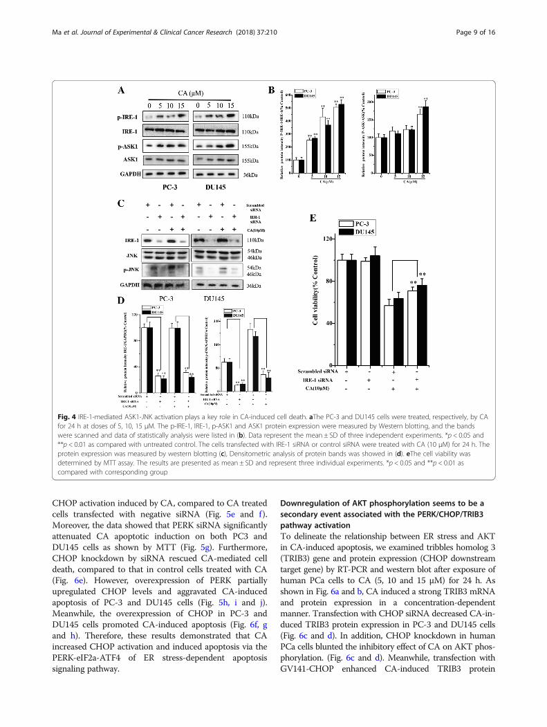
IRE-1-mediated ASK1-JNK activation plays a key role inCA-induced cell deathActivated ER stress induces expression of phospho-JNK,which mediates apoptotic signals. Therefore, to evaluate
whether CA induces ER stress-mediated apoptosis inhuman PCa cells, IRE-1-ASK1-JNK signaling branch ofER stress was investigated in the present research. Resultsin Fig. 4a and b suggested that CA treatment significantlyincreased the phosphorylation of both IRE-1 and ASK1 inboth cell types. However, no significant change in totalIRE-1 and ASK1 expression was observed in PC3 andDU145 cells. IRE-1 recruits ASK1 and leads to JNK activa-tion, which is implicated in ER stress-induced cell death.To validate whether IRE-1 was activated after CA treat-ment and further induced phospho-JNK (a downstreamtarget of IRE-1), we assessed the effect of IRE-1 silencingon CA-mediated activation of JNK. As shown in Fig. 4c, dand e, IRE-1 knockdown resulted in a significant decrease
Fig. 2 Effect of CA on mitochondria dependent apoptosis in human prostate cancer cells. a Mitochondrial membrane potentials (ΔΨm) wereassessed with JC-1 staining by flow cytometry. b The expression of cell key apoptosis-related proteins was detected by Western-blotting and theywere statistically analyzed (c). The PC-3 and DU145 cells was pre-treated with caspase inhibitor (10 μM Z-VAD-FMK) for 3 h and followed byincubation with or without CA for 24 h, respectively. d The cell viability was determined by MTT assay. The results are presented as mean ± SDand described as column chart *p < 0.05 and **p < 0.01 as compared with corresponding group. e Effect of CA induced-Cytochrome c releasewas analyzed by immunofluorescence staining
Ma et al. Journal of Experimental & Clinical Cancer Research (2018) 37:210 Page 7 of 16

in JNK activation and inhibition in the decline of cell via-bility after CA treatment. Taken together, these resultssuggested that CA induced apoptosis was partly mediatedby one of the signaling pathways in ER stress, such asIRE-1-mediated ASK1-JNK activation.
CA activates the PERK-eIF2a-ATF4 pathway leading to thepro-apoptotic CHOP up-regulation in human PCa cellsThe transcription factor CHOP is a key mediator in ERstress–induced apoptosis. Excessive or aberrant ERstress results in CHOP activation to induce cell injury ordeath. Therefore, we assessed whether CA could activateanother route of ER stress-induced apoptosis byPERK-eIF2a-ATF4-CHOP signal pathway. The resultsshowed that Bip, p-PERK, p-eIF2α, and ATF4 proteinexpression, related to ER stress signaling markers, wassignificantly up-regulated at 24 h after CA treatment in
PC-3 and DU145 cells. In addition, no significant differ-ence was found in the total PERK and eIF2α. More im-portantly, an increased expression of the transcriptionfactor CHOP was observed (Fig. 5a and b). Similar toprotein expression, RT-PCR results showed that theamount of CHOP mRNAs was increased 1.33 ± 0.25,6.38 ± 1.62, and 14.34 ± 2.52 -fold in PC-3 as well as1.47 ± 0.36, 5.20 ± 1.83 and 8.64 ± 1.65 -fold in DU145cells at 5, 10 and 15 μM CA, respectively (Fig. 5c). Inaddition, CHOP localization in human PCa cells wasevaluated by immunofluorescence, revealing that CAinduced a strong CHOP expression in cytoplasm andnucleus (Fig. 5a). In order to further verify that ER stressinduced-CHOP plays an important role in the inductionof apoptosis by CA, PERK and CHOP were silencedusing siRNA. Transfection with PERK siRNA led to asignificant decrease of total PERK levels and inhibited
Fig. 3 MAPK and AKT signaling pathways are involved in CA apoptotic effect on human PCa cells. MAPKs (a) and AKT (d) protein expressionswere analyzed using western blot. Densitometry scanning analysis for ratio of p-JNK/total JNK, p-P38/total P38 and p-ERK1/2/total ERK1/2 (b) aswell as p-AKT/total AKT (e). Data represent the mean ± SD of three independent experiments. *p < 0.05 and **p < 0.01 as compared withuntreated control. c Effects of SP600125, SB203580 and LY294002 (f) CA-induced apoptosis in cultured cells for 24 h. Cell viability was determinedby MTT assay and data represent the mean ± SD of six experiments in each group. *p < 0.05, **p < 0.01 as compared with single CA treated cells
Ma et al. Journal of Experimental & Clinical Cancer Research (2018) 37:210 Page 8 of 16
CHOP activation induced by CA, compared to CA treatedcells transfected with negative siRNA (Fig. 5e and f).Moreover, the data showed that PERK siRNA significantlyattenuated CA apoptotic induction on both PC3 andDU145 cells as shown by MTT (Fig. 5g). Furthermore,CHOP knockdown by siRNA rescued CA-mediated celldeath, compared to that in control cells treated with CA(Fig. 6e). However, overexpression of PERK partiallyupregulated CHOP levels and aggravated CA-inducedapoptosis of PC-3 and DU145 cells (Fig. 5h, i and j).Meanwhile, the overexpression of CHOP in PC-3 andDU145 cells promoted CA-induced apoptosis (Fig. 6f, gand h). Therefore, these results demonstrated that CAincreased CHOP activation and induced apoptosis via thePERK-eIF2a-ATF4 of ER stress-dependent apoptosissignaling pathway.
Downregulation of AKT phosphorylation seems to be asecondary event associated with the PERK/CHOP/TRIB3pathway activationTo delineate the relationship between ER stress and AKTin CA-induced apoptosis, we examined tribbles homolog 3(TRIB3) gene and protein expression (CHOP downstreamtarget gene) by RT-PCR and western blot after exposure ofhuman PCa cells to CA (5, 10 and 15 μM) for 24 h. Asshown in Fig. 6a and b, CA induced a strong TRIB3 mRNAand protein expression in a concentration-dependentmanner. Transfection with CHOP siRNA decreased CA-in-duced TRIB3 protein expression in PC-3 and DU145 cells(Fig. 6c and d). In addition, CHOP knockdown in humanPCa cells blunted the inhibitory effect of CA on AKT phos-phorylation. (Fig. 6c and d). Meanwhile, transfection withGV141-CHOP enhanced CA-induced TRIB3 protein
Fig. 4 IRE-1-mediated ASK1-JNK activation plays a key role in CA-induced cell death. aThe PC-3 and DU145 cells were treated, respectively, by CAfor 24 h at doses of 5, 10, 15 μM. The p-IRE-1, IRE-1, p-ASK1 and ASK1 protein expression were measured by Western blotting, and the bandswere scanned and data of statistically analysis were listed in (b). Data represent the mean ± SD of three independent experiments. *p < 0.05 and**p < 0.01 as compared with untreated control. The cells transfected with IRE-1 siRNA or control siRNA were treated with CA (10 μM) for 24 h. Theprotein expression was measured by western blotting (c), Densitometric analysis of protein bands was showed in (d). eThe cell viability wasdetermined by MTT assay. The results are presented as mean ± SD and represent three individual experiments. *p < 0.05 and **p < 0.01 ascompared with corresponding group
Ma et al. Journal of Experimental & Clinical Cancer Research (2018) 37:210 Page 9 of 16

Fig. 5 (See legend on next page.)
Ma et al. Journal of Experimental & Clinical Cancer Research (2018) 37:210 Page 10 of 16

expression and further suppress AKT phosphorylation(Fig. 6f and g). Thus, evidence was also indicated thatTRIB3 was closely correlated with the CHOP levels andCHOP activation resulted in TRIB3 up-regulation to fur-ther suppress AKT activation. Overall, these data sug-gested that CA exerted anti-cancer effects via reduction ofAKT by increasing CHOP/TRIB3 expression, besides CAinduced ER-stress-CHOP dependent apoptosis.
CA retards tumor growth in a murine xenograft model byactivating ER stressTo determine whether CA slows tumor progression invivo, 2 × 10⁶ PC-3 cells were subcutaneously injectedinto the flanks of nude mice. We discovered that CAtreatment markedly attenuated tumor volume comparedwith the control group (Fig. 7a and b). Tumor weight inCA-treated mice (0.550 ± 0.090 g in the 10 mg/kg groupand 0.125 ± 0.028 in the 20 mg/kg group) was signifi-cantly less compared with the tumor in the vehicle con-trol mice (0.759 ± 0.070 g, Fig. 7c). Data indicated thatCA produced a significant inhibition of tumor growthwithout toxicity observed by mean body weight (Fig. 7d)and HE stained tissue sections of heart, liver, spleen, lungand kidney observed at submicroscopic level (Fig. 7e). Anincrease in TUNEL+ cells (green fluorescence) was foundin tumors of the CA-treated mice compared with that oftumors from the vehicle-treated mice (Fig. 7f). Westernblot revealed that CA treatment significantly increasedphosphorylated PERK and phosphorylated eIF2α levels,up-regulated ATF4, CHOP protein expression, andsubsequently activated CHOP downstream target TRIB3(Fig. 7g and h). Further results showed a decrease in theexpression of phosphorylated AKT in tumors, which wasmediated by increased TRIB3 after CA treatment (Fig. 7gand h). Moreover, CA treatment increased AKS1 and JNKphosphorylation due to activated IRE-1 (Fig. 7g and h).PC-3 xenografts/tumors immunohistochemistry showedthat CA induced an increase in ER stress-dependentCHOP expression as shown by immunohistochemistry.
Moreover, significant increase of JNK phosphorylationand cleaved caspase-3 was also found in tumor tissue sec-tion by immunohistochemistry (Fig. 7i). Consistent withwestern blot, remarkable suppression of AKT phosphoryl-ation was observed by immunohistochemistry in tumortissue section after CA treatment (Fig. 7i). The above datasuggested that CA exposure substantially suppressed PC-3xenograft tumor growth in vivo through regulation of ERstress-mediated apoptosis via activation of IRE1α andPERK signaling.
DiscussionTo our knowledge, the current research is the first oneproviding a preclinical rationale of the therapeutic effectof CA against human PCa, especially CRPC in vitro andin vivo, revealing the underlying molecular mechanisms.In this study, we showed that CA elicited dramatic
effects on the inhibition of cell proliferation and stimula-tion of cell apoptosis. JC-1 dye by flow cytometryrevealed that mitochondrial membrane potential wasindeed reduced in human PCa cells after CA treatment.In addition, our data showed that exposure of CA led tonegative effects on anti-apoptotic protein Bcl2 and posi-tive effects pro-apoptotic protein Bax levels, which arebiomarkers for mitochondrial apoptotic pathway. The in-crease of caspase-3 was confirmed with the increased cleav-age of PARP, suggesting that mitochondrion-dependentintrinsic apoptotic pathways participated in CA associatedcytotoxicity. CA-induced apoptosis was not completelyblocked by a caspase inhibitor when CA was used at highconcentration, suggesting that a caspase-independent path-way could be also involved. Therefore, the potential role ofCA in other cell death pathways needs further study.MAPKs expression is closely associated with various sig-
naling pathways critically involved in the regulation of cellproliferation and apoptosis [29]. CA treatment increasedthe induction of p-JNK in a concentration-dependentmanner and a JNK specific inhibitor prevented this effect.Although p-p38 protein was also increased after CA
(See figure on previous page.)Fig. 5 CA activates the PERK-eIF2a-ATF4 pathway leading to the pro-apoptotic CHOP up-regulation in human PCa cells. The PC-3 and DU145cells were treated with CA at the indicated concentrations (5, 10, 15 μM.) for 24 h. a the phosphorylation of PERK, eIF2α and expression levels ofBip, PERK, eIF2α, ATF4 and CHOP were measured by western blot using corresponding antibodies. b And Protein levels were quantified bygrayscale scan and the GAPDH was used as the loading control. c Total RNAs were extracted and subjected to real-time PCR analysis usingspecific primer sets for CHOP and GAPDH, and the data were normalized to GAPDH expression. The results are presented as mean ± SD andrepresent three individual experiments. *p < 0.05 and **p < 0.01 as compared with untreated control. d Effects of CA induced-CHOP activationanalyzed by immunofluorescence staining. The PC-3 and DU145 cells were transfected with PERK siRNA or control siRNA, respectively and treatedwith CA for 24 h. e The protein expression of PERK and CHOP was measured by western blotting. f Protein levels were quantified by grayscalescan and the GAPDH was used as the loading control. g The cell viability was determined by MTT assay. The results are presented as mean ± SDand represent three individual experiments. *p < 0.05 and **p < 0.01 as compared with corresponding group. h Overexpression of PERK in PC-3and DU145 cells that were treated with CA for 24 h. PERK, p-PERK and CHOP protein expressions were determined by western blot. i Proteinlevels were quantified by grayscale scan and the GAPDH was used as the loading control. j MTT assay demonstrated that PERK overexpressionaggravated CA-induced migration inhibition of cell viability in PC-3 and DU145. The results are presented as mean ± SD and represent threeindividual experiments. *p < 0.05 and **p < 0.01 as compared with corresponding group
Ma et al. Journal of Experimental & Clinical Cancer Research (2018) 37:210 Page 11 of 16

treatment, pre-treatment with p38 inhibitor showed noeffect on cell viability, indicating that p38 activation mightnot play a pivotal role in CA-induced cytotoxicity.According to the results showing JNK activation closelyrelated to apoptosis, we further study JNK upstream regu-lation. Previous researches demonstrated that variouscytotoxic drugs inducing JNK-depend cell death were
related to the generation of ROS, and ROS are upstreamJNK promoters. Nevertheless, CA treatments resulted in aweak green fluorescence and no significant difference withthe control group was observed in our experiment (datanot show). Therefore, JNK activation was not due to ROSaccumulation [30]. However, Ca2+ enhancement after CAtreatment suggested Ca2+ release into the cytosol from the
Fig. 6 Downregulation of AKT phosphorylation seems to be a secondary event associated with the PERK/CHOP/TRIB3 pathway activation. Geneand protein levels of TRIB3 in the PC-3 and DU145 cells were detected by RT-PCR (a) and western blot (b) after treatment with (5, 10, 15 μM). Thecells were transfected with CHOP siRNA or control siRNA, respectively and treated with CA for 24 h. c The protein expression of CHOP, TRIB3 andp-AKT was measured by western blotting. d Protein levels were quantified by grayscale scan and the GAPDH was used as the loading control.The results are presented as mean ± SD and represent three individual experiments. *p < 0.05 and **p < 0.01 as compared with correspondinggroup. eThe cell viability was determined by MTT assay. The results are presented as mean ± SD. *p < 0.05 and **p < 0.01 as compared withcorresponding group. f Overexpression of CHOP in PC-3 and DU145 cells that were treated with CA for 24 h. CHOP, AKT, p-AKT and TRIB3 proteinexpressions were determined by western blot. g Protein levels were quantified by grayscale scan and the GAPDH was used as the loadingcontrol. h MTT assay demonstrated that CHOP overexpression aggravated CA-induced inhibition of cell viability in PC-3 and DU145. The resultsare presented as mean ± SD and represent three individual experiments. *p < 0.05 and **p < 0.01 as compared with corresponding group
Ma et al. Journal of Experimental & Clinical Cancer Research (2018) 37:210 Page 12 of 16

ER, which is an important biomarker for ER stress [31](Additional file 5: Figure S2).Activation of JNK can in turn activate apoptotic pathways,
which could be the downstream factor in response to ERstressors. We further investigated the ER stress-dependentapoptosis signaling pathway, revealing that this processplays a key role in the anti-cancer effect of CA. IRE-1 pos-sesses kinase activity by IRE-1-TRAF2-ASK1-JNK, whichinduces mitochondrion-dependent intrinsic apoptotic path-way. In our study, we demonstrated that IRE-1 activation
induced by CA resulted in ASK1 phosphorylation and thenJNK phosphorylation, finally triggering to mitochondrion-dependent intrinsic apoptotic pathway. JNK activity inhib-ition using the inhibitor SP600125 could partially recoverthe cell viability. Transfection with IRE-1 siRNA resulted inthe suppression of phosphorylated JNK and abolishing theapoptotic effect induced by CA, further supporting the roleof JNK signaling in ER stress.Besides activation of IRE-1-ASK1-JNK signaling in re-
sponse to CA treatment, CHOP, a transcriptional target
Fig. 7 CA retards tumor growth in a murine xenograft model by activating ER stress. Typical tumor tissue images after treatment was shown in(a). The tumor volume (b), weight (c) and body weight (d) were recorded after CA treatment for 14 days. e Histological analysis [stained with H&E200×] of normal tissue (heart, liver, spleen, lung and kidney) in mice induced by CA for 14 days. f In situ detection of apoptotic cells in tumorsections after the treatment of CA for 14 days by optical microscope using the TUNEL assay (apoptotic cell: green fluorescence). g The proteinexpression of tumor tissue was measured by western blotting. h Protein levels were quantified by grayscale scan and the GAPDH was used asthe loading control. i Effect of CA on p-JNK, CHOP, p-AKT and Cleaved caspase-3 expressions in male mice tumor sections were detected byimmunocytochemistry (positive cell: claybank). The results are presented as mean ± SD. *p < 0.05 and **p < 0.01 as compared with control group
Ma et al. Journal of Experimental & Clinical Cancer Research (2018) 37:210 Page 13 of 16

of ATF4, is a recognized indicator of ER stress-inducedapoptosis. Its accumulation induces apoptosis directly byrepressing the expression of the anti-apoptotic proteinBcl-2. Activation of CHOP was regulated by the up-stream PERK signaling pathway. The PERK/eIF2a/ATF4/CHOP route has been more widely explored and it playsa crucial role in executing ER stress-induced apoptosisin many anticancer agents [32–34]. However, the role ofthis pathway in CA-induced apoptosis in human PCacells remains unclear. In the present study, our resultsshowed that CA exposure increased Bip expression, amarker protein of ER stress, and then, activated PERK,eIF2a and ATF4 cascade process. Finally, CHOP, the ERstress-induced apoptosis mediator, was induced after CAtreatment. To confirm that CHOP activity occurred inresponse to ER stressors, PERK knockdown by siRNAwas performed, showing that it suppressed CHOP induc-tion by CA. CHOP knockdown resulted in a significantlyless apoptosis, indicating that CHOP is an important
mediator of CA-inducing apoptosis. CHOP-mediatedapoptosis induced by ER stressors occurs by the mito-chondrial apoptotic pathway [35]. Previous observationsby other laboratories indicated that Bim (pro-apoptoticprotein) is transcriptionally induced by CHOP and Bcl-2(anti-apoptotic protein) and was down-regulated at a tran-scriptional level by CHOP [36]. These studies suggestedthat CHOP could induce the pro-apoptotic protein andinhibit the anti-apoptotic protein located in the outermitochondrial membrane to induce apoptosis.In the present study, TRIB3 up-regulated expression
was observed in CA-treated cells. TRIB3, as a novel ERstress-inducible gene, is involved in cell death by induc-tion of ER stress. Its expression could be induced byCHOP, provoking a decrease in phosphorylation of AKT[37]. It is well established that abnormally active AKTplays a crucial role in cancer generation and progression.Previous studies have shown that administration of dif-ferent anticancer agents induces cancer cell death via
Fig. 8 Overview of pathways for CA induced apoptosis of PCa
Ma et al. Journal of Experimental & Clinical Cancer Research (2018) 37:210 Page 14 of 16

TRIB3 upregulation and subsequent inhibition of AKTactivation. M Salazar’s group has also shown that TRIB3deletion is closely associated with a more aggressive pheno-type in various tumor types by enhancing AKTactivity [38].The tumor inhibitory role of TRIB3 is connected to ERstress and AKT signaling pathway. Subsequently, thisdecrease in phospho- AKT caused by TRIB3 resulted in anenhanced Bax expression in the outer mitochondrial mem-brane to further promote apoptosis. Our data demonstratedthat CA exposure increased TRIB3 level and suppressp-AKT, and CHOP knockdown could partly downregulateTRIB3 expression and abolish p-AKT inhibition in vitro(Fig. 8). Therefore, according to the background and our re-sults, we could conclude that CHOP might be one of thecritical target proteins for CA anti-cancer action. On theone hand, CHOP activation directly led to Baxtranslocation into mitochondria, triggering intrinsicapoptosis. On the other hand, CA might induce apop-tosis by interfering with AKT activation throughERS-mediated up-regulation of CHOP/TRIB3.
ConclusionIn conclusion, here we showed for the first time that CA,as a potent antitumor agent, exhibited anti-cancer activityon human CRPC via induction of ER stress-dependentapoptotic signaling. CA-induced cell survival inhibitionwas partly regulated by activation of IRE-1-ASK1-JNKand PERK-CHOP-mediated ER stress, which subsequentlytriggered mitochondrion-dependent intrinsic apoptosis.The present study also presented evidences that the acti-vated CHOP regulated TRIB3 expression resulting inp-AKT inhibition, indirectly display extinction effect incell survival and cancer progression. This research eluci-dating CA anti-cancer effect and its mechanism mightprovide a support in the development of novel therapeuticstrategies against CRPC.
Additional files
Additional file 1: Table S1. Purchase and dilution condition of primaryantibodies and second antibodies. (DOCX 18 kb)
Additional file 2: Table S2. Primer sequences for PCR amplification.(DOC 31 kb)
Additional file 3: CA inhibited cell migration and invasion of humanPCa cells in vitro. (DOCX 1464 kb)
Additional file 4: Figure S1. Morphology of apoptotic cells wasevaluated by fluorescence microscopy following Hoechst 33258 stainingat 200 ×magnification. (TIF 1097 kb)
Additional file 5: Figure S2. Intracellular calcium concentration after CAtreatment for 12 h. (TIF 615 kb)
AbbreviationsASK1: Apoptosis signal regulating kinase 1α; ATF4: Activating transcriptionfactor 4; Bip: Binding immunoglobulin protein; CA: Corosolic acid;CHOP: CCAAT-enhancer-binding protein homologous protein;CRPC: castration-resistant prostate cancer; eIF2a: Eukaryotic initiation factor-
2a; ER Stress: Endoplasmic reticulum stress; IRE-1: Inositolrequiring enzyme 1;JNK: c-jun N-terminal kinase; MAPK: Mitogen-activated protein kinase;PCa: Prostate cancer; PERK: Protein kinase RNA-like endoplasmic reticulumkinase; TRIB3: Tribbles homolog 3; TUNEL: Terminal dexynucleotidyltransferase(TdT)-mediated dUTP nick end labeling
FundingThis study was financially supported by the National Natural ScienceFoundation of China (grant numbers 81773802 and 81672508); The JiangsuSynergetic Innovation Center for Advanced Bio-Manufacture (XTD1819); TheJiangsu Province “Qing-Lan project” for excellent-young-backbone teacher.
Availability of data and materialsAll data analyzed during this study are included in this manuscript.
Authors’ contributionsBM, HZ, YW, AZ, ZZ, XB and YS performed the experiments; QZ and BMrevised the manuscript. BM, LL and YW conceived the ideal of the study,analyzed the data and wrote the manuscript. All authors read and approvedthe final manuscript.
Ethics approval and consent to participateFor the animal study, all procedures performed on the animals in this studywere approved (NO. NJTECH-AE-2017006) the Guidelines for the AnimalEthics Committee of Nanjing Tech University.
Consent for publicationNot applicable.
Competing interestsThe authors declare that they have no competing interests.
Publisher’s NoteSpringer Nature remains neutral with regard to jurisdictional claims inpublished maps and institutional affiliations.
Received: 18 May 2018 Accepted: 17 August 2018
References1. Fallowfield L, Payne H, Jenkins V. Patient-reported outcomes in metastatic
castration-resistant prostate cancer. Nat Rev Clin Oncol. 2016;13(10):643–50.2. Litwin MS, Tan HJ. The diagnosis and treatment of prostate Cancer: a
review. JAMA. 2017;317(24):2532–42.3. Kallifatidis G, Hoy JJ, Lokeshwar BL. Bioactive natural products for
chemoprevention and treatment of castration-resistant prostate cancer.Semin Cancer Biol. 2016;40-41:160–9.
4. Ritch CR, Cookson MS. Advances in the management of castration resistantprostate cancer. BMJ. 2016;355:i4405.
5. Chevet E, Hetz C, Samali A. Endoplasmic reticulum stress-activated cellreprogramming in oncogenesis. Cancer Discov. 2015;5(6):586–97.
6. Ron D, Walter P. Signal integration in the endoplasmic reticulum unfoldedprotein response. Nat Rev Mol Cell Biol. 2007;8(7):519–29.
7. Oakes SA, Papa FR. The role of endoplasmic reticulum stress in humanpathology. Annu Rev Pathol. 2015;10:173–94.
8. Yamamoto K, Ichijo H, Korsmeyer SJ. BCL-2 is phosphorylated andinactivated by an ASK1/Jun N-terminal protein kinase pathway normallyactivated at G(2)/M. Mol Cell Biol. 1999;19(12):8469–78.
9. Yang Y, Liu L, Naik I, Braunstein Z, Zhong J, Ren B. Transcription factor C/EBPhomologous protein in health and diseases. Front Immunol. 2017;8:1612.
10. Lamoureux F, Thomas C, Yin MJ, Fazli L, Zoubeidi A, Gleave ME. Suppressionof heat shock protein 27 using OGX-427 induces endoplasmic reticulumstress and potentiates heat shock protein 90 inhibitors to delay castrate-resistant prostate cancer. Eur Urol. 2014;66(1):145–55.
11. Wang M, Shim JS, Li RJ, Dang Y, He Q, Das M, et al. Identification ofan old antibiotic clofoctol as a novel activator of unfolded proteinresponse pathways and an inhibitor of prostate cancer. Br J Pharmacol.2014;171(19):4478–89.
12. Storm M, Sheng X, Arnoldussen YJ, Saatcioglu F. Prostate cancer and theunfolded protein response. Oncotarget. 2016;7(33):54051–66.
Ma et al. Journal of Experimental & Clinical Cancer Research (2018) 37:210 Page 15 of 16

13. Firczuk M, Gabrysiak M, Barankiewicz J, Domagala A, Nowis D, Kujawa M, etal. GRP78-targeting subtilase cytotoxin sensitizes cancer cells tophotodynamic therapy. Cell Death Dis. 2013;4:e741.
14. Song JH, Kraft AS. Pim kinase inhibitors sensitize prostate cancer cells toapoptosis triggered by Bcl-2 family inhibitor ABT-737. Cancer Res. 2012;72(1):294–303.
15. Gafar AA, Draz HM, Goldberg AA, Bashandy MA, Bakry S, Khalifa MA, et al.Lithocholic acid induces endoplasmic reticulum stress, autophagy andmitochondrial dysfunction in human prostate cancer cells. PeerJ. 2016;4:e2445.
16. Wang Q, Sun Q, Ma X, Rao Z, Li H. Probing the binding interaction ofhuman serum albumin with three bioactive constituents of Eriobotrtajaponica leaves: spectroscopic and molecular modeling approaches. JPhotochem Photobiol B. 2015;148:268–76.
17. Ahn KS, Hahm MS, Park EJ, Lee HK, Kim IH. Corosolic acid isolated from thefruit of Crataegus pinnatifida var. psilosa is a protein kinase C inhibitor aswell as a cytotoxic agent. Planta Med. 1998;64(5):468–70.
18. Ku CY, Wang YR, Lin HY, Lu SC, Lin JY. Corosolic acid inhibits hepatocellularcarcinoma cell migration by targeting the VEGFR2/Src/FAK pathway. PLoSOne. 2015;10(5):e0126725.
19. Sivakumar G, Vail DR, Nair V, Medina-Bolivar F, Lay JO. Plant-based corosolicacid: future anti-diabetic drug. Biotechnol J. 2009;4(12):1704–11.
20. Shi L, Zhang W, Zhou YY, Zhang YN, Li JY, Hu LH, et al. Corosolic acidstimulates glucose uptake via enhancing insulin receptor phosphorylation.Eur J Pharmacol. 2008;584(1):21–9.
21. Fujiwara Y, Komohara Y, Ikeda T, Takeya M. Corosolic acid inhibits glioblastomacell proliferation by suppressing the activation of signal transducer andactivator of transcription-3 and nuclear factor-kappa B in tumor cells andtumor-associated macrophages. Cancer Sci. 2011;102(1):206–11.
22. Nho KJ, Chun JM, Kim HK. Corosolic acid induces apoptotic cell deathin human lung adenocarcinoma A549 cells in vitro. Food Chem Toxicol.2013;56:8–17.
23. Xu Y, Ge R, Du J, Xin H, Yi T, Sheng J, et al. Corosolic acid induces apoptosisthrough mitochondrial pathway and caspase activation in human cervixadenocarcinoma HeLa cells. Cancer Lett. 2009;284(2):229–37.
24. Horlad H, Fujiwara Y, Takemura K, Ohnishi K, Ikeda T, Tsukamoto H, et al.Corosolic acid impairs tumor development and lung metastasis byinhibiting the immunosuppressive activity of myeloid-derived suppressorcells. Mol Nutr Food Res. 2013;57(6):1046–54.
25. Cheng QL, Li HL, Li YC, Liu ZW, Guo XH, Cheng YJ. CRA(Crosolic acid)isolated from Actinidia valvata Dunn.Radix induces apoptosis of humangastric cancer cell line BGC823 in vitro via down-regulation of the NF-κBpathway. Food Chem Toxicol. 2017;105:475–85.
26. Kim JH, Kim YH, Song GY, Kim DE, Jeong YJ, Liu KH, et al. Ursolic acid andits natural derivative corosolic acid suppress the proliferation of APC-mutated colon cancer cells through promotion of β-catenin degradation.Food Chem Toxicol. 2014;67:87–95.
27. Wang Y, Guo SH, Shang XJ, Yu LS, Zhu JW, Zhao A, et al. Triptolide inducesSertoli cell apoptosis in mice via ROS/JNK-dependent activation of themitochondrial pathway and inhibition of Nrf2-mediated antioxidantresponse. Acta Pharmacol Sin. 2018;39(2):311–27.
28. Lien JC, Huang CC, Lu TJ, Tseng CH, Sung PJ, Lee HZ, et al.Naphthoquinone derivative PPE8 induces endoplasmic reticulum stress inp53 null H1299 cells. Oxidative Med Cell Longev. 2015;2015:453679.
29. Chen F. JNK-induced apoptosis, compensatory growth, and cancer stemcells. Cancer Res. 2012;72(2):379–86.
30. Dai X, Wang L, Deivasigamni A, Looi CY, Karthikeyan C, Trivedi P, et al. Anovel benzimidazole derivative, MBIC inhibits tumor growth and promotesapoptosis via activation of ROS-dependent JNK signaling pathway inhepatocellular carcinoma. Oncotarget. 2017;8(8):12831–42.
31. Merlot AM, Shafie NH, Yu Y, Richardson V, Jansson PJ, Sahni S, et al.Mechanism of the induction of endoplasmic reticulum stress by the anti-cancer agent, di-2-pyridylketone 4,4-dimethyl-3-thiosemicarbazone(Dp44mT): activation of PERK/eIF2α, IRE1α, ATF6 and calmodulin kinase.Biochem Pharmacol. 2016;109:27–47.
32. Cao A, Li Q, Yin P, Dong Y, Shi H, Wang L, et al. Curcumin inducesapoptosis in human gastric carcinoma AGS cells and colon carcinoma HT-29 cells through mitochondrial dysfunction and endoplasmic reticulumstress. Apoptosis. 2013;18(11):1391–402.
33. Liu X, Guo S, Liu X, Su L. Chaetocin induces endoplasmic reticulum stressresponse and leads to death receptor 5-dependent apoptosis in humannon-small cell lung cancer cells. Apoptosis. 2015;20(11):1499–507.
34. Zhang B, Han H, Fu S, Yang P, Gu Z, Zhou Q, et al. Dehydroeffusol inhibitsgastric cancer cell growth and tumorigenicity by selectively inducingtumor-suppressive endoplasmic reticulum stress and a moderate apoptosis.Biochem Pharmacol. 2016;104:8–18.
35. Logue SE, Cleary P, Saveljeva S, Samali A. New directions in ER stress-induced cell death. Apoptosis. 2013;18(5):537–46.
36. Iurlaro R, Muñoz-Pinedo C. Cell death induced by endoplasmic reticulumstress. FEBS J. 2016;283(14):2640–52.
37. English BC, Van Prooyen N, Örd T, Örd T, Sil A. The transcription factorCHOP, an effector of the integrated stress response, is required for hostsensitivity to the fungal intracellular pathogen Histoplasma capsulatum.PLoS Pathog. 2017;13(9):e1006589.
38. Salazar M, Lorente M, García-Taboada E, Pérez GE, Dávila D, Zúñiga-García P,et al. Loss of tribbles pseudokinase-3 promotes Akt-driven tumorigenesis viaFOXO inactivation. Cell Death Differ. 2015;22(1):131–44.
Ma et al. Journal of Experimental & Clinical Cancer Research (2018) 37:210 Page 16 of 16