correlating local compositions and structures with …ceweb/faculty/bradc/pdfs...correlating local...
TRANSCRIPT

Correlating Local Compositions and Structures with the MacroscopicOptical Properties of Ce3+-Doped CaSc2O4, an Efficient Green-Emitting PhosphorNathan C. George,†,‡ Jakoah Brgoch,‡,# Andrew J. Pell,§,∇ Clayton Cozzan,‡ Adam Jaffe,‡,○
Geraldine Dantelle,∥ Anna Llobet,⊥ Guido Pintacuda,§ Ram Seshadri,*,‡ and Bradley F. Chmelka*,†
†Department of Chemical Engineering and ‡Materials Department and Materials Research Laboratory, University ofCaliforniaSanta Barbara, Santa Barbara, California 93106, United States§Centre de RMN a Tres Hauts Champs, Universite de Lyon, Institute of Analytical Sciences, CNRS/Ecole Normale Superieure deLyon/Universite Claude Bernard Lyon 1, 5 Rue de la Doua, 69100 Villeurbanne, France∥Institut Neel CNRS, UPR 2940/Universite Grenoble Alpes, 25 Avenue des Martyrs, Grenoble, 38 042 Cedex 09France⊥Lujan Neutron Scattering Center, Los Alamos National Laboratory, Los Alamos, New Mexico 87545, United States
*S Supporting Information
ABSTRACT: Calcium scandate (CaSc2O4) substituted with small amounts (<1%) ofCe3+ is a recently discovered bright-green-emitting phosphor with favorable lightabsorption and emission properties and robust temperature stability that make it well-suited for solid-state white-lighting applications. Combined analyses of scattering, solid-state nuclear magnetic resonance (NMR), electron paramagnetic resonance (EPR), andphotoluminescence measurements establish the compositional and structural origins ofthe macroscopic optical properties of this phosphor material. Simultaneous refinementsof synchrotron X-ray and neutron diffraction data of Ce3+-doped CaSc2O4 enable theaverage crystal structure to be determined, which is shown to correspond to anexceedingly rigid host structure, as corroborated by density functional theory (DFT)calculations. Such structural rigidity leads to high quantum efficiency, which isoptimized by the substitution of as little as 0.5 mol % of Ce3+ for Ca2+ ions, with higherextents of Ce3+ substitution leading to decreased photoluminescent quantum yields.Solid-state 43Ca and 45Sc magic-angle spinning (MAS) NMR spectra are sensitive to theeffects of the paramagnetic Ce3+ dopant ions on nearby atoms in the host structure and yield evidence for local structuraldistortions. EPR measurements provide direct insights on structures of the Ce3+ ions, as a function of Ce3+ substitution. Thecombined scattering and spectroscopic analyses yield detailed new understanding of the local and long-range structures of Ce3+-doped CaSc2O4, which account for the sensitive composition-dependent optical properties of this important phosphor material.
■ INTRODUCTION
The advantages of solid-state lighting (SSL) over conventionallight sources include mercury-free materials, physically robustdevices, long lifetimes, and high efficiencies.1,2 SSL devicesgenerally yield white light from a blue or near-UV light-emittingdiode (LED) by using an inorganic phosphor, such as Ce3+-doped Y3Al5O12 (yttrium−aluminum−garnet, YAG) or calciumscandate (CaSc2O4), to partially down-convert blue or UV LEDemission to longer wavelengths; the combined emissionappears as white light. Several design and performance criteriagovern the selection of phosphor materials for SSL applications.Ideal materials have high quantum efficiencies and goodchemical stabilities, as well as excitation and emission bandsthat are compatible with commercially available LEDs.3 Highefficiencies, even at elevated temperatures (≈500 K), makethem suitable for high-power lighting devices.4 Currently, themost widely investigated compound that meets these designcriteria for white-light phosphors is cerium-substituted YAG
(YAG:Ce3+).5 Although progress has been made in thedevelopment of this and other oxide phosphors for SSL,phosphors with outstanding optical properties are still sought,and the atomic-level origins of their properties remain poorlyunderstood.5 To complement the general criteria for highperformance described above, additional design rules are soughtthat will enable the identification of new candidate phosphorswith improved photoluminescence properties.The pursuit of thermally stable phosphors with high
quantum yields has recently focused on materials with densepolyhedral bonding networks.6 Structures with dense packingof polyhedral units tend to have rigid bonding networks thatprevent nonradiative relaxation from excited states (e.g., viavibrational relaxation). One phosphor that fulfills the design
Received: December 20, 2016Revised: March 20, 2017Published: April 11, 2017
Article
pubs.acs.org/cm
© 2017 American Chemical Society 3538 DOI: 10.1021/acs.chemmater.6b05394Chem. Mater. 2017, 29, 3538−3546
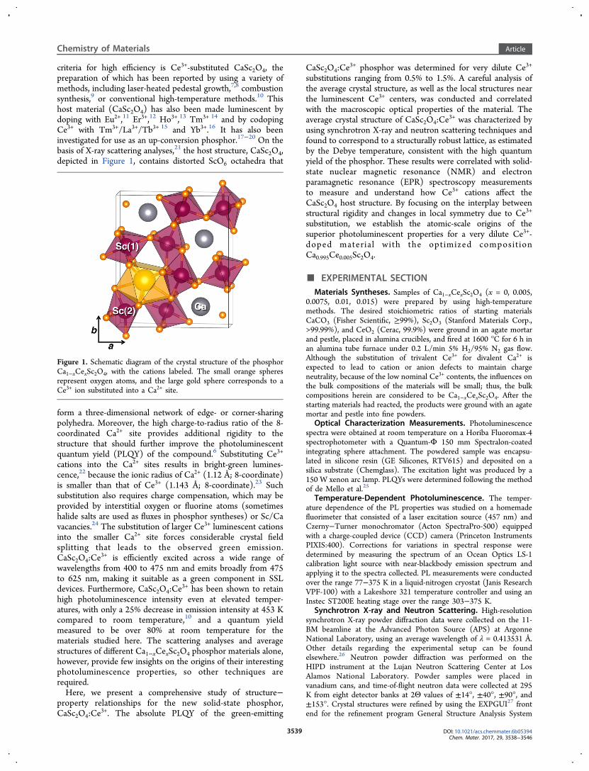
criteria for high efficiency is Ce3+-substituted CaSc2O4, thepreparation of which has been reported by using a variety ofmethods, including laser-heated pedestal growth,7,8 combustionsynthesis,9 or conventional high-temperature methods.10 Thishost material (CaSc2O4) has also been made luminescent bydoping with Eu2+,11 Er3+,12 Ho3+,13 Tm3+ 14 and by codopingCe3+ with Tm3+/La3+/Tb3+ 15 and Yb3+.16 It has also beeninvestigated for use as an up-conversion phosphor.17−20 On thebasis of X-ray scattering analyses,21 the host structure, CaSc2O4,depicted in Figure 1, contains distorted ScO6 octahedra that
form a three-dimensional network of edge- or corner-sharingpolyhedra. Moreover, the high charge-to-radius ratio of the 8-coordinated Ca2+ site provides additional rigidity to thestructure that should further improve the photoluminescentquantum yield (PLQY) of the compound.6 Substituting Ce3+
cations into the Ca2+ sites results in bright-green lumines-cence,22 because the ionic radius of Ca2+ (1.12 Å; 8-coordinate)is smaller than that of Ce3+ (1.143 Å; 8-coordinate).23 Suchsubstitution also requires charge compensation, which may beprovided by interstitial oxygen or fluorine atoms (sometimeshalide salts are used as fluxes in phosphor syntheses) or Sc/Cavacancies.24 The substitution of larger Ce3+ luminescent cationsinto the smaller Ca2+ site forces considerable crystal fieldsplitting that leads to the observed green emission.CaSc2O4:Ce
3+ is efficiently excited across a wide range ofwavelengths from 400 to 475 nm and emits broadly from 475to 625 nm, making it suitable as a green component in SSLdevices. Furthermore, CaSc2O4:Ce
3+ has been shown to retainhigh photoluminescence intensity even at elevated temper-atures, with only a 25% decrease in emission intensity at 453 Kcompared to room temperature,10 and a quantum yieldmeasured to be over 80% at room temperature for thematerials studied here. The scattering analyses and averagestructures of different Ca1−xCexSc2O4 phosphor materials alone,however, provide few insights on the origins of their interestingphotoluminescence properties, so other techniques arerequired.Here, we present a comprehensive study of structure−
property relationships for the new solid-state phosphor,CaSc2O4:Ce
3+. The absolute PLQY of the green-emitting
CaSc2O4:Ce3+ phosphor was determined for very dilute Ce3+
substitutions ranging from 0.5% to 1.5%. A careful analysis ofthe average crystal structure, as well as the local structures nearthe luminescent Ce3+ centers, was conducted and correlatedwith the macroscopic optical properties of the material. Theaverage crystal structure of CaSc2O4:Ce
3+ was characterized byusing synchrotron X-ray and neutron scattering techniques andfound to correspond to a structurally robust lattice, as estimatedby the Debye temperature, consistent with the high quantumyield of the phosphor. These results were correlated with solid-state nuclear magnetic resonance (NMR) and electronparamagnetic resonance (EPR) spectroscopy measurementsto measure and understand how Ce3+ cations affect theCaSc2O4 host structure. By focusing on the interplay betweenstructural rigidity and changes in local symmetry due to Ce3+
substitution, we establish the atomic-scale origins of thesuperior photoluminescent properties for a very dilute Ce3+-doped mater ia l with the optimized composit ionCa0.995Ce0.005Sc2O4.
■ EXPERIMENTAL SECTIONMaterials Syntheses. Samples of Ca1−xCexSc2O4 (x = 0, 0.005,
0.0075, 0.01, 0.015) were prepared by using high-temperaturemethods. The desired stoichiometric ratios of starting materialsCaCO3 (Fisher Scientific, ≥99%), Sc2O3 (Stanford Materials Corp.,>99.99%), and CeO2 (Cerac, 99.9%) were ground in an agate mortarand pestle, placed in alumina crucibles, and fired at 1600 °C for 6 h inan alumina tube furnace under 0.2 L/min 5% H2/95% N2 gas flow.Although the substitution of trivalent Ce3+ for divalent Ca2+ isexpected to lead to cation or anion defects to maintain chargeneutrality, because of the low nominal Ce3+ contents, the influences onthe bulk compositions of the materials will be small; thus, the bulkcompositions herein are considered to be Ca1−xCexSc2O4. After thestarting materials had reacted, the products were ground with an agatemortar and pestle into fine powders.
Optical Characterization Measurements. Photoluminescencespectra were obtained at room temperature on a Horiba Fluoromax-4spectrophotometer with a Quantum-Φ 150 mm Spectralon-coatedintegrating sphere attachment. The powdered sample was encapsu-lated in silicone resin (GE Silicones, RTV615) and deposited on asilica substrate (Chemglass). The excitation light was produced by a150 W xenon arc lamp. PLQYs were determined following the methodof de Mello et al.25
Temperature-Dependent Photoluminescence. The temper-ature dependence of the PL properties was studied on a homemadefluorimeter that consisted of a laser excitation source (457 nm) andCzerny−Turner monochromator (Acton SpectraPro-500) equippedwith a charge-coupled device (CCD) camera (Princeton InstrumentsPIXIS:400). Corrections for variations in spectral response weredetermined by measuring the spectrum of an Ocean Optics LS-1calibration light source with near-blackbody emission spectrum andapplying it to the spectra collected. PL measurements were conductedover the range 77−375 K in a liquid-nitrogen cryostat (Janis ResearchVPF-100) with a Lakeshore 321 temperature controller and using anInstec ST200E heating stage over the range 303−375 K.
Synchrotron X-ray and Neutron Scattering. High-resolutionsynchrotron X-ray powder diffraction data were collected on the 11-BM beamline at the Advanced Photon Source (APS) at ArgonneNational Laboratory, using an average wavelength of λ = 0.413531 Å.Other details regarding the experimental setup can be foundelsewhere.26 Neutron powder diffraction was performed on theHIPD instrument at the Lujan Neutron Scattering Center at LosAlamos National Laboratory. Powder samples were placed invanadium cans, and time-of-flight neutron data were collected at 295K from eight detector banks at 2Θ values of ±14°, ±40°, ±90°, and±153°. Crystal structures were refined by using the EXPGUI27 frontend for the refinement program General Structure Analysis System
Figure 1. Schematic diagram of the crystal structure of the phosphorCa1−xCexSc2O4, with the cations labeled. The small orange spheresrepresent oxygen atoms, and the large gold sphere corresponds to aCe3+ ion substituted into a Ca2+ site.
Chemistry of Materials Article
DOI: 10.1021/acs.chemmater.6b05394Chem. Mater. 2017, 29, 3538−3546
3539

(GSAS).28 Simultaneous refinements to the X-ray and neutronscattering data were completed by adjusting the profile shapes andunit cells during LeBail fits; refining neutron absorption coefficients,instrument parameters, and the backgrounds (10-term shifted-Chebyshev polynomial function); and then refining the atomicpositions and finally the atomic displacement parameters. During thefinal refinement cycle, all appropriate free parameters were allowed torefine simultaneously. Crystal structures were visualized using thesoftware VESTA.29
Solid-State NMR Spectroscopy. High-resolution solid-state 43Caand 45Sc NMR spectroscopy was used to investigate the localcompositions and structural environments of calcium and scandiumatoms in the Ca1−xCexSc2O4 materials. Solid-state
45Sc (nuclear spin I= 7/2, 100% natural abundance) NMR spectra were acquired on aBruker Advance III 1 GHz spectrometer at 23.5 T, operating at a 45ScLarmor frequency of 243 MHz, with a 1.3 mm HX probehead underconditions of magic-angle spinning (MAS) at 60 kHz. One-dimensional (1D) 45Sc NMR spectra were acquired by directexcitation of 45Sc nuclei by using a one-pulse sequence with anexcitation pulse of 44.6 kHz radio frequency (rf) field amplitude and1.4 μs duration. Spin−lattice (T1) relaxation times were measured byusing a saturation-recovery experiment comprised of a saturationsequence of 50 pulses separated by delays of 3 ms and a variablerecovery delay taking 20 values from 10 ms to 60 s. Quantitative 1Dspectra were acquired using a one-pulse sequence with 64 scans andrecycle delays of 54.4 s (x = 0), 53.6 s (x = 0.005), 48.7 s (x = 0.010),and 43.5 s (x = 0.015). All 45Sc shifts were referenced to a 1 Maqueous solution of Sc(NO3)3. Additional
45Sc NMR spectra wereacquired with a higher repetition rate to filter 45Sc signals based ontheir T1 relaxation times. For such spectra, 32 768 transients wereacquired with a recycle delay of 100 ms to enhance the signals fromfaster-relaxing 45Sc nuclei in close proximity to paramagnetic Ce3+
cations, which therefore exhibit shorter T1 values. Two-dimensional(2D) 45Sc MQMAS spectra30 were acquired by using a triple-quantum(3Q) pulse sequence with a z-filter.31 The 3Q excitation pulse and thepulse used to convert triple-quantum coherence back to z-magnet-ization had rf field amplitudes of 141 kHz and were applied for 1.5 and0.5 μs, respectively. The final low-power 90° pulse had a 44.6 kHz rffield amplitude and a duration of 1.4 μs. The time increment in theindirect dimension was 50 ms, 64 increments were acquired using theStates−Haberkorn−Rubens method to obtain pure-phase line shapesand frequency discrimination,32 and 96 transients were acquired per t1increment. The data were sheared in the time domain duringprocessing.33 Spectra were acquired without active temperaturecontrol for all materials, resulting in an estimated sample temperatureof 360 K. To assess the temperature-dependence of the paramagneti-cally displaced 45Sc signals, a spectrum of one material (x = 0.015) wasacquired by using a variable-temperature Bruker BCU-X cooling unitwith a N2 flow of 1335 L/h and a set-point of 243 K, resulting in anestimated sample temperature of 300 K under the fast MAS (60 kHz)conditions used. Solid-state NMR spectra were modeled and the NMRparameters were extracted by using the simulation program DMFIT.34
Solid-state 43Ca (nuclear spin I = 7/2, 0.145% natural abundance)NMR spectra were acquired at the U.S. National High Magnetic FieldLaboratory in Tallahassee, FL, on a narrow-bore Magnex 19.6 Tmagnet with a Bruker console, operating at a 43Ca Larmor frequencyof 56.067 MHz. The measurements were collected at roomtemperature under MAS conditions of 6 kHz using a Samoson 7mm HX triple-resonance MAS probehead with zirconia rotors andKel-F caps. Radio frequency pulses with a 1.5 μs length were used witha recycle delay of 5 s, and 8000 transients were acquired. All 43Ca shiftswere referenced to 1 M CaCl2 (aq).Electron Paramagnetic Resonance (EPR) Spectroscopy. EPR
spectra were collected on a Bruker X-Band EPR spectrometer (ν =9.486 GHz) equipped with a helium flow cryostat. Samples wereplaced in silica tubes, and spectra were acquired at 4 K and with 6309mW microwave power. Spectral simulations employed the EASYSPINcode implemented in MATLAB,35 with a correction included for field-dependent relaxation effects.
■ RESULTS AND DISCUSSIONThe substitution of low concentrations of rare-earth cations,e.g., Ce3+, into calcium scandate (CaSc2O4) leads to materialswith efficient photoluminescent properties. This arises, in part,due to the larger atomic radius of Ce3+, compared to the smallerCa2+ cation, leading to significant crystal field splitting. Theresult is a shift of the absorption bands to longer wavelengthswhen CaSc2O4:Ce
3+ is excited between 400 and 475 nm(maximum intensity at λmax = 455 nm), as manifested by a smallStokes shift of ≈60 nm and emission across the green-yellowregion of the visible spectrum (λmax = 515 nm), as shown inFigure 2a. The photoluminescence quantum yield in the
Ca1−xCexSc2O4 system can be optimized by varying the extentof Ce3+ substitution over a narrow and dilute range, e.g., x = 0−0.015, leading to a high quantum yield of 85% when 0.5% ofCe3+ (x = 0.005) was substituted into calcium scandate (Figure2b, spectra in Supporting Information, Figure S2). Interestingly,the quantum yield decreased dramatically to 60% and 31% with
Figure 2. (a) Two-dimensional excitation and emission scans ofCa0.995Ce0.005Sc2O4 phosphor. The most intense excitation andemission spectra are shown above and beside the plot, respectively.(b) Plot of the PLQY measured at room temperature and λex = 445nm for Ca1−xCexSc2O4 phosphors with different Ce contents of x =0.005, 0.0075, 0.010, and 0.015. The uncertainty bars representdistributions over multiple measurements.
Chemistry of Materials Article
DOI: 10.1021/acs.chemmater.6b05394Chem. Mater. 2017, 29, 3538−3546
3540

substitution of modestly higher loadings of 1% Ce3+ and 1.5%Ce3+, respectively. Such an abrupt decrease in the PLQY withincreasing Ce3+ is likely to be due to either structuraldistortions arising from incorporation of the larger Ce3+ ionsinto smaller Ca2+ sites or the formation of anionic defectsstemming from charge imbalance; these both inhibit photo-emission and are consistent with the lower quantum yieldsobserved. The optical properties of these materials, further-more, exhibit high thermal stabilities, as manifested bytemperature-dependent photoluminescence spectra and anal-yses shown in Supporting Information (Figure S3).Average Crystal Structure and Optical Response of
CaSc2O4:Ce3+. As reported previously,21 the undoped CaSc2O4
host material has an orthorhombic crystal structure (Figure 1,space group Pnma) with one Ca2+ site, two Sc3+ sites, and fourO2− sites. The two Sc3+ ions are in the centers of distortedoctahedra with the Sc(1)O6 units and Sc(2)O6 units sharingedges. The substitution of Ce3+ into the CaSc2O4 crystalstructure occurs only at the Ca2+ site, due to size constraints.This result has similarly been observed by extended X-rayabsorption fine structure (EXAFS) measurements for theanalogous garnet system, Ca3Sc2Si3O12:Ce
3+.36
For calcium scandate containing low loadings of Ce3+ dopantions (<2% substitution), the bulk orthorhombic crystalstructure and space group are retained. For example, Figure 3shows representative synchrotron X-ray and neutron scatteringpatterns for the mater ia l with the composit ionCa0.995Ce0.005Sc2O4, all reflections of which are indexable tothe Pnma structure, indicating a single-phase product. Rietveldrefinements of these data support the previously determinedcrystal structure21 and provide general insights on the average
structure and thermal−mechanical properties of theCa1−xCexSc2O4 phosphor materials. For example, the synchro-tron X-ray scattering analysis (Figure 3a) allows the variation ofthe average unit cell parameters to be accurately determined, asfunctions of small amounts of substituent atoms, such as Ce3+.For the unsubstituted CaSc2O4 material, the unit cellparameters and fit quality indicators associated with Rietveldrefinement of synchrotron X-ray data (Table 1) agree well with
previous X-ray diffraction studies of CaSc2O4.21,37,38 The
substitution of Ce3+ induces a small (<1%) expansion of theunit cell that is consistent with 8-coordinate Ce3+ being slightlylarger than Ca2+. The unit cell parameters of Ca1−xCexSc2O4,monitored as a function of Ce3+ content over the range x = 0−0.01, suggest a maximum for Ce3+ substitution above x = 0.005,which could be due to a solid-solution limit or a chargeimbalance between Ca2+ and Ce3+, which may lead to theformation of defects.On the basis of the scattering data available, however, it is not
possible to determine the occupancies of Ce3+ ions within thephosphor structure, due to the low extents of substitution(≤1.5%) and the lack of long-range Ce3+ order.By comparison, the size of the atomic displacement
parameters, to which neutron scattering is particularly sensitive,have been recently shown to relate to the quantum efficiency ofa phosphor.4 Rietveld corefinements of the synchrotron X-rayscattering results (Figure 3a) with time-of-flight neutron data inFigure 3b enable accurate refinements of atomic displacementparameters (ADPs) of the CaSc2O4 host. The refined atomicpositions and ADPs for Ca0.995Ce0.005Sc2O4, shown in Table 2,indicate that the ADPs have magnitudes similar to those foundin the highly efficient phosphors like YAG:Ce3+ 4 and
Figure 3. Simultaneous Rietveld refinement of the (a) synchrotron X-ray scattering data and (b) time-of-flight neutron scattering dataacquired at 295 K for the phosphor Ca0.995Ce0.005Sc2O4. The expectedreflection positions are shown above the plots. The inset in panel a is apart of the crystal structure, depicted with 99% probability thermalellipsoids from the refinement. Note the exceedingly small ellipsoids,which reflect the rigidity of the structure. The same sample was used inFigures 2a, 5, and 7.
Table 1. Parameters from Combined Rietveld Refinementsof Ca1−xCexSc2O4 of Synchrotron X-ray and NeutronPowder Diffraction Data
x 0 0.005 0.01crystal system orthorhombicspace group; Z Pnma; 4
temp 295 Ka (Å) 9.46337(1) 9.463453(9) 9.463452(9)b (Å) 11.11158(1) 11.11176(1) 11.11176(1)c (Å) 3.141928(4) 3.142051(3) 3.142052(3)V (Å3) 330.3831(7) 330.4043(6) 330.4043(6)
X-ray Rwp (%) 10.5 8.17 8.16neutron Rwp (%) 1.56 1.58 1.59
Table 2. Atomic Positions and Atomic Displacement Factors(Uiso) Refined from a Combined Rietveld Refinement ofSynchrotron X-ray and Neutron Powder Diffraction Data forCa0.995Ce0.005Sc2O4
a
atom occupancy x y Z Uiso × 100
Ca 0.995 0.75651(8) 0.65237(6) 1/4 0.37(1)Ce 0.005 0.75651(8) 0.65237(6) 1/4 0.37(1)Sc(1) 1 0.41954(4) 0.10734(3) 1/4 0.724(7)Sc(2) 1 0.42962(3) 0.61213(3) 1/4 0.425(6)O(1) 1 0.20759(6) 0.17154(5) 1/4 0.20(1)O(2) 1 0.12316(5) 0.47756(5) 1/4 0.16(1)O(3) 1 0.52058(7) 0.78409(5) 1/4 0.20(1)O(4) 1 0.42058(7) 0.42464(5) 1/4 0.12(1)
aThe occupancy ratio Ca2+/Ce3+ was held constant.
Chemistry of Materials Article
DOI: 10.1021/acs.chemmater.6b05394Chem. Mater. 2017, 29, 3538−3546
3541

La3Si6N11:Ce3+.39 This suggests that the CaSc2O4 host also
provides an especially rigid matrix that is consistent with theinterconnected, edge-sharing ScO6 polyhedra. From the ADPs,an estimation of the Debye temperature (ΘD), which correlatesto a material’s photoluminescent quantum yield,6 can becalculated.4,39 For CaSc2O4, ΘD is estimated to be around 900K from the neutron data. The surprisingly high ΘD predictedhere may arise from the correlation between the strong neutronabsorption of Sc3+ and the Rietveld refinements of the atomicdisplacement parameters in the method, which can lead to anoverestimation of the ADP values. On the other hand, even therefined ADPs from X-ray diffraction, which is relativelyinsensitive to light atoms, such as oxygen, still leads to highDebye temperatures ranging between 766 and 790 K for thissystem. As a result, it reasonable to estimate that ΘD for thisphosphor host ranges between 700 and 900 K, which issubstantially higher than for other high-efficiency phosphors,such as YAG:Ce3+ 4 and La3Si6N11:Ce
3+.39
The high Debye temperature ΘD of CaSc2O4 is corroboratedby density functional theory (DFT), which provides a robustapproximation based on the elastic constants (Cij), as has beenpreviously shown.6 Here, the elastic constants for theunsubstituted CaSc2O4 are calculated by using the Vienna abinitio simulation package (VASP)40,41 employing the Perdew−Burke−Ernzerhof generalized gradient approximation (PBE-GGA) to describe exchange and correlation.42 The Debyetemperature from DFT is predicted to be 694 K, which is inagreement with the range of ΘD determined experimentally.The combination of experimental and computational resultssupports the rigidity of the calcium scandate host structure thatleads to its high PLQY.6
Local Structures near Ce3+ Dopants. The opticalproperties of rare-earth-substituted compounds are stronglyaffected by their local coordination environments, which resultin different crystal field splittings.3 However, because thesubstitution of the rare-earth ions occurs at such a low loadings(often <3%), any local distortions that occur are challenging toobserve by scattering analyses of long-range order. Bycomparison, solid-state NMR is sensitive to local bondingenvironments and is also element-selective. For the case ofCa1−xCexSc2O4, the effects of even dilute quantities of Ce3+
incorporated into the calcium scandate structure can be probedby using solid-state 45Sc and 43Ca NMR to detect displacementsof signals and reduced spin−lattice relaxation times that areinduced by nearby paramagnetic Ce3+. Such effects have beenpreviously used to characterize local compositions andstructures near paramagnetic species in pyrochlores,43
stannates,44 garnets,4 and lanthanide-binding proteins.45
Starting first with the undoped parent material, the twocrystallographically distinct Sc(1) and Sc(2) scandium sites inCaSc2O4 are clearly distinguished in 2D 45Sc NMR measure-ments conducted at very high magnetic field (23.5 T).Although the two signals overlap in a conventional 1D 45ScMAS spectrum (Supporting Information, Figure S1, consistentwith similar analyses46 conducted at lower field, 11.7 T), theyare well-resolved in the 2D z-filtered 45Sc triple-quantum (3Q)MAS NMR spectrum in Figure 4. This spectrum was acquiredunder very-high field conditions (23.5 T), which mitigatecontributions from anisotropic second-order quadrupolarinteractions that are associated with the spin I = 7/2
45Scnuclei, yielding resolved signals from the two distinct 45Sc sites.The 2D 45Sc MQ-MAS spectrum correlates anisotropic (MAS)and isotropic spectra, from which the isotropic 45Sc chemical
shift (δiso), the quadrupolar coupling constant (CQ), and thequadrupolar asymmetry parameter (ηQ) were extracted for eachof the two 45Sc sites by fits of each of the signals in theirrespective isotropic and anisotropic dimensions. These results(which are summarized in Table 3) enable the assignments of
the two signals to scandium sites Sc(1) and Sc(2) and provideinsights on the local coordination environments of the 45Scatoms in CaSc2O4. Specifically, site Sc(1) is assigned to thelower frequency 45Sc signal with an isotropic chemical shift of154 ppm and CQ and ηQ values of 8.8 MHz and 0.50,respectively. The second 45Sc signal with the isotropic chemicalshift of 157 ppm, a larger CQ of 14.3 MHz, and ηQ of 0.76, isassigned to Sc(2). The larger CQ value is consistent with theless symmetric local bonding environment of Sc(2), in line withthe scattering analyses (Figure 3 and Table 1). The minordeviation of the fitted line shape near the center (ca. 147 ppm)of the experimental spectrum for Sc(2) (slice ii) is associatedwith uncertainty in the fitted value of ηQ and reflects well-
Figure 4. Solid-state 45Sc z-filtered triple-quantum MAS NMRspectrum of CaSc2O4 acquired at 23.5 T under MAS conditions of60 kHz and at approximately 360 K. Projections are shown along thecorresponding anisotropic and isotropic axes. Horizontal slices i and iiare extracted from the isotropic dimension, as indicated by the orangedashed lines. Fits to the extracted spectra are shown as red lines. Thesample is the same as those used in Figures 5 and 6.
Table 3. 45Sc Isotropic Chemical Shifts, QuadrupolarCoupling Constants, and Asymmetry Parameters for theTwo Scandium Sites in Crystalline CaSc2O4
a
atom δiso (ppm) CQ (MHz) ηQ
Sc(1) 154 ± 0.5 8.8 ± 0.1 0.50 ± 0.1Sc(2) 157 ± 0.5 14.3 ± 0.1 0.76 ± 0.15
aTable values were determined from fits to slices in the isotropicdimension of the 45Sc MQ-MAS spectrum in Figure 4.
Chemistry of Materials Article
DOI: 10.1021/acs.chemmater.6b05394Chem. Mater. 2017, 29, 3538−3546
3542

known difficulties in obtaining precise values of this parameter.Nevertheless, the singularities at the edges of the line shape,which are associated with the value of CQ, are reproduced witha high degree of precision. These results are also consistentwith the larger bond-angle variance of 78° for site Sc(2),compared with 29° for Sc(1), as determined by Rietveldrefinement of the scattering data above.The incorporation of Ce3+ ions into the calcium scandate
structure influences the local environments of nearby scandiumsites, even for low extents of Ce3+ substitution. This is evidentin the solid-state 1D 45Sc MAS NMR spectra acquired at 360 Kin Figure 5 for Ca1−xCexSc2O4 with Ce3+ substitutions for Ca2+
cations in the range of 0−1.5%. The 45Sc MAS spectrum for theundoped CaSc2O4 (x = 0) material exhibits a complicated lineshape in Figure 5a (top), because it contains the twooverlapping central transition signals that were resolved inthe 2D 45Sc MQ-MAS spectrum (Figure 4), as well as twosignals due to the center bands of the satellite-transitionsspinning sideband manifolds that are also associated with eachof the two scandium sites. As small quantities of Ce3+ areincorporated into Ca1−xCexSc2O4, new weak 45Sc signals appearat approximately 177 and 205 ppm, both of which increase inintensity with the extent of Ce3+ substitution and which areassigned to scandium sites Sc(1) and Sc(2), respectively, on thebasis of their relative shifts. The 45Sc signal near 177 ppmappears to partially overlap with the satellite transition at ca.174 ppm, the latter’s intensity and position of which remain
constant with Ce3+ substitution and temperature. It isnoteworthy that the positions of the signals at 177 and 205ppm exhibit marked temperature dependences and are furtherdisplaced to 180 and 208 ppm, respectively, at 300 K (bluetrace in Figure 5b). The displacements of the signals to higherchemical shifts at lower temperatures are consistent with theinverse temperature dependence of paramagnetic shiftsassociated with the Fermi contact and spin-dipolar (pseudo-contact) interactions of the 45Sc nuclei with the nearbyparamagnetic Ce3+ cations.44,47 In addition, the 45Sc spin−lattice relaxation time (T1) associated with the nondisplaceddominant signal at ca. 150 ppm decreases monotonically from11 to 8.7 s as the Ce3+ substitution for Ca2+ increases from 0%to 1.5%. This is consistent with an induced paramagneticrelaxation enhancement48 of the 45Sc polarization, due to long-range dipolar couplings to distant (>1 nm) paramagnetic Ce3+
cations. The widths of the nondisplaced 45Sc peaks also increasewith Ce3+ substitution, reflecting (1) increased local distortionsin the lattice, (2) an increase in the anisotropic bulk magneticsusceptibility broadening,49 and (3) the decrease in the overalltransverse relaxation time constant associated with theincreased Ce3+ content. More importantly, the 45Sc T1 valueassociated with the weak resolved signal at 205 ppm wasmeasured to be approximately 140 ms, which is almost 2 ordersof magnitude shorter than for the 45Sc sites in bulk undopedCaSc2O4. Although a reliable quantitative fit of the T1 of thepeak at 177 ppm is more difficult to obtain, due to the overlapwith the satellite transition, we estimate an approximate valueof 200−300 ms. These substantially shorter T1 values areconsistent with paramagnetic relaxation-enhancement effectsthat arise from the short-range interactions of 45Sc atoms withnearby paramagnetic Ce3+ ions that increase with dopantloading.The incorporation of Ce3+ dopant ions into Ca1−xCexSc2O4
materials is also evident in the effects on nearby unsubstitutedCa2+ sites. As shown in Figure 6a, the 1D solid-state 43Ca MASNMR spectrum of undoped CaSc2O4 reveals a single relativelynarrow signal at 62 ppm with a full width at half-maximum(fwhm) of 3 ppm. This indicates the presence of a single well-
Figure 5. (a) Solid-state single-pulse 45Sc MAS NMR spectra ofCa1−xCexSc2O4 with different Ce3+ compositions (x) acquired at 23.5T and 60 kHz MAS, with a recycle delay of 0.1 s, at approximately 360K. (b) The data are shown with a vertical expansion of the spectra by afactor of 100. Signals from satellite transitions are marked withasterisks (see Supporting Information, Figure S1), and paramagneti-cally displaced peaks are indicated with “+” symbols. The effect oftemperature on the paramagnetic displacement at 300 K is highlightedby dashed lines as a guide for the eye. The samples are the same asthose used in Figures 2, 3, 4, 6, and 7.
Figure 6. Solid-state single-pulse 43Ca MAS NMR spectra ofCa1−xCexSc2O4 phosphor materials with different Ce3+ dopantcompositions acquired at room temperature at 19.6 T, 7 kHz MAS,and with a 5 s recycle delay: (a) undoped CaSc2O4 and (b)Ca0.99Ce0.01Sc2O4. The samples are the same as those used in Figures 4and 5.
Chemistry of Materials Article
DOI: 10.1021/acs.chemmater.6b05394Chem. Mater. 2017, 29, 3538−3546
3543

defined Ca2+ site, which is consistent with the long-rangecrystalline order of the CaSc2O4 parent material. For the Ce3+-doped materials, an additional feature appears at approximately70 ppm. The large displacement of this new signal with respectto the main peak is a consequence of long-range spin−dipolarinteractions of the 43Ca nuclei with nearby paramagnetic Ce3+
dopant species, and its broader line width (ca. 12 ppm fwhm) isconsistent with a progressively weaker paramagnetic effectsensed by 43Ca2+ cations in successive coordination spheres.Given the extremely long T1 of the
43Ca2+ nuclei, both thesesignals were saturated during the NMR experiment, preventingquantitative analysis of their relative intensities.Ce3+ Local Structure from EPR. Although NMR can
indirectly provide details on local structural features of atomsnear the Ce3+ cations, electron paramagnetic resonance (EPR)spectroscopy probes the paramagnetic Ce3+ species directly.The splitting of the fundamental 2F5/2 electron energy levels bythe crystal field reduces the spin system to an effective S = 1/2spin system with an anisotropic g factor. EPR spectra are shownin Figure 7 for several otherwise identical Ce3+-doped calcium
scandates (Ca1−xCexSc2O4), except for their different extents ofCe3+ substitution (x = 0.005, 0.01, 0.015). In each of the EPRspectra, the vast majority of the intensity (90%) is associatedwith one type of Ce3+ site with rhombic symmetry that yieldsthree signal components at g1x = 2.70 (2500 G), g1y = 1.62(4100 G), and g1z = 1.33 (5100 G). This range of g tensors issmaller than those reported for YAG:Ce3+ (gx = 2.73 and gz =0.908)4 and reflects smaller crystal-field splittings in theCa1−xCexSc2O4 materials. Interestingly, a second Ce3+ site
also appears to be present, corresponding to three much weakerCe3+ signals at g2x = 2.38 (2800 G), g2y = 2.07 (3100 G), and g2z= 1.40 (4100 G) that account for 9% of the total signalintensity. This second Ce3+ signal may arise from Ce3+ ions inor near distorted Ca2+ sites, which would be consistent with thebroad 43Ca signal at ca. 70 ppm in the 43Ca MAS NMRspectrum of Figure 6b. Figure 7a shows a fit to the EPRspectrum acquired for the material with the compositionCa0.995Ce0.005Sc2O4, corresponding to the phosphor with thesuperior photoluminescence properties. The dashed red line inFigure 7a is a summation of weighted contributions of the twoindividual fits shown and is in good agreement with theexperimental spectrum, indicating that most of the features inthe spectrum are accounted for by the two types of Ce3+ sites inthe doped calcium scandate structure. A small spectral featureremains near 4800 G. Because the synchrotron X-ray powderdiffraction data did not indicate the presence of significantcrystalline impurity phases, this may be from an additionaldistorted Ce3+ site or associated with a minor amount ofdisordered or defect species. As the concentration of Ce3+
increases, this feature near 4800 G becomes more prominent,as shown in Figure 7b; however, attempts to fit this signal byincluding a third Ce3+ site were unsuccessful. Nevertheless, asdistortions occur with Ce3+ incorporation, the probability ofnonradiative relaxation increases and can explain the rapiddecrease in quantum efficiency with increased Ce3+ content.
■ CONCLUSIONS
Cerium-doped CaSc2O4 has been shown to be a phosphor thatpossesses a rigid host crystal structure, as manifested by its highDebye temperature (>700 K), based on scattering measure-ments and DFT calculations. The high structural rigidity iscorrelated to the high photoluminescent quantum yield of over80% for Ca0.995Ce0.005Sc2O4 (0.5% Ce3+ substitution for Ca2+),as well as the temperature stability of the luminescenceemission. Interestingly, the quantum yield is very sensitive tothe extent of cerium incorporation, dropping abruptly to 31%,as the Ce3+ substitution approaches 1.5%. Variable-temperaturesolid-state 45Sc NMR measurements establish that the decreasein quantum yield is correlated with paramagnetic displacementsof 45Sc signals that are associated with scandium sites Sc(1) andSc(2) near very dilute paramagnetic Ce3+ dopant ions inCa1−xCexSc2O4. Direct EPR measurements of the Ce3+ ionsreveal two types of Ce3+ sites, one relatively ordered site thataccounts for approximately 90% of the Ce3+ ions in the inCa1−xCexSc2O4 materials and the other corresponding to ∼9%of more distorted Ce3+ sites. The EPR spectra indicate that thisfraction of distorted Ce3+ sites increases with Ce3+ loading andcorrelates well with the decrease in quantum efficiency for Ce3+
substitutions above 0.5%. Overall, the combined scattering,solid-state 45Sc and 43Ca NMR, EPR, and DFT analyses yield adetailed new understanding of the structure−propertycorrelations that are associated with the technologicallypromising green-emitting phosphor Ca0.995Ce0.005Sc2O4. Theresults provide insights and design criteria that are expected tobe useful for the discovery, optimization, and development ofnovel and efficient phosphor materials for optoelectronics orsolid-state lighting applications.
Figure 7. (a) Powder EPR spectrum (black solid line) andaccompanying fit (red dashed line) for the phosphor compositionCa0.995Ce0.005Sc2O4 (x = 0.005), with evidence of two distinct Ce3+
sites: site 1 (light blue) and site 2 (dark blue). (b) EPR spectraacquired at 4 K and 9.4 GHz of Ce3+ in Ca1−xCexSc2O4 for x = 0.005,0.01, and 0.015. The samples are the same as those used in Figures 2,3, 5, and 6.
Chemistry of Materials Article
DOI: 10.1021/acs.chemmater.6b05394Chem. Mater. 2017, 29, 3538−3546
3544

■ ASSOCIATED CONTENT*S Supporting InformationThe Supporting Information is available free of charge on theACS Publications website at DOI: 10.1021/acs.chemma-ter.6b05394.
First-principles calculation methods, solid-state single-pulse 45Sc MAS NMR spectra for CaSc2O4 (without Cedopant species), the emission profiles measured fordifferent Ca1−xCexSc2O4 materials with different ceriumcontents (x = 0.005, 0.0075, 0.01, and 0.015), thet em p e r a t u r e - d e p e n d e n t P L s p e c t r a f o rCa1.995Ce0.005Sc2O4 (PDF)
■ AUTHOR INFORMATIONCorresponding Authors*R.S. e-mail: [email protected].*B.F.C. e-mail: [email protected] Brgoch: 0000-0002-1406-1352Adam Jaffe: 0000-0002-9886-0249Ram Seshadri: 0000-0001-5858-4027Bradley F. Chmelka: 0000-0002-4450-6949Present Addresses#J.B.: Department of Chemistry, University of Houston,Houston, Texas 77204, U.S.A.∇A.J.P.: Department of Materials and Environmental Chem-istry, Arrhenius Laboratory, Stockholm University, SvanteArrhenius Vag 16 C, SE-106 91 Stockholm, Sweden○A.J.: Department of Chemistry, Stanford University, Stanford,California 94305, U.S.A.NotesThe authors declare no competing financial interest.
■ ACKNOWLEDGMENTSThe authors thank J. Siewenie for assistance with collection ofthe neutron powder diffraction data and Dr. Z. Gan forassistance with the 43Ca NMR measurements. The work at theENS-Lyon was supported by the Initial Training Network onParamagnetic NMR funded by the European Union’s SeventhFramework Program for research, technological development,and demonstration (FP7-PEOPLE-2012-ITN contract n.317127 “pNMR”). N.C.G. acknowledges support from theConvEne IGERT Program of the U.S. National ScienceFoundation (NSF-DGE 0801627). A.J.P. and G.P. weresupported by the LABEX iMUST (ANR-10-LABX-0064) ofthe Universite de Lyon, within the program “Investissementsd’Avenir” (ANR-11-IDEX-0007) operated by the AgenceNationale de la Recherche (ANR). The research madeextensive use of the Central Facilities of the UCSB MaterialsResearch Laboratory, supported by the MRSEC Program of theNSF under Award DMR-1121053, which is a member of theNSF-funded Materials Research Facilities Network (www.mrfn.org). Use of the Advanced Photon Source at Argonne NationalLaboratory was supported by the U.S. Department of Energy,Office of Science, Office of Basic Energy Sciences, underContract No. DE-AC02-06CH11357. This work benefited fromthe use of the HIPD beamline at the Lujan Center at LosAlamos National Laboratory, funded by the DOE Office ofBasic Energy Sciences; LANL is operated by Los AlamosNational Security LLC under DE-AC52-06NA25396. Thesolid-state NMR measurements at 23.5 T were conducted at
the Centre Europeen de Resonance Magnetique Nucleaire a Tres Hauts Champs in Lyon, France. Solid-state 43Ca NMRmeasurements were conducted at the U.S. National HighMagnetic Field Laboratory, Tallahassee, FL, supported by theU.S. NSF. B.F.C. thanks the ENS-Lyon for support as a“Professeur Invite” in 2013.
■ REFERENCES(1) Service, R. F. The Quest for White LEDs Hits the Home Stretch.Science 2009, 325, 809−809.(2) Pimputkar, S.; Speck, J. S.; DenBaars, S. P.; Nakamura, S.Prospects for LED lighting. Nat. Photonics 2009, 3, 180−182.(3) George, N. C.; Denault, K. A.; Seshadri, R. Phosphors for Solid-State White Lighting. Annu. Rev. Mater. Res. 2013, 43, 481−501.(4) George, N. C.; Pell, A. J.; Dantelle, G.; Page, K.; Llobet, A.;Balasubramanian, M.; Pintacuda, G.; Chmelka, B. F.; Seshadri, R. LocalEnvironments of Dilute Activator Ions in the Solid-State LightingPhosphor Y3-xCexAl5O12. Chem. Mater. 2013, 25, 3979−3995.(5) Hoppe, H. A. Recent Developments in the Field of InorganicPhosphors. Angew. Chem., Int. Ed. 2009, 48, 3572−3582.(6) Brgoch, J.; DenBaars, S. P.; Seshadri, R. Proxies from ab-initioCalculations for Screening Efficient Ce3+ Phosphor Hosts. J. Phys.Chem. C 2013, 117, 17955−17959.(7) Philippen, J.; Guguschev, C.; Bertram, R.; Klimm, D. Laser-Heated Pedestal Growth of Cerium Doped Calcium Scandate CrystalFibers. J. Cryst. Growth 2013, 363, 270−276.(8) Klimm, D., Philippen, J.; Markurt, T.; Kwasniewski, A.Ce3+:CaSc2O4 Crystal Fibers for Green Light Emission: GrowthIssues and Characterization. MRS Online Proc. Libr. 2014, 1655, DOI:10.1557/opl.2014.365(9) Peng, W.; Zou, S.; Liu, G.; Xiao, Q.; Meng, J.; Zhang, R.Combustion Synthesis and Upconversion Luminescence of CaS-c2O4:Yb
3+,Er3+ Nanopowders. J. Rare Earths 2011, 29, 330−334.(10) Chen, Y.; Cheah, K. W.; Gong, M. Low TemperatureQuenching and High Efficiency Tm3+, La3+ or Tb3+ Co-DopedCaSc2O4:Ce
3+ Phosphors for Light-Emitting Diodes. J. Lumin. 2011,131, 1770−1775.(11) Zhijun, W.; Zhi, T.; Jianqi, Y.; Xian, S.; Shuai, Q.; Yamin, L.;Zhiping, Y.; Panlai, L.; Zhenu, X. Recent Development on Single-Phase White Emitting Phosphors for White LEDs. J. Chin. Ceram. Soc.2016, 44, 1.(12) Georgescu, S.; Stefan, A.; Toma, O. Judd−Ofelt and Energy-Transfer Analysis of Er3+ Doped in CaSc2O4 Ceramic Samples. J.Lumin. 2015, 167, 186−192.(13) Georgescu, S.; Stefan, A.; Toma, O.; Voiculescu, A.-M. Judd−Ofelt Analysis of Ho3+ Doped in Ceramic CaSc2O4. J. Lumin. 2015,162, 174−179.(14) Georgescu, S.; Stefan, A.; Voiculescu, A.-M.; Toma, O. Judd−Ofelt Analysis of Tm3+ doped in CaSc2O4 Ceramic Samples. J. Lumin.2015, 166, 130−136.(15) Chen, Y.; Cheah, K. W.; Gong, M. Low TemperatureQuenching and High Efficiency Tm3+, La3+ or Tb3+ Co-DopedCaSc2O4:Ce
3+ Phosphors for Light-Emitting Diodes. J. Lumin. 2011,131, 1770−1775.(16) Li, J.; Hao, Z.; Zhang, X.; Zhang, L.; Luo, Y.; Zhang, J.; Chen, L.Efficient Near-Infrared Downconversion and Energy TransferMechanism of Ce3+/Yb3+ Codoped Calcium Scandate Phosphor.Inorg. Chem. 2015, 54, 4806−4810.(17) Li, J.; Zhang, J.; Hao, Z.; Zhang, X.; Zhao, J.; Luo, Y.Spectroscopic Properties and Upconversion Studies in Ho3+/Yb3+ Co-doped Calcium Scandate with Spectrally Pure Green Emission.ChemPhysChem 2013, 14, 4114−4120.(18) Li, J.; Zhang, J.; Hao, Z.; Chen, L.; Zhang, X.; Luo, Y. IntenseUpconversion Luminescence of CaSc2O4:Ho3+/Yb3+ from LargeAbsorption Cross Section and Energy-Transfer Rate of Yb3+.ChemPhysChem 2015, 16, 1366−1369.
Chemistry of Materials Article
DOI: 10.1021/acs.chemmater.6b05394Chem. Mater. 2017, 29, 3538−3546
3545

(19) Li, J.; Zhang, J.; Hao, Z.; Zhang, X.; Zhao, J.; Luo, Y.Upconversion Properties and Dynamics Study in Tm3+ and Yb3+
Codoped CaSc2O4 Oxide Material. J. Appl. Phys. 2013, 113, 223507.(20) Li, J.; Zhang, J.; Hao, Z.; Zhang, X.; Zhao, J.; Lu, S.; Luo, Y.Synthesis, Morphology, and Upconversion Luminescence of Tm3+/Yb3+ Codoped Bulk and Submicro-Rod CaSc2O4 phosphors. Inorg.Chem. Commun. 2013, 38, 119−122.(21) Muller-Buschbaum, H.; Schnering, H. G. Uber Oxoscandate. I.Zur Kenntnis des CaSc2O4. Z. Anorg. Allg. Chem. 1965, 336, 295−305.(22) Shimomura, Y.; Kurushima, T.; Kijima, N. Photoluminescenceand Crystal Structure of Green-Emitting Phosphor CaSc2O4:Ce
3+. J.Electrochem. Soc. 2007, 154, J234−J238.(23) Shannon, R. D. Revised Effective Ionic Radii and SystematicStudies of Interatomic Distances in Halides and Chalcogenides. ActaCrystallogr., Sect. A: Cryst. Phys., Diffr., Theor. Gen. Crystallogr. 1976, 32,751−767.(24) Kalaji, A.; Mikami, M.; Cheetham, A. K. Ce3+-Activated γ-Ca2SiO4 and Other Olivine-Type ABXO4 Phosphors for Solid-StateLighting. Chem. Mater. 2014, 26, 3966−3975.(25) de Mello, J. C.; Wittmann, H. F.; Friend, R. H. An ImprovedExperimental Determination of External Photoluminescence Quantu-mEfficiency. Adv. Mater. 1997, 9, 230−232.(26) Wang, J.; Toby, B. H.; Lee, P. L.; Ribaud, L.; Antao, S. M.;Kurtz, C.; Ramanathan, M.; Von Dreele, R. B.; Beno, M. A. ADedicated Powder Diffraction Beamline at the Advanced PhotonSource: Commissioning and Early Operational Results. Rev. Sci.Instrum. 2008, 79, 085105.(27) Toby, B. H. EXPGUI, a Graphical User Interface for GSAS. J.Appl. Crystallogr. 2001, 34, 210−213.(28) Larson, A. C.; Dreele, R. B. V. General Structure Analysis System(GSAS); Los Alamos National Laboratory Report LAUR 86-7482000,2000.(29) Momma, K.; Izumi, F. VESTA 3 for Three-DimensionalVisualization of Crystal, Volumetric and Morphology data. J. Appl.Crystallogr. 2011, 44, 1272−1276.(30) Medek, A.; Harwood, J. S.; Frydman, L. Multiple-QuantumMagic-Angle Spinning NMR: A New Method for the Study ofQuadrupolar Nuclei in Solids. J. Am. Chem. Soc. 1995, 117, 12779−12787.(31) Amoureux, J.-P.; Fernandez, C.; Steuernagel, S. J. Z Filtering inMQMAS NMR. J. Magn. Reson., Ser. A 1996, 123, 116−118.(32) States, D. J.; Haberkorn, R. A.; Ruben, D. J. A Two-DimensionalNuclear Overhauser Experiment with Pure Absorption Phase in FourQuadrants. J. Magn. Reson. 1982, 48, 286−292.(33) Grandinetti, P. J.; Ash, J. T.; Trease, N. M. Symmetry Pathwaysin Solid-State NMR. Prog. Nucl. Magn. Reson. Spectrosc. 2011, 59, 121−196.(34) Massiot, D.; Fayon, F.; Capron, M.; King, I.; Le Calve, S.;Alonso, B.; Durand, J. O.; Bujoli, B.; Gan, Z.; Hoatson, G. ModellingOne and Two-Dimensional Solid-State NMR spectra. Magn. Reson.Chem. 2002, 40, 70−76.(35) Stoll, S.; Schweiger, A. EasySpin, a Comprehensive SoftwarePackage for Spectral Simulation and Analysis in EPR. J. Magn. Reson.2006, 178, 42−55.(36) Shimomura, Y.; Honma, T.; Shigeiwa, M.; Akai, T.; Okamoto,K.; Kijima, N. Photoluminescence and Crystal Structure of Green-Emitting Ca3Sc2Si3O12:Ce
3+ Phosphor for White Light EmittingDiodes. J. Electrochem. Soc. 2007, 154, J35−J38.(37) Carter, J. R.; Feigelson, R. S. Preparation and CrystallographicProperties of A2+B2
3+O4, Type Calcium and Strontium Scandates. J.Am. Ceram. Soc. 1964, 47, 141−144.(38) Horyn, R.; Lukaszewicz, K. B. Refinement of the CrystalStructure CaCsO4. Acad. Polym. Sci.-Chim. 1966, 499−504.(39) George, N. C.; Birkel, A.; Brgoch, J.; Hong, B.-C.; Mikhailovsky,A. A.; Page, K.; Llobet, A.; Seshadri, R. Average and Local StructuralOrigins of the Optical Properties of the Nitride PhosphorLa3‑xCexSi6N11(0<x ≤ 3). Inorg. Chem. 2013, 52, 13730−13741.(40) Kresse, G.; Hafner, J. Ab Initio Molecular Dynamics for LiquidMetals. Phys. Rev. B: Condens. Matter Mater. Phys. 1993, 47, 558−561.
(41) Kresse, G.; Furthmuller, J. Efficient Iterative Schemes for AbInitio Total-Energy Calculations Using a Plane-Wave Basis Set. Phys.Rev. B: Condens. Matter Mater. Phys. 1996, 54, 11169−11186.(42) Perdew, J. P.; Burke, K.; Ernzerhof, M. Generalized GradientApproximation Made Simple. Phys. Rev. Lett. 1996, 77, 3865−3868.(43) Cheetham, A. K.; Dobson, C. M.; Grey, C. P.; Jakeman, R. J. B.Paramagnetic Shift Probes in High-Resolution Solid-State NMR.Nature 1987, 328, 706−707.(44) Grey, C. P.; Dobson, C. M.; Cheetham, A. K.; Jakeman, R. J. B.Studies of Rare-Earth Stannates by 119Sn MAS NMR. The Use ofParamagnetic Shift Probes in the Solid State. J. Am. Chem. Soc. 1989,111, 505−511.(45) Pintacuda, G.; John, M.; Su, X. C.; Otting, G. NMR StructureDetermination of Protein−Ligand Complexes by Lanthanide Labeling.Acc. Chem. Res. 2007, 40, 206−212.(46) Brauniger, T.; Hofmann, A. J.; Moudrakovski, I. L.; Hoch, C.;Schnick, W. A 45Sc-NMR and DFT Calculation Study of CrystallineScandium Compounds. Solid State Sci. 2016, 51, 1−7.(47) Bleaney, B. Nuclear Magnetic Resonance Shifts in Solution Dueto Lanthanide Ions. J. Magn. Reson. 1972, 8, 91−100.(48) Pell, A. J.; Pintacuda, G. Broadband Solid-State MAS NMR ofParamagnetic Systems. Prog. Nucl. Magn. Reson. Spectrosc. 2015, 84−85, 33−72.(49) Alla, M.; Lippmaa, E. Resolution Limits in Magic-AngleRotation NMR Spectra of Polycrystalline Solids. Chem. Phys. Lett.1982, 87, 30−33.
Chemistry of Materials Article
DOI: 10.1021/acs.chemmater.6b05394Chem. Mater. 2017, 29, 3538−3546
3546

S1
SUPPORTING INFORMATION
Correlating local compositions and structures with the macroscopic optical properties of Ce3+‐doped CaSc2O4, an efficient green‐emitting phosphor
Nathan C. George,†,⊥ Jakoah Brgoch,⊥,€ Andrew J. Pell,‡,¶ Clayton Cozzan,⊥ Adam Jaffe,⊥,£
Géraldine Dantelle,§ Anna Llobet,$ Guido Pintacuda,‡ Ram Seshadri⊥,* Bradley F. Chmelka,†,* † Department of Chemical Engineering, University of California, Santa Barbara, California, 93106,
U.S.A.
⊥ Materials Department and Materials Research Laboratory, University of California, Santa Barbara, California, 93106, U.S.A.
‡ Centre de RMN à Très Hauts Champs, Université de Lyon, Institute of Analytical Sciences,
CNRS/Ecole Normale Supérieure de Lyon/Université Claude Bernard Lyon 1, 5 rue de la Doua, 69100 Villeurbanne, France
§ Institut Néel CNRS, UPR 2940/Université Grenoble Alpes, 25 Av. des Martyrs, 38 042 Grenoble
Cedex 09, France $ Lujan Neutron Scattering Center, Los Alamos National Laboratory, New Mexico 87545, U.S.A.
* To whom correspondence should be addressed: [email protected];
£ Present address: Department of Chemistry, Stanford University, Stanford, California 94305, U.S.A.
€ Present address: Department of Chemistry, University of Houston, Houston, Texas 77204, U.S.A.
¶ Present address: Department of Materials and Environmental Chemistry, Arrhenius Laboratory,
Stockholm University, Svante Arrhenius Väg 16 C, SE‐106 91 Stockholm, Sweden
S2
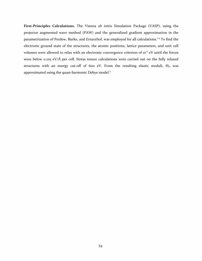
First‐Principles Calculations. The Vienna ab initio Simulation Package (VASP), using the
projector augmented wave method (PAW) and the generalized gradient approximation in the
parametrization of Perdew, Burke, and Ernzerhof, was employed for all calculations.1‐4 To find the
electronic ground state of the structures, the atomic positions, lattice parameters, and unit cell
volumes were allowed to relax with an electronic convergence criterion of 10‐5 eV until the forces
were below 0.015 eV/Å per cell. Stress tensor calculations were carried out on the fully relaxed
structures with an energy cut‐off of 600 eV. From the resulting elastic moduli, ΘD was
approximated using the quasi‐harmonic Debye model.5

S3
Supporting Figure S1. Solid‐state single‐pulse 45Sc MAS NMR spectra for CaSc2O4 (without Ce
dopant species), along with calculated spectra for the two distinct 45Sc sites, based on the CQ, Q,
and isotropic chemical shift values in Table 3 (which were obtained from lineshape fits to the 2D 45Sc MQ‐MAS spectrum in Figure 4. (a) The experimental 45Sc MAS spectrum, simulated
individual spectra of the central transitions for each of the respective 45Sc sites, and the overall
central‐transitions‐only simulated spectrum. Note that the two high frequency signals at ca. 160
and 170 ppm are absent from the simulated spectrum in (a). (b) The same as in (a), except that
the calculated spectra for the two 45Sc sites include contributions from the centerbands of their
respective satellite transitions, which account for the weak signals at ca. 160 and 170 ppm and that
therefore can be concluded to arise from second‐order quadrupolar interactions. The
experimental spectrum, which is the same in both (a) and (b), was acquired at 23.5 T and 60 kHz
MAS, with a recycle delay of 0.1 s, and at approximately 360 K; it is the same as shown in Figure
5a,b(top) of the main paper.
S4
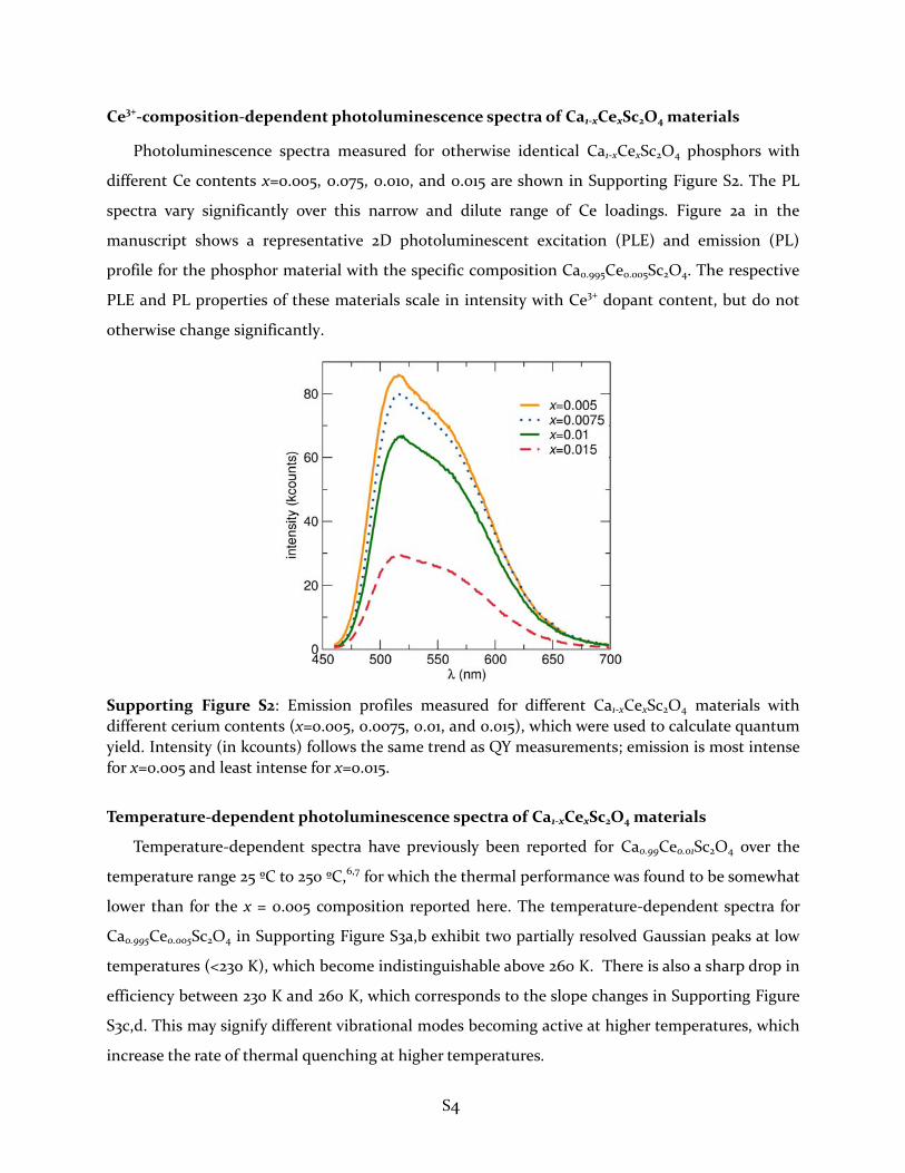
Ce3+‐composition‐dependent photoluminescence spectra of Ca1‐xCexSc2O4 materials
Photoluminescence spectra measured for otherwise identical Ca1‐xCexSc2O4 phosphors with
different Ce contents x=0.005, 0.075, 0.010, and 0.015 are shown in Supporting Figure S2. The PL
spectra vary significantly over this narrow and dilute range of Ce loadings. Figure 2a in the
manuscript shows a representative 2D photoluminescent excitation (PLE) and emission (PL)
profile for the phosphor material with the specific composition Ca0.995Ce0.005Sc2O4. The respective
PLE and PL properties of these materials scale in intensity with Ce3+ dopant content, but do not
otherwise change significantly.
Supporting Figure S2: Emission profiles measured for different Ca1‐xCexSc2O4 materials with
different cerium contents (x=0.005, 0.0075, 0.01, and 0.015), which were used to calculate quantum
yield. Intensity (in kcounts) follows the same trend as QY measurements; emission is most intense
for x=0.005 and least intense for x=0.015.
Temperature‐dependent photoluminescence spectra of Ca1‐xCexSc2O4 materials
Temperature‐dependent spectra have previously been reported for Ca0.99Ce0.01Sc2O4 over the
temperature range 25 ºC to 250 ºC,6,7 for which the thermal performance was found to be somewhat
lower than for the x = 0.005 composition reported here. The temperature‐dependent spectra for
Ca0.995Ce0.005Sc2O4 in Supporting Figure S3a,b exhibit two partially resolved Gaussian peaks at low
temperatures (<230 K), which become indistinguishable above 260 K. There is also a sharp drop in
efficiency between 230 K and 260 K, which corresponds to the slope changes in Supporting Figure
S3c,d. This may signify different vibrational modes becoming active at higher temperatures, which
increase the rate of thermal quenching at higher temperatures.

S5
Supporting Figure S3. Temperature‐dependent photoluminescence (PL) spectra for
Ca0.995Ce0.005Sc2O4: (a) superimposed PL spectra over the temperature range 77 K to 375 K; (b) the same PL spectra plotted as a contour map; (c) an Arrhenius fit to the integrated areas of the temperature dependent PL spectra, and (d) the normalized intensities at λmax of the emission spectra.
Information on the thermal quenching can be extracted from fitting the temperature‐
dependent emission spectra in Supporting Figure S3a,b to an Arrhenius‐like sigmoid equation:8
1
where, I0 is the integrated PL intensity at the lowest temperature, I(T) is the integrated PL intensity
at temperature T, A is a constant, kb is the Boltzmann constant, and E is the activation energy of
the thermal quenching process.8 According to this convention, a lower activation energy

S6
corresponds to less thermal quenching. The activation energy determined from the fit shown in
Supporting Figure 3c,d is E = 0.0622 eV (A=2.71), which differs from a previously reported value of
E=0.282 eV for x = 0.01.38 These data indicate that calcium scandate thermally outperforms (lower
values of E, less quenching) Sr2Ba(AlO4F)1‐x(SiO5)x:Ce3+, for which we have previously reported
activation energies ranging from E= 0.14 eV to 0.31 eV,9 YAG:Ce3+ (E=0.20‒0.24 eV),10 and
Ca2MgSi2O7 (E=0.344 eV).11 The thermal performance and activation energy measured for
Ca0.995Ce0.005Sc2O4 are similar to Ca1.95Eu0.05Si4Al1 N7O1, which was found to have an activation
energy of E = 0.09 eV.12
References:
(1) Kresse, G.; Joubert, D. From Ultrasoft Pseudopotentials to the Projector Augmented‐Wave
Method. Phys. Rev. B. 1999, 59, 1758‐1775.
(2) Kresse, G.; Furthmüller, J. Efficient Iterative Schemes for Ab Initio Total‐Energy Calculations
Using a Plane‐Wave Basis Set. Phys. Rev. B., 1996, 54, 11169‐11186.
(3) Blöchl, P. E. Projector augmented‐wave method. Phys. Rev. B., 1994, 50, 17953‐17979.
(4) Perdew, J. P.; Burke, K.; Ernzerhof, M. Generalized Gradient Approximation Made Simple.
Phys. Rev. Lett., 1996, 77, 3865‐3868.
(5) Brgoch, J.; Denbaars, S. P.; Seshadri, R. Proxies From Ab Initio Calculations for Screening
Efficient Ce3+ Phosphor Hosts. J. Phys. Chem. C., 2013, 117, 17955‐17959.
(6) Zhang, Q.; Ni, H.; Wang, L.; Xiao, F. Effects of BaF2 Flux on the Synthesis of Green Emitting
Phosphor CaSc2O4:Ce3+. ECS J. Solid State Sci. Technol., 2015, 4, R23‐R26.
(7) Zhang, Q.; Ni, H.; Wang, L.; Xiao, F. Thermal Quenching Behavior of the Green‐Emitting
CaSc2O4:Ce3+ Phosphor for LED Application. ECS J. Solid State Sci. Technol., 2016, 5, R34‐R36.
(8) Bushan, S.; Chukichev, M. V. Temperature Dependent Studies of Cathodoluminescence of
Green Band of ZnO crystals. J. Mater. Sci. Lett., 1988, 7, 319‐321.
(9) Denault, K. A.; George, N. C.; Paden, S. R.; Brinkley, S.; Mikhailovsky, A. A.; Neufeind, J.;
DenBaars, S. P.; Seshadri, R. A Green‐Yellow Emitting Oxyfluoride Solid Solution Phosphor
Sr2Ba(AlO4F)1‐x(SiO5)x:Ce3+ for Thermally Stable, High Color Rendition Solid State White
Lighting. J. Mater. Chem., 2012, 22, 18204.

S7
(10) Qiang, Y.; Yu, Y.; Chen, G.; Fang, J. A Flux‐Free Method for Synthesis of Ce3+‐Doped YAG
Phosphor for White LEDs. Mater. Res. Bull., 2016, 74, 353‐359.
(11) Birkel, A.; Lucy, E. D.; Morrison, A.; Lory, L.; George, N. C.; Mikhailovsky, A. A; Birkel, C. S.;
Seshadri, R. Microwave Assisted Preparation of Eu2+‐Doped Åkermanite Ca2MgSi2O7. Solid
State Sci., 2012, 14, 739‐745.
(12) Lin, C. C.; Liu, R.‐S. Thermal Effects in (Oxy)Nitride Phosphors. J. Solid State Lighting, 2014, 1,
16.