correlation between hrct temporal bone findings …
TRANSCRIPT

CORRELATION BETWEEN HRCT TEMPORAL BONE
FINDINGS AND SURGICAL FINDINGS IN PATIENTS
WITH CHRONIC SUPPURATIVE OTITIS MEDIA
Dissertation submitted for
MASTER OF SURGERY
BRANCH IV
(OTO-RHINO-LARYNGOLOGY)
APRIL 2013
THE TAMIL NADU Dr. M.G.R. MEDICAL UNIVERSITY
CHENNAI

CERTIFICATE
This is to certify that this dissertation entitled “ CORRELATION
BETWEEN HRCT TEMPORAL BONE FINDINGS AND SURGICAL
FINDINGS IN PATIENTS WITH CHRONIC SUPPURATIVE OTITIS
MEDIA” presented herewith by Dr. SOWMYA RAJA to the faculty of
otorhinolaryngology in the Tamilnadu Dr. MGR Medical University ,
Chennai, in partial fulfillment of the requirements for the award degree of
the Master of Surgery Branch IV (Otorhinolaryngology) April 2013 session
is a bonafide work carried out by her under my direct supervision and
guidance during the period of 2010-2013.
DR. K.R.
Kannappan,M.S.,MCh.,D.L.O
Professor and Head of the Dept.,
Dept. of Otorhinolaryngology,
Madurai medical college,
Madurai- 625020

DECLARATION
I hereby declare that this dissertation entitled “ CORRELATION
BETWEEN HRCT TEMPORAL BONE FINDINGS AND SURGICAL
FINDINGS IN PATIENTS WITH CHRONIC SUPPURATIVE OTITIS
MEDIA” has been prepared by me under the expert guidance and
supervision of Dr.K.R. KANNAPPAN MS, DLO, M.Ch,
Prof and HOD, Department of ENT Diseases, Govt Rajaji Hospital,Madurai.
This dissertation is submitted to the Tamilnadu Dr. M.G.R Medical
University in partial fulfillment of the university regulations for the award of
“ The Master of Surgery” in Otorhinolaryngology.
This work has not formed the basis of the award of any Degree/ Diploma
previously to me by any other university.
Place :
Date :

ACKNOWLEDGEMENT
I would like to express my deepest sense of gratitude to
PROF. Dr. K.R. KANNAPPAN, M.S., D.L.O., M.Ch., Professor and
Head of the Department of ENT Diseases, Government Rajaji Hospital and
Madurai Medical College, Madurai for his kind encouragement and valuable
guidance during the course of preparation of this dissertation.
I gratefully acknowledge and sincerely thank THE DEAN
Dr.N.MOHAN,M.S.,F.I.C.S.,F.A.I.S Govt Rajaji Hospital, Madurai, for
granting me permission to utilize the resources of this institution for my
study.
I am very much obliged to Dr. RADHAKRISHNAN M.S., D.L.O
and Dr. THANGARAJ M.S., D.L.O, Assistant Prof of Dept of ENT,
who helped me in preparing and bringing a shape to this work.
I would also like to express my sincere gratitude to the Asst Profs of
Dept of ENT, Dr. ALAGUVADIVEL M.S., D.L.O, Dr.
SIVASUBRAMANIAN M.S., D.L.O., and Dr. RAJAGANESH M.S., for
their help and guidance.

I wish to acknowledge my thanks to my postgraduate colleagues and friends
as well.
I am conscious of my indebtedness to all my patients for their kind
cooperation which enabled me to do this study.

CONTENTS
1. INTRODUCTION 1
2. REVIEW OF LITERATURE 3
3. AIMS AND OBJECTIVES 13
4. MATERIALS AND METHODS 14
5. ANATOMY OF THE TEMPORAL BONE 17
6. SURGICAL ANATOMY OF THE MIDDLE EAR CLEFT 20
7. APPLIED PHYSIOLOGY OF THE MIDDLE EAR 37
8. RADIOLOGICAL ANATOMY OF THE TEMPORAL BONE50
9. ANALYSIS OF RESULTS 57
10.DISCUSSION 58
11.CONCLUSION 75
12.BIBLIOGRAPHY
13. ANNEXURES
PROFORMA
MASTER CHART
KEY TO MASTER CHART
ANTIPLAGIARISM CERTIFICATE
ETHICAL COMMITTEE APPROVAL COPY

INTRODUCTION
Chronic otitis media [COM] is an inflammation of the middle ear cleft
of long duration. It involves inflammation of the mastoid air cell system also
due to its anatomical connection to the middle ear. Due to the location of
the tympanomastoid compartment, separated from the middle and posterior
cranial fossae by thin bony partitions, otitis media has the potential for
intracranial extension. So it is very important to know the location and
extent of the disease before planning surgical management. Radiological
examination of the temporal bone helps us to achieve this objective.
The various modalities of temporal bone imaging are conventional
radiography, CT scan and MRI.
The petrous temporal bone is a complex structure containing the
middle and inner ear and various contained structures like the ossicles . This
challenges the limits of resolution by imaging techniques. Good spatial
resolution by imaging to allow adequate demonstration of these bony
structures in the middle and inner ears has made management of otitis media
much simpler these days.

Otitis media can be diagnosed clinically to a certain extent. Radiology
acts as an adjuvant diagnostic modality. It is useful in identifying bony
erosion in acute and chronic mastoiditis ,extent of pneumatization of
temporal bone and relationship of the pathology to adjacent critical
anatomical structures like dura , internal carotid artery , lateral sinus and
facial nerve . Diagnosis of a pathology like acquired cholesteatoma with
attic perforation is considered largely clinical. Radiology was rarely
thought to be required to establish the diagnosis. But nowadays it is being
claimed that a cholesteatoma as small as 3mm in size can be diagnosed
much earlier by the use of CT .
Each patient with chronic otitis media has to be clinically assessed
and managed on an individual basis. Radiological imaging of the temporal
bone would assess the disease extent and determine the type of surgery
suitable for the particular individual.
The present work, has been undertaken to study the role of high
resolution computed tomography temporal bone as a diagnostic modality in
chronic otitis media and its usefulness in determining the management
strategy like the type of surgical intervention required.

REVIEW OF LITERATURE
Nowadays conventional radiography is limited in its use for
evaluation of mastoid pneumatisation . The high resolution computed
tomography (HRCT) of temporal bone provides minute bony details and
excellent demonstration of the location of the soft tissue density but cannot
differentiate the type of substance producing the abnormal density .
Magnetic resonance is superior to CT in the identification of soft tissue
pathology in the temporal bone . However bony structures like ossicles ,
scutum and labyrinthine capsule are better delineated on CT temporal bones.
Hence CT temporal bones has been considered the imaging modality of
choice for assessment of middle ear pathology.1
During the earlier days, X-rays were used, but with its limitations as
an imaging modality for the investigation of diseases of the ear . Nowadays,
improved spatial resolution has meant that high resolution computed
tomography (HRCT) using thin sections gives excellent bone detail in
petrous temporal bone . Proton magnetic resonance imaging (MRI) roduces
sectional images similar to CT, and the reconstruction methods are also

identical. But CT is superior in imaging the temporal bone due to its s ability
to demonstrate both soft tissue abnormalities and fine bone detail.2
HRCT temporal bones is now considered the most useful radiological
imaging modality in demonstration of bony detail in the petrous temporal
bone and soft tissue density in the middle ear and the extension of the
pathology into the cranial cavity. CT scanning is extremely helpful in the
detection of intracranial complications.3
With the introduction of high resolution computed tomography, CT
has become a very useful imaging technique for the temporal bone .4
Jackler RK et al ( 1984 ) conducted a study in forty-two patients
with chronic otitis media who underwent preoperative CT scanning
followed by surgical exploration of the middle ear and mastoid. The CT
finding of abnormal soft tissue density with bony erosion showed high
correlation with the surgical finding of cholesteatoma. On the contrary,

total absence of abnormal soft tissue density on CT essentially excluded
cholesteatoma. They concluded that CT scan does have a role in the
evaluation of selected patients with chronic otitis media, but needed to
be interpreted keeping in mind its associated limitations and pitfalls.5
Mafee MF et al (1986) conducted a study of the microdissections of
250 fresh temporal bones and review of over 1,000 high-resolution
computed tomography (CT) scans of the temporal bones. The anatomy
was described, and the role of the tympanic diaphragm and isthmus in the
determination of the degree of progression of middle ear pathogy stressed.
The appearance of pathological lesions as seen on CT temporal bones like
otomastoiditis, tympanosclerosis,cholesterol granuloma, attic retraction
pocket, and acquired cholesteatoma were illustrated.6
Yamasoba T et al (1991) used axial scans of HRCT temporal bones to
examine the structures of the anterior epitympanic recess and the
surrounding tissues. The length and width of the recess and the cog was
also imaged. The bony structure of the recess was found to be seldom

influenced by inflammatory processes. Chronic otitis media was found to
be associated with suppression of pneumatisation of the temporal bone. The
cells around the recess were also found to be less pneumatised than the
mastoid cells.7
Leighton SE et al (1993) conducted a prospective study on 20
patients suspected to have cholesteatoma in order to establish the
indications for CT imaging in these patients. A management plan was made
in these patients following a thorough clinical evaluation. The plan so made
was altered if needed, on the basis of radiological findings. Surgical
findings were recorded and correlation with CT appearances evaluated. CT
altered the management plan in 10 and was found to be useful in another 6
patients enrolled in the study. They concluded that CT temporal bones could
be used routinely in children, medically unfit patients, only or better
hearing ears, in those patients in whom the tympanic membrane was not
visualized properly during clinical examination, patients who have
undergone previous mastoid surgery but the operative records of the same
not available, and patients with intratemporal or intracranial
complications .8

Garber LZ et al (1994) conducted a retrospective study on 44 patients
who underwent surgery for cholesteatoma to compare CT with the operative
findings. Results showed that though CT could detect abnormalities in
the temporal bone, it could not diagnose cholesteatoma efficiently. They
concluded that CT would be useful in certain special situations as in those
patients presenting diagnostic dilemmas or when an associated pathology
like complications, recurrent disease , etc. is suspected.9
The study conducted by Luchikhin LA et al on 30 patients with
chronic otitis media (1995) compared temporal bone computed
tomography findings with the surgical findings . The study showed that
CT temporal bones provided excellent information on the pathological
process and was found to be of immense value before subjecting the patients
for surgery.10
Walshe P et al (2002) conducted a study on twenty patients awaiting
presenting with chronic suppurative otitis media who underwent
preoperative HRCT of the temporal bones and subsequently mastoid

surgery was done. The HRCT temporal bone findings were compared with
the intraoperative findings. They suggested that CT was useful in
demonstrating the anatomy of the middle ear and mastoid, and the
extent of the pathological disease in the sinus tympani and facial recess.
However, it could not distinguish between cholesteatoma, mucosal disease
and fluid, and it did not contribute much to the surgical management of the
patients. They concluded that CT temporal bones as a routine preoperative
investigation in uncomplicated mastoid surgery was of questionable value.11
Similar studies conducted by Sandeep Berry et al (1998) on 30
patients of unsafe chronic suppurative otitis media with pre-operative CT
scanning and surgical exploration of the middle ear and mastoid, and
comparison of CT findings with the surgical findings. The study showed
that CT scan was highly sensitive for soft tissue density mass in the
tympanomastoid compartment. They concluded that the CT scan of the
temporal bone was best to depict pathology which is not clinically evident.12

Zelikovich EI (2004) used CT of the temporal bone to study thirty
eight patients with chronic otitis media . The study of 52 CTs of the
temporal bone with otoscopic and operative findings helped to
distinguish CT signs of non-cholesteatomic chronic otitis media which
included sclerosis of the mastoid (82.7%), defective pneumatisation of the
tympanic cavity - 80.7% , erosion of the auditory bones (50%), alterations
of the walls of the middle ear cavities (21%). The study also detected
such anomalies as presentation of the sigmoid sinus (36.5%), high
jugular bulb (3.8%), diverticulum of the jugular vein (3.8%), low position
of the middle cranial fossa (7.7%).13
Zelikovich EI (2004),et al used temporal bone CT to examine 87
patients with chronic otitis media . The patients' age ranged from 2 to 74
years. The CT signs of chronic purulent otitis media with and without
cholesteatoma were identified. CT shows changes in the walls of the
middle ear cavity, including the roof and allows labyrinthine fistula and
intracranial complications to be detected .14

Wang LE et al (2007) conducted a study to evaluate the methods
of preoperative diagnosis and differentiation of pathological tissue found
in the middle ear and mastoid. They concluded that CT was not reliable to
diagnose and differentiate pathological tissue in middle ear and mastoid.
But CT value can still be considered to provide significant information.15
Gerami H et al (2009) conducted a cross- sectional study on 80
patients with chronic suppurative otitis media between 2000-2004 and their
preoperative CTtemporal bone findngs were compared with the
intraoperative findings during mastoidectomy. Sensitivity, specificity,
positive and negative predictive value of CT scan temporal bones with
regard to tympano mastoid cholesteatoma, ossicular erosion, tegmen
tympani erosion, dehiscence of facial canal, lateral semicircular canal
(LSCC) fistula were assessed followed by calculation of correlation
between radiological findings and intra-operative findings. They
concluded that preoperative CT scan would be helpful in planning
surgical management in cases of cholesteatoma and ossicular erosion.
Hence CT scanning is a useful adjunct to management of CSOM.16

Firas Q. Alzoubi et al (2008) conducted a retrospective study in 50
patients between January 2003 and December 2007 to compare preoperative
CT scans with surgical findings. They reported that CT scan could not
differentiate cholesteatoma from chronic mucosal disease. It should be used
as a preoperative tool only if complications of the disease suspected.17
Boyraz E et al (2009) conducted a study to show ability of CT
temporal bones to detect tympanosclerotic plaques on 19 tympanoplasty
cases between January 2006 and May 2006. The tympanosclerotic
plaques obtained from surgical specimens were sent for histopathological
examination and preoperative temporal bone CT scans were evaluted.
This study showed that temporal bone CT scan is a valuable tool to
diagnose the localize the tympanosclerosis, in patients with chronic otitis
media and conductive hearing loss. When combined with clinical findings,
CT scans can be useful for preoperative evaluation of tympanosclerosis.18

Shim HJ et al (2010) undertook a study in order to evaluate the
cross-sectional area of the air space in the Eustachian tube (ET) on
computed tomography (CT) images and to predict the postoperative
aeration of the middle ear in 80 patients with chronic otitis media who
underwent tympanomastoid surgery from 2006-2007 and were followed up
for more than 1 yr. The control group had 100 ears of 50 individuals with
normal tympanic membranes and those who had got CT done for other
causes (such as tinnitus or hearing loss).they concluded that the cross-
sectional area of the aerated ET, measured by preoperative coronal
images of temporal bone CT scans, could be useful to predict the
postoperative condition of the tympanic cavity.19
AIMS AND OBJECTIVES OF THE STUDY
1. To study the findings of HRCT temporal bone in patients with chronic
otitis media with and without cholesteatoma .
2. To evaluate the extent of pathological process and sites of involvement
of the middle ear and the mastoid air cell system in these patients .
3. To study the relationship of the tympanomastoid compartment to the
adjacent , critical neurovascular structures .
4. To evaluate the results of our study and compare with similarly
published studies.

MATERIALS AND METHODS
Source of data
The present work was undertaken to study the radiological findings of
temporal bone in patients diagnosed as having chronic otitis media at
Government Rajaji Hospital attached to the department of ENT , Madurai
Medical college, madurai between December 2011 and
November 2012.
Methods of collection of data
Sample size : A minimum of 50 patients were enrolled for the study.
50 patients with Chronic otitis media presenting to ENT outpatient
department at Government Rajaji Hospital attached to Madurai Medical
College were taken up for study.
As soon as the patient presented to the hospital, detailed clinical
history and examination were carried out as per the proforma prepared.

Laboratory investigations were done. All patients were subjected to
HRCT temporal bones, 1mm axial and coronal slices.
Once the radiological findings were noted and extent of disease
established, management was done accordingly.
Inclusion Criteria
50 patients of both sexes and all age groups presenting with chronic
otitis media
Exclusion criteria
1. Patients with previous surgery for chronic otitis media were excluded.
2. Chronic otitis media requiring MRI and
3. Patients with a history of prior temporal bone trauma were excluded.
All patients entering the present study underwent certain
investigations.

Routine investigations : Complete hemogram, bleeding time, clotting
time, urine analysis, RBS, renal function tests Specific investigations : X-
ray mastoids-lateral oblique view, HRCT of temporal bones.
Duration of study- 12 months

ANATOMY OF THE TEMPORAL BONE
The temporal bone consists of the following five parts:
squamous, mastoid, petrous, tympanic, and styloid process.
Squamous Portion
Its external surface is smooth and convex and gives attachment to the
temporalis muscle; zygomatic process arises from the lower portion of the
squama and its lateral surface is convex and lies beneath the skin and
subcutaneous tissue. The medial surface of the zygomatic process gives
origin to the masseter muscle. The anterior portion of the zygomatic process
articulates with the zygomatic bone and its posterior portion is divided into
an anterior and a posterior root. The posterior root of zygoma is continuous
with the suprameatal crest. The anterior rootof zygoma forms the articular
tubercle of the condylar (glenoid or mandibular) fossa.
The internal surface of the squama is grooved by the meningeal
essels. Meningeal vessels groove the inner surface

Mastoid Portion
It gives attachment to the sternocleidomastoid, splenius capitis, and
ongissimus capitis muscles. It is marked on its medial surface by a groove,
the mastoid notch or digastric groove, which gives attachment to the
posterior belly of the digastric muscle. The intracranial surface of the
mastoid presents a deeper groove, the sigmoid sulcus, that contains part of
the transverse sinus.
The mastoid process contains the mastoid cells, that vary greatly in
size and number. The largest of these cells is the tympanic antrum which
communicates with the epitympanum (attic), situated by way of the additus
ad antrum.
Petrous Portion
The petrous pyramid liesbetween the sphenoid bone anteriorly and the
occipital bones. It is a highly dense bone containing the senory organs of the
inner ear. The petrous portion of the temporal bone can be seen from the
superior, medial and posterior views.

SUPERIOR VIEW
Marked by
1. Arcuate eminence ( corresponds to superior semicircular canal)
2. meatal plane( IAC)
3. foramen spinosum
4. facial hiatus
The petrous apex is indicated by transition of petrous to intracranial
portion of internal carotid artery, bony end of Eustachian tube orifice and
trigeminal ganglion in meckels cave.
MEDIAL VIEW
Porus acousticus
POSTERIOR VIEW
Marked by porus acousticus, operculum and subarcuate fossa.
INFERIOR VIEW
Forms a portion of the skull base, a very important landmark being the
jugular foramen.

SURGICAL ANATOMY OF MIDDLE EAR CLEFT
Middle ear cleft consists of tympanic cavity, Eustachian tube, mastoid
air cell system
TYMPANIC CAVITY
Irregular air filled space in the temporal bone lined by mucous
membrane.
LATERAL WALL
Tympanic membrane overlies the mesotympanum while bone
forms the outer lateral walls of the epitympanum & hypotympanum
superiorly and inferiorly respectively. Scutum is a wedge shaped bone that
forms the outer lateral wall of epitympanum. The pearly white tympanic
membrane is 0.1 mm thick, oval in shape forms an angle of 55 degrees with
the floor of the external auditory canal. Its longest diameter from
posterosuperior to anteroinferior is 9.1mm and the shortest diameter
perpendicular to it is 8.9mm. the circumference of pars tensa is thickened to
form a fibrocartilaginous rim, the tympanic annulus which sits in the

tympanic sulcus. The lax area above the anterior and posterior malleolar
folds is the pars flaccid.
Arterial supply- deep auricular branch of maxillary artery, anterior
tympanic branch of maxillary artery, stylomastoid branch of posterior
auricular artery and middle meningeal artery. Venous drainage- external
jugular vein, tranverse sinus, dural veins and venous plexus around
Eustachian tube
Nerve supply- anterior portion: auriculotemporal nerve Posterior portion:
auricular branch of vagus Medial portion: tympanic branch of
glossopharyngeal nerve.
ROOF OF TYMPANIC CAVITY
The tegmen tympani separates the middle cranial fossa from tympanic
cavity

FLOOR OF TYMPANIC CAVITY
Formed by a convex plate of bone separating the tympanic cavity
from the superior bulb of internal jugular vein. Occasionally the bone is
deficient and the uncovered vein may lie in a dangerously exposed position
separated from the middle ear cavity by only mucosa; extremely important
in view of middle ear disease and surgery.
MEDIAL WALL OF TYMPANIC CAVITY
The central portion of middle ear formed by the promontory that
overlies the basal turn of cochlea. The oval window lies below and behind
the promontory which Is occupied by the footplate of stapes surrounded by
the annular ligament. The oval window lies below and behind the oval
window covered by secondary tympanic membrane of 0.7 mm thickness.
The tympanic segment of facial nerve runs above the oval window, maybe
dehiscent canal here; hence its worthy of note during surgery. The sinus
tympani lying posterior to the round window is a hidden site for
cholesteatoma hence needs to be explored meticulously during mastoid
surgery.

ANTERIOR WALL OF TYMPANIC CAVITY
Superiorly, consist of two canals- one for tensor tympani above & for
Eustachian tube below.
POSTERIOR WALL OF TYMPANIC CAVITY
Superiorly lies the aditusad antrum bounded below by the fossa
incudis that houses the short process of incus. Below the fossa incudis and
medial to chorda tympani lies the pyramid that gives attachment to the
stapedius tendon, alo serves as a landmark for the second genu of the facial
nerve . the sinus tympani and facial recess are important areas in posterior
wall of tympanic cavity; they need thorough exploration during mastoid
surgery for complete clearance of cholesteatoma.
CONTENTS OF TYMPANIC CAVITY
The ossicles- malleus, incus and stapesTwo muscles- tensor tympani
and stapedius Nerves- chorda tympani and tympanic plexus of nerves.

MALLEUS:-
The most lateral of the ossicles. It has a head, neck, lateral process,
anterior process and manubrium. The malleus is held in position by five
ligaments, one articulation at the incudiomalleolar joint, tensor tympani
tendon and the tympanic membrane.
THE INCUS:-
The largest of the ossicles, consists of a body, short process, long
process and lenticular process. The body of incus lies in the epitympanumin
association with the head of malleus. The short process of incus occupies the
fossa incudes. Three ligaments provide anchorage for incus namely the
incudal ligament, and the medial & lateral incudomalleal ligaments. The
long process of incus is highly susceptible to osteitic erosion in chronic
otitis media.
THE STAPES:-
The smallest and most medial link of the ossicular chain. It consists of
a head, footplate and two crura. The anterior crus is straighter and more
delicate than the posterior crus.

The muscles of the tympanic cavity help in stabilizing the ossicles,
augmentation of sound signals and protection of inner ear. The tensor
tympani and stapedius exert a dampening effect on the amplitude of the
vibratory wave protecting the cochlea from excess stimulation.
COMPARTMENTS OF MIDDLE EAR
THE EPITYMPANUM
Superiorly bounded by tegmen tympani, medially by the prominence
of the lateral semicircular canal and horizontal portion of facial nerve,
laterally by the scutum and posteriorly by the fossa incudes. It contains the
head of malleus, body of incus and their associated mucosal ligaments and
mucosal folds.
THE MESOTYMPANUM
Contains the stapes, long process of malleus and incus, oval and round
windows. The regions of surgical importance in the mesotympanum as far as
mastoid surgery is concerned are the facial recess and the sinus tympani

THE HYPOTYMPANUM
That portion of middle ear that lies below the level of the floor of the
bony meatal wall. A dehiscent jugular bulb is of considerable importance
here before embarking on surgery
MUCOSAL SPACES OF MIDDLE EAR
The mucous membrane is thrown into a series of folds by the
intratympanic structures dividing the middle ear into mucosal spaces of
surgical importance.
The attic is almost completely separated from the mesotympanum by the
ossicles nd their folds except for two small, constant opening called isthmus
tympani anticus and isthmus tympani posticus.
The superior malleolar fold divides the attic into a small anterior
malleolar space and a larger posterior compartment. The posterior
compartment is further subdivided by the superior incudal fold into a
superior incudal space( lateral to the fold) and a medial incudal space. The
entrance into Prussak‟s space is between lateral malleolar fold and lateral

incudal fold. The latter fold would arrest the passage of cholesteatoma
through a posterosuperior retaction pocket into the attic.
THE INFERIOR INCUDAL SPACE
Bounded superiorly by the lateral incudal fold, medially by the
posterior malleolar foldand anteriorly by the interosseus fold, which lie
between the long process of incus and upper 2/3rd
of handle of malleus.
THE ANTERIOR POUCH OF VON TROLTSCH
Between the anterior malleolar fold and that portion of tympanic
membrane anterior to the handle of malleus.
THE POSTERIOR POUCH OF VON TROLTSCH
Between posterior malleolar fold and that portion of tympanic
membrane posterior to the handle of malleus.
PRUSSACK’S SPACE
Small space lying between pars flaccida laterally and the neck of
malleus medially.it is bounded below by the short process of malleus and

above by the lateral malleolar fold. Cholesteatoma may extend from
Prussack‟s space under the lateral incudal fold into the posterior
mesotympanum.
The mucosal folds limit the spread of infection into adjacent
compartments of middle ear thus preserving the integrity and function of
adjacent structures.
Cholesteatoma from Prussack‟s space can spread in 3 directions.
POSTERIOR ROUTE
Commonest route. There would be extension into superior incudal
space lateral to the body of incus in the posterolateral portion of the attic
and then through the aditus into the mastoid.
INFERIOR ROUTE
Occurs via the inferior incudal space or posterior pouch of Von
Troltsch into the posterior mesotympanum. Then spread occurs into the
region of stapes, round window, sinus tympani and facial recess.

ANTERIOR ROUTE
Less common. Extension anterior to the head of malleus leads to
involvement of anterior epitympanum and supratubal recess.
MASTOID ANTRUM, ADITUS AD ANTRUM AND AIR CELLS
The mastoid antrum and its air cells lie within the petrous part of
temporal bone.
The roof of the mastoid antrum and air cells are related to the middle
cranial fossa. The medial wall is related to the posterior cranial fossa. The
posterior belly of digastric muscle forms a groove in the base of the mastoid
bone. The corresponding ridge inside the mastoid lies lateral to the sigmoid
sinus and the facial nerve and serves as a useful landmark to find the nerve
itself. The periosteum of the digastric groove on the undersurface of the
mastoid bone continues anteriorly and part of it becomes the endosteum of
the tylomastoid foramen and subsequently of the facial nerve canal.
The outer wall of the mastoid lies just below the skin and easily palpable
behind the pinna. Suprameatal triangle (MacEwans triangle) formed by

posterior prolongation of the line of the zygomatic arch and a tangent to this
that passes through the posterior border of the external auditory meatus. The
mastoid antrum lies 15mm deep to this triangle.
ADITUS AD ANTRUM
Narrow communicating passage from the epitympanum into the
mastoid antrum. The horizontal semicircular canal lies between its medial
wall and the floor and the short process of incus lies on its floor. The facial
nerve lies on a plane below and deep to the opening of the aditus fom the
attic.
MASTOID AIR CELLS
Classified into following groups
Zygomatic
Tegmen
Sinodural angle
Marginal: behind the sigmoid sinus
Perisinus
Periantral

Retrofacial
Perilabyrinthine
Supralabyrinthine, infralabyrinthine and retrolabyrinthine
Tip
Peritubal
Radiological evidence of pneumatisation is usually not preent till the
age of 3 years
FACIAL NERVE
Nerve of the second branchial arch. The intratemporal portion of
facial nerve divided into
-labyrinthine portion
- tympanic portion
- mastoid portion
Facial nerve is a mixed nerve containing motor, sensory and
parasympathetic fibres.

The facial nerve enters the temporal bone through the porus acousticus
and internal auditory canal with the cochlear nerve, the nervus intermedius,
and the internal auditory artery artery and veins; all these being ensheathed
in a prolongation of the subarachnoid space with its meninges.
At the fundus of the internal auditory meatus, it enters the bony
Fallopian canal( the labyrinthine portion ; the narrowest part of facial canal,
0.7mm in diameter at the site of entry) . on reaching the medial wall of the
epitympanic recess, it bend sharply backwards above the promontory. This
point is the first genu where it is marked by the geniculate ganglion.
From the geniculate ganglion, it runs posteriorly and slightly
inferiorly in the medial wall of the tympanum forming the tympanic portion
of the nerve. The anterior limit of this segment being formed by the
processus cochleariformis with its emerging tensor tympani tendon, a
valuable landmark.

In the bony floor of the aditus , the facial nerve make a gradual
bend, the second genu, turning inferiorly 1 or 2 mm behind the pyramid to
the commencement of the mastoid segment.
BRANCHES OF FACIAL NERVE
-greater superficial petroal nerve : comes off at the geniculate
ganglion
- nerve to stapedius arises opposite the pyramidal eminenceon the
posterior wall of the mesotympanum.
- the chorda tympani nerve; usually arises 6 mm from the
stylomastoid foramen, but highly variable ; it may be anywhere from
1-2 mm below the nerve to stapedius to the stylomastoid foramen.
- the posterior auricular nerve: supplies the occipitofrontalis and
external auricular muscles
- branches to the posterior belly of digastric and the stylohyoid
muscle; arise close to the stylomastoid foramen

The extratemporal course of the nerve finally gives out a fanwise
branching as follows
-zygomatic
- temporal
-buccal
-mandibular
-cervical branch
FACIAL RECESS
Portion of posterior wall , lies between the pyramid , facial nerve and
annulus of tympanic membrane. Bounded laterally by the chorda tympani,
and the tympanic annulus, and medially by facial nerve. It acts as a window
to the middle ear by the posterior tympanotomy approach.
STAPEDIUS MUSCLE
Arises from apex of pyramid and inserted into the stapes.supplied by
facial nerve.
THE TENSOR TYMPANI MUSCLE
Arises from the bony canal above the Eustachian tube also from its
cartilaginous portion as well as from the greater wing of sphenoid. Then it
enters the processus cochleariformis where it takes a turn and gets inserted
into the medial aspect of the malleus handle. It is supplied by the mandibular
nerve via its branch to medial pterygoid muscle.
THE CHORDA TYMPANI NERVE
It is a branch of facial nerve that supplies the anterior two-thirds of
the tongue. It enters the middle ear cavity through the posterior canaliculus
and runs across the medial surface of tympanic membrane medial to the
handle of malleus and exit the tympanic cavity through the anterior
canaliculus which then joins the petrotympanic fissure.
THE MUCOSA OF THE TYMPANIC CAVITY
Consists of mucus secreting respiratory pseudostratified ciliated
columnar epithelium. There are three mucociliary pathways
-epitympanic
- promontorial
-hypotympanic

THE EUSTACHIAN TUBE
It is 36 mm in length. It consists of two unequal cones connected at
their apices. The lateral 1/3rdis bony while the medial 2/3rd
is cartilaginous,
its narrowest portion being the isthmus. The nasopharyngeal end of the tube
is lined by ciliated respiratory mucoa and towards the tympanic end the
number of cilia decrease a well as the number of goblet cell and glands.
THE TYMPANIC PLEXUS
It is formed by the caroticotympanic nerves derived from the
sympathetic plexus around the internal carotid artery and the tympanic
branch of glossopharyngeal nerve( Jacobson‟s nerve). It forms a plexus on
the promontory and supplies the mucosa of the tympanic cavity.
MUSCLES ATTACHED TO THE EUTACHIAN TUBE
-tensor palati supplied by mandibular nerve
-salpingopharyngeus
- levator veli palatini
The salpingopharyngeus and levator veli palatine derive their blood
supply from the pharyngeal plexus.

APPLIED PHSIOLOGY OF MIDDLE EAR
Acoustic signals are transmitted from air of the external environment
to the fluid filled inner ear which are of entirely different impedance
values. This transfer of sound between two media of very different
impedance values is invariably accompanied by a major loss of energy
since the sound wave gets reflected from the interface.
The middle ear acts as a transformer which helps to increase the
sound pressure at the footplate relative to that at the tympanic membrane.
This occurs at the expense of a reduction in the stapes volume velocity
relative to that of the velocity of the tympanic membrane volume
The transformer mechanism of middle ear can be divided into
1. That provided by the tympanic membrane
2. That provided by the ossicular lever
3. That provided by the area difference between the tympanic
membrane and footplate of the stapes.

CANTENARY LEVER
The curved structure of the head of the tympanic membrane acts as
a cantanery lever which when it is stretched , exerts a large force at its point
of attachment. As the fibrous annulus is immobile, sound energy applied at
the tympanic membrane gets amplified at its central attachment, the malleus.
OSSICULAR LEVER
It refers to the lever action produced due to the different lengths of
the malleus and long process of incus around the axis of rotation of the
ossicles, which is an imaginary line joining the anterior malleal ligament to
the incudal ligament which anchors the short process of incus. This pressure
gain as a result of the area ratio and the ossicular lever , can be quantified
and measured by the ratio of sound pressure in the vestibule to that of the
sound pressure within in the external auditory canal .
AREA RATIO
The SOund pressure that is collected over the large area of the
tympanic membrane and transmitted to the smaller stapes footplate area
results in an increase in the force that is proportional to the ratio of the area.

CANTANERY LEVER- force acting on TM/ force acting on malleus : 2
Ossicular lever- force acting on malleus/ force acting on stapes : 1.15
AREA RATIO- area of TM/ area of stapes footplate : 21
TOTAL LEVER ADVANTAGE- force acting on stapes footplate/ force
acting on the ear drum : 48.3
The theoretical ( ideal) middle ear gain is 28 Db, whereas the
actual(ie the measured) middle ear gain is about 20 dB . the actual middle
ear sound gain is frequency dependent, with a maximum gain of only about
20 dB near 1000 Hz, with lower gains at other frequencies.
BONE CONDUCTION
Occurs when the skull Iis set in vibration by the subject‟s own voice,
by ound waves in the surrounding atmosphere or by a tuning fork applied to
the mastoid.
The vibration of the cochlear fluids and the basilar membrane is due to
1. Inertia of the ossicular chain 2
2. Compression effects on the labyrinth due to deformities of the skull
3. Inertia of the mandible , which causes acoustic vibration of the xternal
auditory canal

The tensor tympani muscle and stapedius regulate the amount of
sound energy incident on the oval window and hence protect the inner ear
from very loud sounds.
Even a simple perforation of the tympanic membrane would hamper
the conduction of sound waves by interfering with the „baffle‟ effect of the
round window. Similarly middle ear pathological lesions likemucosal
edema, granulation tissueosteitis, cholesteatoma and ossicular necrosis
would also interfere with the conduction mechanism of the middle ear.
It has been seen that a patient with chronic otitis media does not only
presnt with conductive hearing loss, but can also come with senorineural
hearing impairment due to diffusion of toxins into the scala tympani through
theround window membrane.
PRESSURE EQUALISATION
This is the important function of the Eustachian tube. The Eustachian
tube causes pressure equalization between the middle ear and the external
environment by the action of the levator palate and tensor palate muscles

which in turn dilate the pharyngeal opening and the cartilaginous end
respectively. The Eustachian tube also plays an important role in the
mucociliary clearance of middle ear secretions.
So a malfunctioning of the Eustachian tube results in retraction of the
tympanic membrane in the early stages due to longstanding negative middle
ear pressure. The retained secretions act as a culture medium for recurrent
infections and otorrhea.
PERCEPTION OF SOUND
Resting conditions
Perilymph has a chemical composition similar to that of other
extracellular fluids and to CSF with minor differences. It is formed as a
blood infiltrate within the labyrinth.
Endolymph has high potassium and low sodium levels like
intracellular fluids. It is secreted and reabsorbed in the stria vascularis.
Circulation of the endolymph is radial through the ductus to the
endolymphatic sac.

Dynamic conditions
1. Hydrodynamic
Vibration of the stapes produces flow of perilymph through the scala
vestibule, helicotrema, scala tympani to the round window. High
frequency sound causes maximal displacement of the basilar
membrane at the basal turn while low frequency sounds cause it at the
apex of the cochlea.
MECHANISM OF EXCITATION OF THE HAIR CELLS
Vibration of the basilar membrane causes a shearing movement
between the tectorial membrane and the reticular lamina. this motion
produces the cochlear microphonic.

ELECTRICAL ACTIVITY IN RESPONSE TO ACOUSTIC
STIMULATION
Microphonic potentials occur in the cochleawhen acoustic stimulation
occurs. They can be analysed into
1. Cochlear microphonic potentials- arise in the vicinity of the hair cells
being stimulated
2. Summation potentials- arise from inner hair cells.
3. Action potentials-
LOCALIZATION OF SOUND
The direction from which a sound comes is perceived by correlation
within the CNS of differences between the sound patterns on both sides of
the head. It occurs by
1. Loudness difference- important for high frequencies
2. Difference in time of reception- for complex sounds
3. Phase difference- for low frequencies

THE VESTIBULAR SYSTEM
It consists of the utricle, saccule and the three semicircular canals
oriented at perpendicular planes to each other. The macular receptors in the
utricle and saccule are concerned with the linear acceleration and static head
position while the semicircular canals sense angular acceleration of the
head, these information being subsequently analysed by the brain to
maintain balance.
PATHOLOGY OF CHRONIC OTITIS MEDIA
Chronic otiti media is an inflammatory process in the middle ear
space that results in long term , permanent changes in the tympanic
membrane, including atelectasis, dimer formation, perforation,
tympanosclerois, retraction pocket development, or cholesteatoma.
CLASSIFICATION OF CHRONIC OTITIS MEDIA
1. Healed otitis media- thinning and/ or local or generalized
opacification of the pars tensa without perforation or retraction
2. Inactive ( mucosal) COM ; Perforation- permanent perforation of
the pars tensa but the middle ear mucosa is not inflamed

3. Inactive ( squamous) COM; retraction-retraction o pars flaccida or
pars tensa ( usually poterosuperior) which has the potential to
become active with retained debris.
4. Active( mucosal) COM; - permanent defect of the pars tensa with
an inflamed middle ear mucosa which produces mucopus that may
discharge.
5. Active ( squamous) COM; Cholesteatoma - retraction of pars
flaccida or tena that has retained squamous epithelial debris and is
associated with inflammation and the production of pus, often from
the adjacent mucosa
PATHOLOGY OF SUBTYPES OF CHRONIC OTITIS MEDIA
1. INACTIVE MUCOSAL COM( DRY PERFORATION)-
perforation is completely surrounded by remnant of pars tensa.
Lamina propria is thickened by fibrous proliferation.
Mucocutaneous junction at the margin of the perforation.

2. ACTIVE MUCOSAL COM( PERFORATION WITH
OTORRHEA)- Chronic inflammation of middle ear mucosa
and mastoid with edeme, submucoal fibrosis, hypervascularity,
infiltration by lymphocytes, plasma cells and histiocytes. Areas
of the mucosa may ulcerate with proliferation of blood vessels,
Fibroblasts and inflammatory cells leading to formation of
granulation tissue. Mucosal changes may progress and coalesce
to form aural polyps. Resorptive osteitis of ossicular chain can
occur; the mechanism of which is similar in both active
mucosal and active squamous types of COM. A nuber of
triggers like infection, inflammation, pressureand keratin leads
to elaboration of cytokines likeIL-6, IL-1, TNF, and non-
protein mediators like prostaglandins, neurotransmitters and
nitric oxide that lead to the recruitment, development and
activation of osteoclasts which results in bone resorption.

3. INACTIVE SQUAMOUS EPITHELIAL COM
(RETRACTION, ATELECTASIS AND
EPIDERMIZATION) - occurs due to negative static middle
ear pressure. Epidermization is an advanced form of retraction
with replacement of middle ear mucosa by keratinizing
squamous epithelium without retention of keratin debris.
4. ACTIVE SQUAMOUS EPITHELIAL COM
(CHOLESTEATOMA)- The hallmark of a cholesteatoma is its
retention of keratin debris. Histologically, the squamous epithelial
lining or „matrix‟ is surrounded by a layer of inflamed, vascular,
subepithelial connective tissue.
5. HEALED COM:
o Healed perforation
o Tympanosclerosis
o Fibrocystic and fibro osseous sclerosis

COMPLICATIONS OF CHRONIC OTITI MEDIA
1. INTRATEMPORAL –
a. Middle ear:
o Facial nerve paralysis
o Ossicular lesions
o Perforation of tympanic membrane
b. Mastoid
o Petrositis
o Reduced pneumatisation
o Coalescent mastoiditis
c. Inner ear
o Labyrinthitis
o Sensorineural hearing loss
d. Extracranial
o Bezold‟s abscess
o Zygomatic abscess
o Postauricular abscess

INTRACRANIAL
Extradural abscess
Subdural abscess
Brain abscess
Meningitis
Lateral sinus thrombophlebitis
Otitic hydrocephalus
1. EXTRACRANIAL
Extratemporal: bezold‟s abscess, subperiosteal abscess
Intratemporal: mastoiditis, labyrinthitis, sensorineural hearing loss,
petrositis, facial paralysis, cholesteatoma, labyrinthine fistula.

RADIOLOGICAL ANATOMY OF TEMPORAL BONE- HRCT
Original plane of sections. The scan angle is chosen such that it
covers the temporal bone but avoids the lens of the eye.
Nowadays, the introduction of multidetector spiral CT Scanner has
meant that an entire volume of the temporal bone can be visualized by
passing the scanner just once in one plane. The data set can be sectioned in
virtually any plane.
The plane of the reformatted image is not governed by ease of patient
positioning, but by the optimal plane for visualizing a Structures. Since the
plane of the final image does not depend on theoriginal scan angle, the scan
needs to be performed avoiding the lens of the eye.
The following images detail the anatomy of the temporal bone in
various planes of section.

ANALYSIS OF RESULTS
The study conducted at our institute within the specified time period
showed the following results.
TABLE 1
Variable CT
scan
Surgery Cases in
agreement
FP FN Sensitivity Specificity PPV NPV
Ossicular
erosion
Malleus
handle
4 5 4 0 1 80 100 100 66
Malleus
head
3 4 3 0 1 75 100 100 97.9
incus 15 18 15 4 3 83.3 87.5 78.9 93.2
FP-false positive
FN- false negative
PPV-positive predictive value
NPV- negative predictive value

TABLE 2
Variable CT
scan
Surgery Cases in
agreement
FP FN Sensitivity Specificity PPV NPV
Facial canal
dehiscence
3 9 3 3 6 66.66 92.6 50 86.3
LSCC erosion 1 1 1 3 0 100 93.7 25 0
Mastoid
cortex
dehiscence
7 9 7 0 2 77.7 100 100 95.3
Cholesteatoma 18 11 11 4 7 61.11 89.7 73.3 83.3
Tegmen
erosion
2 1 1 1 0 100 97.9 50 100
FP- false positive
FN- false negative
PPV- positive predictive value
NPV- negative predictive value
LSCC- lateral semicircular canal

TABLE 3
Variable CTscan Surgery Cases in
agreement
FP FN Sensitivity Specificity PPV NPV
Anat
variations
Korner‟s
septum
2 2 2 0 0 100 100 100 100
High
jugular
bulb
1 1 1 0 0 100 100 100 100
Ant.
Sigm
sinus
8 8 8 0 0 100 100 100 100
Anat variations- anatomical variations
Ant. Sigm sinus- anteriorly placed sigmoid sinus
FP- false positive, FN- false negative
PPV- positive predictive value
NPV- negative predictive value

The results of our study conducted on 50 cases of chronic suppurative
otitis media at GRH, Madurai between December 2011 and November 2012
at the department of otorhinolaryngology were found to be as given below.
The correlation between high resolution CT temporal bone findings and
surgical findings of the following variables were studied over a course of 1
year as mentioned above.
The variables studied were
Ossicular erosion: malleus handle
Malleus head
Incus
Facial canal dehiscence
Lateral semicircular canal erosion
Mastoid cortex dehiscence
Cholesteatoma
Tegmen plate erosion
Anatomical variations : korner‟s septum
High jugular bulb
Anteriorly lying sigmoid sinus

OSSICULAR EROSION
Malleus handle erosion was found in 4 cases on HRCT scans while
surgery revealed the same in 5 patients. . so the sensitivity and specificity of
the investigation were 80% and 100% respectively.
Malleus head erosion was seen intraoperatively in 4 cases . CT scans
detected it in 4 cases. Such that the sensitivity came to be 75% and
specificity of the test being 100% Incus erosion – imaging by HRCT
temporal bones showed erosion of incus in 15 cases while the same was seen
in 18 cases during surgery. Hence it was found that the sensitivity to detect
ossicular erosion of incus was 83.3% and specificity was 87.5%.
As far as ossicular chain disruption on CT temporal bones in cases
of chronic suppurative otitis media was concerned, incus erosion was most
commonly detected followed by malleus and stapes.
FACIAL CANAL DEHISCENCE
Out of the 50 cases studied, p cases showed facial canal dehiscence
during surgery; most of them involving the tympanic segment. HRCT

temporal bones detected the same in only 3 cases. The sensitivity was 66.6%
and specificity was found to be 92.6%
LATERAL SEMICIRCULAR CANAL DEHISCENCE
The most common semicircular canal eroded by cholesteatoma is the
lateral semicircular canal. 4 cases on CT scan were suggestive of LSCC
erosion; intraoperatively only 1 case confirmed the findings. So the
sensitivity and specificity of HRCT temporal bones as an imaging tool in
patients with chronic suppurative otitis media were 100% and 93.7%
respectively.
MASTOID CORTEX DEHISCENCE
The sensitivity of HRCT temporal bones to demonstrate mastoid
cortex dehiscence was 77.7% while the specificity was found to be 100%;
wherein 9 cases revealed the same during surgery , but CT scans could pick
up only 7 of these cases.
CHOLESTEATOMA
A total of 11 cases of cholesteatoma were found on table
intraoperatively, while CT scan was suggestive opf cholesteatoma in 18 of

the total 50 cases studied. Hence it was computed that the sensitivity to
detect cholesteatoma by HRCT temporal bones was 61.11% while the
specificity was 89.7%.
TEGMEN EROSION
A total of 2 cases on CT scan were suggestive of tegmen erosion
while only 1 case showed the same intraoperatively. The reults were such
that sensitivity was 100% and specificity was 97.9%
ANATOMICAL VARIATIONS
Like korner‟s septum, forward lying sigmoid sinus and high jugular
bulb showed good correlation between HRCT scans and surgical findings in
patients with chronic suppurative otitis media.

DISCUSSION
CT scan is a very useful diagnostic tool these days. Slices obtained
by CT can be used to understand the complex relationship of anatomical
structures. Slices less than 1 mm can be used for detailed examinations.
Multislice CT helps to get coronal and sagittal reformatted slices . A CT
with good spatial resolution, slice thickness of 2 mm and high quality
image reformatting programs helps in better evaluation of inflammatory
middle ear pathologies .
The most important advantage of spiral CT in temporal bone imaging
is its better delineation of bony structures and the air in the middle ear. In
addition assessment of soft tissue components can also be made.
But CT cannot differentiate between cholesteatoma, granulation tissue
and neoplastic tissue. Cholesteatoma is better demonstrated on MR images.
Chronic suppurative otitis media can be classified as
tubotympanic type
atticoantral type

TUBOTYMPANIC TYPE
Chronic inflammatory condition of the middle ear cleft cofined to
Eustachian tube, anteroinferior portion of mesotympanum and
hypotympanumdue to unresolved acute suppurative otitis media
characterized by intermittent, mucopurulent, non-foul smelling , non blood
stained dischargeaggravated by episodes of recurrent upper respiratory tract
infection or when water enter the ear.
Pathogenesis is described as below
Bacteria adhere to nasopharyngeal mucosa, enter the Eustachian tube
and then multiply in the middle ear.
It presents with symptoms like ear discharge, deafness and pain with
fever.
The signs are
Ear discharge
Central perforation in tne pars tensa
Tuning fork tests-
Rinne‟s negative
Weber „s lateralized to the diseased ear
Absolute bone conduction test : normal

STAGES OF TUBOTYMPANIC DISEASE
Active stage
Quiescent stage - no discharge for 3 to 6 months
Inactive stage - no discharge for more than 6 months
Healed stage - permanently controlled middle ear inflammation
SEQUELAE OF CSOM
Perforation syndrome
Tympanosclerosis
Resolution
Ossicular necrosis
ATTICOANTRAL DISEASE
It is a condition confined to the posterosuperior part of the
mesotympanum , attic and antrum associated with bone eroding disease or
choleteatoma characterized by thick, purulent, foul smelling, blood tained
persistent discharge associated with perforation in pars flaccida or attic.

CHOLESTEATOMA
A sac of keratinizing squamous epithelium containing desquamated
epithelium in the middle ear cleft, with bone eroding property , which has
lost the self cleaning capacity.
Bone erosion in cholesteatoma is due to pressure necrosis, or
elaboration of various enzymes.
SYMPTOMS
Ear discharge- due to osteitis and saprophytic infection. When water
enters the ear, the growth and desquamation of keratin is accelerated. This
moist keratin is good culture media for bacterial growth and predisposes to
secondary bacterial or fungal infection.
Tinnitus
Bleeding from granulation or polyps
Hearing loss
Earache
Dizziness

SIGNS
Foul smelling discharge in external auditory canal
Attic perforation or marginal perforation
Retraction pocket in attic
Cholesteatoma flakes
Granulation tissue in posterosuperior quadrant of tympanic embrane.
Aural polyp
TECHNIQUES TO VISUALISE FUNDUS OF RETRACTION
Probing with right angled probes
Toynbee or Valsalva maneuver
Otoendoscopic examination
IMPORTANCE OF EXAMINATION UNDER MICROSCOPE
To confirm the diagnosis
For suction cleaning
Confirm site and margins of perforation
Examination of attic and retraction pocket
Site of granulation tissue

Secondary acquired cholesteatoma
CT SCAN FINDING IN CHOLESTEATOMA
1.Soft tissue mass with homogenous opacity
2. mass with sharp defined border
3. blunting of scutum
4. erosion of ossicles
5. erosion of lateral wall of attic- normal figure of 8 pattern is lot
6. Erosion of anterior tympanic spine
7. erosion of sigmod sinus plate
8. erosion of posterior superior meatal wall
9. erosion of facial canal
10. erosion of labyrinth
Chronic suppurative otitis media is a very common pathological
entity that an otorhinolaryngologist encounters in day to day practice. As far
as developing countries like India is concerned it is common in the poor
socioeconomic groups and affects almost all age groups especially the
economically productive strata .

A chronically discharging ear with an offensive smell can also turn
out to be a social stigma which would affect the day to day functioning of an
individual especially at his workplace where the patient has to carry out his
work.
Apart from the patient being affected socially, in developing nations
the affected individuals tend to neglect the disease and ultimately the
relentless progression of the pathology over the years would not only result
in progressive hearing impairment which is initially conductive in nature
then progresses to sensorineural hearing loss. Hence it becomes imperative
that chronic suppurative otitis media is effectively diagnosed and managed
as the need be.
There are various investigations to be done before deciding the line of
management which could be either medical or surgical. They include the
baseline blood and urine investigations and various types of imaging
modality. In the earlier times X ray mastoid lateral oblique view was the
primary radiological investigation that was available and thus used. It could
only give an idea about the extent of pneumatisation of the mastoid and a

rough indication about the level of the dural plate and the sinus plate. But it
could not evaluate the extent of pathology and the presence of early signs of
complications could not be elucidated.
With the passage of time various newer techniques and methods of
radiological investigations came into vogue like the computed tomography,
magnetic resonance imaging, positron emission tomography, single photon
emission tomography.
A thorough evaluation regarding the audiological status of the patient
also is essential which is carried out by a pure tone audiometry, acoustic
reflexes and further specialized audiological tests if indicated.
Initjially , in cases of uncomplicated otitis media, the patients are
treated medically using antibiotics both oral and topical. Most patients
respond well to the medical line of management.
Good response to medical management in otitis media is seen in
tubotympanic type of otitis media. But if atticoantral disease is the

pathology, then treatment with antibiotic alone would not suffice since the
cholesteatoma has bone eroding property which would progress even under
antibiotic cover and result in complications. Hence surgery is the modality
of management indicated in cholesteatoma.
Hence before subjecting a patient with cholesteatoma for surgery, the
location , extent and type of the pathology needs to be delineated , by the
available tools and the best that can done for the patient needs to be fulfilled.
This is important since complete clearance of the disease needs to be done
since cholesteatoma is notorious for its recurrence if residual pathology is
left behind.
The study conducted during the 1 year period on 50 patients of
chronic suppurative otitis media with and without cholesteatoma to evaluate
the correlation of HRCT temporal bone findings with the surgical findings
and find out the usefulness of CT scan as a preoperative imaging tool.
HRCT scan were done with 1mm slices in coronal and axial planes
and the following variables were studied and it was seen whether they were

seen during surgery also and a data was compiled for the specified variables
wherein the specificity and sensitivity of HRCT for each of them was
elucidated. The data so compiled was compared with similarly published
studies.
We would now have a look at the previously published studies.
Gerami et al conducted a study on the usefulness of CT scan in
patients undergoing surgery and the preoperative CT scan findings compared
with that of the surgical findings of cholesteatoma, ossicular chain erosion,
facial canal dehiscence, tegmen erosion and lateral semicircular canal
fistula.
They found that the sensitivity and specificity of detection of
cholesteatoma was 65% and 87%. Hence it showed good correlation . their
study showed the correlation to be moderate to good for ossicular chain
erosion, and it was found to be weak for facial canal dehiscence, tegmen
erosion and lateral semicircular canal fistula.

The results of the aforementioned study was tabulated as follows
Sensitivity Specificity Correlation
Ossicular chain
erosion
92 54 Moderate to
good
Facial canal
dehiscence
0 84 Weak
LSCC fistula 0 95 Weak

Chee et al study also was based on correlation between the radiological and
intraoperative findings . their findings were as follows
Ossicular erosion Correlation excellent
for malleus and
stapes
Correlation good for
incus
Ossicular status Excellent
Anatomical variations Good
Semicircular canal fistula Excellent
Tegmen erosion good
Facial canal dehiscence poor
Extent and location of
disease
good
It was found that incus erosion was more commonly seen than that of
malleus and stapes.
Anatomical variations like anteriorly placed sigmoid sinus, high and
dehiscent jugular bulb and low lying dura were shown very well in the CT
images and proved helpful during surgery to avoid complications.

CT images can be considered suspicious of cholesteatoma when it
presents as a well demarcated soft tissue mass associated with bony erosion.
In such cases the correlation for cholesteatoma was found to be good.
OSSICULAR STATUS
Showed excellent to good correlation in their study. It was seen that
hearing improvement following surgery was better if the stapes
suprastructure of the ossicular chain was found to be intact. Hence when a
surgeon , on preoperative CT temporal bone images sees that the stapes
suprastructure is intact, it gives an indication of a better hearing being likely
following surgery.
LABYRINTHINE FISTULA
The study by Chee et al also showed that a true labyrinthine fistula
was more likely to be detected on CT temporal bones when both the axial
and coronal sections were studied, since partial volume averaging effects
give rise to misinterpretation of the images.

TEGMEN EROSION
Was found to be bet visualize on coronal sections. Volume averaging
effects affect the detection of tegmen plate erosion as well, thus giving rise
to a high number of false positives and false negatives.
FACIAL CANAL DEHISCENCE
It showed a poor radiological correlation with surgical findings. The
soft tissue mass in the tympanic cavity interferes with the detection of
dehiscence of the adjacent bony Fallopian canal of the facial nerve.
CT scan is a very useful tool especially in the detection of
pathological lesion in surgically hidden areas like sinus tympani and facial
recess . If the disease process is found to have been involved in these areas
it implies that these regions need to be explored during surgery though they
are relatively inaccessible ; since residual disease would then linger and
ultimately result in recurrence.
Chronic suppurative otitis media, if neglected has the potential to
result in life threatening complications. CT scan is a very useful diagnostic

modality to detect even impending complications prior to it becoming
frankly apparent and mostly incurable.
The last mentioned study also states that anatomical variations like
high and dehiscent jugulr bulb and an anteriorly placed sigmoid sinus seen
on preoperative HRCT images gives the surgeon a warning before
embarking on surgery so as to be careful not to injure these critical structures
which itself may complicate the procedure planned and affect the surgical
outcome.
Keeping in view the various studies published, our study showed the
following
BONY DESTRUCTION
HRCT temporal bones showed good correlation for ossicular
destruction during surgery , as has been the results of most studies; the incus
being seen to eroded in more cases than malleus or stapes.

Similarly, our study showed moderate correlation for facial canal
dehiscence and tegmen plate erosion and cholesteatoma, good correlation for
lateral semicircular canal erosion and excellent correlation for congenital
anomaly like Korner‟s septum and other anatomical variations.
Hence HRCT temporal bones as a preoperative tool demonstrates fine bony
details and the extent of the disease process along with minute details about
the hidden areas within the tympanic cavity which could not be assessed
adequately prior to the advent of high resolution CT temporal bones.
So complete disease clearance has become a reality these days. In
addition the better understanding of the temporal bone anatomy with
reference to the adjacent critical neurovascular structures results in
avoidance of inadvertent complications.
Our study also found CT scan a good tool to assess the extent of
disease process so that the surgeon could plan preoperatively itself
regarding the best surgical approach for complete exenteration of the

pathology which would ultimately result in better quality of life for the
patient.
Nevertheless, there are certain inherent limitations of CT scan like its
high cost especially in a setting like our setup where the vast majority of
patients constitute the population of the lower socioeconomic strata. And of
course the inevitable radiation exposure and its adverse effects. CT temporal
bones also cannot give a clear cut differentiation between fluid, mucosal
pathology and cholesteatoma.

CONCLUSION
Chronic suppurative otitis media is a disease entity that an
otorhinolaryngologist encounters frequently in his day to day practice.
HRCT temporal bones is emerging as an imaging tool that would guide the
surgeon regarding the extent and location of the pathology in these patients
such that the appropriate line of management can be chalked out in the mind
of the treating surgeon.
The study conducted at our centre regarding the role of CT temporal
bones in patients with CSOM with respect to the variables like ossicular
erosion- malleus handle, malleus head, incus; facial canal dehiscence, LSCC
erosion, mastoid cortex dehiscence, cholesteatoma, and anatomical variants
like Korner‟s septum, high jugular bulb and forward lying sigmoid sinus
were as follows
It showed excellent correlation for anatomical variantions like Korner‟s
septum, anteriorly placed sigmoid sinus; good correlation for ossicular
destruction – incus being the most commonly eroded ossicle.

A moderate correlation was seen in cases of diagnosis of cholesteatoma
on CT and on table. CT was found to be more accurate in detection of
atticoantral pathology when there were associated changes of bony
destruction.
CT temporal bones was not found to be so reliable for predicting facial
canal dehiscence and lateral semicircular canal dehiscence due to the partial
averaging effects of the imaging modality.
HRCT temporal bones is a useful preoperative tool in patients who present
with chronic suppurative otitis media due to
-Its ability to demonstrate fine bony details
- delineation of important adjacent anatomical structures and avoid
inadvertent injury.
- plan the surgical approach
- detect complications
- aids in good and effective surgical clearance.

true positive true negative false negative false positive
OSSICULAR EROSION- MALLEUS
HANDLE
True negative- 45
False negative-1
True positive-4
False positive-0

FALSE NEGATIVE TRUE NEGATIVE FALSE POSITIVE FALSE NEGATIVE
OSSICULAR EROSION- MALLEUS HEAD
True negative-45
False negative- 1
True positive-3
False positive- 0

true positive false positive true negative false negative
OSSICULAR EROSION- INCUS
True positive- 15
True negative-28
False positive-4
False negative- 3

true positive false positive false negative true negative
FACIAL CANAL DEHISCENCE
True positive- 3
False positive-5
False negative- 6
True negative-43

true positive true negative false positive false negative
MASTOID CORTEX
DEHISCENCE
True positive- 7
False positive- 0
True negative-41
False negative-2

true positive true negative false positive fale negative
CHOLESTEATOMA
True positive- 11
False positive-4
True negative-35
False negative-7

true positive true negative false negative false positive
LATERAL SEMICIRCULAR
CANAL EROSION
True positive-1
False positive- 3
False negative- 0
True negative-45

FIGURE 1 HRCT TEMPORAL BONES
Axial images taken from superior to inferior sections

FIGURE 2
AXIAL IMAGES

FIGURE 3
AXIAL IMAGES
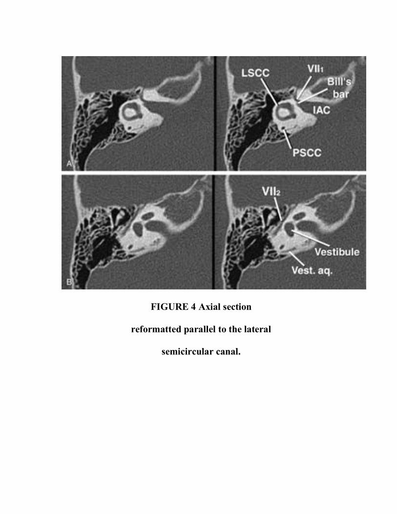
FIGURE 4 Axial section
reformatted parallel to the lateral
semicircular canal.

Coronal plane images through the temporal bone from anteriorly
to posteriorly
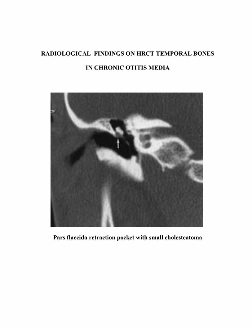
RADIOLOGICAL FINDINGS ON HRCT TEMPORAL BONES
IN CHRONIC OTITIS MEDIA
Pars flaccida retraction pocket with small cholesteatoma

ACQUIRED CHOLESTEATOMA, PRUSSAK’S SPACE. A,
Coronal CT image demonstrates a soft-tissue
mass (asterisk) interposed between the lateral attic wall and the
malleus head with blunted scutum (arrow).
B. Axial CT image demonstrates a soft-tissue mass
Extending posteriorly through the aditus into the mastoid antrum.

CHOLESTEATOMA. A, Cholesteatoma with erosion of ossicles
but sparing of the anterior epitympanic recess, which is protected
by an intact
‘‘cog’’ (arrow). Second case (B) (axial) and (coronal CT) (C).
Cholesteatoma
in the anterior epitympanic recess (supratubal recess), with
involvement of the
proximal tympanic facial nerve canal (arrow).
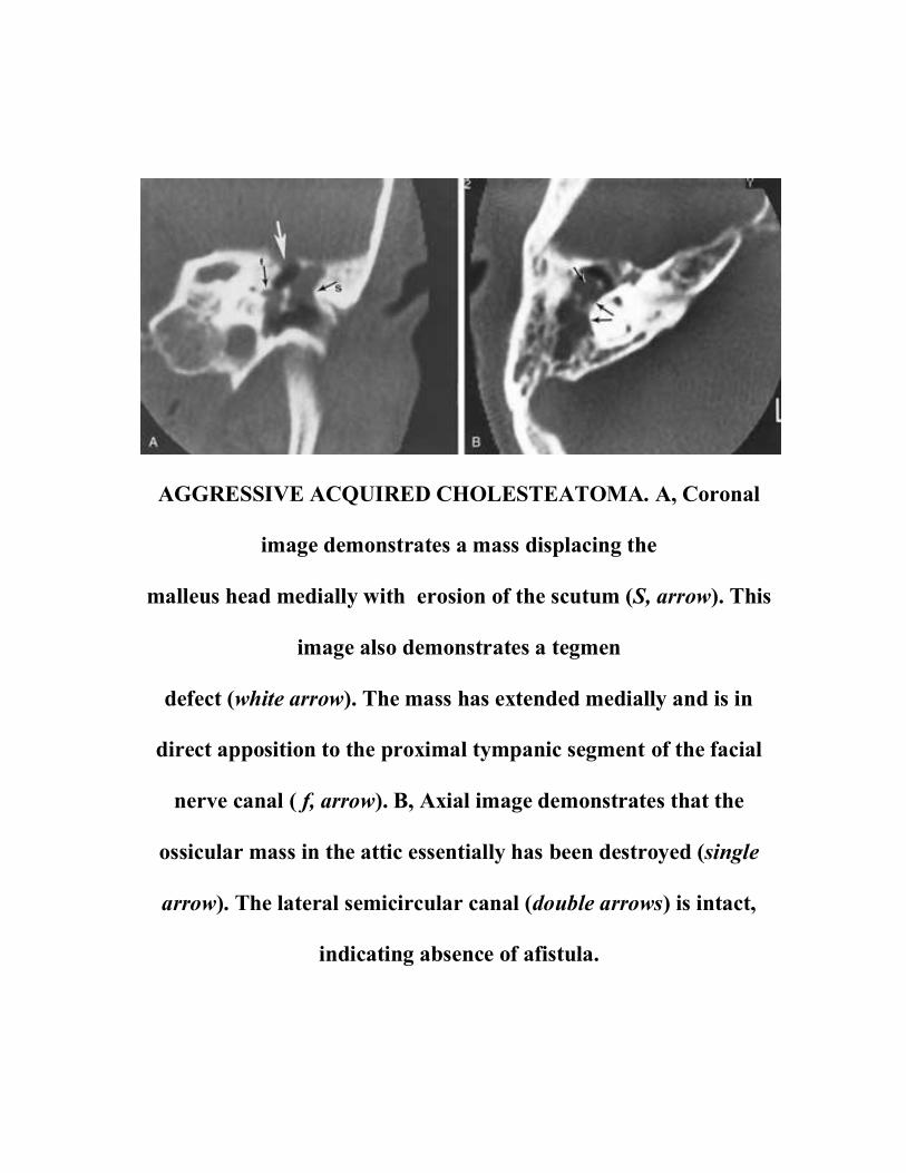
AGGRESSIVE ACQUIRED CHOLESTEATOMA. A, Coronal
image demonstrates a mass displacing the
malleus head medially with erosion of the scutum (S, arrow). This
image also demonstrates a tegmen
defect (white arrow). The mass has extended medially and is in
direct apposition to the proximal tympanic segment of the facial
nerve canal ( f, arrow). B, Axial image demonstrates that the
ossicular mass in the attic essentially has been destroyed (single
arrow). The lateral semicircular canal (double arrows) is intact,
indicating absence of afistula.

EROSIVE CHOLESTEATOMA. A, Coronal CT image shows a
soft-tissue mass eroding the lateral semicircular canal (arrow). B,
Coronal image demonstrates cholesteatoma invading and
expanding the proximal tympanic segment of the facial nerve canal
(curved arrow). C, Different patient with an extensive erosive
cholesteatoma simulating neoplasm

NONCHOLESTEATOMATOUS EROSION. A, There is an
erosion of the head of the malleus and body of the incus in the
vicinity of the malleoincudal articulation (arrow). B, Normal axial
section for comparison (malleoincudal articulation indicated)
(arrow

NONCHOLESTEATOMATOUS EROSION. A, Axial image (right
ear) indicates an absent lenticular process (white arrowhead ). B,
Normal patient for comparison (right ear). Normal lenticular
process and stapes head are present. The incudostapedial
articulation is indicated (arrow).

FORWARD LYING SIGMOID SINUS

BIBLIOGRAPHY
1. Galdino E. Vavassori, MD. „Imaging of Temporal Bone‟. Chapter 11,
Part 2 in Clinical Evaluation, by Glasscock, Shambaugh, 5th
edition,
2003; 227-33.
2. P.D. Phelps.„Imaging and radiology‟. Chapter 17 in Basic sciences,
Volume 1, Scott Brown‟s Otolaryngology, 6th
Edition, 1997; 1-7.
3. P.D. Phelps.„Radiology of ear‟. Chapter 2 in Otology, Volume 3,
Scott Brown‟s Otolaryngology, 6th
Edition, 1997; 1-9.
4. Gerald B. Brookes and John B. Booth. „Diseases of temporal bone‟.
Chapter 15 in Otology, Volume 3, Scott Brown‟s Otolaryngology, 6th
Edition, 1997; 2.
5. Jackler RK , Dillon WP.„Computed Tomography in suppurative ear
disease: a correlation of surgical and radiographic
findings‟.Laryngoscope.1984 Jun; 94(6):746-52.
6. Maffee MF, Almi k, „Chronic otomastoiditis: a conceptual
understanding of CT findings‟.Radiology.1986 Jul; 160(1):193-200.

7. Yamasoba T, Kikuchi S , Takeuchi N , Harada T , Nomura Y.„CT
evaluation of the anterior epitympanic recess-comparison among non-
inflammatory ear,chronic otitis media with central perforation and
cholesteatoma‟. Nippon Jibinkoka Gakkai Kaiho 1991 Feb;
94(2):177-82.
8. Leighton SE, Robson AK „The role of CT imaging in the management
of Chronic suppurative otitis media‟.Clin Otolaryngol Allied Sci.1993
Feb;18(1):23-9.
9. Garber LZ, Dort JC.„Cholesteatoma: diagnosis and staging by CT
scan‟.J.Otolaryngol.1994 Apr; 23(2):121-4.
10. Luchikhin LA. „Effectiveness of CT of temporal bone in diagnosis of
CSOM‟. Vestn Otolaryngol.1995 MAY-Jun; (3):31-4.
11. Walshe P, McConn.„The role of CT in preoperative assessment of
CSOM‟.Clin Otolaryngol. Allied Sci.2002 Apr; 27(2):95-7.
12. Sandeep Berry, S.C.Gandotra and N.C.Saxena.„Role of computed
tomography in unsafe chronic suppurative otitis media‟.Indian journal

Of Otorhinolaryngology and head and neck surgery.1998; 50(2):135-
39.
13. Zelikovich EL.„CT temporal bone in diagnosis of otitis media
chronica purulenta‟.Vestn Otolaryngol.2004 ; (4):25-9
14. Zelikovich EL.„Potentialities of temporal bone CT in the diagnosis of
chronic purulent otitis media and its complications‟.Vestn Rentgenol
Radiol.2004 Jan-Feb; (1):15-22.
15. Wang LE, Gu YF. „Significance of CT in diagnosis of
CSOM‟.Zhonghua ErBi Yan HouJing Wai Ke Za Zhi.2007 Jul;
42(7):494-8.
16. Gerami H, Naghavi E „Comparison of pre-operative computed
tomography scan imaging of temporal bone with the intra-operative
finding in patient undergoing mastoidectomy‟. SaudiMed J.2009 Jan;
30(1):104-8.
17. Firas Q.Alzoubi , Haaitham A.Odat , Hassan A.Al-balas and
S.R.Saeed „The role of preoperative CT scan in patients with chronic

otitis media‟ European Archives of Oto-rhino-laryngology.2009 Jun;
266(6):807-9.
18. Boyraz E, Erdogan, Boyraz I, Kazikdas C, Etit D, Uluc E. „The
importance of computed tomography examination of temporal bone in
detecting tympanosclerosis‟.Kulak Burun Bogaz Lhtis Derg 2009
Nov-Dec; 19(6): 294-8.
19. Shim HJ, Choi AY, Yoon SW, Kwon KH, Yeo SG.„The value of
measuring ET aeration on temporal bone CT in patients with chronic
otitis media‟ Clin Exp Otorhinolaryngol.2010 Jun; 3(2):59-64.
20. 20.Smyth GDL. Treatment of cholesteatoma. Acat Otorhinolarynol
Belg. 1973;25:970–971
21. Dulac GL, Claus E, Barrois J (1973) Otoradiology, x-ray bulletin.
Agfa Gevaert LTd, 137.
22. Mafee MF, Levin BC, Applebaum EL, Campos M, James CF.
Cholesteatoma of the middle ear and mastoid. A comparison of CT
scan and operative findings. OCNA. 1988;21(2):265–293.

23. O‟Reilly BJ, Chevratton EB, Wylie I, Thakkar C, Butler P,
Sathanathan N, Morrison G, Kenyon GS. The Value of CT scanning
in chronic Suppurative otitis media. J Laryngol Otol. 1991;105:990
24. Jackler RK, Dillon WP, Schindler RA. Computed tomography in
suppurative ear disease: a correlation of surgical and radiographic
findings. Laryngoscope. 1984;94:746–25. O‟Donoghue GM. Imaging
the temporal bone (Editorial) Clin Otolaryngol. 1987;12:157–160.
doi: 10.1111/j.1365-2273.1987.tb00181.x.
25. Schwartz JD. High-resolution computed tomography of the middle ear
and mastoid. Part I: Normal radioanatomy including normal
variations. Radiology. 1983;148(2):449–454.
26. Lee Sh, Rao KCVG. Cranial computed tomography and MRI. Mac
Graw-Hill Book Company, New York 1987:477- 508.
27. Littleton JT, Shaffer KA, Callahan WP, Durizch ML. Temporal bone:
Comparison of pluridirectional tomography and high resoluton
computed tomography. AJR 1981; 137:835-45.
28. Shaffer KA, Volz DJ, Haughton VM. Manipulation of CT data for
temporal bone imaging. Radiology 1980;137:825- 9.

29. Winter J. Edge enhancement of computed tomograms by digital
unsharp masking. Radiology 1980;135:234-5.
30. Zonneveld FW. The value non-reconstructive multiplanar CT for the
evaluation of the petrous bone. Neuroradiology 1983;25:1-10.
31. Akan H. Baş ve Boyun Radyolojisi. MN Medikal- Nobel, 2008;2:104.
32. Watts S, Flood LM, CliVord K. A systematic approach to
interpretation of computed tomography scans prior to surgery of
middle ear cholesteatoma. J Laryngol Otol 2000;114:248-53.
33. Mafee MF. MRI and CT in the evaluation of acquired and congenital
cholesteatomas of the temporal bone. J Otolaryngol 1993;22:239-48.
34. O’Donoghue GM, Bates GJ, Anslow P, Rothera MP. The predictive
value of high resolution computerized tomography in chronic
suppurative ear disease. Clin Otolaryngol 1987;12:89-96.
35. Ayache D, Williams MT, Lejeune D, et al. Usefulness of delayed
postcontrast magnetic resonance imaging in the detection of residual
cholesteatoma after canal wall-up tympanoplasty. Laryngoscope
2005;115:607-10.

36. De Foer B, Vercruysse JP, Pouillon M, et al. Value of high-resolution
computed tomography and magnetic resonance imaging in the
detection of residual cholesteatomas in primary bony obliterated
mastoids. Am J Otolaryngol 2007;28:230-34.
37. Williams MT, Ayache D, Alberti C, et al. Detection of postoperative
residual cholesteatoma with delayed contrast-enhanced MR imaging:
initial findings. Eur Radiol 2003; 13:169-74.
38. Krestan C, Czerny C, Gstöttner W, Franz P. The role of high-
resolution computed tomography (HRCT) and magnetic resonance
imaging (MRI) in the diagnosis of preoperative and postoperative
complications caused by acquired cholesteatomas. Radiologe
2003;43(3):207-12.
39. Swartz JD, Wolfson RJ, Marlow FI, Popky GL. Post inflammatory
ossicular fixation: CT analysis with surgical correlation. Radiology
1985;154:697-700.
40. Holliday RA. İnflammatory diseases of the temporal bone: evaluation
with CT and MRI. Seminars in Ultrasound, CT, MRI 1989;10(3):213-
95.

41. Swartz JD, Berger AS, Zwillenberg S, Popky GL. Ossicular erosions
in the dry ear: CT diagnosis. Radiology 1987;


PROFORMA
NAME : AGE :
SEX : ADDRESS :
OCCUPATION :
SOCIOECONOMIC Date of Admission
STATUS
Date of Procedure
Date of Discharge
HISTORY:
PRESENTING COMPLAINTS:
1. Ear discharge : Side, Onset, Duration, Amount, Type of discharge,
Aggravating / Relieving Factors, Response to
treatment
2. Hard of hearing : Side, Acute / Insidious, Degree Progressive
/continuous/
Intermittent , Whether handicapping, Family
history,
H/o trauma to ear,
H/o exposure to loud nice.
3. Earache : Duration, Side, Mild/Server, More on movement of
ear /
during mastication, Increase during ear discharge or
not
4. Giddiness : Duration, Intermittent / Continuous, Associated
with
posture, H/o intake of ototoxic drugs, Aggravating /
relieving factors.
5. Tinnitus : Side, Unilateral / Bilateral, Duration, Sudden /
Gradual,

Progression – Severe / Static / Decreasing,
Continuous / intermittent/ Pulsatile/ Clicking,
Aggravating / Relieving factors.
6. Headache/ vomiting
7. Fever
8. Facial weakness
MENSTRUAL HISTORY
PERSONAL HISTORY:
FAMILY HISTORY :- H/O HOH, H/O Consanguinous marriage
PAST HISTORY :- H/O Previous surgery,
H/O Ototoxic drug intake as in TB.
H/O DM/HTN
EXAMINATION OF EAR:
R L
Pinna
Preauricular area
Postauricular area
EAC
Tympanic Membrane:
Mastoid tenderness
Tragal tenderness
Fistula test
TUNING FORK TESTS
Rinne‟s
Weber‟s
ABC

Facial Nerve:
Romberg‟s:
Examination of Oral Cavity/ Oropharynx / Nose
Palate : Bulge/ Movements of soft palate, postnasal drip, tonsils, posterior
pharyngeal
wall, Dental hygiene.
SYSTEMIC EXAMINATION
RS CNS : Higher functions, Cranial Nerves, Sensory, Motor,
Reflexes, Gait, Cerebellar System, Speech
CVS
Abdomen
INVESTIGATIONS:
Blood - Hb, TC, DC, ESR
Urine - Routine, Microscopy
Pus - Culture/ Sensitivity
CT Temporal bones :- Axial / Coronal views
Plain / Contrast

SL NO. NAME AGE/SEX IP NO. DIAGNOSIS DOA DOD DOS SURGERY OSS.ER- Mhan OSS. ER- INCUS OSS. ER- MH FCD LSCC ER MCD CHOLEST TEG ER KORN SEP HJB FLSS
CT SURG CT SURG CT SURG CT SURG CT SURG CT SURG CT SURG CT SURG CT SURG CT SURG CT SURG
1 SELVARAJ 23/M 75503 L CSOM+CP 11/24/2011 12/1/2011 11/25/2011 CM+T 1 TYM NO NO NO NO NO NO NO NO NO NO NO NO NO NO NO NO NO NO NO NO NO NO
2 DHANALAKSHMI 38/F 69164 LCSOM+ AAD 12/1/2011 12/9/2011 12/2/2011 MRM+WM NO NO YES YES NO YES NO YES NO NO NO YES YES NO NO NO NO NO NO NO NO NO
3 CHITRA 40/F 78234 R CSOM+CP 12/6/2011 12/14/2011 12/7/2011 CM+T 1 TYM NO NO NO NO NO NO NO NO YES NO NO NO YES YES N O NO NO NO NO NO NO NO
4 MALA 40/F 78665 LCSOM+CP 12/8/2011 12/15/2011 12/9/2011 CM+T 1 TYM NO NO NO NO NO NO NO NO NO NO NO NO NO NO NO NO NO NO NO NO NO NO
5 CHELLAMMAL 40/F 5951 R CSOM+ CP 1/5/2012 1/13/2012 1/6/2012 CM+T 1 TYM NO NO NO NO NO NO NO NO NO NO NO NO NO NO NO NO NO NO NO NO YES YES
6 SHANMUGAM 30/M 2210 RCSOM+ CP 1/10/2012 1/18/2012 1/11/2012 CM+T 1 TYM NO NO NO NO NO NO NO NO NO NO NO NO NO NO NO NO NO NO NO NO NO NO
7 VEERAN 21/M 2762 BL CSOM+ CP 1/12/2012 1/19/2012 1/13/2012 ICWM+SCTYM NO NO NO NO NO NO NO NO N O NO YES YES YES YES NO NO NO NO NO NO NO NO
8 PETCHI 32/F 4413 BLCSOM+CP 1/19/2012 1/26/2012 2/5/2012 CM+T 1 TYM NO NO NO NO NO NO NO NO NO NO NO NO NO NO NO NO NO NO NO NO YES YES
9 SUBRAMANIAN 24/M 6192 L CSOM+CP 1/26/2012 2/5/2012 1/27/2012 CM+T 1 TYM NO NO NO NO NO NO NO NO NO NO NO NO NO NO NO NO NO NO NO NO NO NO
10 VIJAY 23/M 7529 R CSOM+CP 1/31/2012 2/8/2012 2/1/2012 MRM+WM NO NO YES YES NO NO NO YES YES NO YES YES YES YES NO NO NO NO NO NO YES YES
11 RAJAMANI 65/M 9210 R CSOM+CP 2/3/2012 2/11/2012 2/4/2012 CM+T 1 TYM NO NO NO NO NO NO NO NO NO NO NO NO NO NO NO NO NO NO NO NO NO NO
12 RAMACHANDRAN 28/M 10252 BL CSOM+ CP 2/12/2012 2/20/2012 2/13/2012 CM+T 1 TYM NO NO NO NO NO NO NO NO NO NO NO NO NO NO NO NO NO NO NO NO NO NO
13 VILVAIRAJA 20/M 10991 R CSOM+CP 2/14/2012 2/22/2012 2/15/2012 CM+T 1 TYM NO NO NO NO NO N O NO NO NO NO NO NO NO NO NO NO NO NO NO NO NO NO
14 MOHD MYDEEN 24/M 11056 BLCSOM+CP 2/14/2012 2/22/2012 2/15/2012 CM+T 1 TYM NO NO NO NO NO NO NO NO NO NO NO NO NO NO NO NO NO NO NO NO YES YES
15 SHAMSHAD BEGUM 24/F 12002 R CSOM+CP 2/19/2012 2/27/2012 2/20/2012 CM+T 1 TYM NO NO NO NO NO NO NO NO NO NO NO N O NO NO NO NO NO NO NO NO NO NO
16 JEYASATHYA 25/F 12201 R CSOM+AAD 2/28/2012 3/8/2012 2/29/2012 MRM+WM YES YES YES YES NO NO NO YES NO NO YES YES YES YES NO NO NO NO NO NO NO NO
17 VELLAISAMY 36/M 13695 R CSOM+CP 2/28/2012 3/8/2012 2/29/2012 CM+T 1 TYM NO NO NO NO NO NO NO NO NO NO NO NO NO NO NO NO NO NO NO NO NO NO
18 VINOD 24/M 14335 R CSOM+CP 3/1/2012 3/9/2012 3/2/2012 CM+T 1 TYM NO NO NO NO NO NO NO NO NO NO NO NO NO NO NO NO NO NO NO NO NO NO
19 SENTHIL KUMAR 32/M 15386 R CSOM+ PSRP 3/4/2012 3/12/2012 3/5/2012 MRM+WM NO NO NO YES NO NO NO NO YES NO NO NO YES YES NO NO NO NO NO NO NO NO
20 JYOTHI 34/F 17939 L CSOM+AAD 3/13/2012 3/21/2012 3/14/2012 MRM+WM NO NO YES YES NO NO NO N O NO NO NO NO YES NO NO NO NO NO NO NO NO NO
21 KRISHNAVENI 63/F 18921 L CSOM+ AAD 3/18/2012 3/26/2012 3/19/2012 MRM+WM NO NO NO YES NO NO NO NO NO NO NO NO YES YES NO NO NO NO NO NO YES YES
22 DHANAPANDI 31/M 19005 R CSOM+CP 3/20/2012 3/28/2012 3/21/2012 CM+T 1 TYM NO NO NO NO NO NO NO NO NO NO NO NO NO NO N O NO NO NO NO NO YES YES
23 MANONMANI 24/F 19651 L CSOM+CP 25-3-12` 4/4/2012 3/26/2012 CM+T 1 TYM NO NO NO NO NO NO NO NO NO NO NO NO NO NO NO NO NO NO NO NO NO NO
24 MURUGANANTHI 18/F 18945 RCSOM+ CP 3/27/2012 4/5/2012 3/28/2012 CM+T 1 TYM NO NO NO NO NO NO NO NO NO NO NO NO NO NO NO NO NO NO NO NO NO NO
25 SUDHARSHAN 46/M 20546 BLCSOM+CP 3/29/2012 4/7/2012 3/30/2012 CM+T 1 TYM NO NO NO NO NO NO NO NO NO NO NO NO NO NO NO NO NO NO NO NO NO NO
26 THANGARAJ 27/M 21263 RCSOM+ CP 4/1/2012 4/9/2012 4/2/2012 CM+T 1 TYM NO NO NO NO NO NO NO NO NO NO NO NO NO NO NO NO NO NO NO NO NO NO
27 SENTHIL KUMAR 31/M 36925 LCSOM+CP 5/31/2012 6/1/2012 6/8/2012 CM+T 1 TYM NO NO NO NO NO NO NO NO NO NO NO NO NO NO NO NO NO NO NO NO NO NO
28 VAITHEESWARI 24/F 37740 LCSOM+CP 6/3/2012 6/12/2012 6/4/2012 CM+T 1 TYM NO NO NO NO NO NO NO NO NO NO NO NO NO NO NO NO NO NO NO NO NO NO
29 SURIYAPRABAVATHI 14/F 38350 R CSOM+AAD 6/7/2012 6/15/2012 6/8/2012 MRM+WM NO NO YES YES NO NO NO NO NO NO NO NO YES NO NO NO NO NO NO NO NO NO
30 UMAR FAROOQ 16/M 39589 BLCSOM+PSRP 6/10/2012 6/18/2012 6/11/2012 L MRM+WM NO NO YES YES YES YES NO YES NO NO NO NO NO NO NO NO NO NO NO NO NO NO
31 MEENAKSHI 30/F 4223 LCSOM+CP 6/11/2012 6/19/2012 6/12/2012 CM+T 1 TYM NO NO NO NO NO NO NO NO NO NO NO NO YES YES NO NO NO NO NO NO NO NO
32 THIRUPATHI 18/M 42542 RCSOM+ CP 6/17/2012 6/27/2012 6/18/2012 CM+T 1 TYM NO NO NO NO NO NO NO NO NO NO NO NO NO NO NO NO NO NO YES YES NO NO
33 RAJENDRAN 57/M 43271 RCSOM+AUR POLYP 6/19/2012 6/28/2012 6/20/2012 MRM+WM YES YES YES YES YES YES NO NO NO NO NO YES NO NO NO NO NO NO NO NO NO NO
34 SOUNDARRAJAN 19/M 19471 RCSOM+AAD 6/21/2012 6/29/2012 6/22/2012 MRM+WM YES NO YES YES NO NO YES YES NO NO NO NO YES YES NO NO NO NO NO NO NO NO
35 KAVIYARASU 23/M 44391 RCSOM+CP 6/23/2012 2-7-112 6/25/2012 CM+T 1 TYM NO NO YES YES NO NO NO NO NO NO NO NO YES YES NO NO NO NO NO NO NO NO
36 CHINNAPONNU 34/F 45135 BLCSOM+CP 6/26/2012 7/4/2012 6/27/2012 CM+T 1 TYM NO NO NO NO NO NO NO NO NO NO NO NO NO NO NO NO NO NO NO NO NO NO
37 SARADHA 40/F 45712 BLCSOM+CP 6/28/2012 7/6/2012 6/29/2012 CM+T 1 TYM NO NO NO NO NO NO NO NO NO NO NO NO YES NO NO NO NO NO NO NO YES YES
38 MADHAVAN 25/M 46214 BLCSOM+CP 6/30/2012 7/10/2012 7/2/2012 CM+T 1 TYM NO NO NO NO NO NO NO NO NO NO NO NO NO NO NO NO YES YES NO NO NO NO
39 SUGANYA 18/F 49190 LCSOM+AAD 7/13/2012 7/22/2012 7/14/2012 MRM+WM NO NO NO YES NO NO NO NO NO NO NO NO NO NO NO NO NO NO NO NO NO NO
40 MAHENDRAN 23/M 50299 BLCSOM+CP 7/17/2012 7/25/2012 7/18/2012 RCM+T 1TYM NO NO NO NO NO NO NO NO NO NO NO NO YES NO NO NO NO NO NO NO NO NO
41 JANNATHIN FIRDOUSE 40/F 53027 L CSOM+CP 7/29/2012 8/8/2012 7/30/2012 CM+T 1 TYM NO NO NO NO NO NO NO NO NO NO NO NO NO NO NO NO NO NO NO NO NO NO
42 MALA 40/F 55454 RCSOM+AAD 8/7/2012 8/15/2012 8/8/2012 MRM+WM NO NO YES YES NO YES NO YES NO NO NO NO NO NO NO NO NO NO NO NO NO NO
43 SHALINI 12/F 49631 L CSOM + CEREBELLAR ABSCESS8/12/2012 8/21/2012 8/13/2012 MRM+WM YES YES YES YES YES YES YES YES YES YES NO YES YES YES YES YES NO NO NO NO NO NO
44 KARUPAYEE 29/F 57638 L CSOM+CP 8/16/2012 8/25/2012 8/17/2012 CM+T 1 TYM NO NO NO NO NO NO NO NO NO NO NO NO NO NO NO NO NO NO NO NO NO NO
45 THANGAPANDI 18/M 58795 R CSOM+AAD 8/21/2012 8/29/2012 8/22/2012 MRM+WM NO NO YES YES NO NO NO NO NO NO NO NO YES YES NO NO NO NO NO NO YES YES
46 DURAIVAIRAVAN 37/M 59777 RCSOM 8/26/2012 9/5/2012 8/27/2012 MRM+WM NO NO YES YES NO NO NO NO NO NO NO NO NO NO NO NO NO NO NO NO NO NO
47 MAHALAKSHMI 55/F 54973 RCSOM+ R LMN FACIAL NERVE PALSY8/28/2012 9/6/2012 8/29/2012 MRM+ FACIAL NERVE DECOMPRESION YES YES YES YES NO NO YES YES NO NO NO NO YES YES YES NO NO NO NO NO NO NO
48 MANIKANDAPRABHU 20/M 53681 BLCSOM+CP 8/2/2012 8/11/2012 8/3/2012 CM+T 1 TYM NO NO NO NO NO NO NO NO NO NO NO NO YES NO NO NO YES YES NO NO NO NO
49 GANESAN 26/M 53728 LCSOM+AAD 7/31/2012 8/9/2012 8/1/2012 MRM+TYPE3 TYMP + WM NO NO YES YES NO NO NO NO NO NO NO NO NO NO NO NO NO NO NO NO NO NO
50 RATHIFA 31/F 54225 LCSOM+CP 8/2/2012 8/11/2012 8/3/2012 CM+T 1 TYM NO NO NO NO NO NO NO NO NO NO NO NO NO NO NO NO NO NO NO NO NO NO

KEY TO MASTER CHART
DOA- date of admission
DOS- date of surgery
DOD- date of discharge
OSS ER- ossicular erosion
M Hand- malleus handle
MH- malleus head
FCD- facial canal dehiscence
LSCC ER- lateral semicircular canal erosion
MCD- mastoid cortex dehiscence
CHOLEST- cholesteatoma
TEG ER- tegmen erosion
KORN SEP- korner’s septum
HJB- high jugular bulb
FLSS- forward lying sigmoid sinus
SURG- surgery
CM+ T 1 TYM- Cortical mastoidectomy+ type 1 tympanoplasty
MRM+ WM- modified radical mastoidectomy + wide meatoplasty
T 3 TYM- type 3 tympanoplasty


Your digital receiptThis receipt acknowledges that Turnitin received your paper. Below you will find the receipt informationregarding your submission.
Paper ID 288989038
Paper titleCORRELATION BETWEEN HRCT TEMPORAL BONE FINDINGS ANDSURGICAL FINDINGS IN PATIENTS WITH CHRONIC SUPPURATIVEOTITIS MEDIA
Assignmenttitle Medical
Author Sowmya rajaE-mail [email protected]
Submissiontime 17-Dec-2012 07:33PM
Total words 13443
First 100 words of your submission
CORRELATION BETWEEN HRCT TEMPORAL BONE AND SURGICAL FINDINGS IN PATIENTSWITH CHRONIC SUPPURATIVE OTITIS MEDIA Dissertation submitted for MASTER OF SURGERYBRANCH IV (OTO-RHINO-LARYNGOLOGY) APRIL 2013 THE TAMIL NADU Dr.M.G.R. MEDICALUNIVERSITY CHENNAI CERTIFICATE This is to certify that this dissertation entitled “CORRELATION BETWEEN HRCT TEMPORAL BONE FINDINGS AND SURGICAL FINDINGS INPATIENTS WITH CHRONIC SUPPURATIVE OTITIS MEDIA” presented herewith by Dr. SOWMYARAJA to the faculty of otorhinolaryngology in the Tamilnadu Dr. MGR Medical University , Chennai, inpartial fulfillment of the requirements for the award degree of the Master of Surgery Branch IV(Otorhinolaryngology) April...
Copyright 2012 Turnitin. All rights reserved.