correspondence postsurgical safety of opioid...
TRANSCRIPT
� CORRESPONDENCE
Anesthesiology 2006; 104:375 © 2006 American Society of Anesthesiologists, Inc. Lippincott Williams & Wilkins, Inc.
Postsurgical Safety of Opioid-sparing Cyclooxygenase-2 Inhibitors
To the Editor:—We read with great interest the excellent meta-analysisby Marret et al.1 and the accompanying editorial by Professor Kehlet2
on the effects of combined opioid and nonsteroidal antiinflammatorydrugs (NSAIDs) use to relieve postsurgical pain. Marret et al. concludethat NSAIDs (cyclooxygenase 2 [COX-2] selective and nonselective), inthe aggregate, provide approximately 30% reduction in morphineconsumption, with associated reductions in postoperative nausea andvomiting and sedation, but not pruritus, urinary retention, and respi-ratory depression. Although the efficacy data support the use of mul-timodal analgesia involving opioids and COX-2 selective NSAIDs, webelieve the safety data on the short-term perioperative use of COX-2selective inhibitors are less than clear for the following reasons.
First, the US New Drug Application for rofecoxib was based almostexclusively on studies in patients with chronic pain. Studies to directlysupport the postsurgical pain indication consisted of 741 patients, ofwhom 85% received one dose for dental pain and 15% received five dosesfor orthopedic pain.* Marret et al. note that the cardiovascular risk ofrofecoxib was associated with “long-term use.” However, in the absenceof robust studies in high-risk surgical patients and based on data onparecoxib, one cannot preclude similar safety risks with short-term post-surgical use of rofecoxib.
Second, although parecoxib (the injectable prodrug of the nowwithdrawn valdecoxib) is an effective analgesic, there remain seriousunanswered questions about the safety of short-term use in the postsur-gical setting. An important early adverse postsurgical safety signal camefrom a coronary artery bypass graft study included in the original US NewDrug Application.† In the parecoxib and valdecoxib group, 19.0% hadserious adverse events, versus 9.9% in the placebo group. Citing deficien-cies in the data, including a numerically higher incidence of myocardialinfarctions (1.9% vs. 0.7%) and cerebrovascular events (2.6% vs. 0.7%) anddeaths (4 vs. 0), parecoxib received a nonapprovable letter from the Foodand Drug Administration in 2001. The Food and Drug Administrationconcluded that “the adverse event profile of parecoxib was generallyworse than that of placebo in this trial. Although not statistically signifi-cantly different, the number of deaths, myocardial infarctions, cerebrovas-cular accidents, pulmonary embolisms, along with renal and pulmonarycomplications were also numerically more frequent for parecoxib duringthis IV dosing period than placebo. In fact, during the entire study period,the incidence of these clinically relevant adverse events associated withparecoxib/valdecoxib was statistically significantly different than placebo.Similarly, during the entire study period, more patients in the parecoxib/valdecoxib versus the placebo group withdrew from the study due to anadverse event.”†
Third, follow-up studies conducted with parecoxib have raised ad-ditional safety issues. In one trial,3 cardiovascular events (including
myocardial infarction, cardiac arrest, stroke, and pulmonary embolism)occurred significantly more frequently in the parecoxib and valde-coxib group than with placebo (2.0% vs. 0.5%; P � 0.03). In anothertrial,4 there were significantly more sternal wound infections withparecoxib than with placebo (3.2% vs. 0%; P � 0.03).
Fourth, 3 yr ago, the European Medicines Evaluation Agency issueda public statement on parecoxib regarding the risk of serious hyper-sensitivity and skin reactions, including Stevens-Johnson syndrome,toxic epidermal necrolysis, erythema multiforme, and exfoliative der-matitis, as well as anaphylaxis and angioedema.5 The European Medi-cines Evaluation Agency has since contraindicated the use of parecoxibin patients with ischemic heart disease and stroke. Excluding individ-uals with silent ischemia, this translates to approximately 20 millionat-risk patients in the United States. Immediately before its withdrawalin the United States, the Food and Drug Administration required asimilarly worded box warning for valdecoxib. Notably, the warningindicated (1) a higher risk of serious skin reactions within the first 2weeks, (2) a greater propensity for such reactions with valdecoxibthan other COX-2 inhibitors, and (3) a recommendation to discontinuevaldecoxib at the first appearance of skin rash. Since self-limitingpruritus and hypersensitivity are common in the postsurgical setting,causality assessment in patients with early signs of serious and unre-lated skin reactions may prove difficult.
Fifth, there are indications that the increased COX-2 levels observedunder pathologic conditions in endothelial cells and atheroscleroticlesions provides atheroprotection and modulates vascular remodelingthrough its principal metabolite, prostacyclin.6 In addition, in patientswith acute pain, there is considerable spinal up-regulation of COX-1,which may provide a further rationale for the use of drugs that inhibitboth isoforms of COX in acute pain.
Finally, survey data continue to support the view that postsurgical painis undertreated. Although the efficacy of COX-2 selective NSAIDs such asparecoxib is comparable to nonselective NSAIDs, further safety data arerequired to support their short-term use in the perioperative setting.
Najib Babul, Pharm.D.,‡ Paul Sloan, M.D., Arthur G. Lipman,Pharm.D. ‡TheraQuest Biosciences, Blue Bell, [email protected]
References
1. Marret E, Kurdi O, Zufferey P, Bonnet F: Effects of nonsteroidal antiinflam-matory drugs on patient-controlled analgesia morphine side effects: Meta-analysisof randomized controlled trials. ANESTHESIOLOGY 2005; 102:1249–60
2. Kehlet H: Postoperative opioid sparing to hasten recovery: What are theissues? ANESTHESIOLOGY 2005; 102:1083–5
3. Nussmeier NA, Whelton AA, Brown MT, Langford RM, Hoeft A, Parlow JL,Boyce SW, Verburg KM: Complications of the COX-2 inhibitors parecoxib andvaldecoxib after cardiac surgery. N Engl J Med 2005; 352:1081–91
4. Ott E, Nussmeier NA, Duke PC, Feneck RO, Alston RP, Snabes MC, HubbardRC, Hsu PH, Saidman LJ, Mangano DT: Efficacy and safety of the cyclooxygenase2 inhibitors parecoxib and valdecoxib in patients undergoing coronary arterybypass surgery. J Thorac Cardiovasc Surg 2003; 125:1481–92
5. EMEA public statement on parecoxib sodium (Dynastat, Rayzon, Xapit):Risk of serious hypersensitivity and skin reactions EMEA/25175/02 October 22,2002
6. Egan KM, Lawson JA, Fries S, Koller B, Rader DJ, Smyth EM, Fitzgerald GA:Cyclooxygenase-2-derived prostacyclin confers atheroprotection on female mice.Obstet Gynecol Surv 2005; 60:309–10
(Accepted for publication September 21, 2005.)
Dr. Babul works at TheraQuest, a pharmaceutical company involved in thedevelopment of opioid and non-opioid analgesics, including nonsteroidal antiin-flammatory drugs, for acute pain and chronic pain. All three authors haveconsulted with and participated as investigators in analgesic clinical trials for avariety of pharmaceutical companies. David C. Warltier, M.D., Ph.D., acted asHandling Editor for this exchange. The above letter was sent to the author of thereferenced Editorial View. The author did not feel that a response was required.—Michael M. Todd, Editor-in-Chief
* Food and Drug Administration Summary Basis for Approval for Vioxx, NDA21-042. Available at: http://www.fda.gov/cder/drug/. Accessed September 21,2005.
† Food and Drug Administration Medical Officer Review: Parecoxib NDA21-294. http://www.fda/gov/cder/drug/. Accessed September 21, 2005.
Anesthesiology, V 104, No 2, Feb 2006 375
Downloaded From: http://anesthesiology.pubs.asahq.org/pdfaccess.ashx?url=/data/journals/jasa/931077/ on 08/11/2018

Anesthesiology 2006; 104:376 © 2006 American Society of Anesthesiologists, Inc. Lippincott Williams & Wilkins, Inc.
In Reply:—We thank Drs. Babul, Sloan, and Lipman for their interestin our meta-analysis,1 which was primarily performed to study theeffects of cyclooxygenase-selective and -nonselective inhibitors onmorphine side effects. We agree with them (as we discussed in thearticle) that some concerns remain about the safety of short-termperioperative prescription of coxibs and that coxibs cannot be admin-istrated to all surgical patients, especially patients with coronary arterydisease or at risk of cerebral infarction. Two studies have indeed clearlydemonstrated that the use of parecoxib and valdecoxib was associatedwith an increased risk of arterial thrombotic adverse events in patientsscheduled to undergo coronary artery bypass surgery.2,3 However,some evidence also suggests that short-term perioperative use ofcoxibs may offer benefits in comparison with nonsteroidal antiinflam-matory drugs. For example, tonsillectomy is one of the most frequentlyperformed ambulatory surgical procedures in children, and nonselec-tive inhibition of cyclooxygenase by nonsteroidal antiinflammatorydrugs increases significantly the rate of reoperation4,5 but also de-creases nausea and vomiting.5 In that setting, celecoxib, has beendemonstrated to relieve posttonsillectomy pain and to decrease bleed-ing risk in comparison with nonsteroidal antiinflammatory drugs.6
Moreover, the risk of adverse cardiovascular events is extremely low inthis population of young patients, as it is in patients devoid of arterialthrombotic pathology scheduled to undergo noncardiac surgery.Therefore, short-term perioperative use of coxibs could have a favor-able risk–benefit ratio compared with nonsteroidal antiinflammatory
drugs in patients without risk factors for arterial thrombotic eventssubmitted to hemorrhagic surgical procedures.
Emmanuel Marret, M.D.,* Francis Bonnet, M.D. *TenonUniversity Hospital, Paris, France. [email protected]
References
1. Marret E, Kurdi O, Zufferey P, Bonnet F: Effects of nonsteroidal antiinflam-matory drugs on patient-controlled analgesia morphine side effects: Meta-analysisof randomized controlled trials. ANESTHESIOLOGY 2005; 102:1249–60
2. Nussmeier NA, Whelton AA, Brown MT, Langford RM, Hoeft A, Parlow JL,Boyce SW, Verburg KM: Complications of the COX-2 inhibitors parecoxib andvaldecoxib after cardiac surgery. N Engl J Med 2005; 352:1081–91
3. Ott E, Nussmeier NA, Duke PC, Feneck RO, Alston RP, Snabes MC, HubbardRC, Hsu PH, Saidman LJ, Mangano DT: Efficacy and safety of the cyclooxygenase2 inhibitors parecoxib and valdecoxib in patients undergoing coronary arterybypass surgery. J Thorac Cardiovasc Surg 2003; 125:1481–92
4. Marret E, Flahault A, Samama CM, Bonnet F,: Effects of postoperative,nonsteroidal, antiinflammatory drugs on bleeding risk after tonsillectomy: Meta-analysis of randomized, controlled trials. Anesthesiology 2003; 98:1497–502
5. Moiniche S, Romsing J, Dahl JB, Tramer MR: Nonsteroidal antiinflammatorydrugs and the risk of operative site bleeding after tonsillectomy: A quantitativesystematic review. Anesth Analg 2003; 96:68–77
6. Nikanne E, Kokki H, Salo J, Linna TJ: Celecoxib and ketoprofen for painmanagement during tonsillectomy: A placebo-controlled clinical trial. Otolaryn-gol Head Neck Surg 2005; 132:287–94
(Accepted for publication September 21, 2005.)
Anesthesiology 2006; 104:376 © 2006 American Society of Anesthesiologists, Inc. Lippincott Williams & Wilkins, Inc.
Facilitating Endotracheal Tube Advancement duringFiberscope-assisted Intubation: Giving Due Credit
To the Editor:—Johnson et al.’s1 pictorial documentation of the struc-tures that obstruct the passage of an endotracheal tube during fiber-optic intubation is brilliant and once again proves the usefulness of a90° counterclockwise rotation of the endotracheal tube to facilitateadvancement.2
Unfortunately, they have, like some authors before them, failed toacknowledge the contribution of Dr. Cossham, who first described thistechnique to facilitate the passage of an endotracheal tube over a gumelastic bougie.3 When passage of an endotracheal tube over a fiber-scope is difficult, most clinicians had, hoping to bypass the obstruc-tion, retried while or after twisting the tube left or right—a somewhathaphazard maneuver also mentioned by Ovassapian et al.4 It was Dr.Cossham, however, who actually clearly illustrated the technique.Whether over a gum elastic bougie or over a flexible bronchoscope, histechnique is elegant and proven,5 and the principles are the same. It issuch a useful “trick” that we believe that the first attempt to advancean endotracheal tube over a flexible bronchoscope should always bemade with it already turned counterclockwise by 90°.6 Our experienceis that the success rate exceeds 90% with only one attempt, thus savingtime and reducing the risk of upper airway trauma and unpleasantnessfor the patient.
Anthony M.-H. Ho, M.S., M.D., F.R.C.P.C., F.C.C.P.,* Manoj K.Karmakar, M.B., M.D., F.R.C.A. *The Chinese University of HongKong, Shatin, NT, Hong Kong Special Administrative Region,Peoples’ Republic of China. [email protected]
References
1. Johnson DM, From AM, Smith RB, From RP, Maktabi MA: Endoscopic studyof mechanisms of failure of endotracheal tube advancement into the tracheaduring awake fiberoptic orotracheal intubation. ANESTHESIOLOGY 2005; 102:910–4
2. Kristensen MS: The Parker Flex-Tip Tube versus a standard tube for fiber-optic orotracheal intubation: A randomized double-blind study. ANESTHESIOLOGY
2003; 98:354–83. Cossham PS. Difficult intubation (letter). Br J Anaesth 1985; 57:2394. Ovassapian A, Yelich SJ, Dykes MH, Edward EB: Fiberoptic nasotracheal
intubation: Incidence and causes of failure. Anesth Analg 1983; 62:692–55. Dogra S, Falconer R, Latto IP: Successful difficult intubation: Tracheal tube
placement over a gum-elastic bougie. Anaesthesia 1990; 45:774–66. Ho AMH, Chung DC, Karmakar MK: Is the Parker Flex-Tip Tube really
superior to the standard tube for fiberoptic orotracheal intubation? (letter).ANESTHESIOLOGY 2003; 99:1236
(Accepted for publication October 11, 2005.)
376 CORRESPONDENCE
Anesthesiology, V 104, No 2, Feb 2006
Downloaded From: http://anesthesiology.pubs.asahq.org/pdfaccess.ashx?url=/data/journals/jasa/931077/ on 08/11/2018

Anesthesiology 2006; 104:377 © 2006 American Society of Anesthesiologists, Inc. Lippincott Williams & Wilkins, Inc.
Corniculate Cartilages Are Wrongly Labeled Arytenoid Cartilages
To the Editor:—The corniculate cartilages are commonly and wronglyreferred to as the arytenoid cartilages. This common misconception iswell illustrated by the labeling of the figure on the cover of the May2005 issue of ANESTHESIOLOGY and its accompanying article.1 The bilat-eral spherical bulges at the five o’clock and seven o’clock positions onthe most proximal part of the laryngeal aperture are the corniculatecartilages,2 not the arytenoid cartilages.
Jonathan L. Benumof, M.D., UCSD Medical Center, San Diego,California. [email protected]
References
1. Johnson D, From AM, Smith RB, From RP, Maktabi MA: Endoscopic study ofmechanisms of failure of endotracheal tube advancement into the trachea duringawake fiberoptic orotracheal intubation. ANESTHESIOLOGY 2005; 102:910–4
2. Gray H: Anatomy of the Human Body. Philadelphia, Lea & Febiger, 1918
(Accepted for publication October 11, 2005.)
Anesthesiology 2006; 104:377 © 2006 American Society of Anesthesiologists, Inc. Lippincott Williams & Wilkins, Inc.
Fiberoptic Intubation
To the Editor:—We read with interest the recent report by Johnson etal.1 about problems encountered during fiberoptic intubations. Thisstudy validated our findings from 1989, where we showed that diffi-culty passing an endotracheal tube over a bronchoscope is mostcommonly due to contact with the right arytenoid.2 Similar to Johnsonet al., we demonstrated that 90° rotation of the tube should be the firstmaneuver to advance the tube over the arytenoid.2,3 We have formallytaught this technique to our residents during the past 10 yr.4 Of furtherinterest, we have reported that contact with the right aryepiglottic foldis also the most common cause for difficulty in advancing an endotra-cheal tube using a Bullard laryngoscope.5
Donald Schwartz, M.D., Neil Roy Connelly, M.D.,* Steven M.Dunn, M.D. *Baystate Medical Center, Springfield, [email protected]
References
1. Johnson D, From AM, Smith RB, From RP, Maktabi MA: Endoscopic study ofmechanisms of failure of endotracheal tube advancement into the trachea duringawake fiberoptic orotracheal intubation. ANESTHESIOLOGY 2005; 102:910–4
2. Schwartz D, Johnson C, Roberts J: A maneuver to facilitate flexible fiber-optic intubation (letter). ANESTHESIOLOGY 1989; 71:470–1
3. Connelly NR, Kyle R, Gotta J, Calimaran A, Robbins LD, Kanter G, Dunn SM:Comparison of wire reinforced tubes with warmed standard tubes to facilitatefiberoptic intubation. J Clin Anesth 2001; 13:3–5
4. Dunn S, Connelly NR, Robbins L: Resident training in advanced airwaymanagement. J Clin Anesth 2004; 16:472–6
5. Shulman GB, Nordin NG, Connelly NR: Teaching with a video systemimproves the training period but not subsequent success of tracheal intubationwith the Bullard laryngoscope. ANESTHESIOLOGY 2003; 98:615–20
(Accepted for publication October 11, 2005.)
Anesthesiology 2006; 104:377–8 © 2006 American Society of Anesthesiologists, Inc. Lippincott Williams & Wilkins, Inc.
Difficulties in Advancing an Endotracheal Tube over aFiberoptic Bronchoscope
To the Editor:—I read with interest the report of Johnson et al.,1
assessing the reasons for difficulties in advancing an endotracheal tubeover a fiberoptic bronchoscope. They state that their study is the firstto provide pictorial evidence of the laryngeal structures that obstructpassage of the endotracheal tube during fiberoptic intubation. I pointout that this statement is not correct: I had already shown pictorialevidence of this in 2002, using a method similar to theirs.2
They stated that the right arytenoid and the interarytenoid softtissues were the sites of resistance to advancement of the endotrachealtube during awake fiberoptic orotracheal intubation.1 This supportsmy statement in a review article on this topic that the main reasons fordifficulty in advancing a tube over a fiberscope is that the tube tends tomove posteriorly to the glottis.3 Another possible reason for the diffi-culty, that they did not observe, but I did, was that the endotrachealtube entered the esophageal inlet.2
There have been reports of esophageal intubation despite correctinsertion of a fiberscope into the trachea.3,4 I have found that a curvedtube was often advanced directly into the esophageal inlet, withoutimpacting on the arytenoid cartilage.2 In such a case, resistance wasfelt, not because the tube was impacting on the arytenoids, but be-cause it was pushing the midsegment of the fiberscope into the
esophagus. These findings can explain why rotation of the tube doesnot always enable the tube into the larynx and why withdrawing thetube for a few centimeters (to remove the tube tip out of the esoph-agus) before rotating and advancing the tube would often facilitatetracheal intubation. I also have shown that cricoid pressure reducesthe difficulty in advancing a tube over a fiberscope, by compressing theesophageal inlet.2
Johnson et al. have shown a variable finding that, when a fiber-scope is located in contact with the arytenoids, it is more likely tobe difficult to advance a tube into the trachea. They also stated thatother factors (e.g., awake vs. anesthetized) may also have played arole. I suggest that the difference between their and my studies inthe incidence of esophageal intubation may be caused by a differ-ence in the head and neck position. Most patients in the study ofJohnson et al.1 were neurosurgical cervical spine patients, andoptimal positioning of the head and neck were limited, whereas inmy study,2 the head was placed on a pillow and mildly extended.The esophageal inlet is more likely to be open when the head isextended (imagine that, when one drinks, one would place the headand neck to a similar position to this, to open the esophageal inlet).Therefore, the incidence of an endotracheal tube migrating into the
377CORRESPONDENCE
Anesthesiology, V 104, No 2, Feb 2006
Downloaded From: http://anesthesiology.pubs.asahq.org/pdfaccess.ashx?url=/data/journals/jasa/931077/ on 08/11/2018

esophageal inlet, in theory, is reduced by placing the head and neckinto the neutral position.
Fiberoptic intubation is an established useful method in patientswith difficult airways. Nevertheless, as Johnson et al.1 pointed out,repetitive attempts at advancing a fiberscope into the trachea andadvancing a tube over the scope increase the risk of injury to the larynxand surrounding tissues, leading to bleeding from, or edema of, thetissues. Because the causes of difficulty in tracheal intubation over afiberscope and the inefficacy of each solution method have not beenelucidated fully, we must continue to study to make fiberoptic intuba-tion safer.
Takashi Asai, M.D., Ph.D., Kansai Medical University, MoriguchiCity, Osaka, Japan. [email protected]
References
1. Johnson DM, From AM, Smith RB, From RP, Maktabi MA: Endoscopic studyof mechanisms of failure of endotracheal tube advancement into the tracheaduring awake fiberoptic orotracheal intubation. ANESTHESIOLOGY 2005; 102:910–4
2. Asai T, Murao K, Johmura S, Shingu K: Effect of cricoid pressure on the easeof fibrescope-aided tracheal intubation. Anaesthesia 2002; 57:909–13
3. Asai T, Shingu K: Difficulty in advancing a tracheal tube over a fibreopticbronchoscope: Incidence, causes and solutions. Br J Anaesth 2004; 92:870–81
4. Koga K, Asai T, Latto IP, Vaughan RS: Effect of the size of a tracheal tube andthe efficacy of the use of the laryngeal mask for fibrescope-aided trachealintubation. Anaesthesia 1997; 52:131–5
(Accepted for publication October 11, 2005.)
Anesthesiology 2006; 104:378 © 2006 American Society of Anesthesiologists, Inc. Lippincott Williams & Wilkins, Inc.
UNDO Your Troubles with the Tube: How to Improve YourSuccess with Endotracheal Tube Passage during
Fiberoptic Intubation
To the Editor:— The article by Johnsen et al.1 once again highlights aproblem commonly faced by practitioners using a fiberoptic scope tointubate the trachea, i.e., resistance to passage of the endotrachealtube. As they discuss, this is usually attributed to the endotracheal tubebeing caught on structures of the supraglottic airway.2–6 Johnson et al.correctly report that when oral fiberoptic intubation is attempted, themost common cause of obstruction to endotracheal tube placement isthe right arytenoid cartilage. The article that best describes the ana-tomical reasons for endotracheal tube obstruction is based on obser-vation of obstruction to endotracheal tube placement in an intubatingmannequin.3 Unfortunately, Johnson et al. neglect to credit this inves-tigation, which found the same cause of obstruction, albeit not inhuman subjects, as they now report. In addition, as we reported in aletter to the editor of this journal, we have had years of experiencewith a high degree of successful oral endotracheal tube passage overthe fiberscope in children and adults using the method of beginningwith the bevel in the down position or facing posteriorly.7 For exam-ple, in one of our recent publications examining the best method toteach fiberoptic intubation to residents, we found a high degree ofsuccessful initial endotracheal tube passage over the fiberscope. Bypaying strict attention to bevel orientation, we had only 3 failures of300 intubation attempts that were secondary to inability to pass theendotracheal tube.8 Overall, intubation was successful in 292 of 300attempts; the 5 additional failures were secondary to being unable tocorrectly place the fiberscope.8 This is a far higher success rate thanthe 50% obstruction Johnson et al. report when the bevel orientationis not down or posterior. It should also be noted that anatomicalobstruction for nasal intubation differs.3 In this case, obstruction isusually secondary to the epiglottis, and, as we have advocated andcontinue to teach, when nasal intubation is performed, the bevel
orientation should be up or facing anteriorly to assure the highest rateof successful endotracheal tube passage. An easy mnemonic to assist inremembering the endotracheal tube orientation is “UNDO your trou-bles with the tube”—i.e., bevel Up for Nasal fiberoptic intubation andbevel Down for Oral fiberoptic intubation.
Melissa Wheeler, M.D.,* Richard M. Dsida, M.D. *FeinbergSchool of Medicine, Northwestern University, Chicago, [email protected]
References
1. Johnson DM, From AM, Smith RB, From RP, Maktabi MA: Endoscopic studyof mechanisms of failure of endotracheal tube advancement into the tracheaduring awake fiberoptic orotracheal intubation. ANESTHESIOLOGY 2005; 102:910–4
2. Ovassapian A, Yelich SJ, Dykes MH, Brunner EE: Fiberoptic nasotrachealintubation: Incidence and causes of failure. Anesth Analg 1983; 62:692–5
3. Katsnelson T, Frost EA, Farcon E, Goldiner PL: When the endotracheal tubewill not pass over the flexible fiberoptic bronchoscope. ANESTHESIOLOGY 1992;76:151–2
4. Kristensen MS: The Parker Flex-Tip tube versus a standard tube for fiber-optic orotracheal intubation: A randomized double-blind study. ANESTHESIOLOGY
2003; 98:354–85. Schwartz D, Johnson C, Roberts J: A maneuver to facilitate flexible fiber-
optic intubation. ANESTHESIOLOGY 1989; 71:470–16. Jones HE, Pearce AC, Moore P: Fibreoptic intubation: Influence of tracheal
tube tip design. Anaesthesia 1993; 48:672–47. Wheeler M, Dsida RM: Fiberoptic intubation: Troubles with the “tube.”
ANESTHESIOLOGY 2003; 99:1236–78. Wheeler M, Roth AG, Dsida RM, Rae B, Seshadri R, Sullivan CL, Heffner CL,
Cote CJ: Teaching residents pediatric fiberoptic intubation of the trachea: Tra-ditional fiberscope with an eyepiece versus a video-assisted technique using afiberscope with an integrated camera. ANESTHESIOLOGY 2004; 101:842–6
(Accepted for publication October 11, 2005.)
Anesthesiology 2006; 104:378–9 © 2006 American Society of Anesthesiologists, Inc. Lippincott Williams & Wilkins, Inc.
Markedly Displaced Arytenoid Cartilage during FiberopticOrotracheal Intubation
To the Editor:—We read with great interest the article by Johnson et al. 1
in which the right arytenoid inhibited the advancement of the endo-tracheal tube (ETT) into the trachea during awake fiberoptic orotra-cheal intubation. We have been observing the process of oral fiberop-
tic intubation during general anesthesia with the use of the similardouble-fiberscope technique and reported a case in which the rightarytenoid cartilage prevented the ETT passage and the tube rotationsolved the problem.2 This problem often occurs not only during awake
378 CORRESPONDENCE
Anesthesiology, V 104, No 2, Feb 2006
Downloaded From: http://anesthesiology.pubs.asahq.org/pdfaccess.ashx?url=/data/journals/jasa/931077/ on 08/11/2018

fiberoptic intubation but during general anesthesia. Moreover, weexperienced a case in which the ETT threaded over the fiberscopedisplaced the right arytenoid markedly despite gentle tube advance-ment (fig. 1). In this case, the operator could not feel the resistanceuntil the ETT displaced the arytenoid markedly because the laryngealtissues were soft, floppy, and relaxed. Therefore, we agree with theauthors that this problem can lead to serious laryngeal injury andshould be solved.
To avoid this problem, the authors recommend that the fiber-scope should be placed in the center of the larynx before tubeadvancement. However, in usual intubation situations, the fiber-scope itself cannot be seen, and the position relative to the larynx
cannot be identified. The fiberscope position may be changedduring tube advancement. When the ETT cannot be passed into thetrachea, the operator cannot identify whether the fiberscope isplaced in the center. Therefore, it would be difficult to control thefiberscope position in the center of the larynx. For successfulintubation, we should carefully consider other factors (i.e., the sizeand type [design] of the ETT,3,4 the fiberscope size,5 and the sleevefor the fiberscope6,7) before the intubation procedure. In any case,the ETT should be rotated at the first tube advancement.
Finally, regarding the study method, when the nasally placedsecond fiberscope for observation of the intubation procedure ispositioned in the center against the larynx, it may be difficult for theoperator to introduce the fiberscope for intubation in the center. Ifthe fiberoptic view for observation is obtained from the left or rightside, the intubating fiberscope position looks near another side. Itseems to be difficult to identify the “true” fiberscope position.
Kazuyoshi Aoyama, M.D.,* Ichiro Takenaka, M.D. *Nippon SteelYawata Memorial Hospital, Kitakyushu, [email protected]
References
1. Johnson DM, From AM, Smith RB, From RP, Maktabi MA: Endoscopic studyof mechanisms of failure of endotracheal tube advancement into the tracheaduring awake fiberoptic orotracheal intubation. ANESTHESIOLOGY 2005; 102:910–4
2. Aoyama K, Takenaka I, Sata T, Shigematsu A: Use of the fibrescope-videocamera system for difficult tracheal intubation. Br J Anaesth 1996; 77:662–4
3. Brull SJ, Wiklund R, Ferris C, Connelly NR, Ehrenwerth J, Silverman DG:Facilitation of fiberoptic orotracheal intubation with a flexible tracheal tube.Anesth Analg 1994; 78:746–8
4. Greer JR, Smith SP, Strang T: A comparison of tracheal tube tip designs onthe passage of an endotracheal tube during oral fiberoptic intubation. ANESTHESI-OLOGY 2001; 94:729–31
5. Hakala P, Randell T: Comparison between two fiberscopes with differentdiameter insertion cords for fibreoptic intubation. Anaesthesia 1995; 50:735–7
6. Ayoub CM, Rizk MS, Yaacoub CI, Baraka AS, Lteif AM: Advancing thetracheal tube over a flexible fiberoptic bronchoscope by a sleeve mounted on theinsertion cord. Anesth Analg 2003; 96:290–2
7. Aoyama K, Yasunaga E, Takenaka I: Another sleeve for fiberoptic trachealintubation. Anesth Analg 2003; 97:1205–6
(Accepted for publication October 11, 2005.)
Anesthesiology 2006; 104:379–80 © 2006 American Society of Anesthesiologists, Inc. Lippincott Williams & Wilkins, Inc.
In Reply:—I am delighted with the interest our recent article1
sparked, and I thank the Editor-in-Chief for this opportunity torespond. I find common themes in all six letters. First, the subjectof fiberoptic intubation is interesting and important. Second, moreresearch is needed in this field. Third, several of the authors re-ported in one way or another in letters to the editor or clinicalreports that endotracheal tubes stop at the arytenoid during fiber-optic intubation. Fourth, clinical observations in correspondencesor clinical reports are good sources of ideas for rigorous scientificstudies. Finally, for the record, Dana Johnson is not an M.D. yet.She is an outstanding medical student at the Carver College ofMedicine at The University of Iowa (Iowa City, Iowa). I thank all ofthe authors for their insightful remarks and their interest in ourarticle.
I agree with Drs. Ho and Karmakar that Cossham, in a letter to theeditor in 1985, described the technique of inserting an endotrachealtube turned 90° counterclockwise over a gum elastic bougie in anes-thetized patients.2 I did refer to Cossham’s letter in a previous articlethat described three cases of trauma to the airway by fiberoptic intu-bation.3 This was not the focus of the study and I certainly do not claimthat I introduced this technique. The focus was to identify the struc-tures that inhibit endotracheal tube advancement over a fiberoptic
bronchoscope. I believe this goal was achieved. I apologize for notreferring again to Dr. Cossham’s contribution.
Dr. Benumof is right, and I am right too. The corniculate cartilage isdifferent from the arytenoid cartilage, although they are intimatelyrelated. According to Gray’s Anatomy, the arytenoid cartilage is de-scribed in this fashion: “The apex of each cartilage is pointed, curvedbackward and medialward, and surmounted by a small conical, carti-laginous nodule, the corniculate cartilage” and also in this fashion:“The corniculate cartilages (cartilagines corniculatæ cartilages ofSantorini) are two small conical nodules consisting of yellow elasticcartilage, which articulate with the summits of the arytenoid cartilagesand serve to prolong them backward and medialward.” Also accordingto Gray’s Anatomy, another small cartilage, the cuneiform cartilage,also sits on the apex of the arytenoid cartilage.4 Both corniculate andcuneiform cartilages may or may not be present in humans. Thearytenoid is the one that dislocates after traumatic intubations, not thecorniculate or the cuneiform. In the medical community, the termarytenoid is the one in common use and refers to the arytenoidcomplex, which encompasses all three structures. When the progressof the endotracheal tube is inhibited by the arytenoid cartilage, the tipof the tube may be stopped at the top of the arytenoids where thecorniculate and the cuneiform cartilages are located, or it may reach all
Fig. 1. View of the larynx during fiberoptic intubation throughthe second fiberscope passed nasally. The right arytenoid (RA)is markedly displaced by the endotracheal tube (ETT) threadedover the fiberscope for intubation. E � epiglottis; LA � leftarytenoid.
379CORRESPONDENCE
Anesthesiology, V 104, No 2, Feb 2006
Downloaded From: http://anesthesiology.pubs.asahq.org/pdfaccess.ashx?url=/data/journals/jasa/931077/ on 08/11/2018
the way to the posterolateral aspect of the arytenoid cartilage at thecricoarytenoid junction, as is well illustrated in figure 1A of our article.1
I am aware of the remark of Schwartz et al.5 in a letter to the editorin 1989, and I apologize for not referring to it.
Dr. Asai, I read your article, Asai et al.,6 as well as your many otherwritings on fiberoptic intubation. In your article, you used two fiber-optic bronchoscopes simultaneously, as I did in my research,1 oneinserted orally and the other inserted through the nose. Having saidthat, I stand behind my statement that “our study is the first to providepictorial evidence of the laryngeal structures that obstruct the passageof the [endotracheal tube] during fiberoptic intubation.” In your arti-cle, you report that in 2 of 10 patients, the arytenoid cartilage stoppedthe advancement of the endotracheal tube. This is a perfectly validclinical observation that was not supported by statistical analysis. I doagree that during the process of threading the tube over a broncho-scope, it is possible for the tube to enter the esophagus, although I didnot make this observation in clinical practice or research.
In the article, I studied only oral fiberoptic intubation. I did not studynasal fiberoptic intubation. I agree with Drs. Wheeler and Dsida thatthe dynamics of threading the endotracheal tube are different in bothtypes of intubations. With regard to oral fiberoptic intubation, thedynamics and the motility of the larynx and threading the endotrachealtube are widely variable between awake and anesthetized patients, letalone adding a plastic static human mannequin model into the com-parison. Therefore, comparisons between a success rate of 298 in 300in your study in threading the endotracheal tube in anesthetizedpatients7 and my success rate (50%) in awake patients should not bemade, because they are two different clinical situations.
In Aoyama et al.,8 you made a valid clinical observation of the
endotracheal tube stopping at the arytenoid cartilage. I did not en-counter significant difficulties in determining the position of the fiber-scope in relation to the arytenoids because, as shown in the pictures,the nasal fiberscope came very close to the oral fiberscope and thelaryngeal structures. As you and Dr. Wheeler mention in your letters,there are many methods to facilitate successful threading of the endo-tracheal tube over the fiberscope.
Mazen A. Maktabi, M.D., The University of Iowa, Iowa City, [email protected]
References
1. Johnson D, From A, From RP, Smith R, Maktabi MA: Endoscopic study oflaryngeal structures that obstruct smooth threading of the endotracheal tubeduring transoral fiberoptic intubation. ANESTHESIOLOGY 2005; 102:910–4
2. Cossham PS: Difficult intubation (letter). Br J Anaesth 1985; 57:2393. Maktabi MA, From RP, Hoffman H, Funk G: Trauma to the larynx during
awake fiberoptic intubation. Anesth Analg 2002; 95:1112–44. Gray H: Anatomy of the Human Body. Philadelphia, Lea & Febiger, 19185. Schwartz D, Johnson C, Roberts J: A maneuver to facilitate flexible fiber-
optic intubation (letter). ANESTHESIOLOGY 1989; 71:470–16. Asai T, Murao K, Johmura S, Shingu K: Effect of cricoid pressure on the ease
of fibrescope-aided tracheal intubation. Anaesthesia 2002; 57:909–137. Wheeler M, Roth AG, Dsida RM, Rae B, Seshadri R, Sullivan CL, Heffner CL,
Cote CJ: Teaching residents pediatric fiberoptic intubation of the trachea: Tra-ditional fiberscope with an eyepiece versus a video-assisted technique using afiberscope with an integrated camera. ANESTHESIOLOGY 2004; 101:842–6
8. Aoyama K, Takenaka I, Sata T, Shigematsu A: Use of the fiberscope-videocamera system for difficult tracheal intubation. Br J Anaesth 1996; 77:662–4
(Accepted for publication October 11, 2005.)
Anesthesiology 2006; 104:380 © 2006 American Society of Anesthesiologists, Inc. Lippincott Williams & Wilkins, Inc.
Preoxygenation during Pregnancy in the Head-up versus theSupine Position
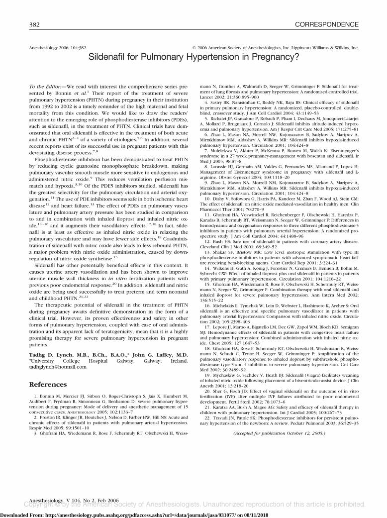
To the Editor:—I read with interest the article of Dixon et al.1 Theresults show in the class III obese patients that preoxygenation in the25° head-up position achieves 23% higher oxygen tensions, allowing aclinical increase in the desaturation safety period. The report postu-lated that preoxygenation in the head-up position may be advanta-geous in many other clinical circumstances in which respiratory func-tion may be impaired in the supine position, e.g., advanced pregnancy,ascites, bowel obstruction.
Baraka et al.2 (1992) reported about “Preoxygenation of Pregnantand Nonpregnant Women in the Head-up versus Supine Position.” Theresults showed that after 3 min of preoxygenation, desaturation to 95%during subsequent apnea, as monitored by pulse oximetry, was morerapid in pregnant than in nonpregnant patients. Also, changing fromthe supine to the 45° head-up position prolonged the desaturation timein the nonpregnant women but had no significant effect in the preg-nant women (table 1). These results were unanticipated because achange from the supine to the sitting position has been shown toincrease the functional residual capacity in both pregnant3 and non-pregnant patients.4 Baraka et al. postulated that adopting the 45°
head-up position rather than the sitting position may not significantlyincrease the functional residual capacity in the pregnant woman atterm, because the gravid uterus at term may not allow a significantdescent of the diaphragm in the head-up position.
Anis S. Baraka, M.D., F.R.C.A.(Hon), American University ofBeirut, Beirut, Lebanon. [email protected]
References
1. Dixon BJ, Dixon JB, Carden JR, Bum AJ, Schachter LM, Playfair JM, LaurieCP, O’Brien PE: Preoxygenation is more effective in the 25° head-up positionthan in the supine position in severely obese patients. ANESTHESIOLOGY 2005;102:1110–5
2. Baraka AS, Hanna MT, Jabbour SI, Nawfal MF, Siba A, Yazbeck VG, KhouryNI, Karam KS: Preoxygenation of pregnant and nonpregnant women in thehead-up versus supine position. Anesth Analg 1992; 75:757–9
3. Russel IF, Chambers WA: Closing volume in normal pregnancy. Br J Anaesth1981; 53:1043–7
4. Nunn JF: Elastic forces and lung volumes, Applied Respiratory Physiology,3rd edition. Edited by Nunn JF. London, Butterworths, 1987, pp 39–40
(Accepted for publication October 12, 2005.)
Table 1. Preoperative Oxygen Saturation (SO2%) and Times to SO2 95% in Nonpregnant versus Pregnant Patients in the Supineand Head-up Positions
Nonpregnant Control Groups Cesarean Delivery Groups
Supine (n � 10) Head-up (n � 10) Supine (n � 10) Head-up (n � 10)
Preoperative SO2% 98.1 � 1.5 98.5 � 9.94 97.5 � 1.3 97.9 � 0.77Time(s) to SO2 95% 243 � 7.4 331 � 7.2 173 � 4.8 156 � 2.8
380 CORRESPONDENCE
Anesthesiology, V 104, No 2, Feb 2006
Downloaded From: http://anesthesiology.pubs.asahq.org/pdfaccess.ashx?url=/data/journals/jasa/931077/ on 08/11/2018

Anesthesiology 2006; 104:381 © 2006 American Society of Anesthesiologists, Inc. Lippincott Williams & Wilkins, Inc.
Mechanism of Benefit of Head-up Preoxygenation inObese Patients
To the Editor:—I read with interest the study report in the June 2005issue of ANESTHESIOLOGY by Dixon et al.1 regarding the benefits ofhead-up preoxygenation in obese patients. Although I do not disputethe basic findings of the study or the benefit of head-up position inobese patients, I question the conclusions drawn in the abstract anddiscussion.
Although there was a strong correlation between oxygen tensionand time to desaturation, it cannot be concluded that the higherarterial oxygen tension (PaO2) itself was protective. The oxygen con-tent of blood in the form of dissolved oxygen under nonhyperbaricpressure conditions is minimal. At the preinduction PaO2 achieved after3 min of preoxygenation in both supine and head-up subjects, hemo-globin would be expected to be 100% saturated, providing maximalblood oxygen content in both study groups. The additional time todesaturation afforded by the small increase in dissolved oxygen reservein the head-up group is unlikely to have been significant. More likely,the benefit of head-up positioning in delaying desaturation (as well as
increasing PaO2) is a result of factors of pulmonary mechanics men-tioned in the study background as they relate to oxygen reserve.
Also for this reason, changing the patient’s position from head-up tosupine at induction as suggested possible to ease intubation maypartially or completely negate the benefits of the head-up preoxygen-ation despite the increased preinduction PaO2. This repositioning ma-neuver might be useful in a follow-up study to test this hypothesis.
David B. Wax, M.D., Mount Sinai School of Medicine, New York,New York. [email protected]
Reference
1. Dixon BJ, Dixon JB, Carden JR, Burn AJ, Schachter LM, Playfair JM, LaurieCP, O’Brien PE: Preoxygenation is more effective in the 25° head-up positionthan in the supine position in severely obese patients. ANESTHESIOLOGY 2005;102:1110–5
(Accepted for publication October 12, 2005.)
Anesthesiology 2006; 104:381 © 2006 American Society of Anesthesiologists, Inc. Lippincott Williams & Wilkins, Inc.
In Reply:—We thank Drs. Wax and Baraka for their interest in ourarticle on the benefits of the head-up position for preoxygenation inclass III obese patients.1 They draw attention to two aspects of ourarticle.
It is interesting that Dr. Baraka et al.2 found, in a small study ofsimilar design, no benefit in desaturation safety with preoxygenation inthe 45° head-up position in pregnant women at term. Perhaps asspeculated, the effect of the gravid uterus on the movement of thediaphragm has a negative impact on lung mechanics. Although thegravid uterus does decrease functional residual capacity, the mecha-nism and distribution of mass are considerably different to those seenin obesity, and therefore, the favorable effects we found with posturechange may not be applicable. Our comments were speculative only,indicating that the head-up position may achieve a prolongation of thedesaturation safety period. The gravid uterus may have a varyingimpact on lung mechanics depending on the posture: supine, 25°, 45°,and sitting up. This is an area for further research because severeobesity and the advanced gravid state are associated with increaseddifficulty in airway management and higher metabolic rates increasingthe risk of hypoxia during anesthetic induction. Oxygen tensions takenin various positions may assist in optimizing preoxygenation, andfurther investigation into the role of position in preoxygenation shouldcontinue in several high-risk groups.
Dr. Wax correctly points out that the dissolved oxygen in bloodunder atmospheric conditions is trivial and unlikely by itself to alter thedesaturation safety period. We agree and indicate within the discussionthat the aim is to optimize lung oxygen content by achieving a posture
that provides optimal respiratory mechanics, lung volumes, functionalresidual capacity, and arterial oxygen tension during preoxygenation.We found a strong correlation between the oxygen tension achievedand the desaturation safety period suggesting that end preoxygenationoxygen tension is an indicator of improved pulmonary oxygen re-serves. We speculate that the extended desaturation safety period inhead-up subjects is due to continued oxygenation of blood from in-creased pulmonary reserves and not directly due to the higher initialoxygen tension. In addition, we caution that after head-up preoxygen-ation, a change to the supine position for intubation may reduce thesefavorable conditions and shorten the desaturation safety period.
Benjamin J. Dixon, M.B.B.S.,* John B. Dixon, M.B.B.S., Ph.D., LindaM. Schachter, M.B.B.S., F.R.A.C.P., Jennifer R. Carden, M.B.B.S.,F.A.N.Z.C.A. *Australian Centre for Obesity Research and Education,Monash University, Melbourne, Australia. [email protected]
References
1. Dixon BJ, Dixon JB, Carden JR, Burn AJ, Schachter LM, Playfair JM, LaurieCP, O’Brien PE: Preoxygenation is more effective in the 25° head-up positionthan in the supine position in severely obese patients: A randomized controlledstudy. ANESTHESIOLOGY 2005; 102:1110–5
2. Baraka AS, Hanna MT, Jabbour SI, Nawfal MF, Sibai AA, Yazbeck VG, KhouryNI, Karam KS: Preoxygenation of pregnant and nonpregnant women in thehead-up versus supine position. Anesth Analg 1992; 75:757–9
(Accepted for publication October 12, 2005.)
381CORRESPONDENCE
Anesthesiology, V 104, No 2, Feb 2006
Downloaded From: http://anesthesiology.pubs.asahq.org/pdfaccess.ashx?url=/data/journals/jasa/931077/ on 08/11/2018
Anesthesiology 2006; 104:382 © 2006 American Society of Anesthesiologists, Inc. Lippincott Williams & Wilkins, Inc.
Sildenafil for Pulmonary Hypertension in Pregnancy?
To the Editor:—We read with interest the comprehensive series pre-sented by Bonnin et al.1 Their report of the treatment of severepulmonary hypertension (PHTN) during pregnancy in their institutionfrom 1992 to 2002 is a timely reminder of the high maternal and fetalmortality from this condition. We would like to draw the readers’attention to the emerging role of phosphodiesterase inhibitors (PDEs),such as sildenafil, in the treatment of PHTN. Clinical trials have dem-onstrated that oral sildenafil is effective in the treatment of both acuteand chronic PHTN2–4 of a variety of etiologies.5,6 In addition, severalrecent reports exist of its successful use in pregnant patients with thisdevastating disease process.7,8
Phosphodiesterase inhibition has been demonstrated to treat PHTNby reducing cyclic guanosine monophosphate breakdown, makingpulmonary vascular smooth muscle more sensitive to endogenous andadministered nitric oxide.9 This reduces ventilation perfusion mis-match and hypoxia.3,10 Of the PDE5 inhibitors studied, sildenafil hasthe greatest selectivity for the pulmonary circulation and arterial oxy-genation.11 The use of PDE inhibitors seems safe in both ischemic heartdisease12 and heart failure.13 The effect of PDEs on pulmonary vascu-lature and pulmonary artery pressure has been studied in comparisonto and in combination with inhaled iloprost and inhaled nitric ox-ide,14–16 and it augments their vasodilatory effects.17,18 In fact, silde-nafil is at least as effective as inhaled nitric oxide in relaxing thepulmonary vasculature and may have fewer side effects.19 Coadminis-tration of sildenafil with nitric oxide also leads to less rebound PHTN,a major problem with nitric oxide administration, caused by down-regulation of nitric oxide synthetase.15
Sildenafil has other potentially beneficial effects in this context. Itcauses uterine artery vasodilation and has been shown to improveuterine muscle wall thickness in in vitro fertilization patients withprevious poor endometrial response.20 In addition, sildenafil and nitricoxide are being used successfully to treat preterm and term neonataland childhood PHTN.21,22
The therapeutic potential of sildenafil in the treatment of PHTNduring pregnancy awaits definitive demonstration in the form of aclinical trial. However, its proven effectiveness and safety in otherforms of pulmonary hypertension, coupled with ease of oral adminis-tration and its apparent lack of teratogenicity, mean that it is a highlypromising therapy for severe pulmonary hypertension in pregnantpatients.
Tadhg D. Lynch, M.B., B.Ch., B.A.O.,* John G. Laffey, M.D.*University College Hospital Galway, Galway, [email protected]
References
1. Bonnin M, Mercier FJ, Sitbon O, Roger-Christoph S, Jais X, Humbert M,Audibert F, Frydman R, Simonneau G, Benhamou D: Severe pulmonary hyper-tension during pregnancy: Mode of delivery and anesthetic management of 15consecutive cases. ANESTHESIOLOGY 2005; 102:1133–7
2. Preston IR, Klinger JR, Houtches J, Nelson D, Farber HW, Hill NS: Acute andchronic effects of sildenafil in patients with pulmonary arterial hypertension.Respir Med 2005; 99:1501–10
3. Ghofrani HA, Wiedemann R, Rose F, Schermuly RT, Olschewski H, Weiss-
mann N, Gunther A, Walmrath D, Seeger W, Grimminger F: Sildenafil for treat-ment of lung fibrosis and pulmonary hypertension: A randomised controlled trial.Lancet 2002; 21:360:895–900
4. Sastry BK, Narasimhan C, Reddy NK, Raju BS: Clinical efficacy of sildenafilin primary pulmonary hypertension: A randomized, placebo-controlled, double-blind, crossover study. J Am Coll Cardiol 2004; 43:1149–53
5. Richalet JP, Gratadour P, Robach P, Pham I, Dechaux M, Joncquiert-LatarjetA, Mollard P, Brugniaux J, Cornolo J: Sildenafil inhibits altitude-induced hypox-emia and pulmonary hypertension. Am J Respir Crit Care Med 2005; 171:275–81
6. Zhao L, Mason NA, Morrell NW, Kojonazarov B, Sadykov A, Maripov A,Mirrakhimov MM, Aldashev A, Wilkins MR: Sildenafil inhibits hypoxia-inducedpulmonary hypertension. Circulation 2001; 104:424–8
7. Molelekwa V, Akhter P, McKenna P, Bowen M, Walsh K: Eisenmenger’ssyndrome in a 27 week pregnancy-management with bosentan and sildenafil. IrMed J 2005; 98:87–8
8. Lacassie HJ, Germain AM, Valdes G, Fernandes MS, Allamand F, Lopez H:Management of Eisenmenger syndrome in pregnancy with sildenafil and L-arginine. Obstet Gynecol 2004; 103:1118–20
9. Zhao L, Mason NA, Morrell NM, Kojonazarov B, Sadykov A, Maripov A,Mirrakhimov MM, Aldashev A, Wilkins MR: Sildenafil inhibits hypoxia-inducedpulmonary hypertension. Circulation 2001; 104:424–8
10. Dishy V, Sofowora G, Harris PA, Kandcer M, Zhan F, Wood AJ, Stein CM:The effect of sildenafil on nitric oxide mediated-vasodilation in healthy men. ClinPharmacol Ther 2001; 70:270–9
11. Ghofrani HA, Voswinckel R, Reichenberger F, Olschewski H, Haredza P,Karadas B, Schermuly RT, Weissmann N, Seeger W, Grimminger F: Differences inhemodynamic and oxygenation responses to three different phosphodiesterase-5inhibitors in patients with pulmonary arterial hypertension: A randomized pro-spective study. J Am Coll Cardiol 2004; 44:1488–96
12. Bush HS: Safe use of sildenafil in patients with coronary artery disease.Cleveland Clin J Med 2001; 68:349–52
13. Shakar SF, Bristow MR: Low level inotropic stimulation with type IIIphosphodiesterase inhibitors in patients with advanced symptomatic heart fail-ure receiving beta-blocking agents. Curr Cardiol Rep 2001; 3:224–31
14. Wilkens H, Guth A, Konig J, Forestier N, Cremers B, Hennen B, Bohm M,Sybrecht GW: Effect of inhaled iloprost plus oral sildenafil in patients in patientswith primary pulmonary hypertension. Circulation 2001; 104:1218–22
15. Ghofrani HA, Wiedemann R, Rose F, Olschewski H, Schermuly RT, Weiss-mann N, Seeger W, Grimminger F: Combination therapy with oral sildenafil andinhaled iloprost for severe pulmonary hypertension. Ann Intern Med 2002;136:515–22
16. Michelakis E, Tymchak W, Lein D, Webster L, Hashimoto K, Archer S: Oralsildenafil is an effective and specific pulmonary vasodilator in patients withpulmonary arterial hypertension: Comparison with inhaled nitric oxide. Circula-tion 2002; 105:2398–403
17. Lepore JJ, Maroo A, Bigatello LM, Dec GW, Zapol WM, Bloch KD, SemigranMJ: Hemodynamic effects of sildenafil in patients with congestive heart failureand pulmonary hypertension: Combined administration with inhaled nitric ox-ide. Chest 2005; 127:1647–53
18. Ghofrani HA, Rose F, Schermuly RT, Olschewski H, Wiedemann R, Weiss-mann N, Schudt C, Tenor H, Seeger W, Grimminger F: Amplification of thepulmonary vasodilatory response to inhaled iloprost by subthreshold phospho-diesterase type 3 and 4 inhibition in severe pulmonary hypertension. Crit CareMed 2002; 30:2489–92
19. Mychaskiw G, Sachdev V, Heath BJ: Sildenafil (Viagra) facilitates weaningof inhaled nitric oxide following placement of a biventricular-assist device. J ClinAnesth 2001; 13:218–20
20. Sher G, Fisch JD: Effect of vaginal sildenafil on the outcome of in vitrofertilization (IVF) after multiple IVF failures attributed to poor endometrialdevelopment. Fertil Steril 2002; 78:1073–6
21. Karatza AA, Bush A, Magee AG: Safety and efficacy of sildenafil therapy inchildren with pulmonary hypertension. Int J Cardiol 2005; 100:267–73
22. Travadi JN, Patole SK: Phosphodiesterase inhibitors for persistent pulmo-nary hypertension of the newborn: A review. Pediatr Pulmonol 2003; 36:529–35
(Accepted for publication October 12, 2005.)
382 CORRESPONDENCE
Anesthesiology, V 104, No 2, Feb 2006
Downloaded From: http://anesthesiology.pubs.asahq.org/pdfaccess.ashx?url=/data/journals/jasa/931077/ on 08/11/2018

Anesthesiology 2006; 104:383 © 2006 American Society of Anesthesiologists, Inc. Lippincott Williams & Wilkins, Inc.
In Reply:—We thank Drs. Lynch and Laffey for their interest in ourseries of 15 cases of severe pulmonary arterial hypertension (PAH)during pregnancy and their useful comment on therapeutic options.1
Our series gathered patients from 1992 to 2002. During this period,therapeutic options have expanded markedly and patient managementhas become more active. Nonetheless, even when considering ourmost recent cases only, pregnancy must be still discouraged undoubt-edly. Therefore, therapeutic abortion is the first-line treatment we offerto patients who are pregnant already. In our experience, however,some patients decline this option. For these patients who are willing tocontinue with their pregnancy, it is particularly important to make surethat an updated optimal treatment is actually implemented. Althoughthere is no curative treatment for idiopathic PAH, several drugs arenow available to target the main dysfunctional pathways of the disease.
These drugs included namely (1) prostaglandin I2 (prostacyclin), (2)endothelin-1 receptor antagonists, and (3) type 5 phosphodiesteraseinhibitors. According to our group2 and to European3 and American4
guidelines, intravenous prostacyclin (epoprostenol) is the treatment ofchoice for patients with PAH in functional class IV. For patients withPAH in functional class III, endothelin-1 receptor antagonists or pros-tacyclin analogs (inhaled iloprost or subcutaneous treprostinil) may beused as an alternative. Guidelines do not provide specific recommen-dations in pregnant patients with regard to drug choice, except thatthe endothelin-1 receptor antagonist bosentan should be contraindi-cated.2,4 This is because animal data indicate that bosentan couldprovide potential major birth defects.5,6
Sildenafil is the first type 5 phosphodiesterase inhibitor approved forclinical use in the United States in patients with PAH, and it is currentlyin registration process in Europe (20 mg three times a day). As pointedout by Drs. Lynch and Laffey, it has several advantages over inhalednitric oxide, and it is also particularly appealing for long-term treatmentbecause of its oral administration, in contrast to other drugs. However,despite a few promising reports, more information is needed in preg-nant patients with functional class III or IV PAH before it could be
considered as a true alternative option to the above-mentioned guide-lines. Meanwhile, we believe that prostacyclin therapy is a morevalidated approach.7 Whatever the drug or combination of drugs cho-sen, it is important to report back both the positive and negativeoutcomes observed, because the experience remains (we hope) par-ticularly scarce in this subpopulation of pregnant patients.
Frederic J. Mercier, M.D., Ph.D.,* Martine Bonnin, M.D., MarcHumbert, M.D., Ph.D. *Antoine Beclere Hospital-Assistance Publiquedes Hopitaux de Paris, France. [email protected]
References
1. Bonnin M, Mercier FJ, Sitbon O, Roger-Christoph S, Jais X, Humbert M,Audibert F, Frydman R, Simonneau G, Benhamou D: Severe pulmonary hyper-tension during pregnancy: Mode of delivery and anesthetic management of 15consecutive cases. ANESTHESIOLOGY 2005; 102:1133–7
2. Humbert M, Sitbon O, Simonneau G: Treatment of pulmonary arterialhypertension. N Engl J Med 2004; 351:1425–36
3. Galie N, Torbicki A, Barst R, Dartevelle P, Haworth S, Higenbottam T,Olschewski H, Peacock A, Pietra G, Rubin LJ, Simonneau G, Priori SG, Garcia MA,Blanc JJ, Budaj A, Cowie M, Dean V, Deckers J, Burgos EF, Lekakis J, Lindahl B,Mazzotta G, McGregor K, Morais J, Oto A, Smiseth OA, Barbera JA, Gibbs S,Hoeper M, Humbert M, Naeije R, Pepke-Zaba J, Task Force: Guidelines ondiagnosis and treatment of pulmonary arterial hypertension. The task force ondiagnosis and treatment of pulmonary arterial hypertension of the EuropeanSociety of Cardiology. Eur Heart J 2004; 25:2243–78
4. Badesch DB, Abman SH, Ahearn GS, Barst RJ, McCrory DC, Simonneau G,McLaughlin VV American College of Chest Physicians: Medical therapy for pul-monary arterial hypertension: ACCP evidence-based clinical practice guidelines.Chest 2004; 126 (suppl):35S–62S
5. Cheng JW: Bosentan. Heart Dis 2003; 5:161–96. Kenyon KW, Nappi JM: Bosentan for the treatment of pulmonary arterial
hypertension. Ann Pharmacother 2003; 37:1055–627. Elliot CA, Stewart P, Webster VJ, Mills GH, Hutchinson SP, Howarth ES,
Bu’lock FA, Lawson RA, Armstrong IJ, Kiely DG: The use of iloprost in earlypregnancy in patients with pulmonary arterial hypertension. Eur Respir J 2005;26:168–73
(Accepted for publication October 12, 2005.)
Anesthesiology 2006; 104:383–4 © 2006 American Society of Anesthesiologists, Inc. Lippincott Williams & Wilkins, Inc.
Effectiveness of Isoflurane in Inducing Delayed Preconditioningagainst Myocardial Infarction In Vivo
To the Editor:—We read with great interest the article by Chiari et al.1
regarding a potential role of endothelial nitric oxide synthase in isoflu-rane-induced delayed preconditioning in rabbit myocardium. The au-thors are to be congratulated for performing an important study aboutthe remote effects of isoflurane in attenuating myocardial infarct afteracute coronary occlusion and reperfusion. In particular, they addressedthe potential role of endothelial nitric oxide synthase and subsequently ofnitric oxide in isoflurane-induced myocardial protection.
Nitric oxide donors have been shown to mimic the protectiveeffects of delayed ischemic preconditioning in rabbits.2 The protectiveeffect of nitric oxide donor was completely abrogated when this agentwas given in conjunction with the peroxynitrite (ONOO-) and hy-droxyl radical (.OH) scavenger mercaptopropionyl glycine,2 suggest-ing nitric oxide induced late preconditioning involved the generationof reactive oxygen species. This is similar in nature to isoflurane in thatisoflurane preconditioning requires the generation of reactive oxygenspecies as a trigger.3 Furthermore, transient systemic ischemic precon-ditioning enhanced early functional recovery of reperfused hearts inthe rabbits, accompanied by an increase in plasma nitric oxide con-centration as well as increases in plasma superoxide dismutase activi-
ty.4 This latter observation seems to suggest nitric oxide as a potentialmediator of ischemic preconditioning in rabbits. This is supportive ofthe study of Chiari et al.1 showing that endogenous nitric oxide mayfunction as a mediator of isoflurane-induced delayed preconditioning.
It should be noted, however, that in all of the studies1–4 mentionedabove, the duration of coronary occlusion was limited to 30 min. Astudy conducted by Kehl et al.5 (from the same research group asChiari et al.1) clearly demonstrated that isoflurane did not produce adelayed preconditioning against myocardial infarction in vivo in dogswhen the duration of coronary occlusion was extended to 60 min.Although the animal species used are different in the two studies,1,5
the duration of coronary occlusion could have played a determinantrole regarding the effectiveness of isoflurane in inducing delayed pre-conditioning against myocardial infarct. It is unexpected that Chiari etal.1 did not comment on this potential limitation of isoflurane precon-ditioning effects. Study has shown that in ischemic-reperfused isolatedguinea pig hearts, the therapeutic time frame for anesthetic precondi-tioning against postischemic contractile dysfunction and infarction isapproximately 25–40 min.6 The protection is maximal when ischemicduration is between 30 and 35 min. This suggests that anesthetic
383CORRESPONDENCE
Anesthesiology, V 104, No 2, Feb 2006
Downloaded From: http://anesthesiology.pubs.asahq.org/pdfaccess.ashx?url=/data/journals/jasa/931077/ on 08/11/2018

preconditioning may be useful therapy only if the typical duration ofischemia during coronary artery bypass falls within this range. There-fore, we have good reason to postulate that isoflurane-induced delayedpreconditioning, if any, is confined to a specific time frame.
As commented by Chiari et al. in the Discussion, aging modulates(reduces the efficiency of) anesthetic preconditioning. One possibleexplanation for this phenomenon is that antioxidant capacity is re-duced with aging. That is, aging is associated with increased formationof reactive oxygen species. Therefore, theoretically, further enhance-ment of oxygen free radical production by volatile anesthetics mayeven prove to be detrimental to an aged population. The oxygen freeradical–induced lipid peroxidation end product 15-F2t-isoprostane perse has been shown to be an independent risk marker of cardiaccomplications and can exacerbate myocardial ischemia–reperfusioninjury.7 In contrast to volatile anesthetics, the intravenous anestheticpropofol has antioxidant property and has been shown experimentallyto better protect hearts of aging animals than hearts of younger animalsagainst postischemic myocardial injury.8 Large prospective clinicaltrials comparing volatile anesthetic preconditioning and intravenous“anesthetic treatment” or trials comparing a combination of the twoare merited, in particular, in the aged population or in those patientswith an expected duration of ischemia during coronary artery bypasslonger than 40 min.
Zhengyuan Xia, M.D., Ph.D.,* Fang Wang, M.D., Tao Luo, M.D.*Renmin Hospital, Wuhan University, Wuhan, China, and Universityof British Columbia, Vancouver, British Columbia, [email protected]
References
1. Chiari PC, Bienengraeber MW, Weihrauch D, Krolikowski JG, Kersten JR,Warltier DC, Pagel PS: Role of endothelial nitric oxide synthase as a trigger andmediator of isoflurane-induced delayed preconditioning in rabbit myocardium.ANESTHESIOLOGY 2005; 103:74–83
2. Takano H, Tang XL, Qiu Y, Guo Y, French BA, Bolli R: Nitric oxidedonors induce late preconditioning against myocardial stunning and infarc-tion in conscious rabbits via an antioxidant-sensitive mechanism. Circ Res1998; 83:73–84
3. Mullenheim J, Ebel D, Frassdorf J, Preckel B, Thamer V, Schlack W: Isoflu-rane preconditions myocardium against infarction via release of free radicals.ANESTHESIOLOGY 2002; 96:934–40
4. Xia Z, Xia R, Lan HT, Luo T, Tang QZ, Xia ZY, Liu XY: Systemic ischemicpreconditioning plus hemodilution enhanced early functional recovery ofreperfused heart in the rabbits. Interactive Cardiovasc Thoracic Surg 2004;3:528–532
5. Kehl F, Pagel PS, Krolikowski JG, Gu W, Toller W, Warltier DC, Kersten JR:Isoflurane does not produce a second window of preconditioning against myo-cardial infarction in vivo. Anesth Analg 2002; 95:1162–8
6. Kevin LG, Katz P, Camara AK, Novalija E, Riess ML, Stowe DF: Anestheticpreconditioning: Effects on latency to ischemic injury in isolated hearts. ANESTHE-SIOLOGY 2003; 99:385–91
7. Xia Z, Kuo KH, Godin DV, Walker MJ, Tao MC, Ansley DM: 15-F2t-isopros-tane exacerbates myocardial ischemia-reperfusion injury of isolated rat hearts.Am J Physiol Heart Circ Physiol 2005; 289:H1366–72
8. Xia Z, Godin DV, Ansle DM: Propofol enhances ischemic tolerance ofmiddle-aged rat hearts: Effects on 15-F2t-isoprostane formation and tissue antiox-idant capacity. Cardiovasc Res 2003; 59:113–21
(Accepted for publication October 20, 2005.)
Anesthesiology 2006; 104:384–5 © 2006 American Society of Anesthesiologists, Inc. Lippincott Williams & Wilkins, Inc.
In Reply:—We thank Xia et al. for their gracious comments aboutour recent work characterizing the role of endothelial nitric oxidesynthase in delayed preconditioning against myocardial infarction pro-duced by isoflurane.1 The authors mention that isoflurane-inducedpreconditioning requires the generation of reactive oxygen species. Infact, we have previously demonstrated that isoflurane produces smallquantities of reactive oxygen species independent of ischemia andreperfusion as detected using dihydroethidium staining and confocallaser microscopy.2 These data provided direct evidence that exposureto isoflurane produces a small burst of reactive oxygen species viaopening of mitochondrial adenosine triphosphate–sensitive potassiumchannels that triggers preconditioning.3
Xia et al. suggest that the duration of coronary artery occlusion maycontribute to the relative efficacy of volatile anesthetics during acute ordelayed preconditioning. Previous data indicated that isoflurane didnot produce delayed preconditioning in dogs exposed to a 60-min leftanterior descending coronary artery occlusion,4 in contrast to thefindings in rabbits when a 30-min coronary occlusion was used.1
Although these results may have been related to the duration ofcoronary occlusion, it is more likely the findings were related todifferences in systemic hemodynamics and coronary collateral bloodflow between species. Coronary artery occlusions of 30 or 60 min induration typically produce myocardial infarct sizes of approximately40 and 33% in rabbits and dogs, respectively. Heart rates in barbiturate-anesthetized rabbits and dogs are approximately 240 and 130 beats/min, respectively. As a result, myocardial oxygen consumption beforeand during coronary occlusion is substantially higher in rabbits ascompared with dogs. In addition to this more pronounced ischemicburden, rabbits have little if any coronary collateral blood flow.5 Incontrast, the canine model of ischemia and reperfusion used in ourprevious investigation4 is complicated by variable degrees of coronarycollateral perfusion, which must be considered when interpreting theresults. We believe that it would also be premature to extrapolate ourfindings in barbiturate-anesthetized, acutely instrumented rabbits1 to
patients with coronary artery disease undergoing cardiac surgery usingcardiopulmonary bypass, as suggested by Xia et al.
In contrast to the arguments of Xia et al., there is little experimentalevidence supporting the hypothesis that propofol produces substantialcardioprotective effects against ischemia–reperfusion injury in vivo.However, a large body of experimental evidence supports the conten-tion that volatile anesthetics exert important protective effects againstreversible and irreversible ischemic injury.6 To date, several clinicaltrials have provided preliminary data to corroborate these experimen-tal findings. In particular, De Hert et al.7 demonstrated that sevofluranebut not propofol preserved myocardial function and attenuated in-creases in troponin I release in patients undergoing coronary arterybypass graft surgery. These data suggested that sevoflurane but notpropofol produces myocardial protection in humans at risk for isch-emic injury.7 Further large-scale, multicenter clinical trials should beperformed to define the utility of volatile anesthetics as cardioprotec-tive agents in humans.
Paul S. Pagel, M.D., Ph.D.,* David C. Warltier, M.D., Ph.D., JudyR. Kersten, M.D. *Medical College of Wisconsin and the Clement J.Zablocki Veterans Affairs Medical Center, Milwaukee, [email protected]
References
1. Chiari PC, Bienengraeber MW, Weihrauch D, Krolikowski JG, Kersten JR,Warltier DC, Pagel PS: Role of endothelial nitric oxide synthase in isoflurane-induced delayed preconditioning in rabbits. ANESTHESIOLOGY 2005; 103:74–83
2. Tanaka K, Weihrauch D, Kehl F, Ludwig LM, LaDisa JF Jr, Kersten JR, PagelPS, Warltier DC: Mechanism of preconditioning by isoflurane in rabbits: A directrole for reactive oxygen species. ANESTHESIOLOGY 2002; 97:1485–90
3. Tanaka K, Weihrauch D, Ludwig LM, Kersten JR, Pagel PS, Warltier DC:Mitochondrial adenosine triphosphate–regulated potassium channel openingacts as a trigger for isoflurane-induced preconditioning by generating reactiveoxygen species. ANESTHESIOLOGY 2003; 98:935–43
4. Kehl F, Pagel PS, Krolikowski JG, Gu W, Toller W, Warltier DC, Kersten JR:Isoflurane does not produce a second window of preconditioning against myo-cardial infarction in vivo. Anesth Analg 2002; 95:1162–8
384 CORRESPONDENCE
Anesthesiology, V 104, No 2, Feb 2006
Downloaded From: http://anesthesiology.pubs.asahq.org/pdfaccess.ashx?url=/data/journals/jasa/931077/ on 08/11/2018

5. Maxwell MP, Hearse DJ, Yellon DM: Species variation in the coronarycollateral circulation during regional myocardial ischaemia: A critical determinantof the rate of evolution and extent of myocardial infarction. Cardiovasc Res 1987;21:737–46
6. Tanaka K, Ludwig LM, Kersten JR, Pagel PS, Warltier DC: Mechanisms ofcardioprotection by volatile anesthetics. ANESTHESIOLOGY 2004; 100:707–21
7. De Hert SG, ten Broeck PW, Mertens E, Van Sommeren EW, De Blier IG,Stockman BA, Rodrigus IE: Sevoflurane but not propofol preserves myocardialfunction in coronary surgery patients. ANESTHESIOLOGY 2002; 97:42–9
(Accepted for publication October 20, 2005.)
Anesthesiology 2006; 104:385 © 2006 American Society of Anesthesiologists, Inc. Lippincott Williams & Wilkins, Inc.
Implications of Postoperative Pruritus
To the Editor:— In a recent Review Article about postoperative pruri-tus regarding anesthesia, Waxler et al.1 discussed in detail the pathway,mechanism, and treatment modalities for postoperative pruritus. How-ever, the saga of postoperative pruritus may not end simply with adiagnosis of pruritus and its treatment. There may be a turning pointafter the exacerbation of the coexisting skin disease as a sequela topruritus and scratching.2–5 In this phenomenon, referred to as theKoebner or isomorphic phenomenon, trauma in a person with certainskin diseases is followed by new lesions in the traumatized but other-wise normal skin, and these new lesions are identical to those in thediseased skin. Although best known in psoriasis, it may also occur inother skin diseases, notably lichen planus, lichen nitidus, pityriasisrubra pilaris, vitiligo, and Darier disease. The Koebner phenomenonbegins 8–10 days after injury. However, it may appear within 3 days ormay be delayed as long as 18 days.3,4 In these diseases, any physical andchemical trauma to skin, including scratching, may precipitate furtherlesions.5 Neuraxial opioids have been implicated to precipitate Koeb-ner phenomenon subsequent to postoperative itching and pruritus.Ideologically, any of the drugs mentioned by the authors that can leadto itching and scratching can precipitate the Koebner phenomenon.1
However, the late manifestation of the Koebner phenomenon afterthe skin trauma and the loss of contact between anesthesiologist andthe patient by this time may lead to ignorance about this important
clinical event after pruritus. It is worth noting that the Koebnerphenomenon after medical therapy may encompass medicolegal im-plications.3,4 Therefore, one must be vigilant regarding this entity andcautious in using any medications or interventions that can lead topruritus, especially in patients with coexisting skin diseases that canmanifest the Koebner phenomenon.
Rajesh Mahajan, M.B.B.S., M.D.,* Rahul Gupta, M.B.B.S., M.D.,Anju Sharma, M.B.B.S. *Acharya Shri Chander College of MedicalSciences, Jammu and Kashmir, India. [email protected] [email protected]
References
1. Waxler B, Dadabhoy ZP, Stojiljkovic L, Rabito SF: Primer of postoperativepruritus for anesthesiologists. ANESTHESIOLOGY 2005; 103:168–78
2. Mahajan R, Grover VK: Neuraxial opioids and Koebner phenomenon: Im-plications for anesthesiologist. ANESTHESIOLOGY 2003; 99:229–30
3. Ramsay DL, Hurlay HJ: Papulosquamous eruptions and exfoliative dermati-tis, Dermatology, 2nd edition. Edited by Moschella SL, Hurley HJ. Philadelphia,Saunders, 1985, pp 499–556
4. Falco OB, Plewig G, Wolff HH, Winkelmann RK: Psoriasis and other exfo-liative skin disorders, Dermatology, 3rd edition. New York, Springer Verlag,1984, pp 403–6
5. Smith MF: Skin and connective tissue diseases, Anesthesia and UncommonPediatric Diseases, 2nd edition. Edited by Ketz J, Steward DJ. Philadelphia,Saunders, 1993, pp 501–62
(Accepted for publication October 20, 2005.)
Anesthesiology 2006; 104:385 © 2006 American Society of Anesthesiologists, Inc. Lippincott Williams & Wilkins, Inc.
In Reply:—We appreciate the interest and comments of Drs. Maha-jan, Gupta, and Sharma regarding our Review Article about postoper-ative pruritus.1 In our Review Article, we summarized coexisting con-ditions that contribute to the choice of anesthesia and treatment ofpostoperative pruritus. Two major purposes for our article were (1) tosummarize up-to-date knowledge about prevention and treatment ofitching after surgery and (2) to illustrate basic principles for diagnosisand treatment for postoperative itching for practicing anesthesiolo-gists. We appreciate the suggestions of Dr. Mahajan et al.2 that anes-thesiologists should be vigilant about the Koebner phenomenon inpatients with certain skin diseases and avoid the use of medicationsthat would lead to pruritus.
In a variety of skin diseases, trauma to the skin may result in the
isomorphic Koebner phenomenon.3–5 We would like to emphasize
that exacerbation of coexisting skin disease is not a sequela to pruritus.
To the best of our knowledge, there is no study that linked mecha-
nisms of pruritus with the Koebner phenomenon. Of course, if itching
is left untreated, it may lead to scratching, and trauma caused by
scratching (but not to itching per se) may cause the Koebner phenom-
enon. Our review focused on early diagnosis and treatment of pruritus
not only to avoid sequelae of itching (scratching, as well as the
isomorphic Koebner phenomenon in certain cutaneous diseases), but
also to improve patients’ satisfaction and to shorten their time in the
recovery room. In addition, we emphasized the increasing need foreffective preventive measures, which are often still missing.
Also, we think that in the future, anesthesiologists will have accessto more specific drugs that will act only at the intended receptor. Withthem, anesthesiologists will be able to modulate specific sites forspecific anesthetic and surgical goals instead of causing undesirableeffects (such as itching).
Beverly Waxler, M.D.,* Zerin P. Dadabhoy, M.D., LjubaStojiljkovic, M.D., Ph.D., Sara F. Rabito, M.D., F.A.H.A. *John H.Stroger Jr. Hospital of Cook County, Chicago, [email protected]
References
1. Waxler B, Dadabhoy ZP, Stojiljkovic L, Rabito SF: Primer of postoperativepruritus for anesthesiologists. ANESTHESIOLOGY 2005; 103:168–78
2. Mahajan R, Kumar Grover V: Neuraxial opioids and Koebner phenomenon:Implications for anesthesiologists. ANESTHESIOLOGY 2003; 99:229–30
3. Rubin AI, Stiller MJ: A listing of skin conditions exhibiting the Koebner andpseudo-Koebner phenomena with eliciting stimuli. J Cutan Med Surg 2002;6:29–34
4. Jolly M: Discoid lupus erythematosus after tattoo: Koebner phenomenon(letter). Arthritis Rheum 2005; 53:627
5. Durani BK, Kurzen H, Hartschuh W, Naeher H: Koebner phenomenon dueto scratch test in scleromyxoedema. Br J Dermatol 2001; 145:306–8
(Accepted for publication October 20, 2005.)
David C. Warltier, M.D., Ph.D., served as Handling Editor for this exchange.
385CORRESPONDENCE
Anesthesiology, V 104, No 2, Feb 2006
Downloaded From: http://anesthesiology.pubs.asahq.org/pdfaccess.ashx?url=/data/journals/jasa/931077/ on 08/11/2018

Anesthesiology 2006; 104:386 © 2006 American Society of Anesthesiologists, Inc. Lippincott Williams & Wilkins, Inc.
Droperidol Has Been Reported to Cause Serious Arrhythmias
To the Editor:—In their recent report on QT interval changes associ-ated with droperidol, White et al.1 state: “Interestingly, despite the useof these high doses of droperidol as part of a neurolept anesthetictechnique for more than 30 yr, there has not been a single report of aserious arrhythmia during or after anesthesia in the peer reviewedliterature.” White made a similar statement in an editorial in 2002.2
However, such a report was published in 2002.3
Mitchel B. Sosis, M.S., M.D., Ph.D., Holy Redeemer Hospital and Med-ical Center, Meadowbrook, Pennsylvania. [email protected]
References
1. White PF, Song D, Abrao J, Klein K, Navarette B: Effect of low-dosedroperidol on the QT interval during and after general anesthesia: A placebo-controlled study. ANESTHESIOLOGY 102:1101–1105.
2. White PF: Droperidol: A cost effective antiemetic for over thirty years.Anesth Analg 2002; 985:789–90
3. Shigeyama T, Yanagidani T: Droperidol causes multifocal ventricular dys-rhythmias. Masui 2002; 51:53–5
(Accepted for publication October 20, 2005.)
Anesthesiology 2006; 104:386 © 2006 American Society of Anesthesiologists, Inc. Lippincott Williams & Wilkins, Inc.
In Reply:—The interest of Dr. Sosis in our recent article1 demon-strating the absence of a clinically significant effect of low-dosedroperidol on the QT interval after a propofol induction is appreciated.As stated in my earlier editorial,2 despite widespread clinical use inanesthesia for more than 30 yr, not a single case report describing adroperidol-induced arrhythmia has appeared in the peer-reviewed an-esthesia literature despite the US Food and Drug Administration–im-posed “black box” warning (excluding the questionable case report3
mentioned by Dr. Sosis, which appeared in a Japanese journal in 2002).Even with extensive use of high-dose droperidol as part of a standard“neurolept” anesthetic technique, there have not been any reportedcases of droperidol-induced dysrhythmias during anesthesia.
In considering the facts of this particular case report,3 Dr. Sosisneglected to mention that the administered dose of droperidol wasmore than 10 times the standard antiemetic dosage. Second, the al-leged case of droperidol-induced multifocal ventricular dysrhythmiasoccurred in a woman with chronic renal failure who was receivinghemodialysis. Importantly, there was no mention of her electrolytestatus at the time of the acute event. Third, she was given a 10-mgintravenous bolus dose of droperidol during general inhalation anes-thesia with a combination of isoflurane and nitrous oxide. This com-bination of inhaled and intravenous drugs would hardly be described asclassic neurolept anesthesia.
I stand by my earlier published statements that there has not been asingle documented case of a serious cardiac arrhythmia occurring in apatient receiving an antiemetic dose (e.g., 0.625–1.25 mg) of droperi-dol or even a large dose of droperidol (e.g., 10 mg) as part of a trueneurolept anesthetic technique. Hopefully, the decision-makers at theFood and Drug Administration will come to their senses and removethe unwarranted black box warning on this highly cost-effective anti-emetic drug.
Paul F. White, Ph.D., M.D., University of Texas Southwestern MedicalCenter, Dallas, Texas. [email protected]
References
1. White PF, Song D, Abrao J, Klein KW, Navarette B: Effect of low-dosedroperidol on the QT interval during general anesthesia: A placebo-controlledstudy. ANESTHESIOLOGY 2005; 102:1101–5
2. White PF: Droperidol: A cost-effective antiemetic for over thirty years.Anesth Analg 2002; 985:89–90
3. Shigeyama T, Yanagidani T: Droperidol causes multifocal ventricular dys-rhythmias. Masui 2002; 51:53–5
(Accepted for publication October 20, 2005.)
Anesthesiology 2006; 104:386–7 © 2006 American Society of Anesthesiologists, Inc. Lippincott Williams & Wilkins, Inc.
Drug-induced Prolongation of the QT Interval: What’s the Point?
To the Editor:—In his recent editorial,1 Scuderi appropriately sug-gested that the action of the US Food and Drug Administration (FDA)in placing a “black box” on the use of low-dose droperidol for thetreatment and prevention of postoperative nausea and vomiting “isclearly specious and does a tremendous disservice to the Americanpublic.”
Although the editorial is excellent, it incorrectly stated that White etal.2 “demonstrated a prolongation of QTc when droperidol (either0.625 or 1.25 mg) is administered intravenously” for antiemetic pro-phylaxis. In fact, the effect of low-dose droperidol was not found to bedifferent from saline (placebo). Given the rigid position taken by the
FDA decision makers in this matter, these findings clearly do not simply“restate the obvious” in their mind. In performing our recently pub-lished study,2 we encountered a patient with sinus bradycardia and acorrected “baseline” QT interval of 419 ms on her 12-lead preoperativescreening electrocardiographic tracing. According to Liu and Juurlink,3
the corrected QT interval is considered to be prolonged if it is greaterthan 450 ms in men or greater than 460 ms in women. Although shewas not entered into the study and did not receive any antiemetic orantibiotic drugs known to prolong the QT interval during surgery, weperformed serial 12-lead electrocardiographic tracings after surgery.On arrival in the recovery room after the patient underwent a super-ficial operation for removal of a small mass in her neck with use of apropofol–remifentanil intravenous anesthetic technique, the QTc wasfound to be prolonged to 685 ms. Subsequent 12-lead electrocardio-graphic tracings at hourly intervals before discharge revealed QTc
The above letter was sent to the author of the referenced Editorial View. Theauthor did not feel that a response was required.—Michael M. Todd, Editor-in-Chief
386 CORRESPONDENCE
Anesthesiology, V 104, No 2, Feb 2006
Downloaded From: http://anesthesiology.pubs.asahq.org/pdfaccess.ashx?url=/data/journals/jasa/931077/ on 08/11/2018
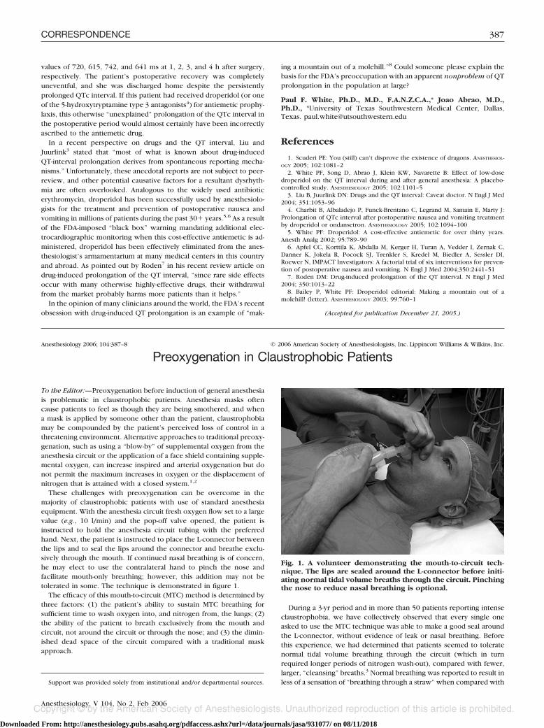
values of 720, 615, 742, and 641 ms at 1, 2, 3, and 4 h after surgery,respectively. The patient’s postoperative recovery was completelyuneventful, and she was discharged home despite the persistentlyprolonged QTc interval. If this patient had received droperidol (or oneof the 5-hydroxytryptamine type 3 antagonists4) for antiemetic prophy-laxis, this otherwise “unexplained” prolongation of the QTc interval inthe postoperative period would almost certainly have been incorrectlyascribed to the antiemetic drug.
In a recent perspective on drugs and the QT interval, Liu andJuurlink3 stated that “most of what is known about drug-inducedQT-interval prolongation derives from spontaneous reporting mecha-nisms.” Unfortunately, these anecdotal reports are not subject to peer-review, and other potential causative factors for a resultant dysrhyth-mia are often overlooked. Analogous to the widely used antibioticerythromycin, droperidol has been successfully used by anesthesiolo-gists for the treatment and prevention of postoperative nausea andvomiting in millions of patients during the past 30� years.5,6 As a resultof the FDA-imposed “black box” warning mandating additional elec-trocardiographic monitoring when this cost-effective antiemetic is ad-ministered, droperidol has been effectively eliminated from the anes-thesiologist’s armamentarium at many medical centers in this countryand abroad. As pointed out by Roden7 in his recent review article ondrug-induced prolongation of the QT interval, “since rare side effectsoccur with many otherwise highly-effective drugs, their withdrawalfrom the market probably harms more patients than it helps.”
In the opinion of many clinicians around the world, the FDA’s recentobsession with drug-induced QT prolongation is an example of “mak-
ing a mountain out of a molehill.”8 Could someone please explain thebasis for the FDA’s preoccupation with an apparent nonproblem of QTprolongation in the population at large?
Paul F. White, Ph.D., M.D., F.A.N.Z.C.A.,* Joao Abrao, M.D.,Ph.D., *University of Texas Southwestern Medical Center, Dallas,Texas. [email protected]
References
1. Scuderi PE: You (still) can’t disprove the existence of dragons. ANESTHESIOL-OGY 2005; 102:1081–2
2. White PF, Song D, Abrao J, Klein KW, Navarette B: Effect of low-dosedroperidol on the QT interval during and after general anesthesia: A placebo-controlled study. ANESTHESIOLOGY 2005; 102:1101–5
3. Liu B, Juurlink DN: Drugs and the QT interval: Caveat doctor. N Engl J Med2004; 351:1053–96
4. Charbit B, Albaladejo P, Funck-Brentano C, Legrand M, Samain E, Marty J:Prolongation of QTc interval after postoperative nausea and vomiting treatmentby droperidol or ondansetron. ANESTHESIOLOGY 2005; 102:1094–100
5. White PF: Droperidol: A cost-effective antiemetic for over thirty years.Anesth Analg 2002; 95:789–90
6. Apfel CC, Korttila K, Abdalla M, Kerger H, Turan A, Vedder I, Zernak C,Danner K, Jokela R, Pocock SJ, Trenkler S, Kredel M, Biedler A, Sessler DI,Roewer N, IMPACT Investigators: A factorial trial of six interventions for preven-tion of postoperative nausea and vomiting. N Engl J Med 2004;350:2441–51
7. Roden DM: Drug-induced prolongation of the QT interval. N Engl J Med2004; 350:1013–22
8. Bailey P, White PF: Droperidol editorial: Making a mountain out of amolehill! (letter). ANESTHESIOLOGY 2003; 99:760–1
(Accepted for publication December 21, 2005.)
Anesthesiology 2006; 104:387–8 © 2006 American Society of Anesthesiologists, Inc. Lippincott Williams & Wilkins, Inc.
Preoxygenation in Claustrophobic Patients
To the Editor:—Preoxygenation before induction of general anesthesiais problematic in claustrophobic patients. Anesthesia masks oftencause patients to feel as though they are being smothered, and whena mask is applied by someone other than the patient, claustrophobiamay be compounded by the patient’s perceived loss of control in athreatening environment. Alternative approaches to traditional preoxy-genation, such as using a “blow-by” of supplemental oxygen from theanesthesia circuit or the application of a face shield containing supple-mental oxygen, can increase inspired and arterial oxygenation but donot permit the maximum increases in oxygen or the displacement ofnitrogen that is attained with a closed system.1,2
These challenges with preoxygenation can be overcome in themajority of claustrophobic patients with use of standard anesthesiaequipment. With the anesthesia circuit fresh oxygen flow set to a largevalue (e.g., 10 l/min) and the pop-off valve opened, the patient isinstructed to hold the anesthesia circuit tubing with the preferredhand. Next, the patient is instructed to place the L-connector betweenthe lips and to seal the lips around the connector and breathe exclu-sively through the mouth. If continued nasal breathing is of concern,he may elect to use the contralateral hand to pinch the nose andfacilitate mouth-only breathing; however, this addition may not betolerated in some. The technique is demonstrated in figure 1.
The efficacy of this mouth-to-circuit (MTC) method is determined bythree factors: (1) the patient’s ability to sustain MTC breathing forsufficient time to wash oxygen into, and nitrogen from, the lungs; (2)the ability of the patient to breath exclusively from the mouth andcircuit, not around the circuit or through the nose; and (3) the dimin-ished dead space of the circuit compared with a traditional maskapproach.
During a 3-yr period and in more than 50 patients reporting intenseclaustrophobia, we have collectively observed that every single oneasked to use the MTC technique was able to make a good seal aroundthe L-connector, without evidence of leak or nasal breathing. Beforethis experience, we had determined that patients seemed to toleratenormal tidal volume breathing through the circuit (which in turnrequired longer periods of nitrogen wash-out), compared with fewer,larger, “cleansing” breaths.3 Normal breathing was reported to result inless of a sensation of “breathing through a straw” when compared withSupport was provided solely from institutional and/or departmental sources.
Fig. 1. A volunteer demonstrating the mouth-to-circuit tech-nique. The lips are sealed around the L-connector before initi-ating normal tidal volume breaths through the circuit. Pinchingthe nose to reduce nasal breathing is optional.
387CORRESPONDENCE
Anesthesiology, V 104, No 2, Feb 2006
Downloaded From: http://anesthesiology.pubs.asahq.org/pdfaccess.ashx?url=/data/journals/jasa/931077/ on 08/11/2018
supranormal tidal volume breaths. There seemed little need to have thepatient manually pinch the nose to prevent nasal breathing, althoughthis may certainly be an option in some patients.
To determine the efficiency of the MTC technique, we conducted aninstitutional review board–approved, minimal risk protocol in 10healthy, nonclaustrophobic volunteers. End-expired oxygen, nitrogen,and carbon dioxide concentrations were measured with a Rascal II gasmonitor (Ohmeda, Louisville, CO) via a sampling catheter attached tothe L-piece of the circuit. Each volunteer pinched his own nose closed.Measurements were recorded before initiation of preoxygenation andat 30, 60, 90,120, 150, and 180 s thereafter. The study divided thevolunteers into two groups and used a crossover design. Mean con-centrations of circuit oxygen, nitrogen, or carbon dioxide were similarbetween the two arms of the study. For example, mean carbon dioxideconcentrations never differed by more than 1 mmHg, denoting con-stant respiratory effort. Circuit nitrogen concentration (mean � SD) forMTC versus facemask breathing were 25 � 5 and 30 � 7%, respec-tively, at 30 s; 11 � 5 and 15 � 6% at 60 s; 3 � 2 and 6 � 5% at 120 s;and 2 � 1 and 4 � 6% at 180 s.
After preoxygenation with this technique and anesthesia inductionwith an intravenous drug, ventilation of the lungs can be rapidlyconverted to a standard mask technique immediately after the patientloses consciousness.
The advantages of the MTC technique for claustrophobic patientsare that it requires no special equipment, the patient’s face is notcovered by a mask, and the patient is in total control of the airway(hence reducing the sense of helplessness). When a complete seal isformed around the L-connector and mouth breathing is used exclu-sively, the oxygen wash-in, nitrogen wash-out properties of the pa-tient–circuit unit should be nearly identical to that attained with themore traditional mask-facilitated preoxygenation used in nonclaustro-phobic patients.
Kirstin M. Erickson, M.D.,* William L. Lanier, M.D. *MayoClinic, Rochester, Minnesota. [email protected]
References
1. Gibson RL, Comer PB, Beckham RW, McGraw CP: Actual tracheal oxygenconcentration with commonly used oxygen equipment. ANESTHESIOLOGY 1976;33:71–3
2. Kory RC, Bergmann JC, Sweet RD, Smith JR: Comparative evaluation ofoxygen therapy techniques. JAMA 1962; 179:767–72
3. Baraka AS, Taha SK, Aouad MT, El-Khatib MF, Kawkabani NI: Preoxygen-ation: Comparison of maximal breathing and tidal volume breathing techniques.ANESTHESIOLOGY 1999; 9:612–6
(Accepted for publication October 24, 2005.)
388 CORRESPONDENCE
Anesthesiology, V 104, No 2, Feb 2006
Downloaded From: http://anesthesiology.pubs.asahq.org/pdfaccess.ashx?url=/data/journals/jasa/931077/ on 08/11/2018