cortical interneuron migration - lab websiteslabs.biology.ucsd.edu/ghosh/ghosh/pdffiles/... · ·...
TRANSCRIPT

INTRODUCTION
While the importance of cell migration in cortical developmenthas long been known, the cellular and molecular basis ofcortical cell migration is only now beginning to be understood.Until recently, the dominant view was that most corticalneurons left the ventricular zone after their last mitosis andmigrated along radial glial fibers to occupy a specific laminarposition within the cortical plate. While this form of migrationcertainly takes place in the developing cortex, recentobservations suggest that radial migration may not provide anadequate description of the cellular events that underlie corticaldevelopment. The first evidence to suggest the need for arevised model came from observations of tangential dispersionof precursors or post-mitotic neurons in the developing cortex(de Carlos et al., 1996; Fishell et al., 1993; O’Rourke et al.,1997; O’Rourke et al., 1992; O’Rourke et al., 1995; Parnavelaset al., 1991; Tan and Breen, 1993; Tan et al., 1995). Thewidespread distribution of clonally related cells also suggestedthe possibility of nonradial migration in the cortex (Walsh andCepko, 1992). The source and destination of these putativetangentially migrating cells, however, remained a mystery untilexperiments by Anderson et al. suggested that neuronsmigrated from the ganglionic eminence (GE) to the cortex andgave rise preferentially to GABAergic interneurons (Andersonet al., 1997). This conclusion was based mainly upon the
observation that there are virtually no neocortical GABAergicneurons in Dlx1/Dlx2 double knockout mice, two homeoboxgenes that are expressed in the ventricular and subventricularzones of the GE (Anderson et al., 1997). It has been suggestedthat the majority of radially migrating neurons generated in thedorsal part of the telencephalon give rise to excitatorypyramidal neurons, whereas neurons migrating tangentiallyfrom the ventral to the dorsal part of the telencephalon giverise to GABAergic interneurons (Parnavelas et al., 1991;Parnavelas, 2000).
Although tangential migration has been extensively studiedin the past few years, little is known about the dynamics andmolecular control of this process. Most of the experimentsperformed so far to study the migration of GE-derived neuronshave studied the role of transcription factors in the specificationof the distinct phenotypes of cortical and striatal interneurons,and have relied on methods such as DiI injection (Anderson etal., 1997; Anderson et al., 2001; Lavdas et al., 1999; Tamamakiet al., 1997) and adenovirus- or electroporation-mediated GFPexpression (Chapouton et al., 1999; Marin et al., 2001). Asonly a small proportion of tangentially migrating cells arelabeled by these methods, we wanted to design a biologicalassay whereby large numbers of tangentially migratingneurons could be visualized, allowing quantitative analysis ofthe dynamics of tangential migration. The sampling limitationin the analysis of migrating cells has hampered molecular
3147Development 129, 3147-3160 (2002)Printed in Great Britain © The Company of Biologists Limited 2002DEV9805
During telencephalic development, cells from the medialganglionic eminence (MGE) are thought to migrate to theneocortex to give rise to a majority of cortical GABAergicinterneurons. By combining time-lapse video-microscopy,immunofluorescence and pharmacological perturbations ina new in vitro migration assay, we find that MGE-derivedcells migrate through the entire extent of the cortex andinto the CA fields of the hippocampus, but avoid the dentategyrus. Migrating neurons initially travel within themarginal zone and intermediate zone, and can enter thecortical plate from either location. Tangential migration isstrongly stimulated by BDNF and NT4 and attenuated bythe Trk-family inhibitor, K252a, suggesting that migration
is regulated by TrkB signaling. Furthermore, TrkB-nullmice show a significant decrease in the number ofcalbindin-positive neurons migrating tangentially in theembryonic cortex. BDNF and NT4 cause rapid activationof PI3-kinase in MGE cells, and inhibition of PI3-kinase(but not of MAP kinase or PLCγ) dramatically attenuatestangential migration. These observations suggest that TrkBsignaling, via PI3-kinase activation, plays an important rolein controlling interneuron migration in the developingcerebral cortex.
Key words: Neurotrophins, Mouse, CNS/migration, Interneuron
SUMMARY
Control of cortical interneuron migration by neurotrophins and PI3-kinase
signaling
Franck Polleux 1, Kristin L. Whitford 2, Paul A. Dijkhuizen 2, Tania Vitalis 3 and Anirvan Ghosh 2,*1INSERM U.371, 18 avenue Doyen Lépine, 69675 Bron, France2Department of Neuroscience, Johns Hopkins University School of Medicine, 725 N Wolfe St, Baltimore, MD 21205, USA3Department of Biomedical Sciences, University of Edinburgh, Hugh Robson Building, Georges Square, Edinburgh EH8 9XD, UK *Author for correspondence (e-mail: [email protected])
Accepted 22 March 2002

3148
investigation of tangential migration, although it is noteworthythat both neurotransmitters released by corticofugal axons aswell as the axonal guidance cues Slit, Sema3a and Sema3f haverecently been implicated in regulating the migration of cellsfrom GE to the neocortex (Métin et al., 2000; Zhu et al., 1999;Marin et al., 2001).
To characterize the migration of neurons from the GE to theneocortex, we have developed a co-culture assay in whichexplants of the GE from GFP-expressing mouse embryos arecultured adjacent to cortical slices from wild-type mice. Atvarious times after culture, the migration of GE cells into theneocortex was analyzed by examining GFP-positive neuronswithin the cortical slice. This assay allows the dynamics ofmigrating cells to be examined by fluorescence videomicroscopy, and also allows the morphology and fate of themigrating cells to be assessed by immunofluorescence. Inaddition, the accessibility of the culture system allowsperturbation experiments that permit investigation of themolecular control of cell migration. Using this assay, we havebeen able to obtain detailed information about the extent anddynamics of tangentially migrating neurons, and havediscovered that TrkB and PI3-kinase signaling play animportant role in the control of the tangential migration ofinterneurons from the GE to the cortex.
MATERIALS AND METHODS
Isochronic and heterochronic co-culturesEmbryonic day 14 (E14) to E16 mouse embryos were isolated fromtimed-mated pregnant wild-type female (E1=midnight after the dayafter the plug is detected), bred with heterozygous transgenic malesexpressing enhanced green fluorescent protein (GFP) under thecontrol of a β-actin promoter and a CMV enhancer (Okabe et al.,1997). The resulting litters contained on average 50% wild-typeand 50% GFP+ heterozygous embryos. Embryos were quicklydissected in ice-cold HBSS (Life Technologies), embedded in 3%low melting point agarose (Sigma) diluted in HBSS and sectionedon a vibratome at 250 µm in the coronal plane. The neocortex orneocortex plus hippocampal formation was trimmed from sectionsfrom wild-type mice and placed adjacent to GE explants from GFP-expressing mice on porous PETC membranes (Falcon 1 µm poresize). The co-cultures were maintained in an air-interface culturesystem as described previously (Polleux et al., 2000). Forheterochromic co-cultures, E14-E16 GE explants from GFP-expressing mice were cultured with rat P2-4 cortical slices isolatedand sectioned as described above. Isochronic and heterochronic GE-GFP co-cultures showed similar patterns and dynamics of cellmigration.
Time-lapse video-microscopyThe migration of GE-GFP cells was monitored using an inverted Zeiss(Axiovert 135) microscope equipped with a heated stage and a 6%CO2 chamber. Images were obtained with a silicon-intensified target(SIT – 2400-08) video-camera (Hamamatsu, Japan) and recorded ontothe hard drive of PC computer before being analyzed using a digitalimage processor (METAMORPH 3.51, Universal Imaging, WestChester, PA) and stored onto CD-ROM. Time-lapse imaging ofmigrating cells was performed on co-cultures on Millipore inserts(Millicell-CM; 0.4 µm pore size) mounted on 60 mm glass-bottomdishes (MatTek Corporation). Multiple locations on several co-cultures were imaged during any one session by piloting a three-axismotorized stage (MultiControl 2000, Marhauzer). The effects ofneurotrophins (BDNF, NT3 and NT4; obtained from Amgen) on
migration was tested directly by pipetting 20 µl (diluted at 20 ng/mlin slice culture medium-control vehicle) onto the explants.
The speed of each migrating cell was computed with theMETAMORPH software, using the ‘track-point’ function to generateseries of points representing the position of each cell body at 15minute intervals. The average rate of migration of each cell wascalculated using Excel 98 software, and the differences of the meanvelocities were analyzed for statistical significance using one-wayANOVA using StatView 5 software.
ImmunofluorescenceAfter 4% paraformaldehyde fixation overnight, doubleimmunofluorescence labeling was performed as described earlier(Polleux et al., 2000). The following antibodies were used. Mousemonoclonals: GFP (1:1000; Molecular Probes), MAP2 (1:2000;Sigma), TuJ1 (1:1000; Sigma), NeuN (1:600; Sigma), BrdU (1:400;Sigma), Nestin (rat 401; 1:20; Developmental Hybridoma Bank) andneurofilament 165 kDa (NF165 kDa; 1:20; DevelopmentalHybridoma Bank). Rabbit polyclonals: GFP (1:3000; MolecularProbes), GABA (1:500; Chemicon) and TrkB (1:600; Oncogene).Primary antibodies were visualized using Cy3- (red) or Cy2- (green)conjugated secondary antibodies (goat anti-mouse or goat anti-rabbit;1:600; Jackson ImmunoResearch). Immunofluorescent labeling wasobserved using a Leica TSC-SP confocal microscope mounted on aLeica DMR microscope. For the detection of NT4 byimmunofluorescence, we used a chick anti-human NT4 polyclonalIgY antibody (1:400; Promega, Madison WI). We then used anaffinity-purified biotin-conjugated anti-chicken IgY secondaryantibody (1:600, Promega, Madison WI) followed by incubation inCy3-conjugated streptavidin (1:1000; CyDye-Amersham PharmaciaBiotech).
PharmacologyWe used LY 294002 [2-(4-Morpholinyl)-8phenyl-4H-1-benzopyran-4-one; Biomol] at a final concentration of 50 µM to inhibit PI3-kinaseactivity (Vlahos et al., 1994); K252a (Calbiochem) at a finalconcentration of 50 nM to inhibit Trk receptor autophosphorylation(Berg et al., 1992; Nye et al., 1992; Tapley et al., 1992); U73122([1-(6-((17b)-3-methoxyestra-1,3,5(10)-trien-17-yl)amino-hexyl)-1H-pyrrole-2,5-dione]; Calbiochem) at a final concentration of 1 µM toinhibit phospholipase C (Smith et al., 1996); and U0126 (1,4-diamino-2,3-dicyano-1,4-bis[2-aminophenylthio]butadiene; Calbiochem) at afinal concentration of 10 µM to inhibit MAP kinase kinase (MEK1/2)(Favata et al., 1998). In each case, the final concentration represented1:1000 dilution of a stock solution in DMSO. Control cultures weretreated with DMSO at a 1:1000 dilution.
Analysis of TrkB mutant miceWild-type mice and mice with a targeted deletion of the TrkB gene(Ntrk2 – Mouse Genome Informatics) (Klein et al., 1993) wereindividually genotyped by PCR performed on proteinase K-digestedtissue. Embryos were fixed at E15 and their brains were immersed infresh paraformaldehyde 4% overnight, then removed from the skull,postfixed overnight and cryoprotected in 30% sucrose in phosphatebuffer for 2 days before sectioning. Serial 46 µm frozen sections wereobtained in the coronal plane. Polyclonal antibodies raised againstcalretinin (1:10,000, Swant, Switzerland) and calbindin 28K(1:20,000, Swant, Switzerland) were used on free-floating sections, aspreviously described (Lebrand et al., 1996). Briefly, sections wereincubated overnight with the primary antibody diluted in PBS+ (0.1M PBS with 0.2% gelatin and 0.25% Triton X-100). Then, sectionswere washed in PBS+ and incubated with secondary antibodies(biotinylated goat anti- rabbit; 1:200; DAKO, Denmark) for 2 hoursat room temperature. Sections were washed in PBS+ and incubatedwith a streptavidin-biotin-peroxidase complex (1:200; Amersham,Arlington Heights, IL) for 2 hours at room temperature. Sections werethen reacted with a solution containing 0.02% diaminobenzidine and
F. Polleux and others

3149Cortical interneuron migration
0.003% H2O2 in PBS, pH 7.6. All sections were mounted on TESPA-coated slides, air-dried overnight, dehydrated and coverslipped inDePeX.
Phospho-AKT western blots E14 GE explants were cultured for 2 days in vitro as described above.They were then pretreated for 30 minutes with 50 µM LY294002(Biomol) or vehicle (DMSO, 1:1000) followed by a 30 minutesstimulation with 50 ng/ml BDNF or NT4. Slices were lysed in Tris-buffered saline containing, 1% nonidet P-40, 10% glycerol, 2 mMEGTA, 1 mM sodium vanadate, 1 mM PMSF, 2 µg/ml pepstatin, 5µg/ml aprotinin, 10 g/ml leupeptin, cleared by centrifugation at13,000 g at 4°C for 10 minutes, and loaded on a 10% polyacrylamidegel for electrophoretic separation. Phosphorylation of Akt wasdetected by western blotting using a rabbit polyclonal phospho-Akt(Ser473) antibody (Cell Signaling Technology). Membranes werestripped and reprobed with rabbit polyclonal Akt antibody (CellSignaling Technology) to control for loading.
RESULTS
Characterization of tangential migration of GE cellsinto the cortexOur initial experiments were directed at characterizing thetrajectory and target specificity of migrating GFP-expressingGE cells (GE-GFP cells) using the GE-cortex co-culture assay(Fig. 1A,B). When E14-E16 GE explants were placed next toisochronic cortical slices, there was a massive influx of GE-GFP cells into the cortical slice within 24 hours. Tangentialmigration was restricted principally to two zones of thecerebral wall: the intermediate zone (IZ) and the marginal zone(MZ) (Fig. 1C,D). Between 2 and 3 days in vitro (DIV), largenumbers of GE-GFP cells could be seen invading the corticalplate (CP) both from the IZ and the MZ (Fig. 1E). Themigrating cells had characteristic and striking morphologies.Virtually all of these cells had an oblong cell body and onemajor leading process that was tipped with a growth cone (Fig.1F,G). The leading process had a gradually taperingmorphology and was typically 10-15 cell body diameters inlength (100-150 µm) (Fig. 1F,G; see Figs 3, 4). In contrast toaxons, which follow stereotypical trajectories to their targets(and often are tightly fasciculated with each other), theorientation of leading processes of migrating neurons washighly variable, suggesting that once within the cerebral wall,the GE-GFP cells often change their direction of localmigration (Fig. 1E).
GE-GFP cells continued to migrate to the cortex in largenumbers during the first few days in culture. To determine theextent of this corticofugal migration, we analyzed thedistribution of GE-GFP cells in co-cultures of GE explants withtelencephalic slices that contained the entire lateral and dorsalextent of the neocortex as well as the hippocampal formation(Fig. 1A,B). In these co-cultures, GE-GFP cells routinelymigrated all the way into the hippocampus, suggesting that atleast some hippocampal neurons originate in the GE (Fig.1B,H-N). Interestingly, there were spatial restrictions tomigration within the hippocampus. Although GE-GFP cellsreadily populated the presumptive CA1-CA3 subfields (pCA,Fig. 1H), there was a clear and consistent lack of GE-GFPneurons within the dentate gyrus (DG, Fig. 1H). The GE-GFPcells avoided the dentate gyrus even when the GE explants
were placed immediately adjacent to a hippocampal slicewhere GE-GFP cells would have easy access to the dentategyrus (Fig. 1I-N). It was also striking that GE-GFP cellsmigrated right up to the border between CA3 and DG withoutinvading the DG proper (Fig. 1H), suggesting that theinhibitory effect of DG on GE-GFP cells was likely to be dueto a cell surface-associated inhibitory signal, rather than adiffusible signal.
To determine the specificity of cell migration out of the GE,we carried out co-culture experiments in which GE explantswere cultured next to the dorsal thalamus, another forebrainstructure. As shown in Fig. 1O, GE-GFP cells showed nomigration into thalamic slices, indicating that GE cells migrateonly into specific target structures such as the cortex andhippocampus. We also examined the ability of neurons fromregions other than the GE to migrate into the cortex. For theseexperiments, explants of various brain regions from GFP-expressing mice were cultured adjacent to neocortical slices.In contrast to the GE explants, which were a rich source ofmigratory cells, cells from other regions showed little or nomigration. For example, thalamic explants from GFP-expressing mice readily extended axons into cortical explants,but thalamic GFP neurons never migrated into the neocortex(data not shown). These experiments indicate that themigration of GE-GFP cells into cortex and hippocampus isboth a consequence of the migratory potential of GE neuronsand the substrate properties of the target tissues.
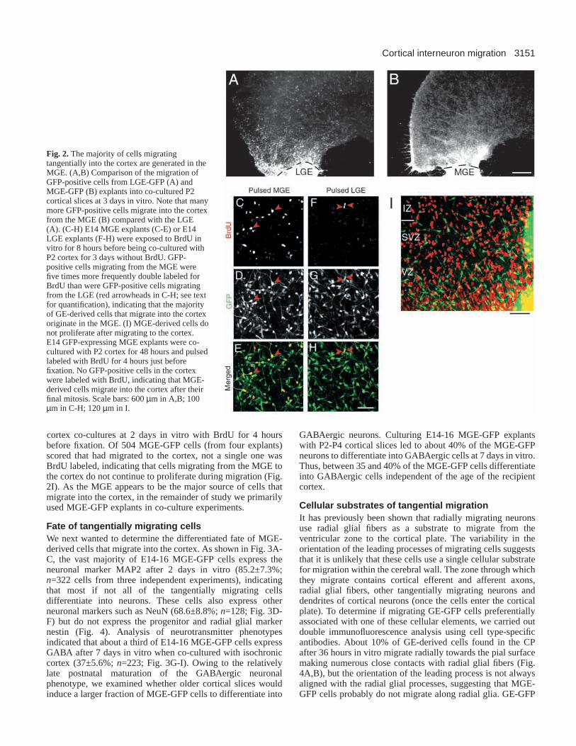
Origin of tangentially migrating cellsWhile the GE-cortex co-culture experiments showed that GEcells migrate in large numbers into the cortex, they did notreveal the contribution of the lateral ganglionic eminence(LGE) and the medial ganglionic eminence (MGE) to thetangentially migrating population. To determine if cells fromthe LGE or MGE migrated equally well to the cortex, wecultured LGE or MGE explants from GFP-expressing micenext to unlabeled cortical slices. As shown in Fig. 2A-B, GFP-expressing cells from both the LGE and MGE were effectivein migrating into the cortical slice, but the number of cellsmigrating out of the MGE was consistently two to three timesgreater than that migrating out of the LGE, indicating that mostof the GE cells that migrate into the cortex at E14-E16 comefrom the MGE.
It is formally possible that the cells we see migrating fromthe MGE or LGE to the cortex are generated in some otherstructure and simply pass through these structures en route tothe cortex. To determine if the cells from the MGE and LGEthat migrate into the cortex undergo their final mitosis in thosestructures, we labeled E14 explants of the LGE or MGE fromGFP-expressing mice with BrdU for 8 hours before washingoff the BrdU and positioning cortical slices next to the explants.After 3 days in culture, we scored the number of BrdU+/GFP+cells in the cortex, which represents the population of cells thatunderwent their final mitotic division during the labelingperiod and then migrated to the cortex. While only about 3%of the LGE-GFP cells in the cortex were double labeled forBrdU, about 15% of the MGE-GFP cells were double labeled(Fig. 2C-H). Thus, at E14-E16, the majority of tangentiallymigrating GE cells originate in the MGE.
To examine whether MGE cells migrating to the cortexcontinue to proliferate while migrating, we labeled MGE-

3150 F. Polleux and others
Fig. 1.Tangentially migrating cells from the ganglioniceminence invade the neocortex and CA fields of thehippocampus, but not the dentate gyrus. (A) A coronal sectionthrough the developing telencephalon showing the locations ofthe medial (M) and lateral (L) ganglionic eminences, cortex andhippocampus (Hipp.) during embryonic development. (B) Lowmagnification photomicrograph of the relative positions of theGE from GFP-expressing mice (GE-GFP), cortex and
hippocampus in a co-culture assay. The white label in the cortex indicates the location of GE-GFP cells that have migrated from the GE tothe cortex. Note the extensive invasion of the cortex and hippocampus by the tangentially migrating cells. (C,D) Analysis of the spatialdistribution of E15 GE-GFP cells that have migrated into the cortex after 20 hours in vitro indicates that GFP cells are found mainly in theintermediate zone (IZ) and the marginal zone (MZ) of co-cultured cortical slices. In these zones, cells are mainly oriented tangentially. Notethat some GFP cells have already invaded the cortical plate (VI, CP). Cells that enter the CP from the IZ typically have leading processesdirected towards the pial surface (arrowhead, C), while those entering the CP from the MZ have leading processes directed away from thepial surface (arrowhead, D), indicating that cell bodies follow the leading process as the cells migrate into the CP. The dorsal (D) and lateral(L) aspects of the cortical slice are indicated in C. In (C,D), the GE explants are located to the right of the cortical explant shown. (E) By 36hours in vitro, large numbers of E16 GE-GFP cells have migrated into the cortex, most of which travel through the IZ. Progressively morecells are found in the CP but they no longer show a clear radial orientation in the CP, suggesting that cells alter their trajectories ofmigration after entering the CP. The broken line in E indicates the edge of the slice on the ventricular side. In C-E, the red channelcorrespond to MAP2 immunofluorescence. (F,G) Examples of the morphology of tangentially migrating cells. The cells typically have aleading process that is 10-15 times the cell body diameter. The leading process is always tipped by a prominent growth cone (arrowheads)and often contains multiple filopodia (arrow in F). Many of the migrating cells also have branched leading processes (G), which mayparticipate in the mechanics of altering trajectories of migration (see also Fig. 5). (H) In GE-cortex co-cultures, E16 GE-GFP cells migratetangentially up to the most medial aspect of the telencephalon by 3 days in vitro where they accumulate in the developing hippocampus butavoid the developing dentate gyrus (DG). Cells in the marginal zone (star) of the putative CA regions (pCA) stop migrating sharply at theinterface between the pCA and DG (arrow) identified as being TuJ1 negative (red channel). (I-K) The region of the developing hippocampusavoided by E16 GE-GFP cells (green channel, I) is a highly proliferative zone (delineated by the broken line in K; red channel, BrdU),which is typical of the DG anlage. (L-N) The avoidance of the developing DG by GE-GFP cells can be observed even when an explant ofE16 MGE-GFP (to the right in L) is placed directly adjacent to the DG for 3 days in vitro. Higher magnification images show a markeddifference in GFP cell density in DG (M) and pCA (N) regions located 250 µm away from the interface with the MGE explant. (O) GE-GFPcells do not migrate into wild-type dorsal thalamus (WT-DT) slices in a co-culture assay, indicating that GE cells are selective about theirtarget zones of migration. All panels in this and other figures are from isochronic co-cultures, unless otherwise indicated. CP: cortical plate;SP: subplate; IZ: intermediate zone; VZ: ventricular zone; DG: developing dentate gyrus; pCA: putative CA regions; VI: cortical layer VI.Scale bars: 150 µm in C-E; 30 µm in F,G; 250 µm in H; 350 µm in I-K; 300 µm in L; 75 µm in M,N; 200 µm in O.
3151Cortical interneuron migration
cortex co-cultures at 2 days in vitro with BrdU for 4 hoursbefore fixation. Of 504 MGE-GFP cells (from four explants)scored that had migrated to the cortex, not a single one wasBrdU labeled, indicating that cells migrating from the MGE tothe cortex do not continue to proliferate during migration (Fig.2I). As the MGE appears to be the major source of cells thatmigrate into the cortex, in the remainder of study we primarilyused MGE-GFP explants in co-culture experiments.
Fate of tangentially migrating cellsWe next wanted to determine the differentiated fate of MGE-derived cells that migrate into the cortex. As shown in Fig. 3A-C, the vast majority of E14-16 MGE-GFP cells express theneuronal marker MAP2 after 2 days in vitro (85.2±7.3%;n=322 cells from three independent experiments), indicatingthat most if not all of the tangentially migrating cellsdifferentiate into neurons. These cells also express otherneuronal markers such as NeuN (68.6±8.8%; n=128; Fig. 3D-F) but do not express the progenitor and radial glial markernestin (Fig. 4). Analysis of neurotransmitter phenotypesindicated that about a third of E14-16 MGE-GFP cells expressGABA after 7 days in vitro when co-cultured with isochroniccortex (37±5.6%; n=223; Fig. 3G-I). Owing to the relativelylate postnatal maturation of the GABAergic neuronalphenotype, we examined whether older cortical slices wouldinduce a larger fraction of MGE-GFP cells to differentiate into
GABAergic neurons. Culturing E14-16 MGE-GFP explantswith P2-P4 cortical slices led to about 40% of the MGE-GFPneurons to differentiate into GABAergic cells at 7 days in vitro.Thus, between 35 and 40% of the MGE-GFP cells differentiateinto GABAergic cells independent of the age of the recipientcortex.
Cellular substrates of tangential migrationIt has previously been shown that radially migrating neuronsuse radial glial fibers as a substrate to migrate from theventricular zone to the cortical plate. The variability in theorientation of the leading processes of migrating cells suggeststhat it is unlikely that these cells use a single cellular substratefor migration within the cerebral wall. The zone through whichthey migrate contains cortical efferent and afferent axons,radial glial fibers, other tangentially migrating neurons anddendrites of cortical neurons (once the cells enter the corticalplate). To determine if migrating GE-GFP cells preferentiallyassociated with one of these cellular elements, we carried outdouble immunofluorescence analysis using cell type-specificantibodies. About 10% of GE-derived cells found in the CPafter 36 hours in vitro migrate radially towards the pial surfacemaking numerous close contacts with radial glial fibers (Fig.4A,B), but the orientation of the leading process is not alwaysaligned with the radial glial processes, suggesting that MGE-GFP cells probably do not migrate along radial glia. GE-GFP
Fig. 2.The majority of cells migratingtangentially into the cortex are generated in theMGE. (A,B) Comparison of the migration ofGFP-positive cells from LGE-GFP (A) andMGE-GFP (B) explants into co-cultured P2cortical slices at 3 days in vitro. Note that manymore GFP-positive cells migrate into the cortexfrom the MGE (B) compared with the LGE(A). (C-H) E14 MGE explants (C-E) or E14LGE explants (F-H) were exposed to BrdU invitro for 8 hours before being co-cultured withP2 cortex for 3 days without BrdU. GFP-positive cells migrating from the MGE werefive times more frequently double labeled forBrdU than were GFP-positive cells migratingfrom the LGE (red arrowheads in C-H; see textfor quantification), indicating that the majorityof GE-derived cells that migrate into the cortexoriginate in the MGE. (I) MGE-derived cells donot proliferate after migrating to the cortex.E14 GFP-expressing MGE explants were co-cultured with P2 cortex for 48 hours and pulsedlabeled with BrdU for 4 hours just beforefixation. No GFP-positive cells in the cortexwere labeled with BrdU, indicating that MGE-derived cells migrate into the cortex after theirfinal mitosis. Scale bars: 600 µm in A,B; 100µm in C-H; 120 µm in I.

3152
cells also make occasional contacts with axons in theintermediate zone (Fig. 4C) and cortical neuron apicaldendrites within the CP (Fig. 4D), but the spatial relationshipbetween the migrating cells and the axons or dendrites suggeststhat GE cells can migrate within the cerebral wall withouthaving to grow along an existing cellular substrate.
Dynamics of tangential migration in the cortexIn the next series of experiments we examined the dynamicsof MGE-GFP neurons as they migrated into the cortex. Forthis, we used time-lapse video-microscopy to image about 300E14-E16 MGE-GFP cells migrating through isochronic wild-type cortex. The period of observation ranged from 3 hours to3 days. The first striking feature of MGE-derived cells is theirfast rate of migration, which averages 58±8.2 µm per hour andcan reach instantaneous rates of migration of about 140µm/hour for periods up to 30 minutes. Fig. 5 shows examplesof the dynamics of migration of MGE-GFP cells as theymigrate from the marginal zone (Fig. 5A-D) or the intermediatezone (Fig. 5E-H) into the cortical plate. We found examples of
MGE-GFP cells migrating from the MZ to the CP (Fig. 5A),and also observed cells migrating from the CP to the MZ (Fig.5B). In both cases, the migratory cells displayed a prolongedpause (50-70 minutes) at the CP/MZ interface before crossingover into the new zone. Many of the migrating cells werecharacterized by a saltatory mode of translocation, alternatingbetween fast and slow instantaneous rates of migration (Fig.5C). Fig. 5D shows that GE-derived cells found in the MZtypically move coherently in a lateral-to-medial direction,indicating that there is a preferred direction of migration withinthe MZ.
MGE-GFP cells in the IZ most frequently invaded the cortexby making sharp 90° turns (Fig. 5E). Unexpectedly, these sharp
F. Polleux and others
Fig. 3.Fate of MGE-derived tangentially migrating cells. (A-C) After 2 daysin vitro, about 85% of E14 MGE-GFP cells (green, B) that migrate into theintermediate zone (IZ) express the neuronal marker MAP2 (A, red in C). Thearrow in C delineates the border between the cortical plate (CP) and the IZ.(D-F) After 3 days in vitro, about 65% of E14 MGE-GFP cells (E, green in F)migrating in the IZ express the nuclear neuronal marker NeuN (D, red in F).(G-I) After 7 days in vitro, about 35% of E14 MGE-GFP cells (H, green in I)found in the cortex express the neurotransmitter GABA (G, red in I). Bluearrowheads indicate examples of double-labeled cells. Scale bars: 150 µm inA-C,G-I; 60 µm in D-F.
Fig. 4.Cellular interactions of MGE-derived tangentiallymigrating neurons. (A,B) Relationship between theleading process of E14 GE-GFP cells and the radial glialfibers in the intermediate zone (red, nestin). Cellsmigrating tangentially in the intermediate zone makeseveral contacts with radial glial fibers, whereas some ofthe cells invading the cortical plate that migrate radially(arrow in A, shown at higher magnification in B) makemultiple close contacts with the nestin+ glial fibers(arrowheads in B). In each image, the dorsal (D) andlateral (L) aspects are indicated. (C) Example of amigrating E14 GE-GFP cell making a few close contactswith axons (red, neurofilament 165 kDa) within thecortical intermediate zone. The insets numbered 1 (red, NF165 kDa), 2 (green, GFP) and 3 (1 and 2 merged)represent a X-Z orthogonal section taken at the levelindicated by the arrowhead in C. The blue arrows (3)indicate the location of close contact between an axon(red) and the leading process (green) of the tangentiallymigrating cell shown in C. (D) Example of an E14 GE-GFP cell migrating radially in the cortical plate towardsthe pia. This GE-GFP neuron makes contacts with corticalapical dendrites, revealed by MAP2 immunofluorescence(red). The insets numbered 1 (red, MAP 2), 2 (green, GFP)and 3 (1 and 2 merged) represent a X-Z orthogonal sectiontaken at the level indicated by the arrowhead in D. Theblue arrows (3) indicate the location of a close contactbetween an apical dendrite and the leading process of theGFP+ cell shown in D. Scale bars: 80 µm in A; 20 µm inB,C; 30 µm in D.

3153Cortical interneuron migration
turns were usually not made by the pre-existing leadingprocess, but instead involved the generation of a new leadingprocess in the new direction of migration (see arrowheads inFig. 5E,F). While the cell changes its direction of migration,the cell body pauses and suspends translocation as a newleading process is generated (Fig. 5E,F). These pauses lastedfor up to 2 hours before migration resumed in the new direction(arrowhead in Fig. 5G). As in the case of cells migrating in theMZ, GE-derived cells migrating in the IZ moved coherently ina lateral-to-medial direction and only a small subpopulationinvaded the CP at any given time (Fig. 5H).
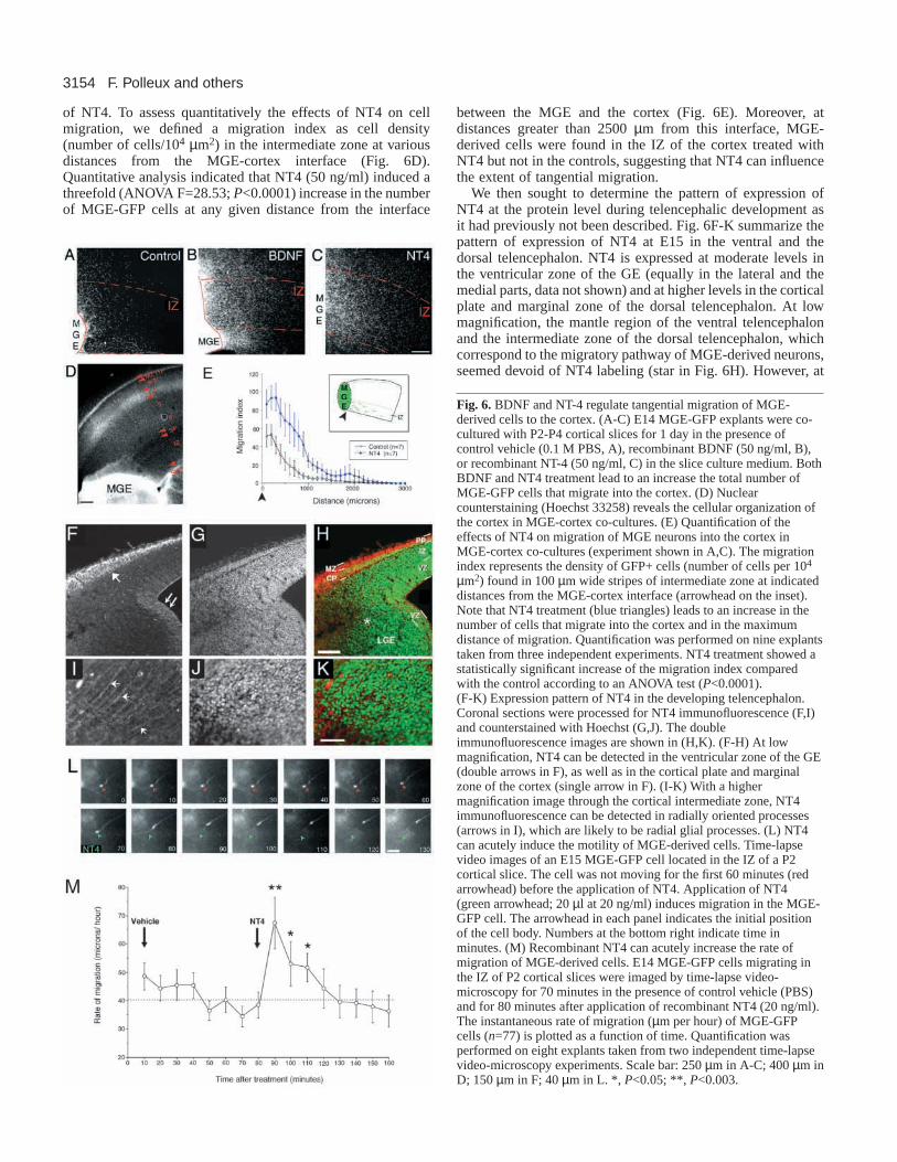
Effects of BDNF and NT4 on tangential cellmigrationTo determine the influence of extracellular factors on the rateand extent of migration of MGE-GFP neurons to the cortex,we examined the effects of various factors on the MGE-GFPcell migration in our co-culture assay. We report on ourinvestigations of the role of neurotrophins in regulatingtangential cell migration. The neurotrophins were tested for
three main reasons: (1) they are expressed in the developingcortex (Maisonpierre et al., 1990; Timmusk et al., 1993;Friedman et al., 1998; Fukumitsu et al., 1998); (2) it haspreviously been suggested that some of their in vivo effectscould be due to a potential action on neuronal migration (Beharet al., 1997; Brunstrom et al., 1997); and (3) their tyrosinekinase receptors, TrkB and TrkC, are expressed in corticalinterneurons (Klein et al., 1990; Gorba and Wahle, 1999). Totest the effects of neurotrophins on the migration of MGE-GFPneurons into the cortex, we treated MGE-cortex co-cultureswith control vehicle solutions or with BDNF, NT3 or NT4 ata concentration of 50 ng/ml. As shown in Fig. 6A-C, just 24hours of stimulation with either BDNF or NT4 led to a markedincrease in the number of MGE-GFP neurons that migrate intothe cortex. NT3 had no effect on tangential migration (data notshown). Thus TrkB ligands potently promote MGE cellmigration into the cortex.
As BDNF and NT4 had similar effects on tangentialmigration and act via similar signaling mechanisms, for theremainder of the study we restricted our analysis to the effects
Fig. 5.Dynamics of tangentially migrationMGE-GFP neurons. (A,B) Time-lapseanalysis of MGE-GFP cells in isochronicco-cultures moving radially down from theMZ to the CP (A) and from the CP to theMZ (B). The position and morphology ofthe migrating cells is shown at 15 minutetime intervals. Note that in each case, thecells pause at the interface between the MZand the CP before crossing into the newzone. (C) Illustration of the kinetics ofmigration of an E15 GE-GFP cell withinthe MZ. Each circle represents the positionof the cell body every 15 minutes andreveals that the cell body translocatesaccording to a saltatory mode of migration.(D) Trajectories of 23 MGE-GFP cellsmigrating within the MZ. Twenty out ofthese 23 cells migrate coherently from thelateral (left-hand side) to medial (right-handside) aspect of the cortex. One cell (#3)migrates mediolaterally, one cell (#1)migrates down into the CP and two cells(#13 and #2) migrate from the CP to theMZ. (E,F) Morphological dynamics of theleading process during a change in thedirection of migration. These cells,migrating within the IZ, make 90° (E) or180° (F) changes in their trajectory in lessthan 2 hours (time interval is indicatedbelow each drawings). Note that in eachcase, the turn is not initiated by the growthcone of the leading process, but instead by asecond leading process that emerges fromthe cell body. (G) Example of an MGE-GFPcells crossing from the IZ to the CP. Notethat the cell initially migrating in the IZpauses for a prolonged period (arrowhead)before changing its trajectory by 90° andentering the CP. The arrows indicate thedirection of migration. Conventions as in C.(H) Trajectories of six GE-GFP cells migrating within the IZ. Note that most cells migrate from a lateral (left) to medial (right) direction.One cell can be seen changing trajectories to enter the CP. Scale bar: 50 µm in A-D,G-H; 100 µm in E,F.
3154
of NT4. To assess quantitatively the effects of NT4 on cellmigration, we defined a migration index as cell density(number of cells/104 µm2) in the intermediate zone at variousdistances from the MGE-cortex interface (Fig. 6D).Quantitative analysis indicated that NT4 (50 ng/ml) induced athreefold (ANOVA F=28.53; P<0.0001) increase in the numberof MGE-GFP cells at any given distance from the interface
between the MGE and the cortex (Fig. 6E). Moreover, atdistances greater than 2500 µm from this interface, MGE-derived cells were found in the IZ of the cortex treated withNT4 but not in the controls, suggesting that NT4 can influencethe extent of tangential migration.
We then sought to determine the pattern of expression ofNT4 at the protein level during telencephalic development asit had previously not been described. Fig. 6F-K summarize thepattern of expression of NT4 at E15 in the ventral and thedorsal telencephalon. NT4 is expressed at moderate levels inthe ventricular zone of the GE (equally in the lateral and themedial parts, data not shown) and at higher levels in the corticalplate and marginal zone of the dorsal telencephalon. At lowmagnification, the mantle region of the ventral telencephalonand the intermediate zone of the dorsal telencephalon, whichcorrespond to the migratory pathway of MGE-derived neurons,seemed devoid of NT4 labeling (star in Fig. 6H). However, at
F. Polleux and others
Fig. 6.BDNF and NT-4 regulate tangential migration of MGE-derived cells to the cortex. (A-C) E14 MGE-GFP explants were co-cultured with P2-P4 cortical slices for 1 day in the presence ofcontrol vehicle (0.1 M PBS, A), recombinant BDNF (50 ng/ml, B),or recombinant NT-4 (50 ng/ml, C) in the slice culture medium. BothBDNF and NT4 treatment lead to an increase the total number ofMGE-GFP cells that migrate into the cortex. (D) Nuclearcounterstaining (Hoechst 33258) reveals the cellular organization ofthe cortex in MGE-cortex co-cultures. (E) Quantification of theeffects of NT4 on migration of MGE neurons into the cortex inMGE-cortex co-cultures (experiment shown in A,C). The migrationindex represents the density of GFP+ cells (number of cells per 104
µm2) found in 100 µm wide stripes of intermediate zone at indicateddistances from the MGE-cortex interface (arrowhead on the inset).Note that NT4 treatment (blue triangles) leads to an increase in thenumber of cells that migrate into the cortex and in the maximumdistance of migration. Quantification was performed on nine explantstaken from three independent experiments. NT4 treatment showed astatistically significant increase of the migration index comparedwith the control according to an ANOVA test (P<0.0001).(F-K) Expression pattern of NT4 in the developing telencephalon.Coronal sections were processed for NT4 immunofluorescence (F,I)and counterstained with Hoechst (G,J). The doubleimmunofluorescence images are shown in (H,K). (F-H) At lowmagnification, NT4 can be detected in the ventricular zone of the GE(double arrows in F), as well as in the cortical plate and marginalzone of the cortex (single arrow in F). (I-K) With a highermagnification image through the cortical intermediate zone, NT4immunofluorescence can be detected in radially oriented processes(arrows in I), which are likely to be radial glial processes. (L) NT4can acutely induce the motility of MGE-derived cells. Time-lapsevideo images of an E15 MGE-GFP cell located in the IZ of a P2cortical slice. The cell was not moving for the first 60 minutes (redarrowhead) before the application of NT4. Application of NT4(green arrowhead; 20 µl at 20 ng/ml) induces migration in the MGE-GFP cell. The arrowhead in each panel indicates the initial positionof the cell body. Numbers at the bottom right indicate time inminutes. (M) Recombinant NT4 can acutely increase the rate ofmigration of MGE-derived cells. E14 MGE-GFP cells migrating inthe IZ of P2 cortical slices were imaged by time-lapse video-microscopy for 70 minutes in the presence of control vehicle (PBS)and for 80 minutes after application of recombinant NT4 (20 ng/ml).The instantaneous rate of migration (µm per hour) of MGE-GFPcells (n=77) is plotted as a function of time. Quantification wasperformed on eight explants taken from two independent time-lapsevideo-microscopy experiments. Scale bar: 250 µm in A-C; 400 µm inD; 150 µm in F; 40 µm in L. *, P<0.05; **, P<0.003.

3155Cortical interneuron migration
higher magnification, NT4 could be detected in radialprocesses, which may be radial glia (Fig. 6I). This is interestingconsidering that tangentially migrating cells make closeorthogonal or longitudinal contacts with nestin+ radial glialprocesses during their migration (Fig. 4A,B). Therefore, NT4is expressed along the migratory path of MGE-derived cells invivo, consistent with a possible role for NT4 in tangentialmigration.
NT4 could influence tangential migration via one or both oftwo mechanisms: (1) by inducing the motility of MGE neuronsas they exit the ventricular zone in the GE; or (2) by acutelyaffecting the rate of cell migration of MGE neurons along theirtangential pathway. To determine if NT4 can acutely influencethe rate of MGE cell migration, we used time-lapse video-microscopy to image MGE-GFP cells before and afterexposure to NT4. In the first set of experiments, we examinedthe ability of NT4 to induce the motility of MGE-GFP cellsthat were not migrating. To do this, we selected E14 MGE-GFP cells in the intermediate zone of P2 cortex after 24-36hours in vitro that had not been migrating for at least 60minutes (Fig. 6L). Cells that respond to this criteria are rare.Sixty minutes after the beginning of recording, the cells werestimulated with 50 ng/ml of recombinant NT4 applied directlyonto the slice. This treatment reliably induced migration incells that were previously stationary (n=7/7 cells; Fig. 6L),indicating that NT4 is sufficient to induce motility on non-migrating MGE cells.
We next tested whether NT4 could induce an increase in therate of migration of MGE-GFP cells. To do this, we performedtime-lapse video analysis of MGE-derived cells migrating inthe IZ of P2 cortical slices. After 80 minutes of observationunder control conditions, we applied recombinant NT4 (20ng/ml) directly onto the slice and monitored changes in the rateof migration of 77 cells (Fig. 6M). This experiment revealedthat as little as 10 minutes of stimulation with recombinantNT4, led to a 70% increase in the rate of migration of MGE-derived cells. An ANOVA analysis revealed that NT4 treatmenthas a significant effect on the rate of migration (F=3.123;P<0.003), whereas the control (vehicle) treatment showed nosignificant effect (F=1.392; P=0.205). Further analysis usingPLSD Fisher test demonstrated that after a brief exposure toNT4 the rate of migration was affected for about 30 minutesafter treatment, and then returned to baseline (broken line inFig. 6M). Thus, NT4 acutely affects the rate of migration ofMGE-derived cells.
Role of Trk-family receptors in controlling tangentialcell migrationAs NT4 and BDNF act as ligands for the TrkB receptortyrosine kinase, we used double immunofluorescence toexamine the expression of TrkB in MGE neurons that migrateto the cortex. As shown in Fig. 7A-C, MGE-GFP neurons thathad migrated into the cortex expressed TrkB. Strikingly, TrkBwas localized in these migrating cells along the entire leadingprocess, while little, if any, was present on the cell bodies.Localization of TrkB on the migrating cells suggests thatendogenous TrkB ligands are likely to be involved in regulatingthe migration of MGE neurons.
To test if activation of Trk-family receptors by endogenousligands was involved in regulating the migration of MGEneurons to the cortex, we examined the effects of treating the
cultures with K252a, an inhibitor of the Trk family receptortyrosine kinases (Knusel and Hefti, 1992). As shown in Fig.7D-E, treatment of MGE-cortex co-cultures with K252asharply reduced the number of MGE-GFP cells that migrateinto the cortex. Quantitative analysis indicated that K252atreatment led to a marked reduction in both the number ofmigrating cells and the distance within the cortex to which thecells migrate (Fig. 7F). These experiments suggest thatactivation of Trk-family receptors by endogenous ligands isinvolved in regulating migration of MGE neurons, and supportsa role for neurotrophins in regulating the migration of MGEcells to the cortex.
To determine if TrkB plays a role in vivo in the control oftangential migration from the MGE to the cortex, we examinedembryos with a targeted deletion of the TrkB gene (Klein etal., 1993). We performed immunocytochemical labeling fortwo Ca2+ binding protein, Calbindin 28K (Fig. 7G-J) andcalretinin (data not shown), which are two early markers oftangentially migrating interneurons (Fonseca et al., 1995), inE15 wild-type and TrkB–/– embryos. Inspection of calbindin28K labeling at low magnification revealed no grossabnormalities in the cytoarchitecture of the ventral and dorsaltelencephalon of TrkB null mice compared with controllittermates (Fig. 7G,H). At higher magnification in both wild-type and TrkB–/–mice, calbindin+ neurons migrate tangentiallyin three distinct layers of the dorsal telencephalic wall (Fig.7I,J): (1) the sub-ventricular zone, (2) the interface between theupper part of the intermediate zone and the subplate, and (3)the marginal zone. At this stage only few calbindin+ neuronshave invaded the cortex, which is reminiscent of our in vitrodata (Fig. 1B-E). Quantitative analysis revealed that there is asignificant reduction (32%) in the number of calbindin+neurons that migrate into the developing cortex in TrkB nullcompared with wild-type cortex (Fig. 7K).
In contrast to the effects of TrkB deletion on the distributionof calbindin+ neurons, we did not find any significantdifference in the distribution of calretinin+ interneuronsmigrating tangentially in the cortex of control (data not shown).The difference in the effects of TrkB deletion on thedistribution of calbindin+ and calretinin+ interneuronssuggests that TrkB signaling may regulate the migration ofspecific subpopulations of interneurons into the cortex.
Role of PI3-kinase signaling in regulating tangentialmigrationStimulation of Trk receptors leads to activation of severalintracellular signaling pathways, of which the best-characterized effectors are (1) MEK1/2-MAP kinase, (2)PLCγ, and (3) PI3-kinase (reviewed by Kaplan and Miller,2000). Pharmacological perturbation experiments show thatinhibition of MAP kinase or PLCγ does not significantly affecttangential migration (Fig. 8F), and raises the possibility thatTrkB signaling might control migration via activation of PI3-kinase. One of the main downstream effectors of PI3-kinase isAKT, which is rapidly phosphorylated upon PI3-kinaseactivation. Phosphorylation of AKT, which can be detected byphospho-AKT-specific antibodies, therefore serves as asensitive assay for PI3-kinase activation. To determine ifstimulation of MGE neurons with BDNF or NT4 led to theactivation of PI3-kinase, we stimulated MGE explants withneurotrophins, and examined AKT phosphorylation by western

3156
blot analysis. BDNF and NT4 stimulation both led to a rapidand robust increase in AKT phosphorylation, which wasprevented by pretreatment of the explant with the PI3-kinaseinhibitor LY294002 (Fig. 8A). Thus, BDNF and NT-4 lead toPI3-kinase activation in MGE cells.
To determine if PI3-kinase function was involved inregulating the migration of cells from the MGE to the cortex,we examined the effects of LY294002 treatment on themigration of MGE-GFP neurons to the cortex in our co-cultureassay. As shown in Fig. 8B-D, LY294002 treatment completelysuppressed the ability of NT4 to stimulate migration of GEneurons into the cortex, indicating that PI3-kinase function isrequired by NT4 to exert its effect on migration. Because theK252a experiment suggested that endogenous neurotrophinsare important in determining the extent of tangential migration,we quantified the LY294002 treatment experiment to see ifmigration was reduced to a level similar to that seen with
K252a. Indeed, quantitative analysis showed that LY294002treatment (with or without NT4, Fig. 8E) reduces migration tolevels comparable to K252a treatment. These experimentsstrongly suggest that TrkB ligands regulate migration of GEcells into the cortex via activation of the PI3-kinase signalingpathway.
Finally, as PI3-kinase has been previously shown to beinvolved in the control of neuronal survival, we wanted to ruleout the possibility that our effect of PI3-kinase inhibition wasdue to an increased level of apoptosis of MGE-derived cells.To do this, we examined the percentage of apoptotic nucleifound in the IZ of P2 cortical slices treated with NT4 andLY294002, as described above. The percentage of nucleishowing condensed chromatin staining by Hoechst 33258 wasless than 1% in control, NT4-treated and NT4+LY294002-treated slices (data not shown). Furthermore, fewer than 0.1%of the MGE-GFP cells show condensed chromatin labeling
F. Polleux and others
Fig. 7.Localization and function of TrkB receptors in tangential migration. (A-C) Double immunofluorescence for GFP (A) and TrkB (B)indicates that E14 MGE-GFP cells migrating in the cortical intermediate zone express the TrkB receptor (merged image in C). Note that theTrkB receptor is localized to the leading processes (arrowheads in C) of MGE-GFP cells. (D-F) Effect of inhibiting Trk receptors on migrationof GE-GFP cells. (D,E) Confocal images of E14 MGE-GFP cells migrating tangentially into the IZ of early postnatal cortex in the presence ofcontrol vehicle (DMSO 1:1000; D) or 50 nM of the Trk tyrosine kinase inhibitor K252a (E). (F) Quantification of the experiments shown inD,E from six slices taken from three independent experiments. Conventions are the same as in Fig. 6E. K252a treatment (red) significantlyinhibits migration according to an ANOVA test (F=28.53; P<0.0001). (G-J) Analysis of tangential migration in wild-type and TrkB null mice.(G-H) Low magnification photographs of coronal sections of E15 wild-type (G) and TrkB–/– mice (H) stained for calbindin 28K. At thisrostrocaudal level of section, the global cytoarchitecture of the telencephalon in the two genotypes is indistinguishable. (I-J) High magnificationphotographs of the lateral cortex of sections shown in G,H. Tangentially migrating calbindin+ cells are found mainly in the subventricular zone(SVZ), the upper part of the intermediate zone (IZ), the subplate (SP) as well as in the marginal zone (MZ) in both wild-type (I) and TrkB–/– (J)cortex. (K) Quantification of the number of calbindin+ cells per 200 µm wide radial column (indicated by double arrows in G,H). This analysisreveals a significant decrease in the number of tangentially migrating calbindin+ cells in TrkB–/– cortex compared with TrkB+/+ cortex at E15[–32%; **P<0.005 – Mann-Whitney test; 12 sections from three wild-type and three knockout mice]. Scale bars: 15 µm in A-C; 250 µm inD,E, 400 µm in G,H; 120 µm in I,J.

3157Cortical interneuron migration
typical of apoptotic cells in the three experimental conditions,suggesting that NT-4 stimulation and inhibition of PI3-kinasedo not affect the survival of migrating MGE cells. Theseexperiments indicate that the effects of PI3-kinase inhibitionon migration are not an indirect consequence of an effect oncell viability, and support a direct role for PI3-kinase in thecontrol of tangential cell migration.
DISCUSSION
We have developed a new in vitro assay that recapitulates thetangential migration of cells from the GE to the cortex, andhave used this assay to explore the cellular and molecularfactors that control tangential migration in the developingtelencephalon. Our observations indicate that GE cells showgreat specificity in their trajectories of migration, and thattangential migration is regulated by neurotrophins and PI3-kinase signaling. Neurotrophins have previously beenimplicated in regulating survival, differentiation and
maturation of cortical neurons. The present results indicate thatin addition to these previously identified cellular functions,BDNF and NT4 play an important role in regulating tangentialmigration of MGE cells into the cortex and thereby contributeto the final distribution of cortical interneurons.
Pattern of migration of GE-derived cells We find that neurons that migrate into the cortex arepreferentially generated in the MGE, and not the LGE. Thisobservation is consistent with recent results that identify theMGE as the principal source of embryonically generatedcortical interneurons. Anderson et al. labeled groups of LGEor MGE neurons in slice cultures and found that MGE-derivedcells preferentially invaded the cortex (Anderson et al., 2001).Similar results were obtained by Wichterle et al., who showedthat transplanted MGE cells, but not LGE cells, migrated intothe cortex in large numbers (Wichterle et al., 2001). Thus,evidence obtained from several independent approachesindicates that the MGE is the principal source of cells thatmigrate into the cortex.
MGE-derived neurons migrate along the IZ and MZ toaccess the dorsal telencephalon. This is also consistent with thefindings of Anderson et al. (Anderson et al., 2001) andWichterle et al. (Wichterle et al., 2001). By time-lapse video-microscopy we were able to follow the trajectories of MGEneurons migrating through the neocortex, and found thattangentially migrating neurons could invade the CP from eitherthe MZ or the IZ. The migration of cells from the MZ to theCP was unexpected and is reminiscent of the migration ofgranule cells from the external granule layer (EGL) to theinternal granule layer (IGL) in the developing cerebellum(reviewed by Hatten and Mason, 1990). It will be of interest to
Fig. 8.Activation of PI3-kinase by neurotrophins and its role intangential migration. (A) AKT phosphorylation in GE explantsinduced by BDNF or NT4 and visualized by western blotting withanti-phospho-AKT antibodies. Note that both BDNF and NT4 induceAKT phosphorylation (top panel), which is suppressed bypreincubation with LY294002 (50 µM), a specific PI3-kinaseinhibitor (lanes 1, 4 and 6). Lower panel indicates western analysisof the same blot with anti-AKT antibodies as a loading control. (B-E) Inhibition of PI3-kinase suppresses tangential migration of MGE-GFP cells. E15 MGE-GFP explants were co-cultured with P2-3cortex for 24 hours in presence of control (B, DMSO), DMSO plusrecombinant NT4 (50 ng/ml, C), or recombinant NT4 (50 ng/ml)plus 50 µM LY294002 (a PI3-kinase inhibitor, D). (E) Quantificationof the results shown in B-D using the migration index as described inFig. 6E (n=8 explants in each condition from four independentexperiments). Note that inhibition of PI3-kinase with LY294002treatment suppresses the NT4 effect, indicating that the effect of NT4on migration requires PI3-kinase activation. Also indicated in green(full squares) is the quantification for treatment with LY294002 alone(ANOVA; F=0.623; not significantly different from LY294002+NT-4, see text). Comparison with Fig. 7F indicates that in LY294002-treated slices, migration is reduced to levels seen with K252a,consistent with the interpretation that endogenous neurotrophinsregulate tangential migration via TrkB and PI3-kinase activation.(F) Quantification of the effect of inhibiting MEK1/2 activity(U0126, 10 µM) and inhibiting phospholipase C (U73122, 1 µM) ontangential migration of MGE-derived cells. Conventions are as inFig. 8E. The migration index curves obtained for U0126 and U73122do not differ significantly for the control (DMSO) curves [ANOVAtest; F=1.021 and F=0.875, respectively]. Scale bar: 500 µm in B-D.

3158
determine if the migration of EGL neurons and MGE neuronsis regulated by similar mechanisms.
We find that MGE-derived neurons migrate throughout thelateromedial extent of the developing telencephalon and invadethe hippocampal primordium. This observation suggests thatsome, if not all, hippocampal interneurons are generated in theGE. Migration of GE cells to the hippocampus has also beenrecently been suggested by Rubenstein and colleagues, based ona lack of GABAergic cells in the hippocampus in Dlx1/Dlx2-null mice (Pleasure et al., 2000). We find that tangentiallymigrating cells specifically avoid the developing dentate gyrus.Although interneurons found in the dentate gyrus are generatedduring largely overlapping periods (E12 to E18 in the mouse)with interneurons found in the rest of the hippocampus (Lubberset al., 1985; Soriano et al., 1989), our experiments suggest thatinterneurons that populate these two hippocampal subdivisionshave distinct origins (Galceran et al., 2000; Grove and Tole,1999; Tole et al., 1997; Tole et al., 2000). The DG also differsfrom the rest of the telencephalon in its ability to produce newneurons throughout adulthood (Eckenhoff and Rakic, 1988;Gould et al., 1999; Rakic, 1985). Our results indicate that signalsexpressed in the DG act as potent chemorepulsive factors forGE-derived migrating neurons, and prevent them from invadingthe DG. The identity of this signal is not yet known, but it isworth noting that several axonal chemorepulsive signals areexpressed exclusively in the dentate gyrus, and may alsofunction as chemorepellants for migrating interneurons.
Dynamics of tangential migrationThe use of GFP-expressing cells in our co-culture assayallowed us to examine the dynamics of tangential migration.The rate of migration of MGE-derived cells with the cerebralwall is 50-60 µm/hour, which is three to ten times faster thancells migrating along radial glial fibers (Komuro and Rakic,1993; Komuro and Rakic, 1992; Miller, 1999). In vivotransplantation experiments also indicate that MGE cells haverapid migration rates in the neocortex (Wichterle et al., 2001).Interestingly, the dynamics of migration described in this studyfor embryonic GE-derived cells are similar to that describedfor migration of cells from the postnatal sub-ventricular zoneof the cortex or striatum, which mainly give rise to astrocytes(Kakita and Goldman, 1999). Therefore common mechanismsmay regulate the motility of non-radially migrating cellsthroughout telencephalic development.
An unexpected feature of the tangentially migrating neuronsis the way they change direction. Instead of following a leadingprocess that turns, in most cases MGE cells change directionby extending a new leading process (originating along the pre-existing leading process or on the cell body). This mode ofturning differs importantly from the mode used by axons wheresteering decisions originate from, and are executed by thegrowth cone at the tip of the axon. It appears that fortangentially migrating neurons the entire leading process canact as a sensor, and that a signal from the leading process canbe propagated to the cell body to initiate a new leading process.The fact that TrkB, which controls the motility of these cells,is expressed along the entire leading process of these migratingneurons, and that membrane ruffles or branches can originateanywhere along the leading process support the notion that theentire leading process is capable of making steering decisionsin tangentially migrating neurons.
The fact that MGE neurons often enter the CP by extendinga new leading process that emerges from the existing leadingprocess raises the possibility that factors that control theinvasion of GE-derived neurons into the CP may act asbranching factors for these cells. A signal that may be involvedin regulating branching of the leading process is Slit, which isexpressed in the developing cortical plate and has a branchingactivity (Wang et al., 1999; Whitford et al., 2002). Slit has alsobeen implicated in regulating LGE cell migration (Zhu et al.,1999), but so far we have not detected a chemotropic effect ofSlit1 or Slit2 on MGE cell migration (K. W. and A. G.,unpublished).
Regulation of migration by neurotrophins and PI3-kinase signalingOur investigations into the molecular control of tangential cellmigration indicate that BDNF and NT4, the high-affinityligands for the TrkB receptor tyrosine kinase, stimulate themigration of MGE-derived neurons. While a role forneurotrophins in cell migration had been previously suggested(Behar et al., 1997; Brunstrom et al., 1997; Schwartz et al.,1997; Ringstedt et al., 1998), there has been little or no directevidence showing that neurotrophins can acutely regulate cellmotility or migration. We find that BDNF and NT4 greatlyincrease the number of MGE cells that migrate into the cortex.Time lapse imaging experiments show that NT4 inducesmotility in stationary cells and acutely increases the rate ofmigration in cells already migrating. We also find thatinhibiting the Trk receptors drastically reduces tangentialmigration and that tangential migration of calbindin+interneurons is reduced in TrkB-null mice. These experimentsstrongly support a role for TrkB ligands in regulating migrationMGE cells to the cortex.
As many of our conclusions are based on the effects ofneurotrophins on the number of MGE-GFP cells present in thecortex, it is important to consider whether these effects may inpart reflect effects of neurotrophins on the survival orproliferation of MGE cells. To examine the effects ofneurotrophin treatment on the survival of MGE cells, wecounted apoptotic profiles (shrunken or fragmented nuclei) inMGE-GFP cells in the absence or presence of NT4.Quantitative analysis showed that less than 1% of MGE-derived cells have apoptotic nuclei under either condition,suggesting that a survival effect of NT4 does not account forthe great increase in MGE-GFP cells found in the cortex afterNT4 treatment (F. P. and A. G., unpublished). Similarly, theaddition of K252a did not increase the percentage of apoptoticprofiles among MGE-GFP cells, indicating that the reductionof MGE-GFP cells in the cortex in the presence of K252a isnot due to increased cell death. BrdU-labeling experimentsindicated that tangentially migrating cells were postmitotic anddid not retain any dividing potential (Fig. 2 and data notshown), suggesting that neurotrophin-induced proliferation ofmigrating cells does not contribute to the increase in the MGE-GFP cells in the cortex after neurotrophin treatment. Ifproliferation were to play a role in the increased number ofMGE-derived cells found in the cortex after NT4 stimulation,this effect would have to be exerted entirely before cells beganmigrating out of the MGE. This seems unlikely because theaverage cycle time of GE precursors in the mouse at E14 isabout 20 hours, and a very significant increase in the number
F. Polleux and others

3159Cortical interneuron migration
of MGE-GFP cells in the cortex is seen within 24 hours. Theseobservations, together with the experiments demonstratingacute effects of neurotrophins on cell motility (Fig. 6), suggestthat the principal effect of BDNF and NT4 on MGE-GFP cellsis to regulate their migration.
Neurotrophin stimulation of the TrkB receptor leads toactivation of the MAP kinase, PLCγ and PI3-kinase signalingpathways (reviewed by Kaplan and Miller, 2000). Ourinvestigations of the contribution of these pathways to neuronalmigration suggest that MAP kinase and PLCγ are not requiredfor MGE cell migration. However, we find that PI3-kinase isactivated in MGE explants by neurotrophins and that PI3-kinase function is required for tangential migration. In recentyears PI3-kinase has been implicated in a large number ofbiological responses downstream of receptor tyrosine kinases,including cell survival, proliferation and migration (reviewedby Vanhaesebroeck et al., 1997). The class Ia PI3-kinases areheterodimers that consist of a catalytic subunit of relativemolecular mass 110 kDa (p110α,β,δ) in complex with anadaptor molecule (p85α, p85β, p55γ or splice variants) thatcontains Src-homology (SH2) domains. It has recently beenshown that in macrophages, colony-stimulating factor 1(CSF1)-induced proliferation requires the activation of p110α,whereas CSF1-induced actin reorganization and migrationdepends on the activity of p110β and p110δ (Vanhaesebroecket al., 1999). It will be of interest to determine if the effects ofTrkB activation on neuronal migration and survival are alsomediated via distinct forms of PI3-kinase. The mechanism bywhich PI3-kinase activation regulates migration of GE cells isnot known, but one possibility is that it might be mediated byRho-family GTPases. PI3-kinase activation leads to theformation of PIP2 and PIP3 at the cell membrane, which canact as docking sites for exchange factors of the Rho GTPasefamily (Han et al., 1998; Ma et al., 1998). Rho proteins, in turn,could locally influence the actin cytoskeleton to regulate cellmotility. Such a mechanism, which is independent of changesin gene expression, would allow local changes in neurotrophinconcentration to acutely influence neuronal migration.
We thank Dr M. Okabe for providing the GFP-expressingtransgenic mice, Dr R. Klein for the TrkB null mice, Dr A. Welcher(Amgen) for the neurotrophins, and Dr A. L. Kolodkin and membersof the Ghosh Laboratory for comments. This work was supported bygrants from INSERM (F. P.), the NIH (NS36176) (A. G.), the Marchof Dimes Birth Defects Foundation (A. G.) and the Pew ScholarsProgram (A. G.).
REFERENCES
Anderson, S. A., Eisenstat, D. D., Shi, L. and Rubenstein, J. L. (1997).Interneuron migration from basal forebrain to neocortex: dependence on Dlxgenes.Science278, 474-476.
Anderson, S. A., Marin, O., Horn, C., Jennings, K. and Rubenstein, J. L.(2001). Distinct cortical migrations from the medial and lateral ganglioniceminences, Development 128, 353-363.
Behar, T. N., Dugich-Djordjevic, M. M., Li, Y. X., Ma, W., Somogyi, R.,Wen, X., Brown, E., Scott, C., McKay, R. D. and Barker, J. L. (1997).Neurotrophins stimulate chemotaxis of embryonic cortical neurons.Eur. J.Neurosci. 9, 2561-2570.
Berg, M. M., Sternberg, D. W., Parada, L. F. and Chao, M. V. (1992). K-252a inhibits nerve growth factor-induced trk proto-oncogene tyrosinephosphorylation and kinase activity.J. Biol. Chem. 267, 13-16.
Brunstrom, J. E., Gray-Swain, M. R., Osborne, P. A. and Pearlman, A. L.
(1997). Neuronal heterotopias in the developing cerebral cortex producedby neurotrophin-4.Neuron18, 505-517.
Chapouton, P., Gartner, A. and Gotz, M. (1999). The role of Pax6 inrestricting cell migration between developing cortex and basal ganglia.Development126, 5569-5579.
de Carlos, J. A., Lopez-Mascaraque, L. and Valverde, F. (1996). Dynamicsof cell migration from the lateral ganglionic eminence in the rat.J. Neurosci.16, 6146-6156.
Eckenhoff, M. F. and Rakic, P. (1988). Nature and fate of proliferative cellsin the hippocampal dentate gyrus during the life span of the rhesus monkey.J. Neurosci. 8, 2729-2747.
Favata, M. F., Horiuchi, K. Y., Manos, E. J., Daulerio, A. J., Stradley, D.A., Feeser, W. S., van Dyk, D. E., Pitts, W. J., Earl, R. A., Hobbs, F. etal. (1998). Identification of a novel inhibitor of mitogen-activated proteinkinase kinase, J. Biol. Chem. 273, 18623-18632.
Fishell, G., Mason, C. A. and Hatten, M. E. (1993). Dispersion of neuralprogenitors within the germinal zones of the forebrain.Nature 362, 636-638.
Fonseca, M., del Rio, J. A., Martinze, A., Gomez, S. and Soriano, E. (1995).Development of Calretinin immunoreactivity in the neocortex of the rat.J.Comp. Neurol. 361, 177-192.
Friedman, W. J., Black, I. B. and Kaplan, D. R. (1998). Distribution of theneurotrophins brain-derived neurotrophic factor, neurotrophin-3, andneurotrophin-4/5 in the postnatal rat brain: an immunocytochemical study.Neuroscience84, 101-114.
Fukumitsu, H., Furukawa, Y., Tsusaka, M., Kinukawa, H., Nitta, A.,Nomoto, H., Mima, T. and Furukawa, S. (1998). Simultaneous expressionof brain-derived neurotrophic factor and neurotrophin-3 in Cajal-Retzius,subplate and ventricular progenitor cells during early development stages ofthe rat cerebral cortex.Neuroscience84, 115-127.
Galceran, J., Miyashita-Lin, E. M., Devaney, E., Rubenstein, J. L. andGrosschedl, R. (2000). Hippocampus development and generation ofdentate gyrus granule cells is regulated by LEF1.Development127, 469-482.
Gorba, T. and Wahle, P. (1999). Expression of TrkB and TrkC but not BDNFmRNA in neurochemically identified interneurons in rat visual cortex andorganotypic cultures.Eur. J. Neurosci. 11, 1179-1190.
Gould, E., Reeves, A. J., Fallah, M., Tanapat, P., Gross, C. G. and Fuchs,E. (1999). Hippocampal neurogenesis in adult Old World primates.Proc.Natl. Acad. Sci. USA96, 5263-5267.
Grove, E. A. and Tole, S. (1999). Patterning events and specification signalsin the developing hippocampus.Cereb. Cortex9, 551-561.
Han, J., Luby-Phelps, K., Das, B., Shu, X., Xia, Y., Mosteller, R. D.,Krishna, U. M., Falck, J. R., White, M. A. and Broek, D. (1998). Roleof substrates and products of PI 3-kinase in regulating activation of Rac-related guanosine triphosphatases by Vav.Science279, 558-560.
Hatten, M. E. and Mason, C. A. (1990). Mechanisms of glial-guidedneuronal migration in vitro and in vivo.Experientia46, 907-916.
Kakita, A. and Goldman, J. E. (1999). Patterns and dynamics of SVZ cellmigration in the postnatal forebrain: monitoring living progenitors in slicepreparations.Neuron23, 461-472.
Kaplan, D. R. and Miller, F. D. (2000). Neurotrophin signal transduction inthe nervous system.Curr. Opin. Neurobiol. 10, 381-391.
Klein, R., Martin-Zanca, D., Barbacid, M. and Parada, L. F. (1990).Expression of the tyrosine kinase receptor gene trkB is confined tothe murine embryonic and adult nervous system.Development109, 845-850.
Klein, R., Smeyne, R. J., Wurst, W., Long, L. K., Auerbach, B. A., Joyner,A. L. and Barbacid, M. (1993). Targeted disruption of the trkBneurotrophin receptor gene results in nervous system lesions and neonataldeath.Cell 75, 113-122.
Knusel, B. and Hefti, F. (1992). K-252 compounds: modulators ofneurotrophin signal transduction.J. Neurochem. 59, 1987-1996.
Komuro, H. and Rakic, P. (1992). Selective role of N-type calcium channelsin neuronal migration.Science257, 806-809.
Komuro, H. and Rakic, P. (1993). Modulation of neuronal migration byNMDA receptors.Science260, 95-97.
Lavdas, A. A., Grigoriou, M., Pachnis, V. and Parnavelas, J. G. (1999). Themedial ganglionic eminence gives rise to a population of early neurons inthe developing cerebral cortex.J. Neurosci. 19, 7881-7888.
Lebrand, C., Cases, O., Adelbrecht, C., Doye, A., Alvarez, C., ElMestikawy, S., Seif, I. and Gaspar, P. (1996). Transient uptake and storageof serotonin in developing thalamic neurons. Neuron 17, 823-835.
Lubbers, K., Wolff, J. R. and Frotscher, M. (1985). Neurogenesis of

3160
GABAergic neurons in the rat dentate gyrus: a combined autoradiographicand immunocytochemical study.Neurosci. Lett. 62, 317-322.
Ma, A. D., Metjian, A., Bagrodia, S., Taylor, S. and Abrams, C. S. (1998).Cytoskeletal reorganization by G protein-coupled receptors is dependent onphosphoinositide 3-kinase gamma, a Rac guanosine exchange factor, andRac.Mol. Cell Biol. 18, 4744-4751.
Maisonpierre, P. C., Belluscio, L., Friedman, B., Alderson, R. F., Wiegand,S. J., Furth, M. E., Lindsay, R. M. and Yancopoulos, G. D. (1990). NT-3, BDNF, and NGF in the developing rat nervous system: parallel as wellas reciprocal patterns of expression.Neuron5, 501-509.
Marin, O., Yaron, A., Bagri, A., Tessier-Lavigne, M. and Rubenstein, J.L. (2001). Sorting of striatal and cortical interneurons regulated bysemaphorin- neuropilin interactions.Science 293, 872-875.
Métin, C., Denizot, J. P. and Ropert, N. (2000). Intermediate zone cellsexpress calcium-permeable AMPA receptors and establish close contactwith growing axons.J. Neurosci. 20, 696-708.
Miller, M. W. (1999). Kinetics of the migration of neurons to ratsomatosensory cortex.Dev. Brain Res. 115, 111-122.
Nye, S. H., Squinto, S. P., Glass, D. J., Stitt, T. N., Hantzopoulos, P.,Macchi, M. J., Lindsay, N. S., Ip, N. Y. and Yancopoulos, G. D. (1992).K-252a and staurosporine selectively block autophosphorylation ofneurotrophin receptors and neurotrophin-mediated responses.Mol. Biol.Cell 3, 677-686.
O’Rourke, N. A., Dailey, M. E., Smith, S. J. and McConnell, S. K. (1992).Diverse migratory pathways in the developing cerebral cortex.Science258,299-302.
O’Rourke, N. A., Sullivan, D. P., Kaznowski, C. E., Jacobs, A. A. andMcConnell, S. K. (1995). Tangential migration of neurons in the developingcerebral cortex.Development121, 2165-2176.
O’Rourke, N. A., Chenn, A. and McConnell, S. K. (1997). Postmitoticneurons migrate tangentially in the cortical ventricular zone.Development124, 997-1005.
Okabe, M., Ikawa, M., Kominami, K., Nakanishi, T. and Nishimune, Y.(1997). ‘Green mice’ as a source of ubiquitous green cells.FEBS Lett. 407,313-319.
Parnavelas, J. G. (2000). The origin and migration of cortical neurones: newvistas.Trends Neurosci. 23, 126-131.
Parnavelas, J. G., Barfield, J. A., Franke, E. and Luskin, M. B. (1991).Separate progenitor cells give rise to pyramidal and nonpyramidal neuronsin the rat telencephalon.Cereb. Cortex1, 463-468.
Pleasure, S., Anderson, A., Hevner, R., Bagri, A., Marin, O., Lowenstein,D. and Rubenstein, J. (2000). Cell migration from the ganglioniceminences is required for the development of hippocampal GABAergicinterneurons.Neuron28, 727-740.
Polleux, F., Morrow, T. and Ghosh, A. (2000). Semaphorin 3A is achemoattractant for cortical apical dendrites.Nature404, 567-573.
Rakic, P. (1985). DNA synthesis and cell division in the adult primate brain.Ann. New York Acad. Sci. 457, 193-211.
Ringstedt, T., Linnarsson, S., Wagner, J., Lendahl, U., Kokaia, Z., Arenas,E., Ernfors, P. and Ibanez, C. F.(1998). BDNF regulates reelin expressionand Cajal-Retzius cell development in the cerebral cortex, Neuron 21, 305-315.
Schwartz, P. M., Borghesani, P. R., Levy, R. L., Pomeroy, S. L. and Segal,R. A. (1997). Abnormal cerebellar development and foliation in BDNF–/–mice reveals a role for neurotrophins in CNS patterning.Neuron19, 269-281.
Soriano, E., Cobas, A. and Fairen, A. (1989). Neurogenesis of glutamic aciddecarboxylase immunoreactive cells in the hippocampus of the mouse. II:Area dentata.J. Comp Neurol. 281, 603-611.
Smith, R. J., Justen, J. M., McNab, A. R., Rosenbloom, C. L., Steele, A.N., Detmers, P. A., Anderson, D. C. and Manning, A. M. (1996). U-73122: a potent inhibitor of human polymorphonuclear neutrophil adhesionon biological surfaces and adhesion-related effector functions.J.Pharmacol. Exp. Ther. 278, 320-329.
Tamamaki, N., Fujimori, K. E. and Takauji, R. (1997). Origin and route oftangentially migrating neurons in the developing neocortical intermediatezone.J. Neurosci. 17, 8313-8323.
Tan, S.-S. and Breen, S. (1993). Radial mosaicism and tangential celldispersion both contribute to mouse neocortical development.Nature362,638-639.
Tan, S.-S., Faulkner-Jones, B., Breen, S. J., Walsh, M., Bertram, J. F. andReese, B. E. (1995). Cell dispersion patterns in different cortical regionsstudied with an X-inactivated transgenic marker.Development121, 1029-1039.
Tapley, P., Lamballe, F. and Barbacid, M. (1992). K252a is a selectiveinhibitor of the tyrosine protein kinase activity of the trk family ofoncogenes and neurotrophin receptors.Oncogene7, 371-381.
Timmusk, T., Belluardo, N., Metsis, M. and Perrson, H. (1993). Widespreadand developmentally regulated expression of NT-4 mRNA in rat brain andperipheral tissues.Eur. J. Neurosci. 5, 605-613.
Tole, S., Christian, C. and Grove, E. A. (1997). Early specification andautonomous development of cortical fields in the mouse hippocampus.Development124, 4959-4970.
Tole, S., Goudreau, G., Assimacopoulos, S. and Grove, E. A. (2000). Emx2is required for growth of the hippocampus but not for hippocampal fieldspecification.J. Neurosci. 20, 2618-2625.
Vanhaesebroeck, B., Leevers, S. J., Panayotou, G. and Waterfield, M. D.(1997). Phosphoinositide 3-kinases: a conserved family of signaltransducers.Trends Biochem. Sci. 22, 267-272.
Vanhaesebroeck, B., Jones, G. E., Allen, W. E., Zicha, D., Hooshmand-Rad, R., Sawyer, C., Wells, C., Waterfield, M. D. and Ridley, A. J. (1999).Distinct PI(3)Ks mediate mitogenic signalling and cell migration inmacrophages.Nat. Cell Biol. 1, 69-71.
Vlahos, C. J., Matter, W. F., Hui, K. Y. and Brown, R. F. (1994). A specificinhibitor of phosphatidylinositol 3-kinase, 2-(4-morpholinyl)-8-phenyl-4H-1-benzopyran-4-one (LY294002). J. Biol. Chem. 269, 5241-5248.
Walsh, C. and Cepko, C. L. (1992). Widespread dispersion of neuronal clonesacross functional regions of the cerebral cortex.Science255, 434-440.
Wang, K. H., Brose, K., Arnott, D., Kidd, T., Goodman, C. S., Henzel, W.and Tessier-Lavigne, M. (1999). Biochemical purification of a mammalianslit protein as a positive regulator of sensory axon elongation and branching.Cell 96, 771-784.
Whitford, K. L., Marillat, V., Stein, E., Goodman, C. S., Tessier-Lavigne,M., Chedotal, A. and Ghosh, A. (2002). Regulation of cortical dendritedevelopment by Slit-Robo interactions.Neuron33, 47-61.
Wichterle, H., Turnbull, D. H., Nery, S., Fishell, G. and Alvarez-Buylla,A. (2001). In utero fate mapping reveals distinct migratory pathways andfates of neurons born in the mammalian basal forebrain.Development 128,3759-3771.
Zhu, Y., Li, H., Zhou, L., Wu, J. Y. and Rao, Y. (1999). Cellular andmolecular guidance of GABAergic neuronal migration from an extracorticalorigin to the neocortex.Neuron23, 473-485.
F. Polleux and others