cortical magnification plus cortical plasticity equals vision? · cortical magnification plus...
TRANSCRIPT
Vision Research 111 (2015) 161–169
Contents lists available at ScienceDirect
Vision Research
journal homepage: www.elsevier .com/locate /v isres
Cortical magnification plus cortical plasticity equals vision? q
http://dx.doi.org/10.1016/j.visres.2014.10.0020042-6989/� 2014 Elsevier Ltd. All rights reserved.
q Scope and purpose of article: We have been asked to provide a perspective oncortical aspects of vision restoration. Our laboratory studies the functionalorganization of early visual cortical areas and their role in visually guided behaviorin alert, nonhuman primates, and no lab member is involved in practical efforts toproduce a visual prosthesis. We approach the problem from a visual neurophysi-ologist’s perspective and thus largely ignore a number of specific, practical issuesrelated to neurosurgical approaches or engineering problems. Instead we focus onissues relevant to attempts to introduce visual signals by the electrical stimulationof primary visual cortex and how the rest of the visual system may or may not beable to make sense of them.⇑ Corresponding author at: Dept. of Neurobiology, Harvard Medical School, 220
Longwood Ave., Boston, MA 02115, United States.E-mail addresses: [email protected] (R.T. Born), [email protected]
(A.R. Trott), [email protected] (T.S. Hartmann).
1 This is a rough estimate. There are a number of different definitions ofbased on factors ranging from anatomical features, such as the ‘‘foveal pit’1941; Schein, 1988), to psychophysical measurements of absent rod visiosubject’s preferred locus of fixation (Putnam et al., 2005).
2 The peripheral retina obviously plays a critical role in alerting the obpotential threats, usually involving moving objects. We largely ignore thishere, but suggest that a visual prosthesis might provide this function throuother means such as an audible warning when out-of-view motion is detec
3 Acuity can be measured in a variety of ways including with sinusoidalletters of various sizes (e.g. Snellen) and a vernier task. Each of these yielddifferent measures of acuity. See Westheimer (1992) for an excellent treatmetopic.
Richard T. Born a,b,⇑, Alexander R. Trott a,c, Till S. Hartmann a
a Dept. of Neurobiology, Harvard Medical School, United Statesb Center for Brain Science, Harvard University, United Statesc Harvard PhD Program in Neuroscience, United States
a r t i c l e i n f o a b s t r a c t
Article history:Received 6 May 2014Received in revised form 15 September 2014Available online 16 October 2014
Keywords:Primary visual cortexV1Magnification factorVision restorationVisual prosthesisPlasticity
Most approaches to visual prostheses have focused on the retina, and for good reasons. The earlier that oneintroduces signals into the visual system, the more one can take advantage of its prodigious computationalabilities. For methods that make use of microelectrodes to introduce electrical signals, however, the limiteddensity and volume occupying nature of the electrodes place severe limits on the image resolution that canbe provided to the brain. In this regard, non-retinal areas in general, and the primary visual cortex in par-ticular, possess one large advantage: ‘‘magnification factor’’ (MF)—a value that represents the distanceacross a sheet of neurons that represents a given angle of the visual field. In the foveal representation of pri-mate primary visual cortex, the MF is enormous—on the order of 15–20 mm/deg in monkeys and humans,whereas on the retina, the MF is limited by the optical design of the eye to around 0.3 mm/deg. This meansthat, for an electrode array of a given density, a much higher-resolution image can be introduced into V1than onto the retina (or any other visual structure). In addition to this tremendous advantage in resolution,visual cortex is plastic at many different levels ranging from a very local ability to learn to better detect elec-trical stimulation to higher levels of learning that permit human observers to adapt to radical changes totheir visual inputs. We argue that the combination of the large magnification factor and the impressive abil-ity of the cerebral cortex to learn to recognize arbitrary patterns, might outweigh the disadvantages ofbypassing earlier processing stages and makes V1 a viable option for the restoration of vision.
� 2014 Elsevier Ltd. All rights reserved.
1. The problem of resolution
It is a remarkable fact that so vital a sensory modality to us pri-mates is dominated by such a tiny sensory epithelium. In fact, mostof what the average person thinks of when she thinks of ‘‘seeing’’ isfoveal vision, subserved by only about one percent of the retina’s
total area.1 One manifestation of this fact is the great surpriseevoked in observers when the extremely poor acuity of their extra-foveal retina is demonstrated to them: more than a few degreesaway from the fovea we are all legally blind, meaning our acuity fallspast 20/200. The bottom line is that when we want to see something,we point our foveae at it.2
The basis for foveal acuity3 begins with the high density of conephotoreceptors in this region of the retina (1.6 � 105 cones/mm2 inthe fovea; Wandell, 1995) and is preserved by the unique circuitrybetween the photoreceptors and the so-called ‘‘midget’’ retinal
the fovea’ (Polyak,n or the
server tofunctiongh someted.gratings,
s slightlynt of this
5.4° 2.3° 1°
HM
45d
135d
135d
45d
HM
5.4° 2.3°
80°40° 20°
10° 5° 2.5°
45d
135d 180d
HMfovea*
* 0d
* *
* *
* *
*
* * *
MonocularRegion
20°10°
40°80° 135d 180d
0d 45d
HM
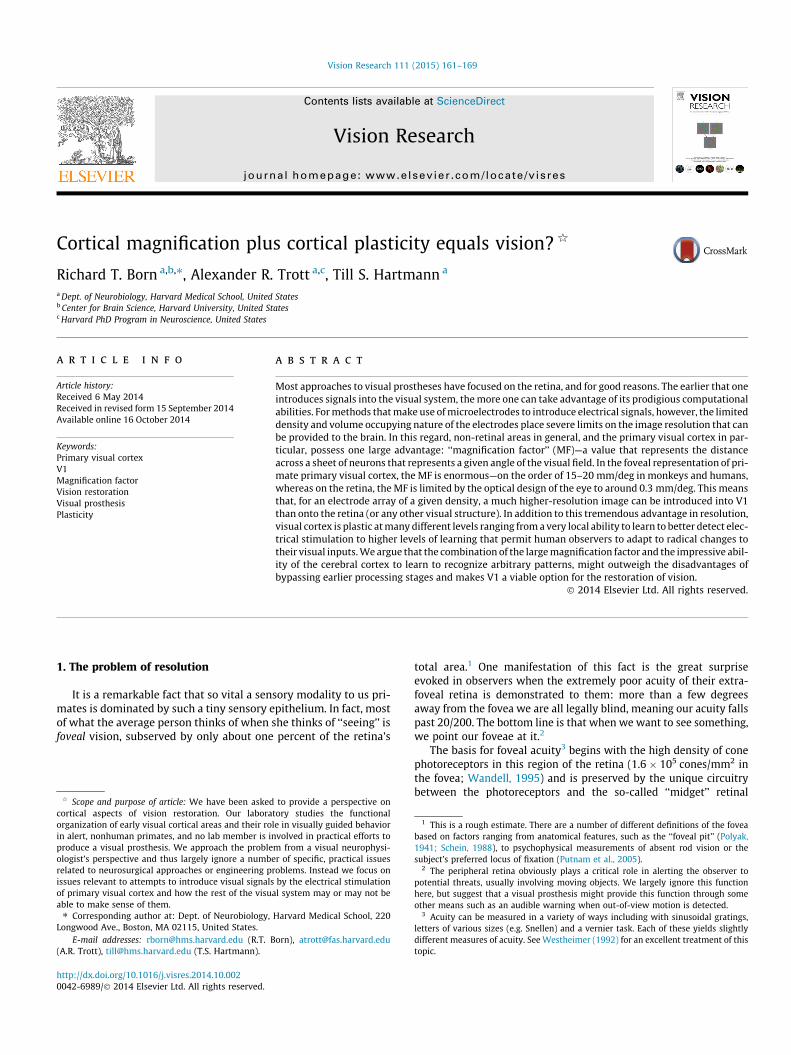
Fig. 1. Retinotopic organization of macaque primary visual cortex. Top: Topographyof V1 mapped with microelectrode recordings, from Van Essen, Newsome, andMaunsell (1984). Bottom: Topography of peri-foveal V1 mapped using 2-deoxy-glucose functional labeling, from Tootell et al. (1988). Scale bars: top left, 1 cm;bottom left, 1 cm; bottom right, 2�. For clarity, polar angles in degrees use the ‘‘d’’symbol, whereas distances in the visual field in degrees use ‘‘�’’.
162 R.T. Born et al. / Vision Research 111 (2015) 161–169
ganglion cells (Kolb & Dekorver, 1991; Kolb & Marshak, 2003). In thelateral geniculate nucleus (LGN), the next stage of visual processing,we already begin to see the over-representation of central vision. Forexample, in layer 6 of the monkey LGN, 20% of the retinotopic map isdevoted to the central 5� (0.5% of the visual field; Connolly & VanEssen, 1984; Malpeli & Baker, 1975). This trend continues and isamplified in the projection from the LGN to V1, where nearly half(42%) of the entire surface of V1 is devoted to the central 5� andabout half of this half represents the fovea (Fig. 1; Tootell et al.,1988; Van Essen, Newsome, & Maunsell, 1984).
For a visual prosthesis, the most relevant consideration deter-mining the necessary electrode density is the distance that onemust travel along neural tissue to cover one degree of the visualfield, the ‘‘magnification factor’’ (MF). While there is variability inthe literature, the most reliable consensus for the maximum MFin monkey V1 is 15–20 mm of cortex per degree of visual field,based on both electrophysiological recordings (Dow et al., 1981;Van Essen, Newsome, & Maunsell, 1984); Fig. 1, top) and 2-deoxy-glucose functional labeling (Tootell et al., 1982, 1988; Fig. 1, bot-tom). And roughly similar values have been reported in humansusing either the mapping of phosphenes produced by direct stim-ulation of visual cortex (Cowey & Rolls, 1974; based on data fromBrindley & Lewin, 1968), mapping of migraine-induced phosph-enes (Grüsser, 1995) or functional magnetic resonance imaging(Duncan & Boynton, 2003; Engel et al., 1994; Sereno et al., 1995).
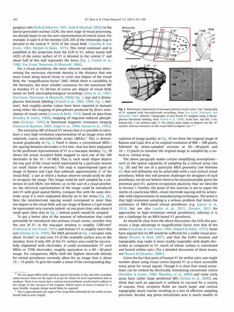
The enormous MF of foveal V1 means that it is possible to intro-duce a very high resolution representation of an image even withrelatively coarse microelectrode arrays (MEAs).4 This is demon-strated graphically in Fig. 2. Panel A shows a conventional MEA—the spacing between electrodes is 0.4 mm—that has been implantedin the perifoveal representation of V1 in a macaque monkey. Fig. 2Bshows the actual receptive fields mapped to each one of the 100electrodes in the 10 � 10 MEA. That is, each small ellipse depictsthe tiny part of the visual world represented by a particular neuron(or small cluster of neurons). This map is superimposed upon animage of Ramon and Cajal that subtends approximately 2� of thevisual field,5 a size at which a human observer would easily be ableto recognize the image. This image could be well sampled by a col-lection of such MEAs (see below), or, in the case of a visual prosthe-sis, the electrical representation of the image could be introducedinto V1 with good spatial fidelity. Compare this with the same elec-trode array if it were implanted directly on to the retina (Fig. 2C).Here the interelectrode spacing would correspond to more thanone degree in the visual field, and our image of Ramon y Cajal wouldbe represented very coarsely indeed—at any given time, only about 4small spots (blue dots in Fig. 2, bottom panel) would be sampled.
To get a better idea of the amount of information that couldpotentially be introduced into primary visual cortex, consider thatthe size of V1 in the macaque monkey is roughly 1200 mm2
(Felleman & Van Essen, 1991) and human V1 is roughly twice thissize (Sereno et al., 1995). The MEA pictured in Fig. 2 occupies onlyabout 16 mm2, or just over 1% of the available surface area in themonkey. Even if only 50% of the V1 surface area could be success-fully implanted with electrodes, it could accommodate 37 suchMEAs or 3700 electrodes, roughly equivalent to a 60 � 60 pixelimage. For comparison, MEAs with the highest electrode densityfor retinal prostheses currently allow for an image that is about15 � 15 pixels. To give the reader a sense of the corresponding deg-
4 We use square MEAs with regularly spaced electrodes in this and other examplessolely because these are the types of arrays for which we have experimental data inmonkeys. There is no reason that a cortical visual prosthesis would need to adhere tothis design. In fact, because of the irregular, folded nature of much of human V1, amore flexible, irregular design would likely be required.
5 This is approximately the angle of the visual field subtended by the width of yourthumb held at arm’s length.
radation of image quality, in Fig. 2D we show the original image ofRamon and Cajal, first at its original resolution of 406 � 300 pixels,followed by down-sampled versions at 58 � 60 pixels and16 � 15 pixels to simulate the original image as sampled by a cor-tical vs. retinal array.
The above paragraph makes certain simplifying assumptions—such as the spatial regularity of sampling by a cortical array (seeFig. 2B) and the use of a particular MEA geometry (see footnote4)—that will definitely not be achievable with a real cortical visualprosthesis. While this will present challenges for designers of suchprostheses, we do not believe them to be insurmountable given thepotential post-V1, pattern-recognition plasticity that we describein Section 5. Further, the point of this exercise is not to argue themerits of a particular MEA—closer electrode spacing will be achiev-able with improvements in manufacturing techniques. The point isthat high resolution sampling is a serious problem that limits theusefulness of MEA-based retinal prostheses (e.g. Jepson et al.,2014; but see also Loudin et al., 2011; Zrenner, 2013 forapproaches to high-resolution retinal prostheses), whereas it isnot a challenge for an MEA-based V1 prosthesis.
As should be clear from the above discussion, the LGN also pos-sesses a large advantage in magnification factor over that of theretina (Connolly & Van Essen, 1984; Malpeli & Baker, 1975). Somehave argued that its MF would be sufficient for a viable visual pros-thesis (Pezaris & Reid, 2007), and that the LGN’s location andtopography may make it more readily targetable with depth elec-trodes as compared to V1, much of whose surface is convolutedand buried within sulci. (For a detailed discussion of these issues,see Pezaris & Eskandar, 2009.)
Given the fact that parts of human V1 lie within sulci, one mightwonder about using visual cortex beyond V1 as a more accessibleentry point for visual signals. Though it is clear that visual sensa-tions can be evoked by electrically stimulating extrastriate cortex(Brindley & Lewin, 1968; Murphey et al., 2009) and some earlyareas have rather large perifoveal MFs (Schira et al., 2009), wethink that such an approach is unlikely to succeed for a varietyof reasons. First, receptive fields are much larger and corticaltopography much coarser resulting in a loss of effective samplingprecision. Second, any given extrastriate area is much smaller in
-3 -2 -1
-2
-1
-10 -5 0
-6
0
L M
A
P
visual field (deg)
retina
V1
V1A
B
C
D
Fig. 2. Cortical versus retinal magnification factors. (A) A 10 � 10 multi-electrodearray (MEA) implanted in primary visual cortex of a macaque monkey. Scale bar,2 mm. (B) Receptive field (RF) map of the MEA shown in the top panel. Each ellipseis the 2-standard deviation size of a 2-dimensional Gaussian fit to the responseprofile for the multi-unit activity on each electrode. The ellipses are color coded toindicate which electrode on the MEA they were recorded from. The 100 electrodesdensely tile an area of the visual field that is roughly 1.5 � 2�. The two blue dots atthe bottom indicate the distance between two electrodes on the MEA in retinalcoordinates (below). (C) The electrode spacing of the MEA projected onto the retina.Each blue dot would approximate the location of a retinal ganglion cell’s receptivefield if the same MEA shown at top were implanted in the retina. The images ofRamon and Cajal and the MEA RF map are shown at the same size, in degrees ofvisual field, in the middle and bottom panels to highlight the large difference inspatial scales. (D) Three images of Ramon and Cajal: the original image of Cajal (left,406 � 300 pixels) and the same image down-sampled to either 58 � 60 pixels(middle) or 16 � 15 pixels (right). Panels A and B are unpublished data from theBorn lab.
R.T. Born et al. / Vision Research 111 (2015) 161–169 163
overall surface area than V1. Third, there is a tremendous fan-outof visual information after V1 with a resulting much greater degreeof specialization of visual response properties, thus making it diffi-cult to imagine how to activate an ensemble of neurons appropri-ate to a given desired percept. And finally, there is a steep declinein subjects’ ability to detect electrical stimulation of visual cortexas one moves anteriorly from the foveal representation in V1
(Murphey et al., 2009). All of these issues notwithstanding, itremains possible that extrastriate visual cortex is much more plas-tic than V1 and thus may be able to learn to extract structure fromcompletely arbitrary patterns of inputs as long as they are lawfullyrelated to the visual world. We address this issue of plasticitybelow, and the reader should keep in mind that all of these argu-ments may also pertain to cortex beyond V1—perhaps even moreso. Only experiments using chronically implanted electrode arraysin extrastriate cortex of nonhuman primates will indicate whetherthis is a fruitful direction to pursue.
2. The problem of computational complexity
By the time normal visual signals reach V1, they have alreadyundergone an enormous amount of processing, largely within theretina. The image input has already been filtered, in parallel, byover 20 separate channels (Masland & Raviola, 2000) each of whichis computing some unique and complementary aspect of the visualscene (Werblin, Roska, & Balya, 2001)—presumably all of these areimportant for brain function at some level, though not all of themare critical for conscious vision. For example, one set of intrinsi-cally photosensitive RGCs projects to hypothalamic areas that con-trol circadian rhythms, and other RGCs are dedicated to measuringoverall illumination levels that control pupil size. While these areimportant functions of the retina, they would not comprise anessential part of a visual prosthesis designed to restore form vision.
Of course, a retinal prosthesis has to deal with this same level ofcomplexity. Even treatments that attempt to create directly light-sensitive bipolar cells or RGCs would suffer from this problem,albeit to a lesser degree. So, in this important sense, any visualprosthesis will sacrifice some or all of the retina’s computationalpower. The good news is that we already possess a sophisticatedunderstanding of retinal circuitry, and modern approaches usingdense electrode arrays combined with optogenetic tools promiseto solve it completely in the near future (e.g. Field et al., 2010). Thismeans that much of what the retina does can be replaced withsmart, front-end processing in the prosthetic camera. However,even if all of the retina’s parallel circuits could be faithfully repro-duced in silico, the problem of appropriately interfacing the differ-ent outputs with the appropriate output channels (i.e. RGCs) wouldremain daunting. However, as we will argue below, it may be pos-sible for the more plastic cortex to make better use of such pre-processed information.
A further difficulty, however, with introducing signals directlyinto V1, is that V1 itself is frighteningly complex. It is beyond thescope of this review to detail what is known about the nature ofV1 circuitry—this has been done previously (e.g. Callaway, 1998;Nassi & Callaway, 2009; Sincich & Horton, 2005). A brief glancemakes it plain that a huge number and variety of computationsare taking place within V1. Within an approximately 2 � 2 mmarea of cortex (including the full thickness across cortical layers)of a monkey is contained a complete range of orientation columns,combinations of inputs from the two eyes and other importantvisual features such as binocular disparity (an important cue fordepth), color, direction of motion and spatial frequency for somesmall neighborhood of the visual field. This unit, named a ‘‘hyper-column’’ by Hubel and Wiesel, is then repeated in a quasi-crystal-line array to tile the entire visual field (Hubel & Wiesel, 1974).While a number of these computations are also reasonably wellunderstood and could thus be reproduced, appropriately insertingsuch highly processed information into the appropriate micro-domains (i.e. the proper layers and columns) of the cortex wouldbe impossible both practically and in principle, since this fine-scaleorganization is not sufficiently stereotyped across individuals.Thus, conceptually, cortical prosthesis designs ignore all of the
164 R.T. Born et al. / Vision Research 111 (2015) 161–169
beautifully detailed circuitry of V1 and essentially treat eachhypercolumn as a pixel. We note, however, that even with a coarseelectrode array (Fig. 2, top), many electrodes within each hypercol-umn would potentially permit both improved spatial representa-tion (Fig. 2, middle) and perhaps allow different electrodes torepresent different features of the image—this latter possibility willbe expanded upon in Section 6.
3. Proof of concept: V1-evoked phosphenes in humans andmonkeys
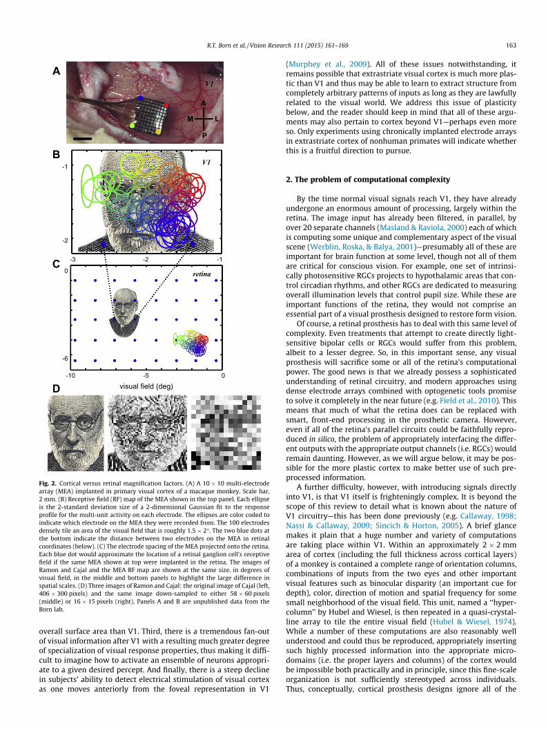
Intracortical microstimulation with currents as low as a fewmicroamperes has been shown to elicit small, retinotopically local-ized visual percepts called ‘‘phosphenes’’ in humans (Schmidtet al., 1996). Similar stimulation protocols in V1 of monkeys canreliably elicit saccades to the region of the visual field correspond-ing to the stimulated neurons’ receptive fields (Fig. 3; Bradley et al.,2005; Tehovnik et al., 2005). Coincidentally, in humans the mini-mal interelectrode spacing that reliably produced two distinctphosphenes was on the order of 0.5 mm (Schmidt et al., 1996), justslightly larger than the spacing on the MEA shown in Fig. 2. Micr-ostimulation with pairs of electrodes in V1 of monkeys also suggestthat sites within approximately 1 mm of each other can be inte-grated for enhanced detection, whereas sites separated by greaterdistances are treated as independent events that can be compared,but not integrated (Ghose & Maunsell, 2012). Taken together, theseresults suggest that the dimensions of the hypercolumn placestrong constraints on the design of MEAs suitable for a V1 pros-thetic device.
There are a number of other important factors that affect thepercept of phosphenes elicited by stimulation of the early visualpathways, such as the fact that they move with the eyes and are
Fig. 3. A monkey makes memory-guided saccades to both visual targets (top two row(bottom two rows). Saccades to phosphenes are less accurate than those to visual targecircles represent either the location at which the visual target was flashed (top) or the recdots depict the saccade end-point on each trial. Used with permission from Bradley et a
scaled in apparent size according to the vergence posture andaccommodation of the subject. In addition, one might also thinkthat it would be necessary to account for geometric distortionsintroduced by the retinotopic organization of V1 (Fig. 1), integra-tion of information from the two eyes and the appropriate dynam-ics of cortical stimulation. These issues have been treated in depthin previous publications (Schiller & Tehovnik, 2008; Schiller et al.,2011, 1996; Tehovnik & Slocum, 2007, 2013; Tehovnik et al., 2009),and we will not elaborate upon them here.
Our point of departure for what follows is that all previousapproaches have been based on the premise that the functional cir-cuitry of the visual system is rather fixed in the adult. However,recent work has indicated a certain degree of plasticity of adultcortex, even in primary sensory areas. In the following section,we briefly review the literature on cortical plasticity with a specialemphasis on that demonstrated in sensory cortex of adult animals.
4. Local cortical plasticity: teaching an old dog to recognize newpatterns
Early in postnatal development, the cortex is remarkably plas-tic. A number of studies document the ability of cortical networksto profoundly and adaptively re-wire following changes in sensoryinput patterns—most noteworthy are those that show develop-ment of novel pattern selectivity in auditory cortex when visualinputs are re-routed to the auditory thalamus (reviewed inHorng & Sur, 2006; O’Leary, 1989). However, there is a limiteddevelopmental window in which this plasticity is observed, givingrise to the concept of a ‘‘critical period’’ (Hensch, 2005; Hubel,1988). Understanding this form of plasticity is crucial for the treat-ment of childhood visual disorders, but it is of limited relevance tothe topic of visual restoration in blind adults, since the critical
s) and to phosphenes elicited by microstimulation of electrodes implanted in V1ts, yet they still reliably track the retinotopic locations of the electrodes. The grayeptive field location of the neurons at the electrode that was stimulated. Small blackl. (2005).
R.T. Born et al. / Vision Research 111 (2015) 161–169 165
period has long since ended. However, these studies imply thatcortical prostheses are likely to be ineffective in patients who havenot had normal vision during the critical period. Moreover, thepossibility that visual cortex can be recruited to perform otherfunctions, such as Braille reading in early blind patients (Cohenet al., 1997), suggests that any visual prosthesis is likely to be mosteffective in people who have more recently lost visual function.
The topic of adult cortical plasticity is more controversial andhas been reviewed extensively (Wandell & Smirnakis, 2009). Earlystudies indicated that following small retinal lesions, V1 neuronswhose receptive fields fell within the induced scotoma6 becameresponsive to adjacent regions of the spared visual field (Bakeret al., 2005; Gilbert & Li, 2012). However, subsequent experiments(e.g. Smirnakis et al., 2005) have challenged the interpretation of thisfinding as true plasticity. It remains possible that some of the appar-ent plasticity could have been due to the existence of rare neuronswith large receptive fields that spanned the border of the scotomaprior to the retinal lesion. Nevertheless, more recent experimentsin which local inhibitory circuits were optogenetically mapped insomatosensory cortex of adult mice both before, during and afterrecovery from sensory deprivation (whisker trimming) haverevealed an impressive degree of plasticity in this circuitry (Kätzel& Miesenböck, 2014). Conversely, and perhaps more relevant to acortical prosthesis, over-activation of local patches of cortex usingmicrostimulation results in an expanded representation of the fea-ture preferred by the stimulated site (reviewed in Histed, Ni, &Maunsell, 2012; see also Talwar & Gerstein, 2001). The examplesprovided by deprivation and over-stimulation likely represent differ-ent sides of the same coin, showing that experience-dependent plas-ticity exists to some degree in the adult brain.
The above studies indicate that local connections in the adultcortex may be at least partially modifiable, but can such mecha-nisms actually support learning? Two sets of studies suggest thatthey can. The first set involves the use of spike-triggered microsti-mulation in which the spiking activity of a nearby reference neuron(measured with a separate, non-stimulating electrode) controls thetiming of the stimulation. Thus the artificially stimulated neuronsare activated contingent upon activity in the reference neuron—acondition that might be expected to produce learning (Rescorla,1968). This manipulation does in fact produce enhanced correla-tions between the reference and target neurons (Jackson,Mavoori, & Fetz, 2006; Rebesco & Miller, 2011; Rebesco et al.,2010; Song et al., 2013) suggesting that cortical plasticity is sensi-tive to the statistical dependencies between the activities of nearbyneurons even if those dependencies are entirely arbitrary.
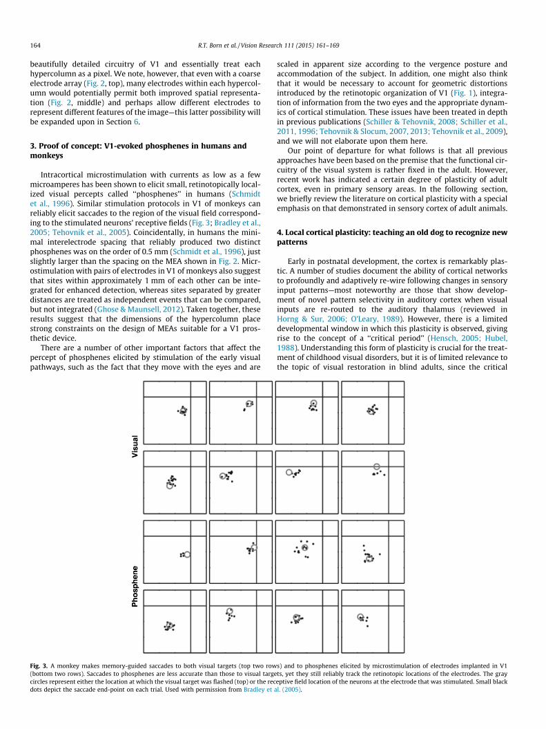
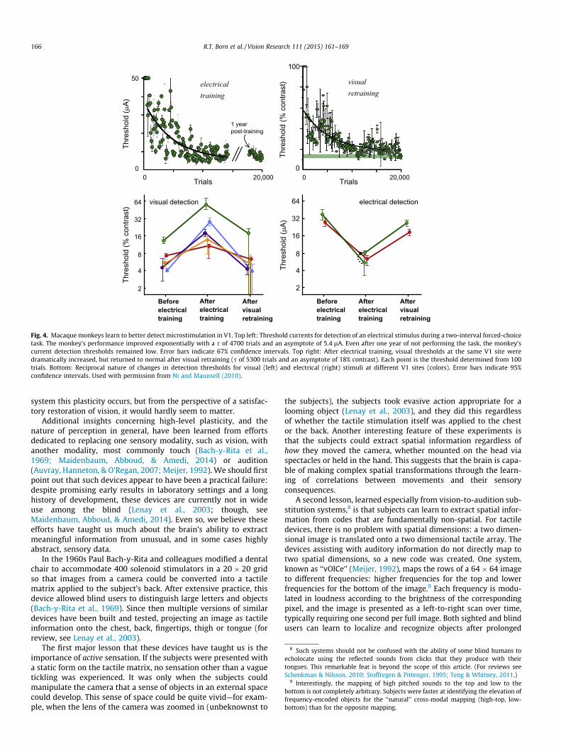
A second set of experiments, performed by Ni and Maunsell(2010), further demonstrate the ability of adult primary sensorycortex to learn. In a two-interval forced-choice task, monkeys wererequired to detect either a small visual stimulus in a given region ofthe visual field or microstimulation in the retinotopically corre-sponding region of V1. For either cue, prolonged training substan-tially lowered detection thresholds (Fig. 4, top). The criticalobservation was that learning to detect the microstimulation dis-rupted the monkeys’ ability to detect the visual stimulus and viceversa (Fig. 4, bottom). These changes were reversible and confinedto roughly the same V1 hypercolumn (i.e. sites more than 2 or3 mm away were unaffected by training), indicating that the localcircuitry had somehow reconfigured to better detect the particularpatterns of activity that were behaviorally relevant. This stronglysuggests that the circuitry of cortex can adapt to consistent inputpatterns, even if those input patterns are distinctly unnatural, asis the case with microstimulation. We believe that this result bodes
6 The ‘‘scotoma’’ is defined as the region of cortex corresponding to the lesion in theretina. For lesions of the early visual pathways, the scotoma is manifest as a localizedregion of blindness in the visual field.
well for visual prostheses in general, but is particularly relevant forthose relying on cortical microstimulation to introduce visualsignals.
Of course, plasticity in response to microstimulation could con-ceivably also produce long-term effects that are maladaptive, as isseen, for example, with the phenomenon of kindling (McNamaraet al., 1980). While primary visual cortex does not appear to beas susceptible to this pathological form of plasticity, it serves toremind us of the necessity of performing additional long-termmicrostimulation experiments in animals to verify that introduc-ing signals to the visual system in this way is both safe and effec-tive. It will be especially important to demonstrate that theelectrically induced phosphenes can be not only detected, as dem-onstrated by Ni and Maunsell (2010), but also serve as the basis forthe recognition of objects. The human subject studied by Schmidtet al. (1996) was able to group multiple phosphenes into the rep-resentation of a vertical line (p. 518), but this is still a long wayfrom true object recognition.
In sum, there is evidence that some forms of experience-depen-dent plasticity continue to exist into adulthood and that they canmediate a form of learning that would be conducive to a V1 visualprosthesis. However, these are purely local mechanisms. The pro-cess of seeing requires the recovery of structure based on thedetection of regularities at higher levels of the visual hierarchyand on the integration of this information with exploratory move-ments (so-called ‘‘active sensing,’’ discussed below) in order toguide behavior. In the next section, we explore results indicatingthat not only can cortex adapt to detect non-natural inputs but thatadaptation also occurs at higher levels of the visual system and iscomplete enough to inform perception and guide behavior.
5. High-level plasticity: ‘‘experiments with goggles’’ and the like
Beginning in the late 1920s, psychologists examined the abilityof human subjects to adapt to various distortions of the visualworld (reviewed in Kohler, 1962; for earlier work with invertingprisms, see Stratton, 1897). Many of their results are nothing shortof astonishing. For example, simple prisms displace the image onthe retina, convert straight edges to curved ones and create rain-bow fringes along contrast borders. All of these distortions arecompletely eliminated over the course of several days (Gibson,1933; Kohler, 1962; R.T. Born, unpublished observations). It isnot simply that subjects learn to ignore the distortions and/orbehaviorally compensate for them—they actually disappear, givingway to normal perception and, when the prisms are removed, thesubject experiences perceptual distortions in the opposite direc-tion (i.e. the fringes are of the complementary color and the direc-tion of curvature reverses). Even the dynamic distortions createdby the prism goggles—such as expansions and contractions alongthe horizontal meridian produced by combinations of head andeye movements—disappear over the course of several weeks, againto return in opposing directions when the goggles are removed. Inthe most extreme versions of these experiments, subjects adaptedto conditions in which their visual world was inverted or right-leftreversed.7 The lesson we take from these experiments is that thebrains of adults who have grown up with normal vision have builta powerful internal model of the external world (Barlow, 1994)and that distortions that are prolonged and consistent can be cor-rected. Again, this sort of result appears to be good news in termsof restoring vision to adults who have become blind after the criticalperiod. It is currently impossible to say at which level of the nervous
7 An original movie of one of Erismann’s subjects adapting to inverting goggles canbe viewed at: http://www.awz.uni-wuerzburg.de/archiv/film_foto_tonarchiv/film-dokumente/th_erismann_ikohler/die_umkehrbrille_und_das_aufrechte_sehen/.
Thre
shol
d (%
con
trast
)
20,0000 0
100
Trials
visual retraining
Thre
shol
d (µ
A)
64
32
16
8
4
2
electrical detection
Beforeelectricaltraining
Afterelectricaltraining
Aftervisualretraining
Beforeelectricaltraining
Afterelectricaltraining
Aftervisualretraining
Thre
shol
d (%
con
trast
) 64
32
16
8
4
2
visual detection
50
0
Thre
shol
d (µ
A)
0 20,000Trials
electrical training
1 year post-training
Fig. 4. Macaque monkeys learn to better detect microstimulation in V1. Top left: Threshold currents for detection of an electrical stimulus during a two-interval forced-choicetask. The monkey’s performance improved exponentially with a s of 4700 trials and an asymptote of 5.4 lA. Even after one year of not performing the task, the monkey’scurrent detection thresholds remained low. Error bars indicate 67% confidence intervals. Top right: After electrical training, visual thresholds at the same V1 site weredramatically increased, but returned to normal after visual retraining (s of 5300 trials and an asymptote of 18% contrast). Each point is the threshold determined from 100trials. Bottom: Reciprocal nature of changes in detection thresholds for visual (left) and electrical (right) stimuli at different V1 sites (colors). Error bars indicate 95%confidence intervals. Used with permission from Ni and Maunsell (2010).
8 Such systems should not be confused with the ability of some blind humans toecholocate using the reflected sounds from clicks that they produce with theirtongues. This remarkable feat is beyond the scope of this article. (For reviews seeSchenkman & Nilsson, 2010; Stoffregen & Pittenger, 1995; Teng & Whitney, 2011.)
9 Interestingly, the mapping of high pitched sounds to the top and low to thebottom is not completely arbitrary. Subjects were faster at identifying the elevation offrequency-encoded objects for the ‘‘natural’’ cross-modal mapping (high-top, low-bottom) than for the opposite mapping.
166 R.T. Born et al. / Vision Research 111 (2015) 161–169
system this plasticity occurs, but from the perspective of a satisfac-tory restoration of vision, it would hardly seem to matter.
Additional insights concerning high-level plasticity, and thenature of perception in general, have been learned from effortsdedicated to replacing one sensory modality, such as vision, withanother modality, most commonly touch (Bach-y-Rita et al.,1969; Maidenbaum, Abboud, & Amedi, 2014) or audition(Auvray, Hanneton, & O’Regan, 2007; Meijer, 1992). We should firstpoint out that such devices appear to have been a practical failure:despite promising early results in laboratory settings and a longhistory of development, these devices are currently not in wideuse among the blind (Lenay et al., 2003; though, seeMaidenbaum, Abboud, & Amedi, 2014). Even so, we believe theseefforts have taught us much about the brain’s ability to extractmeaningful information from unusual, and in some cases highlyabstract, sensory data.
In the 1960s Paul Bach-y-Rita and colleagues modified a dentalchair to accommodate 400 solenoid stimulators in a 20 � 20 gridso that images from a camera could be converted into a tactilematrix applied to the subject’s back. After extensive practice, thisdevice allowed blind users to distinguish large letters and objects(Bach-y-Rita et al., 1969). Since then multiple versions of similardevices have been built and tested, projecting an image as tactileinformation onto the chest, back, fingertips, thigh or tongue (forreview, see Lenay et al., 2003).
The first major lesson that these devices have taught us is theimportance of active sensation. If the subjects were presented witha static form on the tactile matrix, no sensation other than a vaguetickling was experienced. It was only when the subjects couldmanipulate the camera that a sense of objects in an external spacecould develop. This sense of space could be quite vivid—for exam-ple, when the lens of the camera was zoomed in (unbeknownst to
the subjects), the subjects took evasive action appropriate for alooming object (Lenay et al., 2003), and they did this regardlessof whether the tactile stimulation itself was applied to the chestor the back. Another interesting feature of these experiments isthat the subjects could extract spatial information regardless ofhow they moved the camera, whether mounted on the head viaspectacles or held in the hand. This suggests that the brain is capa-ble of making complex spatial transformations through the learn-ing of correlations between movements and their sensoryconsequences.
A second lesson, learned especially from vision-to-audition sub-stitution systems,8 is that subjects can learn to extract spatial infor-mation from codes that are fundamentally non-spatial. For tactiledevices, there is no problem with spatial dimensions: a two dimen-sional image is translated onto a two dimensional tactile array. Thedevices assisting with auditory information do not directly map totwo spatial dimensions, so a new code was created. One system,known as ‘‘vOICe’’ (Meijer, 1992), maps the rows of a 64 � 64 imageto different frequencies: higher frequencies for the top and lowerfrequencies for the bottom of the image.9 Each frequency is modu-lated in loudness according to the brightness of the correspondingpixel, and the image is presented as a left-to-right scan over time,typically requiring one second per full image. Both sighted and blindusers can learn to localize and recognize objects after prolonged

Fig. 5. A new quadrature code for vision. (A) Eight rotations of a contrast edge
R.T. Born et al. / Vision Research 111 (2015) 161–169 167
training periods, though such functionality is typically demonstratedin unnaturally easy conditions, where objects are presented in theabsence of clutter and occlusion, and even under these conditionsperformance is slow and not completely reliable (Auvray,Hanneton, & O’Regan, 2007). Nevertheless, such results are promis-ing because they illustrate the brain’s ability to learn new, abstractcodes even in adults.
Finally, experiments with so-called ‘‘brain–computer inter-faces’’ (BCIs) have revealed the ability of cortical motor networksto learn new codes with which to produce ‘‘movements.’’ In theseexperiments, electrical signals from a population of neurons inmotor cortex of alert monkeys are mapped, via a decoding algo-rithm, to produce movement of a cursor on a computer screen ina virtual space of either two (Chase, Kass, & Schwartz, 2012;Ganguly et al., 2011) or three (Jarosiewicz et al., 2008) dimensions.The experimenter has complete control over the mapping betweenneural activity and cursor movement, allowing arbitrary perturba-tions to be introduced. Remarkably, the animal’s performance andthe underlying neural tuning curves showed partial adaptation tosuch perturbations in just a few hundred trials (Chase, Kass, &Schwartz, 2012; Jarosiewicz et al., 2008), and near perfect perfor-mance was attained after several days, even when the perturbationconsisted of a completely random re-mapping (Ganguly &Carmena, 2009). We do not know whether such results will gener-alize to visual cortex; however, the finding that normal, humanvisual cortex that has been visually deprived for as little as fivedays can be recruited for improved somatosensory discrimination(Merabet et al., 2008), suggests a similar degree of high-level plas-ticity. Moreover, such results provide additional motivation toexplore the possibility that new, more abstract and more efficientcodes might improve the ultimate functionality of visualprostheses.
symbolizing an image fragment from an external stimulus. (B) The default, ‘‘pixel-to-electrode’’ code. In this example the image fragments in A are downsampled intoa 3 pixel by 3 pixel representation. The intensity of each pixel maps onto a distinctelectrode. Top: the portion of each image fragment that feeds into each pixel/electrode. Bottom: the corresponding electrode outputs over the full range ofstimulus rotations. (C) A hypothetical, ‘‘non-natural’’ code. This code calculates thedominant orientation and sign of the stimulus and represents this informationusing 2 electrodes. The first electrode transmits a signal proportional to the cosineof the calculated rotation and the second transmits a signal proportional to its sine;such signals are said to be in quadrature, as they are out of phase by 90�. Top: the‘‘coordinates’’ of the 2 electrode outputs at each of the rotations shown in A.Bottom: the corresponding electrode output over the full range of stimulusrotations.
6. Final speculations: can V1 learn a new code?
The question of plasticity is particularly relevant when consid-ering the code by which signals are introduced into V1, where‘‘code’’ refers to how the visual inputs are pre-processed beforebeing converted into electrical stimulation. Above, we mentionthe possibility of treating each electrode like a pixel. This approachseems reasonably conservative: the retinotopic arrangement of V1roughly parallels the pixel-by-pixel sampling of the interfacingcamera, and treating each electrode like a pixel preserves this rela-tionship. It is a natural code in the sense that it is one that V1already uses.
This makes the reasonable assumption that the best code is theone that V1 will best ‘‘understand.’’ This would certainly entail pre-processing of the image to instantiate retinal operations, such ascenter-surround opponency, as described in section two. But giventhe plasticity of cortex documented in the sections above, we spec-ulate that V1 (and the rest of the visual system) might be able tolearn new codes. And there certainly exist codes that representvisual information in more sparse and efficient ways, which mayoffer a considerable practical advantage in allowing fewer elec-trodes to deliver the same amount of visual information. Fig. 5demonstrates this idea for a simple ‘‘quadrature code’’ that is wellestablished in engineering disciplines. The top row depicts eightpossible visual stimuli, each of which could be effectively summa-rized by the orientation and sign of the contrast edge (the stimulirotated by 0� and 180� have identical orientations but oppositesigns of contrast). An inefficient pixel-to-electrode code (Fig. 5B)maps each different stimulus to a separate electrode, thus requir-ing nine electrodes. A more efficient way to communicate the sameinformation (Fig. 5C) would be to first pre-process the stimulus toextract the dominant orientation and sign of contrast, and then use
this information to vary the stimulation intensity at each of onlytwo electrodes. This essentially produces outputs proportional tothe cosine and sine of the underlying rotation, respectively, andwould increase coding efficiency by a factor of 9/2.
This thought experiment leaves us with two questions. Whichcodes make the best use of the fewest electrodes? And which codeswill V1 understand? The relative importance of these two ques-tions, however, is determined by the answer to the question wehave been discussing: how plastic are the cortical and perceptualmechanisms within and beyond V1? Given sufficient plasticity,prostheses using V1 as an entry point could potentially overcomethe limitation of low electrode density by pre-processing and con-densing the visual information before converting it to electricalstimulation.
We have reviewed studies suggesting that, over time, the adultbrain may be capable of extracting information provided through acondensed, non-natural code. The upshot of these studies is thatplasticity exists into adulthood with regards to both low-level,local mechanisms (Section 4) and higher-level perception andaction (Section 5). In the cortex, these plasticity mechanismsappear to depend on the temporal coincidence between the

168 R.T. Born et al. / Vision Research 111 (2015) 161–169
activities of neighboring neurons (Jackson, Mavoori, & Fetz, 2006;Rebesco & Miller, 2011; Rebesco et al., 2010). A prosthesis thatfaithfully translates the external environment into a cortical inputwill impose patterns of coincidence, and with sufficient experi-ence, the patterns most fundamentally related to the structure ofthe external environment should occur often enough to meaning-fully drive the reorganization of cortical circuits in the vicinity ofthe stimulating array. Provided the external sources of the signalsare stable, a lawful relationship will exist between the activemanipulations of the viewing device (i.e. head movements thatchange the viewpoint of the camera) and the resulting transforma-tions of the signals across the array. In the case of a pixel-to-elec-trode code, the relationship is straightforward. For example, if theuser turns her head to the right slightly, the signals on the elec-trode array will shift some number of electrodes to the left. Anysingle snapshot of the signals on the array may fail to resolve keydescriptions of the visual environment because many possiblestimuli could give rise to the same input. However, the numberof possible stimuli that could give rise to the sequence of snapshotsstrung together by active sensing is comparatively small. Theimportant point is that, as has been demonstrated through exper-iments in sensory substitution (Lenay et al., 2003), the brain is wellequipped to use active sensing to extract the structure of the sig-nals that impinge upon it.
The intersection between the type of learning acquired throughactive sensing and the type of learning permitted through plastic-ity within V1 is not at all clear. The alternative code used as anexample above would require that the brain learn to appropriatelycombine the signals between the pair of electrodes that encode thesame region of visual space. This could be facilitated both by theappropriate electrode spacing (Ghose & Maunsell, 2012) and byactive sensing, which would enforce the tendency for head move-ments to result in translations of related pairs of signals. Alterna-tively, the structure of the inputs themselves (and the patternsthey create across the input array) may drive local plasticitytoward the extraction of those patterns, which may in turn facili-tate higher-order pattern learning, such as that developed throughactive sensing.
To our knowledge, no studies have specifically asked whetherplasticity mechanisms in the adult brain support the learning ofa new code if normal sensory inputs are replaced with visuallyderived inputs through a cortical prosthesis. As such, the questionsraised in this section await empirical answers and the viewpointsoffered above are intended purely as provocative speculation thatwe hope will stimulate future research. Studies that directly inves-tigate the conditions under which adult cortex adaptively reorga-nizes in response to arbitrarily patterned input will go a longway toward resolving the question of whether a cortical entrypoint for visual restoration makes sense. These studies will needto show that any reorganization is specific to the patterns thatdrive it and that this reorganization improves the representationof whatever information those patterns contain.
Acknowledgments
We are grateful to John Maunsell for helpful comments on themanuscript. This work was supported by NIH EY11379 to RTB.
References
Auvray, M., Hanneton, S., & O’Regan, J. K. (2007). Learning to perceive with a visuo-auditory substitution system: Localisation and object recognition with ‘thevOICe’. Perception, 36(3), 416–430.
Bach-y-Rita, P., Collins, C. C., Saunders, F. A., White, B., & Scadden, L. (1969). Visionsubstitution by tactile image projection. Nature, 221(5184), 963–964.
Baker, C. I., Peli, E., Knouf, N., & Kanwisher, N. G. (2005). Reorganization of visualprocessing in macular degeneration. Journal of Neuroscience, 25(3), 614–618.http://dx.doi.org/10.1523/JNEUROSCI.3476-04.2005.
Barlow, H. (1994). What is the computational goal of the neocortex? In C. Koch & J.L. Davis (Eds.), Large-scale neuronal theories of the brain (pp. 1–22). MIT Press.
Bradley, D. C., Troyk, P. R., Berg, J. A., Bak, M., Cogan, S., Erickson, R., et al. (2005).Visuotopic mapping through a multichannel stimulating implant in primate V1.Journal of Neurophysiology, 93(3), 1659–1670. http://dx.doi.org/10.1152/jn.01213.2003.
Brindley, G. S., & Lewin, W. S. (1968). The sensations produced by electricalstimulation of the visual cortex. The Journal of Physiology, 196(2), 479–493.
Callaway, E. M. (1998). Local circuits in primary visual cortex of the macaquemonkey. Annual Review of Neuroscience, 21, 47–74. http://dx.doi.org/10.1146/annurev.neuro.21.1.47.
Chase, S. M., Kass, R. E., & Schwartz, A. B. (2012). Behavioral and neural correlates ofvisuomotor adaptation observed through a brain–computer interface inprimary motor cortex. Journal of Neurophysiology, 108(2), 624–644.
Cohen, L. G., Celnik, P., Pascual-Leone, A., Corwell, B., Falz, L., Dambrosia, J., et al.(1997). Functional relevance of cross-modal plasticity in blind humans. Nature,389(6647), 180–183.
Connolly, M., & Van Essen, D. (1984). The representation of the visual field inparvicellular and magnocellular layers of the lateral geniculate nucleus in themacaque monkey. The Journal of Comparative Neurology, 226(4), 544–564.http://dx.doi.org/10.1002/cne.902260408.
Cowey, A., & Rolls, E. T. (1974). Human cortical magnification factor and its relationto visual acuity. Experimental Brain Research. Experimentelle Hirnforschung.Expérimentation Cérébrale, 21(5), 447–454.
Dow, B. M., Snyder, A. Z., Vautin, R. G., & Bauer, R. (1981). Magnification factor andreceptive field size in foveal striate cortex of the monkey. Experimental BrainResearch. Experimentelle Hirnforschung. Expérimentation Cérébrale, 44(2),213–228.
Duncan, R. O., & Boynton, G. M. (2003). Cortical magnification within humanprimary visual cortex correlates with acuity thresholds. Neuron, 38(4),659–671.
Engel, S. A., Rumelhart, D. E., Wandell, B. A., Lee, A. T., Glover, G. H., Chichilnisky, E. J.,et al. (1994). FMRI of human visual cortex. Nature, 369(6481), 525. http://dx.doi.org/10.1038/369525a0.
Felleman, D. J., & Van Essen, D. C. (1991). Distributed hierarchical processingin the primate cerebral cortex. Cerebral Cortex (New York, N.Y.: 1991), 1(1),1–47.
Field, G. D., Gauthier, J. L., Sher, A., Greschner, M., Machado, T. A., Jepson, L. H., et al.(2010). Functional connectivity in the retina at the resolution of photoreceptors.Nature, 467(7316), 673–677. http://dx.doi.org/10.1038/nature09424.
Ganguly, K., & Carmena, J. M. (2009). Emergence of a stable cortical map forneuroprosthetic control. PLoS Biology, 7(7), e1000153. http://dx.doi.org/10.1371/journal.pbio.1000153.
Ganguly, K., Dimitrov, D. F., Wallis, J. D., & Carmena, J. M. (2011). Reversible large-scale modification of cortical networks during neuroprosthetic control. NatureNeuroscience, 14(5), 662–667.
Ghose, K., & Maunsell, J. H. R. (2012). A strong constraint to the joint processing ofpairs of cortical signals. Journal of Neuroscience, 32(45), 15922–15933. http://dx.doi.org/10.1523/JNEUROSCI.2186-12.2012.
Gibson, J. J. (1933). Adaptation, after-effect and contrast in the perception of curvedlines. Journal of Experimental Psychology, 16(1), 1–31.
Gilbert, C. D., & Li, W. (2012). Adult visual cortical plasticity. Neuron, 75(2), 250–264.http://dx.doi.org/10.1016/j.neuron.2012.06.030.
Grüsser, O. J. (1995). Migraine phosphenes and the retino-cortical magnificationfactor. Vision Research, 35(8), 1125–1134.
Hensch, T. K. (2005). Critical period plasticity in local cortical circuits. NatureReviews Neuroscience, 6(11), 877–888. http://dx.doi.org/10.1038/nrn1787.
Histed, M. H., Ni, A. M., & Maunsell, J. H. R. (2012). Insights into cortical mechanismsof behavior from microstimulation experiments. Progress in Neurobiology, 1–16.http://dx.doi.org/10.1016/j.pneurobio.2012.01.006.
Horng, S. H., & Sur, M. (2006). Visual activity and cortical rewiring: Activity-dependent plasticity of cortical networks. Progress in Brain Research, 157, 3–11.
Hubel, D. H. (1988). Deprivation and development. In Eye, brain, and vision.Scientific American Library Series.
Hubel, D. H., & Wiesel, T. N. (1974). Uniformity of monkey striate cortex: A parallelrelationship between field size, scatter, and magnification factor. The Journal ofComparative Neurology, 158(3), 295–305. http://dx.doi.org/10.1002/cne.901580305.
Jackson, A., Mavoori, J., & Fetz, E. E. (2006). Long-term motor cortex plasticityinduced by an electronic neural implant. Nature, 444(7115), 56–60. http://dx.doi.org/10.1038/nature05226.
Jarosiewicz, B., Chase, S. M., Fraser, G. W., Velliste, M., Kass, R. E., & Schwartz, A. B.(2008). Functional network reorganization during learning in a brain–computerinterface paradigm. Proceedings of the National Academy of Sciences of the UnitedStates of America, 105(49), 19486–19491. http://dx.doi.org/10.1073/pnas.0808113105.
Jepson, L. H., Hottowy, P., Mathieson, K., Gunning, D. E., Dabrowski, W., Litke, A. M.,et al. (2014). Spatially patterned electrical stimulation to enhance resolution ofretinal prostheses. Journal of Neuroscience, 34(14), 4871–4881. http://dx.doi.org/10.1523/JNEUROSCI.2882-13.2014.
Kätzel, D., & Miesenböck, G. (2014). Experience-dependent rewiring of specificinhibitory connections in adult neocortex. PLoS Biology, 12(2), e1001798. http://dx.doi.org/10.1371/journal.pbio.1001798.

R.T. Born et al. / Vision Research 111 (2015) 161–169 169
Kohler, I. (1962, April 29). Experiments with goggles. Scientific American. <http://directlink.lib.harvard.edu/sfx_bypass/sfx_bypass.cgi?target=http%3A%2F%2Fezp-prod1.hul.harvard.edu%2Flogin%3Furl%3Dhttp%3A%2F%2Fdx.doi.org%2F10.1038%2Fscientificamerican0562-62%3Fnosfx%3Dy> Retrieved 29.04.14.
Kolb, H., & Dekorver, L. (1991). Midget ganglion cells of the parafovea of the humanretina: A study by electron microscopy and serial section reconstructions. TheJournal of Comparative Neurology, 303(4), 617–636. http://dx.doi.org/10.1002/cne.903030408.
Kolb, H., & Marshak, D. (2003). The midget pathways of the primate retina.Documenta Ophthalmologica. Advances in Ophthalmology, 106(1), 67–81.
Lenay, C., Gapenne, O., Hanneton, S., Marque, C., & Genoulle, C. (2003). Sensorysubstitution: Limits and perspectives. In Y. Hatwell, S. Arlette, & E. Gentaz(Eds.), Touching for knowing: Cognitive psychology of haptic manual perception(pp. 275–292). Philadelphia, PA: John Benjamins Publishing Company.
Loudin, J. D., Cogan, S. F., Mathieson, K., Sher, A., & Palanker, D. V. (2011). Photodiodecircuits for retinal prostheses. IEEE Transactions on Biomedical Circuits andSystems, 5(5), 468–480.
Maidenbaum, S., Abboud, S., & Amedi, A. (2014). Neuroscience and BiobehavioralReviews, 41, 3–15. http://dx.doi.org/10.1016/j.neubiorev.2013.11.007.
Malpeli, J. G., & Baker, F. H. (1975). The representation of the visual field in thelateral geniculate nucleus of Macaca mulatta. The Journal of ComparativeNeurology, 161(4), 569–594. http://dx.doi.org/10.1002/cne.901610407.
Masland, R. H., & Raviola, E. (2000). Confronting complexity: Strategies forunderstanding the microcircuitry of the retina. Annual Review of Neuroscience,23, 249–284. http://dx.doi.org/10.1146/annurev.neuro.23.1.249.
McNamara, J. O., Byrne, M. C., Dasheiff, R. M., & Fitz, J. G. (1980). The kindling modelof epilepsy: A review. Progress in Neurobiology, 15(2), 139–159.
Meijer, P. B. (1992). An experimental system for auditory image representations.IEEE Transactions on Bio-Medical Engineering, 39(2), 112–121. http://dx.doi.org/10.1109/10.121642.
Merabet, L. B., Hamilton, R., Schlaug, G., Swisher, J. D., Kiriakopoulos, E. T., Pitskel, N.B., et al. (2008). Rapid and reversible recruitment of early visual cortex fortouch. PLoS One, 3(8), e3046.
Murphey, D. K., Maunsell, J. H., Beauchamp, M. S., & Yoshor, D. (2009). Perceivingelectrical stimulation of identified human visual areas. Proceedings of theNational Academy of Sciences of the United States of America, 106(13), 5389–5393.http://dx.doi.org/10.1073/pnas.0804998106.
Nassi, J. J., & Callaway, E. M. (2009). Parallel processing strategies of the primatevisual system. Nature Reviews Neuroscience, 10(5), 360–372. http://dx.doi.org/10.1038/nrn2619.
Ni, A. M., & Maunsell, J. H. R. (2010). Microstimulation reveals limits in detectingdifferent signals from a local cortical region. Current Biology: CB, 20(9), 824–828.http://dx.doi.org/10.1016/j.cub.2010.02.065.
O’Leary, D. D. (1989). Do cortical areas emerge from a protocortex? TrendsNeurosciences, 12(10), 400–406.
Pezaris, J. S., & Eskandar, E. N. (2009). Getting signals into the brain: Visualprosthetics through thalamic microstimulation. Neurosurgical Focus, 27(1), E6.http://dx.doi.org/10.3171/2009.4.FOCUS0986.
Pezaris, J. S., & Reid, R. C. (2007). Demonstration of artificial visual perceptsgenerated through thalamic microstimulation. Proceedings of the NationalAcademy of Sciences of the United States of America, 104(18), 7670–7675.
Polyak, S. L. (1941). The retina. University of Chicago Press.Putnam, N. M., Hofer, H. J., Doble, N., Chen, L., Carroll, J., & Williams, D. R. (2005). The
locus of fixation and the foveal cone mosaic. Journal of Vision, 5(7), 632–639.Rebesco, J. M., & Miller, L. E. (2011). Enhanced detection threshold for in vivo
cortical stimulation produced by Hebbian conditioning. Journal of NeuralEngineering, 8(1), 016011. http://dx.doi.org/10.1088/1741-2560/8/1/016011.
Rebesco, J. M., Stevenson, I. H., Körding, K. P., Solla, S. A., & Miller, L. E. (2010).Rewiring neural interactions by micro-stimulation. Frontiers in SystemsNeuroscience, 4. http://dx.doi.org/10.3389/fnsys.2010.00039.
Rescorla, R. A. (1968). Probability of shock in the presence and absence of CS in fearconditioning. Journal of Comparative and Physiological Psychology, 66(1), 1–5.
Schein, S. J. (1988). Anatomy of macaque fovea and spatial densities of neurons infoveal representation. The Journal of Comparative Neurology, 269(4), 479–505.http://dx.doi.org/10.1002/cne.902690403.
Schenkman, B. N., & Nilsson, M. E. (2010). Human echolocation: Blind and sightedpersons’ ability to detect sounds recorded in the presence of a reflecting object.Perception, 39(4), 483–501.
Schiller, P. H., Slocum, W. M., Kwak, M. C., Kendall, G. L., & Tehovnik, E. J. (2011).New methods devised specify the size and color of the spots monkeyssee when striate cortex (area V1) is electrically stimulated. Proceedingsof the National Academy of Sciences of the United States of America, 108,17809–17814.
Schiller, P. H., & Tehovnik, E. J. (2008). Visual prosthesis. Perception, 37(10),1529–1559. http://dx.doi.org/10.1068/p6100.
Schira, M. M., Tyler, C. W., Breakspear, M., & Spehar, B. (2009). The foveal confluencein human visual cortex. Journal of Neuroscience, 29(28), 9050–9058. http://dx.doi.org/10.1523/JNEUROSCI.1760-09.2009.
Schmidt, E. M., Bak, M. J., Hambrecht, F. T., Kufta, C. V., O’Rourke, D. K., &Vallabhanath, P. (1996). Feasibility of a visual prosthesis for the blind based onintracortical microstimulation of the visual cortex. Brain: A Journal of Neurology,119, 507–522.
Sereno, M. I., Dale, A. M., Reppas, J. B., Kwong, K. K., Belliveau, J. W., Brady, T. J., et al.(1995). Borders of multiple visual areas in humans revealed by functionalmagnetic resonance imaging. Science (New York, N.Y.), 268(5212), 889–893.
Sincich, L. C., & Horton, J. C. (2005). The circuitry of V1 and V2: Integration of color,form, and motion. Annual Review of Neuroscience, 28, 303–326. http://dx.doi.org/10.1146/annurev.neuro.28.061604.135731.
Smirnakis, S. M., Brewer, A. A., Schmid, M. C., Tolias, A. S., Schüz, A., Augath, M., et al.(2005). Lac of long-term cortical reorganization after macaque retinal lesions.Nature, 435(7040), 300–307. PubMed PMID: 15902248.
Song, W., Kerr, C. C., Lytton, W. W., & Francis, J. T. (2013). Cortical plasticity inducedby spike-triggered microstimulation in primate somatosensory cortex. PLoSOne, 8(3), e57453. http://dx.doi.org/10.1371/journal.pone.0057453.
Stoffregen, T. A., & Pittenger, J. B. (1995). Human echolocation as a basic form ofperception and action. Ecological Psychology, 7, 181–216.
Stratton, G. M. (1897). Vision without inversion of the retinal image. PsychologicalReview, 4. 341–360 and 463–481.
Talwar, S. K., & Gerstein, G. L. (2001). Reorganization in awake rat auditory cortex bylocal microstimulation and its effect on frequency-discrimination behavior.Journal of Neurophysiology, 86(4), 1555–1572.
Tehovnik, E. J., & Slocum, W. M. (2007). Phosphene induction by microstimulation ofmacaque V1. Brain Research Reviews, 53(2), 337–343. http://dx.doi.org/10.1016/j.brainresrev.2006.11.001.
Tehovnik, E. J., & Slocum, W. M. (2013). Electrical induction of vision. Neuroscienceand Biobehavioral Reviews, 37, 803–818.
Tehovnik, E. J., Slocum, W. M., Carvey, C. E., & Schiller, P. H. (2005). Phospheneinduction and the generation of saccadic eye movements by striate cortex. Journalof Neurophysiology, 93(1), 1–19. http://dx.doi.org/10.1152/jn.00736.2004.
Tehovnik, E. J., Slocum, W. M., Smirnakis, S. M., & Tolias, A. S. (2009).Microstimulation of visual cortex to restore vision. Progress in Brain Research,175, 347–375.
Teng, S., & Whitney, D. (2011). The acuity of echolocation: Spatial resolution in thesighted compared to expert performance. Journal of Visual Impairment &Blindness, 105(1), 20–32.
Tootell, R. B., Silverman, M. S., Switkes, E., & De Valois, R. L. (1982). Deoxyglucoseanalysis of retinotopic organization in primate striate cortex. Science (New York,N.Y.), 218(4575), 902–904.
Tootell, R. B., Switkes, E., Silverman, M. S., & Hamilton, S. L. (1988). Functionalanatomy of macaque striate cortex. II. Retinotopic organization. TheJournal of Neuroscience: The Official Journal of the Society for Neuroscience, 8(5),1531–1568.
Van Essen, D. C., Newsome, W. T., & Maunsell, J. H. (1984). The visual fieldrepresentation in striate cortex of the macaque monkey: Asymmetries,anisotropies, and individual variability. Vision Research, 24(5), 429–448.
Wandell, B. A. (1995). Foundations of vision. Sunderland, MA: Sinauer Associates Inc.Wandell, B. A., & Smirnakis, S. M. (2009). Plasticity and stability of visual field maps
in adult primary visual cortex. Nature Reviews Neuroscience, 10(12), 873–884.http://dx.doi.org/10.1038/nrn2741.
Werblin, F., Roska, B., & Balya, D. (2001). Parallel processing in the mammalianretina: Lateral and vertical interactions across stacked representations. Progressin Brain Research, 131, 229–238.
Westheimer, G. (1992). Visual acuity. In W. M. Hart, Jr (Ed.), Adler’s psychology of theeye (9th ed., pp. 531–547). St. Louis, MO: Mosby.
Zrenner, E. (2013). Fighting blindness with microelectronics. Science TranslationalMedicine, 5(210). 210ps16.