cortical spreading depression as a target for anti
TRANSCRIPT
Cortical spreading depression asa target for anti-migraine agents
The Harvard community has made thisarticle openly available. Please share howthis access benefits you. Your story matters
Citation Costa, Cinzia, Alessandro Tozzi, Innocenzo Rainero, LetiziaMaria Cupini, Paolo Calabresi, Cenk Ayata, and PaolaSarchielli. 2013. “Cortical spreading depression as atarget for anti-migraine agents.” The Journal of Headacheand Pain 14 (1): 62. doi:10.1186/1129-2377-14-62. http://dx.doi.org/10.1186/1129-2377-14-62.
Published Version doi:10.1186/1129-2377-14-62
Citable link http://nrs.harvard.edu/urn-3:HUL.InstRepos:11717634
Terms of Use This article was downloaded from Harvard University’s DASHrepository, and is made available under the terms and conditionsapplicable to Other Posted Material, as set forth at http://nrs.harvard.edu/urn-3:HUL.InstRepos:dash.current.terms-of-use#LAA
REVIEW ARTICLE Open Access
Cortical spreading depression as a target foranti-migraine agentsCinzia Costa1,3, Alessandro Tozzi1,3, Innocenzo Rainero2, Letizia Maria Cupini5, Paolo Calabresi1,3, Cenk Ayata4
and Paola Sarchielli1*
Abstract
Spreading depression (SD) is a slowly propagating wave of neuronal and glial depolarization lasting a few minutes,that can develop within the cerebral cortex or other brain areas after electrical, mechanical or chemical depolarizingstimulations. Cortical SD (CSD) is considered the neurophysiological correlate of migraine aura. It is characterized bymassive increases in both extracellular K+ and glutamate, as well as rises in intracellular Na+ and Ca2+. These ionicshifts produce slow direct current (DC) potential shifts that can be recorded extracellularly. Moreover, CSD isassociated with changes in cortical parenchymal blood flow.CSD has been shown to be a common therapeutic target for currently prescribed migraine prophylactic drugs. Yet,no effects have been observed for the antiepileptic drugs carbamazepine and oxcarbazepine, consistent with theirlack of efficacy on migraine. Some molecules of interest for migraine have been tested for their effect on CSD.Specifically, blocking CSD may play an enabling role for novel benzopyran derivative tonabersat in preventingmigraine with aura. Additionally, calcitonin gene-related peptide (CGRP) antagonists have been recently reported toinhibit CSD, suggesting the contribution of CGRP receptor activation to the initiation and maintenance of CSD notonly at the classic vascular sites, but also at a central neuronal level. Understanding what may be lying behind thiscontribution, would add further insights into the mechanisms of actions for “gepants”, which may be pivotal for theeffectiveness of these drugs as anti-migraine agents.CSD models are useful tools for testing current and novel prophylactic drugs, providing knowledge on mechanismsof action relevant for migraine.
Keywords: Cortical spreading depression; Calcium channels; Sodium channels; Glutamate; Ionotropic glutamatereceptors; Calcitonin gene-related peptides; Current prophylactic drugs; Antiepileptics; Tonabersat; Gepants
IntroductionSpreading depression (SD) is an intense self-propagatingwave of depolarization involving neuronal and glial cellsin the cerebral cortex, subcortical gray matter or retina,irrespective of functional divisions or arterial territories.This depolarization is followed by a longer lasting waveof inhibition characterised by massive changes in ionicconcentrations and slow chemical waves, propagating ata rate of approximately 3–6 mm/min [1]. In bothlissencephalic and gyrencephalic cortices, SD can beevoked pharmacologically by the application of K+,
glutamate, and Na+/K+-pump inhibitors or by eitherelectrical or mechanical stimulation. SD can developover the course of epileptic crises or can be induced bybrain tissue injury, as in the case of trauma, hemorrhageor ischemia [2-6].Several clinical and neuroimaging findings support the
concept that cortical SD (CSD) is the pathophysiologicalcorrelate of the neurological symptoms in migraine aura[7-9]. Moreover, different experimental models of CSDhave been developed which help to better understandthe underlying neuronal mechanisms and related vascu-lar changes [10,11]. They also, albeit not consistently,suggest that CSD is able to activate central and periph-eral trigemino-vascular nociceptive pathways with a la-tency matching, generally occurring between aura andheadache in migraineurs [12].
* Correspondence: [email protected] Clinic, Department of Public Health and Medical and SurgicalSpecialties, University of Perugia, Ospedale Santa Maria della Misericordia,Sant'Andrea delle Fratte, 06132, Perugia, ItalyFull list of author information is available at the end of the article
© 2013 Costa et al.; licensee Springer. This is an Open Access article distributed under the terms of the Creative CommonsAttribution License (http://creativecommons.org/licenses/by/2.0), which permits unrestricted use, distribution, and reproductionin any medium, provided the original work is properly cited.
Costa et al. The Journal of Headache and Pain 2013, 14:62http://www.thejournalofheadacheandpain.com/content/14/1/62
Familial Hemiplegic Migraine 1 (FHM1) and 2 (FHM2)mutations share the ability to facilitate the induction andpropagation of CSD in mouse models, further supportingthe role of CSD as a key migraine trigger [13]. CSD is fur-ther modulated by endogenous and environmental factors,such as hormones and drugs, and also might be influencedby weather, stress and food [14-16]. Current prophylactictreatments have been investigated for their effects onCSD. Novel drugs can also target CSD and this can ac-count, at least in part, for their mechanisms of action onmigraine [17].
ReviewIn this review, we illustrate the main findings concerningbasic mechanisms underlying CSD as neurophysiologicsubstrate of aura and report results on the effects onCSD from available drugs and new therapeutic options.
Clinical, neurophysiological and neuroimaging evidenceof a link between migraine aura and CSDAbout a third of migraine patients complain of transientfocal aura symptoms beginning from minutes to hoursbefore headache, or occurring during either the head-ache phase or even in its absence [18]. Usually, migraineaura consists of fully reversible visual, sensory and/ordysphasic symptoms [19]. These aura symptoms are ac-companied by fully reversible motor weakness in hemi-plegic migraine. This is referred to as familial (i.e., FHM)subtype when the condition is present in at least onefirst- or second-degree relative, and a sporadic subtypein the absence of family history [20].Specific genetic subtypes of FHM have been identified.
Mutations of three genes all encoding ion-channels ormembrane ionic pumps were discovered from 1996 to2005. They involve a neuronal Ca2+ channel (CACNA1A,FHM1), a glial Na+/K+ pump (ATP1A2, FHM2) and aneuronal Na+ channel (SCN1A, FHM3), respectively [13].However, these mutations have not been identified in themore common types of migraine with typical aura. Theseforms are considered polygenic, with an overall heritabilitynearing 50%, and should be regarded as the result of theinteraction of genetic and environmental factors [21].Descriptions of migraine with aura (MwA) from the
year 1870 onwards have reported a slow, gradual pro-gression of aura symptoms. Specifically, in 1941, Lashley,from the meticulous chartering of his own auras, sug-gested that aura symptoms reflect a cortical processprogressing with a speed of 3 mm/min across the pri-mary visual cortex [22].CSD was first described by Aristides Leão in 1944
[23,24]. Studying experimental epilepsy for his PhD the-sis, Leão came across a depression of electroencephalo-graphic (EEG) activity moving through the rabbit cortexat a rate of 3–6 mm/min after electrical or mechanical
stimulations. The negative wave was sometimes pre-ceded by a small, brief positivity, and always followed bya positive overshoot of 3–5 min [25]. He observed thatthe threshold of CSD varied among cortical areas also inpigeons and cats, and once triggered, it spread in alldirections. During CSD, neither sensory stimulation nordirect cortical stimulation evoked potential waves. Ongoingexperimental seizure discharge was also suppressed byCSD, even if sometimes tonic-clonic activity preceded orfollowed SD [23]. Based on similar propagation of the twoprocesses, Leão hypothesized an association between CSDand seizures [1].Using microscopy and photography of pial vessels to
assess cortical circulation, the researcher was also ableto see both arteries dilated “as scarlet as the arteries”and veins, as a consequence of CSD. This latter obser-vation, for the first time, indicated that the cerebralblood flow increase exceeded the increase in oxygendemand. This topic has become a matter of interest forall investigators studying changes in cerebral circulationover the course of CSD [24].In the early 20th century, aura was considered a vascu-
lar process involving an initial vasoconstriction followedby a reactive vasodilation responsible for head pain[8,26]. Later observations by Olesen et al. modified thisidea by demonstrating a spreading reduction in cerebralblood flow, called “spreading oligemia”, occurring inpatients with MwA [27]. This finding completelyredefined the underlying pathogenesis of aura by at-tributing blood flow changes during aura to changes inneuronal activity [28].Single photon emission computerized tomography
(SPECT) [29] and perfusion-weighted magnetic reson-ance imaging (MRI) [30] studies have further supportedthe hypothesis that “spreading oligemia” observed duringaura is primarily due to changes in neuronal activity.Additionally, perfusion abnormalities have been sug-gested to be a response of the autoregulatory mecha-nisms to underlying neuronal depression. However, inmost SPECT and hyper-emia and subsequent spreadinghypo-perfusion, patients never experienced symptoms oftypical visual auras [7,31].The first-ever study investigating occipital cortex acti-
vation during visual stimulation with functional mag-netic resonance (fMRI) by blood oxygenation level(BOLD)-dependent contrast imaging in MwA patientsdemonstrated that the onset of headache or visualchange (only in 2 patients), or both, were preceded by asuppression of the initial activation. This suppressionslowly propagated into contiguous occipital cortex at arate ranging from 3 to 6 mm/min. and was accompaniedby baseline contrast intensity increases, indicating thatvasodilatation and tissue hyper-oxygenation are associ-ated with the induction of headache [32]. Later, in 2001,
Costa et al. The Journal of Headache and Pain 2013, 14:62 Page 2 of 18http://www.thejournalofheadacheandpain.com/content/14/1/62
Hadjikhani at al. [33], strongly suggested that electro-physiological events consistent with CSD are involved intriggering aura in the human visual cortex. Using high-field fMRI with near-continuous recording during visualaura, the authors identified specific BOLD events in thevisual cortex that were strictly linked to the aura per-cept, in both space (retinotopy) and time. Throughoutthe progression of each aura, unique BOLD perturba-tions were found in the corresponding regions of the ret-inotopic visual cortex. Like the progression of the aurain the visual field, the BOLD perturbations progressedfrom the paracentral to more peripheral eccentricities, inonly the hemisphere corresponding to the aura. Thesource of the aura-related BOLD changes were localizedin the extrastriate visual cortex (area V3A) rather than inthe striate cortex (V1). Strikingly, the spread rate of theBOLD perturbations across the flattened cortical graymatter was consistent with previous measures of CSD.As for diffusion changes on MRI, these have been es-
pecially observed in cases of prolonged complex mi-graine aura, suggesting cytotoxic edema in the absenceof ischemic lesions [34-37]. To this regard, it is note-worthy the finding of a spreading of cortical edema withreversibly restricted water diffusion from the left occipi-tal to the temporo-parietal cortex in a case of persistentvisual migraine aura [38]. In another case of migrainewith prolonged aura, hyper-perfusion with vasogenicleakage was detected by diffusion-weighted MRI [39]. Afurther patient experienced a series of MwA attacksaccompanied by slight pleocytosis and gadolinium (Gd-DTPA) enhancement in proximity of the left middlecerebral artery. In this patient, the migraine attacks andGd-DTPA enhancement were reversed by prophylactictreatment [40].Magneto-electroencephalography (MEG) allows the
study of direct current (DC) neuromagnetic fields inspontaneous and visually induced migraine patients. FewMEG studies have been conducted with this approach inMwA patients due to technical difficulties. The mostrelevant study to date has shown multiple cortical areasactivated in spontaneous and visually induced MwA pa-tients, unlike activation limited to the primary visualcortex in control subjects. This finding supports the hy-pothesis that a CSD-like neuro-electric event arisesspontaneously during migraine aura or can be visuallytriggered in widespread regions of the hyper-excitableoccipital cortex [41].MEG has also been used to directly register changes in
cortical oscillatory power during aura. Specifically, alphaband desynchronization has been demonstrated with thistechnique in both the left extra-striate and temporal cor-tex over the period of reported visual disturbances.These terminated abruptly on the disappearance of scin-tillations, whereas gamma frequency desynchronization
in the left temporal lobe continued for 8 to 10 minutesfollowing the reported end of aura [42].
Neuronal mechanisms of CSD in experimental modelsWhen evoked by local extracellular K+ concentrationsexceeding a critical threshold, CSD is associated with thedisruption of membrane ionic gradients. Both massiveK+ (with extracellular concentration increases up to 60mM) and glutamate effluxes are believed to depolarizeadjacent neurons to facilitate spread [43]. Results fromstudies on ion-selective microelectrodes have shown thatan extracellular K+ rise is accompanied by falls in extra-cellular Na+ and Cl- during CSD, whereas water leavesthe extracellular space with significant changes in extra-cellular pH [44,45].Specifically, in chick retina, SD induced an initial in-
crease in intracellular pH, which was associated with ele-vated levels of ADP, P-Creatine, lactate and pyruvate.This was followed by an intermediary acid shift, in-creases in ATP values and decreases in ADP, a late alka-line rebound, a decrease in P-Creatine levels, andelevations in both ADP and lactate levels. These transi-ent changes in intracellular pH occurring parallel tochanges of energy metabolite levels during SD, may beexpressions of rapidly modifying metabolic activities ofneurons and glial cells. The first alkaline shift was attrib-uted to glial cells, whereas the intermediary acid shiftwas attributed to neurons. No specific cells were thoughtto be responsible for the late alkaline shift, which couldexplain the refractoriness of the neurons in this phase[46]. Accordingly, in rat cerebellum, an initial decreasein [H+] (pH increase) followed by a increase in [H+] (pHdecrease) was observed during SD [45]. Further resultsobtained from a model of CSD showed acidification anda marked depression in the cortical energy status at thewavefront of SD. Afterwards, a residual activation of gly-colysis and an accumulation of cGMP persisted for mi-nutes after relatively rapid restorations of high-energyphosphates and pHi [47]. Recovery from this processoccurs usually within a few minutes without any tissuedamage [43,45].A causative link between enhanced glutamate release
and facilitation of CSD, induced by brief pulses of highK+, has been reported in mouse models of CSD [48-51].In some of these models, CSD could not be recordedafter perfusing cortical slices by Ca2+ free medium orafter blocking Ca2+ channels [50,52-55]. Glutamate in-volvement in CSD is further supported by the findingof CSD blockage by N-methyl- D-aspartate receptor(NMDA-R) antagonists, but not by non-NMDA-R an-tagonists, in vivo [56-59] or in hippocampal and neocor-tical slices of rats [5,55]. CSD has also been reported tobe blocked by the NMDA-R antagonist 2-amino-5-phosphonovaleric acid, in slices of human neocortical
Costa et al. The Journal of Headache and Pain 2013, 14:62 Page 3 of 18http://www.thejournalofheadacheandpain.com/content/14/1/62
tissue [60]. Furthermore, data have demonstrated thatNR2B-containing NMDA-R are key mediators of CSD,providing the theoretical basis for the usefulness ofmemantine and some NR2B-selective antagonists for thetreatment of MwA and other CSD-related disorders,such as stroke or brain injury [50,61].According to the above findings, CSD cannot be in-
duced in brain slices of FHM1 KI mice if either P/Q-type Ca2+ channels or NMDA receptors are blocked.Conversely, blocking N- or R-type Ca2+ channels seemsto have only small inhibitory effects on the CSD thresh-old and velocity of propagation. This suggests that Ca2+
influx through presynaptic P/Q-type Ca2+ channels withconsequent release of glutamate from cortical cell synap-ses and activation of NMDA-R is required for initiationand propagation of the CSD [62]. This is in contrastwith results of in vitro and in vivo pharmacological stud-ies where CSD was induced by perfusing cortical sliceswith a high K+ solution (rather than with brief K+ pulsesor electrical stimulation). In these models, NMDA-Rantagonists only slightly increased CSD threshold with-out affecting its velocity. Accordingly, blocking P/Q-type(or the N-type) Ca2+ did not significantly affect theCSD threshold obtained from perfusing cortical sliceswith progressively increasing K+ concentrations [51,63].Interestingly, removal of extra-cellular Ca2+ did notblock CSD but reduced it to about half the rate of prop-agation [64].Different results have been obtained for multiple CSD
models induced in vivo by continuous K+ microdialysisor topical application of KCl, where P/Q-type (Cav2.1),or N-type, Ca2+ channel blockers and NMDA-R antago-nists led to a strongly reduced frequency, amplitude andduration, but not a complete suppression, of CSD events[50,65,66]. Furthermore, Ca2+ channel blockers have notbeen reported to affect CSD induced by pinprick in vivo[66]. Therefore, results seem to be strongly influencedby the model used. Currently, electrical stimulation and/or brief applications of high K+ are considered to be themost appropriate CSD-inducing stimuli, rather thanprolonged applications of high K+, for the better under-standing of “spontaneous” CSD mechanisms occurringin migraine aura [62]. Specifically, these models best re-veal that the excitatory synaptic transmission, involvedin CSD initiation and propagation at the pyramidal cor-tical cells, predominantly depends on presynaptic P/Q-type Ca2+ channels.Earlier studies reported that the Na+ channel blocker
TTX was not able to consistently inhibit CSD [67-69].More recently, Na+ channels have been shown to be in-volved in the initiation of CSD in hippocampal slices [5].Their contribution to CSD was confirmed by Tozzi et al.[70] in rat neocortical slices by reducing CSD propaga-tion after applying the voltage sensitive Na+ channel
blocker TTX. In another study, Na+ ion channel block-age was also seen to inhibit relative cerebral blood flow(rCBF) changes occurring during CSD induced on bothcats and rats. In the same model, voltage-dependentCa2+ channel blockers had little effect on either the initi-ation or propagation of CSD spread, as was the case forATP-activated K+ channel blockers also [71].It has been demonstrated that the activation of alpha-
amino-3-hydroxy-5-methyl-4-isoxazole propionate (AMPA)receptors (AMPA-R) can suppress the actions of NMDA-Rin the neocortex [72]. Earlier findings, however, suggestedthat NMDA-R blockers, but not AMPA-R antagonists,were able to inhibit CSD in rats [70,72,73]. Conversely, a re-cent study has demonstrated that both 50 μM AMPA, aswell as 10 μM of the NMDA-R antagonist 2-amino-5-phosphono-pentanoic acid (2AP5), significantly reducethe number of CSD cycles. Additionally, the gamma-aminobutyric acid (GABA)-mimetic drug clomethiazole(100 mg/kg i.p.) did not significantly affect the number ofCSD cycles [74]. Being so, the suppression of NMDA-R ac-tions in the neocortex by AMPA-R activation, may repre-sent an intrinsic protective mechanism against CSD andcould, thus, be a potential therapeutic strategy againstCSD-related neurological conditions including migraineaura.In line with the above finding, AMPA-R, as well as
GABA(A) and GABA(B)-R agonists, have been shownto inhibit cerebral blood flow changes associated tomechanically-induced CSD in all rats and in a propor-tion of cats. Furthermore, non-responders showed al-tered speeds of propagation and times to induction [75].In contrast, in a recent investigation, reproducible CSD epi-sodes, induced by high extracellular K+ concentrations inrat neocortical slices, were inhibited by antagonists ofNMDA-R, but not by AMPA-R [70]. Methodologicaldifferences (CSD models, dosages of agonists, outcomemeasures) could explain discrepancy in the results ofthe different studies carried out on this topic.Recent autoradiographic findings suggest that selective
changes in several receptor-binding sites, in both corticaland subcortical regions, are related to the delayed excita-tory phase after CSD. In fact, in neocortical tissues, localincreases of ionotropic glutamate receptors NMDA,AMPA, and kainate receptor binding sites have been ob-served. In addition, receptor binding sites of GABA(A),muscarinic M1 and M2, adrenergic alpha(1) and alpha(2), and serotonergic 5-HT(2) receptors were seen in-creased in the hippocampus. CSD also up-regulatedNMDA, AMPA, kainate, GABA(A), serotonergic 5-HT(2), adrenergic alpha(2) and dopaminergic D1 recep-tor binding sites in the striatum [76]. Therefore, notonly glutamatergic mechanisms, but also changes inmonaminergic and cholinergic pathways seem to be in-volved in CSD.
Costa et al. The Journal of Headache and Pain 2013, 14:62 Page 4 of 18http://www.thejournalofheadacheandpain.com/content/14/1/62
Vascular changes associated to CSD inexperimental modelsCSD has been reported to be associated with changes inthe caliber of surface cortical blood vessels.Leão was the first to report arteriole dilatation accom-
panying electrophysiological changes in CSD of rabbits[24], which was later confirmed in rats and cats [77,78].A further study using laser Doppler flowmetry, focusingon tissue perfusion rather than arterial diameter, hassuggested that CSD is associated with an initial increasein cortical blood flow, which is thought to correspondto arteriolar dilatation [79]. Triggering CSD results in asustained wave of reduced cortical blood flow afterinitial vasodilation, as shown by single modality bloodflow measurements, including autoradiographic methods[80,81] and laser Doppler flowmetry [82]. Moreover,sustained hypo-perfusion was accompanied by a concur-rent reduction in reactivity to vasoactive stimuli [83].Dual modality methods, such as laser Doppler flowmetryand extracellular electrophysiology, allowed for the con-current assessment of changes in neuronal firing andcerebral blood flow in CSD but lacked parallel spatialand temporal resolutions [84]. Optical intrinsic signal(OIS) imaging also enables visualization of CSD on thecortical surface with high temporal and spatial resolution[85-87]. The optical correlates of CSD have been evalu-ated on both a mouse and a rat model by Ayata et al.[88]. Vascular response to CSD propagates with tem-poral and spatial characteristics, which are distinct fromthose of the underlying parenchyma, suggesting a dis-tinct mechanism for vascular conduction.Using OIS imaging and electrophysiology to simultan-
eously examine the vascular and parenchymal changesoccurring with CSD in anesthetized mice and rats, Brennanet al. [89] observed vasomotor changes in the cortexwhich travelled at significantly greater velocities com-pared to neuronal changes. This observation further re-inforces the idea that dissociation between vasomotorand neuronal changes during CSD exists. Specifically,dilatation travelled in a circuitous pattern along indi-vidual arterioles, indicating specific vascular conductionas opposed to concentric propagation of the paren-chymal signal. This should lead to a complete rethinkingof flow-metabolism coupling in the course of CSD. Vas-cular/metabolic uncoupling with CSD has also beenreported by Chang et al. using a combination of OISimaging, electrophysiology, K+-sensitive electrodes andspectroscopy in mice [90]. The authors identified twodistinct phases of altered neurovascular function. In thefirst phase of the propagating CSD wave, the DC shiftwas accompanied by marked arterial constriction anddesaturation of cortical hemoglobin. After recoveryfrom the initial CSD depression wave, a second phasewas identified where a novel DC shift appeared to be
accompanied by arterial constriction and a decrease intissue oxygen supply, lasting at least an hour. Persistentdisruption of neurovascular coupling was supported bya loss of consistency between electrophysiological activ-ity and perfusion.Nitric oxide (NO) may play a relevant role in determining
changes in cerebro-vascular regulation following CSD. Infact, the NO precursor L-arginine prevented the develop-ment of prolonged oligemia after CSD but had no influenceon a marked rise of CBF during CSD. Moreover, ratstreated with L-arginine recovered their vascular reactivityto hyper-capnia after CSD much faster than controls [91].The NO donor, 2-(N,N-diethylamino)-diazenolate-2-oxide(DEA/NO) had little effect on CSD but reversed the effectsof NO synthase (NOS) inhibition by 1 mM L-NAME, in aconcentration-dependent manner, suggesting that the in-creased formation of endogenous NO associated with CSDis critical for subsequent, rapid recovery of cellular ionichomeostasis. Molecular targets for NO may be either braincells, through the suppression of mechanisms directly in-volved in CSD or local blood vessels by means of couplingflow with the increased energy demand associated withCSD.The potent vasoconstrictor endothelin-1 (ET-1) applied
on rat neocortices has been demonstrated to induce CSDsthrough the ET(A) receptor and phospholipase C (PLC)activation. Primary targets of ET-1 mediating CSD seemto be either neurons or vascular smooth muscle cells [92].This finding provides a bridge between the vascular andthe neuronal theories of migraine aura. However, the mi-cro area of selective neuronal necrosis, induced by ET-1application suggests a role by vasoconstriction/ischemiamechanisms. This observation contrasts with the lack ofneuronal damage in several CSD models [93].
Genetic evidence of CSD involvement in migraineGenetic factors are known to enhance susceptibilityto CSD, as results from transgenic mice expressingmutations associated with FHM or cerebral autosomaldominant arteriopathy with subcortical infarcts andleuko-encephalopathy (CADASIL) have shown [94-99].Specifically, P/Q-type Ca2+ channels, located in somato-dendritic membranes and presynaptic terminals in thebrain, play a pivotal role in inducing potential-evokedneurotransmitter release at CNS synapses [100]. Mis-sense mutations in the gene encoding the pore-formingα1 subunit of voltage-gated P/Q-type Ca2+ channel, re-sponsible for the rare autosomal dominant subtype ofMwA FHM1, induce a gain-of-function of human re-combinant P/Q-type Ca2+ channels, due to a shift tochannel activation at lower voltages [101]. Increased P/Q-type Ca2+ current density in cortical pyramidal cellshas been demonstrated in Knock-in (KI) mice carryingFHM1 mutations [101-103]. Furthermore, FHM1 KI
Costa et al. The Journal of Headache and Pain 2013, 14:62 Page 5 of 18http://www.thejournalofheadacheandpain.com/content/14/1/62
mice have shown a reduced threshold for CSD inductionand an increased velocity of CSD propagation [63,104].These mice represent a powerful tool for exploring pre-synaptic regulation associated with expression of P/Q-type Ca2+ channels. Mutated P/Q-type Ca2+ channelsactivate at more hyper-polarizing potentials and lead to again-of-function in synaptic transmission. This gain-of-function might be responsible for alterations in the excita-tory/inhibitory balance of synaptic transmission, favoringa persistent state of hyper-excitability in cortical neuronswhich may increase the susceptibility for CSD [101]. Incontrast, spontaneous CACNA1a mouse carrying muta-tions producing partial loss-of-function of the P/Q-typeCa2+ channel, need approximately a 10 fold higher elec-trical stimulation intensity in order to evoke a CSD com-pared to wild-type mice [105].FHM2, the autosomal dominant form of MwA, is caused
by mutations of the α2-subunit of the Na+,K+-ATPase,an isoform almost exclusively expressed in astrocytesin the adult brain. In a FHM2 KI mouse modelcarrying the human W887R mutation in the Atp1a2orthologous gene, in vivo analysis of CSD in heterozy-gous F Atp1a2 (+/R887) mutants revealed a decreasedinduction threshold and an increased velocity ofpropagation. While several lines of evidence suggest aspecific role on the part of glial α2 Na+/K+ pump in ac-tive reuptake of glutamate from the synaptic cleft, it isplausible that CSD facilitation in the FHM2 mousemodel is sustained by inefficient glutamate clearanceby astrocytes, leading to an increase in cortical excita-tory neurotransmission [106].MwA is often the first manifestation of cerebral auto-
somal dominant arteriopathy with subcortical infarcts andleukoencephalopathy (CADASIL), caused by NOTCH3gene mutations expressed predominantly in vascularsmooth muscles. In a recent study, CSD was reported tobe enhanced in mice expressing either a vascular Notch3 CADASIL mutation (R90C) or a Notch 3 knock-outmutation. These findings further support the role of thetrigeminal neurovascular unit in the development of mi-graine aura [107].
Influence of sexual steroids on CSDA relation between migraine and changes in the level ofsexual steroids has been well documented and both es-trogens and androgens may influence migraine attacks.Accordingly, it has been found that in women withMwA, plasma estrogen concentrations were higher dur-ing normal menstrual cycle. Furthermore, it has alsobeen reported that the occurrence of migraine attacks isassociated with high circulating estrogen levels as duringovulation, pregnancy and the use of certain oral contra-ceptives [18-110]. Notably, sex difference in the presen-tation of attacks has been shown to disappear after
oophorectomy and with senescence [111]. Testosteroneand its synthetic derivatives have also been demon-strated to improve migraine in both men and women[112-116]. Moreover, males treated with gonadotropinsfor infertility experienced a marked improvement intheir MwA attacks [117]. Conversely, anti-androgentherapy increased MwA frequency in a small cohort ofmale-to-female transsexuals [118].Some experimental findings support the excitatory
neuronal effect associated with estradiol and the inhibi-tory effect associated with progesterone. Compared tofemale hormones, mechanisms of androgenic modula-tion of excitability are not as well known. Gonadic hor-mones have been suggested to have a modulating role inCSD susceptibility, which would, at least in part, explainthe gender differences in the prevalence of migraine. Ac-cordingly, female FHM1 mutant mice have been shownto be more susceptible to CSD when compared to theirmale counterparts [119]. On the other hand, testoster-one have been reported to suppress CSD via androgenreceptor-dependent mechanisms and, accordingly, its in-hibitory effect on CSD was prevented by the androgenreceptor blocker flutamide. Furthermore, it has beenshown that chronic testosterone replacement reversedthe effects of orchiectomy on CSD [120].
Astrocytes and gap-junction involvement in CSDAstrocytes, a subset of glial cells, reside next to neurons,establishing together a highly interactive network [121].Astrocytes play a pivotal role in limiting CSD by actingas a buffer for the ionic and neurochemical changeswhich initiate and propagate CSD [122]. On the otherhand, astrocyte interconnections are believed to contrib-ute to propagating the CSD wave, by way of K+ liber-ation, allowed for by an opening of remote K+ channels.Moreover, energy failure in astrocytes increases the vul-nerability of neurons to CSD [123]. There is increasingevidence suggesting that, while synapses connect neur-onal networks, gap-junctions most likely connect astro-cyte networks [124]. Clusters of these tightly packedintercellular channels allow for the direct biochemicaland electrical communications among astrocytes, con-tributing to a syncytium-like organization of these cells[125]. Membranes of adjacent astrocytes have connexin-containing hemi-channels which can bridge an intercel-lular gap to form a gap-junction [126]. This interactionbetween the two hemi-channels opens them both,allowing for the intercellular passage of ions and smallmolecules [127]. Approximately 1.0–1.5 nm in diameter,gap-junctions permit the transport of molecules up toabout 1 kDa in size. Astrocytes express at least three dif-ferent connexins at gap-junctions with regional differ-ences in their distributions.
Costa et al. The Journal of Headache and Pain 2013, 14:62 Page 6 of 18http://www.thejournalofheadacheandpain.com/content/14/1/62
Experimental studies have suggested an involvementof gap-junctions in CSD by regulating the milieu aroundactive neurons including extracellular K+, pH and neuro-transmitter levels (especially glutamate and GABA), aswell as propagating intercellular Ca2+ waves [128]. Non-junctional connexin hemi-channels may also contributeto the release of adenosine triphosphate (ATP). Thisextracellular messenger is able to mediate Ca2+ wavepropagation directly or via the transfer of a messengerwhich triggers ATP release from one cell to another[129]. Generation and propagation of CSD may dependon neuronal activation and Ca2+ influx triggered byNMDA-R. Interestingly, NMDA-R antagonists blockCSD but, unlike the gap-junction blockers, do not in-hibit Ca2+ wave propagation.Astrocytes are known to express several types of glu-
tamate receptors, including NMDA-R. Glutamate releasefrom astrocytes has also have been reported to be medi-ated via the opening of connexin hemi-channels [127].For this, gap-junction-mediated propagation of Ca2+
waves may represent the advancing front of CSD, con-tributing to the triggering of the secondary depolar-ization of the surrounding neurons, leading to furtherreleases of K+ and glutamate into the extracellular space.Glutamate may then stimulate cytosolic Ca2+ oscillationsin astrocytes, providing a feedback loop involved in CSDpropagation. If so, gap-junction blockage would repre-sent a viable pharmacological strategy for MwA preven-tion. Evidence of a gap-junction coupling Ca2+ wavesbetween pia-arachnoid cells and astrocytes has also beenreported, suggesting a transfer of Ca2+ signals from cellsof the cortical parenchyma into the meningeal trigeminalafferents, all of which might mediate the induction ofneurovascular changes responsible for migraine head-ache [130].
Altered blood–brain barrier (BBB) permeability in CSDCSD alters blood–brain barrier (BBB) permeability byactivating matrix metalloproteases (MMPs) [131]. From3 to 6 hours, MMP-9 levels increase within the cortexipsilateral to CSD, reaching a maximum at 24 hoursand persisting for at least 48 hours. At 3–24 hours, im-munoreactive laminin, endothelial barrier antigen, andzona occludens-1 diminish in the ipsilateral cortex,suggesting that CSD altered proteins are critical to theintegrity of BBB.Subclinical infarct-like white matter lesions (WMLs)
in the brain of some migraine patients, especially thosewith aura, have been reported to be consistent withCSD-related BBB disruption. Furthermore, increases inplasma levels of matrix MMPs (especially MMP-9 andMMP-2) in migraine patients, in the headache phase,suggest a potential pathogenic role for MMP elevationin both migraine attacks and WMLs [132-135]. Different
circulating MMP profiles in MwA and migraine withoutaura (MwoA) may reflect pathophysiological differencesbetween these conditions. According to Gupta et al.MMPs are responsible for the loosening of the intercel-lular tight junctions and the expansion of the extracellu-lar matrix of the BBB, consequent to the suddenincrease in cerebral blood flow during migraine attacks[136]. In this condition, WML could result from a tran-sient and discrete breakdown of the BBB followingsustained cerebral hyper-perfusion rather than hypo-perfusion.
The relationship between CSD and headacheRecent electrophysiological data has provided direct evi-dence that CSD is a powerful endogenous process whichcan lead to persistent activation of nociceptors innervat-ing the meninges. Regardless of the method of corticalstimulation, CSD in rat visual cortices induces a two-fold increase in meningeal nociceptor firing rates,persisting for 30 min or more. Meningeal nociceptorsrepresent the first-order neurons of the trigemino-vascular system, whose activation is involved in the initi-ation of migraine headache [137]. CSD waves movingslowly across the cortex can promote the releases of K+,arachidonic acid, hydrogen ions, NO and ATP. Criticallevels of these substances are thought to cause sensi-tization and activation of trigeminal neurons in the affer-ent loop and, in turn, activate second-order neuronsin the trigemino-cervical complex. These second-orderneurons transmit sensory signals to the brainstem andparasympathetic efferents, the latter projecting from thesphenopalatine ganglion. CSD has been suggested topromote persistent sensitization, thereby provoking theactivation of meningeal nociceptors through a mechan-ism involving local neurogenic inflammation, with con-tribution of mast cells, macrophages and the release ofinflammatory mediators. Local action of such nocicep-tive mediators increases the responsiveness of meningealnociceptors. Recent research has provided key experi-mental data suggesting the role of complex meningealimmuno-vascular interactions leading to an enhance-ment in meningeal nociceptor responses [137]. CSD alsoinduces increased neuronal activity of central trigemino-vascular neurons in the spinal trigeminal nucleus (C1-2)as measured by single-unit recording. It thereforerepresents a "nociceptive stimulus" capable of activatingboth peripheral and central trigemino-vascular neuronsunderlying the headache phase of MwA [137].Recent evidence suggests that central trigeminal neu-
rons are activated by CSD. Specifically, an increasein the spontaneous discharge rate, following the induc-tion of CSD by cortical injection of KCl was not reversedthrough the injection of lignocaine into the trigem-inal ganglion 20 min after CSD induction. Lignocaine
Costa et al. The Journal of Headache and Pain 2013, 14:62 Page 7 of 18http://www.thejournalofheadacheandpain.com/content/14/1/62
injection prior to the initiation of CSD also failed to pre-vent the subsequent development of CSD-induced in-creases in discharge rates [138]. In these experiments,lignocaine at a dosage of 10 μg (capable of interruptingstimulus-induced responses to either electrical stimula-tion of the dura mater or mechanical stimulation ofthe craniofacial skin) reduced basal the discharge rateof second-order trigeminovascular neurons. This in-creased traffic in the second-order neurons inducedby CSD, however, was not influenced by the block-age of conduction in first-order neurons which wasdue to lignocaine injection into trigeminal ganglionafter CSD induction by cortical pinprick. A time pointof 20 min post-lignocaine injection was chosen be-cause responses to evoked stimulation reached a mini-mum at this time.It has been suggested that CSD may produce a rapid
sensitization at first sensory neurons which could become“locked-in” and, therefore, would not be influenced by alater reduction in sensory traffic, like that induced by theinjection of lignocaine into the trigeminal ganglion [139].An increase in discharge rate produced by CSD has alsobeen observed when lignocaine is injected into trigeminalganglion, prior to the induction of CSD. This is furtherevidence that CSD does not act solely by increasingcontinuous traffic in primary trigemino-vascular fibersthrough a peripheral action alone, but rather exerts its ef-fect through a mechanism intrinsic to the CNS. Accord-ingly, pain in MwA may not always be the result ofperipheral sensory stimulation, but may arise via a centralmechanism [140].The principal opposition to this hypothesis is based
upon the belief that mediators released as a consequenceof CSD induction cannot be sustained in the perivascu-lar space to induce persistent trigeminal sensitizationand the subsequent hours-lasting headache because ofthe glia limitans barrier (astrocyte foot processes associ-ated with the parenchymal basal lamina surrounding thebrain and spinal cord, regulating the movement of smallmolecules and cells into the brain parenchyma) and thecontinuous cerebrospinal fluid (CSF) flow [141]. Add-itionally, the delay of 20–30 min between aura and head-ache suggests that a time lag is required for thetransduction of algesic signals beyond glia limitans viainflammatory mediators. In support to this rebuttal,Karatas et al. demonstrated that intense depolarizationand NMDA receptor overactivation due to CSD, opensneuronal Pannexin1 (Panx1) mega-channels [142]. Panx1activation induces a downstream inflammasome forma-tion involving caspase-1 activation and the sustained re-lease of pro-inflammatory mediators from glia limitanssuch as high-mobility group box 1 (HMGB1) and IL-1β,both of which take part in the initiation of the inflammatoryresponse [143-146]. A subsequent NF-kB translocation was
observed inside the cortex, involving astrocytes, forming orabutting glia limitans, followed by the activations of bothcyclooxygenase (COX)2 and inducible NOS (iNOS). Theinhibition of Panx1 channels or HMGB1 resulted in a re-versal of this effect.A CSD-induced neuronal megachannel opening may
therefore promote sustained stimulus required for bothsensitization and activation of meningeal trigeminalafferents through the maintenance of inflammatoryresponses which may be involved in the subsequentheadache pain.
The effects of anti-migraine drugs on CSDSymptomatic drugsThere is no evidence that acute anti-migraine drugsaffect CSD due to the fact that they are not able to blockor reduce aura symptoms. In one of the few studies car-ried out on this, sumatriptan was reported to decreasethe amplitude of NO release but was seen to enhanceextracellular superoxide concentrations in both lissen-cephalic and gyrencephalic cortices during CSD [147]. Inanother study, the same drug failed to inhibit CSD andCSD-related events [148].
Preventive treatmentCurrently, numerous drugs are available for the prophy-lactic treatment of migraine, including: tricyclic antide-pressants, beta-blockers, calcium channel blockers andantiepileptics. Many of these have been demonstrated tobe effective for both MwA and MwoA, suggesting thatmultiple targets are involved not only in cortical areasbut also in sub-cortical structures [17,149]. Results fromstudies investigating the effects of currently available ornovel drugs in animal models for CSD are reported inAdditional file 1: Table S1.
Current prophylactic drugsKaube and Goadsby were the first to investigate theeffectiveness of anti-migraine agents in the prevention ofCSD by measuring cortical blood flow with laser Dop-pler flowmetry and cortical single unit activity in alpha-chloralose-anaesthetised cats [150]. None of the testeddrugs, including dihydroergotamine (DHE), acetylsali-cylic acid, lignocaine, metoprolol, clonazepam and val-proate at a single dose prior to CSD induction, were ableto inhibit CSD, reduce the rate of propagation or changethe amplitude of the cortical blood flow increase. CSD,on the other hand, was blocked by both the NMDA-Rblocker MK-801 and halothane. More recently, diverseprophylactic drugs, which are efficacious for the prophy-lactic treatment of MwA and MwoA, have been shownto experimentally suppress SD susceptibility. In particu-lar, chronic daily administration of topiramate (TPM),valproate, propranolol, amitriptyline, and methysergide
Costa et al. The Journal of Headache and Pain 2013, 14:62 Page 8 of 18http://www.thejournalofheadacheandpain.com/content/14/1/62
dose-dependently were seen to inhibit CSD frequency by40 to 80% and increase the cathodal stimulation thresh-old. Longer treatment durations produced stronger CSDsuppression. However, the acute administration of thesedrugs was ineffective [151]. In a further study, peroraladministration of TPM, once-daily for 6 weeks, inhibitedKCl-induced CSD frequency and propagation. This ef-fect emerged for high plasma levels of the drug, whilelow levels were ineffective [152]. According to these re-sults, prophylactic drugs should be administered dailyfor at least 1 month to up 3–6 months, in order to be ef-fective on CSD. This prolonged treatment is necessaryto reduce attack frequency and severity in migraineurs.This is due to the fact that pharmacokinetic factors,which determine the gradual achievement of therapeutictissue levels, as well as the mechanisms involved in geneexpression and ultrastructural changes, require such atime period to be effective. In contrast, Akerman andGoadsby demonstrated that mechanically-induced CSDcould be prevented by a single dose of topiramate 30min after administration [153]. Similarly, Hoffmann ob-served a suppression of CSD susceptibility within 1 hourafter a single intravenous dose of gabapentin [154]. Nodata are available on the effect of high doses of chronicoral gabapentin treatment.Lamotrigine, a potent Na+ channel blocker and glu-
tamate receptor antagonist, has been demonstrated toaffect MwA in open-label studies but not MwoA in con-trolled trials vs placebo or vs an active drug [155-159].The drug has also been tested for its effect on CSD.Chronic treatment with this drug appeared to exert amarked suppressive effect on CSD, which is in line withits selective action on the migraine aura [160]. Specific-ally, lamotrigine was seen to suppress CSD by 37% and60% at proximal and distal electrodes, respectively. Con-versely, valproate had no effect on distal CSD, but re-duced and slowed propagation velocity by 32% at theproximal electrodes. In the same study, riboflavin hadno significant effect. Furthermore, frontal Fos expressionwas decreased by lamotrigine and valproate, but not byriboflavin. Single dose lamotrigine has never been testedfor its specific effects on CSD.Conflicting results have been obtained regarding the
effects of the Ca2+ channel blocker flunarizine on CSDevents [161-166]. These discrepancies are likely due tothe different experimental designs utilized, as well astechnical limitations characteristic of earlier studies.Moreover, this drug has never been tested on currentlyused CSD models.For many years it has been suggested that magnesium
(Mg2+) deficiency could be involved in migraine patho-genic mechanisms by increasing neuronal excitability viaglutamate receptors. To this regard, several studies havereported low values of Mg2+ in serum and blood cells
over the interictal periods, as well as reduced Mg2+ ion-ized levels in more than 50% of migraine patients duringattacks [167-170]. Low brain Mg2+ levels have also beendetected in migraineurs by brain phosphorus spectros-copy [171]. Furthermore, in MwA patients, magnesiumsulphate administration was seen to significantly relievepain and alleviate migraine-associated symptoms [172].Accordingly, systemic administration of Mg2+ has beenshown to reduce CSD frequency induced by topical KClin rat neocortices [173].The predictive values of CSD models, specifically for
the efficacy of drugs in migraine prophylaxis, can be fur-ther reinforced by negative findings from the testing ofmolecules demonstrated to be ineffective in migraine.This was the case for oxcarbazepine which failed to sup-press CSD susceptibility either acutely after a single doseor after chronic treatment for 5 weeks [174] and carba-mazepine tested in vitro on a CSD rat model [70]. D-propranolol, which is anecdotally ineffective in migraine,also did not suppress CSD, indicating an enantiomerspecificity for its efficacy [151].
Novel therapeutic optionsThe novel benzopyran compound tonabersat is a uniquemolecule, which has been demonstrated to exert activityas a gap-junction inhibitor in animal studies. It has beensuggested to be useful in both acute treatment andprophylaxis of migraine [175]. However, conflicting re-sults have been obtained in two-dose ranging, placebo-controlled trials concerning its ability to relieve attacks[176] and also in the two randomized clinical trialsaimed at investigating its effectiveness as a preventivedrug [177,178]. This lack of efficacy was attributed tothe slow absorption of tonabersat, suggesting that a dailyadministration of higher dosages should be tested formigraine prophylaxis. Interestingly, in another randomised,double-blind, placebo-controlled crossover trial, 40 mgtonabersat, administrated once daily, had a significantpreventive effect on MwA but not MwoA, compared toplacebo [179]. These findings concur with results of pre-clinical studies where the drug was reported to markedlyreduce CSD and CSD-associated events, compared to su-matriptan [148]. Additionally, a study using repetitive diffu-sion weighted MR imaging (DWI) detected in vivo CSDmodulation for tonabersat and, in part, also for sumatrip-tan [180]. Another mechanism of action of tonabersat in-cludes its ability to inhibit gap-junction communicationbetween neurons and satellite glial cells in the trigeminalganglion [181]. This mechanism, together with its suppres-sive effect on CSD and its good pharmacokinetic profile,render the drug a potential candidate for preventive treat-ment of MwA.A recent open-labeled pilot study on the Na+/H+ ex-
changer amiloride showed that it was clinically effective
Costa et al. The Journal of Headache and Pain 2013, 14:62 Page 9 of 18http://www.thejournalofheadacheandpain.com/content/14/1/62
by reducing aura and headache symptoms in 4 of 7 pa-tients with intractable MwA. Preclinical findings fromthe same study suggested that the drug blocks CSD andinhibits trigeminal activation in in vivo migraine models,via the acid-sensing ion channel (ASIC)1 mechanism[182]. This mechanism could be a novel therapeutic tar-get for MwA. ASIC3 are expressed in most trigeminalneurons where they mediate proton stimulation of calci-tonin gene-related peptide (CGRP) secretion [183]. Thestimulatory effects of protons (pH 5.5) on CGRP secre-tion, due to ASIC3 activation, appear not to be limitedto peripheral trigeminal neurons, but may also involvedural trigeminal afferents. Activation of dural trigeminalafferents by acidic pH has been shown to be mediatedby ASICs channels (most likely ASIC3) but not TRPV1[184]. Amiloride, due its nonselective inhibitory effect,might influence the activation of these extracellularproton key sensors in trigeminal neurons, therefore re-ducing CGRP release at both peripheral and centrallevels.CGRP is the main mediator of trigeminal pain signals.
This neuropeptide acts at several steps in the cascade,from the trigeminal nerve to the CNS. It is released fromtrigeminal ganglion neurons, both peripherally at thedura and centrally in the spinal trigeminal nucleus andother sites within the CNS. Specifically, both CGRP andCGRP receptors have been observed in structures impli-cated in the pathogenesis of migraine including cortexand meninges [185]. Activation of CGRP receptors onterminals of primary afferent neurons facilitates trans-mitter release on spinal neurons and increases glutamateactivation of AMPA receptors. Both effects are mediatedby cAMP-dependent CGRP mechanisms. CGRP alsoregulates glia activity within the spinal cord and this ac-tivity contributes to central sensitization [186]. Whalet al. investigated for the effect of the CGRP competitiveinhibitor CGRP-(8–37) (10−7 M) and NO inhibitorNOLAG (10−4 M) on dilation of pial arteries accom-panying transient negative DC shift during KCl-inducedCSD in cats. The authors reported a 75% inhibition ofthe CSD-induced dilatation during simultaneous applica-tion of both compounds, indicating that the initial dila-tation during CSD is mediated, at least in part, byreleases of CGRP and NO [187]. The latter two may actas mediators of the coupling between neuronal activityand cerebral blood flow during migraine aura. In afurther study, topical administration of 12.8 μM CGRP-(8–37) reduced CSD-induced pial vasodilation in urethane-anesthetized rabbits. In the same experiment, the re-moval of the receptor antagonist from the brain surfacerestored CSD-induced dilation, further supporting apivotal role for CGRP in determining transient arteri-olar dilation in the first phases of CSD [188]. The aboveresults argue in favor of a local release of CGRP in the
meninges, where this neuropeptide may contribute tosensitizing local sensory neurons. However, a recent in-vestigation on cultured cortical astrocytes has shownthat CGRP possesses a modest proinflammatory action,as does satellite glia [189].CSD does not seem to significantly influence CGRP
outflow in jugular blood, but local increases of CGRP inthe cortex during CSD cannot be excluded [190].Furthermore, peripheral injection of CGRP in MwA pa-tients has triggered a typical aura in 28% of patients, andmigraine-like attacks without aura in the remaining. Ifthis induction of aura can be confirmed, it will indicatethat this neuropeptide has an upstream role in CSD[191]. Recent OIS imaging findings support the releaseof endogenous CGRP during CSD in rat neocorticalslices in a calcium-dependent manner [70]. Additionally,three different CGRP receptor antagonists have showndose-dependent inhibitory effects on CSD events, sug-gesting the critical role of CGRP in CSD. If so, antago-nists targeting central CGRP receptors could be usefulas anti-migraine agents [70].Presently, gepants, a CGRP antagonist class of mole-
cules, might offer a new non-vasoconstrictive approachin the acute treatment of migraine. Four chemically unre-lated CGRP receptor (CGRP-R) antagonists (olcegepant,telcagepant, MK-3207 and BI 44370 TA) have displayedefficacy in the treatment of migraine [192]. They havefewer adverse effects, and act for a longer period thantriptans [193]. Their development has been slowed by aliver toxicity when used as preventives. New CGRP-R an-tagonists, such as BMS-927711 and BI 44370 TA, arecurrently under study. The latter has shown a dose-dependent effectiveness as an acute anti-migraine drug ina recent trial [194]. It remains to be established if thesemolecules are effective in antagonizing MwA.
Other molecules of interestKynurenic acid, a derivative of tryptophan metabolism,is an endogenous NMDA-R antagonist whose cerebralconcentrations can be increased by the systemic admin-istration of its precursor L-kynurenine. L-Kynurenineadministration suppresses CSD in adult Sprague–Dawleyrats, most likely by increasing kynurenic acid levels inthe cortex. Females are more sensitive to the suppressiveeffects of L-kynurenine, further highlighting the role ofsex hormones in migraine [195]. Ketamine, is a drug pri-marily used for the induction and maintenance of gen-eral anesthesia (usually in combination with a sedative)and also for sedation in intensive care, analgesia (par-ticularly in emergency medicine), and the treatment ofbronchospasm. Like other drugs of the same class, suchas tiletamine and phencyclidine, it is also used as a rec-reational drug.
Costa et al. The Journal of Headache and Pain 2013, 14:62 Page 10 of 18http://www.thejournalofheadacheandpain.com/content/14/1/62
From a pharmacologic point of view, ketamine is anNMDA-R antagonist which, at high fully anesthetic leveldoses, binds to μ-opioid receptors type 2, without agon-ist activity and to sigma receptors in rats [196,197]. Thedrug also interacts with muscarinic receptors, monoam-inergic receptors in descending pain pathways andvoltage-gated Ca2+ channels.In earlier experiments, ketamine was shown to block
CSD in rats [56,198]. As for MK-801, tolerance to keta-mine was observed after repeated injections. That is,there was a gradual decline in their CSD blocking ef-fects, which might have been due to some conform-ational changes at binding site(s) in NMDA-R [199].Ketamine has been proposed as a putative treatmentoption for severe and prolonged aura. The first study onketamine was carried out on 11 patients with severe,disabling auras resulting from FHM. In five of these, thedrug reproducibly reduced the severity and duration of
the neurologic deficits, whereas in the remaining 6 patientsno benefits were observed [200]. A recent double-blind,randomized, parallel-group, controlled study including pa-tients with prolonged aura, reported that intranasal 25 mgketamine, but not intranasal 2 mg midazolam, reduced auraseverity but not duration [201]. However, ketamine has sev-eral side effects, including hallucinations, elevated bloodpressure, dissociative anesthesia which strictly limit its usein clinical practice.Early anecdotal evidence indicated that propofol could
have been effective in terminating refractory migraine.This rapidly acting water-insoluble non-barbiturate an-esthetic agent has been recently reported to be effective onCSD. Specifically, intraperitoneal propofol hemisuccinate(PHS), a water-soluble prodrug of propofol, adminis-tered 15 min prior to KCl–induced CSD on the cortexof mice, decreased the number of CSD deflections atdoses of 120 and 200 mg/kg without any effect on CSD
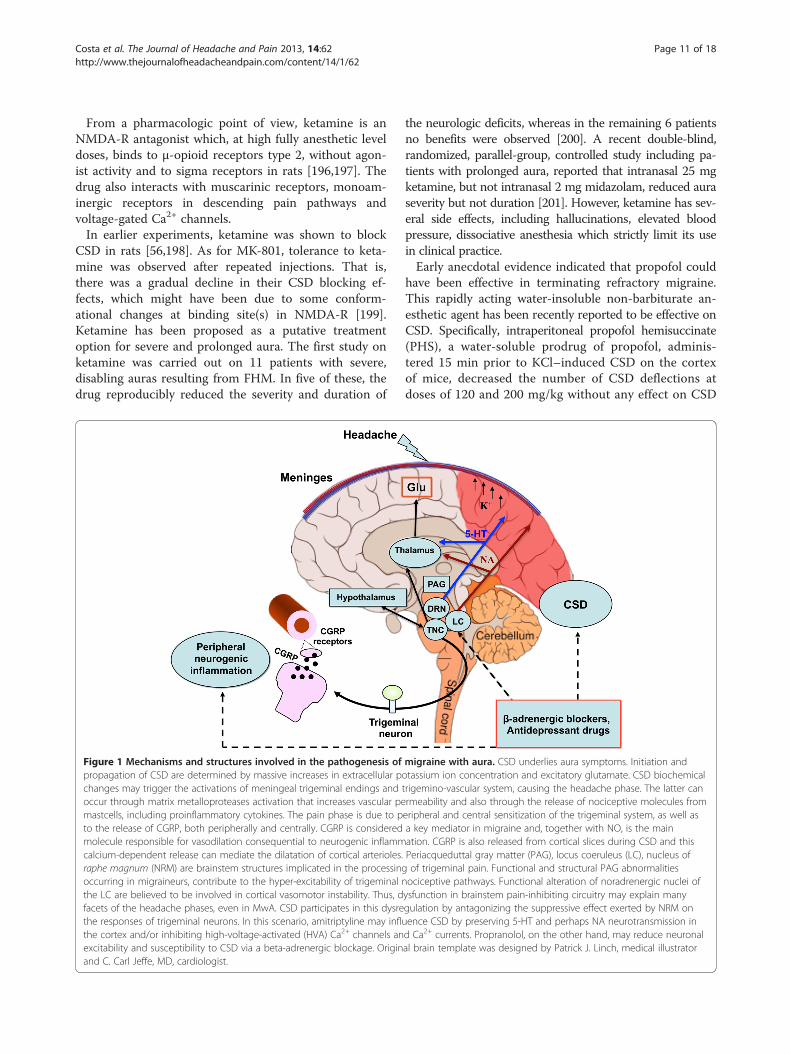
Figure 1 Mechanisms and structures involved in the pathogenesis of migraine with aura. CSD underlies aura symptoms. Initiation andpropagation of CSD are determined by massive increases in extracellular potassium ion concentration and excitatory glutamate. CSD biochemicalchanges may trigger the activations of meningeal trigeminal endings and trigemino-vascular system, causing the headache phase. The latter canoccur through matrix metalloproteases activation that increases vascular permeability and also through the release of nociceptive molecules frommastcells, including proinflammatory cytokines. The pain phase is due to peripheral and central sensitization of the trigeminal system, as well asto the release of CGRP, both peripherally and centrally. CGRP is considered a key mediator in migraine and, together with NO, is the mainmolecule responsible for vasodilation consequential to neurogenic inflammation. CGRP is also released from cortical slices during CSD and thiscalcium-dependent release can mediate the dilatation of cortical arterioles. Periacqueduttal gray matter (PAG), locus coeruleus (LC), nucleus ofraphe magnum (NRM) are brainstem structures implicated in the processing of trigeminal pain. Functional and structural PAG abnormalitiesoccurring in migraineurs, contribute to the hyper-excitability of trigeminal nociceptive pathways. Functional alteration of noradrenergic nuclei ofthe LC are believed to be involved in cortical vasomotor instability. Thus, dysfunction in brainstem pain-inhibiting circuitry may explain manyfacets of the headache phases, even in MwA. CSD participates in this dysregulation by antagonizing the suppressive effect exerted by NRM onthe responses of trigeminal neurons. In this scenario, amitriptyline may influence CSD by preserving 5-HT and perhaps NA neurotransmission inthe cortex and/or inhibiting high-voltage-activated (HVA) Ca2+ channels and Ca2+ currents. Propranolol, on the other hand, may reduce neuronalexcitability and susceptibility to CSD via a beta-adrenergic blockage. Original brain template was designed by Patrick J. Linch, medical illustratorand C. Carl Jeffe, MD, cardiologist.
Costa et al. The Journal of Headache and Pain 2013, 14:62 Page 11 of 18http://www.thejournalofheadacheandpain.com/content/14/1/62
amplitude [202]. In contrast, Kudo et al. failed to dem-onstrate any inhibitory effect of propofol on the fre-quency of KCl-induced CSD in rats [203]. This resultmay have been due to the fact that a water-insolubleformulation of propofol widely administered as a anes-thetic in clinical setting was used. In above mentionedstudy by Dhir et al. a water-soluble, non-commerciallyavailable prodrug PHS, was tested [202]. Consideringthese contrasting findings, it should be investigatedwhether metabolites produced during PHS activation,rather than propofol per se, can mediate an inhibitoryeffect on CSD. Given the varying effects of anestheticson CSD, future studies will have to follow uniform pro-tocols using only anaesthetics having a minimal impacton CSD in order to guarantee reliability and compa-rability of the results [204].
TRPV1 receptors play an important role in modulatingtrigeminal sensory processing and for this have beenproposed as potential targets for migraine treatment.The TRPV1 receptor antagonist, A-993610, has beentested in a model of mechanically-induced CSD but itfailed to show any effects. This lack of effect should beconfirmed for other TRPV1 receptor antagonists in fu-ture research [205].Finally, cannabis has been empirically used for centur-
ies for both symptomatic and prophylactic treatment ofdifferent types of headaches including migraine. Recentfindings have demonstrated a dose-dependent suppres-sion of CSD amplitude, duration and propagation vel-ocity after Delta9-tetrahydrocannabinol (THC) in ratneocortical slices, which had been antagonized by canna-binoid CB1 agonist, WIN 55,212-2 mesylate but not by
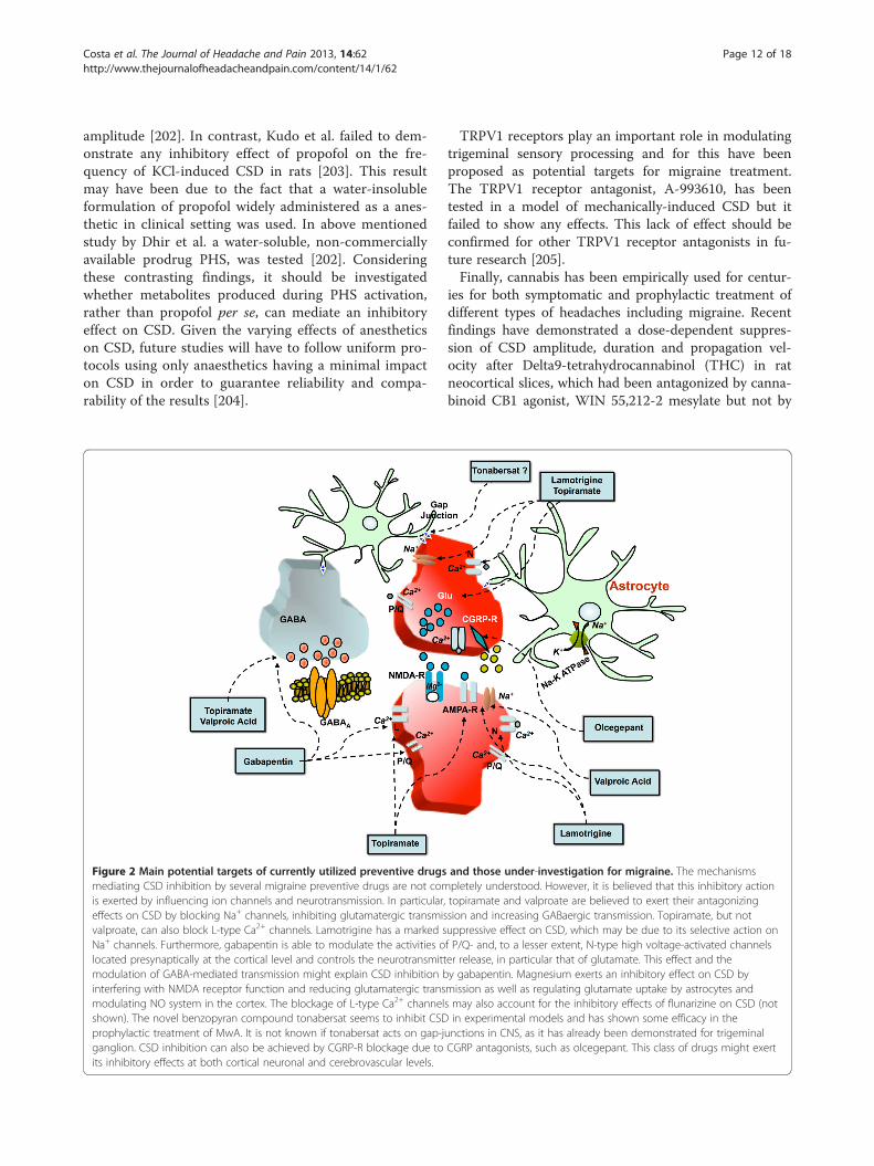
Figure 2 Main potential targets of currently utilized preventive drugs and those under-investigation for migraine. The mechanismsmediating CSD inhibition by several migraine preventive drugs are not completely understood. However, it is believed that this inhibitory actionis exerted by influencing ion channels and neurotransmission. In particular, topiramate and valproate are believed to exert their antagonizingeffects on CSD by blocking Na+ channels, inhibiting glutamatergic transmission and increasing GABaergic transmission. Topiramate, but notvalproate, can also block L-type Ca2+ channels. Lamotrigine has a marked suppressive effect on CSD, which may be due to its selective action onNa+ channels. Furthermore, gabapentin is able to modulate the activities of P/Q- and, to a lesser extent, N-type high voltage-activated channelslocated presynaptically at the cortical level and controls the neurotransmitter release, in particular that of glutamate. This effect and themodulation of GABA-mediated transmission might explain CSD inhibition by gabapentin. Magnesium exerts an inhibitory effect on CSD byinterfering with NMDA receptor function and reducing glutamatergic transmission as well as regulating glutamate uptake by astrocytes andmodulating NO system in the cortex. The blockage of L-type Ca2+ channels may also account for the inhibitory effects of flunarizine on CSD (notshown). The novel benzopyran compound tonabersat seems to inhibit CSD in experimental models and has shown some efficacy in theprophylactic treatment of MwA. It is not known if tonabersat acts on gap-junctions in CNS, as it has already been demonstrated for trigeminalganglion. CSD inhibition can also be achieved by CGRP-R blockage due to CGRP antagonists, such as olcegepant. This class of drugs might exertits inhibitory effects at both cortical neuronal and cerebrovascular levels.
Costa et al. The Journal of Headache and Pain 2013, 14:62 Page 12 of 18http://www.thejournalofheadacheandpain.com/content/14/1/62
cannabinoid CB2 agonist, JWH-133 [206]. These findingsuggests that cannabinoids might have inherent thera-peutic effects for MwA but their known side effects andrisk of dependence must be properly weighed beforecannabinoid being considered for treatment.
ConclusionsA number of mechanisms have been shown to have arole in fostering CSD wave initiation and propagation in-cluding: ion diffusion, membrane ionic currents, osmoticeffects, spatial buffering, neurotransmitter substances,gap junctions, metabolic pumps, and synaptic connec-tions (Figure 1).In spite of this knowledge, CSD remains an enigma
necessitating further theoretical investigations.Experimental findings to date suggest that chronic
daily administration of certain migraine prophylacticdrugs (topiramate, valproate, propranolol, amitryptiline,and methysergide) dose-dependently suppress CSD. Ata molecular level, targets of the inhibitory effects ofantiepileptic drugs tested exert their inhibitory effectson CSD targeting Ca2+ and Na2+ channels, as well asglutamatergic and/or GABAergic transmissions basedon their mechanisms of action. Additionally, the anti-epileptic drug lamotrigine has a proven suppressiveeffect on CSD, which could explain its selective actionon migraine aura. Preservation of 5-HT, and maybeeven NA neurotrasmission in the cortex, could in someway be responsible for the effects of amitryptiline onCSD, while beta-adrenergic blockage by propranololmight facilitate a reduction in cortical neuronal excit-ability and thus in turn reduce susceptibility to CSD(Figure 2).Mechanisms of action for some novel molecules are
currently under investigation. To date, it has been foundthat CGRP-R antagonists exert a dose-dependent inhibi-tory effect on CSD. For this reason, it can be hypothe-sized that CGRP plays a determining role in CSD and itsmodulation may be effective for the preventive treat-ment of MwA (Figure 2). Additionally, tonabersat, anovel benzopyran compound, has been reported to exertan inhibitory action on CSD and on neurogenic inflam-mation in animal models of migraine. These inhibitoryactions might be an effect of the blockage of neuronal-glial cell gap-junctions, as it has only been demonstratedto be for the trigeminal ganglion. The most significantanti-migraine action on the part of tonabersat seems toderive from its inhibitory action on CSD. In fact, clinicaltrials on tonabersat have shown its preventive effect onMwA attacks but not on MwoA attacks.Based on recent clinical findings, intranasal ketamine,
has been shown to be effective in CSD models, andtherefore it has been administered for cases of migrainewith prolonged aura, but its use is limited due to its
relevant side effects. Noteworthy, the effectiveness of keta-mine adds to the existing evidence that glutamatergictransmission plays a role in human aura. The provencapacities of other molecules (i.e. amiloride) in blockingCSD suggests that they might also have a role in preventingMwA.Further investigations on molecules with evidence of
targeting CSD not only would lead to a better under-standing of the underlying mechanisms for CSD, butalso supply meaningful insight into their potential rolesin MwA, as well as other diseases, such as epilepsy, is-chemic stroke, intracranial hemorrhage, and trauma,where CSD is thought to be a pathogenic mechanism.
Additional file
Additional file 1: Table S1. A summary of the most relevant studies onCSD experimental models regarding the effects of currently used drugsand drugs under investigation for migraine prophylaxis.
Abbreviations5-HT: 5-hydroxytryptamine, serotonin; AMPA: Alpha-amino-3-hydroxy-5-methyl-4-isoxazole propionate; AMPA-R: Alpha-amino-3-hydroxy-5-methyl-4-isoxazole propionate receptor; ATP: Adenosine-5'-triphosphate;ATP1A2: ATPase, Na+/K+ transporting, alpha 2 (+) polypeptide; BBB:Blood–brain barrier; BOLD: Blood oxygenation level dependent; Ca2+: Calcium; CACNA: Ca2+ channel alpha 1A; CADASIL: Cerebral autosomaldominant arteriopathy with subcortical infarcts and leukoencephalopathy;CBFLDF: Cortical blood flow laser Doppler flowmetry; CGRP: Calcitoningene-related peptide; COX: Cyclooxygenase; CSD: Cortical spreadingdepression; CSF: Cerebrospinal fluid; DC: Direct current; DEA:(N,N-diethylamino)-diazenolate-2-oxide; DHE: Dihydroergotamine;EEG: Electroencephalographic; ET-1: Endothelin-1; FHM: Familial Hemyplegicmigraine; fMRI: Functional magnetic resonance imaging; GABA:Gamma-aminobutyric acid; GABA-R: Gamma-aminobutyric acid receptor;Gd-DTPA: Gadolinium-diethylenetriaminepenta-acetic acid; HMGB1:High-mobility group box 1; IL-1β: Interleukin-1 β; iNOS: Inducible NOsynthase; K+: Potassium; KCl: Potassium chloride; KI: Knockin; L-NAME:L-NG-Nitroarginine Methyl Ester; MEG: Magnetoelectroencephalography; Mg2+: Magnesium; MMP(s): Metalloprotease(s); MR: Magnetic resonance;MwA: Migraine with aura; MwoA: Migraine without aura; Na+: Sodium;NA: Noradrenalin; NMDA-R: N-methyl- D-aspartate receptor; NO: Nitric oxide;NOLAG: NO-nitro-L-arginine; NOS: NO synthase; OIS: Optical intrinsic signal;Panx1: Pannexin1; PET: Positron Emission Tomography; PHS: Propofolhemisuccinate; PLC: Phospholipase C; SCN1A: Sodium channel, voltage-gated, type I, alpha subunit; SD: Spreading depression; SPECT: Single-photonemission computed tomography; THC: Tetrahydrocannabinol;TPM: Topiramate; TRPV1: Transient receptor potential cation channelsubfamily V member 1; TTX: Tetrodotoxin; WMLs: White matter lesions.
Competing interestsThe authors declare that they have no competing interests.
Authors’ contributionAll authors equally contributed to the conception and drafting of themanuscript. PC, CA, and PS critically revised the manuscript for importantintellectual content. PS, CC, and AT also contributed in the preparation offigures and table. All authors read and approved the final manuscript.
AcknowledgementsWe thank Mr. Thomas Kilcline for editing the English. This Review Article willbe presented at the XXVII National Congress of the Italian Society for theStudy of Headaches – 26–28 September 2013.
Costa et al. The Journal of Headache and Pain 2013, 14:62 Page 13 of 18http://www.thejournalofheadacheandpain.com/content/14/1/62
Author details1Neurologic Clinic, Department of Public Health and Medical and SurgicalSpecialties, University of Perugia, Ospedale Santa Maria della Misericordia,Sant'Andrea delle Fratte, 06132, Perugia, Italy. 2Neurology II, Department ofNeuroscience, University of Torino, Ospedale Molinette, Via Cherasco 15,10126, Turin, Italy. 3Fondazione Santa Lucia I.R.C.C.S., Via del Fosso di Fiorano,00143, Rome, Italy. 4Neurovascular Research Lab., Department of Radiology,Stroke Service and Neuroscience Intensive Unit Department of NeurologyMassachusetts Hospital, Harvard Medical School, 02115, Boston, MA, USA.5Neurologic Clinic, Ospedale S. Eugenio, Piazzale Umanesimo, 00144,Rome, Italy.
Received: 5 June 2013 Accepted: 8 July 2013Published: 23 July 2013
References1. Somjen GG (2005) Aristides Leão's discovery of cortical spreading
depression. J Neurophysiol 94:2–42. Fabricius M, Fuhr S, Bhatia R, Boutelle M, Hashemi P, Strong AJ, Lauritzen M
(2006) Cortical spreading depression and peri-infarct depolarization inacutely injured human cerebral cortex. Brain 129:778–790
3. Lauritzen M, Dreier JP, Fabricius M, Hartings JA, Graf R, Strong AJ (2011)Clinical relevance of cortical spreading depression in neurological disorders:migraine, malignant stroke, subarachnoid and intracranial hemorrhage, andtraumatic brain injury. J Cereb Blood Flow Metab 31:17–35
4. Dreier JP (2011) The role of spreading depression, spreading depolarizationand spreading ischemia in neurological disease. Nat Med 17:439–447
5. Somjen GG (2001) Mechanisms of spreading depression and hypoxicspreading depression-like depolarization. Physiol Rev 81:1065–1096
6. Strong AJ, Dardis R (2005) Depolarisation phenomena in traumatic andischaemic brain injury. In: Pickard JD (ed) Advances and Technical Standardsin Neurosurgery, Springer, Wien, vol 30, Austria vol. pp 3–49
7. Diener HC (1997) Positron emission tomography studies in headache.Headache 37:622–625
8. Tfelt-Hansen PC (2010) History of migraine with aura and cortical spreadingdepression from 1941 and onwards. Cephalalgia 30:780–792
9. Sánchez del Rio M, Alvarez Linera J (2004) Functional neuroimaging ofheadaches. Lancet Neurol 3:645–651
10. Smith JM, Bradley DP, James MF, Huang CL (2006) Physiological studies ofcortical spreading depression. Biol Rev Camb Philos Soc 81:457–481
11. Smith JM, James MF, Fraser JA, Huang CL (2008) Translational imagingstudies of cortical spreading depression in experimental models formigraine aura. Expert Rev Neurother 8:759–768
12. Zhang X, Levy D, Noseda R, Kainz V, Jakubowski M, Burstein R (2010)Activation of meningeal nociceptors by cortical spreading depression:implications for migraine with aura. J Neurosci 30:8807–8814
13. Hansen JM (2010) Familial hemiplegic migraine. Dan Med Bull 57:B418314. Bolay H, Berman NE, Akcali D (2011) Sex-related differences in animal
models of migraine headache. Headache 51:891–90415. Eikermann-Haerter K, Kudo C, Moskowitz MA (2007) Cortical spreading
depression and estrogen. Headache 47(Suppl 2):S79–S8516. Eikermann-Haerter K, Ayata C (2010) Cortical spreading depression and
migraine. Curr Neurol Neurosci Rep 10:167–17317. Eikermann-Haerter K, Can A, Ayata C (2012) Pharmacological targeting of
spreading depression in migraine. Expert Rev Neurother 12:297–30618. Merikangas KR (2013) Contributions of epidemiology to our understanding
of migraine. Headache 53:230–24619. Headache Classification Committee of The International Headache Society
(2004) The International Classification of Headache Disorders: 2nd edition.Cephalalgia 24(1):1–160
20. Montagna P (2008) Migraine genetics. Expert Rev Neurother 8:1321–133021. Pietrobon D (2007) Familial hemiplegic migraine. Neurotherapeutics 4:274–28422. Lashley KS (1941) Patterns of cerebral integration indicated by the scotomas
of migraine. Arch Neurol Psychiatry 46:331–33923. Leão AAP (1944) Spreading depression of activity in the cerebral cortex.
J Neurophysiol 7:359–39024. Leão AAP (1944) Pial circulation and spreading depression of activity in the
cerebral cortex. J Neurophysiol 7:391–39625. Leão AAP (1947) Further observations on the spreading depression of
activity in the cerebral cortex. J Neurophysiol 10:409–41426. Wolff HG (1948) Headache mechanisms. Bull U S Army Med Dep 8:641–653
27. Olesen J, Larsen B, Lauritzen M (1981) Focal hyperemia followed byspreading oligemia and impaired activation of rCBF in classic migraine.Ann Neurol 9:344–352
28. Lauritzen M (1994) Pathophysiology of the migraine aura. The spreadingdepression theory. Brain 117:199–210
29. Olesen J, Friberg L, Olsen TS, Iversen HK, Lassen NA, Andersen AR, Karle A(1990) Timing and topography of cerebral blood flow, aura, and headacheduring migraine attacks. Ann Neurol 28:791–798
30. Cutrer FM, Sorensen AG, Weisskoff RM, Ostergaard L, Sanchez del Rio M,Lee EJ, Rosen BR, Moskowitz MA (1998) Perfusion-weighted imaging defectsduring spontaneous migrainous aura. Ann Neurol 43:25–31
31. Cutrer FM, O'Donnell A, Sanchez del Rio M (2000) Functional neuroimaging:enhanced understanding of migraine pathophysiology. Neurology55(Suppl 2):S36–S45
32. Cao Y, Welch KM, Aurora S, Vikingstad EM (1999) Functional MRI-BOLD of visuallytriggered headache in patients with migraine. Arch Neurol 56:548–554
33. Hadjikhani N, Sanchez Del Rio M, Wu O, Schwartz D, Bakker D, Fischl B,Kwong KK, Cutrer FM, Rosen BR, Tootell RB, Sorensen AG, Moskowitz MA(2001) Mechanisms of migraine aura revealed by functional MRI in humanvisual cortex. Proc Natl Acad Sci USA 98:4687–4692
34. Oberndorfer S, Wöber C, Nasel C, Asenbaum S, Lahrmann H, Fueger B,Grisold W (2004) Familial hemiplegic migraine: follow-up findings ofdiffusion-weighted magnetic resonance imaging (MRI), perfusion-MRI and[99mTc] HMPAO-SPECT in a patient with prolonged hemiplegic aura.Cephalalgia 24:533–539
35. Jäger HR, Giffin NJ, Goadsby PJ (2005) Diffusion- and perfusion-weightedMR imaging in persistent migrainous visual disturbances. Cephalalgia25:323–332
36. Belvís R, Ramos R, Villa C, Segura C, Pagonabarraga J, Ormazabal I, KulisevskyJ (2010) Brain apparent water diffusion coefficient magnetic resonanceimage during a prolonged visual aura. Headache 50:1045–1049
37. Mourand I, Menjot de Champfleur N, Carra-Dallière C, Le Bars E, Roubertie A,Bonafé A, Thouvenot E (2012) Perfusion-weighted MR imaging in persistenthemiplegic migraine. Neuroradiology 54:255–260
38. Bereczki D, Kollár J, Kozák N, Viszokay K, Barta Z, Sikula J, Magyar MT (2008)Cortical spreading edema in persistent visual migraine aura. Headache48:1226–1229
39. Smith M, Cros D, Sheen V (2002) Hyperperfusion with vasogenic leakage byfMRI in migraine with prolonged aura. Neurology 58:1308–1310
40. Arnold G, Reuter U, Kinze S, Wolf T, Einhäupl KM (1998) MWA showsgadolinium enhancement which is reversed following prophylactictreatment. Cephalalgia 18:644–646
41. Bowyer SM, Aurora KS, Moran JE, Tepley N, Welch KM (2001)Magnetoencephalographic fields from patients with spontaneous andinduced migraine aura. Ann Neurol 50:582–587
42. Hall SD, Barnes GR, Hillebrand A, Furlong PL, Singh KD, Holliday IE (2004)Spatio-temporal imaging of cortical desynchronization in migraine visualaura: a magnetoencephalography case study. Headache 44:204–208
43. Grafstein B (1956) Mechanism of spreading cortical depression.J Neurophysiol 19:154–171
44. Strong AJ (2005) Dr. Bernice Grafstein's paper on the mechanism ofspreading depression. J Neurophysiol 94:5–7
45. Martins-Ferreira H, Nedergaard M, Nicholson C (2000) Perspectives onspreading depression. Brain Res Brain Res Rev 32:215–234
46. de Azeredo FA (1991) Transient changes in energy metabolites andintracellular pH during spreading depression in the chick retina. Metab BrainDis 6:75–82
47. Gault LM, Lin CW, LaManna JC, Lust WD (1994) Changes in energymetabolites, cGMP and intracellular pH during cortical spreadingdepression. Brain Res 641:176–180
48. Footitt DR, Newberry NR (1998) Cortical spreading depression induces anLTP-like effect in rat neocortex in vitro. Brain Res 781:339–342
49. Világi I, Klapka N, Luhmann HJ (2001) Optical recording of spreadingdepression in rat neocortical slices. Brain Res 898:288–296
50. Peeters M, Gunthorpe MJ, Strijbos PJ, Goldsmith P, Upton N, James MF(2007) Effects of pan- and subtype-selective N-methyl-D-aspartate receptorantagonists on cortical spreading depression in the rat: therapeuticpotential for migraine. J Pharmacol Exp Ther 321:564–572
51. Petzold GC, Haack S, von Bohlen Und Halbach O, Priller J, Lehmann TN,Heinemann U, Dirnagl U, Dreier JP (2008) Nitric oxide modulates spreadingdepolarization threshold in the human and rodent cortex. Stroke 39:1292–1299
Costa et al. The Journal of Headache and Pain 2013, 14:62 Page 14 of 18http://www.thejournalofheadacheandpain.com/content/14/1/62
52. McLachlan RS (1992) Suppression of spreading depression of Leao in neocortexby an N-methyl-D-aspartate receptor antagonist. Can J Neurol Sci 19:487–491
53. Obrenovitch TP, Zilkha E (1996) Inhibition of cortical spreading depressionby L-701,324, a novel antagonist at the glycine site of the N-methyl-D-aspartate receptor complex. Br J Pharmacol 117:931–937
54. Jing APG, Somjen GG (1993) Role of calcium channels in spreadingdepression in rat hippocampal slices. Brain Res 604:251–259
55. Footitt DR, Newberry NR (1998) Cortical spreading depression induces anLTP-like effect in rat neocortex in vitro. Brain 781:339–342
56. Hernándéz-Cáceres J, Macias-González R, Brozek G, Bures J (1987) Systemicketamine blocks cortical spreading depression but does not delay the onsetof terminal anoxic depolarization in rats. Brain Res 437:360–364
57. Marrannes R, Willems R, De Prins E, Wauquier A (1988) Evidence for a role ofthe N-methyl-D-aspartate (NMDA) receptor in cortical spreading depressionin the rat. Brain Res 457:226–240
58. Lauritzen M, Hansen AJ (1992) The effect of glutamate receptor blockadeon anoxic depolarization and cortical spreading depression. J Cereb BloodFlow Metab 12:223–229
59. Nellgård B, Wieloch T (1992) NMDA-receptor blockers but not NBQX, anAMPA-receptor antagonist, inhibit spreading depression in the rat brain.Acta Physiol Scand 146:497–503
60. Gorji A, Scheller D, Straub H, Tegtmeier F, Köhling R, Höhling JM, Tuxhorn I,Ebner A, Wolf P, Werner Panneck H, Oppel F, Speckmann EJ (2001)Spreading depression in human neocortical slices. Brain Res 906:74–83
61. Berger M, Speckmann EJ, Pape HC, Gorji A (2008) Spreading depressionenhances human neocortical excitability in vitro. Cephalalgia 28:558–562
62. Tottene A, Urbani A, Pietrobon D (2011) Role of different voltage-gated Ca2+ channels in cortical spreading depression: specific requirement of P/Q-type Ca2+ channels. Channels (Austin) 5:110–114
63. Petzold GC, Scheibe F, Braun JS, Freyer D, Priller J, Dirnagl U, Dreier JP(2005) Nitric oxide modulates calcium entry through P/Q-type calciumchannels and N-methyl-d-aspartate receptors in rat cortical neurons. BrainRes 1063:9–14
64. Zhou N, Gordon GR, Feighan D, MacVicar BA (2010) Transient swelling,acidification, and mitochondrial depolarization occurs in neurons but notastrocytes during spreading depression. Cereb Cortex 20:2614–2624
65. Herreras O, Somjen GG (1993) Analysis of potential shifts associated withrecurrent spreading depression and prolonged unstable SD induced bymicrodyalisis of elevated K+ in hippocampus of anesthetized rats. Brain Res610:283–294
66. Richter F, Ebersberger A, Schaible HG (2002) Blockade of voltage-gatedcalcium channels in rat inhibits repetitive cortical spreading depression.Neurosci Lett 334:123–126
67. Sugaya E, Takato M, Noda Y (1975) Neuronal and glial activity duringspreading depression in cerebral cortex of cat. J Neurophysiol 38(4):822–841
68. Tuckwell HC, Hermansen CL (1981) Ion and transmitter movements duringspreading cortical depression. Int J Neurosci 12:109–135
69. Somjen GG, Aitken PG (1984) The ionic and metabolic responses associatedwith neuronal depression of Leão's type in cerebral cortex and inhippocampal formation. An Acad Bras Cienc 56(4):495–504
70. Tozzi A, de Iure A, Di Filippo M, Costa C, Caproni S, Pisani A, Bonsi P, PicconiB, Cupini LM, Materazzi S, Geppetti P, Sarchielli P, Calabresi P (2012) Criticalrole of calcitonin gene-related peptide receptors in cortical spreadingdepression. Proc Natl Acad Sci USA 109:18985–18990
71. Akerman S, Holland PR, Goadsby PJ (2008) Mechanically-induced corticalspreading depression associated regional cerebral blood flow changes areblocked by Na+ ion channel blockade. Brain Res 1229:27–36
72. Addae JI, Evans SM, Ali N, Stone TW (2000) NMDA-induced changes in acortical network in vivo are prevented by AMPA. Brain Res 869:211–215
73. Anderson TR, Andrew RD (2002) Spreading depression: imaging andblockade in the rat neocortical brain slice. J Neurophysiol 88:2713–2725
74. Addae JI, Ali N, Stone TW (2011) Effects of AMPA and clomethiazole on spreadingdepression cycles in the rat neocortex in vivo. Eur J Pharmacol 653:41–46
75. Holland PR, Akerman S, Goadsby PJ (2010) Cortical spreading depression-associated cerebral blood flow changes induced by mechanical stimulationare modulated by AMPA and GABA receptors. Cephalalgia 30:519–527
76. Haghir H, Kovac S, Speckmann EJ, Zilles K, Gorji A (2009) Patterns ofneurotransmitter receptor distributions following cortical spreadingdepression. Neuroscience 163:1340–1352
77. Van Harreveld A, Ochs S (1957) Electrical and vascular concomitants ofspreading depression. Am J Physiol 189:159–166
78. Wahl M, Lauritzen M, Schilling L (1987) Change of cerebrovascular reactivityafter cortical spreading depression in cats and rats. Brain Res 411:72–80
79. Fabricius M, Akgoren N, Lauritzen M (1995) Arginine-nitric oxide pathwayand cerebrovascular regulation in cortical spreading depression. Am JPhysiol 269:H23–H29
80. Duckrow RB (1991) Regional cerebral blood flow during spreading corticaldepression in conscious rats. J Cereb Blood Flow Metab 11:150–154
81. Duckrow RB (1993) A brief hypoperfusion precedes spreading depression ifnitric oxide synthesis is inhibited. Brain Res 618:190–195
82. Piper RD, Lambert GA, Duckworth JW (1991) Cortical blood flow changesduring spreading depression in cats. Am J Physiol 261:H96–H102
83. Lauritzen M, Jørgensen MB, Diemer NH, Gjedde A, Hansen AJ (1982)Persistent oligemia of rat cerebral cortex in the wake of spreadingdepression. Ann Neurol 12:469–474
84. Goadsby PJ (1992) The oligemic phase of cortical spreading depression isnot blocked by tirilazad mesylate (U-74006F). Brain Res 588:140–143
85. Ba AM, Guiou M, Pouratian N, Muthialu A, Rex DE, Cannestra AF, Chen JW,Toga AW (2002) Multiwavelength optical intrinsic signal imaging of corticalspreading depression. J Neurophysiol 88:2726–2735
86. Guiou M, Sheth S, Nemoto M, Walker M, Pouratian N, Ba A, Toga AW (2005)Cortical spreading depression produces long-term disruption of activity-related changes in cerebral blood volume and neurovascular coupling.J Biomed Opt 10(1):11004
87. O'Farrell AM, Rex DE, Muthialu A, Pouratian N, Wong GK, Cannestra AF,Chen JW, Toga AW (2000) Characterization of optical intrinsic signals andblood volume during cortical spreading depression. Neuroreport11:2121–2125
88. Ayata C, Shin HK, Salomone S, Ozdemir-Gursoy Y, Boas DA, Dunn AK,Moskowitz MA (2004) Pronounced hypoperfusion during spreadingdepression in mouse cortex. J Cereb Blood Flow Metab 24:1172–1182
89. Brennan KC, Beltrán-Parrazal L, López-Valdés HE, Theriot J, Toga AW, CharlesAC (2007) Distinct vascular conduction with cortical spreading depression. JNeurophysiol 97:4143–4151
90. Chang JC, Shook LL, Biag J, Nguyen EN, Toga AW, Charles AC, Brennan KC (2010)Biphasic direct current shift, haemoglobin desaturation and neurovascularuncoupling in cortical spreading depression. Brain 133:996–1012
91. Wang M, Obrenovich TP, Ureniak J (2003) Effects of the nitric oxide donor,DEA/NO on cortical spreading depression. Neuropharmacology 44:949–957
92. Kleeberg J, Petzold GC, Major S, Dirnagl U, Dreier JP (2004) ET-1 inducescortical spreading depression via activation of the ETA receptor/phospholipase C pathway in vivo. Am J Physiol Heart Circ Physiol 286:H1339–H1346
93. Dreier JP, Kleeberg J, Alam M, Major S, Kohl-Bareis M, Petzold GC, Victorov I,Dirnagl U, Obrenovitch TP, Priller J (2007) Endothelin-1-induced spreadingdepression in rats is associated with a microarea of selective neuronalnecrosis. Exp Biol Med (Maywood) 232:204–213
94. Ophoff RA, Terwindt GM, Vergouwe MN, van Eijk R, Oefner PJ, Hoffman SM,Lamerdin JE, Mohrenweiser HW, Bulman DE, Ferrari M, Haan J, Lindhout D,van Ommen GJ, Hofker MH, Ferrari MD, Frants RR (1996) Familial hemiplegicmigraine and episodic ataxia type-2 are caused by mutations in the Ca2+channel gene CACNL1A4. Cell 87:543–552
95. van den Maagdenberg AM, Pietrobon D, Pizzorusso T, Kaja S, Broos LA,Cesetti T, van de Ven RC, Tottene A, van der Kaa J, Plomp JJ, Frants RR,Ferrari MD (2004) A Cacna1a knockin migraine mouse model with increasedsusceptibility to cortical spreading depression. Neuron 41:701–710
96. Pietrobon D (2005) Function and dysfunction of synaptic calcium channels:insights from mouse models. Curr Opin Neurobiol 15:257–265
97. Stam AH, Haan J, van den Maagdenberg AM, Ferrari MD, Terwindt GM(2009) Migraine and genetic and acquired vasculopathies. Cephalalgia29:1006–1017
98. Uchitel OD, Inchauspe CG, Urbano FJ, Di Guilmi MN (2012) CaV2.1 voltageactivated calcium channels and synaptic transmission in familial hemiplegicmigraine pathogenesis. J Physiol Paris 106:12–22
99. Inchauspe CG, Urbano FJ, Di Guilmi MN, Forsythe ID, Ferrari MD, van denMaagdenberg AM, Uchitel OD (2010) Gain of function in FHM-1 Cav 2.1knock-in mice is related to the shape of the action potential. J Neurophysiol104:291–299
100. Pietrobon D (2010) Cav2.1 channelopathies. Pflugers Arch 460:375–393101. Pietrobon D (2010) Insights into migraine mechanisms and CaV2.1 calcium
channel function from mouse models of familial hemiplegic migraine.J Physiol 588:1871–1878
Costa et al. The Journal of Headache and Pain 2013, 14:62 Page 15 of 18http://www.thejournalofheadacheandpain.com/content/14/1/62
102. Tottene A, Fellin T, Pagnutti S, Luvisetto S, Striessnig J, Fletcher C, PietrobonD (2002) Familial hemiplegic migraine mutations increase Ca2+ influxthrough single human Cav2.1 channels and decrease maximal Cav2.1current density in neurons. Proc Natl Acad Sci USA 99:13284–13289
103. Tottene A, Conti R, Fabbro A, Vecchia D, Shapovalova M, Santello M, vanden Maagdenberg AM, Ferrari MD, Pietrobon D (2009) Enhanced excitatorytransmission at cortical synapses as the basis for facilitated spreadingdepression in Ca(v)2.1 knockin migraine mice. Neuron 61(5):762–773
104. van den Maagdenberg AM, Pizzorusso T, Kaja S, Terpolilli N, Shapovalova M,Hoebeek FE, Barrett CF, Gherardini L, van de Ven RC, Todorov B, Broos LA,Tottene A, Gao Z, Fodor M, De Zeeuw CI, Frants RR, Plesnila N, Plomp JJ,Pietrobon D, Ferrari MD (2010) High cortical spreading depressionsusceptibility and migraine-associated symptoms in Cav2.1 S218L mice. AnnNeurol 67:85–98
105. Ayata C, Shimizu-Sasamata M, Lo EH, Noebels JL, Moskowitz MA (2000)Impaired neurotransmitter release and elevated threshold for corticalspreading depression in mice with mutations in the α1A subunit of P/Qtype calcium channels. Neuroscience 95:639–645
106. Leo L, Gherardini L, Barone V, De Fusco M, Pietrobon D, Pizzorusso T, CasariG (2011) Increased susceptibility to cortical spreading depression in themouse model of familial hemiplegic migraine type 2. PLoS Genet7(6):e1002129
107. Eikermann-Haerter K, Yuzawa I, Dilekoz E, Joutel A, Moskowitz MA, Ayata C(2011) Cerebral autosomal dominant arteriopathy with subcortical infarctsand leukoencephalopathy syndrome mutations increase susceptibility tospreading depression. Ann Neurol 69:413–418
108. Nagel-Leiby S, Welch KM, Grunfeld S, D’Andrea G (1990) Ovarian steroidlevels in migraine with and without aura. Cephalalgia 10:147–152
109. MacGregor EA (2004) Oestrogen and attacks of migraine with and withoutaura. Lancet Neurol 3:354–361
110. Ertresvag JM, Zwart JA, Helde G, Johnsen HJ, Bovim G (2005) Headache andtransient focal neurological symptoms during pregnancy, a prospectivecohort. Acta Neurol Scand 111:233–237
111. Granella F, Sances G, Zanferrari C, Costa A, Martignoni E, Manzoni GC (1993)Migraine without aura and reproductive life events: a clinicalepidemiological study in 1300 women. Headache 33:385–389
112. Calton GJ, Burnett JW (1984) Danazol and migraine. N Engl J Med 310:721–722113. Jonsson B, Von Reis G, Sahlgren E (1951) Testosterone therapy of headache.
Acta Psychiatr Neurol Scand 74:102–105114. Moehlig RC (1955) Methyl testosterone for migraine of women; report of
sixty cases. J Mich State Med Soc 54:577–579115. Vincent FM (1985) Migraine responsive to danazol. Neurology 35:618116. Lichten EM, Bennett RS, Whitty AJ, Daoud Y (1991) Efficacy of danazol in the
control of hormonal migraine. J Reprod Med 36(6):419–424117. Arango O, Bielsa O, Pascual-Calvet J, Herrero M, Gelabert-Mas A (1996)
Disappearance of migraine crises in two patients with male infertilitytreated with human chorionic gonadotropin/human menopausalgonadotrophin. Rev Neurol 24:977–979
118. Pringsheim T, Gooren L (2004) Migraine prevalence in male to femaletranssexuals on hormone therapy. Neurology 63:593–594
119. Eikermann-Haerter K, Dileköz E, Kudo C, Savitz SI, Waeber C, Baum MJ,Ferrari MD, van den Maagdenberg AM, Moskowitz MA, Ayata C (2009)Genetic and hormonal factors modulate spreading depression and transienthemiparesis in mouse models of familial hemiplegic migraine type 1. J ClinInvest 119:99–109
120. Eikermann-Haerter K, Baum MJ, Ferrari MD, van den Maagdenberg AM,Moskowitz MA, Ayata C (2009) Androgenic suppression of spreading depressionin familial hemiplegic migraine type 1 mutant mice. Ann Neurol 66:564–568
121. Benarroch EE (2005) Neuron-astrocyte interactions: partnership for normalfunction and disease in the central nervous system. Mayo Clin Proc80:1326–1338
122. Lian XY, Stringer JL (2004) Energy failure in astrocytes increases thevulnerability of neurons to spreading depression. Eur J Neurosci 19:2446–2454
123. Lian XY, Stringer JL (2007) Astrocytes contribute to regulation ofextracellular calcium and potassium in the rat cerebral cortex duringspreading depression. J Pharmacol Exp Ther 321:564–572
124. Giaume C, McCarthy KD (1996) Control of gap-junctional communication inastrocytic networks. Trends Neurosci 19:319–325
125. Bennett MVL, Contreras JE, Bukauskas FF, Sáez JC (2003) New roles forastrocytes: gap junction hemichannels have something to communicate.Trends Neurosci 26:610–617
126. Nagy JI, Dudek FE, Rash JE (2004) Update on connexins and gap junctionsin neurons and glia in the mammalian nervous system. Brain Res Rev47:191–215
127. Ye Z-C, Wyeth MS, Baltan-Tekkok S, Ransom BR (2003) Functionalhemichannels in astrocytes: a novel mechanism of glutamate release.J Neurosci 23:3588–3596
128. Peters O, Schipke CG, Hashimoto Y, Kettenmann H (2003) Differentmechanisms promote astrocyte Ca2+ waves and spreading depression inthe mouse neocortex. J Neurosci 23(30):9888–9896
129. Stout CE, Costantin JL, Naus CCG, Charles AC (2002) Intercellular calciumsignaling in astrocytes via ATP release through connexin hemichannels.J Biol Chem 277:10482–10488
130. Grafstein B, Liu S, Cotrina ML, Goldman SA, Nedergaard M (2000) Meningeal cellscan communicate with astrocytes by calcium signaling. Ann Neurol 47:18–25
131. Gursoy-Ozdemir Y, Qiu J, Matsuoka N, Bolay H, Bermpohl D, Jin H, Wang X,Rosenberg GA, Lo EH, Moskowitz MA (2004) Cortical spreading depressionactivates and upregulates MMP-9. J Clin Invest 113:1447–1455
132. Martins-Oliveira A, Speciali JG, Dach F, Marcaccini AM, Gonçalves FM, GerlachRF, Tanus-Santos JE (2009) Different circulating metalloproteinases profiles inwomen with migraine with and without aura. Clin Chim Acta 408:60–64
133. Gao HM, Li L, Zhang KL, Chen XH, Tian SQ, Zhang ZL (2010) Impact ofmigraine attacks on the blood–brain barrier. Chin Med J (Engl)123:2559–2561
134. Martins-Oliveira A, Gonçalves FM, Speciali JG, Fontana V, Izidoro-Toledo TC,Belo VA, Dach F, Tanus-Santos JE (2012) Specific matrix metalloproteinase 9(MMP-9) haplotype affect the circulating MMP-9 levels in women withmigraine. J Neuroimmunol 252:89–94
135. Gonçalves FM, Martins-Oliveira A, Lacchini R, Belo VA, Speciali JG, Dach F,Tanus-Santos JE (2013) Matrix metalloproteinase (MMP)-2 genepolymorphisms affect circulating MMP-2 levels in patients with migrainewith aura. Gene 512:35–40
136. Gupta VK (2009) CSD, BBB and MMP-9 elevations: animal experimentsversus clinical phenomena in migraine. Expert Rev Neurother 9:1595–1614
137. Levy D (2012) Endogenous mechanisms underlying the activation andsensitization of meningeal nociceptors: the role of immuno-vascularinteractions and cortical spreading depression. Curr Pain Headache Rep16:270–277
138. Lambert GA, Truong L, Zagami AS (2011) Effect of cortical spreadingdepression on basal and evoked traffic in the trigeminovascular sensorysystem. Cephalalgia 31(14):1439–1451
139. Ji R-R, Kohno T, Moore KA, Woolf CJ (2003) Central sensitization and LTP: dopain and memory share similar mechanisms? Trends Neurosci 26:696–705
140. Goadsby PJ, Akerman S (2012) The trigeminovascular system does notrequire a peripheral sensory input to be activated–migraine is a centraldisorder. Focus on 'Effect of cortical spreading depression on basal andevoked traffic in the trigeminovascular sensory system'. Cephalalgia 32:3–5
141. Strassman AM, Raimond SA, Burstein R (1996) Sensitization of meningealsensory neurons and the origin of headache. Nature 384:560–564
142. Karatas H, Erdener SE, Gursoy-Ozdemir Y, Lule S, Eren-Koçak E, Sen ZD,Dalkara T (2013) Spreading depression triggers headache by activatingneuronal Panx1 channels. Science 339:1092–1095
143. Silverman WR, de Rivero Vaccari JP, Locovei S, Qiu F, Carlsson SK, Scemes E,Keane RW, Dahl G (2009) The pannexin 1 channel activates theinflammasome in neurons and astrocytes. J Biol Chem 284:18143–18151
144. Müller S, Ronfani L, Bianchi ME (2004) Regulated expression and subcellularlocalization of HMGB1, a chromatin protein with a cytokine function.J Intern Med 255:332–343
145. Faraco G, Fossati S, Bianchi ME, Patrone M, Pedrazzi M, Sparatore B, MoroniF, Chiarugi A (2007) High mobility group box 1 protein is released by neuralcells upon different stresses and worsens ischemic neurodegenerationin vitro and in vivo. J Neurochem 103:590–603
146. Pontremoli S, Melloni E (2007) Selective proinflammatory activation ofastrocytes by high-mobility group box 1 protein signaling. J Immunol179:8525–8532
147. Read SJ, Parsons AA (2000) Sumatriptan modifies cortical free radical releaseduring cortical spreading depression. A novel antimigraine action forsumatriptan? Brain Res 870:44–53
148. Read SJ, Hirst WD, Upton N, Parsons AA (2001) Cortical spreadingdepression produces increased cGMP levels in cortex and brain stemthat is inhibited by tonabersat (SB-220453) but not sumatriptan.Brain Res 891:69–77
Costa et al. The Journal of Headache and Pain 2013, 14:62 Page 16 of 18http://www.thejournalofheadacheandpain.com/content/14/1/62
149. Ayata C (2009) Spreading depression: from serendipity to targeted therapyin migraine prophylaxis. Cephalalgia 29:1095–1114
150. Kaube H, Goadsby PJ (1994) Anti-migraine compounds fail to modulate thepropagation of cortical spreading depression in the cat. Eur Neurol 34:30–35
151. Ayata C, Jin H, Kudo C, Dalkara T, Moskowitz MA (2006) Suppression of corticalspreading depression in migraine prophylaxis. Ann Neurol 59:652–661
152. Unekawa M, Tomita Y, Toriumi H, Suzuki N (2012) Suppressive effect ofchronic peroral topiramate on potassium-induced cortical spreadingdepression in rats. Cephalalgia 32:518–527
153. Akerman S, Goadsby PJ (2005) Topiramate inhibits cortical spreadingdepression in rat and cat: impact in migraine aura. Neuroreport16:1383–1387
154. Hoffmann U, Dileköz E, Kudo C, Ayata C (2010) Gabapentin suppressescortical spreading depression susceptibility. J Cereb Blood Flow Metab30:1588–1592
155. D'Andrea G, Granella F, Cadaldini M, Manzoni GC (1999) Effectiveness oflamotrigine in the prophylaxis of migraine with aura: an open pilot study.Cephalalgia 19:64–66
156. Lampl C, Buzath A, Klinger D, Neumann K (1999) Lamotrigine in theprophylactic treatment of migraine aura–a pilot study. Cephalalgia 19:58–63
157. Lampl C, Katsarava Z, Diener HC, Limmroth V (2005) Lamotrigine reducesmigraine aura and migraine attacks in patients with migraine with aura.J Neurol Neurosurg Psychiatry 76:1730–1732
158. Steiner TJ, Findley LJ, Yuen AW (1997) Lamotrigine versus placebo in theprophylaxis of migraine with and without aura. Cephalalgia 17:109–112
159. Gupta P, Singh S, Goyal V, Shukla G, Behari M (2007) Low-dose topiramateversus lamotrigine in migraine prophylaxis (the Lotolamp study). Headache47:402–412
160. Bogdanov VB, Multon S, Chauvel V, Bogdanova OV, Prodanov D, MakarchukMY, Schoenen J (2011) Migraine preventive drugs differentially affectcortical spreading depression in rat. Neurobiol Dis 41:430–435
161. Marrannes R, Wauquier A, Reid K, De Prins E (1986) The Prelude to TheMigraine Attack. Effects of drugs on cortical spreading depression. BaillièreTindal Oxford, UK, pp 158–173
162. Hansen A, Lauritzen M, Tfelt-Hansen P (1984) Spreading cortical depressionand antimigraine drugs. In: Amery WK, Nueten JM, Waquier A (eds) ThePharmacological Basis of Migraine Therapy. Putman, London, UK, pp 161–170
163. Reid KH, Marrannes R, Wauquier A (1987) Effects of flunarizine on ratcerebral cortex. Drug Dev Res 10:107–116
164. Wauquier A, Ashton D, Marrannes R (1985) The effects of flunarizine inexperimental models related to the pathogenesis of migraine. Cephalalgia 5(2):119–123
165. Marrannes R, De Prins E, Fransen J, Clincke G (1993) Neuropharmacology ofspreading depression. In: Lehmenkuhler A, Grotemeyer KH, Tegtmeier F(eds) Migraine: Basic Mechanisms and Treatment. MD, USA, Urban &Schwarzenberg, pp 431–443
166. Shimazawa M, Hara H, Watano T, Sukamoto T (1995) Effects of Ca2+ channelblockers on cortical hypoperfusion and expression of c-Fos-likeimmunoreactivity after cortical spreading depression in rats. Br J Pharmacol115:1359–1368
167. Sarchielli P, Coata G, Firenze C, Morucci P, Abbritti G, Gallai V (1992) Serumand salivary magnesium levels in migraine and tension-type headache.Results in a group of adult patients. Cephalalgia 12:21–27
168. Gallai V, Sarchielli P, Morucci P, Abbritti G (1994) Magnesium content ofmononuclear blood cells in migraine patients. Headache 34:160–165
169. Gallai V, Sarchielli P, Morucci P, Abbritti G (1993) Red blood cell magnesiumlevels in migraine patients. Cephalalgia 13:94–101
170. Mauskop A, Altura BM (1998) Role of magnesium in the pathogenesis andtreatment of migraines. Clin Neurosci 5:24–27
171. Ramadan NM, Halvorson H, Vande-Linde A, Levine SR, Helpern JA, WelchKM (1989) Low brain magnesium in migraine. Headache 29:590–593
172. Bigal ME, Bordini CA, Tepper SJ, Speciali JG (2002) Intravenous magnesiumsulphate in the acute treatment of migraine without aura and migrainewith aura. A randomized, double-blind, placebo-controlled study.Cephalalgia 22:345–353
173. van der Hel WS, van den Bergh WM, Nicolay K, Tulleken KA, Dijkhuizen RM(1998) Suppression of cortical spreading depressions after magnesiumtreatment in the rat. Neuroreport 9:2179–2182
174. Silberstein S, Saper J, Berenson F, Somogyi M, McCague K, D’Souza J (2008)Oxcarbazepine in migraine headache: a double-blind, randomized, placebo-controlled study. Neurology 70:548–555
175. Durham PL, Garrett FG (2009) Neurological mechanisms of migraine:potential of the gap-junction modulator tonabersat in prevention ofmigraine. Cephalalgia 29(Suppl 2):1–6
176. Silberstein SD, Schoenen J, Göbel H, Diener HC, Elkind AH, Klapper JA,Howard RA (2009) Tonabersat, a gap-junction modulator: efficacy and safetyin two randomized, placebo-controlled, dose-ranging studies of acutemigraine. Cephalalgia 29(Suppl 2):17–27
177. Goadsby PJ, Ferrari MD, Csanyi A, Olesen J, Mills JG (2009)Tonabersat TON-01-05 Study Group. Randomized, double-blind,placebo-controlled, proof-of-concept study of the cortical spreadingdepression inhibiting agent tonabersat in migraine prophylaxis.Cephalalgia 29:742–750
178. Dahlöf CG, Hauge AW, Olesen J (2009) Efficacy and safety of tonabersat, agap-junction modulator, in the acute treatment of migraine: a double-blind,parallel-group, randomized study. Cephalalgia 29(Suppl 2):7–16
179. Hauge AW, Asghar MS, Schytz HW, Christensen K, Olesen J (2009) Effects oftonabersat on migraine with aura: a randomised, double-blind, placebo-controlled crossover study. Lancet Neurol 8:718–723
180. Bradley DP, Smith MI, Netsiri C, Smith JM, Bockhorst KH, Hall LD, Huang CL,Leslie RA, Parsons AA, James MF (2001) Diffusion-Weighted MRI Used toDetect in Vivo Modulation of Cortical Spreading Depression: Comparison ofSumatriptan and Tonabersat. Exp Neurol 172:342–353
181. Damodaram S, Thalakoti S, Freeman SE, Garrett FG, Durham PL (2009)Tonabersat inhibits trigeminal ganglion neuronal-satellite glial cell signaling.Headache 49:5–20
182. Holland PR, Akerman S, Andreou AP, Karsan N, Wemmie JA, Goadsby PJ(2012) Acid-sensing ion channel 1: a novel therapeutic target for migrainewith aura. Ann Neurol 72:559–563
183. Durham PL, Masterson CG (2013) Two mechanisms involved in trigeminalCGRP release: implications for migraine treatment. Headache 53:67–80.
184. Yan J, Edelmayer RM, Wei X, De Felice M, Porreca F, Dussor G (2011) Duralafferents express acid-sensing ion channels: A role for decreased meningealpH in migraine headache. Pain 152:106–113
185. Raddant AC, Russo AF (2011) Calcitonin gene-related peptide in migraine:intersection of peripheral inflammation and central modulation. Expert RevMol Med 13:e36
186. Seybold VS (2009) The role of peptides in central sensitization. Handb ExpPharmacol 194:451–491
187. Wahl M, Schilling L, Parsons AA, Kaumann A (1994) Involvement ofcalcitonin gene-related peptide (CGRP) and nitric oxide (NO) in the pialartery dilatation elicited by cortical spreading depression. Brain Res637:2004–2010
188. Colonna DM, Meng W, Deal DD, Busija DW (1994) Calcitonin gene-relatedpeptide promotes cerebrovascular dilation during cortical spreadingdepression in rabbits. Am J Physiol 266:H1095–H1102
189. De Corato A, Lisi L, Capuano A, Tringali G, Tramutola A, Navarra P, DelloRusso C (2011) Trigeminal satellite cells express functional calcitonin gene-related peptide receptors, whose activation enhances interleukin-1β pro-inflammatory effects. J Neuroimmunol 237:39–46
190. Piper RD P, Edvinsson L, Ekman R, Lambert GA (1993) Cortical spreadingdepression does not result in the release of calcitonin gene-related peptideinto the external jugular vein of the cat: relevance to human migraine.Cephalalgia 13:180–183
191. Hansen JM, Hauge AW, Olesen J, Ashina M (2010) Calcitonin gene-relatedpeptide triggers migraine-like attacks in patients with migraine with aura.Cephalalgia 30:1179–1186
192. Negro A, Lionetto L, Simmaco M, Martelletti P (2012) CGRP receptorantagonists: an expanding drug class for acute migraine? Expert OpinInvestig Drugs 21:807–818
193. Hoffmann J, Goadsby PJ (2012) New Agents for Acute Treatment ofMigraine: CGRP Receptor Antagonists, iNOS Inhibitors. Curr Treat OptionsNeurol 14:50–59
194. Diener HC, Barbanti P, Dahlöf C, Reuter U, Habeck J, Podhorna J (2011) BI44370 TA, an oral CGRP antagonist for the treatment of acute migraineattacks: results from a phase II study. Cephalalgia 31:573–584
195. Chauvel V, Vamos E, Pardutz A, Vecsei L, Schoenen J, Multon S (2012) Effectof systemic kynurenine on cortical spreading depression and its modulationby sex hormones in rat. Exp Neurol 236:207–214
196. Hirota K, Sikand KS, Lambert DG (1999) Interaction of ketamine with mu2opioid receptors in SH-SY5Y human neuroblastoma cells. J Anesth13:107–109
Costa et al. The Journal of Headache and Pain 2013, 14:62 Page 17 of 18http://www.thejournalofheadacheandpain.com/content/14/1/62
197. Narita M, Yoshizawa K, Aoki K, Takagi M, Miyatake M, Suzuki T (2001) Aputative sigma1 receptor antagonist NE-100 attenuates the discriminativestimulus effects of ketamine in rats. Addict Biol 6:373–376
198. Gorelova NA, Koroleva VI, Amemori T, Pavlík V, Burĕs J (1987) Ketamineblockade of cortical spreading depression in rats. Electroencephalogr ClinNeurophysiol 66:440–447
199. Rashidy-Pour A, Motaghed-Larijani Z, Bures J (1995) Tolerance to ketamine-induced blockade of cortical spreading depression transfers to MK-801 butnot to AP5 in rats. Brain Res 693:64–69
200. Kaube H, Herzog J, Käufer T, Dichgans M, Diener HC (2000) Aura in somepatients with familial hemiplegic migraine can be stopped by intranasalketamine. Neurology 55:139–141
201. Afridi SK, Giffin NJ, Kaube H, Goadsby PJ (2013) A randomized controlledtrial of intranasal ketamine in migraine with prolonged aura. Neurology80:642–647
202. Dhir A, Lossin C, Rogawski MA (2012) Propofol hemisuccinate suppressescortical spreading depression. Neurosci Lett 514:67–70
203. Kudo C, Toyama M, Boku A, Hanamoto H, Morimoto Y, Sugimura M, Niwa H(2013) Anesthetic effects on susceptibility to cortical spreading depression.Neuropharmacol 67:32–36
204. Kudo C, Nozari A, Moskowitz MA, Ayata C (2008) The impact of anestheticsand hyperoxia on cortical spreading depression. Exp Neurol 212:201–206
205. Summ O, Holland PR, Akerman S, Goadsby PJ (2011) TRPV1 receptorblockade is ineffective in different in vivo models of migraine. Cephalalgia31:172–180
206. Kazemi H, Rahgozar M, Speckmann EJ, Gorji A (2012) Effect of cannabinoidreceptor activation on spreading depression. Iran J Basic Med Sci15:926–936
doi:10.1186/1129-2377-14-62Cite this article as: Costa et al.: Cortical spreading depression as a targetfor anti-migraine agents. The Journal of Headache and Pain 2013 14:62.
Submit your manuscript to a journal and benefi t from:
7 Convenient online submission
7 Rigorous peer review
7 Immediate publication on acceptance
7 Open access: articles freely available online
7 High visibility within the fi eld
7 Retaining the copyright to your article
Submit your next manuscript at 7 springeropen.com
Costa et al. The Journal of Headache and Pain 2013, 14:62 Page 18 of 18http://www.thejournalofheadacheandpain.com/content/14/1/62