course 8 lumps and bumps: ocular oncology · • acute hordeolum • molluscum contagiosum •...
TRANSCRIPT
SCO HOMECOMING / FALL CE WEEKEND • OCTOBER 9-12, 2014
2014 FallCE@SCO
COURSE 8
Lumps and Bumps: Ocular Oncology
COPE Course 42486-SD

9/16/2014
1
Eyelid and Orbital “Lumps and Bumps”
Anne P. Rowland, M.D.Oculoplastics and Reconstructive Surgeon
Benign Eyelid Lesions
• Nodules• Chalazion• Acute hordeolum• Molluscum contagiosum• Xanthalasma
• Cysts• Hidrocystoma• Sebaceous cyst• Cyst of Zeiss
• Tumors• Actinic Keratosis• Seborrheic Keratosis• Papilloma• Pyogenic granuloma• Melanocytic Nevus• Neurofibroma• Keratoacanthoma
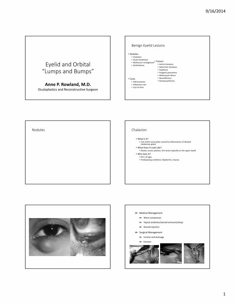
Nodules Chalazion
• What Is It?• Cyst within tarsal plate caused by inflammation of blocked
meibomian gland
• What Does It Look Like?• Raised, round, painless, firm lesion typically on the upper eyelid
• Who Gets It?• M=F, all ages• Predisposing conditions: blepharitis, rosacea
Medical Management
Warm compresses
Topical antibiotic/steroid ointment/drops
Steroid injection
Surgical Management
Incision and drainage
Excision
9/16/2014
2
External Hordoleum
• What Is It?• Staphylococcus abscess of lash follicle and associated gland of Moll or Zeiss
• What Does It Look Like?• Tender bump at lid margin with associated swelling
• Who Gets It?• M=F, all ages• Predisposing conditions: poor nutrition, poor hygeine, history of recurrent
infections
Medical Management
DO NOT forcefully rupture – may cause bacterial spread
Topically or oral antibiotics
Surgical Management
Typically not necessary
Excision after full course antibiotics
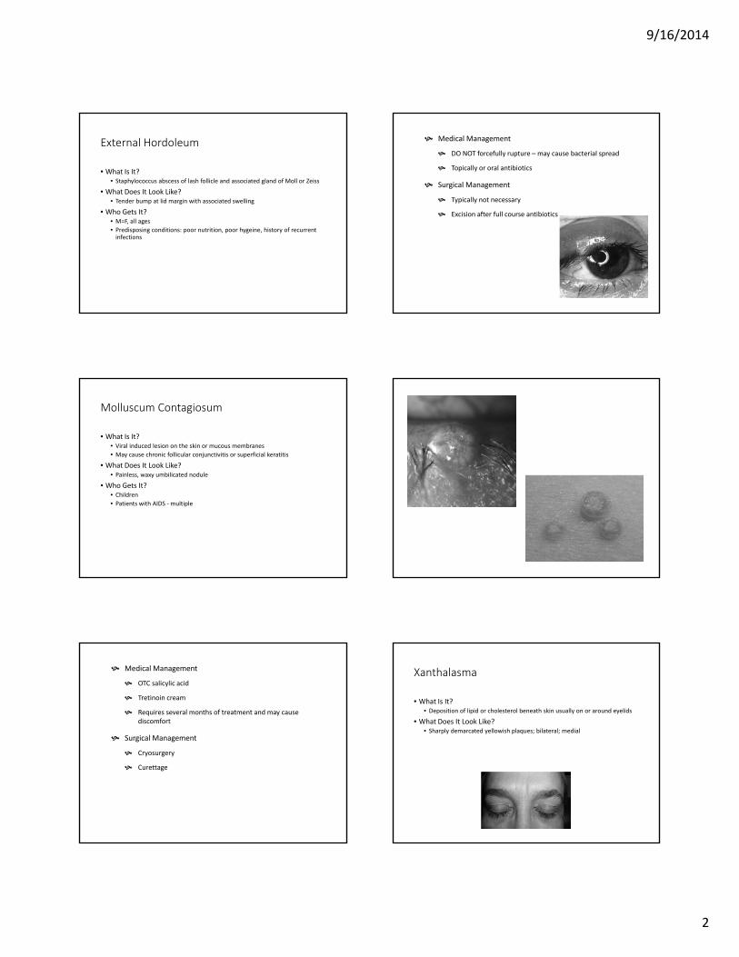
Molluscum Contagiosum
• What Is It?• Viral induced lesion on the skin or mucous membranes• May cause chronic follicular conjunctivitis or superficial keratitis
• What Does It Look Like?• Painless, waxy umbilicated nodule
• Who Gets It?• Children• Patients with AIDS - multiple
Medical Management
OTC salicylic acid
Tretinoin cream
Requires several months of treatment and may cause discomfort
Surgical Management
Cryosurgery
Curettage
Xanthalasma
• What Is It?• Deposition of lipid or cholesterol beneath skin usually on or around eyelids
• What Does It Look Like?• Sharply demarcated yellowish plaques; bilateral; medial
9/16/2014
3
Who Gets It?
Elderly
Mediterranean or Asian descent
Hypercholesterolemia
Medical Management
Deep TCA chemical peels
Lasers
Surgical Management
Cryosurgery
Excision
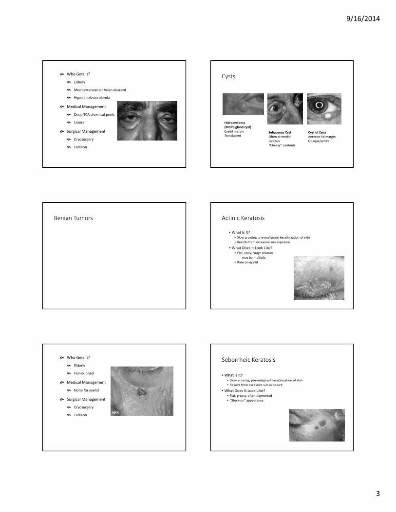
Cysts
Hidrocystoma(Moll’s gland cyst)Eyelid margin Translucent
Sebaceous CystOften at medial canthus“Cheesy” contents
Cyst of ZeissAnterior lid marginOpaque/white
Benign Tumors Actinic Keratosis
• What Is It?• Slow growing, pre-malignant keratinization of skin• Results from excessive sun exposure
• What Does It Look Like?• Flat, scaly, rough plaque;
may be multiple• Rare on eyelid
Who Gets It?
Elderly
Fair-skinned
Medical Management
None for eyelid
Surgical Management
Cryosurgery
Excision
Seborrheic Keratosis
• What Is It?• Slow growing, pre-malignant keratinization of skin• Results from excessive sun exposure
• What Does It Look Like?• Flat, greasy, often pigmented• “Stuck-on” appearance
9/16/2014
4
Who Gets It?
Elderly
Medical Management
None
Surgical Management
Biopsy for definitive diagnosis
Papilloma
• What Is It?• Proliferation of fibrovascular tissue covered by irregular keratinized
squamous epithelium• May be caused by viral infection
(HPV)
• What Does It Look Like?• “Skin tag”• Pedunculated or broad base
Who Gets It?
Anyone
Medical Management
None
Surgical Management
Biopsy for definitive diagnosis
Pyogenic Granuloma
• What Is It?• Overgrowth of vascular tissue
• What Does It Look Like?• Fast-growing, pinkish lesion• Pedunculated or flat based
• Who Gets It?• History of recent surgery or trauma
Medical Management
Topical or intralesional steroid
Surgical Management
Excision
Beware of recurrence
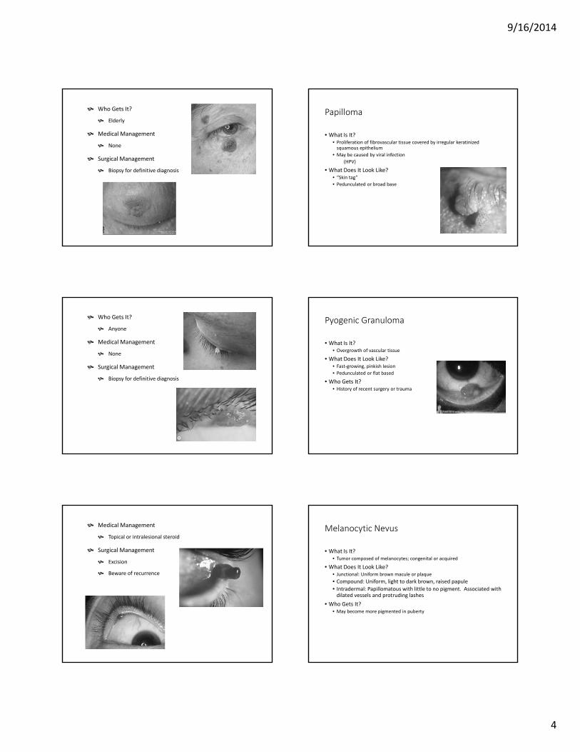
Melanocytic Nevus
• What Is It?• Tumor composed of melanocytes; congenital or acquired
• What Does It Look Like?• Junctional: Uniform brown macule or plaque• Compound: Uniform, light to dark brown, raised papule• Intradermal: Papillomatous with little to no pigment. Associated with
dilated vessels and protruding lashes• Who Gets It?
• May become more pigmented in puberty
9/16/2014
5
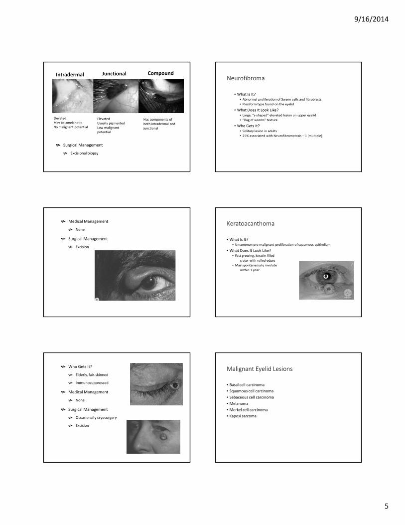
Intradermal Junctional Compound
ElevatedMay be amelanoticNo malignant potential
ElevatedUsually pigmentedLow malignant potential
Has components of both intradermal and junctional
Surgical Management
Excisional biopsy
Neurofibroma
• What Is It?• Abnormal proliferation of Swann cells and fibroblasts• Plexiform type found on the eyelid
• What Does It Look Like?• Large, “s-shaped” elevated lesion on upper eyelid• “Bag of worms” texture
• Who Gets It?• Solitary lesion in adults• 25% associated with Neurofibromatosis – 1 (multiple)
Medical Management
None
Surgical Management
Excision
Keratoacanthoma
• What Is It?• Uncommon pre-malignant proliferation of squamous epithelium
• What Does It Look Like?• Fast growing, keratin-filled
crater with rolled edges• May spontaneously involute
within 1 year
Who Gets It?
Elderly, fair-skinned
Immunosuppressed
Medical Management
None
Surgical Management
Occasionally cryosurgery
Excision
Malignant Eyelid Lesions
• Basal cell carcinoma• Squamous cell carcinoma• Sebaceous cell carcinoma• Melanoma• Merkel cell carcinoma• Kaposi sarcoma
9/16/2014
6
Basal Cell Carcinoma
• What Is It?• Most common eyelid cancer• Slow growling, locally destructive proliferation of basal cells
• What Does It Look Like?• Usually on lower eyelid; destruction of normal architecture• Nodular type: pearl like, with dilated blood vessels on surface• Ulcerative type: central ulcer with raised pearly edges• Sclerosing type: lateral, hardened, infiltration beneath the
epidermis. May be confused with chronic blepharitis
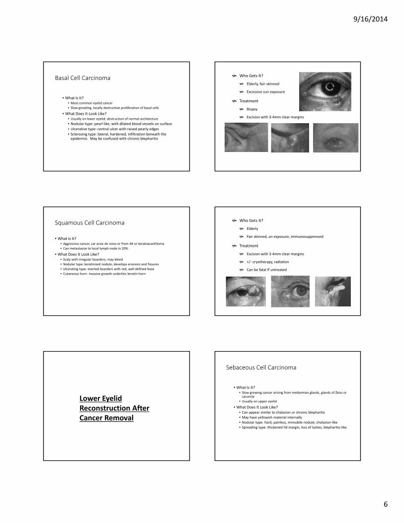
Who Gets It?
Elderly, fair-skinned
Excessive sun exposure
Treatment
Biopsy
Excision with 3-4mm clear margins
Squamous Cell Carcinoma
• What Is It?• Aggressive cancer, car arise de novo or from AK or keratoacanthoma• Can metastasize to local lymph node in 20%
• What Does It Look Like?• Scaly with irregular boarders; may bleed• Nodular type: keratinized nodule; develops erosions and fissures• Ulcerating type: everted boarders with red, well defined base• Cutaneous horn: invasive growth underlies keratin horn
Who Gets It?
Elderly
Fair skinned, un exposure, immunosuppressed
Treatment
Excision with 3-4mm clear margins
+/- cryotherapy, radiation
Can be fatal if untreated
Lower Eyelid Reconstruction After Cancer Removal
Sebaceous Cell Carcinoma
• What Is It?• Slow growing cancer arising from meibomian glands, glands of Zeiss or
caruncle• Usually on upper eyelid
• What Does It Look Like?• Can appear similar to chalazion or chronic blepharitis• May have yellowish material internally• Nodular type: hard, painless, immobile nodule; chalazion-like• Spreading type: thickened lid margin, loss of lashes; blepharitis-like
9/16/2014
7
Who Gets It?
Females, elderly (60’s – 70’s)
Treatment
Cryotherapy and surgical excision are the standard treatments
Recurrence is as high as 33%
Mortality rate is 5-10%
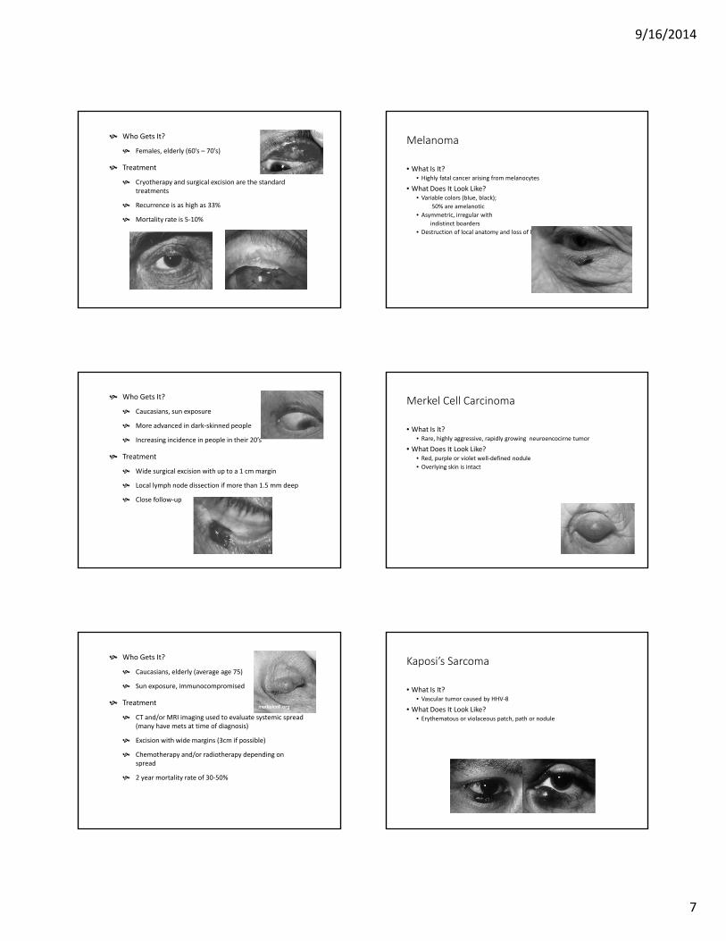
Melanoma
• What Is It?• Highly fatal cancer arising from melanocytes
• What Does It Look Like?• Variable colors (blue, black);
50% are amelanotic• Asymmetric, irregular with
indistinct boarders• Destruction of local anatomy and loss of lashes
Who Gets It?
Caucasians, sun exposure
More advanced in dark-skinned people
Increasing incidence in people in their 20’s
Treatment
Wide surgical excision with up to a 1 cm margin
Local lymph node dissection if more than 1.5 mm deep
Close follow-up
Merkel Cell Carcinoma
• What Is It?• Rare, highly aggressive, rapidly growing neuroencocirne tumor
• What Does It Look Like?• Red, purple or violet well-defined nodule• Overlying skin is intact
Who Gets It?
Caucasians, elderly (average age 75)
Sun exposure, immunocompromised
Treatment
CT and/or MRI imaging used to evaluate systemic spread (many have mets at time of diagnosis)
Excision with wide margins (3cm if possible)
Chemotherapy and/or radiotherapy depending on spread
2 year mortality rate of 30-50%
Kaposi’s Sarcoma
• What Is It?• Vascular tumor caused by HHV-8
• What Does It Look Like?• Erythematous or violaceous patch, path or nodule
9/16/2014
8
Who Gets It?
Middle aged men
Mediterranean or African descent
AIDS
Treatment
Excision, cryotherapy, intralesional injections of vinblastine, radiotherapy, topical immunotherapy (Imiquod)
Extensive disease requires chemo and immunosuppression
Orbital Tumors
Benign Orbital Tumors
• Bone• Osteoma• Fibrous dysplasia
• Well-delineated• Cavernous Hemangioma• Hemangiopericytoma• Dermoid cyst• Mucocele• ON tumors
• Diffuse• Lymphangioma• Benign reactive lymphoid
hyperplasia
• Lacrimal gland (pleomorphic
adenoma)
Osteoma
• What Is It?• Benign skeletal neoplasm of unknown etiology • In the orbit, typically involves the frontal and ethmoid bones• May cause pain, proptosis, decreased vision, or diplopia
What Does It Look Like?
Radiographically, these tumors are well-circumscribed with dense cortical sclerosis surrounding a radiolucent nidus.
Grossly, the lesion has a glistening, white to pink color and is either smooth or with rounded protuberances
Who Gets It?
Younger patients, typically
found incidentally
Treatment
Excisional biopsy
Cavernous Hemangioma
• What Is It?• Benign, noninfiltrative, slowly progressive vascular neoplasm • Composed of endothelial-lined spaces surrounded by a well-delineated
fibrous capsule• Most common benign orbital tumor in adults• Typically located intraconally
9/16/2014
9
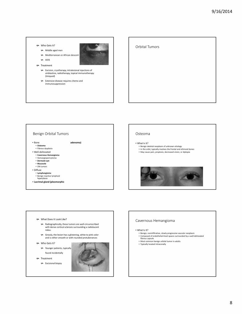
What Does It Look Like?
Presents as slowly progressive, painless proptosis
May have EOM disturbance, induced hyperopia, elevated IOP, choroidal folds or decreased vision
On CT scan - well circumscribed, homogenous mass slightly hyperdense to muscle, located intraconally.
Who Gets It?
Middle-aged adults, F>M
Treatment
May be observed as long as not comprising the eye
Orbital excision indicated for growth, optic ON compression, exposure keratopathy, or evidence of vision loss.
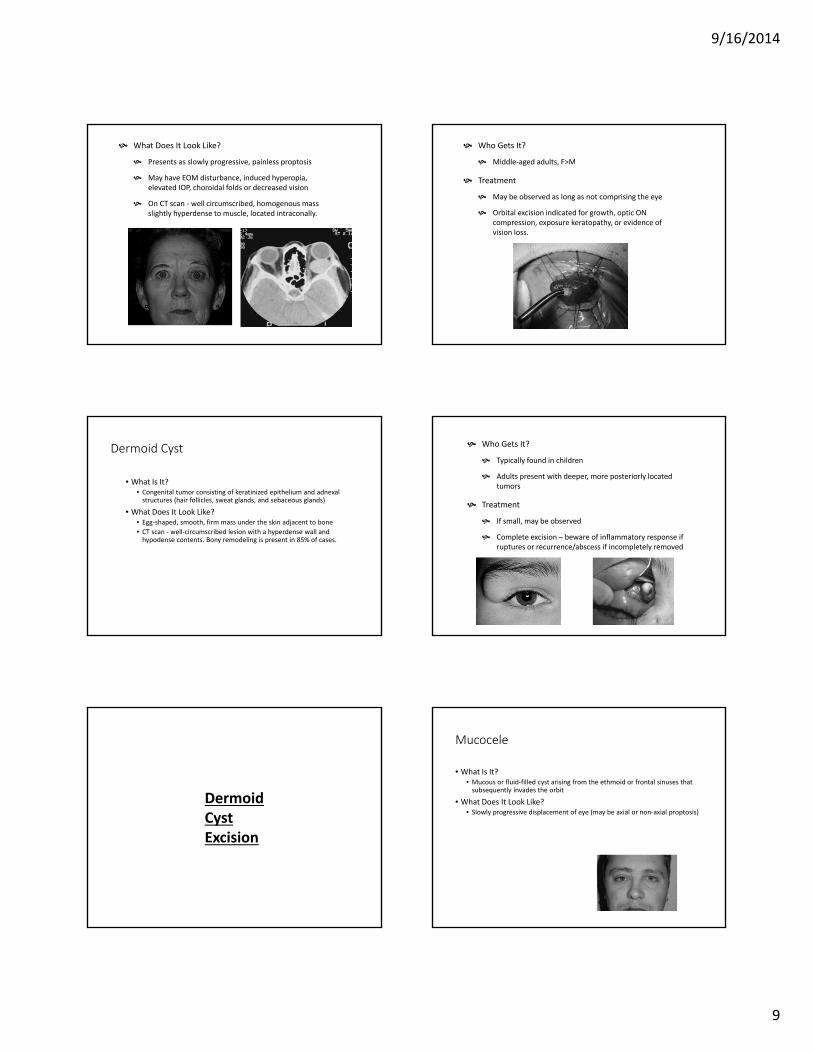
Dermoid Cyst
• What Is It?• Congenital tumor consisting of keratinized epithelium and adnexal
structures (hair follicles, sweat glands, and sebaceous glands)
• What Does It Look Like?• Egg-shaped, smooth, firm mass under the skin adjacent to bone• CT scan - well-circumscribed lesion with a hyperdense wall and
hypodense contents. Bony remodeling is present in 85% of cases.
Who Gets It?
Typically found in children
Adults present with deeper, more posteriorly located tumors
Treatment
If small, may be observed
Complete excision – beware of inflammatory response if ruptures or recurrence/abscess if incompletely removed
DermoidCyst Excision
Mucocele
• What Is It?• Mucous or fluid-filled cyst arising from the ethmoid or frontal sinuses that
subsequently invades the orbit
• What Does It Look Like?• Slowly progressive displacement of eye (may be axial or non-axial proptosis)
9/16/2014
10
Who Gets It?
Middle-age
History of chronic sinus disease or
facial trauma
Treatment
MRI-
Drainage procedure- send fluid for culture
and cytology
Surgery - orbitotomy and sinusectomy;
removal of as much of the cyst and its lining as possible
Lymphangioma
• What Is It?• Diffusely infiltrating benign nonencapsulated vascular tumor
• What Does It Look Like?• Fullness in the superior or nasal quandrant with acute proptosis after minor
head trauma or respiratory URI• >50% affect anterior structures causing bluish discoloration of or blood
vessels within the eyelid skin.• May bleed into itself causing cysts of blood, called chocolate-cysts
Who Gets It?
Any age
Treatment
If small, may be observed
Complete excision – beware of inflammatory response if ruptures or recurrence/abscess if incompletely removed
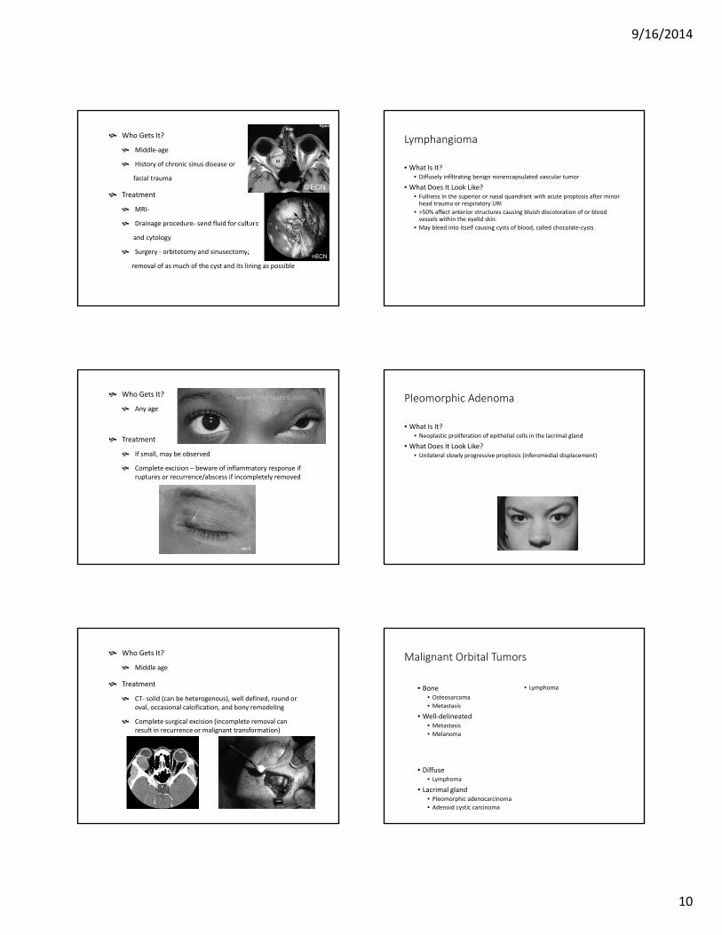
Pleomorphic Adenoma
• What Is It?• Neoplastic proliferation of epithelial cells in the lacrimal gland
• What Does It Look Like?• Unilateral slowly progressive proptosis (inferomedial displacement)
Who Gets It?
Middle age
Treatment
CT- solid (can be heterogenous), well defined, round or oval, occasional calcification, and bony remodeling
Complete surgical excision (incomplete removal can result in recurrence or malignant transformation)
Malignant Orbital Tumors
• Bone• Osteosarcoma• Metastasis
• Well-delineated• Metastasis• Melanoma
• Diffuse• Lymphoma
• Lacrimal gland• Pleomorphic adenocarcinoma• Adenoid cystic carcinoma
• Lymphoma
9/16/2014
11
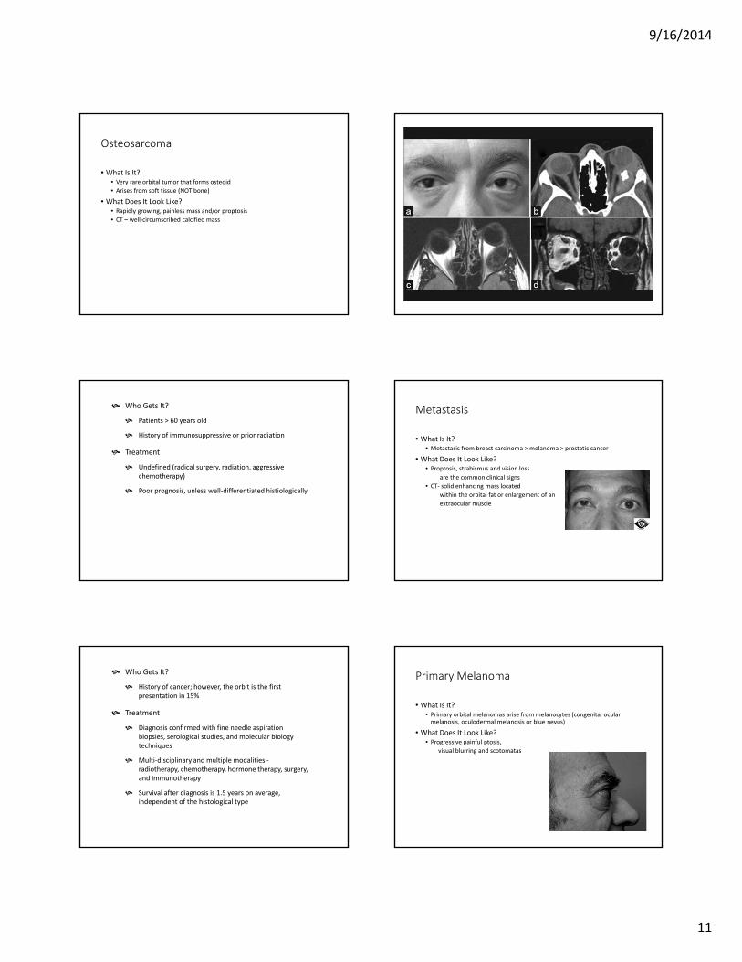
Osteosarcoma
• What Is It?• Very rare orbital tumor that forms osteoid• Arises from soft tissue (NOT bone)
• What Does It Look Like?• Rapidly growing, painless mass and/or proptosis• CT – well-circumscribed calcified mass
Who Gets It?
Patients > 60 years old
History of immunosuppressive or prior radiation
Treatment
Undefined (radical surgery, radiation, aggressive chemotherapy)
Poor prognosis, unless well-differentiated histiologically
Metastasis
• What Is It?• Metastasis from breast carcinoma > melanoma > prostatic cancer
• What Does It Look Like?• Proptosis, strabismus and vision loss
are the common clinical signs• CT- solid enhancing mass located
within the orbital fat or enlargement of an extraocular muscle
Who Gets It?
History of cancer; however, the orbit is the first presentation in 15%
Treatment
Diagnosis confirmed with fine needle aspiration biopsies, serological studies, and molecular biology techniques
Multi-disciplinary and multiple modalities -radiotherapy, chemotherapy, hormone therapy, surgery, and immunotherapy
Survival after diagnosis is 1.5 years on average, independent of the histological type
Primary Melanoma
• What Is It?• Primary orbital melanomas arise from melanocytes (congenital ocular
melanosis, oculodermal melanosis or blue nevus)
• What Does It Look Like?• Progressive painful ptosis,
visual blurring and scotomatas
9/16/2014
12
Who Gets It?
Caucasians, middle age
Treatment
Diagnosis with biopsy and immunophenotyping
Determine primary status with full body imaging and pathologic characteristics
Exenteration +/- radiation and chemotherapy
Lymphoma
• What Is It?• Non-Hodgkins lymphoma of the orbit• May be associated with MALT lymphoma
and Chlamydia psittaci infection (usually the result of exposure to infected birds and household pets)
• What Does It Look Like?• Typically superolateral painless mass causing palpable mas, exophthalmos,
ptosis, diplopia and abnormal ocular movement
Who Gets It?
50-70 years old, M=F
Treatment
Chlamydia psittaci infection- antibiotic therapy reduce the size of the tumor or possible cause remission
Surgical biopsy / resection, radiotherapy and chemotherapy are all used in various combinations
65% 5-year relapse-free rate
Systemic dissemination is only seen in 5-10% of cases
Pleomorphic Adenocarcinoma• What Is It?
• Malignant transformation of a pleomorphic adenoma (either spontaneously or after incomplete excision)
• What Does It Look Like?• Similar to pleomorphic adenoma• EXCEPT: painful growth,
usually more rapid
Who Gets It?
60-70 years old (10-20 years older than those with pleomorphic adenoma)
Treatment
Surgical excision - lateral rhinotomy and medial maxillectomy
Prognosis: survival rate correlates with the size, type, and histologic grade
Undifferentiated carcinoma had the worst survival rate (30%)
Polymorphous low-grade adenocarcinoma has the highest survival rate (96%)
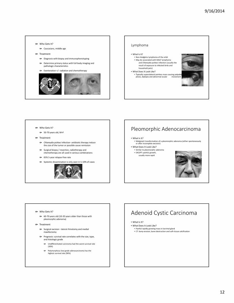
Adenoid Cystic Carcinoma• What Is It?• What Does It Look Like?
• Painful rapidly growing mass in lacrimal gland• CT: bony erosion, bone destruction and soft-tissue calcification
9/16/2014
13
Who Gets It?
M>F
Younger age than other lacrimal malignancies (average age is 41)
Treatment
Radical exenteration + radiation
Prognosis poor
50% recurrence within
2 years
50% mortality rate within
1.5 years
The End!
(For those of you still awake, any questions?)