covid 19 virology and pathophysiology - world critical care
TRANSCRIPT

COVID 19 VirologyandPathophysiology
Jorge Hidalgo, MD, MCCM, MACP, FCCP
World Federation of Intensive Care and Critical Care
Secretary-General

No conflict of Interest


Human Coronaviruses (HCoVs)
▪ Common HCoVs (lower pathogenicity):– HCoV-229E (alpha)
– HCoV-NL63 (alpha)
– HCoV-OC43 (beta)
– HCoV-HKU1 (beta)
▪ XXI Century HCoVs (higher pathogenicity):
– SARS-CoV (beta)
– MERS-CoV (beta)
Mild Respiratory
Infections
Lai & Holmes, Fundamental Virology, 4th Edition 2001.
https://www.who.int/emergencies/diseases/novel-coronavirus-2019/technical-guidance/naming-the-coronavirus-disease-(covid-2019)-and-the-virus-that-causes-it

SARS-CoV & MERS-CoV Emergence in XXI Century
www.who.int access on 2020 Jan 21st
ChinaR0 ≈ 1.8-2.5
Middle
EastR0 ≈ 0.3-1.3
ZOONOSIS

Coronavirus are found in Bats all over the world
Anthony et. al. A strategy to estimate unknown viral diversity in mammals. 2013 mBio. Photo: EcoHealth Alliance

Seven Human Coronaviruses (HCoVs)
▪ Common HCoVs (lower pathogenicity):
– HCoV-229E (alpha)
– HCoV-NL63 (alpha)
– HCoV-OC43 (beta)
– HCoV-HKU1 (beta)
▪ XXI Century HCoVs (higher pathogenicity):
– SARS-CoV (beta)
– MERS-CoV (beta)
– SARS-CoV-2* (beta)
https://www.who.int/emergencies/diseases/novel-coronavirus-2019/technical-guidance/naming-the-coronavirus-disease-(covid-2019)-and-the-virus-that-causes-it

Coronavirus
Nidovirales
Coronaviridae
Enveloped (+) Sense
Single Stranded
Highly Diverse
Linear
Helical Capside
4 groups ⍺, β, γ, δ


Baric RS, CROI, Boston, MA 2020
Drivers of Coronavirus (CoV) Evolution

Lu R et al, Lancet 2020; 395: 565–74
Phylogenetic Analysis of SARS-CoV-2 among Betacoronavirus
2019-nCoV=2019 novel coronavirus. MERS-CoV=Middle East respiratory syndrome coronavirus.
SARS-CoV=severe acute respiratory syndrome coronavirus.

De Wit Nature Rev Microbiol, 2016; Zhou P et al, Nature, 2020; Wrapp D et al, Science. 2020
SARS-CoV-2 (COVID19) Genome
Enveloped RNA virus with a genome size of 32 Kb
SARS-CoV-2
Spike (S) in
the prefusion
conformation

M Vaduganathan et al. N Engl J Med 2020. DOI: 10.1056/NEJMsr2005760
Interaction between SARS-CoV-2 and the Renin–Angiotensin–Aldosterone System.

De Wit Nature Rev Microbiol, 2016; Zhou P et al, Nature, 2020 and bioRxiv, 2020.01.22.914952; Yan R et al. Science. 2020.
SARS-CoV-2 (COVID19) Life Cycle
It uses ACE2 for viral entry
(angiotensin-converting enzyme 2)
ACE2
Viral particles in the ultrathin sections were
imaged using electron microscopy at 200
kV. The sample was from virus-infected
Vero E6 cells. The inset shows the viral
particles in an intra-cytosolic vacuole
TMPRSS2

Hendrickson CM et al. Semin Respir Crit Care Med 2013;34:475–486.
Schematic representation of SARS CoV infection mediating acute lung injury through angiotensin-converting enzyme (ACE) and
ACE2 signaling pathways.
1) SARS CoV binds to ACE2 causing
downregulation of ACE2 through
internalization of this membrane-
bound protein and leading to viral
replication in the cytoplasm.
2) ACE2 inactivates AT II. AT II binds
the angiotensin II receptor 1a
(AT1aR), leading to tissue damage
and lung edema, or it binds the
angiotensin II receptor 2 (AT2R)
reducing tissue damage.

• Channapavar & Perlman. Pathogenic human coronavirus infections: causes and consequences of Cytokine storm and immunopathology. SeminImmunopathol (2017) 39:529-539

• Channapavar & Perlman. Pathogenic human coronavirus infections: causes and consequences of Cytokine storm and immunopathology. SeminImmunopathol (2017) 39:529-539

• Channapavar & Perlman. Pathogenic human coronavirus infections: causes and consequences of Cytokine storm and immunopathology. SeminImmunopathol (2017) 39:529-539

Consequences of Cytokine Storm and immunopathology
Epithelial and endothelial cell apoptosis and vascular leakage.
Cytokine/Chemokines. IFNab, INFy, Fas-FasL or TRAIL-DR5 dependentmechanisms. TNF released by IMMS.
Apoptosis of epithelial and endothelial cells compromises lungmicrovascular and alveolar epithelial cell barrier resulting in endothelialdamage and alveolar edema HYPOXIA
• Channapavar & Perlman. Pathogenic human coronavirus infections: causes and consequences of Cytokine storm and immunopathology. Semin Immunopathol (2017) 39:529-539

Consequences of Cytokine Storm and immunopathology
Suboptimal T cell response:
CoV-specific T cells are crucial for virus clearance and limit further damage to host. Exuberant inflammatory responses caused by pathogenic hCoVdiminish the T Cells response, in the case of SARS-CoV infection via TNF mediated T cell apoptosis, leading to Uncontrolled inflammatory response.
• Channapavar & Perlman. Pathogenic human coronavirus infections: causes and consequences of Cytokine storm and immunopathology. Semin Immunopathol (2017) 39:529-539

Consequences of Cytokine Storm and immunopathology
Accumulation of alternatively activated macrophages and altered tissue homeostasis:
In mice infected with SARS-CoV-Challenged STAT mice on B6 and B129 Background revealed an enhanced perivascular infiltration of alternatively activated macrophages, neutrophils and fibroblast and extensive fibrin deposition and alveolar collapse, features observed during ALI and ARDS in humans.
• Channapavar & Perlman. Pathogenic human coronavirus infections: causes and consequences of Cytokine storm and immunopathology. Semin Immunopathol (2017) 39:529-539
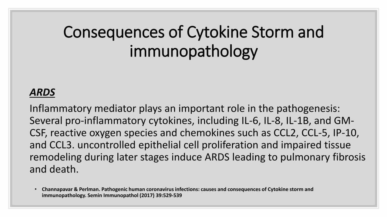
Consequences of Cytokine Storm and immunopathology
ARDS
Inflammatory mediator plays an important role in the pathogenesis: Several pro-inflammatory cytokines, including IL-6, IL-8, IL-1B, and GM-CSF, reactive oxygen species and chemokines such as CCL2, CCL-5, IP-10, and CCL3. uncontrolled epithelial cell proliferation and impaired tissue remodeling during later stages induce ARDS leading to pulmonary fibrosis and death.
• Channapavar & Perlman. Pathogenic human coronavirus infections: causes and consequences of Cytokine storm and immunopathology. Semin Immunopathol (2017) 39:529-539

SARS-CoV-2 (COVID19) Pathogenesis: ARDS
ACE2
Acute Respiratory Distress Sydrome (ARDS) pathology
Acute diffuse alveolar damage, with pulmonary edema and
formation of a hyaline membrane in a SARS-CoV patientThe airspaces are indicated by asterisks and some of the hyaline membranes lining the alveolar
spaces are highlighted by arrows (hematoxylin and eosin stain; originalmagnification,x100).
Tse GMK et al. J Clin Pathol 2004;57:260–265


Thompson BT et al. N Engl J Med 2017;377:562-572.
Th
e H
ealt
hy L
un
g a
nd
th
e
Exu
dati
ve P
ha
se o
f A
RD
S.

Endothelial Injury and loss of auto-regulation
(Adapted from Go ́ mez H, Kellum JA. Sepsis-induced acute kidney injury. Curr Opin Crit Care 2016;22(6):546–53.)

Endothelial Injury and loss of Auto-regulation
(Adapted from Go ́ mez H, Kellum JA. Sepsis-induced acute kidney injury. Curr Opin Crit Care 2016;22(6):546–53.)

Endothelial Injury and loss of auto-regulation
(Adapted from Go ́ mez H, Kellum JA. Sepsis-induced acute kidney injury. Curr Opin Crit Care 2016;22(6):546–53.)

Endothelial Injury and loss of auto-regulation
Such mechanisms revolve around the loss of auto-regulation secondary to endothelial cell injury
and altered retrograde endothelial cell-cell communication; impaired red blood cell
deformability and increased blood viscosity
(Adapted from Go ́ mez H, Kellum JA. Sepsis-induced acute kidney injury. Curr Opin Crit Care 2016;22(6):546–53.)

Endothelial Injury and loss of auto-regulation
denudation of the glycocalyx ( key biomechanical activities, including maintenance of blood flow and
protection of the barrier function); platelet activation; leukocyte adhesion and rolling; and activation of the
coagulation and complement systems.
(Adapted from Go ́ mez H, Kellum JA. Sepsis-induced acute kidney injury. Curr Opin Crit Care 2016;22(6):546–53.)

Met
abo
lic R
epro
gram
ing
as c
ell
surv
ival
str
ateg
y

Regulation of the cell cycle
Adapted from Go ́ mez H, KellumJA. Sepsis-induced acute kidney injury. Curr Opin Crit Care
2016;22(6):546–53


