cpy document - international agency for research on...
TRANSCRIPT
CLOFIBRATE
This substance was considered by previous working groups, in February 1980 (lARC,1980) and March 1987 (lARC, 1987). Since that time, new data have become available,and these have been incorporated in the monograph and taken into consideration in theevaluation.
1. Exposure Data
1.1 Chemical and physical data
1.1.1 Nomel1clature
Chem. Abstr. Serv. Reg. No.: 637-07-0
Chem. Abstr. Name: 2-(4-Chlorophenoxy)-2-methylpropanoic acid, ethyl ester
IUPAC Systematic Name: Ethyl 2-(para-chlorophenoxy)-2-methylpropionate
SYl1ol1yms: para-Chlorophenoxyisobutyric acid ethyl ester; 2-(para-chlorophenoxy)-
2-methylpropionic acid ethyl ester; ethyl para-chlorophenoxyisobutyrate; ethyl
2-(para-chlorophenox y )isobutyrate; ethy 1 2-( 4-chlorophenox y )isobutyrate; ethy ia-(para-chlorophenoxy)isobutyrate; ethy 1 a-( 4-chlorophenox y )isobutyrate; ethy 1a-(para-chlorophenoxy)-a-methylpropionate; ethyl a-( 4-chlorophenox y )-a-methyl-propionate; ethyl 2-(4-chlorophenoxy)-2-methylpropionate; ethyl clofibrate
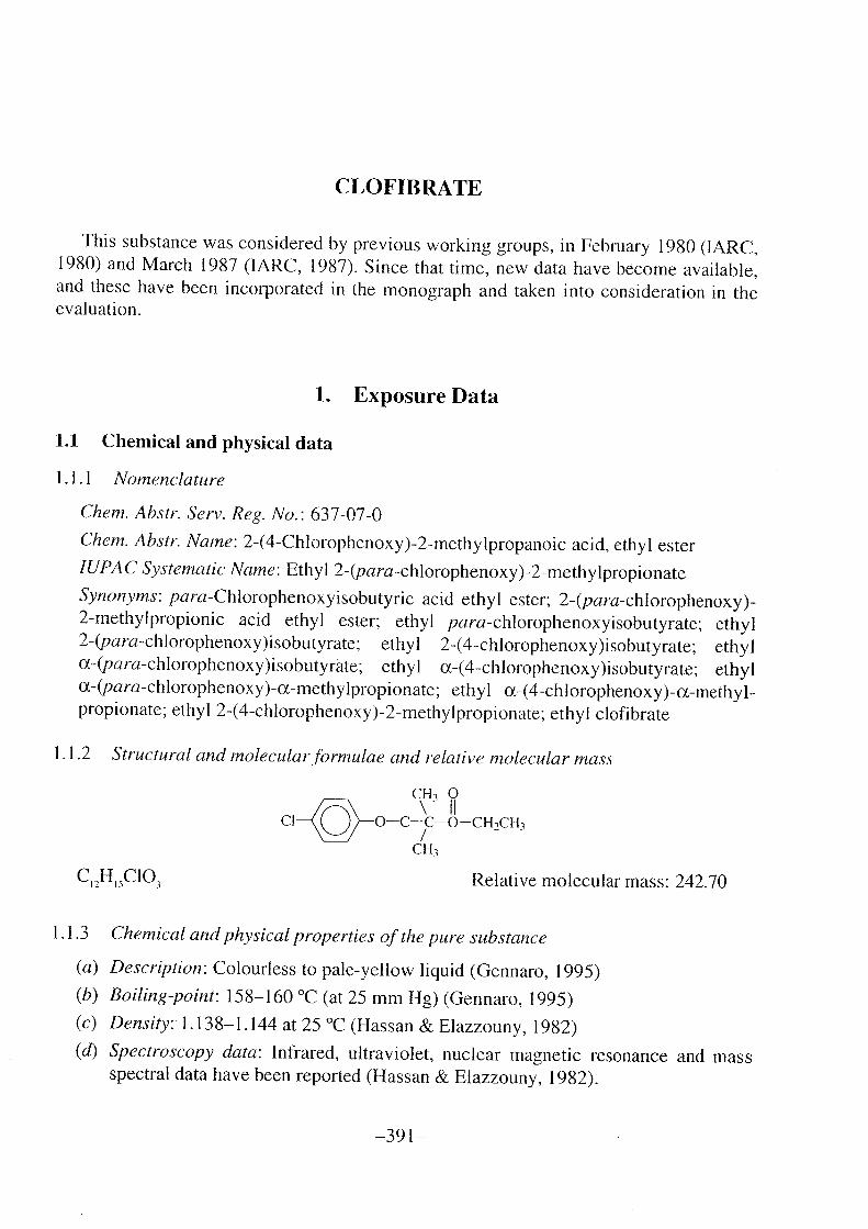
1.1.2 Structural al1d molecular formulae al1d relative molecular mass
CH, 0
-0 \"11CI 0 O-C-C-O-CH,CH,/ -"CH,
Ci2H1sCIO, Relative molecular mass: 242.70
1.1.3 Chemical al1d physical properties of the pure substal1ce
(a) Descriptiol1: Colourless to pale-yellow liquid (Gennaro, 1995)
(b) Boilil1g-poil1t: 158-160 °C (at 25 mm Hg) (Gennaro, 1995)
(c) Del1sity: 1.138-1.144 at 25°C (Hassan & Elazzouny, 1982)
(d) Spectroscopy data: lnfrared, ultraviolet, nuclear magnetic resonance and massspectral data have been reported (Hassan & Elazzouny, 1982).
-391-

392 IARC MONOGRAPHS VOLUME 66
(e) Solubility: Practically insoluble in water; miscible with acetone, chloroform,diethyl ether and ethanol (Budavari, 1995; Gennaro, 1995)
l,lA Techl1ical products al1d impurities
Clofibrate is available as 500-mg capsules which may also contain gelatin, D&C Red28, D&C Red 30, D&C Yellow 10 (Quinoline Yellow), FD&C Blue 1 (Brilliant BlueFCF), FD&C Red 3 (Erythrosine) or FD&C Yellow 6 (Sunset Yellow FCF) (Thomas,1991; Medical Economics, 1996).
Trade names and designations of the chemical and its pharmaceutical preparationsinclude: Amotril; Anparton; Apolan; Arterioflexin; Artes; Artevil; Ateculon; Ateriosan;Aterosol; Atheromide; Atheropront; Atrofort; Atrolen; Atromid; Atromid-S; Atromidin;Atrovis; A Y 61 123; Azionyl; Bioscleran; Cartagyl; Cititlus; Claripex; Claripex CPIB;Cloberat; Clobrat; Clobren SF; Clof; Clofibral; Clofibrat; Clofinit; Clofipront; Clofirem;CPIB; Deliva; ECPlB; EPlB; Estaprol; Geromid; Healthstyle; Hyclorate; LCI 28257;Ipolipid; Klofiran; Levatrom; Lipavil; Lipavlon; Lipilim; Lipomid; Liponorm; Liporan;Liprinal; Lobetrin; Lostat; MG 46; Miscleron; Misclerone; Neo-Atromid; NSC 79389;Normet; Normolipol; Novofibrate; Recolip; Regelan; Sclerovasal; Serotinex; Sklerolip;Skleromexe; Sklero- Tablinen; Ticlobran; Xyduril; y oclo.
1.1.5 A l1alysis
Methods for the analysis of clofibrate have been reviewed (Hassan & Elazzouny,1982).
Several international pharmacopoeias specify high-performance liquid chromato-graphy (HPLC) or titration with hydrochloric acid as the assays for purity of clofibrate,and HPLC or gas chromatography with flame ionization detection (GC/FID) for deter-mining impurities and decomposition products. Methods are also specified for determi-ning acid, heavy metal, arsenic and para-chlorophenol content. The assays for clofibratein capsules apply titration with hydrochloric acid, or HPLC or GC/FlD methods usingstandards (Council of Europe, 1984; Society of Japanese Pharmacopoeia, 1992; BritishPharmacopoeial Commission, 1993; United States Pharmacopeial Con ven tion, 1994).
1.2 Production and use
1.2.1 ProdUCtiOI1
Clofibric acid was first synthesized in 1947 (Windholz, 1976), but the ethyl ester,clofibrate, was not reported until 1961 (Budavari, i 995). Clofibrate is prepared bycondensing phenol with ethyl 2-chloro-2-methylpropionate in the presence of a suitabledehydrochlorinating agent and then chlorinating the aromatic ring (Gennaro, 1995).
1.2.2 Use
The efficacy of clofibrate in reducing serum cholesterol levels was first reported in1962 (Thorp & Waring, 1962). Clofibrate was first marketed in the United States ofAmerica in 1967 (Wysowski et al., 1990).
CLOFIBRA TE 393
Clofibrate is used as a hypolipidaemic drug. lt reduces elevated plasma concentrationsof triglycerides by reduction of elevated concentrations of very low-density lipoproteins(VLDLs) within two to five days after initiation of therapy. It is less effective in reducinglow-density lipoprotein (LDL) cholesterol and the plasma concentration of total choles-terol. It is mostly effective in the treatment of type III hyperlipoproteinaemia. It may alsobe helpful in some patients with type IIb, type lV or type V hyperlipoproteinaemia (seeGlossary, p. 448) (Goodman Gilman et al., 1990; Reynolds, 1993; Larsen et al., 1994).
The usual daily dose is 2 g (20-30 mg/kg bw per day) taken orally in two or threedivided doses (Reynolds, 1993; Vidal, 1995),
The cellular mechanisms responsible for the hypolipidaemic effects of fibrate drugshave not been clarified fully but include: activation of lipoprotein lipase, suppression offree fatty acid release from adipose tissue, inhibition of hepatic triglyceride synthesis andincreased secretion of cholesterol (see IARC, 1983) into bile. Therapy with clofibratedoes not significantly reduce the rate of synthesis of VLDL triglycerides, but suchtreatment is associated with an increase in the rate of catabolIsm of VLDL particles(Larsen et al., 1994). The mobilization of deposits of cholesterol in tissues is accom-panied by regression and disappearance of xanthomas (Goodman Gilman et al., 1990).
Clofibrate has been used in the prophylaxis of ischaemic heart diseases but it is nolonger recommended for this purpose, because of adverse effects se en during long-term
treatment: increased incidences of cholecystitis, gallstones and in some cases of certaincardiovascular disorders and excess deaths found in the WHO Cooperative Trial on theuse of clofibrate in the primary prevention of ischaemic heart disease (Reynolds, 1993).Some patients have also shown a paradoxical rise in LDL (Goodman Gilman et al.,1990).
Clofibrate has been used in the treatment of neonatal jaundice (Gabilan et aL., 1990;Erkul et al., i 99 1; Gabilan et al., 1991).
Following the report of a WHO-sponsored cooperative study of the use ofclofibratein the primary prevention of ischaemic heart disease (Committee of Principal Investi-gators, 1978), it was withdrawn in the Federal Republic of Germany and Norway in early1979. ln a number of other countries, including France, Italy, Sweden, Switzerland, theUnited Kingdom and the United States, practitioners were advised to reserve its use forpatients with high plasma lipid concentrations that are refractory to dietary measures andto consider carefully the risks and benefits of the treatment (United States Food and DrugAdministration, 1979; WHO, 1979a; Expert Panel, 1988). It was reintroduced in theFederal Republic of Germany in August 1979 (WHO, 1979b). ln the United Kingdom,clofibrate is now rarely prescribed (Dunnigan, 1992).
ln the United States, clofibrate represented 80.9% of the cholesterol-Iowering medi-cations used in 1978, 41.2% in 1983 and 3.5% in 1988. Gemfibrozil (see monograph,pp.428-429), lovastatin and cholestyramine are now used more commonly (Wysowskiet al., 1990).

394 IARC MONOGRAPHS VOLUME 66
1.3 Occurrence
Clofibrate is not known to occur as a natural product.
No quantitative data on occupational exposure levels were available to the WorkingGroup.
The National Occupation al Exposure Survey conducted between 1981 and 1983 inthe United States by the National lnstitute for Occupational Safety and Health indicatedthat about 325 employees were potentially occupationally exposed to clofibrate. Theestimate was based on a survey of companies and did not involve measurements of actualexposure (United States National Library of Medicine, 1996).
1.4 Regulations and guidelines
Clofibrate is lIsted in the following pharmacopoeias: British, Brazilian, Chinese,
Czech, Egyptian, European, French, Greek, Hungarian, Indian, Italian, Japanese,Netherlands, Nordic, Portuguese, Romanian, Swiss and United States (Reynolds, 1993).
2. Studies of Cancer in Humans
2.1 Case-control study
A population-based case-control study in Kansas, United States, investigated a largenumber of possible risk factors for soft-tissue sarcoma (Hoar Zahm et aL., 1989). One ofthe factors examined was medical treatment with cholesterol-lowering drugs (amongwhich was clofibrate). Among white males, aged 21 years or older, a total of 139 newlydiagnosed (1976-82) and histologically confirmed cases of soft-tissue sarcomas wereidentified through the University of Kansas Cancer Data Service (50% deceased).Deceased cases were not excluded from the study. Three controls were matched to eachcase by age and vital status. For living cases, controls were selected either from theHealth Care Financing Administration files or by telephone random digit diallng. Fordeceased cases, controls were selected from Kansas state mortality files. Exposureinformation was obtained from interviews with study subjects or with their next-of-kin.The response rate was 93%. The distribution of proxy type was similar among the casesand controls. Among users of cholesterol-lowering drugs (5 cases and 20 controls), anonsignificant excess of soft-tissue sarcoma was seen (odds ratio, 1.7; 95% confidenceinterval (CI), 0.5-5.0). The increased risk was found only among deceased subjects (oddsratio,' 1.9; 95% CI, 0.5-6.4; 4 cases, 15 controls). (The Working Group noted that noadjustment was made for confounders, that ail medical data, such as on use ofcholesterol-Iowering drugs, were self- or proxy-reported and that the inclusion of
deceased controls may have overrepresented the prevalence of their use.)

CLOFIBRA TE 395
2.2 Clinical trials
A randomized trial of the W orld Health Organization, started in i 965 to determinewhether clofibrate would lower the incidence of ischaemic heart disease in men, raisedconcern over a nonsignificant excess of cancer deaths in treated subjects (58 versus 42 inplacebo-treated controls) (Committee of Principal lnvestigators, 1978, 1980; lARC,1980). The greatest excesses were for cancers of the gastrointestinal and respiratorytracts. Results of a further four years of follow-up of this trial to the end of 1982 sub-sequently became available (Committee of Principal Investigators, l 984). On average,the total follow-up period was 13.2 years, 5.3 of which were during the actual treatmentphase (range, four to eight years) and 7,9 thereafter. Three groups of men, dividedaccording to their cholesterol level, were studied, comprising 208 000 man-years ofobservation. The first two groups included subjects in the upper third of the serumcholesterol distribution, randomly allocated either to treatment with clofibrate (1.6 gdaily) or to receive an olive oil-containing placebo. The third group was composed ofhalf of the men in the lowest third of the distribution, who received an olive oil-containing placebo. At the conclusion of follow-up, the age-standardized rates of deathfrom malignant neoplasms per 1000 per annum were 2.4; 2.4 and 2.3, respectively (basedon 206, 197 and 173 deaths from malignant neoplasms). However, the age-standardizeddeath rates for malignant neoplasms during the treatment phase had been 2.0 (42 deaths),1.2 (25 deaths) and 1.7 (30 deaths), respectively.
The Coronary Drug Project, a randomized and double blind trial in the United Statesand Puerto Rico, started in i 966, investigated the effects of lipid-Iowering drugs on 8341men, aged 30-64 years with a history of myocardial infarction. The first results, with amean follow-up of 6.2 years study (5-8.5 years), showed no increase in the cancer deathrate in the clofibrate (1.8 g/day)-treated group (10 deaths in 1 i 03 patients) compared
with that of a placebo-treated group (24 deaths in 2789 patients) (Coronary Drug ProjectResearch Group, i 975). After a mean follow-up of 15 years (including 8.8 years aftertermination of the trial), with definite information about vital status for 98.9% ofsubjects, the clofibrate group had somewhat lower cancer mortality (3.4%) than did theplacebo group (4.4%). This was also the case for lung cancer (13 deaths in 1103
clofibrate-treated men (12/1000) and 53 deaths in 2789 placebo-treated men (19/l000))and for cancer of the gastrointestinal tract (4/l 000 versus 6/l 000) (Canner et al., 1986).
ln the Stockholm Ischaemic Heart Disease study (Carlson & Rosenhamer, 1988), 555patients with ischaemic heart disease, under 70 years of age, were treated with clofibrateand nicotinic acid (11 = 279) or with a placebo (11 = 276) (not blind). Because of cancer,10 subjects among the treated group and 6 among the controls withdrew from the triaL.The numbers of cancer deaths during the five years of treatment were four in thetreatment group and six in the control group.
Recently, Law et al. (1994) conducted a meta-analysis to assess whether low serumcholesterol concentration increases mortality from causes other th an ischaemic heartdisease. The data were derived from the 10 largest cohort studies, two internationalstudies and 28 randomized trials, supplemented by unpublished data. Only the trialsprovided information about cholesterol-Iowering drugs. Extended observation after the

396 rARC MONOGRAPHS VOLUME 66
trial period had ended was available for six of the trials and provided information on therisk for cancer 5-10 years after treatment with cholesterol-lowering drugs (about 15
years after the start of treatment). The overall relative odds estimate of the risk for cancerwas 0.9 (95% CI, 0.7- l. 1; based on 232 treated patients. The meta-analysis did notprovide estimates of relative risk for cancer mortality for the clofibrate trial separately.(The Working Group noted that the numbers of cancer deaths provided cannot becompared directly because of differences in survival between clofibrate-treated subjectsand those who did not receive the drug.)
3. Studies of Cancer in Experimental Animais
3.1 Oral administration
3.1.1 Mouse
ln a study reported as a summary in a monograph, groups of 25 male and 25 femaleAlderley Park mice (age not specified) were given 0 (control), 1000 (therapeutic level inhumans), 2500 or 5000 (maximum tolerated dose, MTO) mg/kg diet (ppm) clofibrate(purity not specified) in the diet for 18 months. Major organs and abnormalities wereexamined histologicaIly. Mortality was similar in aIl groups. No difference in the inci-dence of any tumour type between control and treated groups was reported (Tucker &Orton, 1995).
ln another study reported as a summary in a monograph, groups of 51 male and 5 ifemale C57Bl/IOJ mice, six weeks of age, were given clofibrate (purity not specified) atdaily dose levels of 150, 250 and 350 mg/kg bw in the diet for 18 months. The untreatedcontrol groups comprised 151 males and 151 females. There was body-weight reductionof about 10% the higher-dose group. Mortality was similar in aIl groups (70-80%survival at 18 months). There was a significant increase in lIver weights in treated malesand females in the two higher-dose groups. Full necropsy and histological examinationswere carried out on aIl animais. No difference in tumour incidence between treated andcontrol groups was reported (Tucker & Orton, 1995).
3.1.2 Rat
A group of 15 male Fischer 344 rats, weighing 84- 100 g (age not specified), wasgiven 0.5% (v/w) clofibrate (purity not specified; equivalent to about 250 mg/kg bw perday) in the diet for up to 28 months. A group of 15 untreated males served as controls. Ofthe treated animais, one rat was kiIled at 13 months and three more between 17 and 21months. The remaining 1 1 rats were kiled between 24 and 28 months. One or morehepatocellular carcinoma developed in 10/1 1 rats compared with 0/14 controls whichsurvived to 28 months (p .. 0.001); five of the animaIs with hepatocellular carcinomas
showed pulmonary metastases. ln addition, among the treated animaIs, pancreaticexocrine acinar carcinomas were found in 2/1 1 rats, whereas none was found in controls(Reddy & Qureshi, 1979). (The Working Group noted the small number of animaIs.)

CLOFIBRA TE 397
A group of 25 male weanling Fischer 344 rats, weighing approximately 100 g, wasfed 5000 mg/kg diet (ppm) cIofibrate (purity not specified) in the diet for 72 (when thefirst tumour appeared)-97 weeks (total intake, 25-33 g/rat). A group of 25 untreatedmales served as controls. The study was terminated at 129 weeks, when aIl survivinganimaIs were killed. Among the treated rats, malignant tumours developed at varioussites. Hepatocellular carcinomas were found in 4/25 treated rats; among the othertumours observed in treated rats were a pancreatic exocrine acinar carcinoma in one ratand pancreatic exocrine acinar adenomas in three rats. Treated and control rats developedsimilar numbers of leukaemias and tumours of the testis (Svoboda & Azarnoff, i 979).
Groups of 70 (control) and 74 (continuous treatment) male Sprague-Dawley rats,se ven weeks of age, were given 0 or 400 mg/kg bw cIofibrate (purity not specified) dailyin the diet for up to 1 13 weeks. A third group of 28 (recovery group) male Sprague-Dawley rats was given 400 mg/kg bw clofibrate in the diet for 42-95 weeks and thenheld for a further 16- 18 weeks before killing. Three to five rats in each group weresacrificed at 4- 1 O-week intervals beginning at week 4 and ending at week i 13, whenzero to five animais remained per group. Only the liver and abnormal organs wereexamined histologically. Hyperplastic (neoplastic) nodules did not occur before week 68of treatment. ln rats treated for 68 weeks or longer, the incidence of hyperplastic (neo-plastic) nodules was: controls, 0/36; continuous treatment, 19/36; and recovery group,1/16. The only hepatocellular carcinomas found were in two rats in the continuoustreatment group at week 95 (Greaves et aL., 1986).
3.1.3 Marmoset
ln a study reported as a summary in a monograph, groups of 1 1 - 16 male and i 1 - i 6female marmosets were given clofibrate (purity not specified) in water containing 0.5%w/w polysorbate 80 by gastric instillation at intended dose levels of 100, 150, 250 and300 mg/kg bw per day. Effective doses were 94, 157, 213 and 263 mg/kg bw per day.Groups of 20 males and 20 females were untreated or treated with the vehicle only. Thestudy was terminated after 6.5 years due to premature deaths. Necropsy was performedand all major organs were examined histologically. Causes of death varied and were notrelated to cIofibrate treatment. There was no effect on liver weight in any of thecIofibrate-treated groups. A statistically significant (p .( 0.01) increase in kidney weightwas seen in the higher-dose groups. There were no histological changes in the liver thatcou Id be attributed to clofibrate. No liver tumour or other treatment-related tumour wasfound in clofibrate-treated marmosets (Tucker & Orton, 1995).
3.2 Administration with known carcinogens
3.2.1 Mouse
Groups of 7-20 male C3H/en, C57Bl/6N and BALB/cA mice, six weeks of age,were subjected to partial hepatectomy. After 24 h, mice were given 20 mg/kg bw N-nitrosodiethylamine (NDEA) by intraperitoneal injection. Six ho urs later, they weregiven basal diet or a diet containing 1000 mg/kg diet (ppm) clofibrate (purity notspecified) until week 20, at which time the experiment was terminated and aIl surviving

398 IARC MONOGRAPHS VOLUME 66
animaIs were killed. Livers were analysed for the number and size distribution ofglucose-6-phosphatase-deficient enzyme-altered islands by a stereological method (for
statistical comparisons, WeIch's test was used). NDEA alone induced many more andlarger enzyme-altered islands in C3H mice than in the other two strains. ln C3H mice,administration of c10fibrate in addition to NDEA increased the number and volume ofenzyme-altered islands. ln the other two strains, clofibrate had no enhancing effect (Leeet al., 1989). (The Working Group noted the absence of a group given clofibrate alone.)
3.2.2 Rat
Groups of 30 male Fischer rats weighing 135- 1 50 g (age not specified) were given100 mg/L (ppm) NDEA in the drinking-water for two weeks. One week later the ratswere fed 0 or 5000 mg/kg diet (ppm) clofibrate (purity not specified) in the diet for 48weeks, at which time the experiment was terminated. Clofibrate significantly (p 0( 0.001)enhanced the development of liver tumours in rats previously exposed to NDEA: 25/28rats given NDEA plus clofibrate had liver-cell tumours (type not specified) versus 5/18(three hepatocellular carcinomas) in the group given NDEA alone (Reddy & Rao, 1978).(The Working Group noted the absence of a group given clofibrate alone.)
A group of 54 female rats (strain and age not specified) was treated twice with50 mg/kg bw N-methyl-N-nitrosourea (MNU) in citrate buffer (pH 6.0) intravenously atone-week intervals. Twenty-six treated rats were given 20.8 mg/day c10fibrate in milk(route and volume not specified) on five days per week for one year. The remaining 28animaIs received milk only. One year after the first injection of MNU, aIl animais werekilled. Complete necropsy and histological examination were performed. The authorsreported that clofibrate had no effect on the incidence of tumours induced by MNU(Anisimov et aL., 1981).
A group of 68 female rats (strain and age not specified) was treated intravenouslywith 1.5 mg 7,1 2-dimethylbenz(a)anthracene (DMBA) in water/lipid emulsion threetimes at one-week intervals. Thirty-six rats were then given 20.8 mg/day per rat clo-fibrate in milk (route and volume not specified) five days per week for one year at whichtime the experiment was terminated. The remaining 32 DMBA-treated animaIs weregiven milk only. One year after the first DMBA injection, animais were killed andcomplete necropsy and histological examination were performed. There was a decreasedincidence (1.8 times lower) and a decreased multiplicity of mammary adenocarcinoma inclofibrate-treated animaIs compared with the animaIs treated with DMBA alone (1.21versus 1.77, respectively). There was no difference in the incidence of other tumourtypes between the two groups (Anisimov et aL., 1981).
Three groups of 13- 17 male rats (strain and age not specifiedJ, weighing 200-220 g,were given 14 mg/kg bw dimethylhydrazine dihydrochloride (DMH) weekly bysubcutaneous injection for 20 weeks. Two of the se groups were given 25 mg/animal
clofibrate (purity not specified) by gastric instillation on five days per week eitherbeginning 10 days before or concomitantly with DMH treatment, and the third group wasgiven water. Ali rats were kiled 25 weeks after the start of DMH treatment. The inci-dence of intestinal tumours was 100% in ail three groups. There was no difference in the
CLOFIBRA TE 399
number of intestinal tumours per animaL. The mean volume of intestinal tumours wassignificantly smaller in groups treated with DMH and clofibrate compared with the grouptreated with DMH al one (p 0( 0.05, Students t test). The percentage of tumours withoutinvasion was considerably higher in animais that began clofibrate treatment before DMHtreatment than in those only treated concomitantly or given no clofibrate (Berstein et al.,1982). (The W orking Group noted that the strong carcinogenic effect of DMH in allgroups may have precluded the detection of modulating effects.)
Groups of 15-25 male Fischer 344 rats, weighing 80-90 g (age not specifiedJ, weregiven 40 mg/L (ppm) NDEA in the drinking-water for five weeks (total dose, 32 mg/rat).One week later, the rats were given 0, 1000, 2500, 5000 or 10 000 mg/kg diet (ppm)clofibrate (purity not specified) in the diet for 19 weeks. At the end of clofibratetreatment, all surviving animaIs were kiled. Body-weight gain was depressed, especiallyin the two highest-dose groups., Livers were fixed and sliced at 2 mm intervals andtumours larger than 1 mm in diameter were counted visually. At the lower-dose levels,clofibrate significantly increased the multiplicity of liver tumours initiated by NDEA:12.5 :! 5.7 (0% clofibrate, 20/20 survivors), 22.2 :! 15.1 (p 0( 0.025, Students t test)(1000 ppm clofibrate, 13/15 survivors) and 19.1 :! 8.3 (p 0( 0.005) (2500 ppm clofibrate,23/25 survivors); 5000 ppm clofibrate had no effect (12.0 :! 4.6, 11/15 survivors) and10 000 ppm c10fibrate significantly decreased (p 0( 0.05) the multiplicity of liver tumours(7.8 :! 5.3, 17/20 survivors) (Mochizuki et al., 1982). (The Working Group noted theabsence of a group given clofibrate alone and that the counting technique used is.susceptible to multiple counting of large lesions.)
Three groups of 10 male Fischer 344 rats, weighing 90-100 g (age not specifiedJ,were concomitantly given 40 mg/L (ppm) NDEA in the drinking-water and fed 0, 1000or 2500 mg/kg diet (ppm) clofibrate (purity not specified) for five weeks. The total intakeof NDEA was 31, 26.5 and 25.9 mg/rat, respectively, in the three groups. Ali ratssurvived and were killed 25 weeks after the start of the experiment. Livers were fixedand sliced at 2 mm intervals and tumours larger than 1 mm in diameter were countedvisually. Clofibrate significantly increased the multiplicity of liver tumours initiated byNDEA: 12.4:! 5.4 (0% clofibrate), 25.3:: 14.1 (p 0( 0.025, Students t test) (1000 ppmc1ofibrate) and 22.6:: 8.7 (p 0( 0.01) (2500 ppm clofibrate) (Mochizuki et al., 1983). (TheWorking Group noted the absence of a group given clofibrate alone and that the countingtechnique used is susceptible to multiple counting of large lesions.) .
Two groups of 15 male Fischer 344 rats, eight weeks of age, were given basal di etcontaining 200 mg/kg diet (ppm) 2-acetylaminofluorene (2-AAF) for eight weeks, thenmaintained on basal diet for a further two weeks, after which the animaIs received
730 mg/kg diet (ppm) clofibrate (purity not specified) for 24 weeks or the basal diet.Another two groups of 9 or 12 rats were given basal di et without 2-AAF for 10 weeksand then given 730 ppm clofibrate in the di et for 24 weeks or the basal diet. SomeanimaIs from each group were kiled at six weeks after the start of clofibrate treatmentand the remainder were killed after 24 weeks of clofibrate treatment. The livers wereanalysed for altered foci and neoplasms. Clofibrate slightly enhanced the incidence of2-AAF-induced foci at week 34. The incidence of foci/cm2 was 5.4 :! 1.5 in animaIsgiven 2-AAF and clofibrate and 4.3 :! 2.2 in those given 2-AAF only. No significant

400 IARC MONOGRAPHS VOLUME 66
increase in the incidence of liver neoplasms (nodules) was observed in the cIofibrate-treated group (2/9 versus 0/9 in controls) (Numoto et aL., 1984)
Groups of 20 male Fischer 344 rats, five weeks of age, were given 0 or 500 mg/kgdiet (ppm) N-nitrosoethylhydroxyethylamine in the di et for two weeks, followed by3500 mg/kg diet (ppm) cIofibrate (purity not specified) in the diet for 24 weeks. Ail ani-maIs survived to the end of the ex peri ment at week 27 when they were killed. Clofibratedid not increase the incidence or multiplicity of renal tubular-cell adenomas and adeno-carcinomas (Kurokawa et al., 1988).
Groups of 60-70 male Fischer 344 rats, four weeks of age, were gi ven 0 or 200 mg/kgbw NDEA as a single intraperitoneal injection in physiological saline. Two weeks later,the animaIs were fed diets containing 0 or 3000 mg/kg diet (ppm) cIofibrate (pu
rit y notspecified) for up to 64 weeks. Ail animais were subjected to partial hepatectomy atweek 3. At weeks 8, 20, 32, 49 and 64, 7-22 rats were killed from the various groups.Clofibrate al one did not induce hepatocellular carcinomas and only a few, small preneo-plastic foci were observed at the end of the study. However, in animais treated withNDEA, cIofibrate increased the total number of glutathione S-transferase placental form(GST-P)-positive and -negative preneoplastic lesions from week 32 onward (p c: 0.05,Student s t test) and the incidence of hepatocellular carcinomas: 12/26 (NDEA plus3000 ppm clofibrate) versus 4/17 (NDEA alone) (Hosokawa et al., 1989).
Groups of 15 male Fischer 344 rats, six weeks of age, were given 0 or 500 mg/L(ppm) N-nitrosobutyl(4-hydroxybutyl)amine (NBHBA) in the drinking-water for fourweeks. Subsequently, rats were given 2500, 5000 or 10000 mg/kg di et (ppm) cIofibrate(purity not specified) in the diet for four weeks, followed by a three-week interval duringwhich they were fed 30 000 mg/kg (ppm) uracil in the diet. The clofibrate treatment wasthen resumed for a further nine weeks. A further group of animaIs treated with NBHBAand uracIl only served as controls. The experiment was terminated at 20 weeks. Theincidence of urinary bladder hyperplasias and papillomas in control animais (NBHBAonly) and in animaIs treated with NBHBA and clofibrate was similar. The density ofhyperplasias (number of lesions/l 0 cm basement membrane) was significantly increased(p c: 0.01, Student s t test) in ail clofibrate-treated groups (Hagiwara et al., i 990).
Groups of male Fischer 344 rats (exact numbers not specified), seven weeks of age,were given 0 or 3000 mg/kg diet (ppm) clofibrate (purity not specified) in the diet for30 weeks, followed by a basal diet or a diet containing 100 ppm 2-AAF for up to78 weeks. Three weeks after the start of the experiment, partial hepatectomy wasperformed on aIl animaIs. Five rats fed clofibrate were kiled at week 30 and three toseven rats from each group were killed at week 48; ail surviving animaIs were killed at78 weeks. The authors reported that clofibrate inhibited the development of GST-P-positive focal lesions and hepatocellular carcinomas induced by subsequent feeding of 2-AAP (Mutai et al., 1990). (The Working Group noted the unusual design.)
Groups of 13-14 male Nagase analbuminaemic and 13-14 Sprague-Dawley rats,seven weeks of age, were given 200 mg/kg bw NDEA as a single intraperitonealinjection. Two weeks later, the rats were given 0 or i 0 000 mg/kg diet (ppm) clofibrate(purity not specified) in the diet for six weeks. Pour Nagase analbuminaemic and five

CLOFIBRA TE 401
Sprague-Dawley rats were given the diet containing clofibrate without prior treatmentwith NDEA. Three weeks after the start of the experiment, partial hepatectomy wasperformed on ail animais. The rats were killed at week 8. No GST-P-positive foci werefound in the animaIs fed clofibrate without prior treatment with NDEA. NDEA aloneinduced significantly more and larger GST-P-positive foci in Nagase analbuminaemicrats than in Sprague-Dawley rats (p , 0.001, Student s t test). Clofibrate significantlydecreased the number of GST-P-positive foci induced by NDEA in both strains(p ,0.002) (de Camargo et al., 1993), (The Working Group noted that some studies
suggest that peroxisome proliferators inhibit the histochemical detection of foci.)Groups of 7- 1 0 male Fischer 344 rats, 12 weeks of age, were given 150 mg/kg bw
NDEA as a single intraperitoneal injection or were given 200 mg/kg diet (ppm) 2-AAFin the diet for eight weeks or were untreated. Two weeks later, rats were fed 1000 mg/kgdiet (ppm) clofibrate (purity not ,specified) in the diet for up to 37 weeks or received nofurther treatment. Clofibrate increased the incidence of hepatocellular adenomas
following treatment with NDEA (4/8 versus 0/8; p , 0.05, Fisher's exact test), but notafter treatment with 2-AAF (Cattley et aL., 1994).
Groups of four male Sprague-Dawley rats, seven weeks of age, were given 200 mg/kgbw NDEA as a single intraperitoneal injection. After two weeks on basal diet, they weregiven 200 mg/kg diet (ppm) 2-AAF in the diet for two weeks and were subjected topartial hepatectomy at week 3. Subsequently, two groups of animais were given3000 mg/kg diet (ppm) clofibrate (purity not specified) for two or four weeks. Anothertwo groups were given basal diet for either two or four weeks. The numbers and areas ofGST-P-positive foci of diametergreater than 0.2 mm were measured. Administration ofclofibrate for two or four weeks significantly reduced the number and areas of GST-P-positive foci (Y okoyama et aL., 1993). (The Working Group noted that some studies havesuggested that peroxisome proliferators inhibit the histochemical detection of foci.)
3.2.3 Hamster
Groups of 16-22 male Syrian golden hamsters, six weeks of age, were given500 mg/kg bw N-nitrosobis(2-hydroxypropyl)amine (NBHP A) or 0.9% NaCI by sub-cutaneous injection weekly for five weeks, after which they were given 0, 2500 or5000 mg/kg diet (ppm) clofibrate (purity not specified) in the diet for 30 weeks, at whichtime the experiment was terminated. Clofibrate significantly (p , 0.001) increased themultiplicity of hepatocellular les ions (including hyperplastic nodules and hepatocellularcarcinomas) as measured by the number of lesions/cm" in histological sections: 0.5 :: 0.3(NBHP A alone, 17 hamsters), 1.4:: 0.5 (NBHP A plus 2500 ppm clofibrate, 16 hamsters)and 1.0 :: 0.3 (NBHP A plus 5000 ppm clofibrate, 17 hamsters). ln contrast, 5000 ppmclofibrate significantly (p , 0.05) inhibited the development of pancreatic adeno-carcinomas and lung neoplasms induced by NBHP A (Mizumoto et al., 1988).
402 IARC MONOGRAPHS VOLUME 66
4. Other Data Relevant to an Evaluation of
Carcinogenicity and its Mechanisms
4.1 Absorption, distribution, metabolism, and excretion
4.1.1 Humal1s
Absorption of clofibrate is typically monitored as circulating c10fibric acid (2-(4-chlorophenoxy)isobutyric acid), since the ethyl ester is rapidly hydrolysed by tissue andserum esterases, both il1 vivo and il1 vitro, to the acid (Thorp, 1962). Gugler and Hartlapp(1978) evaluated plasma levels of clofibric acid following single and repeated doses ofclofibrate in human volunteers (four men and one woman). Single oral doses of 500-2000 mg (7-28 mg/kg bw est.) resulted in mean peak plasma concentrations of 53-151 Ilg/mL, that were observed 4-6 h after dosing. At doses of 1000 mg (14 mg/kg bwest.) given twice daily for eight days, peak plasma concentrations of c10fibric acid rangedbetween 200 and 240 Ilg/mL on the last day. Elimination appeared to be similar for asingle dose and for multiple dosing regimens, with mean half-lives of 15-18 h followingsingle doses of 500-2000 mg and 1000 mg twice daily for eight days. Similar plasmalevels of clofibric acid were observed in seven male volunteers receiving a single oraladministration of 1.3 g (19 mglkg bw est.) clofibrate (Harvengt & Desager, 1976) and infour men and six women after single oral dosing of 500 mg (7 mglkg bw est.) clofibrate(Männistö et al., 1975). Cayen et al. (1977) reported protein binding levels of 98.5% and96.8% at serum concentrations of 10 and 100 Ilg/mL cIofibric acid, respectively.
Clofibric acid can undergo conjugation with glucuronic acid in man (Thorp, 1962).The metabolism and elimination of cIofibrate and cIofibric acid in three and five malesubjects, respectively, were described by Emudianughe et aL. (1983). Elimination wasmainly via the urine, with 48-h recoveries of 56% (clofibrate, 565 mg oral dose)(8 mg/kg bw est.) and 80% (clofibric acid, 500 mg oral dose) (7 mglkg bw est.). Theprincipal urinary metabolite was clofibryl glucuronide, with only approximately 2% ofthe dose excreted as clofibric acid. ln four male and one female subjects, the total plasmaclearance of clofibric acid was greater (6.8 mL/min) following a single oral dose of2000 mg (29 mglkg bw est.) than following a single oral dose of 500 mg (7 mglkg bwest.) (5.6 mL/min) (Gugler & Hartlapp, 1978). The more rapid elimination following thehigher dose of clofibrate was attributed to reduced plasma protein binding at higherplasma concentrations (see Figure 1).
4.1.2 Experimel1tal systems
The absorption and distribution of orally administered clofibrate in rats appear to besimilar to those in humans. Cayen et aL. (1977) studied the serum levels of radioactivityin male albino Charles River rats given a single oral dose of 0.3 mmol/g (73 mglkg) bwC4C)cIofibrate or 0.3 mmol/kg (64 mglkg) bw ('4C)clofibric acid. Peak serum concen-trations of clofibric acid equivalents between 500 and 1000 nmol/mL (approximately100-200 Ilg/mL) were similar to those in humans following single oral doses of

CLOFIBRA TE 403
Figure 1. Postulated metabolic pathways of cIofibrate
CH, °\ ILCI-lO-C-C-O-CHoCH,~ / _.
CH,
Clofibrate
\CH, °\ ilCInO-r-C-O-HCH3
Clofibric acid
/ \Clofibryl glucuronide
CH, °\ IlCInO-iC-C- NH2(CH212S03H
CH3
Tauroclofibric acid
From Emiidianiighe et al, (1983)Clofibryl glliclIronide is the only conjiigated form observed in rat, giiineapig, rabbit and man. ln the dog and ferret, taiiroclofibric acid is alsoformed, which in cat, was the only conjiigate excreted.
clofibrate. Analysis of serum in rats given oral doses of C4CJcIofibrate indicated that over
90% of the drug was in the form of unconjugated cIofibric acid, consistent with resultsafter oral administration to humans. Groups of five male and five female rats aged threeweeks and similar groups aged eight weeks were given 100 or 250 mg/kg bw cIofibrateper day by gastric instillation for 16 days (Tucker & Orton, 1995). Blood concentrationsof clofibric acid 4 h after the final dose (Table 1) were higher in the older rats than in theweanlings, possibly because of differences in esterase activity. The lack of an intra-venous formulation of clofibrate precludes total plasma clearance determinations.
Table 1. Blood cIofibric acid (¡.g/mL) concen-trations in rats after c10fibrate administration
Dose Three-week-old rats(mg/kg)
Eight-week-old rats
Male Female Male Female
100
250158 ::4l
301 :: 5598:: 56
170::96328 :: 42 279 :: 81
509 :: IL 0 336:: 29
From TlIcker & Grton, 1995

404 IARC MONOGRAPHS VOLUME 66
Following a single intraperitoneal injection to rats of i 13 mg/kg bw C:lC)clofibrate,85% of the I:lC dose was recovered in the urine within 24 h of administration (70% asclofibryl glucuronide and 15% as clofibric acid) (Emudianughe et al., 1983). ln compa-rison with the same authors' study in humans, a higher dose rate on a mg/kg basis wasadministered to rats and a different route of administration used. A higher percentage ofthe dose appeared as unconjugated cIofibric acid in the urine of rats than that seen withhumans. Odum and Orton (1983) measured hepatic microsomal glucuronyl transferaseactivity towards clofibric acid in male Alderley Park strain rats, noting an increase inenzyme activity associated with postnatal maturation. Feeding rats with 4000 mg/kg diet(ppm) cIofibrate for 14 days did not appear to increase the activity of glucuronyltransferase towards clofibric acid. Bronfman et aL. (i 986) characterized the activity ofmicrosomal fractions from male Sprague-Dawley rats with respect to formation ofcoenzyme A (CoA) thioesters of clofibric acid il1 vitro. It has been speculated thatformation of CoA thioesters of clofibric acid il1 vivo may mediate the pharmacological ortoxic effects of clofibrate (Tomaszewski & Melnick, 1994).
Baldwin et al. (1980) measured the distribution of I:lC following administration of0.4 mmol/kg bw C4C)clofibrate (97 mg/kg) given either as a single intragastric or intra-peritoneal dose (acute) or twice daily for 14 days (chronic) in rats. The units reportedwere 10-5 mmol clofibric acid equivalents per gram of tissue (CFE). Twelve hours afterthe last dose, levels of radioactivity were similar in liver (6 CFE) and a variety of othertissues for both dosing regimens. Notable exceptions were blood (14 CFE acute versus 7CFE chronic) and epididymal fat (3 CFE acute versus 1 1 CFE chronic).
Differences in rates of elimination between humans and rats for clofibric acid havebeen observed. Baldwin et aL. (1980) calculated a half-life of 4.1 h in rats, based on eli-mination of 14C-labelled cIofibrate in male HarIan Sprague-Dawley rats given a singledose of 97 mg/kg bw by the intraperitoneal or oral route. The authors attributed thehigher elimination rate in rats to the differences in serum protein binding reported byCayen et aL. (1977) who found that at concentrations of 10 and 1 00 ~g/mL clofibric acid,the proportions of protein binding in rat serum were 87.2% and 75.4%, respectively,lower than that reported for human serum (see Section 4.1.1).
4.2 Toxic effects
Because clofibrate is readily metabolized to clofibric acid in humans and animaIs, thetoxic effects of clofibric acid are summarized together with those of clofibrate in thissection.
4.2.1 Humal1s
Several studies have documented the pharmacological reduction In plasma levels ofserum triglycerides and cholesterol in humans treated with clofibrate. Larsen et aL.(1994) reported the effects of oral clofibrate in 12 human patients with hyperlipoprotein-aemia type III treated with clofibrate for eight weeks (1 mg/kg bw est.). Reductions incirculating total cholesterol (approx. 40%), in VLDL cholesterol (approx. 60%) andtriglycerides (approx. 50%) were observed, as was an increase in circulating high-density

CLOFIBRA TE 405
lipoprotein cholesterol (approx. 9%). The mechanism of this pharmacological effect isunclear.
Various adverse effects have been attributed to the administration of clofibrate. Themost common is cholelithiasis. Bateson et aL. (1978) found a strong association betweenclofibrate therapy and gallstones in patients with hyperlipidaemia (the prevalence of gall-bladder disease was about four times that expected, p .: 0.001). This effect was asso-ciated with the elevated cholesterol concentration of the bile in the clofibrate-treatedpatients, (The route of exposure was probably oral, but the dose levels and duration ofadministration were not specified.)
An association of clofibrate therapy with skeletal myopathy has been described inseveral case reports, summarized in reviews by Rush et aL. (1986) and London et aL.(1991), Exposures ranged from 750 to 4000 mg (11-57 mg/kg bw est.) per day andduration of treatment from 3 to 730 days (mean, 56 days). Clofibrate-associated myo-
pathy was characterized by muscle pain, elevated levels of leakage enzymes such ascreatine phosphokinase, aspartate aminotransferase and lactate dehydrogenase, and
muscle weakness. ln some cases, cardiac myopathy accompanied the effect on skeletalmuscle (McGarvey, 1973; Smals et aL., 1977; Scionti et aL., 1984). Single cases ofassociation between clofibrate treatment and eosinophilic pneumonia (Hendrickson &Simpson, 1982), erythema multiforme (Murata et aL., 1988) and interstitial nephritis(Cumming, 1980) have been reported.
Because of effects noted in rodents, the effects of clofibrate and clofibric acid onhuman hepatocytes have been studied following in-vivo and in-vitro exposures.Schwandt et al. (1978) studied liver biopsies from 40 patients before and after adminis-tration of c10fibrate (1.5 g/day in 27 patients and 0.5 g/day in 13 patients). A tendency todecreased fatty infiltration was the only effect noted by light microscopy. Hanefeld et aL.(1980) also described a regression of fatty infiltration after 3-5 months of clofibratetreatment (2 g/day) (29 mg/kg bw est.). Ultrastructural evaluation revealed an increase insmooth endoplasmic reticulum as well as increased inner membranes of mitochondria.Selective alterations in microbodies (peroxisomes) were not observed. The effect oflonger-term treatment (six months to seven years) with cIofibrate elicited similar butmore marked ultrastructural alterations, with an increase in microbodies (peroxisomes),although peroxisomal ultrastructural effects were not quantified. A second study(Hanefeld et al., 1983) compared biopsies before and during clofibrate therapy (2 g/dayfor 3-94 months) (29 mg/kg bw est.). Volume density and numbers of mitochondria bothincreased by approximately 30%. The numerical density of peroxisomes (a stereologicalestimation) was increased by approximately 50%, but there was no statistically signi~ficant increase in the volume density of peroxisomes (a direct measurement).
4.2.2 E\pe ri 11 enta 1 systems
Many of the pharmacological effects of c10fibrate observed in experimental animaisare qualitatively similar to those observed in humans. Reductions in serum cholesteroland/or triglyceride levels in rats treated with cIofibrate have been reported from severalstudies (Anthony el al., 1978: Barnard et al., 1980; Watanabe et al., 1987). ln the study
406 lARe MONOGRAPHS VOLUME 66
described in Section 4.1.1 (Tucker & Grton, 1995), in which weanling and mature ratswere dosed by gastric instillation with clofibrate, this effect was not statistically signi-ficant and measurements other than those of alkaline phosphatase (Increase) were notreported. A dose-dependent increase in liver weights and, at the high dose, a loss ofglycogen from hepatocytes and hypertrophy and eosinophilia of centrilobular cells wereobserved.
Hepatic effects of clofibrate treatment in experimental animais include peroxisomeproliferation, which is characterized by increases in numbers of hepatocellular pero-xisomes and levels of peroxisomal enzymes, and hepatocellular hyperplasia. ln onestudy, male and female Sprague-Dawley rats were given 1500-9000 mg/kg diet (ppm)clofibrate in the diet (90-540 mg/kg bw est.) for up to 13 weeks. Increases inperoxisomal fatty acyl CoA oxidase activity were observed after one week (at ail dosesin males and at the highest dose in females) and after 13 weeks (at 4500 or 9000 ppmdoses in males and females). Increases in relative liver weights and peroxisomal volumedensities (measured only after 13 weeks) were observed at similar levels of exposure. lnthe male rats ingesting 9000 ppm clofibrate for 13 weeks, both peroxisomal fatty acylCoA oxidase activities and peroxisomal volume densities were increased approximatelyseven-fold. Hepatocyte nuclear bromodeoxyuridine-labelling indices, an indirect measureof cell replication, were increased at one week of exposure, but decreased at 13 weeks ofexposure. These effects of clofibrate did not produce any increase in circulating alanineaminotransferase or aspartate aminotransferase activities, which are markers of hepato-cellular injury (Tanaka et al., 1992). Similar effects on liver weights, peroxisomal
enzymes and/or cell replication have been reported for male Wistar rats (Priee et aL.,1986) gi ven clofibrate, as weIl as male Fischer 344 rats (Eacho et al., 1991; Marsmanet aL., 1992) and male Sprague-Dawley rats (Barrass et aL., 1993) given clofibric acid.Statistically significant increases in peroxisomal ß-oxidation and/or lauroyl CoA oxidaseactivities were observed in male C57Bl/6, A TL/OLA, C3H/He, BALB/c and NJ strainmice but not in male C57Bl/l 0 or CBA/Ca mice given 5000 ppm clofibrate (625 mg/kgbw est.) in the diet for 10 days. However, peroxisomal catalase activity and relative liverweights were increased in ail of the strains examined (Lundgren & DePierre, 1989). lnNMRI mice treated similarly for up to 25 days, increased relative liver weights andincreased cytoplasmic volume density of peroxisomes were measured (Meijer et aL.,1991).
Accumulation of lipofuscin pigment was observed to increase markedly over theduration of exposure in male Fischer 344 rats given 5000 mg/kg diet (ppm) clofibric acidin the diet (300 mg/kg bw est.) for up to 22 weeks (Mars man et aL., 1992). ln maleSprague-Dawley rats given approximately 500 mg/kg bw clofibrate in the diet for 22days, increases in hepatic lipofuscin (three to four times control levels) were completelyprevented by simultaneous feeding with vitamin E, although no effect on peroxisomal
ß-oxidation activity was observed (Stanko et aL., 1995). However, in male Wistar andFischer 344 rats given 2500 mg/kg diet (ppm) clofibrate (150 mg/kg bw est.) for 78-79weeks, only minimal changes in hepatic hydrogen peroxide concentration were observed(Tamura et aL., 1990a,b). A slight (two- to three-fold) increase was observed in levels of8-hydroxydeoxyguanosine, a marker of oxidative DNA damage, in hepatic DNA of male
CLOFIBRATE 407
Fischer 344 rats fed 5000 ppm c10fibric acid for 22 weeks in the diet (Cattley & Glover,1993). Similar feeding with c10fibrate gave rise to a two-fold increase in levels of thisadduct, compared with controls, after one month. Levels were also slightly elevated at 2,3, 6, 9 and 12 months, but the increases were statIstically significant (p .: 0.05) only at 2and 12 months (Takagi et aL., 1990). Peroxisomal hydrogen peroxide may injure cellularconstituents via the production of hydroxyl radical, as demonstrated in liver fractionsprepared from male Alpk/ Ap (Wistar-derived) rats given 200 mg/kg bw clofibrate dailyby gastric instillation for nine days (Elliott et aL., 1986).
The induction of peroxisome proliferation appears to be a direct result of the action ofclofibric acid on hepatocytes. Incubation of primary cultures of rat hepatocytes withclofibric acid (50-250 ~M) for up to 72 h resulted in an up to six-fold increase in theinduction of peroxisomal ß-oxidation (Foxworthy & Eacho, 1986). This response isprobably mediated by the peroxisome proliferator-activated receptor a (PPARa), amember of the nuclear steroid hormone receptor superfamily. ln the presence of clofibricacid, PP ARa and retinoic acid-X-receptor a form a heterodimer which binds to theresponse elements located in the promo ter region of several peroxisomal genes, su ch as
that of the rat acyl CoA oxidase gene, and facilitate transcription al activity (Issemann etaL., 1993). The critical role of PPARa in mediating responses to clofibrate has beendemonstrated with knockout mice (derived from Sv/129 strain embryonic stem cells) thatdo not express the receptor (Lee et aL., 1995). These mPP ARa-l- mice (F2 homozygotes;hybrids of Sv/129xC57Bl/6N), when given 5000 mg/kg diet (ppm) clofibrate in the dietfor two weeks (625 mg/kg bw est.), failed to show the increases in liver weight, inperoxisome and in mRNA levels for peroxisomal enzymes, including acyl CoA oxidase,that are seen with wild-type mice.
Because of the ditticulty in conducting repeat biopsy studies of hepatic responses inhuman patients, investigators have studied the response to c10fibrate in primary humanhepatocytes and in human hepatoma cell lines. Treatment of primary cultures of humanhepatocytes with clofibric acid at concentrations of 1 - 1 000 ~M for up to 72 h did notinduce peroxisomal ß-oxidation activity or increase numbers of peroxisomes (Blaauboeret aL., 1990). Another marker enzyme of peroxisome proliferation, carnitine acetyltrans-ferase, was not induced in human primary hepatocyte cultures exposed to 500 /.Mclofibric acid for 48 h (Butterworth et aL., 1989). The lack of response in these primaryhuman hepatocyte cultures contrasts with results obtained with human hepatoma celllines. Treatment of human hepatoma (Hep) EBNA2 cells with 100-1000 ~M clofibratefor up to five days resulted in increased peroxisomal acyl CoA oxidase activity and acylCoA oxidase mRNA content (Scotto et aL., 1995). Treatment of human HepG2 cells with250- 1 000 ~M clofibric acid for two days increased the activities of peroxisomal pal mi-toyl CoA oxidase and catalase (Chance et aL., 1995). The significance of these results isunclear because of the low magnitude of the response (s 3-fold) seen and uncertaintyabout how weIl the se cells model potential responses in human tissues.
The central role of PP ARa in mediating the hepatic effects of fibrate drugs in rodentsindicated that characterization of human PP ARa could be important for the extrapolation.of effects in rodents to humans. Tugwood et al. (1996) found generally low (but variable)expression of PPARa mRNA in 10 human liver samples compared with rodent liver

408 lARe MONOGRAPHS VOLUME 66
samples. They characterized the function of human PPARa cDNA clones isolated fromtwo livers. One had a deleted segment leading to aC-terminal truncation of the receptor;the other had non-conservative codon substitutions at amino acid positions 71 and 123.Both clones failed to activate transcription under conditions in which the mouse wild-type PP ARa clone is active, indicating a non-functional human receptor. Thus, the insen-sitivity of human liver to the adaptive effects of peroxisome proliferators may be attribu-table to low expression of PP ARa and/or genetic variations in the PP ARa gene thatresult in lack of response to peroxisome proliferators.
Comparison of laboratory animal species suggests that sensitivity to induction ofperoxisome proliferation is species-dependent. The hepatic effects of 300-350 mg/kg bwclofibrate administered orally for two weeks to male marmosets (11 = 12), C57Bl/l 01mice (11 = 3) and AP rats (11 = 3) were reported briefly (Tucker & Orton, 1995). For themice, the replicate was' the pooled IIvers from five mi ce per cage. Serum concentrationsof clofibric acid were: marmoset, 1 17 :t 34 ¡.g/mL at 4 h after the final dose; mice,undetectable at autopsy (between 10 h and 12 h); rats, 268 :t 35 ¡.g/mL at autopsy
(between 10 h and 12 h). The limit of detection of clofibric acid was 30 ¡.g/mL. Thefailure to detect c10fibric acid in mice may be a function of the time of last consumptionof medicated diet and the short half-life (.. 4 h) of the compound in this species. Theresults of the liver analyses are presented in Table 2. No effect upon any of the para-meters was observed in marmosets. Liver weight was increased in comparison withconcurrent controls by 18% in mice and 54% in rats. The parameters indicative ofmicrosomal oxidase activity and, especially, peroxisomal activity were clearly morestrongly affected in rats than in mice. Palmitoyl CoA reduction activity was increased27.5-fold in rats and 3.9-fold in mice.
A long-term study in marmosets also suggests the relative insensitivity to the hepaticeffects of clofibrate of this primate species compared with rodents. Groups of male andfemale marmosets (Callthrix jacchus) were given clofibrate by gastric instillation for upto 343 weeks at doses of 0 (undosed), 0 (vehicle), 94, 157, 213 and 263 mg/kg bw perday (Tucker & Orton, 1995). Initially, there were 20 marmosets of each sex in eachcontrol group and 10 of each sex in each dose group. A substantial number of prematuredeaths occurred. The numbers of survivors at week 343 (sexes combined and controlscombined) in each of the groups were 61, 14, 14, 9 and 8. Causes of death were unrelatedto c10fibrate treatment. Because of the premature deaths, the numbers were supplementedat weeks 30 and 143. An increase in kidney weight was observed in the higher-dosegroups, but no corresponding change in renal histopathology to account for this obser-vation. No change in relative liver weight was observed and no histological change inany tissue was attributable to treatment. ln particular, there was no evidence of changesin the levels of hepatic peroxisomes (by transmission electron microscopy) on three
animaIs per sex in control and highest -dose groups. (Methods used for the evaluation ofthis end-point were not described.) Hepatic iron deposits of unknown etiology wereobserved in ail marmosets.
ln rodents, modulation of enzyme activities by clofibrate is not limited to pero-xisomes, but may also extend to mitochondrial, cytosolic and endoplasmic reticulumenzymes within the cell. For ex ample, c10fibrate increases the hepatic expression of an
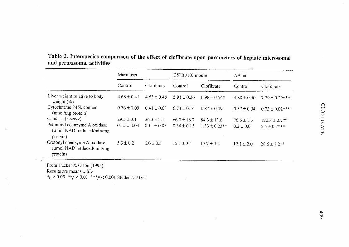
Table 2. Interspecies comparison of the effect of clofibrate upon parameters of hepatic microsomaland peroxisomal activities
Marmoset C57Bl/l0J mouse AP rat
Control Clofibrate Control Clofibrate Control Clofibrate
Liver weight relative to body 4.68 :t 0.41 4.63 :t 0.48 5.91 :t 0.36 6.98 :t 0.54* 4.80 :t 0.50 7.39:t 0.29***weight (%) (lCytochrome P450 content 0.36 :t 0.09 0.41 :t 0.08 0.74:t0.14 0.87 :t 0.09 0.37 :t 0.04 0.73:t 0.02*** r0(nmol/mg protein) 'T..Catalase (k.sec/g) 29.5 :t 3.1 36.3:: J.l 66,O:t 16,7 84.3:t 13,6 76.6:t 1.3 120.3 :t 2.7** Ci
::Palmitoyl coenzyme A oxidase 0.15 :t 0.03 0.1 1 :t 0.03 0.34 :t 0.13 1.33 :t 0.23** 0.2 :t 0.0 5.5 :t 0.7*** ;i(¡.mol NAD+ reduced/min/mg -i
t'protein)
Crotonyl coenzyme A oxidase 5.3 :t 0.2 6.0 :t 0.3 15.1:t3.4 17.7 :t 3.5 12.1:t2.0 28.6:t 1.2**
(¡.mol NAD+ reduced/min/mgprotein)
From Tucker & Orton (1995)Results are means :t SD*p 0( 0.05 **p 0( 0.01 ***p 0( 0.001 Students t test
.to\D

410 lARe MONOGRAPHS VOLUME 66
enzyme of the CYP4A family associated with fatty acid (O-hydroxylase activity. MaleLong-Evans rats given 80 mg/kg bw of the enantiomers or racemix mixture of the c10-fibrate analogue, 2-( 4-para-chlorophenyloxy)2-phenyl ethanoic acid, by gastric instilla-tion for three consecutive days, had elevated hepatic CYP4Al and lauric acid 12-hydro-xylase activity (Chin je & Gibson, 1990). Clofibrate (730 mg/kg diet (ppm) for 24 weeks)inhibited the expression of y-glutamyl transpeptidase in liver homogenates of maleFischer 344 rats in which this enzyme activity had been induced by prior feeding of2-AAF (Nu moto et al., 1984).
Some extrahepatic effects of clofibrate in experimental animaIs are analogous to thoseobserved in the lIver. For example, in male but not female Fischer 344 rats given 400mg/kg bw c10fibrate daily by intraperitoneal injection for three consecutive days,increased renal content of CYP4A2 mRNA was observed (Sundseth & Waxman, 1992).Clofibric acid caused an increase in renal peroxisomal palmitoyl CoA oxidase activity inmale Wistar rats given 200 mg/kg bw clofibric acid daily by gastric instillation for 10consecutive days (Chandoga et aL., 1994). Administration of 400 mg/kg bw c10fibrate perday in the diet to male Wistar rats for 3 months or longer resulted in diminished size ofthyroid follicles, with calcium deposition in the colloid and hypertrophy of the Golgiapparatus (Price et aL., 1988).
4.3 Reproductive and developmental effects
4.3.1 Humal1s
Schneider and Kaffarnik (1975) reported three cases of male impotence in patientswith type lV hyperlipoproteinaemia who were treated with a controlled di et and clo-fibrate. The complaints of impotence were made within one year of beginning treatmentwith the drug. Two of the patients reported improvement of the symptoms three and fourweeks after interruption of clofibrate therapy; one patient again complained of impotencewhen clofibrate therapy was resumed.
4.3.2 Experimental systems
ln a study reported only in abstract, no change in number of resorption sites, littersize, fetal weight or no teratogenic effect was found in rats when dams were given0.6 mg/kg bw c10fibrate per day in feed or 1 or 140 mg/kg bw clofibrate per day by
gastric instillation from day 6 to day 20 of gestation (Diener & Hsu, 1966). When dosesof 200 mg/kg bw per day were given to both male and female rats by gastric instilation,both before and during gestation, a significant decrease in litter size was observed, andwith a dose of 500 mg/kg bw, the number of pregnancies decreased from 7/8 in con troisto 0/8 in treated \ animaIs. No such effect was found when female rabbits were treatedsimilarly (Pantaleoni & Valeri, 1974). ln female albino rats given 50 mg c10fibrate perday orally during the en tire period of mating, gestation and lactation, the liver weight atbirth of the offspring was significantly higher than that of control pups, while there wasno difference in birth weight between the groups (Chhabra & Kurup, 1978). Theoffspring of Wistar rats given 8000 mg/kg diet (ppm) c10fibrate in the diet for one week
CLOFIBRATE 411
on gestational days 13, 15, 17, 19 or 21 weighed significantly less than the offspring ofcontrol rats. Maternai weight gain was reduced in treated animaIs compared with controls(Cibelli et al., 1988). An abnormal postnatal fetal thrombosis syndrome in rats has beendescribed, consisting of an extension of the normal thrombosis in the umbilical arteriesand causing necrosis of the tail or parts of the hindlimbs (Dange et al., 1975). ln pregnantDutch rabbits given 0 or 5000 ppm clofibrate in the diet throughout pregnancy, no effecton fertility or litter size and no skeletal abnormality were detected (Tucker & Orton,1995). Nishimura and Tanimura (1976) found that the rabbit fetus serum accumulates ahigher concentration of clofibrate th an maternai serum.
ln albino rats, the serum of newborn pups of dams that had received clofibrate(50 mg/day) orally during mating, gestation and lactation contained 93 nmoI/mL clo-fibric acid. This decreased to 48 nmoI/mL on day 12 and 31 nmoI/mL at the time ofweaning. Placenta collected before birth from clofibrate-fed dams contained about80 nmoI/g clofibric acid. This indicates that the drug crosses the placenta. The activity ofmitochondrial glycerol phosphate dehydrogenase in hepatic mitochondria isolated fromnewborn rats of dams that were fed the drug was almost three times the level observedfor control offspring. The activity increased and remained at a higher level duringlactation but, when the young animais were weaned, it rapidly decreased to about thesame level as that seen in control animaIs. This suggests that the drug may also pass tothe offspring via the mother's milk (Chhabra & Kurup, 1978).
Clofibrate (150 mg/kg bw per day) given continuously to female Wistar/H-Riop ratsfrom gestational day 16 to the end of lactation (22nd day post-partum) produced a
decrease in birth weight, an increase in perinatal mortality and an increase in liver weightat the age of 22 days. Investigations in which the dam received doses of 150 mg/kg bwper day during four time intervals between gestational day 16 and postnatal day 22showed that the increase in liver weight was associated with exposure between deliveryand postnatal day 15, When the drug was administered in the last week of pregnancy andthe young were dissected on postnatal days l, 8, 15 or 22, increased liver weight wasobserved in neonates but not subsequently. The authors suggested that this transientincrease in liver weight might be related to enzyme induction rather th an to hepato-toxicity (Nyitray et al., 1980).
Pregnant Swiss lCR mice were given clofibrate by subcutaneous injections at variousdosages (480 and 960 mg/kg bw) and time intervals, and embryos were removed on days17 or 18 of gestation. ln embryos removed on day 17, the level of intestinal catalase acti-
vit Y of the proximal and distal halves did not differ between treated groups and controls.ln embryos removed on day 18, a dose-dependent rise in catalase activities in the proxi-mal half of the small intestine in treated groups was observed, but a plateau was attainedwith repeated injections (Calvert et aL., 1979).
Clofibrate treatment of pregnant female rats has been found to increase the number ofliver peroxisomes and the levels of fatty acid oxidation enzymes in fetuses, suggestingthat the treatment induces fetal peroxisome proliferation (Cibelli et al., 1988; Stefaniniet al., 1989). ln mice, 400 mg/kg bw oral clofibrate treatment initiated at day 6 of gesta-tion produced a 4-5-fold Increase in levels of peroxisomal membrane protein 70, a 1.5-
412 rARC MONOGRAPHS VOLUME 66
to 2-fold increase in dihydroxyacetone phosphate acyltransferase specifie activity and aL.2-L.8-fold increase in catalase specific activity in fetal liver of 19 days gestation.
Electron microscopy showed amplification of endoplasmic reticulum and peroxisomes inthe fetalliver. There was a general increase in peroxisomal proteins between gestationaldays 13 and 19 in ail fetal tissues except the placenta, and the effect of clofibrate in thelung and the placenta was evident by gestational day 13 (Wilson et al. 1991).
4.4 Genetic and related effects
4.4,1 Humal1s
No data were available to the Working Group.
4.4.2 Experimel1tal systems (see also Table 3 for references and Appendices 1 and 2)
Clofibrate is not mutagenic in Salmol1ella typhimurium in the presence or absence ofmicrosomal preparations. ln the yeast Saccharomyces cerevisiae, clofibrate inducedneither gene conversion nor mitotic recombination.
ln single studies, clofibrate did not induce unscheduled DNA synthesis in culturedhepatocytes or DNA strand breaks in LI 210 cells. However, the ability of N-ethyl-N-nitrosourea to produce single-strand DNA breaks and of N,N'-bis(2-chloroethyl)-N-nitro-sourea to produce both single-strand DNA breaks and interstrand cross-links in LI 210cells was enhanced by prior treatment of the cells with clofibrate (Lawson & Gwilt,1993).
Clofibrate was not mutagenic in Chinese hamster lung V79 cells, in the presence of arat hepatocyte metabolic activation system. As reported in an abstract, clofibrate did notinduce resistance ta 6-thioguanine in the granuloma pouch assay in rats.
Clofibrate did not induce chromosomal aberrations in three studies with culturedmammalian cell lines nor micronucleus formation in a study with cultured rat hepa-toc
Y tes.
ln morphological transformation studies with Syrian hamster embryo (SHE) ceIls,clofibrate had no effect in one study, but was reported in another study to have increasedthe frequency of transformation. The administration of clofibrate al one was also withouteffect in the C3H/l OTl/2 Ci 8 cell transformation system, whereas it did enhance thefrequency of transformation produced by prior treatment with 3-methylcholanthrene.
Weak inhibition of gap-junctional intercellular communication in Chinese hamster V79cells was reported to occur with high concentrations of cIofibrate.
No evidence was seen of DNA adduct formation by clofibrate in the livers of maleFischer 344 rats given three doses of 250 mg/kg at 24-h intervals and killed 2 h after thefinal dose. DNA was analysed by 32P-postlabellng with an estimated limit of detection of1 adduct in 1010 nucleotides and no adduct was detected in hepatocytes treated II1 vitrowith 10-1 M clofibrate for 4 h (Gupta et aL., 1985).
III vivo, clofibrate did not induce unscheduled DNA synthesis in rat hepatocytes.Neither did it induce sister chromatid exchange in rat peripheral blood lymphocytes orbone marrow cells of Chinese hamsters.

Q)
..c:i...!:o-~'+oCI
..~æêQ)
"'Q)
..c:-Q
)i."'=c:~.....Q
)=Q
)
~.
('Q)
:ec:E
-
IluCIl..~Ilcc
6'5:--Il Cl
'" ~o ..Cl '-
'":: ,S:
g Õ a
Il .r-5 bi ro Il~
0 '0 ~~~ar;
::::'"Ilcc
'":: ,S:
'5 0 Õo~.ra.. bi ro Il
.. 0 +- +-
~ ~ š ~
aIl..'";;'"..'"Ilt-
CL
OFIB
RA
TE
~o 0 ~
~ ~
~O
~ooooo~o~o~oooo~~0\ 0\ 00 ir 00 tr 00 ir 00 0\ 00 ir ir ;;
~~~~~~~~~~~~~:g'- -- '- .. '- -- '- '- ~ .. Q)
~ è: ~ '- -. "- -. '- .. ~ ~ '- '- 0:;: ;: .: ~ .: ~ .: ~ .: ;: .: ~ ~ a(:.:. ~.. ~.. \U "-~:. ~......
uuc~c~c~c c~~~
ro ro Il C Il C
Il C Il ~ Il C
C ~
~ ~
l: ~ l: ~
l: ~ l: ~
l: ~ ~
';:ro ro ro ro ro ~,~
~~~8~8~8~~~88rJoo0:0:00000000:0000Z
Z S; 0 Ss Ss S; S; S; z Ss S; S; ~
CC C C C C .g
Co C
o C ,S
.S .S
,S .S
~._ ,S
~ ~
.. .. .. C C
C ::
ò.~ ~ ~ ¡g ~ .: .s .s .§ .g.,8 8"G
'5 '5 '5 ::a a:: ::a ::a ::a ro B ~ Il _
"~ a a a '5 :: :: '" ~
8 Il Il Il ~ ~ ~ ~ ~ a a a ~ ,S..~~~~~~~~~IlIl~~,~
Il Il Il ? ? ? ~ ? .. ~
~ .. ~
..~~?1l1l1l1l1l1l1l1l ~c
CIlIlIl..........???~o
~ o..~ o..~ ..~ tr tr r- r- 00 ~ ~ ~ ¡: u
Il 0 M (" M M M ~ 0 Il
¡t 0 0 0 tr tr tr ir ir 00 00 00 0 C
~~~~~~~~~~~~~ ~
ig t- t- t- t- t- t- t- t- t- t- t- L' ~
.~ E: E: E: E: E: E: E: E: E: E: E: E: .~
b ~ ~ ~ ~ ~ ~ ~ ~ ~ ~ ~ ~ .~
~ .~
.~ .~
.~ .~
.~ .~
.~ .~
.~ .~
.~ ~
u~~~~~~~~~~~~~~
E: E
: E: E
: E: E
: E: E
: E: E
: E: .E
: ~,~
~ ~
~ ~
~ ~
~ ~
~ ~
~ ~
'"~ B
BB
BB
BB
BB
BB
B~
¡; .. .. .. .. .. .. .. .. ~ ~ .. .. E:
c.;)~~~~~~~~~~~ ~
~~~~~~~~~~~~~~~aaaaaaaaaaaa~'g .§ .§ .§ .§ .§ .§ .§ .§ .§ .§ .§ .§ ~
CQ c5 c5 c5 c5 c5 r5 c5 c5 r5 c5 c5 c5 c5
d ó ó ó ir ir r- r- 00 0\ 0\ 0\ e/ Ó:a~....~~~..~~......u_ rr rr rr rr rr ¡z rr rr rr rr rr rr rr
v~~'"o Q)
0\ ,-0\ ..
.. u
'- rr
413
;;"e"eIl0:a(
~M0\ ~
0\ 0\ ~o~~
.. ~~oo ~oo 00 M 0\
'- ir tr 0\ "' r- 0\ 0\ ..
....oooo-ooO\..O
\",0\ 0\ '- 0\ :: '-.. .
.~ .. .. .. .. . '- ~r .. '- '- ~
'- -. ~ ~
'V-;~ ro\:~~~
~~~ro..~'"~a( ;: ;: CI a ~:. ;: OJ
~ § C cac ~ u"5 ~
o '" ro ro .~ '~ ro ro '" ..0\ ~ ;;;;:: c"e ~ ~~0\ ro ro ro'- c :. ro ëi ~
~..ClC
l~:J~~t-:J
ooooN
oQ
Q~Q
S;O:N
01M
NC
"INN
NZ
r-
t-Zt- ¡. t- f-
ZZZ Z
c,S..roc:BaouQ)
...S:..o..ï§~~'C
;.~~~(.'"~(.fj2~~(.(.c5:JU¡z
j2..j2 a'~ j2
.~ ~
.5.~.5 os: en .s
'S ~~ ..CJ~~
~ Q) ..0 Q
).; ü Q) I- U
'S: ;; u .. 0
.. .. 4: ?-
a ~ ~
.S ~
:: bi È.tuu~~U
§a~'~ 0\ 0\ ;; ~ ~.. Q) -V
~r-r-u-""''''u'- ? .. 0 '" '" Q) Q) lU
'" ....aa~~CI
.. blbiC' ro ro a s::
Q) c i: o..... ro C' 0
u.2.2~ ~ Q).... S
S .. .. ~
Il ~ i: i: ,N
Q) Q
) i: i:. ro .~ -'
N t: t: ..-:. .- i: .. f-
..ssv.UU
..;;;;o..~ ro ro ';; ¡z rr -
'" .. .. ~ Q) u: u: u: :i
-a Q) Q).. OJi: i: i:C"
Il",,,i:i:ooou.. ~
~ r;.ß
';: ';: ';:.r ,_ ._ u ê ro ro ci
"d Õ õ.. :x Q
) l: l: .S§ ~ ~~~~.22~b i: i: ~ ,_ ro ro S
'f .S .S "d ~ c; c; c; ,~o
~~~~aasa-O
J~""::OO
oovii: :: ::"d .. '" '" '" i:';; s a ~
-8 0 0 0 ~..Q
)Q)u..soas.:
i:i:CIIl o0-v
ZQ
)Q)i:~..""''u
Cl~~~ü3uÕÕ ~
.¿ :i 0 0. ~ c. e/ e/ ;¿
..O\O
\cc;d..__uQ
oo;:rrUU
Uf-
'"::U i:
.Q .~
.. ..~ ro
t: ::
.. 0

.t-Table 3 (contd) .t
Test system Result" Doseb Reference(LED/HID)
Without Withexogenous exogenousmetabolic metabolicsystem system
MIA, Micronucleus test, rat hepatocytes in vitro - NT 243 Müller et al. (1993) -TCS, Cell transformation, Syrian hamster embryo cells + NT 12 Mikalsen et al. (1990) ;i::TCS, Cell transformation, Syrian hamster embryo cells - - 72 Tsutsui et al. (1993) nDV A, DNA strand breaks, rat hepatocytes in vivo - 200 po x 6 Elliott & E1combe ~
( 1987) 0ZDV A, DNA strand breaks, rat hepatocytes in vivo -
750 di et x Nilsson et al. (1991) 014 d a::DVA, DNA strand breaks, Fischer rat liver in vivo -100 di et x Tamura et al. (1991) ;i78 wk
'":i
UPR, Unscheduled DNA synthesis, rat hepatocytes in vivo 750 di et x Nilsson et al. (1991) C/--c
14 d 0GV A, Gene mutation, rat granuloma pouch 6- TG resistance in vivo ? 4 sc x 1 Maier (1984) (abstract) l'
c:SV A, Sister chromatid exchange, rat peripheral lymphocytes in vivo - 200 po x 14 Linnainmaa (1984) ~
t'SV A, Sister chromatid exchange, Chinese hamster bone-marrow cells - 200 po x 14 Linnainmaa (1984) 0\in vivo 0\
CBA, Chromosomal aberrations, rat bone marrow in vivo - NG Kawachi et al. (1980)BIO, Binding (covalent) to DNA, male F344 rat hepatocytes in vitro - NT 243 Gupta et al. (1985)BVD, Binding (covalent) to DNA, male F344 rat Ii ver in vivo - 250 po x 3 Gupta et al. (1985)
(P-postlabelling)ICR, Inhibition of intercellu1ar communication, V79 cells (+) NT 24 A wogi et aL. (1984)
(abstract)
"+, positive; (+), weak positive; -, negative; NT, not tested; ?, inconclusiveh LED, lowest effective dose; HID, highest ineffective dose; in-vitro tests, l.g/mL; in-vivo tests, mg/kg bw; NG, dose not given
-'I'

CLOFIBRA TE 415
Slightly elevated levels of 8-hydroxydeoxyguanosine have been detected in liverDNA of rats fed diets containing clofibrate (see Section 4.2.2).
Oral treatment of Sprague-Dawley-derived SIV 50 rats with 14C-labelled clofibric acid(225 mg/kg) did not lead to detectable radioactivity associated with liver DNA, althoughprotein binding was clearly demonstrated (von Däniken et aL., 1981).
4.5 Mechanistic considerations
The role of data on peroxisome proliferation in evaluating carcinogenicity in humanshas been discussed. When the data support the conclusion that a tumour response in mi ceor rats is secondary only to peroxisome proliferation, this should be considered inaddressing the potential carcinogenicity of an agent in humans. The report of theWorking Group on Peroxisome Proliferation and its Role in Carcinogenesis (IARC,1995) indicates that the following issues should be considered:
"(a) Information is available to exclude mechanisms of carcinogenesis other th anthose related to peroxisome proliferation.
(b) Peroxisome proliferation (increases in peroxisome volume density or fatty acid~-oxidation activity) and hepatocellular proliferation have been demonstratedunder the conditions of the bioassay.
(c) Such ettects have not been found in adequately designed and conducted investi-
gations of human groups and systems."
The weight of evidence indicates that clofibrate, and peroxisome proliferators ingeneral, do not act as direct DNA-damaging agents and that their mechanism of tumourinitiation is indirect. Two responses have been proposed to account for liver carcino-genesis by peroxisome proliferators in rodents. These include (i) induction of peroxisomeproliferation and (ii) increased hepatocellular proliferation. These responses are notmutually exclusive with respect to tumour formation.
Chronic administration of peroxisome proliferators produces a sustained oxidativestress in rodent hepatocytes due to overproduction of hydrogen peroxide. This cantheoretically generate reactive oxygen species which can attack DNA or may affect cellsin other ways. There is also evidence froID in-vitro experiments that fatty acid meta-bolism in peroxisomal fractions can result in hydroxyl radical formation and DNAdamage. ln-vivo observations in support of this hypothesis include increased lipidperoxidation, increased lipofuscin deposition, the effects on levels of hepatic antioxidantsand inhibition of tumour formation by antioxidants (Lake 1995). However, sorne of theevidence suggests that the level of oxidative damage in vivo may be too low to accountentirely for the carcinogenicity of peroxisome proliferators.
During the first few days of administration, peroxisome proliferators induce celldivision in rodent hepatocytes; in some, but not all, studies sustained stimulation ofreplicative DNA synthesis has also been observed (Lake, 1995). An enhanced rate of cellproliferation can be a critical effect both in tumour initiation, by increasing the frequencyof spontaneous mutations and the rate of conversion of DNA adducts into mutationsbefore they are repaired, and in tumour promotion by facilitating clonaI expansion of

416 IARC MONOGRAPHS VOLUME 66
initiated cells. The promoting activity of cIofibrate has been demonstrated in rodentmodels of multistage hepatocarcinogenesis.
There are clear species differences in the responses of mammalian cells to peroxisome ,proliferators (Lake, 1995). Biopsy studies have clearly indicated that the responsivenessof human livers to the peroxisome proliferation produced by fibrate drugs is lacking or ismuch less than that seen in the livers of treated rodents, although similar levels of drugare achieved in the circulation. The striking hepatomegalic effect of peroxisome proli-feration is similarly not observed in patients receiving fibrate drugs. ln cultures ofhepatocytes, peroxisome proliferation and cell proliferation occur with rodent but nothuman hepatocytes. ln rodent liver, hepatomegaly and peroxisome proliferation requireexpression of functional PPARa, a member of the steroid hormone receptor superfamily.Clofibrate activates rodent PPARa il1 vitro. The insensitivity of human liver to theeffects of peroxisome proliferators is consistent with the low level of PP ARa in humanlivers, as weil as observations of genetic variations that render the human PP ARareceptor inactive as compared to PPARa expressed in rodent liver (Tugwood et al.,1996). ln non-human primates, administration of cIofibrate and other peroxisome proli-ferators has also failed to elicit the hepatomegaly and peroxisome proliferation inducedin rodent liver.
Clofibrate-induced peroxisome proliferation and cell proliferation have been demons-trated in feeding studies in rats conducted under bioassay conditions. Peroxisome proli-feration has not been found in studies of human groups and systems using c1ofibrate.Taken together, these findings indicate that the increased incidence of liver tumours inrodents treated with cIofibrate results from a mechanism that wou Id not be operative in
humans.
5. Summary of Data Reported and Evaluation
5.1 Exposure data
Clofibrate was introduced in the 1960s to reduce plasma concentrations of tri-glycerides and cholesterol in patients at high risk of coronary heart disease. Since the late1970s, its use has decreased considerably.
5.2 Human carcinogenicity data
ln 1978, a randomized trial of the World Health Organization, conducted to determinewhether clofibrate treatment would lower the incidence of ischaemic heart disease inmen, raised concern over a nonsignificant excess of deaths from cancer in treatedsubjects.
Subsequently the association between cIofibrate and cancer risk was examined inthree randomized trials and a small case-control study. A further four-year follow-up ofthe WHO trial showed no difference in the age-standardized death rates from malignant',neoplasms. ln two other trials, there was also no difference in cancer deaths between
CLOFIBRA TE 417
clofibrate-treated patients and a placebo-treated group. A meta-analysis of results fromsix trials also found no excess cancer mortality due to use of c10fibrate as a cholesterol-10wering drug. The case-control study, that had several methodological limitations,showed a nonsignificant excess of soft-tissue sarcoma.
5.3 Animal carcinogenicity data
Clofibrate was tested for carcinogenicity by oral administration in the diet in twoexperiments in mice and in three experiments in rats, and in one experiment in
marmosets by gastric instillation. No increase in incidence of tumours was reported inmice or marmosets. ln rats, clofibrate produced hepatocellular carcinomas.
Clofibrate was tested in several experiments by combined administration with otherchemicals. It enhanced the hepatocarcinogenicity of N-nitrosamines in rats and hamsters.It did not enhance the carcinogenicity of 2-acetylaminofluorene in rat liver.
5.4 Other relevant data
Clofibrate exerts similar pharmacological responses in humans and rodents. Absorp-tion and metabolism of clofibrate are similar in humans and rats. Elimination of clofibricacid, the free acid form of the drug as it appears in the circulation, is more rapid in rats,possibly due to lower binding to plasma proteins.
Clofibrate-induced peroxisome proliferation and cell proliferation have been demons-trated in feeding studies in rats. Peroxisome proliferation has not been found in studies ofclofibrate in human livers or hepatocytes.
There are a number of case reports of reversible impotence in men treated withclofibrate. No noteworthy effect on ,the fetus has been observed in studies in rats orrabbits.
Clofibrate is inactive in most tests for genetic activity, although it induced cell
transformation in one study.
M echal1istic cOl1side ratiol1s
The weight of evidence indicates that c10fibrate does not act as a direct DNA-damaging agent and that its mechanism of tumour induction is indirect. Two biologicalresponses have been proposed to account for liver carcinogenesis by peroxisome proli-ferators in rodents. These are (i) induction of peroxisome proliferation and (ii) increasedhepatocellular proliferation. Upon exposure to clofibrate, proliferation of both pero-xisomes and cells occurs in rat" liver and of peroxisomes in cultured rat hepatocytes,whereas peroxisome proliferation does not occur in human liver or cultured hepatocytes.These observations suggest that the mechanism of liver carcinogenesis in c1ofibrate-treated rats would not be operative in humans.
~,,'-~.

418 IARC MONOGRAPHS VOLUME 66
5.5 Evaluation 1
There is il1adequate evidel1ce in humans for the carcinogenicity of clofibrate.There is limited evidel1ce in experimental animais for the carcinogenicity of
clofibrate.
Overall evaluation
Clofibrate is 110t classifable as to its carcil1ogel1icity in humal1s (Croup 3).
6. References
Anisimov, V.N., Danetskaya, E.V., Miretsky, G.I., Troitskaya, M.N. & Dilman, V.M. (1981)Influence of misc1eron (c1ofibrate) on carcinogenic effect of 7, 12-dimethylbenz(a)anthraceneand methylnitrosourea in female rats. Vopr. Onkol., 27, 64-67 (in Russian)
Anthony, L.E., Schmucker, D.L., Mooney, J.S. & Jones, AL. (1978) A quantitative analysis offine structure and drug metabolism in lIvers of clofibrate-treated young adult and retiredbreeder rats. J. Lipid Res., 19, 154-165
Awogi, T., Kato, K., Itoh, T. & Tsushimoto, G. (1984) Comparison of inhibiting activity on cell-cell communication between some tumor promoters in Chinese hamster V79 cells (Abstractno. 1). Mutat. Res., 130, 361
Baldwin, J.R., Witiak, D.T. & Feller, D.R. (1980) Disposition of c10fibrate in the rat: acute andchronic administration. Biochem. Pharmacol., 29, 3143-3154
Barnard, S.D., Molello, J.A., Caldwell, WJ. & LeBeau, J.E. (1980) Comparative ultrastructuralstudy of rat hepatocytes after treatment with the hypolipidemic agents probucol, clofibrate,and fenofibrate. J. ToxicoL. environ. Health, 6, 547-557
Barrass, N.C., Priee, RJ., Lake, B.G. & Orton, T.C. (1993) Comparison of the acute and chronicmitogenic effects of the peroxisome proliferators methylc10fenapate and c10fibric acid in ratlIver. Carcinogenesis, 14, 1451-1456
Bateson, M.C., Mac1ean, D., Ross, P.E. & Bouchier, LAD. (1978) Clofibrate therapy and gall-stone induction. Digest. Dis., 23, 623-628
Berstein, L.M., Pozharisski, K.M. & Dilman, V.M. (1982) Effects of misclerone (c1ofibrate) ondimethylhydrazine-induced intestinal carcinogenesis in rats. Oncology, 39,331-335
Blaauboer, BJ., van Holsteijn, C.W.M., Bleumink, R., Mennes, W.c., van Pelt, F.N.A.M., Yap,S.H., van Pelt, J.F., van Iersel, AA.J., Timmerman, A & Schmid, B.P. (1990) The effect ofbec10bric acid and c10fibric acid on peroxisomal ß-oxidation and peroxisome proliferation inprimary cultures ofrat, monkey and human hepatocytes. Biochem. Pharmacol., 40,521-528
British Pharmacopoeial Commission (1993) British Pharmacopoeia 1993, Vols. 1 & II, London,Her Majesty's Stationery Office, pp. 167-168,839-840
Bronfman, M., Amigo, L. & Morales, M.N. (1986) Activation of hypolipidaemic drugs to acyl-coenzyme A thioesters. Biochem. J., 239, 781-784
IFor definition of the italicized terms, see Preamble, pp. 22-25.

CLOFIBRATE 419
Budavari, S., ed. (1995) The Merck Index, l2th Ed., Rahway, NJ, Merck & Co.
Butterworth, B.E., Smith-Oliver, T., Earle, L., Loury, D.1., White, R.D., Doolittle, D.1.,Working, P.K., Cattley, R.C., Jirtle, R., Michalopoulos, G. & Strom, S. (1989) Use ofprimary cultures of human hepatocytes in toxicology studies. Cancer Res., 49, 1075-1084
Cal vert, R., Malka, D. & Ménard, D. (1979) Effect of c10fibrate on the small intestine of fetalmice. Histochemistry, 63, 7-14
de Camargo, J,L.V" Tsuda, H., Asamoto, M., Tagawa, y" Wada, S., Nagase, S. & Ito, N. (1993)Modifying effects of chemicals on the development of liver preneoplastic placental gluta-thione S-transferase positive foci in analbuminemic and Sprague-Dawley rats. Toxico!.PathoL., 21, 409-416
Canner, PL., Berge, K.B., Wenger, N.K., Stamler, J., Friedman, L., Prineas, R.J. & Friedewald,W. (1986) Cooperative studies. Fifteen year mortality in coronary drug project patients:10ng-term benefit with niacin. J. Am. Coll. Cardio!., 8, 1245-1255
Carlson, L.A, & Rosenhamer, G. (1988) Reduction of mortality in the Stockholm Ischaemic HeartDisease Secondar Prevention Study by combined treatment with clofibrate and nicotInic acid.Acta med. scand., 223,405-418
Cattley, R.e. & Glover, S.E. (1993) Elevated 8-hydroxydeoxyguanosine in hepatic DNA of ratsfollowing exposure to peroxisome proliferators: relationship to carcinogenesis and nuc1earlocalization. Carcinogenesis, 14, 2495-2499
Cattley, R.e., Kato, M., Popp, J.A., Teets, V.J. & Voss, K.S. (1994) Initiator-specific promotionof hepatocarcinogenesis by WY -14,643 and c1ofibrate. Carcinogenesis, 15, 1763-1766
Cayen, M.N., Ferdinandi, E.S., Greselin, E., Robinson, W.T. & Dvornik, D. (1977) Clofibrateand clofibric acid: comparison of the metabolic disposition in rats and dogs. J. Pharmaco!.exp. Ther" 200, 33-43
Chance, D.S., Wu, S.-M, & McIntosh, M.K. (1995) Inverse relationship between peroxisomaland mitochondrial ß-oxidation in HepG2 cells treated with dehydroepiandrosterone and c10-fibric acid (43865). Proc. Soc. exp. Bio!. Med., 208, 378-384
Chandoga, J., Rojeková, 1., Hampl, L. & Hocman, G. (1994) Cetaben and fibrates both influencethe activities of peroxisomal enzymes in different ways. Biochem. Pharmacol" 47, 515-519
Chhabra, S. & Kurup, e.K.R. (1978) MaternaI transport of chlorophenoxyisobutyrate at thefoetal and neonatal stages of development (Short communication). Biochem. Pharmacol., 27,2063--2065
Chin je, E.e. & Gibson, G.G. (1990) Stereochemical induction of cytochrome P4501V Al (P452)and peroxisome proliferation in male rat. Adv. exp. Med. Bio!., 283,267-270
Cibelli, A., Stefanini, S. & Ceru, M.P. (1988) Peroxisomal ß-oxidation and catalase activities infetal rat liver: effect of maternaI treatment with clofibrate. Cel!. ma!. Biol., 34, 191-205
Committee of Principal Investigators (1978) A cooperative trial in the primary prevention ofischaemic heart disease using clofibrate. Br. Heart J., 40, 1069- 1 1 18
Committee of Principal Investigators (1980) WHO cooperative trial on primary prevention ofischaemic heart disease using clofibrate to lower serum cholesterol: mortality follow-up.Lancet, ii, 379-385
Committee of Principal Investigators (1984) WHO cooperative trial on primary prevention ofischaemic heart disease with clofibrate to 10wer serum cholesterol: final mortality follow-up.Lancet, ii, 600-604

420 rARC MONOGRAPHS VOLUME 66
Coronary Drug Project Research Group (1975) Clofibrate and niacin in coronary heart disease. 1.Am, med. Assoc., 231, 360-381
Council of Europe (1984) European Pharmaeopoeia, 2nd Ed., Sainte-Ruffine, France,Maisonneuve S.A., pp. 318-1-318-4
Cumming, A. (1980) Acute renal failure and interstitial nephritis after cIofibrate treatment. Br,med. J., 281, 1529-1530
Dange, M., Junghani, J., Nachbaur, J., Perraud, J. & Reinert, H. (1975) Postnatal thrombosis(PNT) in newborn rats by hypolipidemic agents. Preliminary results (Abstract). Terata logy,12, 328
von Däniken, A., Lutz, W.K. & Schlatter, C. (1981) Lack of covalent binding to rat liver DNAof the hypolipidemic drugs clofibrate and fenofibrate. Toxieol. LeU., 7,305-310
Dayan, J., Deguingand, S. & Truzman, C. (1985) Study of the mutagenic activity of 6 hepato-toxic pharmaceutical drugs in the Salmol1ella typhimurium microsome test, and the HGPRTand Na+/K+ ATPase system in cultured mammalian cells. Mu
ta t. Res., 157, 1-12Diener, R.M. & Hsu, B. (1966) Effects of certain aryloxisobutyrates on the rat fetus (Abstract
no. 12). ToxieoL. appL. Pharmaeol., 8,338
Dunnigan, M.G. (1992) Should clofibrate still be prescribed? Further trials with harder endpoints are needed for all lipid lowering drugs. Br. med. 1.,305, 379-380
Eacho, P.I., Lanier, T.L. & Brodhecker, C.A. (1991) Hepatocellular DNA synthesis in rats giventhe peroxisome proliferating agents: comparison of WY -14,643 to cIofibric acid, nafenopinand L Y171883. Carcinogenesis, 12, 1557-1561
Elliott, B.M. & Elcombe, C.R. (1987) Lack of DNA damage or lipid peroxidation measuredin vivo in the rat liver following treatment with peroxisomal proliferators. Careinogenesis, 8,1213-1218
Elliott, B.M., Dodd, N.J.F. & Elcombe, c.R. (1986) Increased hydroxyl radical production inliver peroxisomal fractions from rats treated with peroxisome proliferators. Carcinogenesis,7, 795-799
Emudianughe, T.S., Caldwell, J., Sinclair, K.A. & Smith, R.L. (1983) Species differences in themetabolic conjugation of clofibric acid and c10fibrate in laboratory animaIs and man. DrugMetab. Dispos., 11,97-102
Erkul, L, Yavuz, H. & Özel, A. (1991) Clofibrate treatment of neonatal jaundice (Letter to theEditor). Pediatries, 88, 1292
Expert Panel (1988) Report of the National Cholesterol Education Program Expert Panel ondetection, evaluation, and treatment of high blood cholesterol in adults. Arch. intern. Med.,148, 36-69
Foxworthy, P.S. & Eacho, P.L (1986) Conditions influencing the induction of peroxisomalß-oxidation in cultured rat hepatocytes. ToxieoL. Leu., 30, 189- 1 96
Gabilan, J.C., Benattar, C. & Lindenbaum, A. (1990) Clofibrate treatment of neonatal jaundice(Letter to the Editor). Pediatries, 86, 647-648
Gabilan, J.c., Benattar, C. & Lindenbaum, A. (1991) Clofibrate treatment of neonatal jaundice(Letter to the Editor). Pediatries, 88, 1292-1294
Gennaro, A.R., ed. (1995) Remington: The Science and Praetice of Pharmaey, 19th Ed., VoL. II,Easton, PA, Mack Publishing Co., p. 967

CLOFIBRA TE 421
Goodman Gilman, A., RaIl, T.W., Nies, A.S. & Taylor, P., eds (1990) Goodman and Gilman 's.The Pharmacological Rasis of Therapeutics, 8th Ed., New York, Pergamon Press, pp. 886-889
Greaves, P., Irisarri, E. & Monro, AM. (1986) Hepatic foci of cellular and enzymatic alterationand nodules in rats treated with c10fibrate or diethylnitrosamine followed by phenobarbital:their rate of onset and their reversibility. 1. natl Cancer lnst., 76, 475-484
Gugler, R. & Hartlapp, J. (1978) Clofibrate kinetics after single and multiple doses.. Clin.Pharmacol. Ther., 24,432-438
Gupta, R.C., Goel, S.K., Earley, K., Singh, B, & Reddy, J.K. (1985) 32P-Postlabeling analysis of
peroxisome proliferator-DNA adduct formation in rat liver in vivo and hepatocytes in vitro.Carcinogenesis, 6, 933-936
Hagiwara, A, Tamano, S., Ogiso, T., Asakawa, E, & Fukushima, S. (1990) Promoting effect ofthe peroxisome proliferator, clofibrate, but not di(2-ethylhexyl)phthalate, on urinary bladdercarcinogenesis in F344 rats initiated by N-butyl-N-( 4-hydroxybutyl)nitrosamine. Jpn. 1.Cal1cer Res., 81, 1232-1238
Hanefeld, M., Kemmer, c., Leonhardt~ W., Kunze, K.D., Jaross, W.& Haller, H. (1980) Effectsof p-chlorophenoxyisobutyric acid (CPIB) on the human liver. Atherosclerosis, 36, 159-172
Hanefeld, M., Kemmer, C. & Kadner, E. (1983) Relationship between morphological changesand lipid-lowering action of p-chlorophenoxyisobutyric acid (CPIB) on hepatic mitochondriaand peroxisomes in man: Atherosclerosis, 46, 239-246
Harvengt, C. & Desager, J .P. (1976) Pharmacokinetic study and bioavailability of threemarketed compounds releasing p-chlorophenoxyisobutyric acid (CPIB) in volunteers. lnt. J.clin. Pharmacol., 14, 113-118
Hassan, M.M.A. & Elazzouny, A.A. (198,2) Clofibrate. ln: Florey, K., ed., Analytical Profiles ofDrug Substances, VoL. 11, New York, Academic Press, pp.197-224
Hendrickson, R.M. & Simpson, F. (1982) Clofibrate and eosinophilic pneumonia (Letter to theEditor). J. Am. med. Assac., 247, 3082
Hoar Zahm, 5., Blair, A, Holmes, F.F., Boysen, C.D., Robel, R.J. & Fraumeni, J.F., Jr (1989) Acase-control study of soft-tissue sarcoma. Am. J. Epidemiol., 130, 665-674
Hosokawa, S., Tatematsu, M., Aoki, T., Nakanowatari, J., Igarashi, T. & Ito, N. (1989) Modu-lation of diethylnitrosamine-initiated placental glutathione S-transferase positive preneo-plastic and neoplastic lesions by c1ofibrate, a hepatic peroxisome proliferator. Cat-cino-gel1esis, 10, 2237-2241
IARC (1980) lARC Monographs 011 the Evaluation of the Carcinogenic Risk of Chemicals toHumal1S, Vol. 24, Some Pharmaceutical Drugs, Lyon, pp. 39-58
IARC (1983) IARC Monographs on the Evaluation of the Carcinogenic Risk of Chemicals taHumal1S, VoL. 31, Some Food Additives, Feed Additives and Naturally OccurringSubstances, Lyon, pp. 95-132
IARC (1987) IARC Monographs on the Evaluation of Carcil10genic Risks to Humal1S, Suppl. 7,Overall Evaluations of Carcinogenicity: An Update of IARC Monographs Volumes 1 ta 42,Lyon, pp. 171-172
IARC (1995) Peroxisome Proliferation and its Role in Carcinogenesis (lARC Technical ReportNo. 24), Lyon

422 IARC MONOGRAPHS VOLUME 66
Ishidate, M., Jr, Hayashi, M., Sawada, M., Matsuoka, A., Yoshikawa, K., Ono, M, & Nakadate,M. (1978) Cytotoxicity test on medical drugs - chromosome aberration tests with Chinesehamster cells in vitro. Eisei Shikenjo Hokoku, 96, 55-61 (in Japanese)
Issemann, 1., Prince, R.A., Tugwood, J.D, & Green, S. (1993) The peroxisome proliferator-activated receptor:retinoid X receptor heterodimer is activated by fatty acids and fibratehypolipidaemic drugs. 1. moL. Endocrinol., 11,37-47
Kawachi, T., Komatsu, T., Kada, T., Ishidate, L, Sasaki, M., Sugiyama, T. & Tazima, y, (1980)Results of recent studies on the relevance of various short-term screening tests in Japan.Appl. Methods Oncol., 3, 253-267
Kurokawa, y., Takamura, N., Matushima, y., Imazawa, T. & Hayashi, Y. (1988) Promoting
effect of peroxisome proliferators in two-stage rat renal tumorigenesis, Cancer Leu., 43,145-149
Lake, B.G. (1995) Mechanisms of hepatocarcinogenicity of peroxisome-proliferating drugs andchemicals. Anl1u. Rev. Pharmacol. Toxicol., 35, 483-507
Larsen, Mol., Ilingworth, D.R. & O'Malley, J.P. (1994) Comparative effects of gemfibrozil andc10fibrate in type III hyperlipoproteinemia. Atherosclerosis, 106, 235-240
Law, M.R., Thompson, S.G. & Wald, N.J. (1994) Assessing possible hazards of reducing serumcholesteroL. Br. med. J., 308, 373-379
Lawson, T. & Gwilt, P.R. (1993) Clofibrate enhances the DNA damaging action and cyto-toxicity of nitrosoureas. Cancer Leu., 70, 1 19-122
Lee, G.-H., Nomura, K, & Kitagawa, T. (1989) Comparative study of diethylnitrosamine-ini-tiated two-stage hepatocarcinogenesis in C3H, C57Bl and BALB mice promoted by varioushepatopromoters. Carcinogenesis, 10, 2227-2230
Lee, S.S.-T., Pineau, T., Drago, 1., Lee, EJ., Owens, J.W., Kroetz, D.L., Fernandez-Salguero,P.M., Westphal, H. & Gonzalez, FJ. (1995) Targeted disruption of the Ci isoform of theperoxisome proliferator-activated receptor gene in mice results in abolishment of the pleio-tropic effects of peroxisome proliferators. MoL. cell. BioL., 15, 3012-3022
Lilehaug,' J.R., Aarsaether, N., Berge, RK. & Male, R (1986) Peroxisome proliferators showtumor-promoting but no direct transforming activity in vitro. l11t. 1. Cancer, 37, 97-100
Linnainmaa, K. (1984) The effects of hypolipidemic peroxisome proliferators on the inductionof sister chromatid exchanges. Basic Life Sei., 29B, 965-974
London, S.F, Gross, K.F. & Ringel, S.P. (1991) Cholesterol-lowering agent myopathy
(CLAM). Neurology, 41, 1159-1160Lundgren, B. & DePierre, J.W. (1989) Proliferation of peroxisomes and induction of cytosolic
and microsomal epoxide hydrolases in different strains of mice and rats after dietarytreatment with c1ofibrate. Xenobiotica, 19, 867-881
Maier, P., Schawalder, H.P. & Zbinden, G. (1984) Specifie-locus mutations induced withhepatocarcinogens in a subcutaneous granulation tissue in rats (Abstract no. 1.3.4). Mutat.Res., 130, 187
Männistö, PT., Tuomisto, J., Jounela, A. & Penttilä, O. (1975) Pharmacokinetics of c10fiQrateand chlorophenoxy isobutyric acid. 1. Cross-over studies on human volunteers. Acta phar-macot. toxicol., 36,353-365
,Marsman, D.S., Goldsworthy, T.L. & Popp, J.A. (1992) Contrasting hepatocytic peroxisomeproliferation, lipofuscin accumulation and cell turnover for the hepatocarcinogens
Wy-14,643 and clofibric acid. Carcil1ogenesis, 13, 1011-1017
CLOFIBRA TE 423
McGarvey, J .FX. (1973) Premature contractions and clofibrate (Letter to the Editor). 1. Am.med. Assoc., 225, 638
Medical Economies (1996) PDR'8: Physicians' Desk Reference, 50th Ed., Montvale, NJ,Medical Economies Data Production Co., pp. 2701-2703
Meijer, J" Starkerud, C., Granell, 1. & Afzelius, B.A. (1991) Time-dependent effects of thehypolipidemic agent clofibrate on peroxisomes and mitochondria in mouse hepatocytes. 1.submicrosc. Cytol. PathoL., 23, 185-194
Mikalsen, S.-O., Holen, 1. & Sanner, T. (1990) Morphological transformation and catalase
activity of Syrian hamster embryo cells treated with hepatic peroxisome proliferators, TP Aand nickel sulphate. Cel! BioL. ToxicoL., 6, 1-14
Mizumoto, K., Kitazawa, S., Eguchi, T., Nakajima, A., Tsutsumi, M., Ito, S., Danda, A. &Konishi, Y. (1988) Modulation of N-nitrosobis(2-hydroxypropyl)amine-induced carcino-
genesis by clofibrate in hamsters. Carcinogel1esis, 9, 1421-1425
Mochizuki, Y., Furukawa, K. & Sawada, N. (1982) Effects of various concentrations of ethyl-a-p-chlorophenoxyisobutyrate (clofibrate) on diethylnitrosamine-induced hepatic tumori-genesis in the rat. Carcinogenesis,3, 1027-1029
Mochizuki, Y., Furukawa, K. & Sawada, N. (1983) Effect of simultaneous administration ofclofibrate with diethylnitrosamine on hepatic tumorigenesis in the rat. Cancer LeU., 19, 99-105
Müller, K., Kasper, P. & Müller, L. (1993) An assessment of the in vitro hepatocyte micro-nucleus assay. Mutation Res., 292, 213-224
Murata, Y., Tani, M. & Amano, M. (1988) Erythema multiforme due to clofibrate (Letter to theEditor).1. Am. Acad. Dermatol., 18,381-382
Mutai, M., Tatematsu, M., Aoki, T., Wada, S. & Ito, N. (1990) Modulatory interaction betweeninitial clofibrate treatment and subsequent administration of 2-acetylaminofluorene orsodium phenobarbital on glutathione S-transferase positive les ion development. Cancer Lett.,49, 127-132
Nilsson, R., Beije, 8., Préat, V., Erixon, K. & Ramel, C. (1991) On the mechanism of thehepatocarcinogenicity of peroxisome proliferators. Chem. -bioL. 1l1teractions, 78, 235-250
Nishimura, A. & Tanimura, T. (1976) Clil1ical Aspects of the Teratogenicity of Drugs, NewYork, Excerpta Medica, pp. 256-257
Numuto, S., Furukawa, K., Furuya, K. & Willams, G.M. (1984) Effeets of the hepatocarcino-genic peroxisome-proliferating hypolipidemic agents clofibrate and nafenopin on the rat livtrcell membrane enzymes y-glutamyltranspeptidase and alkaline phosphatase and on the earlystages of liver carcinogenesis. Carcinogenesis, 5, 1603-1611
Nyitray, M., Szaszovszky, E. & Druga, A. (1980) Clofibrate and the development of rats. Arch.ToxicoL., Suppl. 4, 463-465
Odum, J. & Orton, T.C. (1983) Hepatic microsomal glucuronidation of clofibric acid in the adultand neonate albino rat. Biochem. Pharmacol., 32,3565-3569
Pantaleoni, G.c. & Valeri, P. (1974) Investigation on the interactions of clofibrate with repro-ductive funetion. Clin. Terap., 69, 321-328 (in Italian)
Priee, S.C., Hinton, R.H., Mitchell, FE., Hall, D.E., Grasso, P., Blane, G.F & Bridges, J.W.(1986) Time and dose study on the response of rats to the hypo1ipidaemic drug fenofibrate.Toxicology,41, 169-191
424 IARC MONOGRAPHS VOLUME 66
Priee, S.c., Chescoe, D., Grasso, P., Wright, M. & Hinton, R,H. (1988) Alterations in thethyroids of rats treated for long periods with di-(2-ethylhexyl)phthalate or with hypolipi-daemic agents. Toxicol. Leu., 40,37-46
Reddy, J.K. & Qureshi, S.A. (1979) Tumorigenicity of the hypolipidaemic peroxisome proli-ferator ethyl-a-p-chlorophenoxyisobutyrate (clofibrate) in rats. Br. 1. Cancer, 40, 476-482
Reddy, J.K. & Rao, M.S. (1978) Enhancement by Wy- 14,643, a hepatic peroxisome proliferator,of diethylnitrosamine-initiated hepatic tumorigenesis in the rat. Br. 1. Cancer, 38,537-543
Reynolds, J.E.F., ed. (1993) Martindale: The Extra Pharmacopoeia, 30th Ed., London, ThePharmaceutical Press, pp. 986-987
Rush, P., Baron, M. & Kapusta, M. (1986) Clofibrate myopathy: a case repoi1 and a review ofthe literature. Sem. Arthrit. Rheumat., 15, 226-229
Schiestl, R.H. & Reddy, J.K. (1990) Effect of peroxisome proliferators on intrachromosomaland interchromosomal recombination in yeast. Carcinogenesis, 11, 173-176
Schneider, J. & Kaffarnik, H. (1975) Impotence in patients treated with clofibrate. Athero-sclerosis, 21, 455-457
Schwandt, P., Klinge, O. & Immich, H. (1978) Clofibrate and the liver (Letter to the Editor).Lancet, ii, 325
Scionti, L., Calafiore, R., Coaccioli, S., Bellomo, G., Berrettini, M. & Puxeddu, A. (1984) Clo-fibrate-induced myocardial injury and disseminated intravascular coagulation in a patientwith chronic renal failure. Pan. Med., 26,45-47
Scotto, c., Keller, J.-M., Schohn, H. & Dauça, M. (1995) Comparative effects of clofibrate onperoxisomal enzymes of human (Hep EBNA2) and rat (FaO) hepatoma cell lines. Eur. 1.Cell BioL., 66, 375-381
Smals, A.G.H., Beex, L.V.A.M. & Kloppenborg, P.W.c. (1977) Clofibrate-induced muscledamage with myoglobinuria and cardiomyopathy (Letter to the Editor). New Engl. J. Med.,296, 942
Society of Japanese Pharmacopoeia (1992) The Pharmacopoeia of Japal1 JP XII, l2th Ed.,Tokyo, pp. 231-232
Stanko, R.T., Sekas, G., Isaacson, LA., Clarke, M.R., Billiar, T.R. & Paul, H.S. (1995) Pyruvateinhibits c1ofibrate-induced hepatic peroxisomal proliferation and free radical production inrats. Metabolism, 44, 166-171
Stefanini, S., Mauriello, A., Farrace, M.G., Cibelli, A. & Ceru, M.P. (1989) Proliferativeresponse of foetal liver peroxisomes to clofibrate treatment of pregnant rats. A quantitiveevaluation. BioL. Cell, 67, 299-305
Sundseth, S.S. & Waxman, D.J. (1992) Sex-dependent expression and c10fibrate inducibility ofcytochrome P450 4A fatty acid û)-hydroxylases~ 1. biol. Chem., 267, 3915-3921
Svoboda, D.J. & Azarnoff, D.L. (1979) Tumors in male rats fed ethyl chlorophenoxyisobutyrate,a hypolipidemicdrug. Cancer Res., 39,3419-3428
Takagi, A., Sai, K., Umemura, T., Hasegawa, R. & Kurokawa, Y. (1990) Relationship betweenhepatic peroxisome proliferation and 8-hydroxydeoxyguanosine formation in liver DNA ofrats following 10ng-term exposure to three peroxisome proliferators; di(2-ethylhexyl)-phthalate, aluminium c10fibrate and simfibrate. Cancer Leu., 53, 33-38

CLOFIBRA TE 425
Tamura, H., lida, T., Watanabe, T & Suga, T. (1990a) Long-term effects of peroxisome proli-ferators on the balance between hydrogen peroxide-generating and scavenging capacities inthe liver of Fischer-344 rats. Toxicology, 63, 199-213
Tamura, H" Iida, T, Watanabe, T & Suga, T. (1990b) Long-tenu effects of hypolipidemicperoxisome proliferator administration of hepatic hydrogen peroxide metabolism in rats.Carcinogenesis, 11, 445-450
Tamura, H., Iida, T, Watanabe, T. & Suga, T (1991) Lack of induction of hepatic DNA damageon long-term administration of peroxisome proliferators in male F-344 rats, Toxicology, 69,55-62
Tanaka, K., Smith, P.F., Stromberg, P.c., Eydelloth, R.S., Herold, E.G., Grossman, SJ., Frank,J.D" Hertzog, P.R., Soper, KA. & Keenan, KP. (1992) Studies of early hepatocellular proli-feration and peroxisomal proliferation in Sprague-Dawley rats treated with tumorigenicdoses of clofibrate. Toxicol. appL. Pharmacol., 116, 71-77
Thomas, J., ed. (1991) Prescription Products Guide 1991, 20th Ed., Victoria, Australian Phar-maceutical Publishing Co., pp. 320-321,333-334
Thorp, J.M. (1962) Experimental evaluation of an orally active combination of androsteronewith ethyl chlorophenoxyisobutyrate, Lancet, i, 1323-1326
Thorp, J.M. & Waring, W.S. (1962) Modification of metabolism and distribution of lipids byethyl chlorophenoxyisobutyrate. Nature, 194,948-949
Tomaszewski, KE. & Melnick, R.L. (1994) ln vitro evidence for involvement of CoA thioestersin peroxisome proliferation and hypolipidaemia. Biochim. biophys. Acta, 1120, 1 18- 1 24
Tsutsui, T., Watanabe, E. & Barrett, J.c. (1993) Ability of peroxisome proliferators to inducecell transformation, chromosome aberrations and peroxisome proliferation in cultured Syrianhamster embryo cells, Carcinogenesis, 14,611 -618
Tucker, M.J. & Orton, T.C. (1995) Comparative Toxicology of Hypolipidaemic Fibrates,
London, Taylor & Francis, pp. 1-22Tugwood, J.D., A1dridge, T.C., Lambe, K.G., Macdonald, N. & Woodyatt, NJ. (1996) Peroxi-
some proliferator-activated receptors - Structures and function. Ann. N. Y. Acad. Sei. (inpress)
United States Food and Drug Administration (1979) New label restrictions and boxed wamingon clofibrate. FDA Drug Bulletin, Rockville, MD, August, p. 14
United States National Library of Medicine (1996) RTEC Database, Bethesda, MDUnited States Pharmacopeial Convention (1994) The 1995 US Pharmacopeia, 23rd Rev./Tne
Natiol1al Formulary, 18th Rev., Rockville, MD, pp. 399-400
Vidal (1995) Dictionnaire Vidal, 71th Ed., Paris, Editions du Vidal, pp. 837-838
Warren, J.R., Simmon, V.F. & Reddy, J.K. (1980) Properties of hypolipidemic peroxisomepro1iferators in the lymphocyte CH)thymidine and Salmonella mutagenesis assays, CancerRes., 40,36-41
Watanabe, T., Mitsukawa, M., Horie, S., Suga, T& Seki, K (1987) Effects of sorne hypo-lipidemic agents on biochemical values and hepatic peroxisomal enzymes in rats: compa-rison of probucol, CGA, KCD-232, MLM- 160, AL-369 and clinofibrate with clofibrate. J.Pharmacobio-Dyn., 10, 142-147
WHO (1979a) Drug Informatiol1 - January-March (PDT/DI/79. 1), Geneva, p. 19. WHO (1979b) Drug Informatiol1 - July-September 1979 (PDT/DI/79.3), Geneva, p. 17
426 IARC MONOGRAPHS VOLUME 66
Williams, G.M., Mori, H, & McQueen, c.A. (1989) Structure-activity relationships in the rathepatocyte DNA-repair test for 300 chemicals, Mutat, Res" 221, 263-286
Wilson, G.N., King, T., Argyle, J.c. & Garcia, R.F. (1991) MaternaI clofibrate administrationamplifies fetal peroxisomes. Pediatr. Res., 29, 256-262
Windholz, M., ed. (1976) The Merck Index, 9th Ed., Rathway, N.J., Merck & Co., Inc., p. 305Wysowski, D.K., Kennedy, D.L. & Gross, T.P. (1990) Prescribed use of cholesterol-lowering
drugs in the United States, 1978 through 1988. 1. Am. med. Assac., 263, 2185-2188
Y okoyama, H., Tsuchida, S., Hatayama, I. & Sato, K. (1993) Lack of peroxisomal enzyme indu-cibility in rat hepatic preneoplastic lesions induced by mutagenic carcinogens: contrastedexpression of glutathione S-transferase P form and enoyl CoA hydratase. Carcinogenesis,14, 393-398