craig s. radnay, md, mph - foundation for orthopaedic ... · coetzee , foot ankle int 2010...
TRANSCRIPT

Craig S. Radnay, M.D. 1/30/2017
1
Insall Scott Kelly® Institute for Orthopaedics and Sports Medicine
NYU Hospital for Joint DiseasesJanuary 27, 2017
Total Ankle Replacement for Ankle Arthritis with Deformity
Craig S. Radnay, MD, MPH
Disclosures
• Consultant, speaker bureau– Wright Medical
– Integra
Successful TAR
•Patient selection• Surgeon experience
• Prosthesis design
• Alignment
• Ligament stability
• Rehabilitation

Craig S. Radnay, M.D. 1/30/2017
2
TAR and Deformity
• Most end-stage arthritic ankles– Post-traumatic
– Instability
– Deformity (33-44% >10°)
– Soft tissue contracture
• Asymmetric loss of articular cartilage
• Neighboring joint arthritis and deformity
TAR with Persistent Angular Deformity
• Progressive edge loading
• Polyethylene wear, subluxation
• Osteolysis
• Premature failure
• Neutrally aligned prosthesis components have the best clinical outcomes including ROM and pain relief(Barg et al, JBJS 2011)
TAR with Coronal Plane DeformityChallenge to Regain Anatomical Alignment
• Joint replacement is a soft tissue procedure
• Ligament balancing needs to be predictable and has to be done before patient leaves OR to ensure component parallelism
• Need to achieve a plantigrade foot and ankle

Craig S. Radnay, M.D. 1/30/2017
3
TAR with Coronal Plane DeformityChallenge to Regain Anatomical Alignment
• Release contracted tissues on concave side
• Reinforce tissues on convex side
TAR with Coronal Plane DeformityChallenge to Regain Anatomical Alignment
• Controversy on limits:– Coetzee : >20° contraindication
– Hobson : 30° can be managed
– Kim, Queen, Sung: no difference
• Must achieve neutral alignment & stability intra-op to reduce subsequent wear
Coetzee, Foot Ankle Clin N Am, 2008. Hobson et al, JBJS Br, 2009. Kim et al, JBJS Br 2010. Queen et al, JBJS 2013; Sung et al, FAI 2014.
TAR with DeformityPre-op
• Ankle ROM, contractures
• Scars from previous operations
• Neurovascular status
• Examine gait, alignment, instability– Coleman block testing
– Test deltoid competence!

Craig S. Radnay, M.D. 1/30/2017
4
TAR with Deformity Radiographic Analysis
• Standing foot, ankle XR
• Alignment b/w tibia anatomic axis and perpendicular to talar dome– >10° = varus/valgus
• Talar tilt angle is the tibialand talar articular surfaces– >10° = incongruent joint
Kim et al, JBJS Br 2010
Joint Congruity
• Congruent:<10° difference between talar
and tibial joint lines
• Incongruent:>10° difference between talar
and tibia joint lines
TAR with DeformityRadiographic Analysis
• 3 joint standing film with large deformity
• HF alignment view
• MRI helpful for soft-tissue pathology, vascularity
• CT: deformity, bone loss, cysts, impingement– Use for patient-specific blocks

Craig S. Radnay, M.D. 1/30/2017
5
Radiographic Analysis
• Location of deformity determines procedure(s)
• At the joint– Bony cuts, soft tissue
• Proximal to the joint– Corrective osteotomy
• Distal to joint– Osteotomy, fusion, soft tissue
Balance Above or Below the Ankle JointGoals
• In patients with early OA (not TAR)
• Osteotomy to alter WB axis of LE to offload areas of asymmetric wear
• Concomitant intra-articular procedures– Osteophyte debridement
– ? Chondral resurfacing
– Injection
– Otherwise ineffective w/o proper mechanical alignment!
Balance Above or Below the Ankle JointGoals
• In patients who need TAR– Adjuvant procedures to produce a
neutral alignment
• Correct deformity– Especially with focal wear changes
• Simultaneous vs staged
Barg et al, Foot Ankle Clin NA, 2012

Craig S. Radnay, M.D. 1/30/2017
6
Balance Above or Below the Ankle JointGoals
• Proximal tibia osteotomy (rotational)
• Distal tibia +/- fibular osteotomy
– Oblique, opening/closing, dome
• Calcaneus osteotomy
• Tendon releases, transfers
• Ligament reconstruction
TAR with DeformityTechnique
• Anterior incision
• Adequate length (10-12 cm)
• Avoid self-retaining retractors that crush skin edges
• Avoid subcutaneous dissection to preserve skin vascularity
TAR with DeformityTechnique
• Debride osteophytes
• Assess intra-articulardeformity– Ability to correct
Craig S. Radnay, M.D. 1/30/2017
7
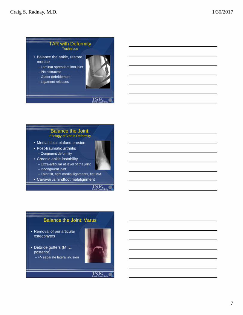
TAR with DeformityTechnique
• Balance the ankle, restore mortise– Laminar spreaders into joint
– Pin distractor
– Gutter debridement
– Ligament releases
Balance the Joint:Etiology of Varus Deformity
• Medial tibial plafond erosion
• Post-traumatic arthritis– Congruent deformity
• Chronic ankle instability– Extra-articular at level of the joint
– Incongruent joint
– Talar tilt, tight medial ligaments, flat MM
• Cavovarus hindfoot malalignment
Balance the Joint: Varus
• Removal of periarticularosteophytes
• Debride gutters (M, L, posterior)– +/- separate lateral incision

Craig S. Radnay, M.D. 1/30/2017
8
Balancing the Joint:Congruent Varus
• Parallel joint lines
• Bony>soft tissue pathology
• Stiff subtalar joints
• Address deformity before cuts are made
• Often just adjust TAR cut– Slightly higher tibial cut
Coetzee , Foot Ankle Int 2010
Balancing the Joint:Incongruent Varus
• Greater than 10° difference in tibial and talar joint lines
• Obvious soft tissue imbalance
• Cannot be corrected by bony cuts alone
• Deltoid ligament release (deep +/- sup)
Easley, Advanced Reconstruction Foot and Ankle 2, 2015
Balance the Joint: Varus
• Laminar spreader into medial joint
• Posterior capsule release
• Medial malleolar osteotomy– When deltoid lengthening is not
enough
– With severe intra-articular varusdeformity

Craig S. Radnay, M.D. 1/30/2017
9
Varus CorrectionMedial Malleolar Osteotomy
• Doets et al, FAI 2008
• Medial gutter debridement
• Osteotomy is done half way down the malleolus
• Medial to shoulder
• Hindfoot deformity correction
DeOrio, Clin Podr Med Surg, 2013.
60 yo male, history of instability
DeOrio, Clin Podr Med Surg, 2013
6 wks
Varus CorrectionLateral Malleolar Shortening Osteotomy,
Medial Malleolar Lowering Ostoetomy
Trincat et al, Orth & Traum, 2012

Craig S. Radnay, M.D. 1/30/2017
10
Balance the Joint: Varus
• Talonavicular capsule release
• Posterior tibial tendon recession at m-t
• Flexor retinaculum release
TAR, deltoid release, PTT/PLPB, cavovarus correction, ST/TN fusion
Schuberth et al, J Ft Ankle Surg 2016
Beware Persistent Instability, Malalignment60 yo male, history of instability
DeOrio, Clin Podr Med Surg, 2013
24° varus 6 wks 6 mos

Craig S. Radnay, M.D. 1/30/2017
11
Balancing the Joint: Varus
Balance the Joint: Etiology of Valgus Deformity
• Severe pes planovalgus
– Deltoid insufficiency
– PTT insufficiency
– Correct the foot to prevent implant valgus tilt
• Osseous deformities
– Fibular fracture malunion (short, ER)
– Posterior distal tibia, plafond fracture
• Identify medial ligament incompetence pre-op
Balance the Joint: Mild Valgus (<10°)
• Removal of periarticular osteophytes
• Debride gutters (M, L, posterior)– Modify bone cuts
– Fibula lengthening/rotational osteotomy
– Medializing calcaneus osteotomy or LCL
– Possible corrective subtalar/triple if rigid
– Reconstruction/repair of medial ligaments
– Reconstruction of lateral ligaments

Craig S. Radnay, M.D. 1/30/2017
12
Balance the Joint: Mild Valgus (<10°)
Balance the Joint: Severe Valgus (>10°), supramalleolar
• Supramalleolar osteotomy
• TAR
• Hindfoot still in valgus?– Fibula lenthening/rotational osteotomy
Medializing calc osteotomy or HF fusion
– Cotton osteotomy
– Reconstruction of medial ligaments
– Reconstruction of lateral ligaments
Gougoulias, Maffuli, Clin Podr Med 2015. Trincat et al, Orth & Traum, 2012
Balance the Joint:Severe valgus (>10°) inframalleolar
• (PTTD)– Reconstruction vs arthrodesis
• PF osteotomy medial cuneiform
• PBPL
– TAR
– Fibula lengthening/rotational osteotomy
– Reconstruction of medial ligaments
– Reconstruction of lateral ligaments

Craig S. Radnay, M.D. 1/30/2017
13
TAR with DeformityTechnique
• Once joint is balanced, can make cuts– +/- place in jig
– Heel cord lengthening
– Deformity correction in jig
– Maintain correction with IM rod
– Precise tibial, talar cuts
– If anything, under resect bone!
– Posterior capsulectomy
TAR with DeformityTechnique
• Implant components– Maximize cortical support, stems?
– Stability
• Test ankle stability with trial components in place– Favor fixed bearing implants
TAR with DeformityTechnique
• Peri-articular bony procedures– Hindfoot
• Ligament reconstruction
• Foot alignment correction

Craig S. Radnay, M.D. 1/30/2017
14
TAR with DeformityTechnique
• Hindfoot/midfoot deformity?– Reconstruction, osteotomy
• If peroneals,eversion weak• PTT/FDL/FHLPB
• AT transfer to middle cuneiform– With severe varus
Easley, Advanced Reconstruction Foot and Ankle 2, 2015
TAR with DeformityTechnique
• Test full ROM with final components, trial poly• Layered closure over Hemovac drain
Balanced TAR with Varus Deformity

Craig S. Radnay, M.D. 1/30/2017
15
JD: Varus
JD: 3 mos
JD: 3 mos

Craig S. Radnay, M.D. 1/30/2017
16
HT:Varus
HT: Varus
Balanced TAR with Valgus Deformity

Craig S. Radnay, M.D. 1/30/2017
17
AC: Valgus
CB: Valgus

Craig S. Radnay, M.D. 1/30/2017
18
CB: Valgus
CB: Valgus
TAR with DeformityPatient-Specific Total Ankle Arthroplasty

Craig S. Radnay, M.D. 1/30/2017
19
TAR with DeformityPatient-Specific Total Ankle Arthroplasty
• Goal is to restore mechanical, kinematic joint axes
• Improved implant alignment– Neutral coronal and sagittal alignments obtained for all
TAR cases regardless of preoperative deformity (Hsu et al, FAI 2015)
• Improved accuracy, reproducibility
• Decreased surgical time
• Decreased flouro time (12 min)
TAR with DeformityPatient-Specific Total Ankle Arthroplasty
Patient-Specific Total Ankle Arthroplasty: Cannot Blindly Rely on Technology
• MRI: avascular necrosis 50% talus, plafond
• Erosion anterior tibial plafond

Craig S. Radnay, M.D. 1/30/2017
20
Patient-Specific Total Ankle Arthroplasty: Cannot Blindly Rely on Technology
Patient-Specific Total Ankle Arthroplasty: Cannot Blindly Rely on Technology
My Preference With Deformity
• With severe foot deformity I prefer to stage
• With moderate HF arthritis with minimal symptoms, I prefer TAR by itself– Provides good pain relief and function
– TAR usually markedly improves foot alignment
• Always try to protect the talar blood supply– Limit ST preparation to the posterior facet avoids
inferior talar neck blood supply
– Also avoid inferior talar head w/TN preparation

Craig S. Radnay, M.D. 1/30/2017
21
Conclusions
• Tibiotalar coronal malalignment is not uncommon in end-stage arthritis and should not be a contraindication to TAR
• But still a challenge to correct
• Balancing is vital to outcomes– Surgeon experience is important
– Different constraints with different prostheses
– More refined techniques of ligament balancing
Conclusions
• Figure out pre-operatively where the deformity is coming from– Above the ankle
– The ankle joint itself
– Below the ankle
• Address deformities systematically either concurrently or staged– Soft-tissue, bony, fusion
• Beware of an incompetent deltoid!
Conclusions
• Stable neutral alignment
• Achieve a plantigrade, balanced foot & ankle
• Restore component parallelism
– Reduce eccentric wear
– Reduce component loosening
– Reduce subsidence
– Reduce reoperation and failure

Craig S. Radnay, M.D. 1/30/2017
22
Thank You For Your Attention