cranial sonographic findings in meningomyelocele
TRANSCRIPT

Caffey Award
D. S. Babcock' B. K. Han'
Received April 28. 1980; accepted after revision July 3, 1980.
Presented at the annual meeting of the Society for Pediatric Radiology, Salt Lake City, UT, April 1980.
1 Division of Roentgenology, Children's Hospital Medical Center, Eiland & Bethesda Ave., Cincinnati, OH 45229. Address reprint requests to D. S, Babcock.
This article appears in November/ December 1980 AJNR and March 1981 AJR.
AJNR 1 :493-499, November/December 1980 0195-6108/ 80/ 0016-0493 $00.00 © American Roentgen Ray Society
Cranial Sonographic Findings in Meningomyelocele
493
Children with meningomyeloceles have problems involving several organ systems and often require repeated radiologic examinations. They have multiple cranial defects, particularly the Arnold-Chiari II malformation and associated hydrocephalus. Cranial sonographic findings in 29 patients included hydrocephalus with pointing of the frontal horns, asymmetry of the lateral ventricles, and relative enlargement of the occipital horns compared with the frontal horns. Partial absence of the septum pellucidum, abnormalities of the third ventricle including a prominent massa intermedia, prominent anterior commissure, and prominent suprapineal recess were also seen. A V-shaped tentorium cerebelli was demonstrated on axial scan as well as low position of the tent on coronal scan. The posterior fossa pseudomass was demonstrated. A prominent interhemispheric fissure was also seen in some patients. Sonography is an excellent method for following ventricular size and shunt function since repeated examinations can readily be performed.
Children with meningomyeloceles are a special group with problems involving several organ systems and repeated radiologic examinations are often required. They have numerous cranial defects, particularly the Arnold-Chiari (type II) malformation [1 - 3], and associated hydrocephalus, wh ich can be evaluated with newer imag ing techniques. Several recent reports have demonstrated the cranial computed tomography (CT) findings in these patients [4 - 7]. Sonography of the head is now a recognized diagnostic imaging method that offers a noninvasive, safe, inexpensive way of studying these meningomyelocele patients without the danger of ionizing radiation, while providing useful neuroanatomic information. We report our experience with B-mode gray scale sonography in the meningomyelocele patient.
Materials and Methods
We retrospectively reviewed 65 sonograms obtained on 29 meningomyelocele patients. The Cincinnati Center for Developmental Disorders of the University of Cincinnati is a referral center for patients with this abnormal ity and infants are referred for multidisciplinary treatment within a few hours after birth. The spinal defect is repaired, if possible, within 24
hr after birth. The initial cranial sonogram is usually obtained within 2-4 days after birth. The sonograms are obtained with a commercially available contact B-mode gray scale ultrasonoscope or a high-resolution mechanical sector, real-time ultrasonoscope and recorded on video tape and x-ray film. The examinations are usually performed in the radiology department in the ultrasound room , which is equipped so that critical life support measures can be maintained. Bedside examinations are occasionally performed on severely ill infants using the real-time equipment; 3.5 and 5.0 MHz medium-focused transducers are used. Sedation is usually unnecessary, although chloral hydrate in a dose of 50 mg / kg is used on older infants. Technically adequate head examinations are performed only in children under the age of about 2 years who have open sutures and fontanelles . Serial scans of the entire head are obtained at 5 mm intervals in axial , coronal, posterior fossa, and sagittal projections. Our technique was described previously [8].

494 BABCOCK AND HAN AJNR: 1 , November/ December 1980
Results
Table 1 lists abnormalities we found associated with meningomyelocele. The findings represent the frequency with which we detected the abnormality, not necessarily its frequency of occurrence in this group of patients.
Lateral Ventricular Size
Although the spinal defect had been repaired, most patients were not shunted at the time of the initial examination and the size of the lateral ventricles varied considerably. In nine patients there was no or mild ventricular dilatation (fig. 1). There was moderate to marked dilatation in 15 patients and severe dilatation in five patients (fig. 2). In many patients, the ventricular size was shown to gradually increase on serial examinations until the patient was shunted. Examinations were not routinely performed after shunting; however, in several patients the ventricles decreased markedly in size after shunting. The lateral ventricles were mild to moderately asymmetrical in 17 of the 29 patients (fig. 2).
TABLE 1: Summary of Findings in Meningomyelocele
Hydrocephalus: Not present Mild Moderate to marked .
Finding
Marked to severe . . . . . . . . . . . . . . . . . . . ..... Anterior pointing of the frontal horns on axial scans .. Inferior pointing of the frontal horns on coronal scans . Occipital horns more dilated than frontal horns . Asymmetry of the lateral ventricles . Cava sept i pellucidi present Partial absence of the septum pellucidum Dilated third ventricle Prominent massa intermedia ..... Prominent anterior commissu re of the third ~~~t~i~ie' .. Prominent suprapineal recess Low position of the tent on coronal view . V-shaped tent on axial view Tentorial-cerebellar pseudomass Prominent interhemispheric fissure . . . . . . . .. . Herniation of third ventricle into parasella cistern
LV
A
No. (n - 29)
1 8
15 5
23 25 19 17
4 16 21
5 2 3
23 8 9
12 1
The occipital horns and atria were dilated more than the frontal and temporal horns in 19 of the patients (figs. 3 and 7). This has been noted in congenital hydrocephalus from other etiologies as well. The cerebral mantle was thinnest over the occipital horns (fig. 3).
Configuration of the Lateral Ventricles
On axial view, a characteristic anterior pOinting and medial concavity of the frontal horns has been described on CT examination [4]. This was seen in 23 of our patients (fig. 4). Inferior pointing of the frontal horns resulting from prominence of the caudate nuclei, which bulge medially into the lateral walls of the lateral ventricles has been described on ventriculography [2] and was seen in 25 of our patients (fig. 5).
Evalua...tion of the Septum Pellucidum
The septum pellucidum was completely or partially absent in 16 of the patients (figs. 4, 7, and 11). A cavum septi pellucidi, or visible separation between the leaves of the septum pellucidum, was identified in four of the patients (fig . 6). This was the same incidence as seen in other patients with hydrocephalus and may not be related to the ArnoldChiari " malformation.
Third Ventricle Abnormalities
The third ventricle was dilated in all but seven of the patients with hydrocephalus. It was noted to be only mildly dilated in most patients, although it may become large in some cases (fig. 7). The massa intermedia has been described as being unusually enlarged in 82%-90% of ArnoldChiari " brains [1]. The massa intermedia was not demonstrated in many of our patients, however, a prominent massa intermedia was identified in five (figs. 7 and 8) and appeared to lie unusually close to the foramen of Monro.
A prominent anterior commissure has been described by ventriculography and was identified in two patients (fig . 8A). Herniation of the third ventricle into the parasellar cistern (fig. 8B) was seen in one patient and an enlarged suprapi-
Fig. 1 .-Mild hydrocephalus. Axial (A) and coronal (6) scans. Mild dilatation of lateral ventricles (LV). FC = falx cerebri, ChP = choroid plexus.

AJNR:1 , November/ December 1980 SONOGRAPHY OF MENINGOMYELOCELE 495
2 3 Fig. 2.-Asymmetric, marked hydrocephalus. Low axial scan . Asymmetric
dilatation of lateral ventricles. LV,f = frontal horn, LV,o = occipital horn . Fig. 3 .-Lateral ventricles. Axial scan. More dilatation of occipital horns
(i...V,o) than frontal horns (LV,I) and thinnest cerebral mantle over occipital
Fig. 5.-Lateral ventricles. Coronal scan through frontal horns of lateral ventricles. Inferior pointing of frontal horns (arrows) .
Fig. 5.-Cavum septi pellucidi. Coronal scan. Cavum septi pellucidi (CSP) between dilated frontal horns of lateral ventricles (LV,I).
Fig. 7.-Absent septum pellucidum. Dilated third ventricle with prominent massa intermedia. Axial (A) and coronal (B) scans. Absent septum pellucidum and moderate dilatation of lateral and third ventricles (V3). Occipital horns (Lv,o) more dilated than frontal horns (LV,I) . MI = massa intermedia.
5
MI
1cm -A
4 horns (arrows).
Fig. 4.-Lateral ventricles. Axial scan. Characteristic anterior pointing and medial concavity of frontal horns (arrow).
6
B

496 BABCOCK AND HAN AJNR: 1 , November / December 1980
A
w 9
ChP
B
10
Fig. 9.-Low position of tentorium cerebelli. Coronal scan. Relatively low position of tentorium cerebelli (arrows) and small posterior fossa. Dilated lateral ventricles (LV) with choroid plexuses (ChP).
Fig. 10.- Tentorial-cerebellar pseudomass. Low axial scan. Heart-shaped tentorial-cerebellar pseudomass (open arrow) and V-shaped tentorium cere-
neal recess was noted in three patients (fig. 8A and 8B). Most of these abnormalities of the third ventricle were identified only on sagittal views, and this view was not available on all of the patients. Beaking of the tectum and herniation of the third ventricle into the quadrigeminal cistern has been described but was not demonstrated on any of our patients .
Tentorial-Cerebellar Pseudomass
The posterior fossa abnormalities in the Arnold-Chiari II malformation include downward displacement of the cerebellum, medulla, and fourth ventricle , with a relatively small posterior fossa. In 23 of our patients, a relatively low position of the tentorium cerebelli was seen on coronal scans, with a small posterior fossa (fig . 9). The fourth ventricle was not identified in any of our patients, probably because of its low position in the posterior fossa and because it is not dilated in this malformation.
The tentorial-cerebellar pseudomass recently described on CT examinations [4, 6] was seen in nine of our patients (fig . 10). On axial scans the leaves of the tentorium come
Fig. 8 .- Third ventric le abnormalities. Sagittal scans on two different patients. Enlarged massa intermedia (black arrows), prominent anterior commissure (curved arrow) , herniation of third ventricle into the parasellar cistern (straight white
arrow), and enlarged suprapineal recess (open arrows).
LV
11
belli posteriorly (solid arrow) . CP = cerebral peduncles. Fig. 11.- Prominent interhemispheric fissure . Coronal scan. Prominent
interhemispheric fissure (arrow) and moderate di latation of lateral ventricles (LV) with absence of septum pelluc idum.
together to form a sharp angle posteriorly, producing a Vshaped configuration, rather than the normal U shape. The tentorium is hypoplastic, with a wide incisura through which the cerebellum bulges upward, producing the heart-shaped tentorial-cerebellar pseudomass.
Interhemispheric Fissure
The interhemispheric fissure was identified as being prominent in 12 of the patients and was associated with hydrocephalus (fig . 11). It has been postulated that communicating hydrocephalus with distal block at the foramen magnum may result in simultaneous dilatation of both the ventricles and the interhemispheric fissure [7]. It was noted in several cases that the interhemispheric fissure enlarged when the ventricles diminished after ventricular shunting.
Ventricular Shunt Function
Occasional patients returned for study after ventricular shunting with symptoms of shunt malfunction . Figure 12 [8]
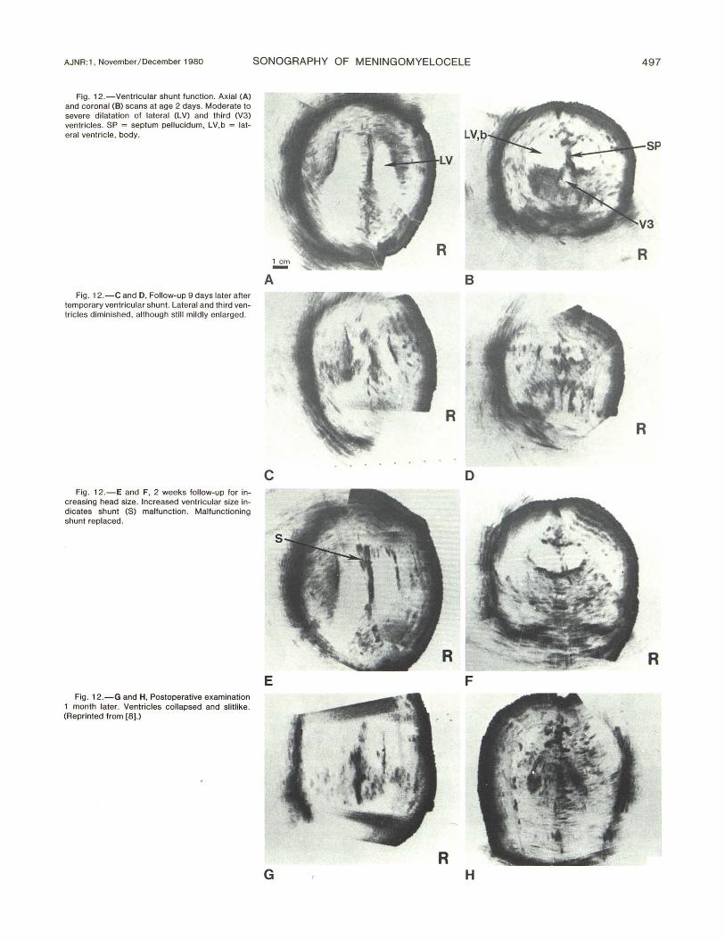
AJNR:1, November/ December 1980
Fig. 12.-Ventricular shunt function. Axial (A) and coronal (B) scans at age 2 days. Moderate to severe dilatation of lateral (LV) and third (V3) ventric les. SP = septum pellucidum, LV,b = lateral ventric le, body.
Fig . 12.-C and D, Follow-up 9 days later after temporary ventricular shunt. Lateral and th ird ventricles diminished, although sti ll mildly enlarged .
Fig. 12.-E and F, 2 weeks follow-up for increasing head size. Increased ventricu lar size indicates shunt (S) malfunction. Malfunctioning shunt replaced .
Fig. 12.-G and H, Postoperative examination 1 month later. Ventricles collapsed and slitlike. (Reprinted from [8].)
SONOGRAPHY OF MENINGOMYELOCELE 497
R
A B
R R
c D
R E F
R G H

498 BABCOCK AND HAN AJNR:1, November/December 1980
LV
A
c
E
shows a 37 week gestation infant with mild head enlargement whose first sonographic examination at 2 days of age demonstrated moderate to severe dilatation of the lateral and third ventricles. After a temporary ventricular shunt, a follow-up examination 9 days later demonstrated diminished lateral and third ventricles, although they were still mildly enlarged . The patient returned for follow-up examination 2 weeks later because of increasing head size suggesting shunt malfunction. Increasing ventricular size since the last examination was demonstrated. The malfunctioning shunt was replaced and a final postoperative examination 1 month later showed that the ventricles had collapsed and become
B
D
LV
Fig . 13.-0bstruction of foramen of Monro. Axial (A) and coronal (8) scans before shunting. Moderate dilatation of lateral ventricles (l V). Third ventricle obliterated by enlarged massa intermedia. Axial (C) and coronal (0) scans after unilateral ventricular shunting . Collapse of ipsilateral ventricle and further enlargement of contralateral ventricle. Displaced fal x cerebri (Fe). E, Ventricu logram. Enlarged massa intermedia (arrow) may be obstructing foramen of Monro.
slitlike. When the ventricles collapse. the skull diminishes and the sutures close and even tend to overlap, causing technical problems in obtaining good quality scans. Zimmerman et al. [4] and Naidich et al. [5-7] reported that even after shunting, the ventricles tend to remain moderately enlarged and on serial studies their size seems to correlate with the variations in shunt function rather than the underlying disease process.
Another patient with a ventricular shunt developed progressive enlargement of the contralateral ventricle indicating obstruction of the foramen of Monro (fig . 13). Ventriculography demonstrated only an enlarged massa intermedia.

AJNR:1 , November/ December 1980 SONOGRAPHY OF MENINGOMYELOCELE 499
which may have been obstructing the foramen of Monro sufficiently to require bilateral ventricular shunts.
Discussion
Meningomyelocele patients with Arnold-Chiari II malformation have been reported to show a variety of abnormalities on cranial CT and ventriculographic examinations [2-7]. We found that many of these features can also be demonstrated by cranial sonographic examination. The primary indication for performing cranial sonography in our patients was to determine ventricular size, and this was successfully determined on all examinations. Therefore, cranial CT and ventriculography were unnecessary in most patients and performed only rarely in unusual or complicated situations such as shunt malfunction and unilateral ventricular enlargement (fig. 13) to evaluate the patency of the third ventricle and foramina of Monro.
Sonography has the advantage of being a rapid, safe, and less expensive method for studying these patients with no radiation exposure with repeated examinations. This is particularly important for following ventricular size and shunt function. The need for deep sedation and anesthesia is also avoided. Sonography has one disadvantage in that only patients with open sutures and fontanelles can be evaluated by this method.
Since the advent of cranial sonography at our institution, ventriculography and CT are rarely performed on infants with this malformation .
ACKNOWLEDGMENTS
We thank Marsha Ellington and Theresa Skelley for technical assistance, Marlena Tyre for secretarial assistance, and Thomas Tomsick and J. Scott Dunbar for editorial assistance.
REFERENCES
1. Peach B. Arnold-Chiari malformation. Anatomic features of 20 cases. Arch Neuro/1965; 12 : 613-621
2. Gooding CA, Carter A, Hoare RD. New ventriculographic aspects of the Arnold-Chiari malformation. Radiology 1967;89: 626-632
3. Harwood-Nash DC, Fitz CR. Neuroradiology in infants and children. St. Louis: Mosby, 1976: 1000-1014
4. Zimmerman RD, Breckbill 0, Dennis MW, David DO. Cranial CT findings in patients with meningomyelocele . AJR 1979; 132 : 623-629
5. Naidich TP, Pudlowski RM, Naidich JB, Gornish M, Rodriguez FJ. Computed tomographic signs of the Chiari II malformation. Part I: skull and dural partitions. Radiology 1980;134: 65-71
6. Naidich TP, Pudlowski RM, Naidich JB, Gornish M, Rodriguez FJ. Computed tomographic signs of the Chiari II malformation . Part II: midbrain and cerebellum . Radiology 1980;134:391-398
7. Naidich TP, Pudlowski RM, Naidich JB, Garnish M, Rodriguez FJ . Computed tomographic signs of the Chiari II malformation. III: Ventricles and cisterns. Radiology 1980; 134: 657 -663
8 . Babcock OS, Han BK, LeQuesne GW. B-mode gray scale ultrasound of the head in the newborn and young infant. AJNR 1980;1 : 181-192