craniofacial tissue engineeringperspectivesinmedicine.cshlp.org/content/8/1/a025775.full.pdf · and...
TRANSCRIPT

Craniofacial Tissue Engineering
Weibo Zhang and Pamela Crotty Yelick
Department of Orthodontics, School of Medicine, School of Engineering, Tufts University, Boston,Massachusetts 02111
Correspondence: [email protected]
The craniofacial complex is composed of fundamental components such as bloodvessels andnerves,andalsoavarietyof specialized tissues suchascraniofacialbones,cartilages,muscles,ligaments, and the highly specialized and unique organs, the teeth. Together, these structuresprovide many functions including speech, mastication, and aesthetics of the craniofacialcomplex. Craniofacial defects not only influence the structure and function of the jaws andface, but may also result in deleterious psychosocial issues, emphasizing the need for rapidand effective, precise, and aesthetic reconstruction of craniofacial tissues. In a broad sense,craniofacial tissue reconstructions share many of the same issues as noncraniofacial tissuereconstructions. Therefore, many concepts and therapies for general tissue engineering canand have been used for craniofacial tissue regeneration. Still, repair of craniofacial defectspresents unique challenges, mainly because of their complex and unique 3D geometry.
The most common causes of craniofacial de-fects are congenital birth defects (1/700
live births), trauma, inflammation, and cancersurgeries (Miura et al. 2006). Among these, themost prevalent is acute trauma, including falls,assaults, sports injuries, and vehicle crashes(Rocchi et al. 2007; Grayson et al. 2015; Hunteret al. 2015). Significant facial trauma can alsoresult from battlefield injuries, particularlywhen combined with an increased survivabilityof wounded soldiers because of improved bat-tlefield medical care and body armor. A recentreport indicated that craniomaxillofacial inju-ries can represent up to 26% of all battlefieldinjuries, as occurred in Operation Iraqi Free-dom/Operation Enduring Freedom (Afghani-stan) (Lew et al. 2010). Congenital anomalies(CAs) are major causes of infant mortality andchildhood morbidity, affecting 2%–3% of all
babies (Mossey and Castilla 2003). Geneticbirth defects, environmental exposure, and folicacid deficiency are the main causes of CA.
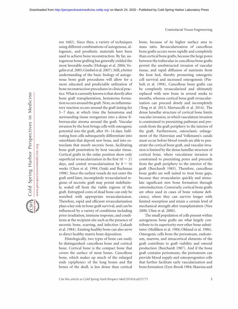
Craniofacial bone reconstruction plays acrucial role in craniofacial repairs, becausethey provide support for adjacent soft tissuesand anchorage for dental structures, therebyguiding the structural stability and appearanceof the face (Fig. 1) (Petrovic et al. 2012). Espe-cially for extensive craniofacial injuries, success-ful regeneration of craniofacial bone is necessaryto restore normal function of the craniofacialcomplex (Genden 2010; Ward et al. 2010).With sufficient bone structure, it is compara-tively easier to restore the soft tissues of the cra-niofacial complex to form the facial features. Anextreme example of this is the world’s first par-tial face transplant from a cadaver to a livinghuman, which was performed on November
Editor: Joseph P. Vacanti
Additional Perspectives on Tissue Engineering and Regenerative Medicine available at www.perspectivesinmedicine.org
Copyright # 2018 Cold Spring Harbor Laboratory Press; all rights reserved; doi: 10.1101/cshperspect.a025775
Cite this article as Cold Spring Harb Perspect Med 2018;8:a025775
1
ww
w.p
ersp
ecti
vesi
nm
edic
ine.
org
on March 24, 2020 - Published by Cold Spring Harbor Laboratory Press http://perspectivesinmedicine.cshlp.org/Downloaded from

27, 2005, and which has been performed glob-ally since then (Petrini 2015). In addition, inMarch 2010, a team of 30 Spanish doctors per-formed the first full-face transplant in the world(Garrett et al. 2015). These accomplishmentsstress the importance of craniofacial bone defectreconstruction as a fundamental first step forsuccessful craniofacial regeneration.
CRANIOFACIAL BONE ENGINEERING
As described above, bone repair is a crucial andfundamental step of craniofacial reconstruc-tion. Living bone is in a continuous state ofdynamic equilibrium consisting of bone resorp-
tion, regeneration, and remodeling. Bone has aninnate ability for a limited amount of self-repairfollowing traumatic injury. In 1982, Enlow hy-pothesized that some areas of the face remodelby bony deposition, whereas others remodel bybony resorption (Enlow 1982).
Bone Reconstruction
Bone reconstruction has a long history. The ear-liest attempt to repair bone defects was reportedin Edgar Smith papyrus, ca. 2000 BC, using met-als (Frommelt 1987). And, in 1668, the first bonegraft was performed by Van Meekren on a patientwith a cranial defect, via a xenograft (Van Meek-
Figure 1. Schematic of the layered structure of craniofacial tissues. The skull and craniofacial bones provide thestructural support for the muscle, vascular network, and skin (from catalog.biodigital.com/storeImages/detail/cranio_dvd.jpg).
W. Zhang and P.C. Yelick
2 Cite this article as Cold Spring Harb Perspect Med 2018;8:a025775
ww
w.p
ersp
ecti
vesi
nm
edic
ine.
org
on March 24, 2020 - Published by Cold Spring Harbor Laboratory Press http://perspectivesinmedicine.cshlp.org/Downloaded from
ren 1682). Since then, a variety of techniquesusing different combinations of autogenous, al-logeneic, and prosthetic materials have beenused to achieve bone reconstruction. By far, au-togenous bone grafting has generally yielded themost favorable results (Hokugo et al. 2004; Vo-gelin et al. 2005; Gimbel et al. 2007). Still, a betterunderstanding of the basic biology of autoge-nous bone graft procedures will allow for amore educated and predictable utilization ofbone reconstruction procedures in clinical prac-tice. What is currently known is that shortly afterbone graft transplantation, hematoma forma-tion occurs around the graft. Next, an inflamma-tory reaction occurs around the graft lasting for5–7 days, at which time the hematoma andsurrounding tissue reorganizes into a dense fi-brovascular stroma around the graft. Vascularinvasion by the host brings cells with osteogenicpotential into the graft, after 10–14 days. Infil-trating host cells subsequently differentiate intoosteoblasts that deposit new bone, and into os-teoclasts that resorb necrotic bone, facilitatingbone graft penetration by host vascular tissue.Cortical grafts in the onlay position show onlysuperficial revascularization in the first 10 � 21days, and central revascularization by 8 � 16weeks (Chen et al. 1994; Ozaki and Buchman1998). Since the earliest vessels do not enter thegraft until later, incompletely revascularized re-gions of necrotic graft may persist indefinite-ly, sealed off from the viable regions of thegraft. Entrapped cores of dead bone can only beresorbed with appropriate revascularizaion.Therefore, rapid and efficient revascularizationplays a key role in bone graft survival, and can beinfluenced by a variety of conditions includingprior irradiation, immune response, and condi-tions at the recipient site such as the presence ofnecrotic bone, scarring, and infection (Lukashet al. 1984). Existing healthy bone can also serveto direct healthy matrix bone deposition.
Histologically, two types of bone can easilybe distinguished: cancellous bone and corticalbone. Cortical bone is the compact bone thatcovers the surface of most bones. Cancellousbone, which makes up much of the enlargedends (epiphyses) of the long bones and flatbones of the skull, is less dense than cortical
bone, because of its higher surface area tomass ratio. Revascularization of cancellousbone grafts occurs more rapidly and completelythan cortical bone grafts, because the large poresbetween the trabeculae in cancellous bone graftspermit the unobstructed invasion of vasculartissue, and rapid diffusion of nutrients fromthe host bed, thereby promoting osteogeniccell survival and increased osteogenesis (Pin-holt et al. 1994). Cancellous bone grafts canbe completely revascularized and ultimatelyreplaced with new bone in several weeks tomonths, whereas cortical bone graft revascular-ization can proceed slowly and incompletely(Teng et al. 2013; Martuscelli et al. 2014). Thedense lamellar structure of cortical bone limitsvascular invasion, in which vasculature invasionis constrained to preexisting pathways and pro-ceeds from the graft periphery to the interior ofthe graft. Furthermore, osteoclastic enlarge-ment of the Haversian and Volkmann’s canalsmust occur before blood vessels are able to pen-etrate the cortical bone graft, and vascular inva-sion is limited by the dense lamellar structure ofcortical bone, where vasculature invasion isconstrained to preexisting pores and proceedsfrom the graft periphery to the interior of thegraft (Burchardt 1983). Therefore, cancellousbone grafts are well suited to treat bone gaps,because they revascularize quickly and stimu-late significant new bone formation throughosteoinduction. Conversely, cortical bone graftsare often used in cases of bone volume defi-ciency, where they can survive longer withlimited resorption and retain a certain level ofmechanical strength after transplantation (Neu2000; Uhm et al. 2000).
The small population of cells present withinautogenous bone grafts are what largely con-tribute to its superiority over other bone substi-tutes (Mulliken et al. 1984; Oklund et al. 1986).Osteogenic cells from the periosteum, endoste-um, marrow, and intracortical elements of thegraft contribute to graft viability and osteoidproduction (Burchardt 1987). And if the bonegraft contains periosteum, the periosteum canprovide blood supply and osteoprogenitor cellsthat further facilitate early vascularization andbone formation (Eyre-Brook 1984; Skawina and
Craniofacial Tissue Engineering
Cite this article as Cold Spring Harb Perspect Med 2018;8:a025775 3
ww
w.p
ersp
ecti
vesi
nm
edic
ine.
org
on March 24, 2020 - Published by Cold Spring Harbor Laboratory Press http://perspectivesinmedicine.cshlp.org/Downloaded from

Gorczyca 1984). Unfortunately, adult perioste-um loses this osteogenic ability after being de-tached from the ordinal bone surface (Melcherand Accursi 1971; Weng et al. 2000). Shortlyafter transplantation, most of the osteocytespresent within the graft undergo necrosis, there-by rendering the graft relatively inert. One of theapproaches used in clinic to increase the sur-vival rate of large bone grafts is to first implantthe grafts into highly vascularized muscle tissue,to facilitate vascular ingrowth and to develop avascular pedicle suitable for microsurgical anas-tomosis (Warnke et al. 2004; Mesimaki et al.2009). This practice is somewhat limited be-cause of donor-site morbidity and the amountof bone that can be harvested (Burchardt 1987).
To overcome these issues, stem-cell-basedbone-tissue engineering has been recognizedas a promising alternative for bone reconstruc-tions. As far back as 1968, Friedenstein’s teamfirst reported that fibroblast-like cells isolatedfrom bone marrow not only had the ability todifferentiate to haematopoietic cells, but alsohad the potential for osteogenic differentiation(Friedenstein et al. 1968; Friedenstein 1976). Atthat time, the cells were named “mechanocytes.”Subsequent studies showed that these cells werechondrogenic and adipogenic (Owen and Frie-denstein 1988). To date, these cells are com-monly called mesenchymal stem cells (MSCs)(Beyer Nardi and da Silva Meirelles 2006). Inaddition to bone marrow, cells with MSC-likecharacteristics have been isolated from a varietyof tissues including adult adipose, dental, andskeletal muscle tissues (Zuk et al. 2001; Qu-Pe-tersen et al. 2002; Feisst et al. 2014). MSC-me-diated bone regeneration has been widely testedin several clinical trials, demonstrating that localdelivery of MSCs can enhance bone regenera-tion (Grayson et al. 2015).
Comparison of Craniofacialand Noncraniofacial Bone
Generally speaking, craniofacial bones are flatbones that share similar turnover and injury re-pair mechanisms of other skeletal bones, butwith their own specific properties (Leuchtet al. 2008). Whereas most of the knowledge
gained from skeletal bone reconstruction canbe applied to the craniofacial bone field, theunique features and properties of craniofacialbone regeneration require some additional con-siderations.
One of the major differences between cra-niofacial and noncraniofacial bones is their em-bryonic origin. The cranial neural crest gives riseto branchial arch–derived craniofacial bonesand cartilages, whereas the axial skeleton is de-rived from the somites, and the lateral plate me-soderm forms the limb skeleton. Bone forma-tion, or osteogenesis, is the transformation ofpreexisting mesenchymal tissue to calcifiedbone tissue. There are two major modes ofbone formation: intramembranous and endo-chondral ossification. Intramembranous ossifi-cation is the direct conversion of mesenchymaltissue into bone, whereas endochondral ossifi-cation consists of mesenchymal cells that firstdifferentiate to form a cartilage template that islater replaced by bone. Bones of the skull pri-marily form through intramembranous boneformation, with some contributions by endo-chondral ossification (Bilezikian et al. 2002).For example, Meckel’s cartilage forms at theproximal end of the mandible then largely dis-appears as the mandible ossifies, thereby playingan important role in mandibular morphogene-sis (Ramaesh and Bard 2003; Tsutsui et al. 2008).During intramembranous ossification of theskull, neural crest–derived mesenchymal cellsproliferate into compact condensations. Someof these cells develop into capillaries, whereasothers change their shape to become osteoblasts,which secrete a collagen–proteoglycan matrixthat is able to bind calcium salts. As calcificationproceeds, bony spicules radiate out from regionswhere ossification began. Eventually, the entireregion of calcified spicules becomes surroundedby compact mesenchymal cells that form theperiosteum. The cells on the inner surface ofthe periosteum also become osteoblasts and de-posit osteoid matrix parallel to that of the exist-ing spicules. In this manner, many layers of boneare formed.
Craniofacial and noncraniofacial bones alsoshow different homeostatic mechanisms. Sever-al publications showed that bone grafts from the
W. Zhang and P.C. Yelick
4 Cite this article as Cold Spring Harb Perspect Med 2018;8:a025775
ww
w.p
ersp
ecti
vesi
nm
edic
ine.
org
on March 24, 2020 - Published by Cold Spring Harbor Laboratory Press http://perspectivesinmedicine.cshlp.org/Downloaded from

craniofacial skeleton had better survival andlonger volumetric maintenance as comparedto bone harvested from the iliac crest, rib, andtibia (Peer 1951; Sullivan and Szwajkun 1991).In addition, reports have indicated that mem-branous bone grafts retained their volume bet-ter than endochondral bone grafts (Zins andWhitaker 1979, 1983). In 1999, Buchman andOzaki’s results suggested that bone volumemaintenance may be the result of microarchi-tecture of the bone graft, based on the fact thatintramembranous-derived craniofacial bone hasa higher proportion of cortical bone as com-pared to endochondral bone (Buchman andOzaki 1999). Evidence for this includes thefact that, under external stimuli such as ovari-ectomy and malnutrition, rat mandibular tra-becular bone and bone mineral density lossoccurs at a lower rate than in tibial primaryspongiosa (Mavropoulos et al. 2007). Further-more, the fact that certain bone diseases onlyaffect the maxilla and mandible, such as bis-phosphonate-related osteonecrosis of the jaw(BRONJ) (Price et al. 2004) and hyperparathy-roid jaw tumor syndrome (Simonds et al. 2002),also suggest that different homeostatic mecha-nisms exist between craniofacial and noncra-niofacial bones.
It remains ambiguous as to what causesthese differences between craniofacial and skel-etal bones, because osteoblast differentiationin both types of bone is regulated by the samekey factors, including the transcription factorsRunx2 and osterix (Ducy et al. 1997; Otto et al.1997; Nakashima et al. 2002). Still, it has beenshown that several growth factors, receptors,and associated signaling cascades play distinctroles in the craniofacial versus axial and appen-dicular skeleton (Abzhanov et al. 2007; De Cos-ter et al. 2007; Kimmel et al. 2007).
CRANIOFACIAL BONE REGENERATION
Taken together, craniofacial and long bonesare not only derived from different germ layers,but also show distinct characteristics. These re-sults emphasize the need to take these differenc-es into account when considering craniofacialbone–regeneration strategies.
Craniofacial Bone Graft
Similar to other bone reconstructions, autog-enous bone grafts are considered to be thegold standard for reconstructing craniofacialbone defects (Gruss et al. 1985; Manson et al.1985). The iliac crest and rib bones, all derivedfrom the mesoderm, are among the more com-monly used donor sites for bone grafting incraniofacial surgeries because they can be read-ily harvested while minimally impacting thehost. Other than the common shortcomingsof bone grafts, such as limited harvest amountand donor site morbidity, the major concernwith using these bones is that they do notshow the characteristics of natural jawbone.
As previously discussed, lack of vasculariza-tion can lead to graft resorption with resultantloss of the geometric structure of the bone graft(Peer 1951). Periosteum preservation can im-prove graft survival in the craniofacial region(Thompson and Casson 1970) by facilitatingearly revascularization (Knize 1974). Condi-tions at the implant site also influence graft re-vascularization (Zins et al. 1984). Althoughmuscle coverage results in increased bone graftrevascularization (Ermis and Poole 1992), facialmuscles generally originate from the surface ofthe skull and craniofacial bones, and craniofa-cial injuries are commonly associated with mus-cle damage, which negatively influences the in-growth of blood vessels. Previously reports haveshown that for mandibular defects small bonydefects (,4 to 6 cm) can successfully be treatedwith nonvascularized cortical-cancellous bonegrafts, whereas larger continuity defects requirevascularized grafts (Hidalgo 1989; Pogrel et al.1997).
Another issue is that most craniofacial bonesshow extremely complex 3D shapes as com-pared to long bones. It is, therefore, exceedinglydifficult to select and reshape vascularized au-tologous iliac crest, fibula, or ribs to precisely fitcraniofacial bone defects.
Previous publications have shown that rigidfixation can improve bone graft volume reten-tion, by facilitating primary bone healing andrapid revascularization, especially in bones re-quired for motion such as the femur (Phillips
Craniofacial Tissue Engineering
Cite this article as Cold Spring Harb Perspect Med 2018;8:a025775 5
ww
w.p
ersp
ecti
vesi
nm
edic
ine.
org
on March 24, 2020 - Published by Cold Spring Harbor Laboratory Press http://perspectivesinmedicine.cshlp.org/Downloaded from

and Rahn 1988; Lin et al. 1990). In contrast, mostcraniofacial bones show little motion, with theexception of the mandible. Even for the mandi-ble, liquid food can be provided to restrain man-dibular movement during the bone-healingprocess. While it may seem that rigid fixationwould not significantly improve fixed craniofa-cial bone graft survival, a clinical study of 363patients undergoing nasal reconstruction overa 14-year period showed that exceptional bonegraft survival occurred when rigid interosseousstabilization was used (Jackson et al. 1998).
Craniofacial Bone Substitute
Stem Cells for Craniofacial Reconstruction
The main obstacles of using autologous bonefor grafting is that it is only available in limitedamounts (Neovius and Engstrand 2010). Toovercome this critical shortcoming, and to cre-ate bone grafts of sufficient complex geometry,the field of bone-tissue engineering has beencreated as a practical approach to treat cranio-facial skeletal defects by combining bioactivecarriers, cells, and growth factors. Stem-cell-mediated bone repair has been used in clinicaltrials to regenerate large craniomaxillofacial de-fects to slow the process of bone degeneration inpatients with osteonecrosis of the femoral headand for prophylactic treatment of distal tibialfractures (Grayson et al. 2015). To date, all ofthe MSC sources that have displayed promisingosteogenic potential for bone regeneration alsohave been proposed as potential cell sources forcraniofacial bone–tissue engineering (Cowanet al. 2004; Griffin et al. 2014).
The most well-characterized and used stemcells for bone regeneration are adult bone mar-row stem cells (BMSCs). BMSCs are also thefirst type of cell tested for craniofacial bone re-generation. In 2001, augmented repair of cranialdefects was observed by combining autologoussheep BMSCs with calcium alginate gel (Shanget al. 2001). Since then, MSCs from a variety ofspecies, including mouse, rat, rabbit, canine,and porcine, have been confirmed as suitablecells for craniofacial bone repair (Abukawaet al. 2009; Zou et al. 2011; Lin et al. 2012).
Successful craniofacial ossifications have beenachieved using autologous BMSC-seeded bio-scaffolds in several clinical studies (Kaigler et al.2010; Behnia et al. 2012).
Adipose-derived mesenchymal cells (AMCs)are another type of commonly used cells forbone regeneration (Gimble et al. 2006; Sandorand Suuronen 2008). Recently, AMCs haveemerged as a potential cell source for craniofa-cial tissue engineering (Zuk et al. 2001, 2002).Compared to BMCs, AMCs are more accessibleand can be harvested in larger amounts. It hasbeen shown that AMCs and BMCs isolated fromthe same donor showed similar growth kineticsand cell senescence (De Ugarte et al. 2003). In2004, Longaker’s group first showed that theAMCs promoted bone regeneration of critical-size calvarial defects, when seeded onto hy-droxyapatite-coated poly-lactic-co-glycolic acidscaffolds (Cowan et al. 2004). Many subsequentpublications have shown that AMCs are a prom-ising cell source for craniofacial bone regenera-tion when combined with a variety of scaffoldmaterials (Gomes et al. 2012; Azevedo-Netoet al. 2013; Jin et al. 2014).
Certain types of perinatal stem cells, includ-ing umbilical cord–derived mesenchymal stemcells (Chen et al. 2013), amniotic epithelial cells(Barboni et al. 2013), and amniotic fluid mes-enchymal cells (Berardinelli et al. 2013), havealso shown osteogenic differentiation capacityand, therefore, the potential for craniofacialbone regeneration. One of the main advantagesof these cells for promoting craniofacial boneregeneration is their ability to promote bloodvessel formation (Maraldi et al. 2013).
As described above, most craniofacial tis-sues are derived from the neural crest. Paraxialmesoderm–derived iliac crest bone and osteo-blasts show distinctly different properties thanneural crest cell (NCC)-derived craniofacialbones and osteoblasts (Chai and Maxson2006; Oppenheimer et al. 2008; Aghaloo et al.2010), making them less desirable for craniofa-cial reparative bone applications. Therefore, avariety of cell populations harvested from thecraniofacial complex have been examined forutility in craniofacial bone regeneration (Zhaoand Chai 2015).
W. Zhang and P.C. Yelick
6 Cite this article as Cold Spring Harb Perspect Med 2018;8:a025775
ww
w.p
ersp
ecti
vesi
nm
edic
ine.
org
on March 24, 2020 - Published by Cold Spring Harbor Laboratory Press http://perspectivesinmedicine.cshlp.org/Downloaded from

These studies showed that, similar to longbones, the periosteum of craniofacial bonescontains progenitor cells that support craniofa-cial bone repair (Ochareon and Herring 2011).Craniofacial bone marrow cells have also beenwell characterized. A recent study using labeledNCC and mesoderm-derived bone marrow cellsshowed bone defect healing via the selective re-cruitment of cells from their respective tissuesembryonic origin (Leucht et al. 2008), therebyreinforcing the presence of embryonic site-spe-cific differences in craniofacial versus appendic-ular skeleton–derived BMSCs. A rat studyshowed that in vitro cultured mandible–de-rived BMSCs showed higher alkaline phospha-tase activity, mineralization, and osteoblast geneexpression, and formed 70% larger bone nod-ules containing threefold more mineralizedbone, as compared to long-bone BMSCs (Agha-loo et al. 2010). It has also been shown thathuman stromal cells isolated from mandibularor maxillary marrow showed increased cell pro-liferation, stronger expression of osteoblasticmarkers, and delayed senescence as comparedto iliac crest–derived marrow cells harvestedfrom the same patient (Akintoye et al. 2006).But the relatively small size and anatomic com-plexity of the maxilla and mandible render theefficient isolation of bone marrow stem cells adifficult process (Yang et al. 2014).
Recently, a new type of BMSC, isolated fromcalvarial bone sutures, has shown utility forcraniofacial bone regeneration (Zhao et al.2015). In 2015, Chai’s group identified Gli1þ
cells present within the calvarial suture mesen-chyme of adult mice that can give rise to both anosteogenic front, periosteum, dura, and cranio-facial bones. In humans, cranial sutures normal-ly fuse between 20 and 30 years of age, whereasfacial sutures fuse after �50 years of age (Se-narath-Yapa et al. 2012; Badve et al. 2013), sug-gesting the possibility of isolating these cells foruse in craniofacial bone regeneration. Still, thelimited amount of suture mesenchyme preventsharvest of large numbers of cells.
All of the tissues found in teeth originatefrom the neural crest. Stem cells isolated fromadult dental tissues, including dental pulp stemcells (DPSCs) (Gronthos et al. 2000; Papaccio
et al. 2006; Zhang et al. 2008), stem cells fromhuman exfoliated deciduous teeth (SHED)(Miura et al. 2003), periodontal ligament stemcells (PDLSCs) (Seo et al. 2004), and periapicalstem cells (PSCs) (Han et al. 2010), all showedpotential for osteo/odontogenic differentiationand the ability to form calcified dentin or os-teodentin-like tissue. Therefore, these cell pop-ulations can be used not only to regeneratethese same dental tissues, but also for craniofa-cial and noncraniofacial bone–tissue regene-ration. In particular, DPSCs, which can be easilyisolated from extracted wisdom teeth, showeda higher proliferation rate as compared tobone marrow–derived MSCs under the sameculture conditions, potentially attributable tothe strong expression of the cell-cycle activator,cyclin-dependent kinase 6 (Shi et al. 2001).
Widespread use of dental tissue–derivedstem cells is somewhat limited by the size ofthe available tissues that can be used. Compar-atively, it may be easier to harvest gingiva-de-rived mesenchymal stem cells (GMSCs), whichalso share the multilineage differentiation po-tential (Zhang et al. 2009). It has been shownthat human GMSCs can support new bone for-mation in both mandibular and calvarial de-fects at 2 months in rat postsurgical reconstruc-tions (Wang et al. 2011).
Cleft lip and palate (CL/P) are among themost common congenital malformations, oc-curring in 1/700 live births (Tolarova and Cer-venka 1998). Standard surgical treatment ofCL/P patients involves the removal of smallpieces of orbicular oris muscle (Shkoukaniet al. 2013). Bone reconstructive repair of a crit-ical-size cranial defect was successfully achievedusing muscle cells derived from these discardedtissues (Bueno et al. 2009).
Stem cells and/or scaffolds loaded withactive growth factors, which stimulate osteopro-genitor cells to differentiate toward osteogenicsupport of the new bone formation, are also com-monly used for bone-tissue engineering (Fried-laender et al. 2001; Terheyden et al. 2004). One ofthe most commonly used growth factors for cra-niofacial bone regeneration is bone morphogen-ic protein (BMP) (Terheyden et al. 2001a,b;Warnke and Coren 2003; Warnke et al. 2004).
Craniofacial Tissue Engineering
Cite this article as Cold Spring Harb Perspect Med 2018;8:a025775 7
ww
w.p
ersp
ecti
vesi
nm
edic
ine.
org
on March 24, 2020 - Published by Cold Spring Harbor Laboratory Press http://perspectivesinmedicine.cshlp.org/Downloaded from
Challenges for the Use of Stem Cellsin Craniofacial Tissue–EngineeringApplications
One of the main obstacles in translating exper-imental observations into clinical practice is therelatively poor mechanistic understanding ofstem-cell-mediated therapies. In vivo stem-celltherapy requires large numbers of cells; insuffi-cient cell numbers will not provide positive out-comes (Habisch et al. 2007). Despite the obvi-ous benefits associated with cell delivery forbone regeneration, very few transplanted cellssurvive long term following transplantation(Dupont et al. 2010). The current lack of reliablemethods to control MSC fate, especially in thein vivo condition, is one of the main problemsthat needs to be addressed. Better understand-ing of methods to optimize regenerative ap-proaches is required before stem-cell-mediatedtherapies can be implemented as standard carein regenerative medicine.
MSC isolation and selection is another chal-lenge. For most clinical applications, only a lim-ited amount of viable tissue is available forstem-cell isolation (Sodek and McKee 2000).In addition, MSC selection and enrichment ishampered by lack of a single definitive stem-cellmarker (Gronthos et al. 2003). Current isolationmethods can achieve heterogeneous cell popu-lations at best, and the proportions of MSC pro-genitors in these enriched MSC populations canvary even when the samples are obtained fromthe same donor at the same time (Digirolamoet al. 1999). Previous reports have indicated thatonly �30% of the initial adherent bone mar-row–derived MSC clones showed trilineage (os-teo/adipo/chondro) differentiation potential(Pittenger et al. 1999; Muraglia et al. 2000).Furthermore, only half of the single colony–derived clones transplanted with hydroxyapa-tite-tricalcium phosphate ceramic scaffoldsshowed bone formation in vivo (Kuznetsov etal. 1997). Although current clinical trials haveshown that it is not necessary to generate puri-fied cell populations to achieve positive resultsin the clinic (Prockop 2007), it is possible thatmore purified MSC populations will help toachieve more reliable clinical outcomes.
There also is a need for optimized methodsto store and expand enriched MSC populationswhile maintaining the “stemness” of the MSCs,as required for advanced clinical application.This is because MSCs tend to spontaneouslydifferentiate into various cell types over timein culture (Banfi et al. 2000; Izadpanah et al.2008) and MSCs lose their multipotentialityafter six or seven passages in in vitro cell culture(Colter et al. 2000; Sekiya et al. 2002). One of themost promising solutions is to create 3D cultureconditions that resemble the in vivo stem-cellniche (Bara et al. 2015; Xiong et al. 2015). Todate, no standardized methods have beenachieved for this approach, despite many di-verse attempts (Chow et al. 2001a,b). For exam-ple, a variety of tissue culture media have beentested, including those with low serum, varioussubstitutes for fetal bovine serum (FBS) such asautologous serum, fresh frozen plasma, and hu-man platelet lysates (Lange et al. 2007; Le Blancet al. 2007), and a variety of serum-free media(Sekiya et al. 2002; Muller et al. 2006; Agata et al.2009; Lindroos et al. 2009). Avariety of 3D scaf-fold tissue culture methods have also been test-ed as in vitro 3D stem-cell niche systems, in-cluding the 3D hanging drop model (Schmalet al. 2013), bioreactor (Santos et al. 2016),and 3D printing (Alessandri et al. 2016).
CRANIOFACIAL RECONSTRUCTION
Craniofacial reconstruction involves the regen-eration of multiple hard and soft tissues that arewell vascularized and innervated. For betteroutcomes, it is ideal to regenerate the craniofa-cial tissues “all-in-one,” including both soft andhard tissues. A concept of the “biological boun-dary” was introduced in 1989 (Whitaker 1989),which emphasized that the soft tissue envelopehas a genetically predetermined shape that isinclined to remain constant. However, soft con-nective tissues show faster growth rates as com-pared to hard tissues, because of the fact thatsoft tissue cellular components show higherrates of migration. Therefore, one of the mainchallenges for successful bone healing is pre-venting the ingrowth of connective tissue intothe bone space, especially for large and complex
W. Zhang and P.C. Yelick
8 Cite this article as Cold Spring Harb Perspect Med 2018;8:a025775
ww
w.p
ersp
ecti
vesi
nm
edic
ine.
org
on March 24, 2020 - Published by Cold Spring Harbor Laboratory Press http://perspectivesinmedicine.cshlp.org/Downloaded from

craniofacial bone repair. To achieve this, theconcept of guided tissue regeneration, whichcan be achieved by placing an inert membranebarrier over the defect to block the ingrowth ofconnective tissue, has been widely used for suc-cessful craniofacial reconstruction (Dahlin et al.1988, 1990).
Computer-Aided Design (CAD)
One central challenge for successful craniofacialreconstruction is how to mimic the complex 3Dstructure and multicellular interactions thatnaturally occur in complex craniofacial struc-tures (Thesleff et al. 2011). Resorption of bonegrafts or regenerated bones often occurs afterbone reconstruction. Minor bone resorptionmay not significantly influence the successfulrepair of long bones, but can seriously affectcraniofacial reconstruction outcomes, again be-cause of the complex geometry and small sizesof many craniofacial bones. Craniofacial recon-struction requires more accurate regenerativeapproaches to achieve desirable outcomes.
Currently, with the development of 3Dcomputer tomography (CT) and CAD tech-niques, CT/CAD-based scaffold design cangreatly benefit the accurate regeneration of cra-niofacial bones. One of the most commonlyused strategies for accurate craniofacial recon-struction is the use of titanium scaffolds manu-factured by the CT/CAD technique, based onthe fact that nonabsorbable titanium scaffoldscan achieve appropriately detailed complex cra-niofacial anatomical geometry. Terheyden’sgroup has used a titanium mesh cage filledwith bone mineral blocks infiltrated with 7 mgrecombinant hBMP7 and 20 ml of the patient’sbone marrow to successfully restore mandibulardefects greater than 7 cm (Warnke et al. 2004).The titanium mesh cage was first implanted intothe patient’s right latissimus dorsi muscle for 7weeks before being transplanted as a free bonemuscle flap. Eventually, the reconstructed man-dible achieved the desired aesthetic and masti-cative functional levels. In another case, a tita-nium mesh was prefabricated and filled with b
tricalcium phosphate (b-TCP) granules thatwere soaked in rhBMP-2 for 48 h before infil-
trating with cultured adipose-derived stem cells(ASCs). In this instance, sufficient new bonedeveloped to support dental implants in 10months (Sandor et al. 2013). A combinationof milled bone from the iliac crest, ASCs isolatedfrom fat tissue harvested from the gluteal region,and autologous fibrin glue have also been usedto treat multiple fractures (Lendeckel et al.2004). Bone regeneration was observed within3 months of surgery in these cases. Functionaland anatomically correct repair of a maxillarydefect was achieved using a microvascular cus-tom-made ectopic bone flap developed from atitanium cage filled with b-TCP granules mixedautologous ASCs (Thesleff et al. 2011). To date,titanium cages filled with granules, cancellousbone chips, or bone blocks have been shown byfar to be the best strategy for bone restoration oflarge craniofacial defects.
While conventional regenerative strategieshave failed to regenerate or mimic the 3D com-plexity of the multicellular interactions occur-ring in native craniofacial tissues, 3D cell culturein bioreactors combined with gene therapy orgrowth factors have shown promise for in-creased survival of bone substitutes (Scherbe-rich et al. 2007; Cordonnier et al. 2010; Sohieret al. 2010).
3D Printing
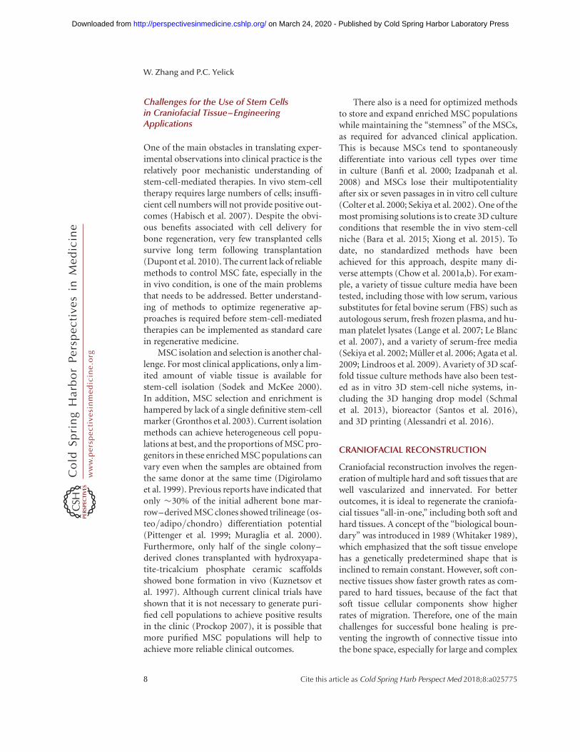
3D bioprinting of tissue provides a promisingapproach to customize various combinations ofbiomaterials and cell sources to achieve complex3D architectures. 3D printing is a method thatfabricates objects by building consecutive layersof scaffold material plus cells layer by layer, untilthe desired 3D volumetric structures areachieved (Derby 2012). In this way, 3D printingtechniques can provide precise spatial control ofthe functional components. For the purpose oftissue regeneration, it is possible to print cellsdirectly, to print cell-laden biomaterials, or toprint scaffold-free cell aggregates. Currently,there are three types of commercially available3D printers for tissue engineering: inkjet print-ers (Klebe 1988), laser-based printers (Guille-mot et al. 2010), and microextrusion printers(Fig. 2) (Cohen et al. 2006). Inkjet printers, the
Craniofacial Tissue Engineering
Cite this article as Cold Spring Harb Perspect Med 2018;8:a025775 9
ww
w.p
ersp
ecti
vesi
nm
edic
ine.
org
on March 24, 2020 - Published by Cold Spring Harbor Laboratory Press http://perspectivesinmedicine.cshlp.org/Downloaded from

most commonly used type of printer for bothnonbiological and biological applications, printstructures using a drop-on-demand process(Murphy and Atala 2014). Inkjet bioprintershave successfully been used to fabricate bone.This technique can greatly benefit craniofacialbone constructs because it provides fairly rapidand “customer-fit” reconstruction of complexsurface bone (Saijo et al. 2009; Azuma et al.2014). Inkjet bioprinting techniques have alsobeen tested for functional in situ skin regener-ation (Skardal et al. 2012). Microextrusionprinters use pneumatic or mechanical (pistonor screw) dispensing systems to extrude contin-uous beads of material and/or cells (Changet al. 2008; Visser et al. 2013). Microextrusionbioprinters have been used to fabricate multipletissue types, including branched vascular trees(Norotte et al. 2009), islet tissue (Xu et al. 2010),and aortic valves (Duan et al. 2013). Laser-based bioprinter design is based on a laser-in-duced forward transfer technique using a laserbeam (Chrisey 2000). To date, laser-based bio-printers have been successfully used to printcellularized skin (Colina et al. 2005).
Current 3D techniques for craniofacial re-generation are largely limited to bone and car-tilage tissues (Schek et al. 2005; Reichert et al.2012). Hierarchical composite scaffolds, con-sisting of both soft and hard tissue components,have successfully been used for periodontalcomplex regeneration (Vaquette et al. 2012;Costa et al. 2014; Ivanovski et al. 2014). Laser-based bioprinters have been used to depositnanohydroxyapatite in a mouse calvarial 3Ddefect model (Keriquel et al. 2010). Similarly,3D-printed biphasic scaffolds containing poly-(epsilon)-caprolactone and hydroxyapatite inthe size and shape of teeth have been tested, aswell as 3D-printed dental epithelial (DE) anddental mesenchymal (DM) cell aggregates, touse as in vitro models for DE–DM cell interac-tions observed in natural tooth development(Kim et al. 2010).
The 3D printing techniques designed toachieve functional organ regeneration is still inits infancy (Ho et al. 2015). 3D-printed scaf-folds, tissue analogs, and organs have been pro-posed as exciting alternatives to address some of
these key challenges now facing the fields ofregenerative medicine and dentistry (Derby2012; Murphy and Atala 2014). This techniquehas the advantages of enabling the precise posi-tioning of cells and biomaterials in 3D withfinely tuned internal and external architectures,while being customizable to patient-specificneeds (Obregon et al. 2015).
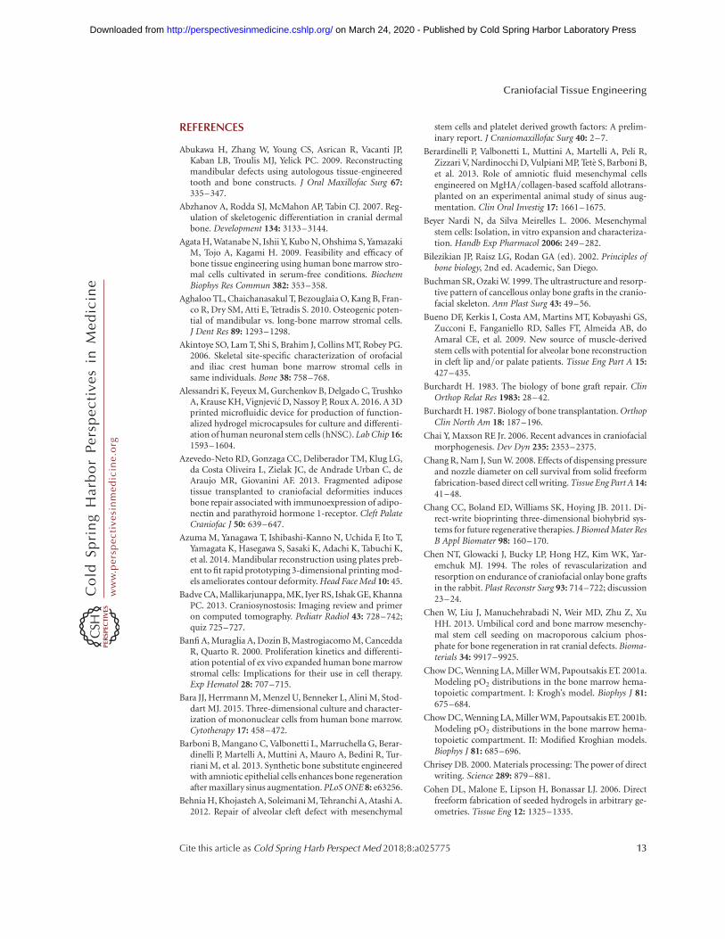
The craniofacial complex contains multipletypes of highly integrated hard and soft tissues.It would be beneficial to regenerate compositehard and soft tissues at the same time, to achieverapid functional recovery of regenerated cranio-facial tissues. Teeth are somewhat unique com-ponents of the craniofacial complex. Since teethare anchored in maxillary and mandibular jaw-bones, methods to generate jawbone togetherwith dental tissues, in addition to 3D printing,are currently being investigated, with some suc-cess (Fig. 3) (Duailibi et al. 2008; Abukawa et al.2009). A major obstacle in this approach is thedesign of methods and approaches that main-tain the integrity of both the bone and toothstructures, while allowing them to be function-ally integrated. Prior reports showed that bio-engineered tooth constructs were surroundedby newly formed bone, precluding tooth erup-tion and proper function (Cowan et al. 2004).The clinical use of composite tissue regenera-tion, such as cartilage and bone and vascular-ized muscle, is still in its infancy, and furtherstudies are needed to perfect these highly prom-ising approaches.
One of the main obstacles of current 3Dprinting techniques is the size limit of the print-out structure, mainly because hydrogel, themost commonly used injecting material, pro-vides inadequate structural support (Changet al. 2011). Recently, a new integrated tissue-organ printer (ITOP) has been developed totarget the generation of stable human-scale tis-sue constructs (Kang et al. 2016). The key to thismethod is to print a sacrificial scaffold adjacentto the cell-laden hydrogel. The cell-laden hydro-gel protects cell viability and supports cell ex-pansion and differentiation. The sacrificial scaf-folding material provides the initial structuraland architectural integrity and, on removal, alsoprovides diffusable microchannels to facilitate
W. Zhang and P.C. Yelick
10 Cite this article as Cold Spring Harb Perspect Med 2018;8:a025775
ww
w.p
ersp
ecti
vesi
nm
edic
ine.
org
on March 24, 2020 - Published by Cold Spring Harbor Laboratory Press http://perspectivesinmedicine.cshlp.org/Downloaded from

The
rmal
Hea
ter
Vap
orbu
bble
Pie
zoel
etric
Pie
zoel
ectr
icac
tuat
er
Inkj
et b
iopr
inte
rA
BC
Mic
roex
trus
ion
biop
rinte
r
Pne
umat
icP
isto
nS
crew
Don
orsl
ide
Lase
rpu
lse
Ene
rgy-
abso
rbin
gla
yer
Val
ve
Lase
r-as
sist
ed b
iopr
inte
r
Figu
re2.
Co
mp
on
ents
of
inkj
et,
mic
roex
tru
sio
n,
and
lase
r-as
sist
edb
iop
rin
ters
.(A
)T
her
mal
inkj
etp
rin
ters
elec
tric
ally
hea
tth
ep
rin
thea
dto
pro
du
ceai
r-p
ress
ure
pu
lses
that
forc
ed
rop
lets
fro
mth
en
ozz
le,w
her
eas
aco
ust
icp
rin
ters
use
pu
lses
form
edb
yp
iezo
elec
tric
or
ult
raso
un
dp
ress
ure
.(B
)M
icro
extr
usi
on
pri
nte
rsu
sep
neu
mat
ico
rm
ech
anic
al(p
isto
no
rsc
rew
)d
isp
ensi
ng
syst
ems
toex
tru
de
con
tin
uo
us
bea
ds
of
mat
eria
lan
d/
or
cell
s.(C
)L
aser
-ass
iste
dp
rin
ters
use
lase
rsfo
cuse
do
nan
abso
rbin
gsu
bst
rate
toge
ner
ate
pre
ssu
res
that
pro
pel
cell
-co
nta
inin
gm
ater
ials
on
toa
coll
ecto
rsu
bst
rate
.(F
rom
Mu
rph
yan
dA
tala
2014
;ad
apte
d,
wit
hp
erm
issi
on
,fr
om
Nat
ure
Pu
bli
shin
gG
rou
p#
2014
.)
Craniofacial Tissue Engineering
Cite this article as Cold Spring Harb Perspect Med 2018;8:a025775 11
ww
w.p
ersp
ecti
vesi
nm
edic
ine.
org
on March 24, 2020 - Published by Cold Spring Harbor Laboratory Press http://perspectivesinmedicine.cshlp.org/Downloaded from

nutrient perfusion. This technique has beenused to fabricate human-scale mandibularbone, ear-shaped cartilage, and organized skel-etal muscle.
Another major challenge of the 3D printingtechnique is how to precisely integrate biologi-cal components and gradients for compositetissue engineering. Compared to other tissue-regeneration methods, 3D printing can providefairly accurate control of different tissues. Still,3D printing has its own limitations, mainly be-cause the formation of 3D structures is based onthe accumulation of 2D structures. For example,accurately mimicking the branching patterns ofthe vascular tree has remained a big challenge of3D printing (Kolesky et al. 2016).
CONCLUSIONS
Successful reconstruction of the craniofacialcomplex requires full restoration of both func-
tional and aesthetic aspects of the face. Stem-cell-mediated tissue-regeneration approachesprovide the potential for highly successful cra-niofacial tissue-regeneration. Despite increasedknowledge and characterization of MSCs,and the mounting enthusiasm for the use ofMSCs in regenerative therapies in humans,the detailed mechanisms of MSCs proliferationand differentiation are still not fully under-stood.
This lack of understanding has not permit-ted the full use of stromal cells to facilitate orenhance tissue repair in clinical practice be-cause of fear of potential unwarranted deleteri-ous behaviors of the transplanted cells (Graysonet al. 2015). Despite these concerns, stem-cell-mediated regenerative strategies, combinedwith precise CAD approaches, are anticipated toeventually provide many new and promisingmethods for successful craniofacial reparativetherapies.
A B
D Eb
d
pc
pdl
C
Figure 3. Composite bone–tooth constructs. (A) Tooth scaffolds composed of a dental mesenchymal (DM) cellseeded polyglycolide/poly-L-lactide (PGA/PLLA) scaffold sphere to mimic the dental papilla, and a dentalepithelial (DE) cell seeded gelfoam strip to mimic the enamel organ. (B) Lattice bone scaffolds made by poly-DL-lactic-co-glycolic acid (PLGA), seeded with iliac crest–derived mesenchymal stem cells (MSCs) and grown inthe rotational oxygen-permeable bioreactor system (ROBS) for 6 weeks. (C) Fabricated tooth–bone constructseeded with cells before implantation. (D) Surgical implant site before wound closure. (E) Bioengineered dentaltissues that closely resembled those of naturally formed pig tooth tissues surrounded by alveolar bone. Scale bar¼ 100 mm. b, bone; bm, bone marrow; d, dentin; e, enamel; p, pulp; pdl, periodontal ligament.
W. Zhang and P.C. Yelick
12 Cite this article as Cold Spring Harb Perspect Med 2018;8:a025775
ww
w.p
ersp
ecti
vesi
nm
edic
ine.
org
on March 24, 2020 - Published by Cold Spring Harbor Laboratory Press http://perspectivesinmedicine.cshlp.org/Downloaded from
REFERENCES
Abukawa H, Zhang W, Young CS, Asrican R, Vacanti JP,Kaban LB, Troulis MJ, Yelick PC. 2009. Reconstructingmandibular defects using autologous tissue-engineeredtooth and bone constructs. J Oral Maxillofac Surg 67:335–347.
Abzhanov A, Rodda SJ, McMahon AP, Tabin CJ. 2007. Reg-ulation of skeletogenic differentiation in cranial dermalbone. Development 134: 3133–3144.
Agata H, Watanabe N, Ishii Y, Kubo N, Ohshima S, YamazakiM, Tojo A, Kagami H. 2009. Feasibility and efficacy ofbone tissue engineering using human bone marrow stro-mal cells cultivated in serum-free conditions. BiochemBiophys Res Commun 382: 353–358.
Aghaloo TL, Chaichanasakul T, Bezouglaia O, Kang B, Fran-co R, Dry SM, Atti E, Tetradis S. 2010. Osteogenic poten-tial of mandibular vs. long-bone marrow stromal cells.J Dent Res 89: 1293–1298.
Akintoye SO, Lam T, Shi S, Brahim J, Collins MT, Robey PG.2006. Skeletal site-specific characterization of orofacialand iliac crest human bone marrow stromal cells insame individuals. Bone 38: 758–768.
Alessandri K, Feyeux M, Gurchenkov B, Delgado C, TrushkoA, Krause KH, Vignjevic D, Nassoy P, Roux A. 2016. A 3Dprinted microfluidic device for production of function-alized hydrogel microcapsules for culture and differenti-ation of human neuronal stem cells (hNSC). Lab Chip 16:1593–1604.
Azevedo-Neto RD, Gonzaga CC, Deliberador TM, Klug LG,da Costa Oliveira L, Zielak JC, de Andrade Urban C, deAraujo MR, Giovanini AF. 2013. Fragmented adiposetissue transplanted to craniofacial deformities inducesbone repair associated with immunoexpression of adipo-nectin and parathyroid hormone 1-receptor. Cleft PalateCraniofac J 50: 639–647.
Azuma M, Yanagawa T, Ishibashi-Kanno N, Uchida F, Ito T,Yamagata K, Hasegawa S, Sasaki K, Adachi K, Tabuchi K,et al. 2014. Mandibular reconstruction using plates preb-ent to fit rapid prototyping 3-dimensional printing mod-els ameliorates contour deformity. Head Face Med 10: 45.
Badve CA, Mallikarjunappa, MK, Iyer RS, Ishak GE, KhannaPC. 2013. Craniosynostosis: Imaging review and primeron computed tomography. Pediatr Radiol 43: 728–742;quiz 725–727.
Banfi A, Muraglia A, Dozin B, Mastrogiacomo M, CanceddaR, Quarto R. 2000. Proliferation kinetics and differenti-ation potential of ex vivo expanded human bone marrowstromal cells: Implications for their use in cell therapy.Exp Hematol 28: 707–715.
Bara JJ, Herrmann M, Menzel U, Benneker L, Alini M, Stod-dart MJ. 2015. Three-dimensional culture and character-ization of mononuclear cells from human bone marrow.Cytotherapy 17: 458–472.
Barboni B, Mangano C, Valbonetti L, Marruchella G, Berar-dinelli P, Martelli A, Muttini A, Mauro A, Bedini R, Tur-riani M, et al. 2013. Synthetic bone substitute engineeredwith amniotic epithelial cells enhances bone regenerationafter maxillary sinus augmentation. PLoS ONE 8: e63256.
Behnia H, Khojasteh A, Soleimani M, Tehranchi A, Atashi A.2012. Repair of alveolar cleft defect with mesenchymal
stem cells and platelet derived growth factors: A prelim-inary report. J Craniomaxillofac Surg 40: 2–7.
Berardinelli P, Valbonetti L, Muttini A, Martelli A, Peli R,Zizzari V, Nardinocchi D, Vulpiani MP, Tete S, Barboni B,et al. 2013. Role of amniotic fluid mesenchymal cellsengineered on MgHA/collagen-based scaffold allotrans-planted on an experimental animal study of sinus aug-mentation. Clin Oral Investig 17: 1661–1675.
Beyer Nardi N, da Silva Meirelles L. 2006. Mesenchymalstem cells: Isolation, in vitro expansion and characteriza-tion. Handb Exp Pharmacol 2006: 249–282.
Bilezikian JP, Raisz LG, Rodan GA (ed). 2002. Principles ofbone biology, 2nd ed. Academic, San Diego.
Buchman SR, Ozaki W. 1999. The ultrastructure and resorp-tive pattern of cancellous onlay bone grafts in the cranio-facial skeleton. Ann Plast Surg 43: 49–56.
Bueno DF, Kerkis I, Costa AM, Martins MT, Kobayashi GS,Zucconi E, Fanganiello RD, Salles FT, Almeida AB, doAmaral CE, et al. 2009. New source of muscle-derivedstem cells with potential for alveolar bone reconstructionin cleft lip and/or palate patients. Tissue Eng Part A 15:427–435.
Burchardt H. 1983. The biology of bone graft repair. ClinOrthop Relat Res 1983: 28–42.
Burchardt H. 1987. Biology of bone transplantation. OrthopClin North Am 18: 187–196.
Chai Y, Maxson RE Jr. 2006. Recent advances in craniofacialmorphogenesis. Dev Dyn 235: 2353–2375.
Chang R, Nam J, Sun W. 2008. Effects of dispensing pressureand nozzle diameter on cell survival from solid freeformfabrication-based direct cell writing. Tissue Eng Part A 14:41–48.
Chang CC, Boland ED, Williams SK, Hoying JB. 2011. Di-rect-write bioprinting three-dimensional biohybrid sys-tems for future regenerative therapies. J Biomed Mater ResB Appl Biomater 98: 160–170.
Chen NT, Glowacki J, Bucky LP, Hong HZ, Kim WK, Yar-emchuk MJ. 1994. The roles of revascularization andresorption on endurance of craniofacial onlay bone graftsin the rabbit. Plast Reconstr Surg 93: 714–722; discussion23–24.
Chen W, Liu J, Manuchehrabadi N, Weir MD, Zhu Z, XuHH. 2013. Umbilical cord and bone marrow mesenchy-mal stem cell seeding on macroporous calcium phos-phate for bone regeneration in rat cranial defects. Bioma-terials 34: 9917–9925.
Chow DC, Wenning LA, Miller WM, Papoutsakis ET. 2001a.Modeling pO2 distributions in the bone marrow hema-topoietic compartment. I: Krogh’s model. Biophys J 81:675–684.
Chow DC, Wenning LA, Miller WM, Papoutsakis ET. 2001b.Modeling pO2 distributions in the bone marrow hema-topoietic compartment. II: Modified Kroghian models.Biophys J 81: 685–696.
Chrisey DB. 2000. Materials processing: The power of directwriting. Science 289: 879–881.
Cohen DL, Malone E, Lipson H, Bonassar LJ. 2006. Directfreeform fabrication of seeded hydrogels in arbitrary ge-ometries. Tissue Eng 12: 1325–1335.
Craniofacial Tissue Engineering
Cite this article as Cold Spring Harb Perspect Med 2018;8:a025775 13
ww
w.p
ersp
ecti
vesi
nm
edic
ine.
org
on March 24, 2020 - Published by Cold Spring Harbor Laboratory Press http://perspectivesinmedicine.cshlp.org/Downloaded from

Colina M, Serra P, Fernandez-Pradas JM, Sevilla L, MorenzaJL. 2005. DNA deposition through laser induced forwardtransfer. Biosens Bioelectron 20: 1638–1642.
Colter DC, Class R, DiGirolamo CM, Prockop DJ. 2000.Rapid expansion of recycling stem cells in cultures ofplastic-adherent cells from human bone marrow. ProcNatl Acad Sci 97: 3213–3218.
Cordonnier T, Layrolle P, Gaillard J, Langonne A, Sensebe L,Rosset P, Sohier J. 2010. 3D environment on human mes-enchymal stem cells differentiation for bone tissue engi-neering. J Mater Sci Mater Med 21: 981–987.
Costa PF, Vaquette C, Zhang Q, Reis RL, Ivanovski S, Hut-macher DW. 2014. Advanced tissue engineering scaffolddesign for regeneration of the complex hierarchical peri-odontal structure. J Clin Periodontol 41: 283–294.
Cowan CM, Shi YY, Aalami OO, Chou YF, Mari C, ThomasR, Quarto N, Contag CH, Wu B, Longaker MT. 2004.Adipose-derived adult stromal cells heal critical-sizemouse calvarial defects. Nat Biotechnol 22: 560–567.
Dahlin C, Linde A, Gottlow J, Nyman S. 1988. Healing ofbone defects by guided tissue regeneration. Plast ReconstrSurg 81: 672–676.
Dahlin C, Gottlow J, Linde A, Nyman S. 1990. Healing ofmaxillary and mandibular bone defects using a mem-brane technique. An experimental study in monkeys.Scand J Plast Reconstr Surg Hand Surg 24: 13–19.
De Coster PJ, Mortier G, Marks LA, Martens LC. 2007.Cranial suture biology and dental development: Geneticand clinical perspectives. J Oral Pathol Med 36: 447–455.
Derby B. 2012. Printing and prototyping of tissues and scaf-folds. Science 338: 921–926.
De Ugarte DA, Morizono K, Elbarbary A, Alfonso Z, ZukPA, Zhu M, Draggo JL, Ashjian P, Thomas B, Benhaim P,et al. 2003. Comparison of multi-lineage cells from hu-man adipose tissue and bone marrow. Cells Tissues Or-gans 174: 101–109.
Digirolamo CM, Stokes D, Colter D, Phinney DG, Class R,Prockop DJ. 1999. Propagation and senescence of humanmarrow stromal cells in culture: A simple colony-formingassay identifies samples with the greatest potential topropagate and differentiate. Br J Haematol 107: 275–281.
Duailibi SE, Duailibi MT, Zhang W, Asrican R, Vacanti JP,Yelick PC. 2008. Bioengineered dental tissues grown inthe rat jaw. J Dent Res 87: 745–750.
Duan B, Hockaday LA, Kang KH, Butcher JT. 2013. 3Dbioprinting of heterogeneous aortic valve conduits withalginate/gelatin hydrogels. J Biomed Mater Res A 101:1255–1264.
Ducy P, Zhang R, Geoffroy V, Ridall AL, Karsenty G. 1997.Osf2/Cbfa1: A transcriptional activator of osteoblast dif-ferentiation. Cell 89: 747–754.
Dupont KM, Sharma K, Stevens HY, Boerckel JD, Garcia AJ,Guldberg RE. 2010. Human stem cell delivery for treat-ment of large segmental bone defects. Proc Natl Acad Sci107: 3305–3310.
Enlow DH. 1982. The handbook of facial growth. WB Saun-ders, Philadelphia.
Ermis I, Poole M. 1992. The effects of soft tissue coverage onbone graft resorption in the craniofacial region. Br J PlastSurg 45: 26–29.
Eyre-Brook AL. 1984. The periosteum: Its function reas-sessed. Clin Orthop Relat Res 1984: 300–307.
Feisst V, Brooks AE, Chen CJ, Dunbar PR. 2014. Character-ization of mesenchymal progenitor cell populations di-rectly derived from human dermis. Stem Cells Dev 23:631–642.
Friedenstein AJ. 1976. Precursor cells of mechanocytes. IntRev Cytol 47: 327–359.
Friedenstein AJ, Petrakova KV, Kurolesova AI, Frolova GP.1968. Heterotopic of bone marrow. Analysis of precursorcells for osteogenic and hematopoietic tissues. Transplan-tation 6: 230–247.
Friedlaender GE, Perry CR, Cole JD, Cook SD, Cierny G,Muschler GF, Zych GA, Calhoun JH, LaForte AJ, Yin S.2001. Osteogenic protein-1 (bone morphogenetic pro-tein-7) in the treatment of tibial nonunions. J Bone JointSurg Am 83-A: S151–S158.
Frommelt H. 1987. Polymers for medical applications. Mak-romol Chem Macromol Symp 12: 281–301.
Garrett GL, Beegun I, D’Souza A. 2015. Facial transplanta-tion: Historical developments and future directions.J Laryngol Otol 129: 206–211.
Genden EM. 2010. Reconstruction of the mandible and themaxilla: The evolution of surgical technique. Arch FacialPlast Surg 12: 87–90.
Gimbel M, Ashley RK, Sisodia M, Gabbay JS, Wasson KL,Heller J, Wilson L, Kawamoto HK, Bradley JP. 2007. Re-pair of alveolar cleft defects: Reduced morbidity withbone marrow stem cells in a resorbable matrix. J Cranio-fac Surg 18: 895–901.
Gimble JM, Zvonic S, Floyd ZE, Kassem M, Nuttall ME.2006. Playing with bone and fat. J Cell Biochem 98:251–266.
Gomes SP, Deliberador TM, Gonzaga CC, Klug LG, da CostaOliveira L, de Andrade Urban C, Zielak JC, Giovanini AF.2012. Bone healing in critical-size defects treated withimmediate transplant of fragmented autogenous whiteadipose tissue. J Craniofac Surg 23: 1239–1244.
Grayson WL, Bunnell BA, Martin E, Frazier T, Hung BP,Gimble JM. 2015. Stromal cells and stem cells in clinicalbone regeneration. Nat Rev Endocrinol 11: 140–150.
Griffin M, Kalaskar DM, Butler PE, Seifalian AM. 2014. Theuse of adipose stem cells in cranial facial surgery. StemCell Rev 10: 671–685.
Gronthos S, Mankani M, Brahim J, Robey PG, Shi S. 2000.Postnatal human dental pulp stem cells (DPSCs) in vitroand in vivo. Proc Natl Acad Sci 97: 13625–13630.
Gronthos S, Zannettino AC, Hay SJ, Shi S, Graves SE, Kor-tesidis A, Simmons PJ. 2003. Molecular and cellular char-acterisation of highly purified stromal stem cells derivedfrom human bone marrow. J Cell Sci 116: 1827–1835.
Gruss JS, Mackinnon SE, Kassel EE, Cooper PW. 1985. Therole of primary bone grafting in complex craniomaxillo-facial trauma. Plast Reconstr Surg 75: 17–24.
Guillemot F, Souquet A, Catros S, Guillotin B, Lopez J, Fau-con M, Pippenger B, Bareille R, Remy M, Bellance S, et al.2010. High-throughput laser printing of cells and bioma-terials for tissue engineering. Acta Biomater 6: 2494–2500.
Habisch HJ, Janowski M, Binder D, Kuzma-Kozakiewicz M,Widmann A, Habich A, Schwalenstocker B, Hermann A,
W. Zhang and P.C. Yelick
14 Cite this article as Cold Spring Harb Perspect Med 2018;8:a025775
ww
w.p
ersp
ecti
vesi
nm
edic
ine.
org
on March 24, 2020 - Published by Cold Spring Harbor Laboratory Press http://perspectivesinmedicine.cshlp.org/Downloaded from

Brenner R, Lukomska B, et al. 2007. Intrathecal applica-tion of neuroectodermally converted stem cells into amouse model of ALS: Limited intraparenchymal migra-tion and survival narrows therapeutic effects. J NeuralTransm 114: 1395–1406.
Han C, Yang Z, Zhou W, Jin F, Song Y, Wang Y, Huo N, ChenL, Qian H, Hou R, et al. 2010. Periapical follicle stem cell:A promising candidate for cementum/periodontal liga-ment regeneration and bio-root engineering. Stem CellsDev 19: 1405–1415.
Hidalgo DA. 1989. Fibula free flap: A new method of man-dible reconstruction. Plast Reconstr Surg 84: 71–79.
Ho TV, Iwata J, Ho HA, Grimes WC, Park S, Sanchez-LaraPA, Chai Y. 2015. Integration of comprehensive 3D mi-croCTand signaling analysis reveals differential regulato-ry mechanisms of craniofacial bone development. DevBiol 400: 180–190.
Hokugo A, Kubo Y, Takahashi Y, Fukuda A, Horiuchi K,Mushimoto K, Morita S, Tabata Y. 2004. Prefabricationof vascularized bone graft using guided bone regenera-tion. Tissue Eng 10: 978–986.
Hunter C, Januszyk M, Wan DC, Momeni A. 2015. System-atic reviews in craniofacial trauma-strengths and weak-nesses. Ann Plast Surg 77: 363–368.
Ivanovski S, Vaquette C, Gronthos S, Hutmacher DW, Bar-told PM. 2014. Multiphasic scaffolds for periodontal tis-sue engineering. J Dent Res 93: 1212–1221.
Izadpanah R, Kaushal D, Kriedt C, Tsien F, Patel B, Dufour J,Bunnell BA. 2008. Long-term in vitro expansion altersthe biology of adult mesenchymal stem cells. Cancer Res68: 4229–4238.
Jackson IT, Choi HY, Clay R, Bevilacqua R, TerKonda S,Celik M, Smith AW. 1998. Long-term follow-up of cranialbone graft in dorsal nasal augmentation. Plast ReconstrSurg 102: 1869–1873.
Jin Y, Zhang W, Liu Y, Zhang M, Xu L, Wu Q, Zhang X, ZhuZ, Huang Q, Jiang X. 2014. rhPDGF-BB via ERK pathwayosteogenesis and adipogenesis balancing in ADSCs forcritical-sized calvarial defect repair. Tissue Eng Part A20: 3303–3313.
Kaigler D, Pagni G, Park CH, Tarle SA, Bartel RL, GiannobileWV. 2010. Angiogenic and osteogenic potential of bonerepair cells for craniofacial regeneration. Tissue Eng Part A16: 2809–2820.
Kang HW, Lee SJ, Ko IK, Kengla C, Yoo JJ, Atala A. 2016. A3D bioprinting system to produce human-scale tissueconstructs with structural integrity. Nat Biotechnol 34:312–319.
Keriquel V, Guillemot F, Arnault I, Guillotin B, Miraux S,Amedee J, Fricain JC, Catros S. 2010. In vivo bioprintingfor computer- and robotic-assisted medical intervention:Preliminary study in mice. Biofabrication 2: 014101.
Kim K, Lee CH, Kim BK, Mao JJ. 2010. Anatomically shapedtooth and periodontal regeneration by cell homing.J Dent Res 89: 842–847.
Kimmel CB, Walker MB, Miller CT. 2007. Morphing thehyomandibular skeleton in development and evolution.J Exp Zool B Mol Dev Evol 308: 609–624.
Klebe RJ. 1988. Cytoscribing: A method for microposition-ing cells and the construction of two- and three-dimen-sional synthetic tissues. Exp Cell Res 179: 362–373.
Knize DM. 1974. The influence of periosteum and calcito-nin on onlay bone graft survival. A roentgenographicstudy. Plast Reconstr Surg 53: 190–199.
Kolesky DB, Homan KA, Skylar-Scott MA, Lewis JA. 2016.Three-dimensional bioprinting of thick vascularized tis-sues. Proc Natl Acad Sci 113: 3179–3184.
Kuznetsov SA, Krebsbach PH, Satomura K, Kerr J, Rimi-nucci M, Benayahu D, Robey PG. 1997. Single-colonyderived strains of human marrow stromal fibroblastsform bone after transplantation in vivo. J Bone MinerRes 12: 1335–1347.
Lange C, Cakiroglu F, Spiess AN, Cappallo-Obermann H,Dierlamm J, Zander AR. 2007. Accelerated and safe ex-pansion of human mesenchymal stromal cells in animalserum-free medium for transplantation and regenerativemedicine. J Cell Physiol 213: 18–26.
Le Blanc K, Samuelsson H, Lonnies L, Sundin M, RingdenO. 2007. Generation of immunosuppressive mesenchy-mal stem cells in allogeneic human serum. Transplanta-tion 84: 1055–1059.
Lendeckel S, Jodicke A, Christophis P, Heidinger K, Wolff J,Fraser JK, Hedrick MH, Berthold L, Howaldt HP. 2004.Autologous stem cells (adipose) and fibrin glue used totreat widespread traumatic calvarial defects: Case report.J Craniomaxillofac Surg 32: 370–373.
Leucht P, Kim JB, Amasha R, James AW, Girod S, Helms JA.2008. Embryonic origin and Hox status determine pro-genitor cell fate during adult bone regeneration. Devel-opment 135: 2845–2854.
Lew TA, Walker JA, Wenke JC, Blackbourne LH, Hale RG.2010. Characterization of craniomaxillofacial battle inju-ries sustained by United States service members in thecurrent conflicts of Iraq and Afghanistan. J Oral Maxil-lofac Surg 68: 3–7.
Lin KY, Bartlett SP, Yaremchuk MJ, Fallon M, Grossman RF,Whitaker LA. 1990. The effect of rigid fixation on thesurvival of onlay bone grafts: An experimental study.Plast Reconstr Surg 86: 449–456.
Lin CY, Chang YH, Kao CY, Lu CH, Sung LY, Yen TC, Lin KJ,Hu YC. 2012. Augmented healing of critical-size calvarialdefects by baculovirus-engineered MSCs that persistentlyexpress growth factors. Biomaterials 33: 3682–3692.
Lindroos B, Boucher S, Chase L, Kuokkanen H, Huhtala H,Haataja R, Vemuri M, Suuronen R, Miettinen S. 2009.Serum-free, xeno-free culture media maintain the prolif-eration rate and multipotentiality of adipose stem cells invitro. Cytotherapy 11: 958–972.
Lukash FN, Zingaro EA, Salig J. 1984. The survival of freenonvascularized bone grafts in irradiated areas by wrap-ping in muscle flaps. Plast Reconstr Surg 74: 783–788.
Manson PN, Crawley WA, Yaremchuk MJ, Rochman GM,Hoopes JE, French JHJr. 1985. Midface fractures: Advan-tages of immediate extended open reduction and bonegrafting. Plast Reconstr Surg 76: 1–12.
Maraldi T, Riccio M, Pisciotta A, Zavatti M, Carnevale G,Beretti F, La Sala GB, Motta A, De Pol A. 2013. Humanamniotic fluid-derived and dental pulp-derived stemcells seeded into collagen scaffold repair critical-sizebone defects promoting vascularization. Stem Cell ResTher 4: 53.
Martuscelli R, Toti P, Sbordone L, Guidetti F, Ramaglia L,Sbordone C. 2014. Five-year outcome of bone remodel-
Craniofacial Tissue Engineering
Cite this article as Cold Spring Harb Perspect Med 2018;8:a025775 15
ww
w.p
ersp
ecti
vesi
nm
edic
ine.
org
on March 24, 2020 - Published by Cold Spring Harbor Laboratory Press http://perspectivesinmedicine.cshlp.org/Downloaded from

ling around implants in the maxillary sinus: Assessmentof differences between implants placed in autogenousinlay bone blocks and in ungrafted maxilla. Int J OralMaxillofac Surg 43: 1117–1126.
Mavropoulos A, Rizzoli R, Ammann P. 2007. Different re-sponsiveness of alveolar and tibial bone to bone lossstimuli. J Bone Miner Res 22: 403–410.
Melcher AH, Accursi GE. 1971. Osteogenic capacity of peri-osteal and osteoperiosteal flaps elevated from the parietalbone of the rat. Arch Oral Biol 16: 573–580.
Mesimaki K, Lindroos B, Tornwall J, Mauno J, Lindqvist C,Kontio R, Miettinen S, Suuronen R. 2009. Novel maxil-lary reconstruction with ectopic bone formation by GMPadipose stem cells. Int J Oral Maxillofac Surg 38: 201–209.
Miura M, Gronthos S, Zhao M, Lu B, Fisher LW, Robey PG,Robey PG, Shi S. 2003. SHED: Stem cells from humanexfoliated deciduous teeth. Proc Natl Acad Sci 100: 5807–5812.
Miura M, Miura Y, Sonoyama W, Yamaza T, Gronthos S, ShiS. 2006. Bone marrow-derived mesenchymal stem cellsfor regenerative medicine in craniofacial region. Oral Dis12: 514–522.
Mossey P, Castilla E (ed.). 2003. Global registry and databaseon craniofacial anomalies. In Report of a WHO registrymeeting on craniofacial anomalies. World Health Organi-zation, Geneva, Switzerland.
Muller I, Kordowich S, Holzwarth C, Spano C, Isensee G,Staiber A, Viebahn S, Gieseke F, Langer H, Gawaz MP, etal. 2006. Animal serum-free culture conditions for isola-tion and expansion of multipotent mesenchymal stromalcells from human BM. Cytotherapy 8: 437–444.
Mulliken JB, Kaban LB, Glowacki J. 1984. Induced osteo-genesis—The biological principle and clinical applica-tions. J Surg Res 37: 487–496.
Muraglia A, Cancedda R, Quarto R. 2000. Clonal mesen-chymal progenitors from human bone marrow differen-tiate in vitro according to a hierarchical model. J Cell Sci113: 1161–1166.
Murphy SV, Atala A. 2014. 3D bioprinting of tissues andorgans. Nat Biotechnol 32: 773–785.
Nakashima M, Mizunuma K, Murakami T, Akamine A.2002. Induction of dental pulp stem cell differentiationinto odontoblasts by electroporation-mediated gene de-livery of growth/differentiation factor 11 (Gdf11). GeneTher 9: 814–818.
Neovius E, Engstrand T. 2010. Craniofacial reconstructionwith bone and biomaterials: Review over the last 11 years.J Plast Reconstr Aesthet Surg 63: 1615–1623.
Neu BR. 2000. Segmental bone and cartilage reconstructionof major nasal dorsal defects. Plast Reconstr Surg 106:160–170.
Norotte C, Marga FS, Niklason LE, Forgacs G. 2009. Scaf-fold-free vascular tissue engineering using bioprinting.Biomaterials 30: 5910–5917.
Obregon F, Vaquette C, Ivanovski S, Hutmacher DW, Ber-tassoni LE. 2015. Three-dimensional bioprinting for re-generative dentistry and craniofacial tissue engineering.J Dent Res 94: 143S–152S.
Ochareon P, Herring SW. 2011. Cell replication in craniofa-cial periosteum: Appositional vs. resorptive sites. J Anat218: 285–297.
Oklund SA, Prolo DJ, Gutierrez RV, King SE. 1986. Quan-titative comparisons of healing in cranial fresh autografts,frozen autografts and processed autografts, and allograftsin canine skull defects. Clin Orthop Relat Res 1986: 269–291.
Oppenheimer AJ, Tong L, Buchman SR. 2008. Craniofacialbone grafting: Wolff ’s Law revisited. CraniomaxillofacTrauma Reconstr 1: 49–61.
Otto F, Thornell AP, Crompton T, Denzel A, Gilmour KC,Rosewell IR, Stamp GW, Beddington RS, Mundlos S,Olsen BR, et al. 1997. Cbfa1, a candidate gene for clei-docranial dysplasia syndrome, is essential for osteoblastdifferentiation and bone development. Cell 89: 765–771.
Owen M, Friedenstein AJ. 1988. Stromal stem cells: Marrow-derived osteogenic precursors. Ciba Found Symp 136:42–60.
Ozaki W, Buchman SR. 1998. Volume maintenance of onlaybone grafts in the craniofacial skeleton: Micro-architec-ture versus embryologic origin. Plast Reconstr Surg 102:291–299.
Papaccio G, Graziano A, d’Aquino R, Graziano MF, PirozziG, Menditti D, De Rosa A, Carinci F, Laino G. 2006.Long-term cryopreservation of dental pulp stem cells(SBP-DPSCs) and their differentiated osteoblasts: A cellsource for tissue repair. J Cell Physiol 208: 319–325.
Peer LA. 1951. Fate of autogenous human bone grafts. Br JPlast Surg 3: 233–243.
Petrini C. 2015. Facial transplants: Current situation andethical issues. Clin Ter 166: 215–217.
Petrovic V, Zivkovic P, Petrovic D, Stefanovic V. 2012. Cra-niofacial bone tissue engineering. Oral Surg Oral MedOral Pathol Oral Radiol 114: e1–e9.
Phillips JH, Rahn BA. 1988. Fixation effects on membra-nous and endochondral onlay bone-graft resorption.Plast Reconstr Surg 82: 872–877.
Pinholt EM, Solheim E, Talsnes O, Larsen TB, Bang G, Kir-keby OJ. 1994. Revascularization of calvarial, mandibu-lar, tibial, and iliac bone grafts in rats. Ann Plast Surg 33:193–197.
Pittenger MF, Mackay AM, Beck SC, Jaiswal RK, Douglas R,Mosca JD, Moorman MA, Simonetti DW, Craig S,Marshak DR. 1999. Multilineage potential of adult hu-man mesenchymal stem cells. Science 284: 143–147.
Pogrel MA, Podlesh S, Anthony JP, Alexander J. 1997. Acomparison of vascularized and nonvascularized bonegrafts for reconstruction of mandibular continuity de-fects. J Oral Maxillofac Surg 55: 1200–1206.
Price N, Lipton A, Jain VK, Ruggiero S. 2004. Preventionand management of osteonecrosis of the jaw associatedwith bisphosphonate therapy. Support Cancer Ther 2:14–17.
Prockop DJ. 2007. “Stemness” does not explain the repair ofmany tissues by mesenchymal stem/multipotent stromalcells (MSCs). Clin Pharmacol Ther 82: 241–243.
Qu-Petersen Z, Deasy B, Jankowski R, Ikezawa M, CumminsJ, Pruchnic R, Mytinger J, Cao B, Gates C, Wernig A, et al.2002. Identification of a novel population of muscle stem
W. Zhang and P.C. Yelick
16 Cite this article as Cold Spring Harb Perspect Med 2018;8:a025775
ww
w.p
ersp
ecti
vesi
nm
edic
ine.
org
on March 24, 2020 - Published by Cold Spring Harbor Laboratory Press http://perspectivesinmedicine.cshlp.org/Downloaded from

cells in mice: Potential for muscle regeneration. J Cell Biol157: 851–864.
Ramaesh T, Bard JB. 2003. The growth and morphogenesisof the early mouse mandible: A quantitative analysis.J Anat 203: 213–222.
Reichert JC, Cipitria A, Epari DR, Saifzadeh S, KrishnakanthP, Berner A, Woodruff MA, Schell H, Mehta M, SchuetzMA, et al. 2012. A tissue engineering solution for seg-mental defect regeneration in load-bearing long bones.Sci Transl Med 4: 141ra93.
Rocchi G, Fadda MT, Marianetti TM, Reale G, Iannetti G.2007. Craniofacial trauma in adolescents: Incidence, eti-ology, and prevention. J Trauma 62: 404–409.
Saijo H, Igawa K, Kanno Y, Mori Y, Kondo K, Shimizu K,Suzuki S, Chikazu D, Lino M, Anzai M, et al. 2009. Max-illofacial reconstruction using custom-made artificialbones fabricated by inkjet printing technology. J ArtifOrgans 12: 200–205.
Sandor GK, Suuronen R. 2008. Combining adipose-derivedstem cells, resorbable scaffolds and growth factors: Anoverview of tissue engineering. J Can Dent Assoc 74:167–170.
Sandor GK, Tuovinen VJ, Wolff J, Patrikoski M, Jokinen J,Nieminen E, Mannerstrom B, Lappalainen OP, SeppanenR, Miettinen S. 2013. Adipose stem cell tissue-engineeredconstruct used to treat large anterior mandibular defect:A case report and review of the clinical application ofgood manufacturing practice-level adipose stem cellsfor bone regeneration. J Oral Maxillofac Surg 71: 938–950.
Santos CA, Andrade LR, Costa MH, Souza HS, GranjeiroJM, Takiya CM, Borojevic R, Nasciutti LE. 2016. Gastro-spheres of human gastric mucosa cells: An in vitro modelof stromal and epithelial stem cell niche reconstruction.Histol Histopathol 31: 879–895.
Schek RM, Taboas JM, Hollister SJ, Krebsbach PH. 2005.Tissue engineering osteochondral implants for temporo-mandibular joint repair. Orthod Craniofac Res 8: 313–319.
Scherberich A, Galli R, Jaquiery C, Farhadi J, Martin I. 2007.Three-dimensional perfusion culture of human adiposetissue-derived endothelial and osteoblastic progenitorsgenerates osteogenic constructs with intrinsic vasculari-zation capacity. Stem Cells 25: 1823–1829.
Schmal O, Seifert J, Schaffer TE, Walter CB, Aicher WK,Klein G. 2013. Hematopoietic stem and progenitor cellexpansion in contact with mesenchymal stromal cells in ahanging drop model uncovers disadvantages of 3D cul-ture. Stem Cells Int 2016: 4148093.
Sekiya I, Larson BL, Smith JR, Pochampally R, Cui JG,Prockop DJ. 2002. Expansion of human adult stem cellsfrom bone marrow stroma: Conditions that maximize theyields of early progenitors and evaluate their quality.Stem Cells 20: 530–541.
Senarath-Yapa K, Chung MT, McArdle A, Wong VW, QuartoN, Longaker MT, Wan DC. 2012. Craniosynostosis: Mo-lecular pathways and future pharmacologic therapy. Or-ganogenesis 8: 103–113.
Seo BM, Miura M, Gronthos S, Bartold PM, Batouli S,Brahim J, Young M, Robey PG, Wang CY, Shi S. 2004.Investigation of multipotent postnatal stem cells fromhuman periodontal ligament. Lancet 364: 149–155.
Shang Q, Wang Z, Liu W, Shi Y, Cui L, Cao Y. 2001. Tissue-engineered bone repair of sheep cranial defects with au-tologous bone marrow stromal cells. J Craniofac Surg 12:586–593; discussion 94–95.
Shi S, Robey PG, Gronthos S. 2001. Comparison of humandental pulp and bone marrow stromal stem cells by cDNAmicroarray analysis. Bone 29: 532–539.
Shkoukani MA, Chen M, Vong A. 2013. Cleft lip—A com-prehensive review. Front Pediatr 1: 53.
Simonds WF, James-Newton LA, Agarwal SK, Yang B, Skar-ulis MC, Hendy GN, Marx SJ. 2002. Familial isolatedhyperparathyroidism: Clinical and genetic characteristicsof 36 kindreds. Medicine (Baltimore) 81: 1–26.
Skawina A, Gorczyca W. 1984. The role of nutrient andperiosteal blood vessels in the vascularization of the cor-tex of shafts of the long bones in human fetuses. FoliaMorphol (Warsz) 43: 159–164.
Skardal A, Mack D, Kapetanovic E, Atala A, Jackson JD, YooJ, Soker S. 2012. Bioprinted amniotic fluid-derived stemcells accelerate healing of large skin wounds. Stem CellsTransl Med 1: 792–802.
Sodek J, McKee MD. 2000. Molecular and cellular biology ofalveolar bone. Periodontol 2000 24: 99–126.
Sohier J, Corre P, Weiss P, Layrolle P. 2010. Hydrogel/calci-um phosphate composites require specific properties forthree-dimensional culture of human bone mesenchymalcells. Acta Biomater 6: 2932–2939.
Sullivan WG, Szwajkun PR. 1991. Revascularization of cra-nial versus iliac crest bone grafts in the rat. Plast ReconstrSurg 87: 1105–1109.
Teng M, Liang X, Yuan Q, Nie J, Ye J, Cheng Q, Zhai J, Liao J,Sun X, Wen C, et al. 2013. The inlay osteotome sinusaugmentation technique for placing short implants si-multaneously with reduced crestal bone height. Ashort-term follow-up. Clin Implant Dent Relat Res 15:918–926.
Terheyden H, Knak C, Jepsen S, Palmie S, Rueger DR. 2001a.Mandibular reconstruction with a prefabricated vascu-larized bone graft using recombinant human osteogenicprotein.1: An experimental study in miniature pigs. PartI: Prefabrication. Int J Oral Maxillofac Surg 30: 373–379.
Terheyden H, Warnke P, Dunsche A, Jepsen S, Brenner W,Palmie S, Toth C, Rueger DR. 2001b. Mandibular recon-struction with prefabricated vascularized bone grafts us-ing recombinant human osteogenic protein.1: An exper-imental study in miniature pigs. Part II: Transplantation.Int J Oral Maxillofac Surg 30: 469–478.
Terheyden H, Menzel C, Wang H, Springer IN, Rueger DR,Acil Y. 2004. Prefabrication of vascularized bone graftsusing recombinant human osteogenic protein.1: Part 3:Dosage of rhOP-1, the use of external and internal scaf-folds. Int J Oral Maxillofac Surg 33: 164–172.
Thesleff T, Lehtimaki K, Niskakangas T, Mannerstrom B,Miettinen S, Suuronen R, Ohman J. 2011. Cranioplastywith adipose-derived stem cells and biomaterial: A novelmethod for cranial reconstruction. Neurosurgery 68:1535–1540.
Thompson N, Casson JA. 1970. Experimental onlay bonegrafts to the jaws. A preliminary study in dogs. Plast Re-constr Surg 46: 341–349.
Craniofacial Tissue Engineering
Cite this article as Cold Spring Harb Perspect Med 2018;8:a025775 17
ww
w.p
ersp
ecti
vesi
nm
edic
ine.
org
on March 24, 2020 - Published by Cold Spring Harbor Laboratory Press http://perspectivesinmedicine.cshlp.org/Downloaded from

Tolarova MM, Cervenka J. 1998. Classification and birthprevalence of orofacial clefts. Am J Med Genet 75: 126–137.
Tsutsui TW, Riminucci M, Holmbeck K, Bianco P, RobeyPG. 2008. Development of craniofacial structures intransgenic mice with constitutively active PTH/PTHrPreceptor. Bone 42: 321–331.
Uhm KI, Hwang SH, Choi BG. 2000. Cleft lip nose correc-tion with onlay calvarial bone graft and suture suspensionin Oriental patients. Plast Reconstr Surg 105: 499–503.
Van Meekran MJ. 1682. Observationes medicochirurgicae[Observations of medicine and surgery]. Henrici andBloom, Amsterdam.
Vaquette C, Fan W, Xiao Y, Hamlet S, Hutmacher DW, Iva-novski S. 2012. A biphasic scaffold design combined withcell sheet technology for simultaneous regeneration ofalveolar bone/periodontal ligament complex. Biomateri-als 33: 5560–5573.
Visser J, Peters B, Burger TJ, Boomstra J, Dhert WJ, MelchelsFP, Malda J. 2013. Biofabrication of multi-material ana-tomically shaped tissue constructs. Biofabrication 5:035007.
Vogelin E, Jones NF, Huang JI, Brekke JH, Lieberman JR.2005. Healing of a critical-sized defect in the rat femurwith use of a vascularized periosteal flap, a biodegradablematrix, and bone morphogenetic protein. J Bone JointSurg Am 87: 1323–1331.
Wang F, Yu M, Yan X, Wen Y, Zeng Q, Yue W, Yang P, Pei X.2011. Gingiva-derived mesenchymal stem cell-mediatedtherapeutic approach for bone tissue regeneration. StemCells Dev 20: 2093–2102.
Ward BB, Brown SE, Krebsbach PH. 2010. Bioengineeringstrategies for regeneration of craniofacial bone: A reviewof emerging technologies. Oral Dis 16: 709–716.
Warnke PH, Coren AJ. 2003. First experiences with recom-binant human bone morphogenetic protein 7 (osteogen-ic protein 1) in a human case in maxillofacial surgery.Plast Reconstr Surg 111: 2471–2472.
Warnke PH, Springer IN, Wiltfang J, Acil Y, Eufinger H,Wehmoller M, Russo PA, Bolte H, Sherry E, Behrens E,et al. 2004. Growth and transplantation of a custom vas-cularised bone graft in a man. Lancet 364: 766–770.
Weng D, Hurzeler MB, Quinones CR, Ohlms A, CaffesseRG. 2000. Contribution of the periosteum to bone for-mation in guided bone regeneration. A study in monkeys.Clin Oral Implants Res 11: 546–554.
Whitaker LA. 1989. Biological boundaries: A concept infacial skeletal restructuring. Clin Plast Surg 16: 1–10.
Xiong Y, He J, Zhang W, Zhou G, Cao Y, Liu W. 2015.Retention of the stemness of mouse adipose-derived
stem cells by their expansion on human bone marrowstromal cell-derived extracellular matrix. Tissue EngPart A 21: 1886–1894.
Xu M, Wang X, Yan Y, Yao R, Ge Y. 2010. A cell-assemblyderived physiological 3D model of the metabolic syn-drome, based on adipose-derived stromal cells and a gel-atin/alginate/fibrinogen matrix. Biomaterials 31: 3868–3877.
Yang M, Zhang H, Gangolli R. 2014. Advances of mesen-chymal stem cells derived from bone marrow and dentaltissue in craniofacial tissue engineering. Curr Stem CellRes Ther 9: 150–161.
Zhang W, Walboomers XF, van Osch GJ, van den Dolder J,Jansen JA. 2008. Hard tissue formation in a porous HA/TCP ceramic scaffold loaded with stromal cells derivedfrom dental pulp and bone marrow. Tissue Eng Part A 14:285–294.
Zhang Q, Shi S, Liu Y, Uyanne J, Shi Y, Shi S, Le AD. 2009.Mesenchymal stem cells derived from human gingiva arecapable of immunomodulatory functions and ameliorateinflammation-related tissue destruction in experimentalcolitis. J Immunol 183: 7787–7798.
Zhao H, Chai Y. 2015. Stem cells in teeth and craniofacialbones. J Dent Res 94: 1495–1501.
Zhao H, Feng J, Ho TV, Grimes W, Urata M, Chai Y. 2015.The suture provides a niche for mesenchymal stem cellsof craniofacial bones. Nat Cell Biol 17: 386–396.
Zins JE, Whitaker LA. 1979. Membranous vs endochondralbone autografts: Implications for craniofacial reconstruc-tion. Surg Forum 30: 521–523.
Zins JE, Whitaker LA. 1983. Membranous versus endochon-dral bone: Implications for craniofacial reconstruction.Plast Reconstr Surg 72: 778–785.
Zins JE, Kusiak JF, Whitaker LA, Enlow DH. 1984. Theinfluence of the recipient site on bone grafts to the face.Plast Reconstr Surg 73: 371–381.
Zou D, Zhang Z, Ye D, Tang A, Deng L, Han W, Zhao J, WangS, Zhang W, Zhu C, et al. 2011. Repair of critical-sized ratcalvarial defects using genetically engineered bone mar-row-derived mesenchymal stem cells overexpressing hyp-oxia-inducible factor-1a. Stem Cells 29: 1380–1390.
Zuk PA, Zhu M, Mizuno H, Huang J, Futrell JW, Katz AJ,Benhaim P, Lorenz HP, Hedrick MH. 2001. Multilineagecells from human adipose tissue: Implications for cell-based therapies. Tissue Eng 7: 211–228.
Zuk PA, Zhu M, Ashjian P, De Ugarte DA, Huang JI, MizunoH, Alfonso ZC, Fraser JK, Benhaim P, Hendrick MH.2002. Human adipose tissue is a source of multipotentstem cells. Mol Biol Cell 13: 4279–4295.
W. Zhang and P.C. Yelick
18 Cite this article as Cold Spring Harb Perspect Med 2018;8:a025775
ww
w.p
ersp
ecti
vesi
nm
edic
ine.
org
on March 24, 2020 - Published by Cold Spring Harbor Laboratory Press http://perspectivesinmedicine.cshlp.org/Downloaded from

March 27, 20172018; doi: 10.1101/cshperspect.a025775 originally published onlineCold Spring Harb Perspect Med
Weibo Zhang and Pamela Crotty Yelick Craniofacial Tissue Engineering
Subject Collection Tissue Engineering and Regenerative Medicine
The Heart and Great VesselsEkene Onwuka, Nakesha King, Eric Heuer, et al. Blood Vessel Developments
Historical Perspective and Future Direction of
Sashka Dimitrievska and Laura E. Niklason
Tissue EngineeringThree-Dimensional Bioprinting Strategies for
Dokmeci, et al.Yu Shrike Zhang, Rahmi Oklu, Mehmet Remzi
Craniofacial Tissue EngineeringWeibo Zhang and Pamela Crotty Yelick
Bone and Cartilage Tissue EngineeringHoning Cell and Tissue Culture Conditions for
Johnny Lam, Esther J. Lee, Elisa C. Clark, et al.Tissue EngineeringThe Self-Assembling Process and Applications in
Jennifer K. Lee, Jarrett M. Link, Jerry C.Y. Hu, et al.
CellsRegions: The Importance of Stem and Progenitor Tissue Engineering Functional Gastrointestinal
Andrew Trecartin and Tracy Grikscheit
Biologic Scaffolds
Londono, et al.Alessandra Costa, Juan Diego Naranjo, Ricardo
http://perspectivesinmedicine.cshlp.org/cgi/collection/ For additional articles in this collection, see
Copyright © 2018 Cold Spring Harbor Laboratory Press; all rights reserved
on March 24, 2020 - Published by Cold Spring Harbor Laboratory Press http://perspectivesinmedicine.cshlp.org/Downloaded from