creating brighter futures - aso · second orthodontic phase may be needed as the central incisor...
TRANSCRIPT

Creating Brighter Futures
University of Sydney
Missing Maxillary Lateral Incisors

Fig 1. Mesial drift of canine and posterior teeth on right side determines canine substitution as the more practical option.
Patient’s Age. Because cessation of growth is essential, implant placement may have to be deferred until the patient is in their early to mid twenties. However, space opening, which also aims to establish correct root angulation, is usually completed in the mid teen years. Therefore, temporary prosthetic replacement, usually a partial denture, could be necessary for several years. Not only is this inconvenient, but, if compliance is poor, some of the space will be lost, indicating a second course of orthodontics. Even with good compliance, a second orthodontic phase may be needed as the central incisor and canine roots can converge during the interim period. In addition to these short-term difficulties, an implant will require, in the long- term, considerably more care and maintenance than canine substitution. We know that canines substituted for lateral incisors remain in a balanced and favourable position even after decades of growth and maturation. However, as discussed in a previous issue of “Brighter Futures”, there is concern about implant position after long-term continued vertical facial growth. (Behrents 1985). Facial Profile. A balanced or slightly convex facial profile is ideal for canine substitution. A retrusive midface, as in some Class III malocclusions, is less favourable.
WRITTEN IN CO-OPERATION WITH THE DENTAL HYGIENIST ASSOCIATION OF AUSTRALIA INC.
EROSION - this generation’s challenge PART 3Tooth wear from dental erosion is an ever increasing problem and monitoring this can be difficult. Although progressive study casts and photographs can be taken at regular intervals, charting of erosion may prove more cost effective and simpler for the patient. This charting can be incorporated into the patient’s regular visits and can then be used for patient education and treatment planning.
The following classification has been developed as a simple aid in recording the progression of tooth wear and erosion. This was devised by Prof Bill Young, Oral Pathologist QLD, and is printed with his kind permission.
Attrition and Erosion noted on Occlusal Surfaces:
IntroductionManagement of missing maxillary lateral incisors requires thorough treatment planning and an interdisciplinary approach. Occlusion and alignment are significant considerations. However issues such as patient age, available space, facial profile, tooth display when smiling and particularly, the condition, shape and size of the canine and adjacent teeth must also be evaluated.
Treatment Considerations The two main treatment options involve either space closure and canine substitution, or alternatively, space opening and maintenance for prosthetic replacement. While both options have advantages and disadvantages, consideration of the following factors will help determine the more suitable option.
Occlusion. Space opening can restore or maintain canine guidance, optimum overbite and overjet, and provide Class I canine and molar relationships. This should, in theory, provide the best functional occlusion. However, there is ample evidence that a first premolar functions well in the canine position and that the posterior teeth function well after canine substitution. (Nordquist 1975, Thornton 1990, Robertsson 2000).
Alignment. If the canine is in an ideal position and all the other teeth are well aligned and in good occlusion, the patient may prefer to avoid orthodontic treatment and have implant replacement therapy. However, if orthodontic treatment is necessary and the canine is suitable, canine substitution may be more appropriate. Often canines erupt mesially into the missing lateral incisor position resulting in mesial drift of the premolars and molars. This could determine space closure as the more practical option (Fig 1).
CARE COLUMN
Stage “A”: Attrition matching the wear facets. This is generally the first appearance and is often overlooked.
Stage “B”: Bowl-shaped lesions on the occlusal surfaces of premolars and molars and visible scooping out on the incisal edges of incisors and canines.
Note the four cusp tips of the first molar which is frequently diagnosed solely as “grinding”. It is theorized that the enamel prisms in this area “spiral” into a conical shape and are predisposed to acid dissolution.
Erosion and Abrasion noted on Buccal or Lingual Surfaces:
Stage “C”: Cervical. Record whether buccal or lingual.
Stage “D”: Degradation involves an occlusal and a buccal or lingual surface which is most commonly the palatal of the upper incisors. This is often seen in patients with regurgitation habits.
Stage “E”: Near pulpal exposure which is not seen often.
Stage “F”: Pulpal exposure requiring RCT is rare in adults.
Missing Maxillary Lateral Incisors

Aesthetics. The most significant advantage of the implant restoration, when compared to canine substitution, is the potential to provide a more aesthetic result (Fig 2). Although canines can be reshaped to look more like lateral incisors, they invariably remain larger in size and darker in colour.
Fig 2. Implant replacement provides an excellent result.
Gingival Considerations. Crown size and shape are not the only aesthetic determinants. The relative height of the gingival margins of the upper anterior teeth is significant, particularly for patients with a high smile line, and is relevant to both implants and substituted canines. A substituted canine, where the gingival margin is slightly lower or is orthodontically repositioned more incisally than the adjacent central incisor and premolar is preferable to a canine where the gingival margin is left more apically. For implants, where there is concern that there will be some gingival recession or insufficiency and the crown margin will be visible, the implant may not be the more aesthetic option.
Canine width at the cemento-enamel junction (CEJ). Canines with narrow mesio-distal widths at the CEJ produce a more aesthetic emergence profile and can be more aesthetically reshaped to look like a lateral incisor.
Reliability and Viability. Although implants and bridges are reliable and have relatively low failure rates, a healthy natural tooth is preferable long-term. We can be confident that a canine substituted for a missing lateral incisor will still be healthy and functional well into old age, probably requiring minimal restorative and periodontal care throughout the patient’s lifetime (Fig 3). There are, however, concerns about long-term bone and gingival health around implants, particularly where oral hygiene is not optimal. Even with the best of care, some implants do fail, placing considerable physical and emotional burden on the patient and the dental team.
Cost. The cost of orthodontic treatment to either open or close the space is similar. Therefore the increased cost of maintaining the space with a partial denture, the possible need for supplementary orthodontic treatment just prior to implant placement, the cost of the implant itself, and the cost of replacing the crown on the implant several times during the life of the patient, as compared to the smaller cost of placing and renewing periodically an adhesive composite or veneer on a substituted canine, must be considered. If a significantly superior aesthetic result can be achieved with an implant, or if the canine is already in an ideal Class I position and orthodontic treatment is not otherwise indicated, the extra cost and inconvenience of an implant can be justified.
.
Fig 3. Canine substitution provides an aesthetic, reliable, biologically compatible and cost effective result.
Space Closure - Canine Substitution ConsiderationsSmaller, shorter, less angular canines are more favourable for substitution (Fig 4). Long and pointed or very broad and rounded canines can be difficult to reshape.
Fig 4. Smaller and less pointed canines are more favourable for canine substitution.
A small amount of canine tooth reduction and recontouring is often required to achieve better aesthetics and a more balanced occlusion (Fig 5).
Fig 5. Reshaping maxillary canines to resemble and function as lateral incisors. (Tuverson 1970)
Aesthetics can be compromised if the canine is a darker shade, or a significant amount of enamel must be removed, thereby displaying underlying dentine (Zachrisson 1975). Individual bleaching of the canine can resolve a mild colour discrepancy. If the discrepancy is more significant, a composite resin or porcelain veneer restoration may be indicated.
Restoration of the mesio-incisal and sometimes disto-incisal edges with adhesive composite can shape the canine to look more like a lateral incisor. If more significant recontouring is required, a veneer or even a full coverage restoration can be placed.
Occasionally gingival recontouring can be indicated to aesthetically position the gingival margins. For example the gingival margin of the first premolar can be raised apically to make the crown appear longer and a little more like a canine. To further improve the aesthetics the root of the premolar can be torqued out slightly to give the appearance of canine root eminence while the canine root can be torqued in to reduce its eminence. Furthermore, the palatal cusp tip of the first premolar may need reduction to minimise visual impact and avoid occlusal interferences, while mesial rotation of the buccal surface can also be beneficial.
Opening Space – ProstheticReplacement ConsiderationsAs discussed in the previous issue of “Brighter Futures”, critical to successful space opening is provision of sufficient space and paralleling the adjacent tooth roots. Ensuring adequate labio-palatal alveolar bone thickness as well as bone height is also important (Fig 6). Compliance with these criteria will satisfy the strict requirements for implant placement as well as establishing favourable force distribution to the abutment teeth if a bridge is chosen.
2006-4B
RIG
HT
ER
FU
TU
RE
S
Creating Brighter Futures
YOU MAY WISH TO SHARE THIS ISSUE OF BRIGHTER FUTURES WITH YOUR HYGIENISTS AND OTHER STAFF MEMBERS
BRIGHTER FUTURES
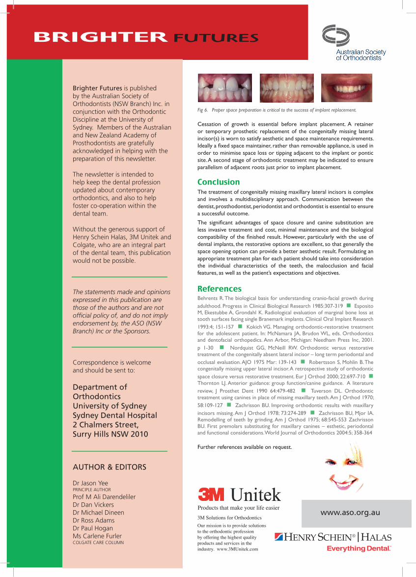
Fig 6. Proper space preparation is critical to the success of implant replacement.
Cessation of growth is essential before implant placement. A retainer or temporary prosthetic replacement of the congenitally missing lateral incisor(s) is worn to satisfy aesthetic and space maintenance requirements. Ideally a fi xed space maintainer, rather than removable appliance, is used in order to minimise space loss or tipping adjacent to the implant or pontic site. A second stage of orthodontic treatment may be indicated to ensure parallelism of adjacent roots just prior to implant placement.
ConclusionThe treatment of congenitally missing maxillary lateral incisors is complex and involves a multidisciplinary approach. Communication between the dentist, prosthodontist, periodontist and orthodontist is essential to ensure a successful outcome.
The signifi cant advantages of space closure and canine substitution are less invasive treatment and cost, minimal maintenance and the biological compatibility of the fi nished result. However, particularly with the use of dental implants, the restorative options are excellent, so that generally the space opening option can provide a better aesthetic result. Formulating an appropriate treatment plan for each patient should take into consideration the individual characteristics of the teeth, the malocclusion and facial features, as well as the patient’s expectations and objectives.
ReferencesBehrents R. The biological basis for understanding cranio-facial growth during adulthood. Progress in Clinical Biological Research 1985:307-319 n Esposito M, Ekestubbe A, Grondahl K. Radiological evaluation of marginal bone loss at tooth surfaces facing single Branemark implants. Clinical Oral Implant Research 1993:4; 151-157 n Kokich VG. Managing orthodontic-restorative treatment for the adolescent patient. In: McNamara JA, Brudon WL, eds. Orthodontics and dentofacial orthopedics. Ann Arbor, Michigan: Needham Press Inc, 2001. p 1-30 n Nordquist GG, McNeill RW. Orthodontic versus restorative treatment of the congenitally absent lateral incisor – long term periodontal and occlusal evaluation. AJO 1975 Mar: 139-143 n Robertsson S, Mohlin B. The congenitally missing upper lateral incisor. A retrospective study of orthodontic space closure versus restorative treatment. Eur J Orthod 2000; 22:697-710 n Thornton LJ. Anterior guidance: group function/canine guidance. A literature review, J Prosthet Dent 1990 64:479-482 n Tuverson DL. Orthodontic treatment using canines in place of missing maxillary teeth. Am J Orthod 1970; 58:109-127 n Zachrisson BU. Improving orthodontic results with maxillary incisors missing. Am J Orthod 1978; 73:274-289 n Zachrisson BU, Mjor IA. Remodelling of teeth by grinding. Am J Orthod 1975; 68:545-553 Zachrisson BU. First premolars substituting for maxillary canines – esthetic, periodontal and functional considerations. World Journal of Orthodontics 2004:5; 358-364
Further references available on request.
Brighter Futures is published by the Australian Society of Orthodontists (NSW Branch) Inc. in conjunction with the Orthodontic Discipline at the University of Sydney. Members of the Australian and New Zealand Academy of Prosthodontists are gratefully acknowledged in helping with the preparation of this newsletter.
The newsletter is intended to help keep the dental profession updated about contemporary orthodontics, and also to help foster co-operation within the dental team.
Without the generous support of Henry Schein Halas, 3M Unitek and Colgate, who are an integral part of the dental team, this publication would not be possible.
The statements made and opinions expressed in this publication are those of the authors and are not offi cial policy of, and do not imply endorsement by, the ASO (NSW Branch) Inc or the Sponsors.
Correspondence is welcome and should be sent to:
Department of OrthodonticsUniversity of SydneySydney Dental Hospital2 Chalmers Street, Surry Hills NSW 2010
AUTHOR & EDITORS
Dr Jason YeePRINCIPLE AUTHOR
Prof M Ali DarendelilerDr Dan VickersDr Michael DineenDr Ross AdamsDr Paul HoganMs Carlene Furler COLGATE CARE COLUMN
Products that make your life easierwww.aso.org.au
3M Solutions for Orthodontics Our mission is to provide solutions to the orthodontic profession by offering the highest quality products and services in the industry. www.3MUnitek.com
b