critical care of the lung transplant patient: the surgical ... · critical care of the lung...
TRANSCRIPT

Critical Care of the Lung Transplant Patient:
The Surgical Perspective
Shaf Keshavjee MD MSc FRCSC FACS
Program Medical Director, Surgery and Critical Care, UHN
Director, Toronto Lung Transplant Program
James Wallace McCutcheon Chair in Surgery
Professor, Division of Thoracic Surgery & Institute of
Biomaterials and Biomedical Engineering,
Vice Chair, Innovation, Department of Surgery
University of Toronto.
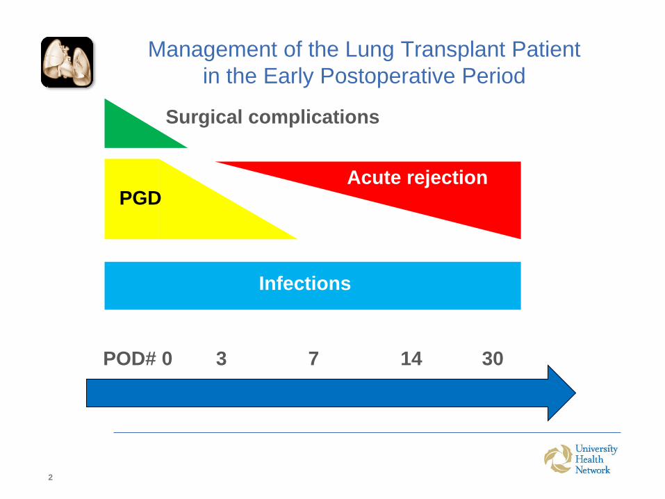
Management of the Lung Transplant Patient
in the Early Postoperative Period
2
POD# 0 3 7 14 30
PGDAcute rejection
Surgical complications
Infections

Critical Care of the Lung Transplant Patient
1. Preoperative management Donor lung, bridge to
lung transplant: ventilator, ECMO
2. Intraoperative management
3. Postoperative Management
4. Principles of Primary Graft Dysfunction
5. ECLS and Lung Transplantation in an Advanced
Lung Failure Unit

Munshi L, Keshavjee S, Cypel M. Lancet RM Feb 2013
Injury to the Donor Lung:
A Multifactorial Process

Clinical Problem - PGD
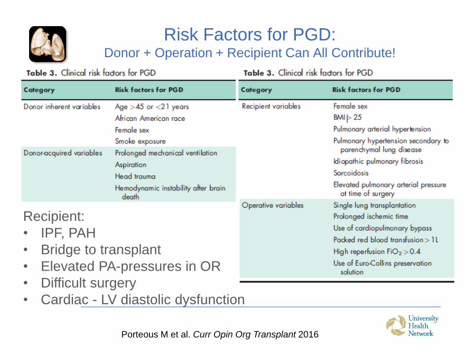
Risk Factors for PGD:Donor + Operation + Recipient Can All Contribute!
Recipient:
• IPF, PAH
• Bridge to transplant
• Elevated PA-pressures in OR
• Difficult surgery
• Cardiac - LV diastolic dysfunction
Porteous M et al. Curr Opin Org Transplant 2016

Primary Graft Dysfunction: Case
• 47 year old man
• Sarcoidosis, RVSP 83
• Bilateral lung transplant
• Difficult dissection, technically challenging operation
• Required cardiopulmonary bypass support
• Came off bypass easily
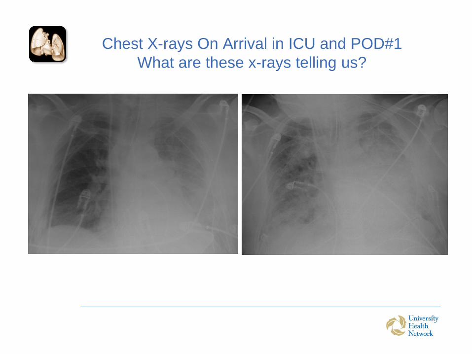
Chest X-rays On Arrival in ICU and POD#1
What are these x-rays telling us?

Primary Graft Dysfunction: Case
• 47 year old man
• Sarcoidosis, RVSP 83
• Bilateral (sequential) lung transplant
• Difficult dissection, technically challenging operation
• Required cardiopulmonary bypasssupport
• Came off bypass easily

Chest X-rays On Arrival in ICU and POD#1
What are these x-rays telling us?

Definition of Primary Graft DysfunctionInternational Society for Heart and Lung Transplantation
Grade PaO2/FiO2 Infiltrates
0 >300 Absent
1 >300 Present
2 200-300 Present
3 <200 Present
• Assessed at: T0, T24, T48, T72 hours
• Automatic Grade 3: ECMO or iNO and FiO2 > 0.5
• Nasal prongs or FiO2< 0.3: Grade 0 or 1
Christie et al, J Heart Lung Transplant 2005;24:1454-659

Primary Graft Dysfunction:
A Multifactorial Injury
• A relatively common early complication
•Severe PGD (PGD3) in 20 - 30%
• Increased : 20%
•Negative long-term effects
• Increased Chronic Lung Allograft Dysfunction (CLAD)
• Link between early innate immune injury and late
acquired immune response (rejection)
• Exclude:
•Infections
•Technical issues (e.g.pulmonary venous obstruction)
•Volume overload
•Cardiac issues
Porteous M et al. Curr Opin Org Transplant 2016

• THIS IS A LEAKY CAPILLARY SYNDROME LUNG INJURY!
• Careful fluid management to avoid lung edema, BUT
preserving adequate end-organ perfusion
• Adequate pain management
• Early weaning of sedation Early extubation
• Early mobilization
• Immunosuppression
• Antibiotic prophylaxis
ICU Management during the first 72 h
Overall Goals

RV
Pre-load
After-load
Contractility
PVR (RV after-load)
Post LTx
LV
Pre-load
After-load
Contractility
SVR
PAS and PAD
PA flow=cardiac output
CVP (RV pre-load)
PCWP (LV pre-load)
Hemodynamic monitoring – You Need a
Swan Ganz Catheter
LV-RV Interaction

RV Contractility
(inotropes)
PVR (RV after-load)
Post DLTx
PGD 3
LV After-load: MAP 65-75 mmHg
(vasopressors, vasoldilators, sedation,
PEEP)
Contractility (inotropes)
mPAP < 20 mmHg
CI= 2.2-2.5
CVP (RV pre-load) <8
mmHg(fluids/furosemide, PEEP)
PAOP (LV pre-load) < 10
mmHg
(fluids/furosemide, PEEP)
LV-RV interaction
PAD-PAOP, pO2, pCO2, MV
settings, sedation, iNO
End organ perfusion monitoring:SvO2, lactate, u/o, pH, HCO3-, temperature
Lung function monitoring: P/F, Vd/Vt, compliance, CXR, EVLW (?)

Monitoring
Monitor and record the following parameters as indicated below or more
frequently if clinically required:
Parameter Target
MAP (SAP, DAP) (q1h) 65 - 75 mmHg
HR (q1h) 60 - 100 b/min
CI (q4h) 2.2 – 2.5 L/min/m2
mPAP (PAS, PAD) (q1h) ≤ 20 mmHg
CVP (q1h) < 8 mmHg
PCWP (q4h) < 10 mmHg
Calculate - and look at - CI, PVR and SVR q 4h
PAD-PCWP (q4h) < 5 mmHg
SvO2 (q8h) > 60%
pH, PaO2, PaCO2 (HCO3) (q8h) > 7.30, > 60 mmHg, 30 - 45 mmHg
Lactate (q8h) ≤ 2.5 mmol/L
U/O (q1h) > 0.5 ml/kg/hr
Temperature (q1h) 35 - 38 °C

Goals of Mechanical Ventilation Management
• Provide adequate support for gas exchange
• Minimize respiratory distress
• Protective - minimize ventilator-induce lung injury
• Optimize alveolar recruitment
• Facilitate bronchial toilet
• Facilitate early weaning

Initial mechanical ventilation settings
Strategy Goals
Pressure controlled ventilation Early weaning
Minimize tidal volume/distending pressure Vt ≤ 6 cc/kg (PBW D/R)
Pplat < 30 cmH2O
Minimize FiO2 SpO2 ≥ 90%
Optimize PEEP Maintain alveolar
recruitment
PEEP/FiO2 table: FiO2 0.3 0.4 0.5 0.6 0.7 0.8-1
PEEP 5 5-8 8-10 10-12 12-14 14-15
Minimize respiratory rate: RR < 25 breaths/min
pH > 7.30
Maximize expiratory time: I:E ≤ 1:2 (1:3, 1:4, etc.)

PGD Treatment
• Manage leaky capillary syndrome
• pRBCs (when needed) and 25% Albumin are agents of choice
for intravascular volume expansion in the early phase
• Diuresis (euvolemia – NB nephrotoxic immunosuppressants
and antibiotics)
• Protective ventilation
• iNO (10 ppm for oxygenation, 40ppm for PAP)
• PGE1, other agents in the pipeline (targetting inflammation,
free radicals, early innate and acquired immune responses)
• ECLS

Ventilator Management - WeaningP/F ≥ 200:
1. Optimize analgesia
2. Assess CXR, endotracheal secretions (suctioning), and chest tube output/leak
3. Optimize hemodynamic management
4. Wean sedation: manage agitation / sedation
5. Wean iNO (5 ppm/hr, when iNO is 5 ppm proceed with slower wean)
6. Wean MV to assisted modality of MV (e.g. PSV) if tolerated, and target:
1. 6-8 cc/kg PBW D/R
2. RR < 25 breaths/min
7. Follow the spontaneous breathing trial (SBT) policy (twice daily )
8. After failure of > 3 SBT, after > 72 hours of continuous MV, or after failed
extubation: consider tracheostomy allows easier mobility, improved patient
comfort, oral hygiene, and clearance of pulmonary secretions

ManagementP/F < 200:
• Optimize sedation/analgesia
• Assess CXR, endotracheal secretions (suctioning), and chest tube output/leak
• Optimize hemodynamic management
• Manage / treat agitation
• Maintain controlled modality of MV with protective settings (see initial MV
settings)
• Consider iNO – it works in this population to improve V/Q matching
P/F < 150:
• Contact lung transplant surgeon / team
• Consider neuro-muscular blocking agents
P/F < 100:
• ECLS
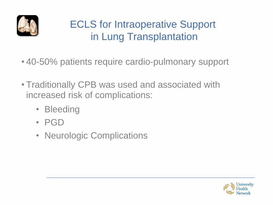
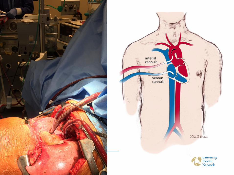
ECLS for Intraoperative Support
in Lung Transplantation
• 40-50% patients require cardio-pulmonary support
• Traditionally CPB was used and associated with increased risk of complications:
• Bleeding
• PGD
• Neurologic Complications

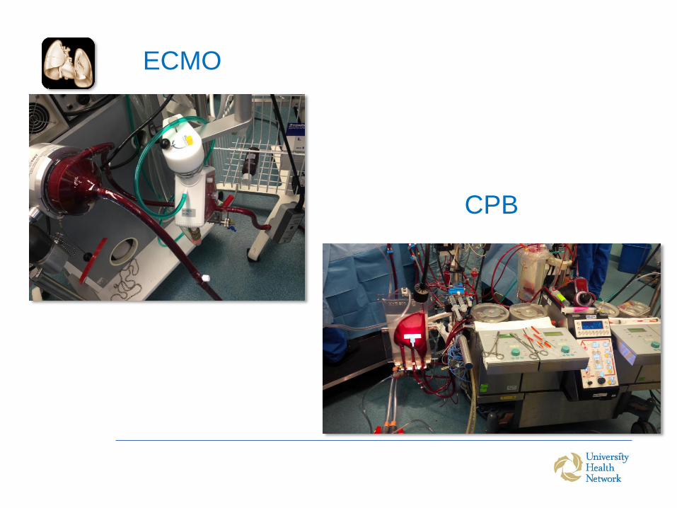
ECMO
CPB

Outcome CPB ECMO p
Length of MV (days) 7.5 (2-18) 3 (2.5-5) 0.005
ICU stay (days) 9.5 (3-210 5 (3-9) 0.026
Hospital Stay (days) 27 (17-42) 19 (14-30) 0.029
ECLS post-op 5 (7.5%) 0 0.166
Dialysis requirement 12 (18%) 3 (9%) 0.37
Reoperation (bleeding) 18 (27%) 3 (9%) 0.04
90-day mortality 10 (15%) 2 (6%) 0.32
J Thor Cardiovasc Surgery 2015

12-month Survival After Lung Transplant

ECLS for Primary Graft
Dysfunction (PGD)

Hypercapnic failure Hypoxemic failure
Veno-Venous
(high flow)
Veno-ArterialPA-LA iLA
(pumpless)
PAH (severe RV
dysfunction)
Veno-Venous
(low flow)
Current Algorithm for Extracorporeal
Lung Support (ECLS)
VV + Septostomy

Case 1
• 30Y, male with CF
• BLT
• After reperfusion of 1st lung patient become hypoxemic
• VA ECMO for intraoperative support
• Wean VA ECMO at the end of transplant
• Severe hypoxemia and respiratory acidosis develops next few
minutes
• PAP 52/30 mmHg
• Moderate requirements for vasopressors and inotropes
• Pulmonary edema evident on bronchoscopy
• What’s next?

Some options…
• 1) Trial of iNO
• 2) Leave Chest Open
• 3) Initiate VV ECMO
• 4) Initiate VA ECMO

VV or VA ECMO for PGD?

VA ECMO Advantages
• Both respiratory and hemodynamic
support
• Ability to decrease pulmonary flow and
pulmonary pressure
• Ultimate protection in severe leaky
capillary syndrome – IR injury
Marasco et al. 2011
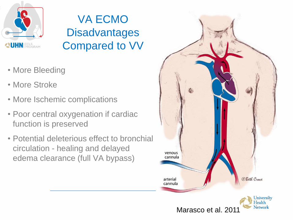
VA ECMO
Disadvantages
Compared to VV
• More Bleeding
• More Stroke
• More Ischemic complications
• Poor central oxygenation if cardiac
function is preserved
• Potential deleterious effect to bronchial
circulation - healing and delayed
edema clearance (full VA bypass)
Marasco et al. 2011
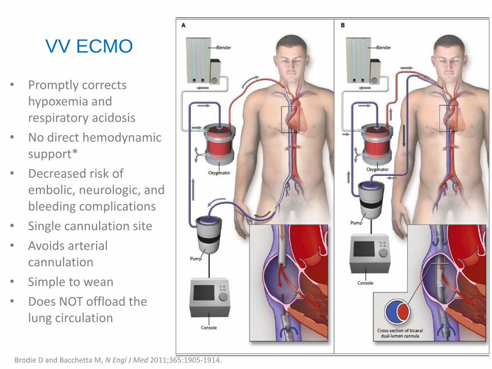
• Promptly corrects hypoxemia and respiratory acidosis
• No direct hemodynamic support*
• Decreased risk of embolic, neurologic, and bleeding complications
• Single cannulation site
• Avoids arterial cannulation
• Simple to wean
• Does NOT offload the lung circulation
VV ECMO
Brodie D and Bacchetta M, N Engl J Med 2011;365:1905-1914.
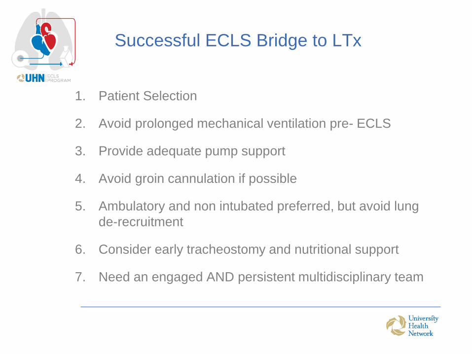
Successful ECLS Bridge to LTx
1. Patient Selection
2. Avoid prolonged mechanical ventilation pre- ECLS
3. Provide adequate pump support
4. Avoid groin cannulation if possible
5. Ambulatory and non intubated preferred, but avoid lung
de-recruitment
6. Consider early tracheostomy and nutritional support
7. Need an engaged AND persistent multidisciplinary team

32 yo female with Cystic Fibrosis
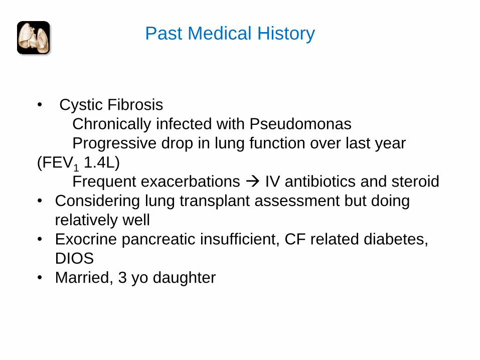
• Cystic Fibrosis
Chronically infected with Pseudomonas
Progressive drop in lung function over last year
(FEV1 1.4L)
Frequent exacerbations IV antibiotics and steroid
• Considering lung transplant assessment but doing
relatively well
• Exocrine pancreatic insufficient, CF related diabetes,
DIOS
• Married, 3 yo daughter
Past Medical History
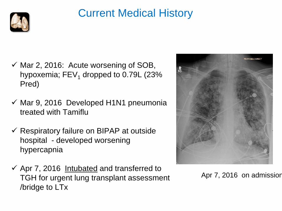
Mar 2, 2016: Acute worsening of SOB,
hypoxemia; FEV1 dropped to 0.79L (23%
Pred)
Mar 9, 2016 Developed H1N1 pneumonia
treated with Tamiflu
Respiratory failure on BIPAP at outside
hospital - developed worsening
hypercapnia
Apr 7, 2016 Intubated and transferred to
TGH for urgent lung transplant assessment
/bridge to LTx
Current Medical History
Apr 7, 2016 on admission
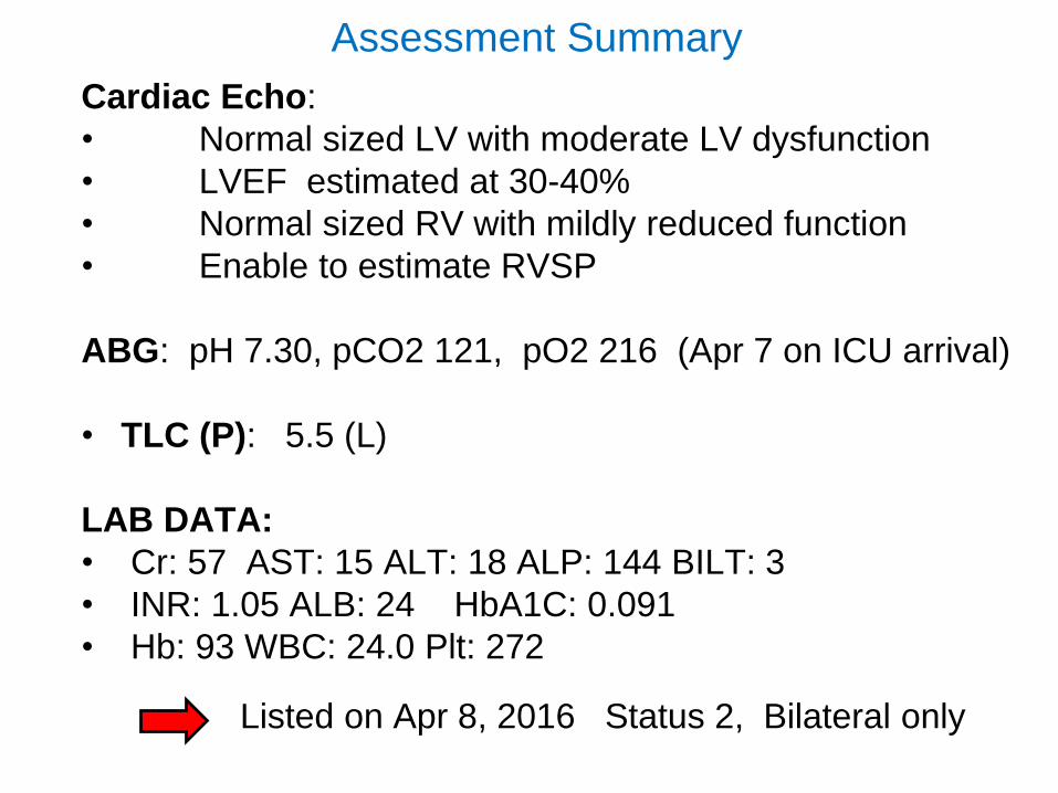
Assessment Summary
Listed on Apr 8, 2016 Status 2, Bilateral only
Cardiac Echo:
• Normal sized LV with moderate LV dysfunction
• LVEF estimated at 30-40%
• Normal sized RV with mildly reduced function
• Enable to estimate RVSP
ABG: pH 7.30, pCO2 121, pO2 216 (Apr 7 on ICU arrival)
• TLC (P): 5.5 (L)
LAB DATA:
• Cr: 57 AST: 15 ALT: 18 ALP: 144 BILT: 3
• INR: 1.05 ALB: 24 HbA1C: 0.091
• Hb: 93 WBC: 24.0 Plt: 272
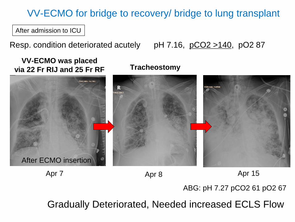
VV-ECMO was placed
via 22 Fr RIJ and 25 Fr RF
VV-ECMO for bridge to recovery/ bridge to lung transplant
Resp. condition deteriorated acutely pH 7.16, pCO2 >140, pO2 87
Apr 7
Gradually Deteriorated, Needed increased ECLS Flow
Apr 8 Apr 15
After admission to ICU
Tracheostomy
ABG: pH 7.27 pCO2 61 pO2 67
After ECMO insertion

• Developed septic shock
• Bacteremia with gram-negative
Pseudomonas
• 3 vasopressors at maximum dose
VASOPRESSIN, LEVOPHED, and
ADRENALIN
• Despite VV ECMO (flow of 7 L/min),
Significant hypoxemia (without pump
recirculation)
• ABG: pH 7.14 pCO2 52, pO2 60
• Refractory vasodilation - sepsis
Septic shock and bacteremia
April 17, 2016

Remove the Septic Source: Bilateral Pneumonectomy
Switch to Central VA-ECMO (22Fr
aortic cannula, 34/46 two-stage IVC
venous cannula
Right-sided pneumonectomy first, then
left pneumonectomy
Insertion of Right lung PA-LA
Novalung
Outflow: Pulmonary arterial (34 Fr
single-stage venous cannula)
Inflow: right superior pulmonary
vein (28 Fr Pacifico)
April 17, 2016
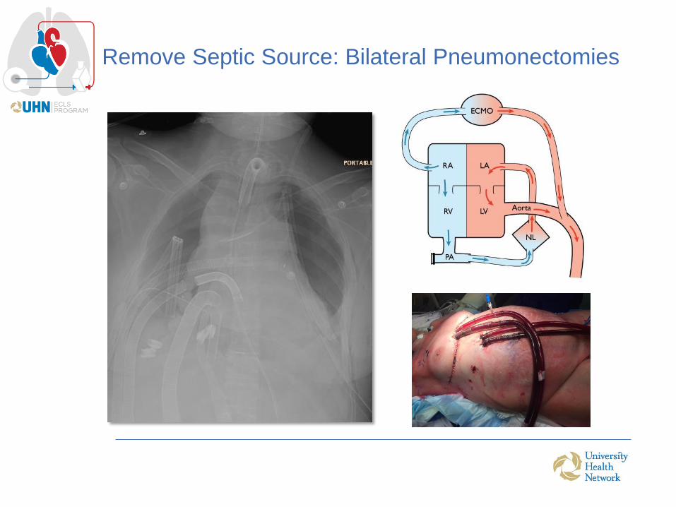
Remove Septic Source: Bilateral Pneumonectomies

Immediate Postop BLT
Bilateral Lung Transplant
• On central VA-ECMO
• Left side implantation first ( CIT: 3h 15
min, WIT: 49 m)
• Removed the PA cannula from right
PA (for the PA-LA Novalung)
• Removed the LA cannula from right
superior pulmonary vein
• Right lung implantation ( CIT: 4h 45
min, WIT: 50 min)
• 6 U pRBC and 2 U platelets
April 22, 2016 (5 days after pneumonectomy)
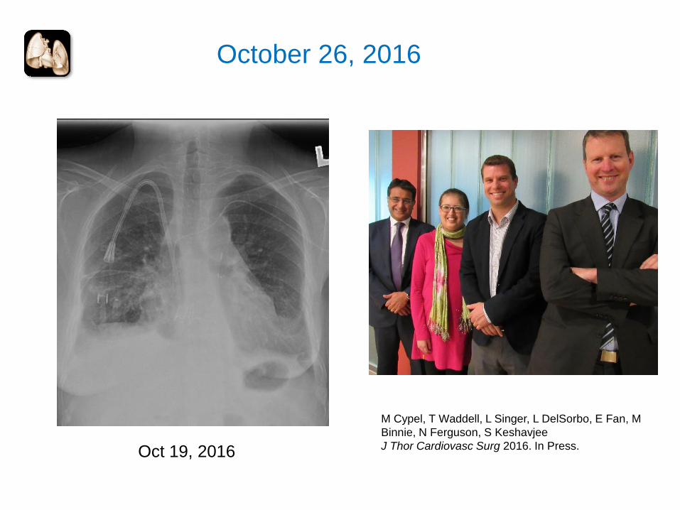
October 26, 2016
Oct 19, 2016
M Cypel, T Waddell, L Singer, L DelSorbo, E Fan, M
Binnie, N Ferguson, S Keshavjee
J Thor Cardiovasc Surg 2016. In Press.

The Toronto Lung Transplant Team
2016
