criticalroleofthe -subunitcdc50ainthestable expression ... · criticalroleofthe...
TRANSCRIPT

Critical Role of the �-Subunit CDC50A in the StableExpression, Assembly, Subcellular Localization, and LipidTransport Activity of the P4-ATPase ATP8A2*□S
Received for publication, February 10, 2011, and in revised form, March 14, 2011 Published, JBC Papers in Press, March 18, 2011, DOI 10.1074/jbc.M111.229419
Jonathan A. Coleman‡1 and Robert S. Molday‡§2
From the Departments of ‡Biochemistry and Molecular Biology and §Ophthalmology and Visual Sciences, Centre for MacularResearch, University of British Columbia, Vancouver, British Columbia V6T 1Z3, Canada
P4-ATPases have been implicated in the transport of lipidsacross cellular membranes. Some P4-ATPases are known toassociate with members of the CDC50 protein family. Previ-ously,wehave shown that theP4-ATPaseATP8A2purified fromphotoreceptor membranes and reconstituted into liposomescatalyzes the active transport of phosphatidylserine acrossmembranes. However, it was unclear whether ATP8A2 func-tioned alone or as a complex with a CDC50 protein. Here, weshow bymass spectrometry andWestern blotting using newlygenerated anti-CDC50A antibodies that CDC50A is associ-ated with ATP8A2 purified from photoreceptor membranes.ATP8A2 expressed in HEK293T cells assembles with endoge-nous or expressed CDC50A, but not CDC50B, to generate a het-eromeric complex that actively transports phosphatidylserineand to a lesser extent phosphatidylethanolamine across mem-branes. Chimera CDC50 proteins in which various domains ofCDC50B were replaced with the corresponding domains ofCDC50Awere used to identify domains important in the forma-tion of a functional ATP8A2-CDC50 complex. These studiesindicate that both the transmembrane and exocytoplasmicdomains of CDC50A are required to generate a functionallyactive complex. The N-terminal cytoplasmic domain ofCDC50A appears to play a direct role in the reaction cycle.Mutagenesis studies further indicate that theN-linked oligosac-charide chains of CDC50A are required for stable expression ofan active ATP8A2-CDC50A lipid transport complex. Together,our studies indicate that CDC50A is the �-subunit of ATP8A2and is crucial for the correct folding, stable expression, exportfrom endoplasmic reticulum, and phosphatidylserine flippaseactivity of ATP8A2.
P4-ATPases comprise a subfamily of P-type ATPases impli-cated in the active transport or flipping of aminophospholipids
from the exocytoplasmic to the cytoplasmic side of cell mem-branes (1–6). This activity generates and maintains amino-phospholipid asymmetry, which plays a crucial role in such cel-lular processes as membrane stability and impermeability,vesicle-mediated protein transport, blood coagulation, estab-lishment of cellular polarity, recognition of apoptotic cells, cel-lular division, sperm capacitation, and regulation of membraneprotein function (7–9). The importance of P4-ATPases is high-lighted by the finding that mutations in several P4-ATPasesincluding ATP8B1 and ATP8A2 have been linked to severehuman diseases (10–13).A number of yeast, plant, and mammalian P4-ATPases are
known to associate with members of the Cdc50 family of pro-teins (14–17). There are three members of this family in yeast(Cdc50p, Crflp, and Lem3/Ros3p) and three in mammals(CDC50A, CDC50B, and CDC50C). These proteins containtwo transmembrane segments separated by a relatively largeglycosylated exocytoplasmic domain. Several cellular studiesindicate that CDC50 proteins play a crucial role in the export ofspecific P4-ATPases from the ER of cells (14–19). CDC50 pro-teins also have been suggested to play a direct role in the reac-tion cycle of P4-ATPases (20, 21). In yeast, the P4-ATPaseDrs2p requiresCdc50p for phosphorylation of the key aspartateresidue involved in the ATPase catalytic reaction. The affinityof Drs2p for Cdc50pwas also found to change during the trans-port cycle. Additionally, phosphorylation of the catalyticallyimportant aspartate residue of the human P4-ATPases,ATP8B1 and ATP8B2, was dependent on the CDC50 subunit.On the basis of these studies, it has been proposed that theCDC50 proteins serve as the �-subunit of P4-ATPases akin tothe �-subunit of the Na�/K� ATPase (9). However, althoughan association between the catalytic subunit of P4-ATPases andCDC50 proteins has been observed in heterologous cell expres-sion systems, their direct association in mammalian tissues hasnot been demonstrated, and the domains of CDC50 proteinsresponsible for their interactionwith P4-ATPases have yet to bedetermined.Recently, we have purified, localized, and characterized the
functional properties of mammalian ATP8A2, a member of theP4-ATPase family that shares a 67% amino acid sequence iden-tity with ATP8A1, the founding member of mammalian P4-ATPases (5). ATP8A2 expression was detected in the retina,testes, and brain (5, 13). In the retina,ATP8A2 is localized in theouter segment compartment of rod and cone photoreceptors
* This work was supported by Canadian Institutes of Health Research GrantMOP-106667.
□S The on-line version of this article (available at http://www.jbc.org) containssupplemental Tables S1 and S2 and Figs. S1–S4.
1 Supported by a University of British Columbia and National Sciences andEngineering Council predoctoral studentship.
2 Canada Research Chair in Vision and Macular Degeneration. To whom cor-respondence should be addressed: Dept. of Biochemistry and MolecularBiology, 2350 Health Sciences Mall, University of British Columbia, Vancou-ver, British Columbia V6T 1Z3, Canada. Tel.: 604-822-6173; Fax: 604-822-5227; E-mail: [email protected].
THE JOURNAL OF BIOLOGICAL CHEMISTRY VOL. 286, NO. 19, pp. 17205–17216, May 13, 2011© 2011 by The American Society for Biochemistry and Molecular Biology, Inc. Printed in the U.S.A.
MAY 13, 2011 • VOLUME 286 • NUMBER 19 JOURNAL OF BIOLOGICAL CHEMISTRY 17205
by guest on June 21, 2018http://w
ww
.jbc.org/D
ownloaded from

where it functions in the transport of PS3 and to a lesser extentPE across the photoreceptor discmembrane (5). In a proteomicstudy, unique peptides belonging to CDC50A were detected inphotoreceptor outer segment preparations (22), but the possi-ble association of CDC50A with ATP8A2 was not investigated.In this study, we show that ATP8A2 is present as a hetero-
meric complex with CDC50A in photoreceptor outer segmentsaswell asHEK293T cells expressingATP8A2. Co-expression ofATP8A2 with CDC50A, but not CDC50B, not only promotesthe translocation of ATP8A2 from the endoplasmic reticulum(ER) to the Golgi but also significantly enhances the yield of afunctional ATP8A2-CDC50A aminophospholipid transporter.Chimera proteins in which various domains of CDC50B havebeen replaced with corresponding domains of CDC50A havebeen used to define regions ofCDC50A that are required for theformation of a functionally active ATP8A2-CDC50A complex.Finally, we have investigated the effect of N-linked glycosyla-tion of CDC50A on the stable expression, subunit association,and functional properties of the ATP8A2-CDC50A complex.
EXPERIMENTAL PROCEDURES
Materials—1,2-Dioleoyl-sn-glycero-3-phospho-ethanol-amine, 1,2-dioleoyl-sn-glycero-3-phosphoserine, L-�-phosph-atidylcholine (egg, chicken), C6 NBD-PS, C6 NBD-PE, and C6NBD-PC were purchased from Avanti Polar Lipids (Alabaster,AL). ATP, AMP-PNP, sodiumorthovanadate, and n-octyl-�-D-glucopyranoside were purchased from Sigma, dithionite wasfrom Fisher (Waltham, MA), CHAPS was from Anatrace(Maumee, OH), and the synthetic 6C11 and 7F4 peptides, Ac-RDRLLKRLS and Ac-AKDEVDGGP, respectively, were pur-chased from Biomatik (Cambridge, Canada). Complete inhibi-tor was from Roche Applied Science. The Rho 1D4 antibodywas obtained from UBC-UILO (Vancouver, Canada), and the1D4 peptide was from the Nucleic Acid Protein Service Unit(Vancouver, Canada). The restriction enzymes were fromNewEngland Biolabs (Ipswich, MA). The primers were purchasedfrom Integrated DNA Technologies (Coralville, IA).Solutions—The compositions of the buffers were as follows:
Buffer A: 50 mM HEPES, pH 7.5, 150 mM NaCl, 5 mM MgCl2, 1mM DTT, 20 mM CHAPS, 0.5 mg/ml egg PC, complete inhibi-tor; Buffer B: 50mMHEPES, pH7.5, 150mMNaCl, 5mMMgCl2,1 mMDTT, 10 mMCHAPS, 0.5 mg/ml egg PC; Buffer C, 50 mM
HEPES, pH 7.5, 150 mM NaCl, 5 mM MgCl2, 1 mM DTT, 0.75%n-octyl-�-D-glucopyranoside, 0.5 mg/ml egg PC; Buffer D: 50mM HEPES, pH 7.5, 150 mM NaCl, 12.5 mM MgCl2, 10 mM
CHAPS, and 1 mM DTT; Buffer E: 50 mM HEPES, pH 7.5, 150mM NaCl, 5 mM MgCl2, 1 mM DTT, 1% n-octyl-�-D-glucopy-ranoside, 5mg/ml egg PC, 10% sucrose; Buffer F: 10mMHEPES,
pH 7.5, 150 mM NaCl, 5 mM MgCl2, 1 mM DTT, 10% sucrose;and PBS: 10 mM phosphate, pH 7.4, 140 mM NaCl, 3 mM KCl.DNA Constructs—Bovine Atp8a2 containing a 1D4 tag in
pcDNA3 (Invitrogen) was described previously (5). This con-struct was used as a PCR template to generate each Atp8a2construct described. The cDNA of bovine Cdc50a (IMAGE:8284976) and human Cdc50b (IMAGE: 8322611) were pur-chased from Open Biosystems (Huntsville, AL). Restrictionsites and epitope tags were introduced by PCR. Full-lengthAtp8a2 without a tag was PCR-amplified and cloned intopcDNA3 using BamHI and NotI restriction sites. Full-lengthCdc50a with a 1D4 or Myc tag was cloned into pcDNA3 usingthe HindIII and XhoI restrictions sites. Cdc50b with a 1D4 tagwas also cloned into pcDNA3 usingHindIII andXhoI sites. The1D4-tagged constructs contained a 9-amino acid C-terminaltag (TETSQVAPA). The Myc-tagged constructs contained a10-amino acid C-terminal tag (EQKLISEEDL). Chimeras ofCdc50a and Cdc50b were constructed from a 1D4-taggedCdc50b construct that had been cloned into a modifiedpcDNA3 vector by EcoRI and NotI. Silent restriction sites wereinserted into Cdc50b by mutagenesis to facilitate cloning ofindividual domains. The ECD chimera contained amino acids1–57 and 301–351 of CDC50B and amino acids 73–308 ofCDC50A. The ECD/TM chimera contained amino acids 1–33and 341–351 of CDC50B and amino acids 49–348 of CDC50A.The ECD/TM/C chimera contained amino acids 1–33 ofCDC50B and amino acids 49–361 of CDC50A. TheN-terminalchimera contained amino acids 1–47 of CDC50A and aminoacids 33–351 of CDC50B. The M1 chimera contained aminoacids 1–33 and 55–351 of CDC50B and amino acids 49–69 ofCDC50A. The M2 chimera contained amino acids 1–301 and341–351 of CDC50B and amino acids 310–348 of CDC50A.TheM1/M2 chimera contained amino acids 1–33, 55–301, and341–351 of CDC50B and amino acids 49–69 and 310–348 ofCDC50A. GST fusion constructs containing amino acids 1–40and 194–283 of CDC50A were cloned into pGEX-4T-1 (GEHealthcare) using the BamHI and EcoRI restriction sites. Site-directed mutations were created using the QuikChangemutagenesis kit from Agilent Technologies (Santa Clara, CA).All of the constructs were verified by DNA sequencing (Euro-fins MWGOperon, Huntsville, AL).Gene Expression by RT-PCR—RNAwas isolated from tissues
of 6-month-old C57/B6 mice and HEK293T cells (AmericanTypeCultureCollection,Manassas, VA) using the guanidiniumthiocyanate phenol chloroform method (23). Random primedcDNA was made using the first strand cDNA synthesis kit (GEHealthcare). Cdc50a, Cdc50b, and Cdc50c gene expression wasmeasured using gene specific primers. GapdH was used as aloading control. GapdHPCRswere run for 25 cycles. The cdc50genes were amplified for 25 cycles, and then 2�l of the reactionwas removed and reamplified for an additional 25 cycles. Taqpolymerase (New England Biolabs) was used for amplification,and the primers were annealed at 55 °C. Primer sequences areavailable (supplemental Table S1).Generation of Monoclonal Antibodies against CDC50A—
Fragments of bovine CDC50A (amino acids 1–40 and 194–283) were cloned in framewithGST and expressed and purifiedfrom Escherichia coli BL21 on a glutathione-Sepharose-4B col-
3 The abbreviations used are: PS, phosphatidylserine; PE, phosphatidyletha-nolamine; PC, phosphatidylcholine; ROS, rod outer segments; NBD, 7-nit-robenz-2-oxa-1,3-diazol-4-yl; NBD-PS, 1-oleoyl-2-{6-[7-nitro-2–1,3-benzoxadiazol-4-yl)amino]hexanoyl}-sn-glycero-3-phosphoserine; NBD-PE, 1-oleoyl-2-{6-[7-nitro-2–1,3-benzoxadiazol-4-yl)amino]hexanoyl}-sn-glycero-3-phosphoethanolamine; NBD-PC, 1-oleoyl-2-{6-[7-nitro-2–1,3-benzoxadiazol-4-yl)amino]hexanoyl}-sn-glycero-3-phosphocholine; AMP-PNP, adenylyl-imidodiphosphate; CHAPS, 3-[(3-cholamidopropyl)dimet-hylammonio]-1-propanesulfonic acid; ER, endoplasmic reticulum, TM,transmembrane; ECD, exocytoplasmic domain.
Interaction of CDC50A with ATP8A2
17206 JOURNAL OF BIOLOGICAL CHEMISTRY VOLUME 286 • NUMBER 19 • MAY 13, 2011
by guest on June 21, 2018http://w
ww
.jbc.org/D
ownloaded from

umn (GE Healthcare). Hybridoma cell lines were generatedfrom Swiss Webster mice (Charles River, Wilmington, MA)immunized with a mixture of the GST fusion proteins as previ-ously described (24). Positive clones were identified by screen-ing for immunoreactivity against purified 1D4-taggedCDC50Aprotein expressed inHEK293T cells by ELISA. Immunoreactiv-ity was confirmed onWestern blots of bovine ROS. The epitopefor the Cdc50–7F4 antibody was identified by measuring thereactivity of the antibody to a series of overlapping 9-aminoacid N-terminal peptides using the SPOTs kit (Sigma).Expression of ATP8A2 and CDC50A in HEK293T and Cos-7
Cells—The cells were maintained in Dulbecco’s modifiedEagle’s medium supplemented with 10% fetal bovine serum,100 units/ml penicillin, 100 �g/ml streptomycin, 2 mM L-glu-tamine, and 1.25 �g/ml fungizone. HEK293T cells in 10-cmdishes were transfected at 30% confluence with 10 �g ofpcDNA3 containing either Atp8a2 or Cdc50a by the calciumphosphate method (25) and harvested 48 h later. In some cases,the cells were co-transfected with both Atp8a2 and Cdc50a bymixing 10 �g of each plasmid together followed by calciumphosphate precipitation. The membranes from HEK293T cellswere prepared as described (26). Cos-7 cells (American TypeTissue Collection) were transfected in six-well plates contain-ing polylysine-treated coverslips with 2.5 �g of each plasmid.Immunofluorescence Microscopy—Cryosections of bovine
retina tissue were prepared by fixing bovine eyes in 4% para-formaldehyde, 100 mM phosphate buffer, pH 7.4, for 1 h. Cryo-sections (10 �m) were cut followed by blocking and permeabi-lization with 10% normal goat serum and 0.2% Triton X-100 inphosphate buffer for 30 min. The sections were then labeledovernight at room temperature with Atp2F6 hybridoma super-natant diluted 2:3 in phosphate buffer containing 2.5% normalgoat serum and 0.1%TritonX-100 or purifiedCdc50–7F4 anti-body at a concentration of 1 �g/ml. In control studies, Cdc50–7F4 antibody labeling was blocked by preabsorption with 7F4peptide at a concentration of 0.2 �g/�l for 30 min. Cos-7 cellstransfected with 1D4-tagged Atp8a2 or Myc-tagged Cdc50awere labeled with either Rho-1D4 hybridoma culture fluid(1:500) or Myc antibody diluted 1:200 (ab10910; Abcam, Cam-bridge,MA). The cells were also labeled with antibodies againstcalnexin (Abcam, ab13504) or GM130 (Sigma; G7295) diluted1:200 and 1:300, respectively. Cos-7 cells transfected withuntagged Atp8a2 and 1D4 tagged CDC50A chimeras werelabeled with Atp2F6 and GM130 antibodies. The samples werewashed with phosphate buffer and labeled for 1 h with Cy3- orAlexa-488 tagged goat anti-mouse Ig secondary antibody(diluted 1:1000) and counterstained with DAPI. Double label-ing studies were performed with anti-mouse secondary andAlexa 568 goat anti-rat Ig, Alexa 594 goat anti-rabbit Ig, orAlexa 488 anti-rabbit (diluted 1:1000). The samples were exam-ined on a Zeiss LSM 700 confocal scanning microscope.Purification of ATP8A2-CDC50AComplex—ROSwere puri-
fied by sucrose gradient centrifugation (27). The Rho-1D4,Atp6C11, and Cdc50–7F4 immunoaffinity columns were pre-pared as previously described (28). Purification of ATP8A2wasperformed as described earlier (5). Briefly, 3.5 mg of ROS or a10-cm dish of HEK293T cells were lysed in 1 ml of Buffer A for30 min with stirring at 4 °C. Following solubilization, insoluble
material was removed by centrifugation at 100,000 � g for 10min. The detergent-solubilized fraction was added to�30�l ofpacked immunoaffinity matrix that had been pre-equilibratedin Buffer A. After a 2-h incubation, the matrix was washed sixtimes with 500 �l of Buffer B and eluted twice for 30 min in 25�l of Buffer B containing 0.2 mg/ml 1D4 or 6C11 peptideswhere appropriate. Protein was eluted from the Cdc50–7F4column in Buffer B containing 2% SDS instead of 10 mM
CHAPS. For reconstitution experiments, the columns werewashed and eluted in Buffer C with the appropriate amount ofpeptide.ATPase Activity Assay—ATPase activity was measured as
described (5) at 37 °C for 15min and stopped with 6% SDS. Theamount of phosphate release wasmeasured by the colorimetricmethod (29). Unless otherwise stated, ATP8A2 was assayed inthe presence of 2.5 mg/ml lipid containing 100% PC, 10% PS,and 90% PC or 40% PE and 60% PC (w/w) in Buffer Dwith 5mM
ATP.Reconstitution of ATP8A2-CDC50A Complex—Reconstitu-
tion was performed as described (5). Briefly, �5 �g of purifiedATP8A2-CDC50A in 150 �l of Buffer C was mixed 1:1 withBuffer E containing either 2.5% (w/w) NBD-PC, NBD-PE, orNBD-PS followed by dialysis overnight against 1 liter of Buffer Fto remove the detergent.FlippaseAssay—Flippase assayswere performed as described
(5, 30, 31). Reconstituted ATP8A2 in 30 �l was mixed witheither ATP or AMP-PNP to a final concentration of 0.5 mM
with 50 �l of Buffer F and incubated at 23 °C for 2.5 min. Fol-lowing incubation, 0.95 ml of Buffer F was added, and the sam-ple was transferred to a cuvette (path length, 1 cm) and read ina fluorescence spectrophotometer (Varian, Palo Alto, CA)using excitation and emission wavelengths of 478 and 540 nm,respectively. After 2.5min, sodiumdithionite in 1 MTris buffer,pH10,was added to a final concentration of 2mM.After 10min,a stable base line was reached. Triton X-100 was added to finalconcentration of 1%, and the sample was read for an additional1.5 min. The percentage of NBD lipid on the outside of theproteoliposomes was calculated using the following formula,
%NBDout � ��FT � FD���FT � F0�� � 100 (Eq. 1)
where FT is the total fluorescence of the sample before dithio-nite treatment, FD is the fluorescence of the sample after dithio-nite treatment, and F0 is the fluorescence after detergent solu-bilization. The percentage of flipped lipid was calculated fromthe difference between the transbilayer distribution of NBDlipids with ATP and AMP-PNP treatment.Endoglycosidase Treatment of Rod Outer Segments—Ap-
proximately 50�g of ROSwere denatured at room temperatureand subjected to PNGase F treatment at 37 °C for 1 h accordingto the manufacturer’s instructions (New England Biolabs). Thesame amount of solubilized ROS in 10 mM CHAPS and 50 mM
NaH2PO4, pH 5.0, was treated with neuraminidase (RocheApplied Science) for 1 h followed by O-glycosidase (Sigma) for1 h according to the manufacturer’s instructions.Membrane Topology and Glycosylation Site Prediction—
Membrane topology prediction was made using the TMHMMserver, version 2.0 (32). Prediction of N-linked and O-linked
Interaction of CDC50A with ATP8A2
MAY 13, 2011 • VOLUME 286 • NUMBER 19 JOURNAL OF BIOLOGICAL CHEMISTRY 17207
by guest on June 21, 2018http://w
ww
.jbc.org/D
ownloaded from

glycosylation was made using NetNGlyc 1.0 and NetOGlyc 3.1servers, respectively (33, 34).SDS-PAGE andWestern Blots—The proteins were separated
by SDS gel electrophoresis on 9% polyacrylamide gels andeither stained with Coomassie Blue or transferred onto Immo-bilon FL membranes (Millipore, Bedford, MA) in buffer con-taining 25 mM Tris, pH 8.3, 192 mM glycine, 10% methanol.Immobilon membranes were blocked with 1% milk in PBS for30 min; incubated with antibody supernatant typically diluted1:100 for the Rho 1D4, 1:25 for Cdc50–9C9 and Atp6C11, and1:10 for Cdc50–7F4 in PBS for 40min; washed thoroughly withPBST (PBS containing 0.05% Tween 20); incubated for 40 minwith secondary antibody, goat anti-mouse conjugated with IRdye 680, or IR dye 800 (LI-COR, Lincoln, NE) diluted 1:20,000in PBST containing 0.5% milk; and washed with PBST prior todata collection on a LI-COR Odyssey infrared imaging system.The relative amounts of ATP8A2 were quantified on Westernblots by LI-COR densitometry measurements and normalizedto �-actin (Abcam, ab1801). Protein concentrations were esti-mated by Coomassie Blue staining of SDS-PAGE gels usingknown amounts of bovine serum albumin as a standard.Tryptic Digestion and Mass Spectrometry—SDS-PAGE gels
were divided into several equal slices and digested with trypsinaccording to established protocol (35). Peptides were concen-trated using C18 stop and go extraction (STAGE) tips. The pep-tides were subjected to LC-MS/MS as described (22) using anLTQ-Orbitrap system (ThermoFisher, Bremen, Germany).The peptides were identified using the Mascot search program(36).
RESULTS
Identification of CDC50A as the �-Subunit for ATP8A2 byMass Spectrometry—In previous studies, immunoaffinity-puri-fied ATP8A2 from bovine ROSs was observed to migrate as asingle 130-kDa protein on SDS gels stained with CoomassieBlue (5). However, a number of P4-ATPases are known to asso-ciate with members of the Cdc50 protein family (14, 15, 18).Furthermore, CDC50A was identified as a protein present inphotoreceptor outer segment preparations in a recent pro-teomic study (22). On this basis, we speculated that CDC50Amay be associated with ATP8A2 but was undetectable in ourpurified ATP8A2 preparations by conventional protein stain-ing techniques because of its highly glycosylated extracellulardomain. To test this hypothesis, we digested immunoaffinity-purified ATP8A2 from photoreceptor membranes with trypsinfor analysis by mass spectrometry. Tryptic peptides from bothATP8A2 and CDC50A were identified with a high level of con-fidence as shown in supplemental Table S2. Several proteinsknown to be highly abundant in outer segment preparationswere also present including rhodopsin and tubulin. However,these proteins are commonly detected in immunoprecipitatedprotein samples fromphotoreceptor outer segments and there-fore may represent residual contaminants in the preparation.Monoclonal Antibodies to CDC50A—Because highly specific
antibodies to CDC50A are not commercially available, we gen-erated anti-Cdc50a monoclonal antibodies for use as probes tostudy the biochemical properties and localization of CDC50Aand its interaction with ATP8A2. Two distinct monoclonal
antibodies were obtained from mice immunized with GSTfusion proteins comprising the N-terminal domain and part ofthe nonglycosylated portion of the exocytoplasmic domain ofbovine CDC50A. One antibody designated as Cdc50–7F4 rec-ognized the GST fusion protein comprising the N-terminaldomain and another monoclonal antibody Cdc50–9C9 boundto the GST fusion protein containing part of the extracellulardomain.The specificity of these antibodies was confirmed on West-
ern blots of CHAPS solubilized bovine ROS andHEK293T cellsexpressing 1D4 epitope-tagged bovine CDC50A (Fig. 1A). Boththe Cdc50–7F4 and Cdc50–9C9 antibodies strongly labeled a50-kDa protein in bovine ROS- and CDC50A-transfectedHEK293T cells. The identity of the 50-kDa protein as CDC50Awas further confirmed on Western blots of HEK293T cellsexpressing the 1D4-tagged CDC50A and labeled with the Rho-1D4 antibody. In addition to the 50-kDa protein, a 100-kDaprotein was also observed in HEK293T cell extracts labeledwith the Cdc50a and Rho 1D4 antibodies. This protein mostlikely represents a CDC50A dimer. CDC50A was not detectedin HEK293T cells that had been mock transfected with emptyplasmid. This further confirms the specificity of the monoclo-nal antibodies and further indicates that endogenous CDC50Aexpression in HEK293T cells is extremely low.The anti-bovineCdc50–7F4 antibody cross-reactedwith the
mouse and human orthologs, whereas the Cdc50–9C9 anti-body was specific for the bovine protein (data not shown).Chemically synthesized peptides were used to map the epitopeof the Cdc50–7F4 antibody to a 9-amino acid segment(AKDEVDGGP) encompassing positions 7–15 of bovineCDC50A. Finally, both CDC50A monoclonal antibodies didnot label expressed CDC50B (data not shown).Co-immunoprecipitation of ATP8A2 and CDC50A on
Atp6C11 and Cdc50–7F4 Immunoaffinity Column—To con-firm the association of ATP8A2 with CDC50A observed bymass spectrometry, ATP8A2was purified fromCHAPS solubi-lized ROS for analysis by Western blotting. Fig. 1B shows thatthe purified ATP8A2 ran as a 130-kDa protein as observed byCoomassie Blue staining and confirmed by Western blotting.The Cdc50–9C9 antibody intensely labeled the 50-kDaCDC50A protein, which was not visible in Coomassie Blue-stained gels. Essentially all of the CDC50A was associated withATP8A2, because it was largely absent in the unbound fractionfrom the immunoaffinity column.The ability of the Cdc50–7F4 antibody to immunoprecipi-
tate the ATP8A2-CDC50A complex from ROS was investi-gated. As shown in Fig. 1B, the eluted fraction showed the 130-kDa Coomassie Blue-stained ATP8A2 protein and a smallamount of contaminating antibody.Western blots labeled withthe CDC50A and ATP8A2 specific antibodies confirmed thepresence of both proteins in the elution fraction from theCdc50–7F4 immunoaffinity support.Expression and Localization of CDC50A in the Retina—The
cellular and subcellular distribution of CDC50A and ATP8A2in cryosections of bovine retina were compared by immunoflu-orescence microscopy. CDC50A labeling with the Cdc50–7F4antibody was observed in the photoreceptor outer segments, aswell as other retinal layers including a subset of ganglion cells
Interaction of CDC50A with ATP8A2
17208 JOURNAL OF BIOLOGICAL CHEMISTRY VOLUME 286 • NUMBER 19 • MAY 13, 2011
by guest on June 21, 2018http://w
ww
.jbc.org/D
ownloaded from

(Fig. 2A). In contrast, ATP8A2 labeling with the Atp2F6 anti-body was restricted to the rod and cone outer segments asreported previously (5). No significant labeling of the Cdc50–7F4 antibody was observed in control samples treated with the
competing 7F4 peptide. The presence of CDC50A in the retinalcell layers outside the photoreceptor outer segments suggeststhat CDC50A associates with other P4-ATPases expressed inphotoreceptors and other retinal cells.To determine the gene expression profile of various mem-
bers of the Cdc50 family, RNA was extracted from retina andother tissues of 6-month-old C57/B6 mice for analysis by RT-PCR. A 600-bp fragment was amplified in all tissues usingcdc50a-specific primers (Fig. 2B), confirming its ubiquitous tis-sue expression (37). High levels of expression of cdc50a weredetected in the brain, retina, liver, lung, testis, and spleen withlower levels in the heart and kidney. Using cdc50b gene-specificprimers, high mRNA levels were observed in the liver, kidney,lung, testis, and spleen but not the brain, retina, and heart (Fig.2B). Finally, cdc50c expression was only seen in the testis, con-sistent with earlier reports indicating that cdc50c gene expres-sion is restricted to spermatocytes and spermatids (38, 39). Onthe basis of these studies, cdc50a appears to be the onlymemberof the cdc50 family with detectable expression in adult mam-malian retina.The expression of the cdc50 family in HEK293T cells, a
human kidney cell line commonly used for overexpression ofmammalian membrane proteins, was also investigated. cdc50abut not cdc50b or cdc50c expression was observed in this cellline as shown in supplemental Fig. S1.ATP8A2 Expressed in HEK293T Cells Forms a Functional
Complex with Endogenous CDC50A—Because HEK293T cellsexpress low levels of CDC50A but not ATP8A2, we determinedwhether endogenous human CDC50A from HEK293T cellsinteracts with expressed ATP8A2 to form a functional com-plex. A detergent-solubilized extract from HEK293T cellsexpressing ATP8A2 was incubated with Atp6C11-Sepharose,and the bound protein was subsequently eluted with the com-peting 6C11 peptide. A relatively small amount of ATP8A2waspurified from these cells as visualized by Coomassie stainingand Western blots (Fig. 3A). Western blots labeled with theCdc50–7F4 antibody showed the presence of the endogenous50-kDaCDC50Aprotein in the eluted fraction, whichmigratedas a broad band. Despite the low yield of purified ATP8A2, theATPase activity of the protein complex was measured. TheATP8A2-CDC50A complex from HEK293T cells exhibited ahigh ATPase activity with PS, significantly lower levels with PE,and minimal activity in PC (Fig. 3B). The specific activity of58� 1 �mol/min/mg for expressed ATP8A2 in the presence ofPS was similar to the specific activity of 57 �mol/min/mgreported for the ATP8A2 complex isolated from bovine ROS(5).Next, we investigated the effect of co-expression of CDC50A
and ATP8A2 in HEK293T cells. In these studies, ATP8A2 wasexpressed without a tag, and CDC50A was expressed with a1D4 tag to facilitate the identification and purification of theexpressed complex. Significantly higher levels of ATP8A2 wereobtained from cells co-expressing ATP8A2 and CDC50A (Fig.3C). A 6-fold increase in ATP8A2 was observed for cells thathad been co-transfected with Atp8a2 and Cdc50a relative tocells transfected with only Atp8a2 as determined after solubili-zation in SDS. Importantly, when membranes were solubilized
FIGURE 1. CDC50A monoclonal antibodies and immunoprecipitation ofthe ATP8A2-CDC50A complex of rod outer segments. A, Western blots ofextracts from HEK293T cells transfected with Cdc50a-1D4 plasmid (CDC50A)or empty plasmid (Mock) and bovine ROS membranes were labeled withmonoclonal antibodies to CDC50A (Cdc50 –7F4 and Cdc50 –9C9) and Rho1D4. B, Western blots of the ATP8A2-CDC50A complex immunoprecipitated(IP) from ROS membranes. CHAPS-solubilized ROS membranes (Input) wereincubated with an immunoaffinity support consisting of either the ATP8A2Atp6C11 antibody (ATP8A2 IP) or the CDC50A Cdc50 –7F4 antibody (CDC50AIP) coupled to a Sepharose matrix. The fraction that did not bind to the col-umn (Unbound) and the fraction that was eluted from the column (Elution)using either competing peptide 6C11 peptide (ATP8A2 IP) or 2% SDS(CDCC50A IP) were analyzed on SDS gels stained with Coomassie Blue andWestern blots labeled with either the ATP8A2 or CDC50A specific antibodies.The asterisks indicate the presence of antibody present in the SDS-elutedfraction. Approximately 50 �g of input and unbound and 200 ng of elutionwere applied to the gel.
Interaction of CDC50A with ATP8A2
MAY 13, 2011 • VOLUME 286 • NUMBER 19 JOURNAL OF BIOLOGICAL CHEMISTRY 17209
by guest on June 21, 2018http://w
ww
.jbc.org/D
ownloaded from

with the nondenaturing detergentCHAPS, a 15-fold increase inATP8A2 was obtained from co-transfected cells.ATPase and Lipid Flippase Activity of Expressed and Purified
ATP8A2-CDC50A Complex—A dual immunoaffinity proce-dure was devised to study the functional activity of theATP8A2-CDC50A complex from HEK293T cells co-express-ing ATP8A2 and CDC50A with a 1D4 tag. A detergent-solubi-lized cell extract was first subjected to purification on anAtp6C11 immunoaffinity column. After elution with the com-peting 6C11 peptide, the complex was subjected to a secondround of purification on a Rho-1D4 immunoaffinity column.This purification procedure has the potential of removingany free ATP8A2 or ATP8A2 associated with endogenousCDC50A.Fig. 4A compares the purification of the ATP8A2-CDC50A
complex on the Atp6C11 column with the complex isolated onthe dual Atp6C11 and Rho 1D4 columns. A relatively high yieldof the ATP8A2-CDC50A complex was obtained by either pro-cedure as visualized in the Coomassie Blue-stained eluted frac-tions (Fig. 4A). Western blots indicated that the CDC50A-1D4protein co-purified with ATP8A2 and ran as a broad band. In atypical experiment, the expressed complex purified only on anAtp6C11 immunoaffinity matrix resulted in a specific activityof 67 � 2 �mol/min/mg, and the complex isolated on theAtp6C11 and Rho1D4 matrix resulted in a slightly higher spe-cific activity of 76 � 3 �mol/min/mg (Fig. 4B). The smallincrease in activity found after the dual immunoaffinity purifi-cation procedure most likely results from the removal of resid-ual nonfunctional forms of ATP8A2 not associated withCDC50A-1D4.The aminophospholipid flippase activity of the ATP8A2-
CDC50A complex expressed in HEK293T cells and isolated bythe dual immunoaffinity procedure was measured using the
fluorescence-based NBD-labeled lipid assay (30, 31). The puri-fied complex reconstituted into liposomes containing NBD-labeled lipid was incubated with ATP or the nonhydrolyzableATP analog, AMP-PNP as a control. Dithionite was then addedto bleach the NBD lipids on the outer surface of the liposomes,resulting in a decrease in the fluorescence signal. The extent oflipid transport or flipping was determined from the differencein fluorescence observed with ATP versus AMP-PNP afterdithionite treatment.A typical fluorescence trace for liposomes reconstituted with
ATP8A2-CDC50A and containingNBD-labeled PS is shown insupplemental Fig. S2A. Liposomes incubatedwithATP showeda 6% decrease in fluorescence relative to liposomes incubatedwith AMP-PNP after treatment with dithionite. This reflectsATP8A2-CDC50A-mediated ATP-dependent transport ofNBD-labeled PS from the inner to the outer leaflet of the lipo-somes. In control experiments, vesicles lacking ATP8A2-CDC50A showed no difference in fluorescence signal withATPorAMP-PNP after the addition of dithionite (supplemental Fig.S2B).The flippase activity of the purified and reconstituted
ATP8A2-CDC50A complex from co-transfected HEK293Tcells was measured for various aminophospholipid substrates(Fig. 4C). The complex transported up to 6% of the total NBD-labeled PS, but only 1% of the NBD-labeled PE, and essentiallyno NBD-labeled PC. In an earlier study, no transport ofNBD-PE was observed for ATP8A2 complex purified fromROS (5). This was likely due to the lower amounts of proteinpresent in the reconstituted vesicles. As expected, the PSflippase activity of ATP8A2-CDC50A was found to bedependent on the amount of protein reconstituted into theliposomes (Fig. 4C).
FIGURE 2. Immunofluorescence localization of CDC50A in the retina and gene expression of CDC50 variants in the retina and other tissues. A, retinalcryosections labeled with Atp2F6 or Cdc50 –7F4 antibodies to ATP8A2 and CDC50A, respectively (green), and counterstained with the nuclear stain 4,6-diamidino-2-phenylindole (blue). In the control sample, the CDC50A antibody was treated with excess 7F4 peptide prior to immunolabeling. CDC50A isabundantly localized in photoreceptor outer segments and other retinal layers. ATP8A2 is primarily restricted to the photoreceptor outer segment layer. OS,outer segments; IS, inner segments; ONL, outer nuclear layer; OPL, outer plexiform layer; INL, inner nuclear layer; IPL, inner plexiform layer; GCL, ganglion celllayer. Bar, 25 �m. B, gene expression of the cdc50 family members in the retina and other tissues of adult mice by RT-PCR. Gapdh was used as a loading control.Cdc50a is the only member of the cdc50 family detectable in the retina.
Interaction of CDC50A with ATP8A2
17210 JOURNAL OF BIOLOGICAL CHEMISTRY VOLUME 286 • NUMBER 19 • MAY 13, 2011
by guest on June 21, 2018http://w
ww
.jbc.org/D
ownloaded from

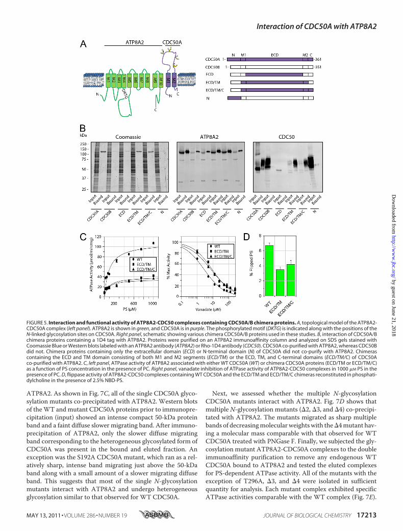
Expression, Co-immunoprecipitation and Functional Char-acterization of CDC50A/CDC50B Chimera Proteins—TheCDC50B variant is 55% identical to CDC50A and displays asimilar membrane topology (Fig. 5A). However, as previouslyreported (19) and confirmed in this study (Fig. 5B), CDC50B,unlike CDC50A, does not interact with ATP8A2. We havetaken advantage of this property to begin to define regions ofCDC50A that are required for the formation of a functionallyactive ATP8A2-CDC50A complex. Chimera proteins consis-ting of a combination of CDC50A and CDC50B domains (Fig.5A) were constructed and co-expressed with ATP8A2 inHEK293T cells. The interaction of ATP8A2 with the CDC50chimera proteins was determined by co-immunoprecipitationof these proteins on an anti-ATP8A2 immunoaffinity matrix.As shown in Fig. 5B, CDC50 chimera proteins in which eitherthe ECD or the N-terminal domain of CDC50B was replacedwith corresponding domain of CDC50A failed to interact withATP8A2. In contrast, the CDC50 chimera protein in which theexocytoplasmic domain and the transmembrane domain con-sisting of both M1 and M2 (ECD/TM) of CBC50B werereplaced with the corresponding CDC50A domains expressed
at relatively high levels and co-immunoprecipitated with theATP8A2. A chimera protein containing the exocytoplasmicdomain, the transmembrane domain, and the C-terminaldomain (ECD/TM/C) of CDC50A also interacted withATP8A2. However, chimera proteins in which one or bothtransmembrane segments (M1/M2) of CDC50B were replacedwith the transmembrane segments of CDC50A failed to inter-act with ATP8A2 (supplemental Fig. S3).The PS-dependent ATPase activities of the purified
ATP8A2-CDC50 chimera protein complexes were studied. Asshown in Fig. 5C, the ECD/TM and ECD/TM/C chimera pro-tein complexes displayed substantially lower PS-dependentATPase activities compared with the WT ATP8A2-CDC50Acomplex. The WT ATP8A2-CDC50A complex had a Vmax of107 � 8 �mol/min/mg, whereas the ATP8A2-ECD/TM andATP8A2-ECD/TM/C complexes had Vmax of 40 � 5 and 35 �5 �mol/min/mg, respectively. The Km values of the ECD/TMand ECD/TM/C chimera complexes (Km of 86 � 35 and 73 �32 �M, respectively), however, were similar to the WT protein(Km of 98 � 24 �M).Vanadate is known to be a potent inhibitor of P-type
ATPases and is often used to measure changes in the equilib-rium between the E1 and E2 conformational states of ATPasesduring the reaction cycle. Fig. 5C shows that the ATPase activ-ity of purified WT ATP8A2-CDC50A complex is stronglyinhibited by vanadate with Ki of 2.7 � 0.3 �M. The chimeracomplexes showed a decrease in sensitivity to vanadate exhib-iting aKi for the ECD/TM and ECD/TM/C chimera complexesof 8.7 � 0.8 and 7.3 � 0.7 �M, respectively.
The aminophospholipid flippase activity of equal amounts ofpurified and reconstituted ATP8A2-CDC50 chimeras wasstudied (Fig. 5D). TheWT complex was able to transport 6.7 �0.3% of total NBD-PS. In contrast, the chimeras were able totransport less NBD-PS consistent with the PS-dependentATPase studies. The ECD/TM and ECD/TM/C chimerastransported 3.5 � 0.4 and 4.2 � 0.5%, respectively.Localization of ATP8A2-CDC50 Complexes in Transfected
Cos-7 Cells—The subcellular distribution of ATP8A2 andCDC50A individually and co-expressed in Cos-7 cells wasinvestigated in double labeling studies using calnexin as an ERmarker and GM130 as a Golgi marker. For these studies,ATP8A2 and CDC50A contained C-terminal 1D4 and Myctags, respectively, to facilitate double labeling. Immunoprecipi-tation studies and ATPase assays indicated that the C-terminaltags had no effect on the interaction of ATP8A2 with CDC50Aor functional activity of the complex. The majority of ATP8A2and CDC50A co-localized with calnexin in the ER in singlytransfected cells (supplemental Fig. S4). In contrast, ATP8A2and CDC50A co-localized with GM130 in the Golgi ofco-transfected cells. This indicates that the association ofATP8A2 with CDC50A resulted in the translocation of thecomplex from the ER to the Golgi. A similar distribution pat-tern has been recently reported in cultured U20S cells express-ingATP8A2 andCDC50A (19), indicating that this distributionis not specific to Cos-7 cells.The effect of CDC50 chimera proteins on the localization of
ATP8A2 was investigated. When ATP8A2 was co-expressedwith the noninteracting CDC50 chimera proteins, ECD or
FIGURE 3. ATP8A2 expressed in HEK293T cells interacts with endogenousCDC50A to form a catalytically active ATP8A2-CDC50A complex.A, immunoaffinity purification of ATP8A2-CDC50A complex from HEK293Tcells transfected with the Atp8A2 plasmid. Solubilized HEK293T cell extract(Input) was incubated with the Atp6C11 immunoaffinity matrix, and theunbound fraction (Unbound) and the 6C11 peptide eluted fraction (Elution)were analyzed on SDS gels stained with Coomassie Blue, and Western blotswere labeled with the Atp6C11 antibody (ATP8A2) and the Cdc50 –7F4 anti-body (CDC50A). B, ATPase activity of the purified ATP8A2-CDC50A complex in100% phosphatidylcholine (PC) or 10% phosphatidylserine (PS) or 40% phos-phatidylethanolamine (PE) each containing the corresponding concentra-tions of PC. C, quantitative analysis of expressed and solubilized ATP8A2 andCDC50A. ATP8A2 and CDC50A containing a 1D4 tag were expressed individ-ually (ATP8A2 or CDC50A) or together (ATP8A2/CDC50A) in HEK293T cells. Theamounts of ATP8A2 and CDC50A solubilized with either SDS or CHAPS weremeasured by Western blotting (left), and the relative quantity of solubilizedATP8A2 was determined from Western blots of three experiments (right).Approximately, 30 �g of protein was loaded into each lane.
Interaction of CDC50A with ATP8A2
MAY 13, 2011 • VOLUME 286 • NUMBER 19 JOURNAL OF BIOLOGICAL CHEMISTRY 17211
by guest on June 21, 2018http://w
ww
.jbc.org/D
ownloaded from

N-terminal domain, ATP8A2 co-localized with calnexin in ERsimilar to that observed with noninteracting CDC50B (Fig. 6).However, when ATP8A2 was co-expressed with the ECD/TMor ECD/TM/C chimera proteins, it co-localizedwithGM130 tothe Golgi similar to that observed for the WT ATP8A2-CDC50A complex. These results suggest that the ECD/TMandECD/TM/C chimera proteins promote the folding of ATP8A2into a native-like conformation, allowing the complex to exitthe ER and translocate to the Golgi.N-Linked Glycosylation of CDC50A Is Required for the Stable
Expression of ATP8A2—The exocytoplasmic domain ofCDC50A contains four consensus sequences for N-linked gly-cosylation and one possible site for O-linked glycosylation. Todetermine whether endogenous bovine CDC50A is glyco-sylated, detergent-solubilized ROS membranes were treatedwith enzymes known to remove oligosaccharide chains fromglycoproteins and analyzed on Western blots labeled with theCdc50–9C9 antibody. Treatment with PNGase F, whichremoves N-linked sugars, resulted in a marked decrease in theapparent molecular mass from 50 to 37 kDa close to the pre-dictedmolecularmass of the CDC50A polypeptide (Fig. 7A). Incontrast, no detectable shift in molecular mass was observed
after treatment with neuraminidase alone or in combinationwith O-glycosidase.The effect of N-glycosylation of CDC50A on the expression
and interaction with ATP8A2 was studied with mutants inwhich single and multiple consensus sequences forN-glycosyl-ation were abolished. Co-expression of single N-glycosylationdefective mutants of CDC50A (T109A, T182A, S192A, andT296A) with ATP8A2 showed a reduction in ATP8A2 expres-sion levels relative to co-expression with WT CDC50A (Fig.7B). In contrast, the mutation that abolished the putativeO-linked glycosylation (T285A) showed no reduction inATP8A2 expression.Multiple glycosylation site mutants 2 (T109A,T182A), 3
(T109A,T182A,S192A), and4 (T109A,T182A,S192A,T296A)showed an inverse additive effect on the ATP8A2 expressionlevels (Fig. 7B). The CDC50A 2 mutant resulted in a 60%reduction in ATP8A2 expression relative to WT CDC50A,whereas the 3 mutant showed an 80% reduction, and the 4mutant reduced ATP8A2 expression to levels that were essen-tially undetectable in cell lysates.Co-immunoprecipitation studies were carried out to deter-
mine whether the N-glycosylation mutants interact with
FIGURE 4. ATPase activity and phospholipid flippase activity of the expressed and purified ATP8A2-CDC50A complex. A, Coomassie Blue-stained geland Western blots of the immunoaffinity-purified ATP8A2-CDC50A complex from HEK293T cells co-expressing both ATP8A2 and CDC50A-1D4. The complexwas purified on either an Atp6C11 column alone (ATP8A2 IP) or sequentially on Atp6C11 and Rho-1D4 columns (ATP8A2/CDC50A IP). B, ATP8A2 and CDC50Acontaining a 1D4 tag were co-expressed in HEK293T cells, and the complex was purified by the dual immunoaffinity procedure. The ATPase activity of thecomplex was measured in the presence of 100% PC or 10% PS or 40% PE lipids each containing the appropriate amount of PC. C, the ATP8A2-CDC50A complexpurified by the dual immunoaffinity procedure was reconstituted into liposomes for phospholipid flippase measurements. The percentage of lipid flipped bythe ATP8A2-CDC50A complex was measured in phosphatidylcholine proteoliposomes for 2.5% NBD-labeled PC, PE, or PS. Right panel, the PS flippase activityof the ATP8A2-CDC50A complex was measured for decreasing amounts of reconstituted protein. IP, immunoprecipitation.
Interaction of CDC50A with ATP8A2
17212 JOURNAL OF BIOLOGICAL CHEMISTRY VOLUME 286 • NUMBER 19 • MAY 13, 2011
by guest on June 21, 2018http://w
ww
.jbc.org/D
ownloaded from
ATP8A2. As shown in Fig. 7C, all of the single CDC50A glyco-sylation mutants co-precipitated with ATP8A2. Western blotsof theWT andmutant CDC50A proteins prior to immunopre-cipitation (input) showed an intense compact 50-kDa proteinband and a faint diffuse slower migrating band. After immuno-precipitation of ATP8A2, only the slower diffuse migratingband corresponding to the heterogeneous glycosylated form ofCDC50A was present in the bound and eluted fraction. Anexception was the S192A CDC50A mutant, which ran as a rel-atively sharp, intense band migrating just above the 50-kDaband along with a small amount of a slower migrating diffuseband. This suggests that most of the single N-glycosylationmutants interact with ATP8A2 and undergo heterogeneousglycosylation similar to that observed for WT CDC50A.
Next, we assessed whether the multiple N-glycosylationCDC50A mutants interact with ATP8A2. Fig. 7D shows thatmultiple N-glycosylation mutants (2, 3, and 4) co-precipi-tated with ATP8A2. The mutants migrated as sharp multiplebands of decreasingmolecular weights with the4mutant hav-ing a molecular mass comparable with that observed for WTCDC50A treated with PNGase F. Finally, we subjected the gly-cosylation mutant ATP8A2-CDC50A complexes to the doubleimmunoaffinity purification to remove any endogenous WTCDC50A bound to ATP8A2 and tested the eluted complexesfor PS-dependent ATPase activity. All of the mutants with theexception of T296A, 3, and 4 were isolated in sufficientquantity for analysis. Each mutant complex exhibited specificATPase activities comparable with the WT complex (Fig. 7E).
FIGURE 5. Interaction and functional activity of ATP8A2-CDC50 complexes containing CDC50A/B chimera proteins. A, topological model of the ATP8A2-CDC50A complex (left panel). ATP8A2 is shown in green, and CDC50A is in purple. The phosphorylated motif (DKTG) is indicated along with the positions of theN-linked glycosylation sites on CDC50A. Right panel, schematic showing various chimera CDC50A/B proteins used in these studies. B, interaction of CDC50A/Bchimera proteins containing a 1D4 tag with ATP8A2. Proteins were purified on an ATP8A2 immunoaffinity column and analyzed on SDS gels stained withCoomassie Blue or Western blots labeled with an ATP8A2 antibody (ATP8A2) or Rho-1D4 antibody (CDC50). CDC50A co-purified with ATP8A2, whereas CDC50Bdid not. Chimera proteins containing only the extracellular domain (ECD) or N-terminal domain (N) of CDC50A did not co-purify with ATP8A2. Chimerascontaining the ECD and TM domain consisting of both M1 and M2 segments (ECD/TM) or the ECD, TM, and C-terminal domains (ECD/TM/C) of CDC50Aco-purified with ATP8A2. C, left panel, ATPase activity of ATP8A2 associated with either WT CDC50A (WT) or chimera CDC50A proteins (ECD/TM or ECD/TM/C)as a function of PS concentration in the presence of PC. Right panel, vanadate inhibition of ATPase activity of ATP8A2-CDC50 complexes in 1000 �M PS in thepresence of PC. D, flippase activity of ATP8A2-CDC50 complexes containing WT CDC50A and the ECD/TM and ECD/TM/C chimeras reconstituted in phosphati-dylcholine in the presence of 2.5% NBD-PS.
Interaction of CDC50A with ATP8A2
MAY 13, 2011 • VOLUME 286 • NUMBER 19 JOURNAL OF BIOLOGICAL CHEMISTRY 17213
by guest on June 21, 2018http://w
ww
.jbc.org/D
ownloaded from

The flippase activity for the T109A glycosylation mutants wasalso found to be similar to WT (Fig. 7F).
DISCUSSION
Although the interaction of P4-ATPases with CDC50 pro-teins has been observed in cells co-expressing these proteins,the association of endogenous CDC50 proteins with endoge-nous P4-ATPases in animal cells has not been reported to date.In the present study, we show that ATP8A2 purified from pho-toreceptor outer segment membranes exists as a heteromericcomplex with CDC50A by both mass spectrometry and West-ern blotting. The purified and reconstituted complex functionsas a PS and to a lesser degree PE lipid transporter as previouslyreported (5). Immunofluorescence labeling studies using thehighly specific ATP8A2 monoclonal antibody Atp2F6 and theCDC50A monoclonal antibody Cdc50–7F4 generated as part
FIGURE 6. Immunofluorescence localization of ATP8A2 co-expressedwith WT CDC50A, WT CDC50B, or chimera CDC50A/B containing a 1D4tag in Cos-7 cells. The cells were double labeled for ATP8A2 with the Atp2F6monoclonal antibody (green) and the GM130 polyclonal antibody as a Golgimarker (red). Co-expression of ATP8A2 with CDC50B or chimeric CDC50A pro-teins containing the ECD or N-terminal domain (N) domains resulted in areticular staining pattern of ATP8A2 characteristic of ER localization. Co-ex-pression of ATP8A2 with either WT CDC50A or chimera proteins containingthe ECD/TM or ECD/TM/C domains of CDC50A resulted in localization ofATP8A2 to the Golgi as shown in merged images. Expression of CDC50 pro-teins was confirmed independently using the Rho 1D4 antibody (data notshown). The nuclei (blue) were labeled with 4,6-diamidino-2-phenylindole.Bar, 10 �m.
FIGURE 7. N-Linked glycosylation of CDC50A is necessary for stabilizationof ATP8A2. A, approximately 50 �g of rod outer segment membranes weretreated with PNGase F or neuraminidase with or without O-glycosidase andlabeled with the Cdc50 –9C9 antibody. B, expression levels of ATP8A2 in thepresence of CDC50A glycosylation mutants containing a 1D4 tag. ATP8A2levels were normalized to �-actin as a loading control. C, co-immunoprecipi-tation of single CDC50A glycosylation mutants with ATP8A2 on Atp6C11-Sepharose. ATP8A2 was detected with Atp6C11, and the CDC50A-1D4mutants were detected with the Rho 1D4 antibody. Solubilized HEK293T cells(Input) were incubated in the column, and the bound proteins (Bound) wereeluted with 6C11 peptide. D, immunoprecipitation of multiple CDC50A gly-cosylation mutants with ATP8A2 on Atp6C11-Sepharose. Multiple siteCDC50A glycosylation mutants expressed at low levels relative to WTCDC50A. Approximately 30 �g of input and 5–200 ng of the bound fractionswere applied to the gel. E, specific ATPase activity of ATP8A2 expressed withvarious CDC50A N-linked glycosylation mutants and purified on an immuno-affinity column. ATPase activity was measured in the presence of 10% PS and90% PC. F, lipid flippase activity of WT and T109A mutant reconstituted in PCin the presence of 2.5% NBD-PS. The difference in activities between WT andT109A is not significant.
Interaction of CDC50A with ATP8A2
17214 JOURNAL OF BIOLOGICAL CHEMISTRY VOLUME 286 • NUMBER 19 • MAY 13, 2011
by guest on June 21, 2018http://w
ww
.jbc.org/D
ownloaded from

of this study show that these proteins co-localize to the outersegment compartment of photoreceptor cells. AlthoughATP8A2 is restricted to the outer segments, CDC50A is alsopresent in other retinal cells and other cellular compartmentsof photoreceptor cells. This suggests that CDC50A also forms aheteromeric complex with other P4-ATPases in the retina. Itshould be possible to use the CDC50A specificmonoclonal anti-bodies in conjunction with immunoprecipitation and mass spec-trometry to identify and characterize the various P4-ATPases inthe retina and other tissues.In a recent study, ATP8A2 and other class 1 P4-ATPases
were found to associate with CDC50 variants when co-ex-pressed in U2OS cells and promote the export of these com-plexes from the ER (19). However, the function of these com-plexes as phospholipid-dependent ATPases or ATP-dependentlipid transporters was not determined. Here, we have con-firmed that ATP8A2 forms a heteromeric complex withCDC50A, but not CDC50B, in HEK293T and Cos-7 cells, andthis complex is translocated from the ER to the Golgi. Impor-tantly, we show for the first time that this expressed complex isfunctionally active as an aminophospholipid transporter whenreconstituted into lipid vesicles.HEK293T cells express low levels of endogeneous CDC50A.
When cells are transfected with Atp8a2 alone, a small fractionof the heterologously expressed ATP8A2 interacts with endog-enous CDC50A. Although the yield of this ATP8A2-CDC50Acomplex is low, nonetheless it shows PS- and PE-dependentATPase activity similar to that previously observed forATP8A2-CDC50A complex isolated from photoreceptor outersegments (5). The low quantity of the complex is likely due tothe limiting expression of endogenous CDC50A in HEK293Tcells because co-expression of ATP8A2 with CDC50A greatlyenhances the yield of functionally active complex.The role of CDC50A in promoting a stable active complex
is supported by immunolabeling studies of ATP8A2 andCDC50A expressed in Cos-7 cells. In the absence of expressedCDC50A, the majority of the expressed ATP8A2 is retained inthe ER, most likely as a misfolded protein that is poorly solublein mild detergent. Co-expression of ATP8A2 with CDC50Aresults in the formation of properly folded heteromeric com-plex that is transported from the ER and to the Golgi compart-ment and is readily solubilized in CHAPS detergent as a func-tionally active PS-dependent ATPase.CDC50A does not appear to direct the subcellular targeting
of P4-ATPases. It has been previously reported that ATP8B1associated with CDC50A localizes to the plasma membrane ofculture cells (15), whereas ATP8A2 associated with CDC50Alocalizes to the Golgi compartment. Hence, the cell targetingsignals of most, if not all, mammalian P4-ATPases appear toreside in the catalytic P4-ATPase subunit. This is in agreementwith studies of Lopez-Marques et al. (18), who showed thatArabidopsis P4-ATPases ALA2 and ALA3 and not CDC50 pro-teins are responsible for subcellular targeting of the complex.In HEK293T cells, CDC50A expressed by itself is retained in
the ER. It migrates as a relatively compact 50-kDa protein onSDS gels. In contrast, CDC50A associated with ATP8A2 and ispresent in the Golgi migrates as a diffuse band. This suggeststhat the N-linked oligosaccharide chains of CDC50A undergo
extensive heterogeneous modification within the Golgi ofHEK293T cells. This highly heterogeneous glycosylation ap-pears to be a characteristic of ATP8A2-CDC50A overexpres-sion in culture cells because CDC50A associated with ATP8A2in photoreceptor outer segments migrates as a more compactband. A similar difference in migration pattern has beenreported for the N-linked glycoprotein rhodopsin. Rhodopsinfrom rod outer segments migrates as a relatively tight band onSDS gels, whereas rhodopsin expressed in culture cellsmigratesas a diffuse band characteristic of heterogeneous N-linked gly-cosylation (40). This pattern most likely reflects a difference inthe processing of oligosaccharide chains in these distinct celltypes.The four highly conservedN-linked glycosylation consensus
sequences in the exocytoplasmic domain of CDC50A allundergo glycosylation. Analysis of single and multiple glycosy-lation mutants indicate that each oligosaccharide chain con-tributes to the optimal, stable expression of the ATP8A2-CDC50A complex. Although the loss of one or moreglycosylation sites reduces the yield of the complex, it does notappear to affect the functional activity of theATP8A2-CDC50Acomplex as measured by PS-stimulated ATPase and flippaseassays.In this study chimera CDC50A/B proteins were generated
and used to investigate the role of the various domains ofCDC50A in the binding and functional activity of ATP8A2.Chimera proteins inwhich either theTMor the ECDdomain ofCDC50B was replaced with the corresponding CDC50Adomain on a CDC50B backbone failed to interact withATP8A2. In contrast, the chimera protein with both domains(ECD/TM) replaced assembled with ATP8A2 into a function-ally active complex capable of actively transporting PS acrossthe lipid bilayer. Like the WT protein complex, this chimeracomplex was also exported from the ER to the Golgi. This indi-cates that both ECD and TMdomains of CDC50A are requiredfor the proper folding, subunit assembly, and function of theATP8A2-CDC50 complex as a PS flippase. A similar situationexists for the Na/K-ATPase. Both the transmembrane andextracellular domains of the �-subunit of Na/K-ATPase havebeen implicated in its interaction with the catalytic �-subunitand contribute to the transport mechanism (41–43).The ECD/TM/C chimera protein containing the C-terminal
as well as the ECD and TM domains of CDC50A exhibitedsimilar properties as the ECD/TM chimera. This indicateseither that theC terminus is not involved in the interactionwithATP8A2 or that the C-terminal domain of CDC50B can effec-tively substitute for the C-terminal domain of CDC50A in thiscapacity. Interestingly, although the ECD/TM and ECD/TM/Cchimera interact with ATP8A2 to form a functional complex,the PS-dependent ATPase activity and lipid flippase activity aresignificantly reduced. Likewise, these chimeric complexes havea decreased sensitivity to vanadate, a P-type ATPase inhibitorthat interacts with the E2 state. Collectively, these studies sug-gest that the N-terminal domain plays a role in the transportcycle of ATP8A2. One possibility is that CDC50A plays a sim-ilar role as the �-subunit of the H�/K� ATPase stabilizing theE2P state of the enzyme through its N-terminal domain (44).Our finding that CDC50A participates in the ATPase reaction
Interaction of CDC50A with ATP8A2
MAY 13, 2011 • VOLUME 286 • NUMBER 19 JOURNAL OF BIOLOGICAL CHEMISTRY 17215
by guest on June 21, 2018http://w
ww
.jbc.org/D
ownloaded from

cycle of ATP8A2 is in general agreement with recent studies onthe yeast Drs2p-Cdc50p and the ATP8B1-CDC50 P4-ATPases(20, 21).In summary, our studies show that CDC50A is the �-subunit
of ATP8A2 in photoreceptor outer segment membranes and isessential for the proper folding, assembly, and exit of theATP8A2-CDC50A complex from the ER to theGolgi in culturecells. Importantly, CDC50A is also critical for the formation ofa functionally active complex that can catalyze the transport ofPS and to a lesser extent PE across cell membranes. Both thetransmembrane and exocytoplasmic domains of CDC50A areessential for the formation of a functionally active ATP8A2-CDC50A complex, whereas the N-terminal domain ofCDC50A participates in the reaction cycle of ATP8A2, possiblystabilizing the E2P state. Finally, N-linked glycosylation ofCDC50A plays an important role in the formation of a stableATP8A2-CDC50A protein complex.
Acknowledgments—We thank Laurie Molday and Theresa Hii fortechnical assistance in the generation andmaintenance of hybridomacell lines.
REFERENCES1. Alder-Baerens, N., Lisman, Q., Luong, L., Pomorski, T., andHolthuis, J. C.
(2006)Mol. Biol. Cell 17, 1632–16422. Darland-Ransom, M., Wang, X., Sun, C. L., Mapes, J., Gengyo-Ando, K.,
Mitani, S., and Xue, D. (2008) Science 320, 528–5313. Natarajan, P.,Wang, J., Hua, Z., andGraham,T. R. (2004)Proc. Natl. Acad.
Sci. U.S.A. 101, 10614–106194. Wang, L., Beserra, C., and Garbers, D. L. (2004) Dev. Biol. 267, 203–2155. Coleman, J. A., Kwok, M. C., and Molday, R. S. (2009) J. Biol. Chem. 284,
32670–326796. Zhou, X., and Graham, T. R. (2009) Proc. Natl. Acad. Sci. U.S.A. 106,
16586–165917. Folmer, D. E., Elferink, R. P., and Paulusma, C. C. (2009) Biochim. Biophys.
Acta 1791, 628–6358. Muthusamy, B. P., Natarajan, P., Zhou, X., and Graham, T. R. (2009)
Biochim. Biophys. Acta 1791, 612–6199. Puts, C. F., and Holthuis, J. C. (2009) Biochim. Biophys. Acta 1791,
603–61110. Bull, L. N., van Eijk,M. J., Pawlikowska, L., DeYoung, J. A., Juijn, J. A., Liao,
M., Klomp, L.W., Lomri, N., Berger, R., Scharschmidt, B. F., Knisely, A. S.,Houwen, R. H., and Freimer, N. B. (1998) Nat. Genet. 18, 219–224
11. Meguro, M., Kashiwagi, A., Mitsuya, K., Nakao, M., Kondo, I., Saitoh, S.,and Oshimura, M. (2001) Nat. Genet. 28, 19–20
12. Stapelbroek, J. M., Peters, T. A., van Beurden, D. H., Curfs, J. H., Joosten,A., Beynon, A. J., van Leeuwen, B.M., van der Velden, L.M., Bull, L., OudeElferink, R. P., van Zanten, B. A., Klomp, L. W., and Houwen, R. H. (2009)Proc. Natl. Acad. Sci. U.S.A. 106, 9709–9714
13. Cacciagli, P., Haddad, M. R., Mignon-Ravix, C., El-Waly, B., Moncla, A.,Missirian, C., Chabrol, B., and Villard, L. (2010) Eur. J. Hum. Genet. 18,1360–1363
14. Saito, K., Fujimura-Kamada, K., Furuta, N., Kato, U., Umeda, M., and
Tanaka, K. (2004)Mol. Biol. Cell 15, 3418–343215. Paulusma, C. C., Folmer, D. E., Ho-Mok, K. S., de Waart, D. R., Hilarius,
P. M., Verhoeven, A. J., and Oude Elferink, R. P. (2008) Hepatology 47,268–278
16. Poulsen, L. R., Lopez-Marques, R. L., McDowell, S. C., Okkeri, J., Licht, D.,Schulz, A., Pomorski, T., Harper, J. F., and Palmgren, M. G. (2008) PlantCell 20, 658–676
17. Chen, S., Wang, J., Muthusamy, B. P., Liu, K., Zare, S., Andersen, R. J., andGraham, T. R. (2006) Traffic 7, 1503–1517
18. Lopez-Marques, R. L., Poulsen, L. R., Hanisch, S.,Meffert, K., Buch-Peder-sen, M. J., Jakobsen, M. K., Pomorski, T. G., and Palmgren, M. G. (2010)Mol. Biol. Cell 21, 791–801
19. van der Velden, L.M.,Wichers, C. G., van Breevoort, A. E., Coleman, J. A.,Molday, R. S., Berger, R., Klomp, L. W., and van de Graaf, S. F. (2010)J. Biol. Chem. 285, 40088–40096
20. Lenoir, G., Williamson, P., Puts, C. F., and Holthuis, J. C. (2009) J. Biol.Chem. 284, 17956–17967
21. Bryde, S., Hennrich, H., Verhulst, P. M., Devaux, P. F., Lenoir, G., andHolthuis, J. C. (2010) J. Biol. Chem. 285, 40562–40572
22. Kwok, M. C., Holopainen, J. M., Molday, L. L., Foster, L. J., and Molday,R. S. (2008)Mol. Cell Proteomics 7, 1053–1066
23. Chomczynski, P., and Sacchi, N. (1987) Anal. Biochem. 162, 156–15924. MacKenzie, D., and Molday, R. S. (1982) J. Biol. Chem. 257, 7100–710525. Chen, C., and Okayama, H. (1987)Mol. Cell. Biol. 7, 2745–275226. Bungert, S., Molday, L. L., and Molday, R. S. (2001) J. Biol. Chem. 276,
23539–2354627. Papermaster, D. S., and Dreyer, W. J. (1974) Biochemistry 13, 2438–244428. Molday, L. L., Cook, N. J., Kaupp, U. B., and Molday, R. S. (1990) J. Biol.
Chem. 265, 18690–1869529. Gonzalez-Romo, P., Sanchez-Nieto, S., and Gavilanes-Ruíz, M. (1992)
Anal. Biochem. 200, 235–23830. McIntyre, J. C., and Sleight, R. G. (1991) Biochemistry 30, 11819–1182731. Romsicki, Y., and Sharom, F. J. (2001) Biochemistry 40, 6937–694732. Krogh, A., Larsson, B., von Heijne, G., and Sonnhammer, E. L. (2001) J.
Mol. Biol. 305, 567–58033. Gupta, R., and Brunak, S. (2002) Pac Symp Biocomput. 7, 310–32234. Julenius, K., Mølgaard, A., Gupta, R., and Brunak, S. (2005) Glycobiology
15, 153–16435. Shevchenko,A.,Wilm,M., Vorm,O., andMann,M. (1996)AnalChem68,
850–85836. Perkins, D. N., Pappin, D. J., Creasy, D. M., and Cottrell, J. S. (1999) Elec-
trophoresis 20, 3551–356737. Katoh, Y., and Katoh, M. (2004) Oncol Rep. 12, 939–94338. Osada, N., Hashimoto, K., Hirai, M., and Kusuda, J. (2007) Gene 392,
151–15639. Xu, P., and Ding, X. (2007) Acta Biochim. Biophys. Sin. 39, 739–74440. Doi, T., Molday, R. S., and Khorana, H. G. (1990) Proc. Natl. Acad. Sci.
U.S.A. 87, 4991–499541. Jaunin, P., Jaisser, F., Beggah, A. T., Takeyasu, K., Mangeat, P., Rossier,
B. C.,Horisberger, J. D., andGeering, K. (1993) J. Cell Biol.123, 1751–175942. Hasler, U., Wang, X., Crambert, G., Beguin, P., Jaisser, F., Horisberger,
J. D., and Geering, K. (1998) J. Biol. Chem. 273, 30826–3083543. Hasler, U., Crambert, G., Horisberger, J. D., and Geering, K. (2001) J. Biol.
Chem. 276, 16356–1636444. Abe, K., Tani, K., Nishizawa, T., and Fujiyoshi, Y. (2009) EMBO J. 28,
1637–1643
Interaction of CDC50A with ATP8A2
17216 JOURNAL OF BIOLOGICAL CHEMISTRY VOLUME 286 • NUMBER 19 • MAY 13, 2011
by guest on June 21, 2018http://w
ww
.jbc.org/D
ownloaded from

Jonathan A. Coleman and Robert S. Molday-ATPase ATP8A24Subcellular Localization, and Lipid Transport Activity of the P
-Subunit CDC50A in the Stable Expression, Assembly,βCritical Role of the
doi: 10.1074/jbc.M111.229419 originally published online March 18, 20112011, 286:17205-17216.J. Biol. Chem.
10.1074/jbc.M111.229419Access the most updated version of this article at doi:
Alerts:
When a correction for this article is posted•
When this article is cited•
to choose from all of JBC's e-mail alertsClick here
Supplemental material:
http://www.jbc.org/content/suppl/2011/03/18/M111.229419.DC1
http://www.jbc.org/content/286/19/17205.full.html#ref-list-1
This article cites 44 references, 21 of which can be accessed free at
by guest on June 21, 2018http://w
ww
.jbc.org/D
ownloaded from