crocodile oil enhances cutaneous burn wound healing and reduces scar formation in rats
TRANSCRIPT
ORIGINAL RESEARCH CONTRIBUTION
Crocodile Oil Enhances Cutaneous BurnWound Healing and Reduces Scar Formationin RatsHua-Liang Li, PhD, Li-Ping Chen, MS, Yong-Hua Hu, PhD, Yan Qin, MS, Ge Liang, MS,You-Xiong Xiong, and Qing-Xi Chen, PhD
AbstractObjectives: This study was performed to evaluate the burn wound–healing efficacy of crocodile oil fromCrocodylus siamensis by employing deep second-degree burns in a Wistar rat model.
Methods: Twenty-four rats were assigned equally into four groups using a random-number table, andtwo burns were created on the dorsum of each animal except for the sham group. The three treatmentgroups received with saline solution (12 burns, served as negative control), silver sulfadiazine (12 burns,served as positive control), or crocodile oil (12 burns). Silver sulfadiazine cream was used as standardcare, and the treatments were repeated twice daily for 28 days. After day 28 the animals were eutha-nized and the wounds were removed for quantitative real-time polymerase chain reaction, histologic,and immunohistochemical study.
Results: Crocodile oil accelerated the wound-healing process as indicated by a significant decrease inwound closure time in comparison to the burn control and silver sulfadiazine treatment groups. Histo-logic results showed well-organized and distributed skin structure and collagen deposition in the ani-mals treated with crocodile oil. Transforming growth factor-b1 (TGF-b1), a key cytokine promotingscarring, was also observed to play a role in the burn wound healing. Immunohistochemical stainingresults showed the negative expression of TGF-b1 and Smad3 in the 28-days-postburn skin of crocodileoil group versus positive in the epidermis of burn controls. Compared to the burn control group, expres-sions of TGF-b1 and Smad3 mRNA decreased significantly (p < 0.01) in the 28-days-postburn skin of thecrocodile oil group.
Conclusions: Our results showed that crocodile oil could enhance cutaneous burn wound healing andreduce scar formation in rats, which might be related to TGF-b1 ⁄ Smad3 signaling.
ACADEMIC EMERGENCY MEDICINE 2012; 19:265–273 ª 2012 by the Society for Academic EmergencyMedicine
B urns are one of the most widespread injuries inaccidents and remain a global public healthissue.1,2 The burn wound is a continuous and
severe threat against the rest of the body due to invasionof infectious agents, antigen challenge, and repeatedadditional trauma caused by wound cleaning.3 Althoughmany advances have been made in our understandingand care of burn injuries, there are still many burnshealed with scar formation, resulting in significant aes-thetic disfigurement and dysfunction.4,5
The sequence of events repairing the wound is cate-gorized into three overlapping phases: inflammation,proliferation and tissue remodeling, and scar matura-tion.6 Burn wound healing involves a sequence ofmolecular and cellular events including inflammation,cell migration, angiogenesis, extracellular matrixsynthesis, and reepithelialization.7 Some studies havedemonstrated that a number of cytokines are involvedin the progression of the wound healing.8,9 Transform-ing growth factor-b (TGF-b), known as a potent stimulus
ª 2012 by the Society for Academic Emergency Medicine ISSN 1069-6563doi: 10.1111/j.1553-2712.2012.01300.x PII ISSN 1069-6563583 265
From the State Key Laboratory of Stress Cell Biology, School of Life Sciences (HLL, LPC, YHH, YQ, GL, QXC), Xiamen Univer-sity, Xiamen, China; and the Thailand SriRaCha Tiger Zoo Co., Ltd (YXX), SriRaCha, Thailand.Received July 28, 2011; revision received September 19, 2011; accepted September 23, 2011.Presented at the 9th Academic Meeting of the Chinese Nautical Medicine Association, Yinchuan, Ningxia, China, August 2011.The present investigation was supported by SriRaCha Tiger Zoo Co., Ltd, SriRaCha, Thailand. Silver sulfadiazine cream (1% wt ⁄ wt)was donated by the first affiliated hospital of Xiamen University, Xiamen, China.The authors have no relevant financial information or potential conflicts of interest to disclose.Supervising Editor: Richard Sinert, DO.Address for correspondence and reprints: Qing-Xi Chen, PhD; e-mail: [email protected].
of connective tissue accumulation, is implicated in thepathogenesis of fibrotic disorders.10 In terms of repair,TGF-b1 and TGF-b2 are known to promote scar tissue,while TGF-b3 may reduce scar formation.11,12 The Smadsignal transduction pathways are crucial in mediatingseveral TGF-b responses in fibroblasts,10 such as stimu-lation of collagens13,14 and a-smooth muscle actin.15,16
Smad3 is a key intracellular signal transducer in profi-brotic TGF-b responses. Some researchers have foundthat targeted disruption of TGF-b ⁄ Smad3 signalingcould modulate skin fibrosis.10
Oils extracted from plants or animal fats that areabundant with fatty acids are used to treat burn inju-ries.1,17,18 Crocodile is a general term of animals ofCrocodilia, Reptilia, which have high economic andmedical value.19 Crocodile oil extracted from the fattytissues of crocodiles is rich in monounsaturated andpolyunsaturated fats.20 Crocodile oil and its productsare used as ointments for burns and scalds in the tradi-tional medicines, such as traditional Chinese and South-east Asia medicine.19 However, few studies on the roleof crocodile oil in burn wound healing have beenreported.
The aim of this study was to investigate the therapeu-tic effect of crocodile oil on experimental cutaneousburn wounds in Wistar rats. The mechanism of reduc-ing scar formation was also evaluated by analyzing theexpression of TGF-b1 and Smad3 in rat skins.
METHODS
Study DesignThis was a randomized controlled trial to compare theburn wound–healing efficacy of deep second-degree ratburns treated with crocodile oil, silver sulfadiazine(positive control), and saline solution (negative control).The animal experiments were performed in accordancewith the ‘‘Animal Welfare Act and the Guide for theCare and Use of Laboratory Animals’’ formulated byXiamen University’s Animal Ethics Committee. Thestudy was approved by the institutional review boardof Xiamen University.
Animal Handling and PreparationThis study was conducted in the Laboratory AnimalCenter of Xiamen University. Twenty-four SPF-classmale Wistar rats were kept on 12-hour-light ⁄ 12-hour-dark cycle with a constant temperature of 21 to 22�Cand 60% to 65% humidity. The rats were caged individ-ually with free access to a commercial balanced rat dietand tap water. The animals were assigned equally intofour groups using a random-number table: sham (nor-mal group, no burn), burn control group treated withsaline solution (12 burns, served as negative control),silver sulfadiazine–treated group (12 burns, served aspositive control), and crocodile oil–treated group (12burns). Silver sulfadiazine cream (1% wt ⁄ wt) was usedas standard care.
Preparation of Crocodile OilOil from Crocodylus siamensis was extracted using aprevious described method.21 Briefly, the fats wereextracted with petroleum ether (boiling process 30–60�C)
in an ultrasonic apparatus at room temperature (threetimes, 2 hours each). The extractions were collectedtogether and concentrated in vacuum at 50�C by a rotaryevaporator. The oil was then evaporated by a vacuumdryer until it reached a constant weight.22
Gas Chromatography Analysis of Crocodile OilThe gas chromatography analyses were accomplishedusing a Varian 1200 gas chromatograph equippedwith a mass spectrometer and CP-8713 capillarycolumns (Varian Medical Systems, Palo Alto, CA;30 m · 0.25 mm, i.d. · 0.25 lm) following a temperatureprogram of: 50�C for 5 minutes, rising at 10�C ⁄ minuteto 170�C, holding for 10 minutes, rising 2�C ⁄ minutes to210�C; holding for 25 minutes, then rising at 10�C ⁄ min-utes again to 225�C; and injector, ion, and transfer linetemperatures of 230, 200, and 250�C, respectively. Thecarrier gas was nitrogen (1 mL ⁄ min). The percentagecomposition was obtained from electronic integrationmeasurements using flame ionization detection.23
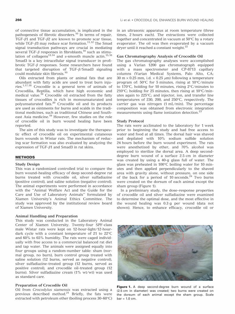
Study ProtocolThe rats were acclimated to the laboratory for 1 weekprior to beginning the study and had free access towater and food at all times. The dorsal hair was shavedand depilated with 10% sodium sulfide solution24 hours before the burn wound experiment. The ratswere anesthetized by ether, and 70% alcohol wasemployed to sterilize the dorsal area. A deep second-degree burn wound of a surface 2.5 cm in diameterwas created by using a 40-g glass full of water. Theglass was preheated in 100�C boiling water for 10 min-utes and then applied perpendicularly to the shavedarea with gravity alone, without pressure, on one sideof the back for a period of 10 seconds.24 Two burnswere created on the dorsum of each animal except thesham group (Figure 1).
In a preliminary study, the dose–response propertiesof crocodile oil and silver sulfadiazine were examinedto determine the optimal dose, and the most effective inthe wound healing was 0.3 g per wound (data notshown). Hence, to enhance efficacy, crocodile oil or
Figure 1. A deep second-degree burn wound of a surface(2.5 cm in diameter) was created; two burns were created onthe dorsum of each animal except the sham group. Scalebar = 1.0 cm.
266 Li et al. • CROCODILE OIL ENHANCES BURN WOUND HEALING
silver sulfadiazine was administrated at the dose of0.3 g per wound in the experiment. The 0.3 g of croco-dile oil or silver sulfadiazine was applied slowly to theburn wound area and extended slightly outside thewound area to ensure inclusion of the wound edges.The rats in the burn control group were treated with0.3 g of saline solution per wound under the standardconditions. Treatments were repeated twice daily for28 days. The first application was done directly afterthe wound injury.
Macroscopic Analysis of Wound ClosureOptical photographs were taken from the burn woundarea at an equal distance from the wound and rightangle to the wound surface on days 3, 7, 10, 17, 21, and28. The wound surface areas were measured by tracingtheir contours using a transparent paper to evaluatewound contraction.1 The area (mm2) within the bound-ary was measured planimetrically.25 Wound closuretime, exudation, and the firmness of the wound surfacewere also observed.
Histologic and Immunohistochemical AnalysisOn the 28th day, the experiment was terminated andthe granulation tissues were excised for histologicexamination. The excisional skin biopsies were fixed in4% neutral buffered formaldehyde solution for24 hours. To determine the structure and collagendeposition of the 28-days-postburn skin, deparaffinizedsections were processed for Van Gieson stain. Five–micrometer-thickness sections were observed for histo-pathologic changes under microscope, such as thethickness of the newly formed epidermis and dermis.
For immunohistochemistry, 5-lm-thickness cross-sec-tions from 28-days-postburn skin samples were depa-raffinized, rehydrated in descending alcohol dilutions,and immersed in phosphate-buffered saline (PBS). Thesections were treated with 0.01 mol ⁄ L, pH 6.0, citratebuffer solution at 95�C for 15 minutes and then withserum-free blocking agent (MaiXin Bio., Fuzhou, China)for 10 minutes, followed by incubation with polyclonalrabbit anti-human primary antibody anti-TGF-b1 (theidentity of protein sequences between Rattus norvegi-cus and Homo sapiens is 99%; dilution, 1:50 [MaiXinBio.]) and monoclonal rabbit anti-rat primary antibodyanti-Smad3 (dilution, 1:150; Epitomics, Inc., Burlingame,CA) in PBS overnight at 4�C. For detection of boundprimary antibodies, UltraSensitive S-P kit (MaiXin Bio.)was used according to the manufacturers’ instructions.Substitution of the primary antibody with PBS servedas negative control. Peroxidase activity was visualizedby incubation with AEC (3-amino-9-ethylcarbazole;Boster Biological Technology, Ltd., Wuhan, China).Nuclei were counterstained with Mayer’s hematoxylin(Boster) and coverslipped.
RNA Extraction and Quantitative Real-time PolymeraseChain ReactionTotal RNA was extracted from the granulation tissuesusing the RNAiso Plus kit (Takara Bio Inc., Shiga,Japan) following the manufacturer’s instructions andreverse-transcribed in 20 lL total volume using theSYBR PrimeScript real-time polymerase chain reaction
(RT-PCR) kit. The process of reverse transcription was37�C for 15 minutes (reverse transcription reaction) andone cycle at 85�C for 5 seconds (denaturation of reversetranscriptase). The synthesized cDNA was stored at)80�C. The mRNA levels for TGF-b1 and Smad3 in eachsample were determined by a quantitative RT-PCR.Primers were synthesized by Takara Bio, Inc. The prim-ers used for RT-PCR were as follows: TGF-b1 primers(forward 5¢-TGGCGTTACTTGGTAACC-3¢ and reverse5¢-GGTGTTGAGCCCTTTCCAG-3¢); Smad 3 primers(forward 5¢-AGCACACAATAACTTGGACC-3¢ andreverse 5¢-TAAGACACACTGGAACAGCGGATG-3¢);26
and b-actin (internal standard) primers (forward5¢-TGGAATCCTGTGGCATCCAT-3¢ and reverse 5¢-TA-AAACGCAGCTCAGTAACA-3¢). Real-time quantitativePCR was performed using the SYBR PrimeScript RT–PCR kit in accordance with the manufacturer’s instruc-tions. Then, PCR was carried out in a Rotor-Gene 6000(Corbett Research, Sydney, Australia) according to thefollowing protocol: 30 seconds at 95�C, one cycle; 5 sec-onds at 95�C; and 20 seconds at 57�C, 45 cycles. Fluo-rescence was detected at the annealing stage of eachcycle. A melting curve was generated during the reac-tions to check for the possibility of primer–dimer for-mation. The 2–44Ct method was used to calculate therelative mRNA level of each gene.27
Measures and OutcomesAll morphometric parameters were done with ImageAnalyzer (Olympus Microscope BX51, Olympus, CenterValley, PA) by using an image analyzing computer pro-gram (Image-Pro Plus 6.0, Media Cybernetics, Inc., Beth-esda, MD). The primary outcome was the percentage ofwound contraction. The sizes of the wound areas on the7th, 10th, 17th, 21st, and 28th days were determined by abalance, and the percentage of wound contraction wascalculated by subtracting the surface area of individualburns from the surface area of the largest burn anddividing this value by the surface area of the largest burnmultiplied by 100.28 This outcome had an interobservercorrelation of 0.99 on a subset of burns. A secondaryoutcome was the epidermal and dermal thickness ofthe granulation tissues in different groups at 28 dayspostburn. The thickness of the newly formed epidermiswas measured at 1-mm intervals, and the mean wascalculated.1 Dermal thickness was determined bymeasuring the distance from the epidermal–dermal junc-tion to the dermal–fat junction for eight randomlyselected sites ⁄ fields from two or more skin samples ineach animal.29 The collagen was stained red using VanGieson staining, and its deposition was observed.Another secondary outcome was TGF-b1 and Smad3mRNA expression in the 28-days-postburn rat skins.Quantitative RT-PCR analysis was used to examine TGF-b1 and Smad3 mRNA levels. The net intensity values forTGF-b1 and Smad3 bands were normalized to the house-keeping gene b-actin, and the data were presented as thepercentage difference in TGF-b1 ⁄ b-actin or Smad3 ⁄ b-actin gene expression from the age-matchedunwounded values (mean ± SEM). We also measuredthe TGF-b1 and Smad3 protein expressions in the 28-days-postburn rat skins by using immunohistochemicalstaining.
ACADEMIC EMERGENCY MEDICINE • March 2012, Vol. 19, No. 3 • www.aemj.org 267
Data AnalysisDescriptive statistics were used to describe outcomes.Continuous variables are presented as means with stan-dard deviations (SDs). A one-way analysis of variance(ANOVA) was used to assess the differences in the burnwound healing time and percent wound contraction ofthe groups. Additionally, we used a one-way ANOVAto assess differences in epidermal and dermal thicknessof crocodile oil treated and untreated burnwounds after 28 days of treatment. The percentage dif-ferences in TGF-b1 ⁄ b-actin and Smad3 ⁄ b-actin geneexpression of the burn control group and crocodile oilgroup were also assessed with one-way ANOVA.Statistical analysis was performed using SPSS 14.0for Windows (SPSS, Inc., Chicago, IL). Normality ofdistribution was assessed through the use of theKolmogorov-Smirnov test, and for multiple group com-parisons, homogeneity of variance was assessed by theLevene test. A p-value <0.05 was considered statisticallysignificant. A sample size of 12 burns in each groupprovided a power of 80% to detect a 33-percentage-point difference in the percentage of wound contractionbetween the groups30 and was determined a prioriusing PASS: Power Analysis and Sample Size for Win-dows (version 11, NCSS, LLC, Kaysville, UT).
RESULTS
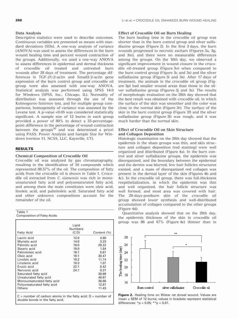
Chemical Composition of Crocodile OilCrocodile oil was analyzed by gas chromatography,resulting in the identification of 10 compounds whichrepresented 88.57% of the oil. The composition of fattyacids from the crocodile oil is shown in Table 1. Croco-dile oil extracted from C. siamensis was rich in mono-unsaturated fatty acid and polyunsaturated fatty acid,and among them the main constitutes were oleic acid,linoleic acid, and palmitoleic acid. Saturated fatty acidand other unknown compositions account for theremainder of the oil.
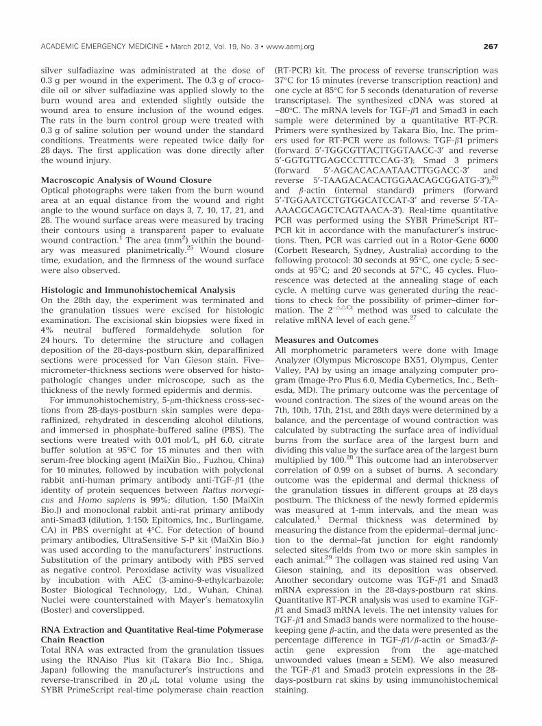
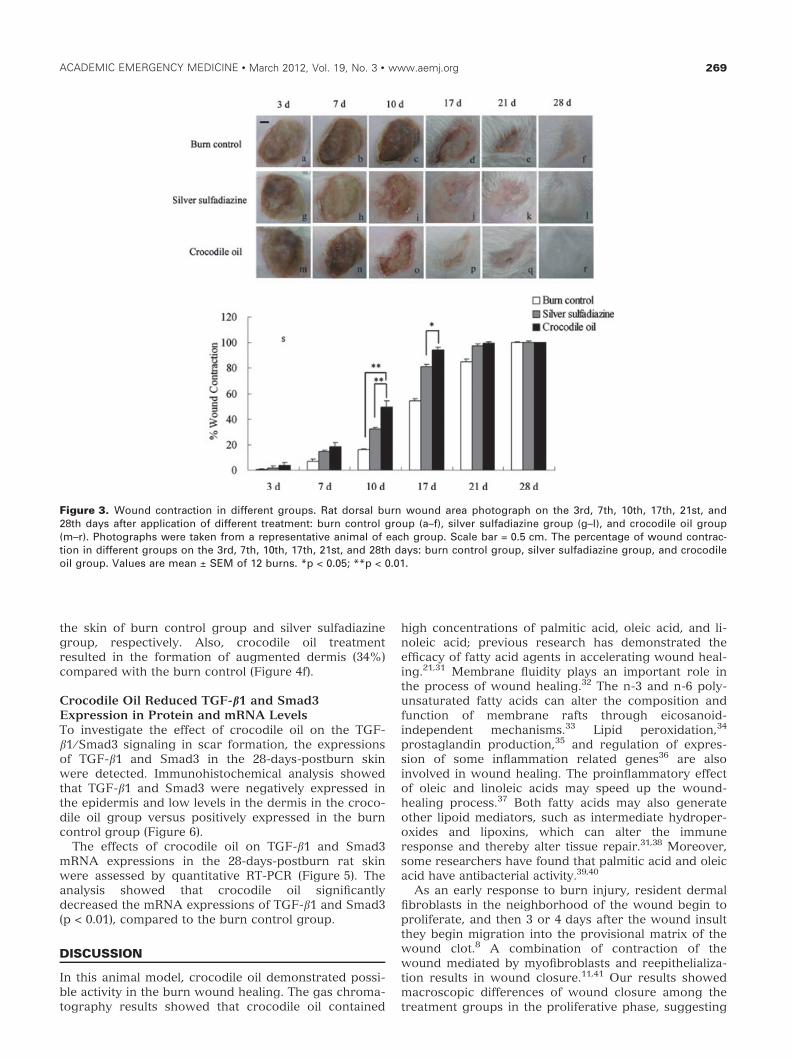
Effect of Crocodile Oil on Burn HealingThe burn healing time in the crocodile oil group wasshorter than in the burn control group and silver sulfa-diazine groups (Figure 2). In the first 3 days, the burnwounds progressed to necrotic eschars (Figures 3a, 3g,and 3m), and there were no measurable differencesamong the groups. On the 10th day, we observed asignificant improvement in wound closure in the croco-dile oil–treated group (Figure 3o) when compared tothe burn control group (Figure 3c and 3s) and the silversulfadiazine group (Figure 3i and 3s). After 17 days oftreatment, the animals in the crocodile oil group (Fig-ure 3p) had smaller wound areas than those in the sil-ver sulfadiazine group (Figures 3j and 3s). The resultsof morphologic evaluation on the 28th day showed thatthe best result was obtained with crocodile oil, in whichthe surface of the skin was smoother and the color wasclose to the normal skin (Figure 3r). The surface of theskin in the burn control group (Figure 3f) and the silversulfadiazine group (Figure 3l) was rough, and it wasmuch harder than the normal skin.
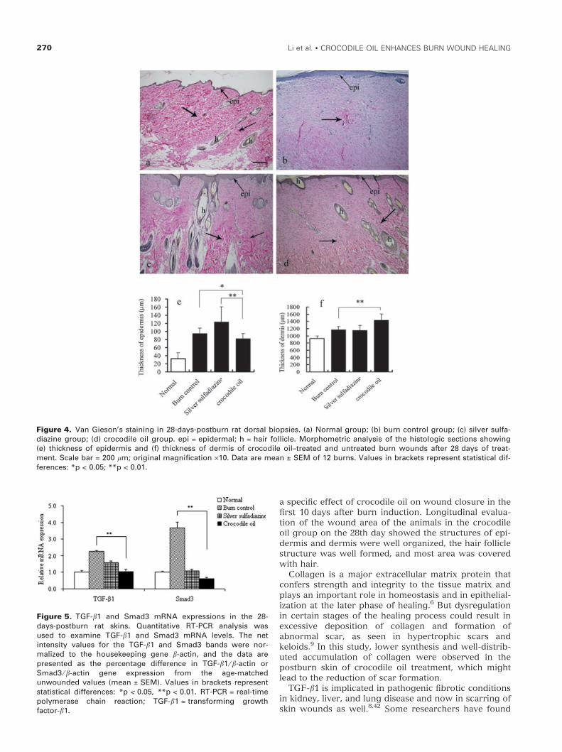
Effect of Crocodile Oil on Skin Structureand Collagen DepositionHistologic examination on the 28th day showed that theepidermis in the sham groups was thin, and skin struc-ture and collagen deposition (red staining) were wellorganized and distributed (Figure 4a). In the burn con-trol and silver sulfadiazine groups, the epidermis wasdisorganized, and the boundary between the epidermaland the dermis was blurred, few hair follicles structuresexisted, and a mass of disorganized red collagen waspresent in the dermal layer of the skin (Figures 4b and4c). In the crocodile oil group, there was full-thicknessreepithelialization, in which the epidermis was thinand well organized, the hair follicle structure waswell formed, and most area was covered with hair.The 28-days-postburn skin of the crocodile oilgroup showed lower synthesis and well-distributedaccumulation of collagen compared to the other groups(Figure 4d).
Quantitative analysis showed that on the 28th day,the epidermis thickness of the skin in crocodile oilgroup was 86 and 67% (Figure 4e) thinner than in
Table 1Composition of Fatty Acids
Fatty Acid
LipidNumbers
(C:D) Content (%)
Lauric acid 12:0 4.60Myristic acid 14:0 3.23Palmitic acid 16:0 29.22Stearic acid 18:0 1.84Palmitoleic acid 16:1 5.67Oleic acid 18:1 30.47Linoleic acid 18:2 11.74Linolenic acid 18:3 1.07Erucic acid 22:1 0.42Nervonic acid 24:1 0.31Saturated fatty acid 38.89Unsaturated fatty acid 49.67Monounsaturated fatty acid 36.86Polyunsaturated fatty acid 12.81Others 11.43
C = number of carbon atoms in the fatty acid; D = number ofdouble bonds in the fatty acid.
Figure 2. Healing time on Wistar rat dorsal wound. Values aremean ± SEM of 12 burns; values in brackets represent statisticaldifferences: *p < 0.05; **p < 0.01.
268 Li et al. • CROCODILE OIL ENHANCES BURN WOUND HEALING
the skin of burn control group and silver sulfadiazinegroup, respectively. Also, crocodile oil treatmentresulted in the formation of augmented dermis (34%)compared with the burn control (Figure 4f).
Crocodile Oil Reduced TGF-b1 and Smad3Expression in Protein and mRNA LevelsTo investigate the effect of crocodile oil on the TGF-b1 ⁄ Smad3 signaling in scar formation, the expressionsof TGF-b1 and Smad3 in the 28-days-postburn skinwere detected. Immunohistochemical analysis showedthat TGF-b1 and Smad3 were negatively expressed inthe epidermis and low levels in the dermis in the croco-dile oil group versus positively expressed in the burncontrol group (Figure 6).
The effects of crocodile oil on TGF-b1 and Smad3mRNA expressions in the 28-days-postburn rat skinwere assessed by quantitative RT-PCR (Figure 5). Theanalysis showed that crocodile oil significantlydecreased the mRNA expressions of TGF-b1 and Smad3(p < 0.01), compared to the burn control group.
DISCUSSION
In this animal model, crocodile oil demonstrated possi-ble activity in the burn wound healing. The gas chroma-tography results showed that crocodile oil contained
high concentrations of palmitic acid, oleic acid, and li-noleic acid; previous research has demonstrated theefficacy of fatty acid agents in accelerating wound heal-ing.21,31 Membrane fluidity plays an important role inthe process of wound healing.32 The n-3 and n-6 poly-unsaturated fatty acids can alter the composition andfunction of membrane rafts through eicosanoid-independent mechanisms.33 Lipid peroxidation,34
prostaglandin production,35 and regulation of expres-sion of some inflammation related genes36 are alsoinvolved in wound healing. The proinflammatory effectof oleic and linoleic acids may speed up the wound-healing process.37 Both fatty acids may also generateother lipoid mediators, such as intermediate hydroper-oxides and lipoxins, which can alter the immuneresponse and thereby alter tissue repair.31,38 Moreover,some researchers have found that palmitic acid and oleicacid have antibacterial activity.39,40
As an early response to burn injury, resident dermalfibroblasts in the neighborhood of the wound begin toproliferate, and then 3 or 4 days after the wound insultthey begin migration into the provisional matrix of thewound clot.8 A combination of contraction of thewound mediated by myofibroblasts and reepithelializa-tion results in wound closure.11,41 Our results showedmacroscopic differences of wound closure among thetreatment groups in the proliferative phase, suggesting
Figure 3. Wound contraction in different groups. Rat dorsal burn wound area photograph on the 3rd, 7th, 10th, 17th, 21st, and28th days after application of different treatment: burn control group (a–f), silver sulfadiazine group (g–l), and crocodile oil group(m–r). Photographs were taken from a representative animal of each group. Scale bar = 0.5 cm. The percentage of wound contrac-tion in different groups on the 3rd, 7th, 10th, 17th, 21st, and 28th days: burn control group, silver sulfadiazine group, and crocodileoil group. Values are mean ± SEM of 12 burns. *p < 0.05; **p < 0.01.
ACADEMIC EMERGENCY MEDICINE • March 2012, Vol. 19, No. 3 • www.aemj.org 269
a specific effect of crocodile oil on wound closure in thefirst 10 days after burn induction. Longitudinal evalua-tion of the wound area of the animals in the crocodileoil group on the 28th day showed the structures of epi-dermis and dermis were well organized, the hair folliclestructure was well formed, and most area was coveredwith hair.
Collagen is a major extracellular matrix protein thatconfers strength and integrity to the tissue matrix andplays an important role in homeostasis and in epithelial-ization at the later phase of healing.6 But dysregulationin certain stages of the healing process could result inexcessive deposition of collagen and formation ofabnormal scar, as seen in hypertrophic scars andkeloids.9 In this study, lower synthesis and well-distrib-uted accumulation of collagen were observed in thepostburn skin of crocodile oil treatment, which mightlead to the reduction of scar formation.
TGF-b1 is implicated in pathogenic fibrotic conditionsin kidney, liver, and lung disease and now in scarring ofskin wounds as well.8,42 Some researchers have found
Figure 4. Van Gieson’s staining in 28-days-postburn rat dorsal biopsies. (a) Normal group; (b) burn control group; (c) silver sulfa-diazine group; (d) crocodile oil group. epi = epidermal; h = hair follicle. Morphometric analysis of the histologic sections showing(e) thickness of epidermis and (f) thickness of dermis of crocodile oil–treated and untreated burn wounds after 28 days of treat-ment. Scale bar = 200 lm; original magnification ·10. Data are mean ± SEM of 12 burns. Values in brackets represent statistical dif-ferences: *p < 0.05; **p < 0.01.
Figure 5. TGF-b1 and Smad3 mRNA expressions in the 28-days-postburn rat skins. Quantitative RT-PCR analysis wasused to examine TGF-b1 and Smad3 mRNA levels. The netintensity values for the TGF-b1 and Smad3 bands were nor-malized to the housekeeping gene b-actin, and the data arepresented as the percentage difference in TGF-b1 ⁄ b-actin orSmad3 ⁄ b-actin gene expression from the age-matchedunwounded values (mean ± SEM). Values in brackets representstatistical differences: *p < 0.05, **p < 0.01. RT-PCR = real-timepolymerase chain reaction; TGF-b1 = transforming growthfactor-b1.
270 Li et al. • CROCODILE OIL ENHANCES BURN WOUND HEALING
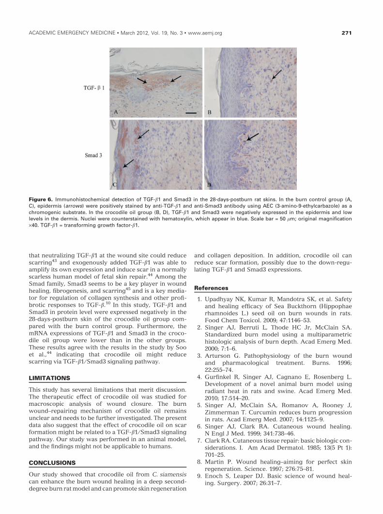
that neutralizing TGF-b1 at the wound site could reducescarring43 and exogenously added TGF-b1 was able toamplify its own expression and induce scar in a normallyscarless human model of fetal skin repair.44 Among theSmad family, Smad3 seems to be a key player in woundhealing, fibrogenesis, and scarring45 and is a key media-tor for regulation of collagen synthesis and other profi-brotic responses to TGF-b.10 In this study, TGF-b1 andSmad3 in protein level were expressed negatively in the28-days-postburn skin of the crocodile oil group com-pared with the burn control group. Furthermore, themRNA expressions of TGF-b1 and Smad3 in the croco-dile oil group were lower than in the other groups.These results agree with the results in the study by Sooet al.,44 indicating that crocodile oil might reducescarring via TGF-b1 ⁄ Smad3 signaling pathway.
LIMITATIONS
This study has several limitations that merit discussion.The therapeutic effect of crocodile oil was studied formacroscopic analysis of wound closure. The burnwound–repairing mechanism of crocodile oil remainsunclear and needs to be further investigated. The presentdata also suggest that the effect of crocodile oil on scarformation might be related to a TGF-b1 ⁄ Smad3 signalingpathway. Our study was performed in an animal model,and the findings might not be applicable to humans.
CONCLUSIONS
Our study showed that crocodile oil from C. siamensiscan enhance the burn wound healing in a deep second-degree burn rat model and can promote skin regeneration
and collagen deposition. In addition, crocodile oil canreduce scar formation, possibly due to the down-regu-lating TGF-b1 and Smad3 expressions.
References
1. Upadhyay NK, Kumar R, Mandotra SK, et al. Safetyand healing efficacy of Sea Buckthorn (Hippophaerhamnoides L.) seed oil on burn wounds in rats.Food Chem Toxicol. 2009; 47:1146–53.
2. Singer AJ, Berruti L, Thode HC Jr, McClain SA.Standardized burn model using a multiparametrichistologic analysis of burn depth. Acad Emerg Med.2000; 7:1–6.
3. Arturson G. Pathophysiology of the burn woundand pharmacological treatment. Burns. 1996;22:255–74.
4. Gurfinkel R, Singer AJ, Cagnano E, Rosenberg L.Development of a novel animal burn model usingradiant heat in rats and swine. Acad Emerg Med.2010; 17:514–20.
5. Singer AJ, McClain SA, Romanov A, Rooney J,Zimmerman T. Curcumin reduces burn progressionin rats. Acad Emerg Med. 2007; 14:1125–9.
6. Singer AJ, Clark RA. Cutaneous wound healing.N Engl J Med. 1999; 341:738–46.
7. Clark RA. Cutaneous tissue repair: basic biologic con-siderations. I. Am Acad Dermatol. 1985; 13(5 Pt 1):701–25.
8. Martin P. Wound healing–aiming for perfect skinregeneration. Science. 1997; 276:75–81.
9. Enoch S, Leaper DJ. Basic science of wound heal-ing. Surgery. 2007; 26:31–7.
Figure 6. Immunohistochemical detection of TGF-b1 and Smad3 in the 28-days-postburn rat skins. In the burn control group (A,C), epidermis (arrows) were positively stained by anti-TGF-b1 and anti-Smad3 antibody using AEC (3-amino-9-ethylcarbazole) as achromogenic substrate. In the crocodile oil group (B, D), TGF-b1 and Smad3 were negatively expressed in the epidermis and lowlevels in the dermis. Nuclei were counterstained with hematoxylin, which appear in blue. Scale bar = 50 lm; original magnification·40. TGF-b1 = transforming growth factor-b1.
ACADEMIC EMERGENCY MEDICINE • March 2012, Vol. 19, No. 3 • www.aemj.org 271
10. Lakos G, Takagawa S, Chen SJ, et al. Targeted dis-ruption of TGF-beta ⁄ Smad3 signaling modulatesskin fibrosis in a mouse model of scleroderma. AmJ Pathol. 2004; 165:203–17.
11. Lin RY, Sullivan KM, Argenta PA, et al. Exogenoustransforming growth factor-beta amplifies its ownexpression and induces scar formation in a modelof human fetal skin repair. Ann Surg. 1995;222:146–54.
12. Shah M, Foreman DM, Ferguson MW. Neutralisa-tion of TGF-beta 1 and TGF-beta 2 or exogenousaddition of TGF-beta 3 to cutaneous rat woundsreduces scarring. J Cell Sci. 1995; 108(Pt 3):985–1002.
13. Kavsak P, Rasmussen RK, Causing CG, et al. Smad7binds to Smurf2 to form an E3 ubiquitin ligase thattargets the TGF beta receptor for degradation. MolCell. 2000; 6:1365–75.
14. Vindevoghel L, Kon A, Lechleider RJ, et al. Smad-dependent transcriptional activation of human typeVII collagen gene (COL7A1) promoter by TGF-b.J Biol Chem. 1998; 37:13053–7.
15. Hu B, Wu Z, Phan S. Smad3 mediates TGF-b-induced a-smooth muscle actin expression. AmJ Respir Cell Mol Biol. 2003; 29:397–404.
16. Evans RA, Tian YC, Steadman R, Phillips AO. TGF-beta1-mediated fibroblast- myofibroblast terminaldifferentiation-the role of Smad proteins. Exp CellRes. 2003; 282:90–100.
17. Valacchi G, Lim Y, Belmonte G, et al. Ozonatedsesame oil enhances cutaneous wound healing inSKH1 mice. Wound Repair Regen. 2011; 19:107–15.
18. Li ZQ, Wang JH, Ren JL, Yi ZH. Effects of topicalemu oil on wound healing in scalded rats. Di Yi JunYi Da Xue Xue Bao. 2004; 24:1255–6.
19. Tang L, Qin MZ. The medicinal research and devel-opment prospects of crocodile. World Health Digest(in Chinese). 2007; 4:66–8.
20. Gunstone FD, Russell WC. Animal fats. The compo-nent acids of crocodile fat. Biochem J. 1954;57:462–5.
21. Feng X, Cheng G, Chen SY, Yang H, Huang W.Evaluation of the burn healing properties of oilextraction from housefly larva in mice. J Ethno-pharmacol. 2010; 130:586–92.
22. Wang FR, Ai H, Chen XM, Lei CL. Hepatoprotec-tive effect of a protein-enriched fraction from themaggots (Musca domestica) against CCl4-inducedhepatic damage in rats. Biotechnol Lett. 2007;29:853–8.
23. Ozturk S, Ercisli S. The chemical composition ofessential oil and in vitro antibacterial activities ofessential oil and methanol extract of Ziziphora per-sica Bunge. J Ethnopharmacol. 2006; 106:372–6.
24. Lee JA, Jeong HJ, Park HJ, Jeon S, Hong SU. Acu-puncture accelerates wound healing in burn-injuredmice. Burns. 2011; 37:117–25.
25. Gupta A, Kumar R, Upadhyay NK, Pal K, SawhneyRC. Effects of Rhodiola imbricata on dermal woundhealing. Planta Med. 2007; 73:774–7.
26. Zhou J, Zhong DW, Wang QW, Miao XY, Xu XD.Paclitaxel ameliorates fibrosis in hepatic stellate
cells via inhibition of TGF-beta ⁄ Smad activity.World J Gastroenterol. 2010; 16:3330–4.
27. Livak K. Analysis of relative gene expression datausing real-time quantitative PCR and the 2)DDCT
method. Methods. 2001; 25:402–8.28. Singer A, Thode HC Jr, McClain S. The effects of
Ctylcyanoacrylate on scarring after burns. AcadEmerg Med. 2001; 8:107–11.
29. Takagawa S, Lakos G, Mori Y, et al. Sustainedactivation of fibroblast transforming growth factor-beta ⁄ Smad signaling in a murine model of sclero-derma. J Invest Dermatol. 2003; 121:41–50.
30. Cohen J. Statistical Power Analysis for the Behav-ioral Sciences. Hillsdale, NJ: Lawrence ErlbaumAssociates, 1988.
31. Cardoso CR, Souza MA, Ferro EA, Favoreto S,Pena JD. Influence of topical administration of n-3and n-6 essential and n-9 nonessential fatty acids onthe healing of cutaneous wounds. Wound RepairRegen. 2004; 12:235–43.
32. Choi JH, Yu BP. Brain synaptosomal aging: freeradicals and membrane fluidity. Free Radic BiolMed. 1995; 18:133–9.
33. Jump DB, Clarke SD. Regulation of gene expressionby dietary fat. Annu Rev Nutr. 1999; 19:63–90.
34. Dursun N, Liman N, Özyazgan _I, Günes I, SaraymenR. Role of thymus oil in burn wound healing. J BurnCare Rehabil. 2003; 24:395–9.
35. James MJ, Gibson RA, Cleland LG. Dietary polyun-saturated fatty acids and inflammatory mediator pro-duction. Am J Clin Nutr. 2000; 71(1 Suppl):343S–8S.
36. Kumagai T, Kawamoto Y, Nakamura Y, et al.4-hydroxy-2-nonenal, the end product of lipidperoxidation, is a specific inducer of cyclooxy-genase-2 gene expression. Biochem Biophys ResCommun. 2000; 273:437–41.
37. Pereira LM, Hatanaka E, Martins EF, et al. Effect ofoleic and linoleic acids on the inflammatory phaseof wound healing in rats. Cell Biochem Funct. 2008;26:197–204.
38. Aliberti J, Hieny S, Reise SC, Serhan CN, Sher A.Lipoxin-mediated inhibition of IL-12 production byDCs: a mechanism for regulation of microbialimmunity. Nat Immunol. 2002; 3:76–82.
39. Hinton A Jr, Ingram KD. Use of oleic acid to reducethe population of the bacterial flora of poultry skin.J Food Prot. 2000; 63:1282–6.
40. Yff BT, Lindsey KL, Taylor MB, Erasmus DG, JagerAK. The pharmacological screening of Pentanisiaprunelloides and the isolation of the antibacterialcompound palmitic acid. J Ethnopharmacol. 2002;79:101–7.
41. Kietzmann M. Improvement and retardation ofwound healing: effects of pharmacological agents inlaboratory animal studies. Vet Dermatol. 1999;10:83–8.
42. Border WA, Noble NA. Transforming growth factorbeta in tissue fibrosis. N Engl J Med. 1994;331:1286–92.
43. Shah M, Foreman DM, Ferguson MW. Control ofscarring in adult wounds by neutralising antibodyto transforming growth factor beta. Lancet. 1992;339:213–4.
272 Li et al. • CROCODILE OIL ENHANCES BURN WOUND HEALING
44. Soo C, Beanes SR, Hu FY, et al. Ontogenetic transi-tion in fetal wound transforming growth factor-betaregulation correlates with collagen organization.Am J Pathol. 2003; 163:2459–76.
45. Phan TT, Lim IJ, Aalami O, et al. Smad3 signallingplays an important role in keloid pathogenesis viaepithelial–mesenchymal interactions. J Pathol. 2005;207:232–42.
ACADEMIC EMERGENCY MEDICINE • March 2012, Vol. 19, No. 3 • www.aemj.org 273