cronicon open access dental science research · pdf filemandibular saggital piezo-osteotomy...
TRANSCRIPT

CroniconO P E N A C C E S S DENTAL SCIENCE
Research Article
Eber Luis de Lima Stevao* Department of the Orthognathic Surgery, Institute of Curitiba, Brazil
Received: December 02, 2014; Published: February 09, 2015
*Corresponding Author: Eber Luis de Lima Stevao, Department of the Orthognathic Surgery Institute of Curitiba, Curitiba, Parana, Brazil.
Piezosurgery Applied to Orthognathic Surgery-Retrospective Study with New Mandibular Saggital Piezo-Osteotomy Technique Description
Abstract
Piezoelectric surgery, known as Piezosurgery, is a relatively new technique which was initiated in oral surgery in the year 2000 with the classic publication of Dr. Tomaso Vercellotti’s article, and in orthognathic surgery only in 2004 and 2005. This technique is safe and useful for facial osteotomies and or osteoplasties with excellent results. Piezosurgery functioning principle is an ultrasonic trans-ducer which modifies electric energy into vibrational one (mechanics). Connected to its handpiece is an ultrasonic tip (insert). The ultrasonic insert oscillates with microvibrations resulted from ceramics expansions and contractions. The ultrasonic tip vibration, along with a gentle manual pressure on its handpiece, and a manual back and forth movement upon the bony tissue will result in bone cavitation. Irrigation for tip refrigeration is an essential part of the procedure. This new method of bone cutting involves a total different learning curve from the one necessary for reciprocating saw usage. Although there is a small increase in the intra-operative time for bone procedures with Piezosurgery, its low morbidity (safety) due to mineralized tissue selectivity, associated to other ad-vantages such as less bleeding, minor postoperative edema, rapidity in bone recovery (faster bone repair) reveals the potential of this new ultrasound bone cutting technique.
This article reports the author’s five-year Piezosurgery experience in orthognathic surgical procedures. The results obtained until the present moment are promising, since the Piezosurgery manufacturer companies will continue to develop better ultrasonic inserts with specific geometrics on account of the characteristics of the various types of osteotomies, and illuminated handpiece by optic fiber or light emitting diode (LED). Above of all, with this new technology it is possible to perform very precise and micrometric osteotomy lines (precision) giving absolute confidence to surgeons, mainly to those who are new beginners in orthognathic surgery field where the saw cutting instrument generate imprecise cuts and increases the possibility for soft tissue (mucosae, periosteum and nerves) injuries. It is believed in the next years this will be the standard technique for bone procedures as far as the demanding art of orthognathic surgery is concerned.
Keywords: Piezosurgery; Piezoelectric surgery; Piezo-orthognathic surgery; Ultrasonic osteotomy; Ultrasonic osteoplasty
Abbreviations: LED: Light Emitting Diode; IAN: Inferior Alveolar Nerve; SARPE: Surgically Assisted Rapid Expansions; MSTDO: Man-dibular Symphyseal Transverse Distraction Osteogenesis; ANSp: Anterior Nasal Spine-plasties; BIT: Bilateral Inferior Turbinectomies; PS: Partial Septoplasties; AMxSO: Anterior Maxillary Subapical Osteotomy; PMPCO: Pterigomaxillary Process Chisel Osteotomies; PMPPDA: Pterigomaxillary Process Piezo-disjunction Attempts; BSSO: Bilateral Saggital Split Osteotomies; USSO: Unilateral Saggital Split Osteot-omy; MSTDO: Mandibular Symphyseal Transverse Distraction Osteogenesis; TMG: Tenon and Mortise Genioplasties; HSOG: Horizontal Straight Osteotomy for Genioplasties; AMdSO: Anterior Mandibular Subapical Osteotomy; BPMdSO: Bilateral Posterior Mandibular Sub-apical Osteotomy; UPMdSO: Unilateral Posterior Mandibular Subapical Osteotomy
Citation: Eber Luis de Lima Stevao. “Piezosurgery applied to Orthognathic Surgery-Retrospective Study with New Mandibular Saggital Piezo-Osteotomy Technique Description”. EC Dental Science 1.2 (2015): 56-79.

Piezosurgery Applied to Orthognathic Surgery-Retrospective Study with New Mandibular Saggital Piezo-Osteoto-my Technique Description
57
Citation: Eber Luis de Lima Stevao. “Piezosurgery applied to Orthognathic Surgery-Retrospective Study with New Mandibular Sag-gital Piezo-Osteotomy Technique Description”. EC Dental Science 1.2 (2015): 56-79.
Piezo is composed of a Piezoeletric device called central unity which works with a frequency of 24 to 32 kHz reaching up to 30 Hz through a digital modulation by a central unity that controls oscillation movements introducing small periods of pause to avoid heating of ultrasonic insert, however maintaining an excellent cutting capacity.
A handpiece is coupled to a central unity and it receives an ultrasonic insert specially made to resist micro-oscillations which are generated by ceramic distensions and contractions.
The handpiece tip can offer an auxiliary lighting for operating field with LED (Light Emitting Diode) or optic fiber light coupled de-pending on the manufacturer.
There are several inserts that can be connected to the handpiece through simple clockwise twisting and final deadlocking with a special torquimeter key.
IntroductionAs an initial explanation it is important to emphasize that the term Piezosurgery throughout this article has two meanings: a. First one, referred to Piezosurgery System, the appliance itself, which is mentioned along this study simply as Piezo.b. Second one, concerning to piezoelectric surgery technique cited here as Piezosurgery or Piezo - Osteotomy.
Figure 1: Digital central unity of a Piezo system, Surgystar Plus (Dmetec ®, Seoul, South Korea).
Figure 2: Handpiece with two plug connections: the metal one for distilled water coupling and other one with gray intermediary for central unity connection.
Piezosurgery Applied to Orthognathic Surgery-Retrospective Study with New Mandibular Saggital Piezo-Osteoto-my Technique Description
58
Citation: Eber Luis de Lima Stevao. “Piezosurgery applied to Orthognathic Surgery-Retrospective Study with New Mandibular Saggital Piezo-Osteotomy Technique Description”. EC Dental Science 1.2 (2015): 56-79.
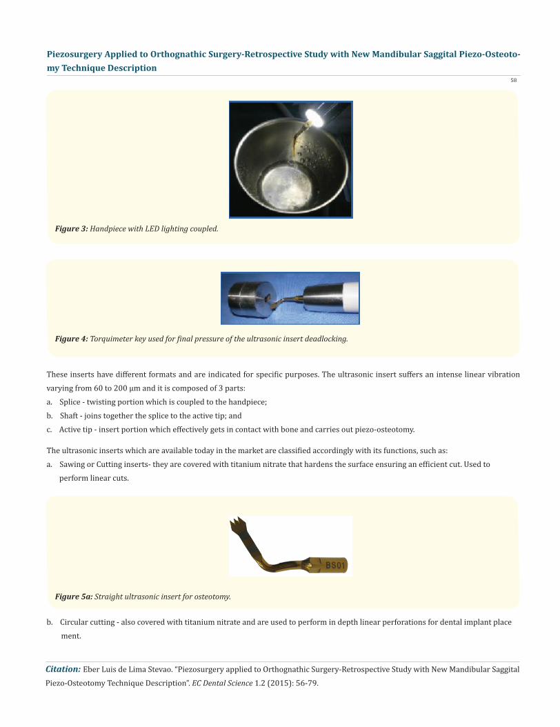
Figure 3: Handpiece with LED lighting coupled.
Figure 5a: Straight ultrasonic insert for osteotomy.
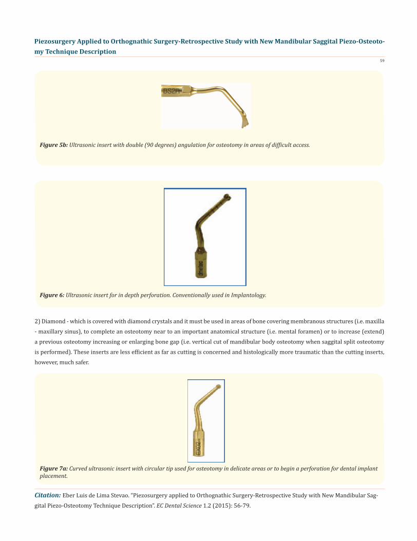
Figure 4: Torquimeter key used for final pressure of the ultrasonic insert deadlocking.
These inserts have different formats and are indicated for specific purposes. The ultrasonic insert suffers an intense linear vibration varying from 60 to 200 µm and it is composed of 3 parts: a. Splice - twisting portion which is coupled to the handpiece; b. Shaft - joins together the splice to the active tip; and c. Active tip - insert portion which effectively gets in contact with bone and carries out piezo-osteotomy.
The ultrasonic inserts which are available today in the market are classified accordingly with its functions, such as:a. Sawing or Cutting inserts- they are covered with titanium nitrate that hardens the surface ensuring an efficient cut. Used to perform linear cuts.
b. Circular cutting - also covered with titanium nitrate and are used to perform in depth linear perforations for dental implant place ment.
Piezosurgery Applied to Orthognathic Surgery-Retrospective Study with New Mandibular Saggital Piezo-Osteoto-my Technique Description
59
Citation: Eber Luis de Lima Stevao. “Piezosurgery applied to Orthognathic Surgery-Retrospective Study with New Mandibular Sag-gital Piezo-Osteotomy Technique Description”. EC Dental Science 1.2 (2015): 56-79.
2) Diamond - which is covered with diamond crystals and it must be used in areas of bone covering membranous structures (i.e. maxilla - maxillary sinus), to complete an osteotomy near to an important anatomical structure (i.e. mental foramen) or to increase (extend) a previous osteotomy increasing or enlarging bone gap (i.e. vertical cut of mandibular body osteotomy when saggital split osteotomy is performed). These inserts are less efficient as far as cutting is concerned and histologically more traumatic than the cutting inserts, however, much safer.
Figure 5b: Ultrasonic insert with double (90 degrees) angulation for osteotomy in areas of difficult access.
Figure 6: Ultrasonic insert for in depth perforation. Conventionally used in Implantology.
Figure 7a: Curved ultrasonic insert with circular tip used for osteotomy in delicate areas or to begin a perforation for dental implant placement.

Piezosurgery Applied to Orthognathic Surgery-Retrospective Study with New Mandibular Saggital Piezo-Osteot-omy Technique Description
60
Citation: Eber Luis de Lima Stevao. “Piezosurgery applied to Orthognathic Surgery-Retrospective Study with New Mandibular Sag-gital Piezo-Osteotomy Technique Description”. EC Dental Science 1.2 (2015): 56-79.
Piezo through microvibrations and by ultrasonic frequency produced offers three mechanical aspects that are superior to rotating or oscillatory instruments. Firstly, it allows a micrometric cut offering a superior precision without bone loss. Secondly, instrument se-lects mineralized structures without causing damages to adjacent soft tissues which remain intact even in case of accidental ultrasonic insert contact with delicate structures. Thirdly, physical cavitation phenomenon (ostectomy) which guarantees less bleeding as well demonstrated by Stubinger et al. [1].
Based on literature finds about Piezosurgery for orthognathic surgery this work critically values Piezo - Osteotomy viability as a better alternative method than rotating and oscillatory ones for dentofacial deformities surgical correction. Advantages and disadvan-tages are objectively and realistically presented here bringing an opposite point of view to an apparent consensus in oral and maxil-lofacial surgery centres which is Piezo has not power nor necessary boost for its destined aim.
Dentistry all around the world is suffering a radical transformation in the areas of Oral and Maxillofacial Surgery, Implantology, Or-thodontics and Periodontics with Piezo arise mostly in oral surgery scenario in spite of ultrasound early beginning back to 1952 when it was introduced for tooth cavity preparations. In the interim high speed rotating instruments were introduced and ultrasound became latent for some decades as reported by Escoda - Francoli et al. [2].
Even though Piezo had its applicability in teeth during those years ultrasound for bone tissue procedures had already been de-fended by Maintz [3] who had published the very first study showing the beneficial ultrasound effects on bone surgery.
c. Rasp - with a sketched surface giving a characteristic of bone file to this insert. It can be used to shape a bone surface or complete an osteoplasty.
Figure 7b: Curved ultrasonic insert to enlarge previous osteotomies.
Figure 8: Straight ultrasonic insert for scraping (osteoplasty) bone.
Literature Review

Piezosurgery Applied to Orthognathic Surgery-Retrospective Study with New Mandibular Saggital Piezo-Osteot-omy Technique Description
61
Citation: Eber Luis de Lima Stevao. “Piezosurgery applied to Orthognathic Surgery-Retrospective Study with New Mandibular Sag-gital Piezo-Osteotomy Technique Description”. EC Dental Science 1.2 (2015): 56-79.
But piezoelectric technique was only retaken in 1988 and later several published articles showed its applicability in minor oral surgery for alveolar crest expansion by Vercellotti [4], removal of impacted teeth by Eggers et al. [5] and Vercellotti [6], in parendo-dontic surgery by Marti - Bowen et al. [7], in periodontal surgery by Penarrocha et al. [8]. In major oral surgery its use was described for distraction osteogenesis by Politi et al. [9], maxillary orthognathic surgery by Robiony et al. [10], mandibular orthognathic surgery with a pilot study described by Gruber et al. [11], in lateralization (skeletization) of inferior alveolar nerve (IAN) by Metzger et al. [12], and in surgically assisted rapid maxillary palatal expansion by Robiony et al. [13].
Schlee et al. [14] referred that Piezosurgery technique offers a micrometric (precise and safe action limiting the damages to vital tissues specially to osteocytes), selective (affecting mineralized tissues but not surrounding soft tissues) and clean cut (result of cavity effect created by refrigerating/irrigating solution and fast oscillatory insert). These authors alerted that because insert vibrates in dif-ferent ultrasonic frequencies its selective action enables surgeons to cut hard tissue while preserving delicate anatomical structures such as Schneiderian membrane and nerves. As a result of this selectivity the above authors acknowledged that Implantology surgical techniques such as bone harvesting, alveolar ridge splitting and maxillary sinus floor elevation can be executed with greater easiness and security.
Preti et al. [15] carried out a biomolecular and histological analysis study to compare dental implant osseointegration installed in bone perforations performed with conventional drills and piezoelectric techniques. They evaluated histomorphological and bone mor-phogenetic protein (BMP-4), transformer growth factor (TGF-b2), necrosis factor alpha (TNF-alpha), interleucin-1b and interleucin-10 responses. The obtained results showed higher quantity of inflammatory cells present in perforations carried out with conventional implant drills. Also neo-osteogenesis was more active in bone sites which had been prepared with Piezosurgery, plus earlier increase of BMP-4 and TGF-b2 proteins and reduction of the proinflammatory cytokines were evident. These authors concluded that Piezosurgery for bone perforation is more efficient in first phases of bone repair, induced to increased levels of BMPs, better controlled inflammation and early stimulated bone remodeling on 56 days of post-operative when compared with conventional bone drilling.
Heiland et al. [16] described a seven-week old patient with Pierre - Robin syndrome who underwent Piezosurgery bone distrac-tion. In spite of such a tender age and low ossification level it was possible to carry out mandibular osteotomies and transcutaneous distractors placement. After follow up and devices removal those authors suggested Piezosurgery usage for the same syndrome cases even in patients with less than 2 months of life because of the success obtained with ultrasound bone surgery technique.
Stübinger and Goethe [17] exposed that Piezosurgery definitely increases careful handling of structures in oral and maxillofacial region. Regarding osteotomy of fine and fragile bones, ultrasound application offers superior results when compared with other me-chanical instruments because it is of easier handling, has an efficient bone ablation and it causes least accidental damage to adjacent soft tissue structures. They concluded asserting that once bone repair is not disturbed by Piezosurgery and even it seems to be im-proved, this method will have a great influence in minimally invasive bone surgery techniques with special attention to its biomechan-ics.
Studies of neurossensorial response after facial piezo-osteotomies were carried out and very positively reported in oral and maxil-lofacial literature.
Geha et al. [18] accomplished a study to access neurossensorial clinical function of Inferior Alveolar Nerve (IAN) after bilateral saggital split osteotomy with Piezosurgery. They performed forty procedures in twenty patients with dentoskeletal deformities. IAN was valued subjectively and objectively through pin-prick and light touch lip sensation, and two points discrimination tests, at pre-operatively, 5, 7, 10 days and 2 months after treatment. A combination of those tests revealed 75 to 80% complete neurossensorial recovery at the second postoperative month. Recovery of IAN function could be seen in 77.5% of sides assessed by neurossensory tests

Piezosurgery Applied to Orthognathic Surgery-Retrospective Study with New Mandibular Saggital Piezo-Osteot-omy Technique Description
62
Citation: Eber Luis de Lima Stevao. “Piezosurgery applied to Orthognathic Surgery-Retrospective Study with New Mandibular Sag-gital Piezo-Osteotomy Technique Description”. EC Dental Science 1.2 (2015): 56-79.
and 55% of sides assessed subjectively. Piezo device was able to split the mandible (down to the basilar border) in twenty six out of forty saggital splits or thirteen (65%) of cases. In the remaining patients, the split was incomplete and reached varying depths without attaining the posteroinferior basilar border. The authors simply concluded that Piezosurgery for saggital split osteotomy allows IAN recuperation within two months.
Schaeren et al. [19] observed that peripheral nerve direct exposition to Piezosurgery, even in the worst case scenarios, did not dis-sect the nerve but did induce some structural and functional damages. The perineurium of the nerve remained intact even after nerve contact at peak force, thus enhancing the potential for functional recovery. They emphasized the extent of damage was significantly higher with application of increased force on the nerve but not by activation of the ultrasonic vibration. Because the proper use of Pi-ezosurgery technique calls for application of limited pressure, the safety margins are greater than those when using instruments that are operated at higher force (i.e. bur) or are likely to cut the nerve on direct contact (i.e. oscillatory saw). In conclusion they found Pi-ezosurgery to be a promising tool in the maxillofacial surgery for performing osteotomy in close proximity to a nerve (i.e. orthognathic surgery for mandible).
Landes et al. [20] described their results after operating fifty patients with Piezosurgery osteotomies. Twenty two one-segment maxilla (44%), twenty six multi-segmented maxilla (52%), forty eight saggital split osteotomies (48%), six symphyseal osteotomies (12%) and four mandibular body osteotomies (4%). They reported the need of chisel and hammer for 46% of the cases when pterigo-maxillary process osteotomy (disjunction) was necessary due to the fact that these procedures had not been completed with Piezo-surgery. The pterigomaxillary suture was weakened by angled insert OP1 (Mectron ®, Carasco, Italy) initially and consecutively using OT8L and OT8R inserts. The sensibility of the Nerve Alveolar Inferior (IAN) returned to normality in 95% after 3 months of post-operative follow up. The authors reported that piezoelectric surgery reduces blood loss and injuries to IAN.
Figure 9a: Ultrasonic insert BS1S used for maxillary, and internal aspect of mandible ascending ramus and body osteotomies.
Figure 9b: Piezo-osteotomy for maxillary expansion carried out with insert BS1S and enlarged with insert EX03 following the design of bilateral step osteotomy described Wolford and Epker [23].

Piezosurgery Applied to Orthognathic Surgery-Retrospective Study with New Mandibular Saggital Piezo-Osteot-omy Technique Description
63
Citation: Eber Luis de Lima Stevao. “Piezosurgery applied to Orthognathic Surgery-Retrospective Study with New Mandibular Sag-gital Piezo-Osteotomy Technique Description”. EC Dental Science 1.2 (2015): 56-79.
All patients were instructed and oriented about the surgical procedures and free will signed the routine informed consent of the Institute of Orthognathic Surgery of Curitiba, in Parana State, Brazil, authorizing the use of its materials for research and or publication. The retrospective survey data project for future publication of this research had its approval by the Committee of Ethics of the same Institution.
In a retrospective series seventy four patients, forty seven women (63%) and twenty seven men (37%), were exclusively submit-ted to orthognathic surgery where exclusively maxillary, mandibular and chin piezo-osteotomy technique were performed from May 2007 to May 2012. Also, maxillary surgically assisted rapid expansions (SARPE) and mandibular symphyseal transverse distraction
Signs and Symptoms
Figure 9c: Mandibular piezo-osteotomy. The photo shows the beginning of horizontal osteotomy accordingly the design described by Schuchardt [24], its (modified) extension through retromolar area accordingly the design described by Dal Pont [25].
Figure 9d: Mandibular Piezo-osteotomy. The photo shows the curvilinear step carried out at the level of second molar roots, then its osteotomy extension in 90 degrees following the design described by Wolford and Davis [22] and the vertical cut at the first molar level to complete this piezo-osteotomy.
Materials and Methods

Piezosurgery Applied to Orthognathic Surgery-Retrospective Study with New Mandibular Saggital Piezo-Osteot-omy Technique Description
64
Citation: Eber Luis de Lima Stevao. “Piezosurgery applied to Orthognathic Surgery-Retrospective Study with New Mandibular Sag-gital Piezo-Osteotomy Technique Description”. EC Dental Science 1.2 (2015): 56-79.
Technical aspects and Piezo Surgical Procedures - Piezo-osteotomies
osteogenesis (MSTDO), as well as anterior nasal spine-plasties (ANSp), bilateral inferior turbinectomies (BIT) and partial septoplasties (PS) were mentioned in this research as secondary minor procedures.
All patients were instructed and oriented about their surgical procedures and free will signed the informed consent of the Orthog-nathic Surgery Institute of Curitiba - OSIC - in Parana State, Brazil, authorizing the usage of his/her material for research/publication. The retrospective data collection project had its approval by the OSIC and DSP Biomedical Ethics Committees, under protocol 03/08.
The technical aspects described below oriented all surgical procedures for this study and they became like rules to be based upon.
During Piezosurgery a surgeon has better hand control than handling any other bone cutting instrument whereas its demanded control force is less if compared to any rotating or oscillatory instruments which demand higher manual pressure. Every time surgical force is needed, sensibility is reduced and this per se is a very strong indicative the ultrasonic insert must be changed by a new one. An ultrasonic insert usage must not exceed 3 to 5 piezo-osteotomies as recommended by Dmetec® [21].
Also, cuts carried out with Piezosurgery allows in depth control (i.e. genioplasty), a difficult aspect to be commended by beginners when using oscillatory instruments such as reciprocating saw, which vibrates more in operator’s hand and, maybe, because of exagger-ated care, stress or surgeon’s fear, an osteotomy ends up with chisel.
The central unity boost mode was always on when osteotomy or osteoplasty were intended. The undesirable contact with soft tissues (vascular-nervous bundle, mucous membrane or periosteum) does not mean that they will be immediately cut or lacerated as it usually happens with rotating (drills) or oscillatory (saws) instruments. As soon as any contact occurred Piezosurgery movement was interrupted to avoid tissue heating. Even though piezoelectric energy is inactive on soft tissues concerning the mechanical aspect, however heat is dispersed on the structure(s). For this reason, the author used on this study and recommends irrigation to a hundred per cent to be always appropriate to reducing thermal trauma.
Even if a professional is an experienced surgeon with rotating and oscillatory saws it does not mean much thing concerning to piezo-osteotomy. On contrary these operators normally become frustrated with longer time for piezo-osteotomy to be carried out and they leave the technique for lacking patience and perseverance so needed for developing an optimal and perfected art which will result in reduction of piezo-operating time. Humility is necessary to recognize one’s own limits and lack of familiarity with this new technol-ogy. Perhaps this psychological obstacle is the biggest barrier to be overcome and the biggest reason, up to this present moment, why piezoelectric surgery is not yet leading bone surgical procedures all around the world. So, it is necessary that a novice in ultrasound osteotomy understands Piezosurgery is a completely different method for osteotomy from the other ones, and a learning curve will be necessary for reaching the best level of usage for any piezoelectric device. The movements repetition for this type of osteotomy com-poses the basis of piezo-osteotomy and patience becomes a virtue.
The rational for performing a piezo-osteotomy is simple and follows a pattern. For example, a surgeon leading with a higher dif-ficulty for cutting a thicker cortical bone has a tendency to increase pressure when using drills or saw but rationalization and attitude cannot be the same for piezo-osteotomy because any pressure increase inhibits oscillatory ultrasonic insert micro movements result-ing in heat or metal fatigue leading to fracture. This insert fracture normally occurs between insert’s shaft and its active part.
Along the development and refine of this new piezoelectric technique the author was able to create a modification of the traditional mandibular SSO and named it Modified Mandibular Saggital Split Piezo-Osteotomy or Obwegeser-Wolford-Stevao piezo-osteotomy design for mandible. This technique will be described below because it is part of the material and methods of this study.

Piezosurgery Applied to Orthognathic Surgery-Retrospective Study with New Mandibular Saggital Piezo-Osteot-omy Technique Description
65
Citation: Eber Luis de Lima Stevao. “Piezosurgery applied to Orthognathic Surgery-Retrospective Study with New Mandibular Sag-gital Piezo-Osteotomy Technique Description”. EC Dental Science 1.2 (2015): 56-79.
The Obwegeser-Wolford-Stevao mandibular piezo-osteotomy begins with the horizontal osteotomy of the internal face of the as-cending ramus using ultrasonic insert BS01, crossed and angled downward (approximately 30 degrees), and slightly positioned (1 to 2 mm) above the mandibular Spix process, accordingly to the traditional technique described by Schuchardt [22] which was modified by Trauner and Obwegeser [23]. However this cut cannot be performed caudally or buccally in its total extension. This initial piezo-osteot-omy must be carefully extended 3 to 5 mm beyond the mandibular channel entrance in its posterior-anterior aspect, as no medullary bone is to be found between the two mandibular cortices (external and internal) at this level of the ascending ramus.
Using the same ultrasonic insert piezo-osteotomy now progresses inferiorly and longitudinally to the ascending ramus and the insert tip is dislocated towards the external cortex always deepening it as much as possible. Cortical orientation during this procedure is vital and this movement intention is to avoid bone marrow adhered to the proximal segment.
In the author’s experience it was possible to notice when performing osteotomy closer from the external mandibular cortex at retromolar region to the descending curve at mandibular body, BS01 total deepening helped to maintain cancellous bone on distal seg-ment upholding the IAN always protected. This procedure correctly performed all along the mandible oblique line weakens this area of great bone resistance facilitating segments separation in an ideal way without undesirable fractures.
The piezo-osteotomy follows anteriorly along retromolar trigone as described by Dal Pont [24], however in a quite short form not extending it to molar region nor medially to cortex (internal and external) but directing the insert more externally to follow the begin-ning of mandibular oblique line.
Through an analysis of an anterior-posterior standard radiography it is possible to identify where the area of greater bone resis-tance is located which is at the level of mandibular body and ascending ramus.
Subsequently piezo-osteotomy assumes a curvilinear pattern described by Wolford and Davis [25], however with a very important modification. Throughout this curvilinear extension ultrasonic insert was not tilted 90 degrees perpendicular to bone cortex but it was longitudinally orientated strictly following Obwegeser’s [23] technique. The piezo-osteotomy was spread out more inferiorly than the one originally described by Wolford and Davis [25], reducing bone height of the mandibular body horizontal cut to the inferior
Obwegeser-Wolford-Stevao Piezo-Osteotomy design - Technique description
Figure 10a: Conventional X-ray in standard anterior-posterior projection. The arrows indicate the line of bone reinforcement (corti-cal bone line).
Piezosurgery Applied to Orthognathic Surgery-Retrospective Study with New Mandibular Saggital Piezo-Osteot-omy Technique Description
66
Citation: Eber Luis de Lima Stevao. “Piezosurgery applied to Orthognathic Surgery-Retrospective Study with New Mandibular Sag-gital Piezo-Osteotomy Technique Description”. EC Dental Science 1.2 (2015): 56-79.
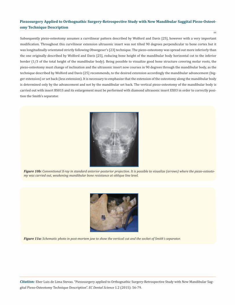
Subsequently piezo-osteotomy assumes a curvilinear pattern described by Wolford and Davis [25], however with a very important modification. Throughout this curvilinear extension ultrasonic insert was not tilted 90 degrees perpendicular to bone cortex but it was longitudinally orientated strictly following Obwegeser’s [23] technique. The piezo-osteotomy was spread out more inferiorly than the one originally described by Wolford and Davis [25], reducing bone height of the mandibular body horizontal cut to the inferior border (1/3 of the total height of the mandibular body). Being possible to visualize good bone structure covering molar roots, the piezo-osteotomy must change of inclination and the ultrasonic insert now courses in 90 degrees through the mandibular body, as the technique described by Wolford and Davis [25] recommends, to the desired extension accordingly the mandibular advancement (big-ger extension) or set back (less extension). It is necessary to emphasize that the extension of the osteotomy along the mandibular body is determined only by the advancement and not by the mandibular set back. The vertical piezo-osteotomy of the mandibular body is carried out with insert BS01S and its enlargement must be performed with diamond ultrasonic insert EX03 in order to correctly posi-tion the Smith’s separator.
Figure 10b: Conventional X-ray in standard anterior-posterior projection. It is possible to visualize (arrows) where the piezo-osteoto-my was carried out, weakening mandibular bone resistance at oblique line level.
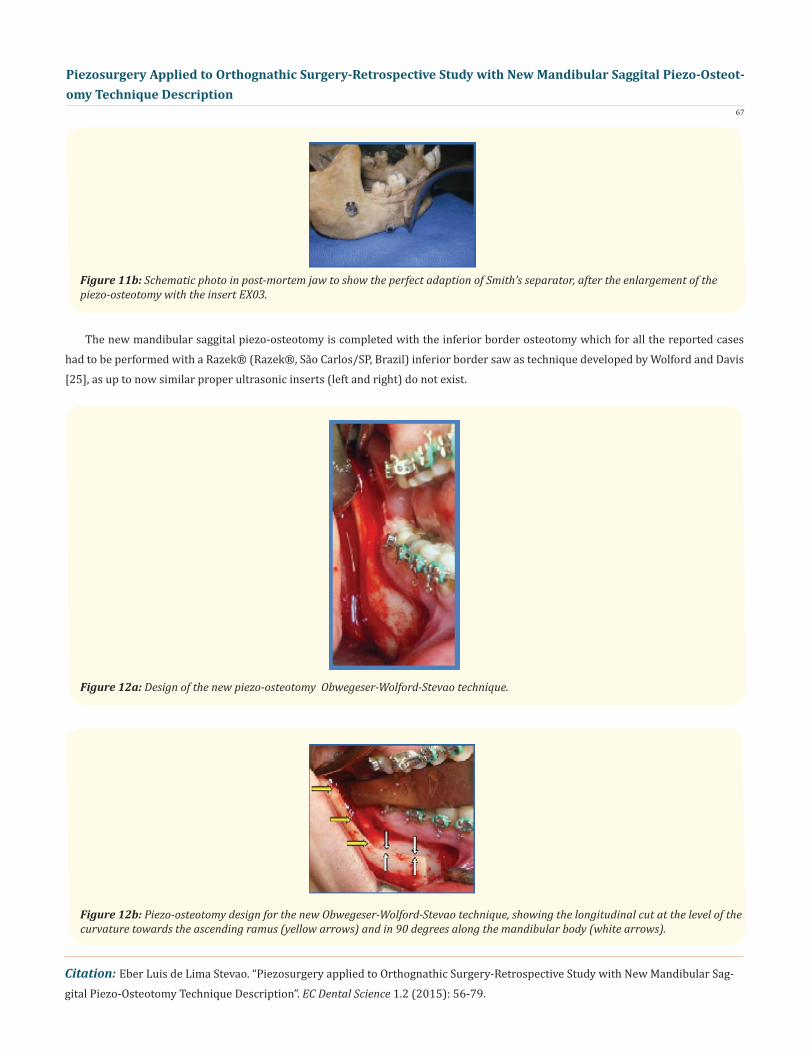
Figure 11a: Schematic photo in post-mortem jaw to show the vertical cut and the socket of Smith’s separator.
Piezosurgery Applied to Orthognathic Surgery-Retrospective Study with New Mandibular Saggital Piezo-Osteot-omy Technique Description
67
Citation: Eber Luis de Lima Stevao. “Piezosurgery applied to Orthognathic Surgery-Retrospective Study with New Mandibular Sag-gital Piezo-Osteotomy Technique Description”. EC Dental Science 1.2 (2015): 56-79.
The new mandibular saggital piezo-osteotomy is completed with the inferior border osteotomy which for all the reported cases had to be performed with a Razek® (Razek®, São Carlos/SP, Brazil) inferior border saw as technique developed by Wolford and Davis [25], as up to now similar proper ultrasonic inserts (left and right) do not exist.
Figure 11b: Schematic photo in post-mortem jaw to show the perfect adaption of Smith’s separator, after the enlargement of the piezo-osteotomy with the insert EX03.
Figure 12a: Design of the new piezo-osteotomy Obwegeser-Wolford-Stevao technique.
Figure 12b: Piezo-osteotomy design for the new Obwegeser-Wolford-Stevao technique, showing the longitudinal cut at the level of the curvature towards the ascending ramus (yellow arrows) and in 90 degrees along the mandibular body (white arrows).

Piezosurgery Applied to Orthognathic Surgery-Retrospective Study with New Mandibular Saggital Piezo-Osteot-omy Technique Description
68
Citation: Eber Luis de Lima Stevao. “Piezosurgery applied to Orthognathic Surgery-Retrospective Study with New Mandibular Sag-gital Piezo-Osteotomy Technique Description”. EC Dental Science 1.2 (2015): 56-79.
After proximal and distal segments separation an osteoplasty of the internal face of the proximal segment is done using insert HB01. This ultrasonic insert appeared to be propitious and with excellent cut power for cleaning procedure of these bone segments when rasping movements were done. This procedure is to be done to avoid compression of the IAN during rigid bone fixation phase- RBFP.
Due to non-existence of a specific ultrasonic insert for precise bone perforation for posterior screws placement during RBFP, all cases included in this retrospective study followed traditional bone perforation with W. Lorenz® (W. Lorenz Surgical Inc., Florida, USA) 1.5 mm drills, plates and screws.
Forty three maxilla were operated: eight Le Fort I (LF I), fifteen Le Fort I multi-segmented, one modified Le Fort II (LF II), twelve Le Fort I for SARPE and six anterior maxillary subapical osteotomy (AMxSO). Thirty six pterigomaxillary process chisel osteotomies (PMPCO) and eleven pterigomaxillary process piezo-disjunction attempts (PMPPDA).
Sixty one mandibles received the following procedures: fifty three bilateral saggital split osteotomies (BSSO), one unilateral saggi-tal split osteotomy (USSO), three mandibular midline straight osteotomy for mandibular symphyseal transverse distraction osteogen-esis (MSTDO), twenty five Tenon & Mortise genioplasties (TMG), three horizontal straight osteotomy for genioplasties (HSOG), three anterior mandibular subapical osteotomy (AMdSO), three bilateral posterior mandibular subapical osteotomy (BPMdSO) and one unilateral posterior mandibular subapical osteotomy (UPMdSO).
Some concomitant surgical procedures were carried out: twenty nine ENAp, seventeen PS and twenty four BIT. These procedures are presented in Table 1.
Ultrasonic inserts BS01 and BS1S were used for maxillary and mandibular osteotomies.
No trans-operative complications or accidents were observed and all saggital splits for proximal and distal segments separation were performed satisfactorily with any undesirable split. For all saggital split osteotomies an inferior border saw (Razek®, São Carlos/SP, Brazil) was used attached to an Aesculap® reciprocating handpiece (Aesculap®, Melsungen, Germany), as a complementary pro-cedure accordingly with the technique described by Wolford and Davis [25].
Figure 13: Ultrasonic insert HB01 used to carry out osteoplasty on internal aspect of mandibular proximal segment.
Results
Piezosurgery Applied to Orthognathic Surgery-Retrospective Study with New Mandibular Saggital Piezo-Osteot-omy Technique Description
69
Citation: Eber Luis de Lima Stevao. “Piezosurgery applied to Orthognathic Surgery-Retrospective Study with New Mandibular Sag-gital Piezo-Osteotomy Technique Description”. EC Dental Science 1.2 (2015): 56-79.
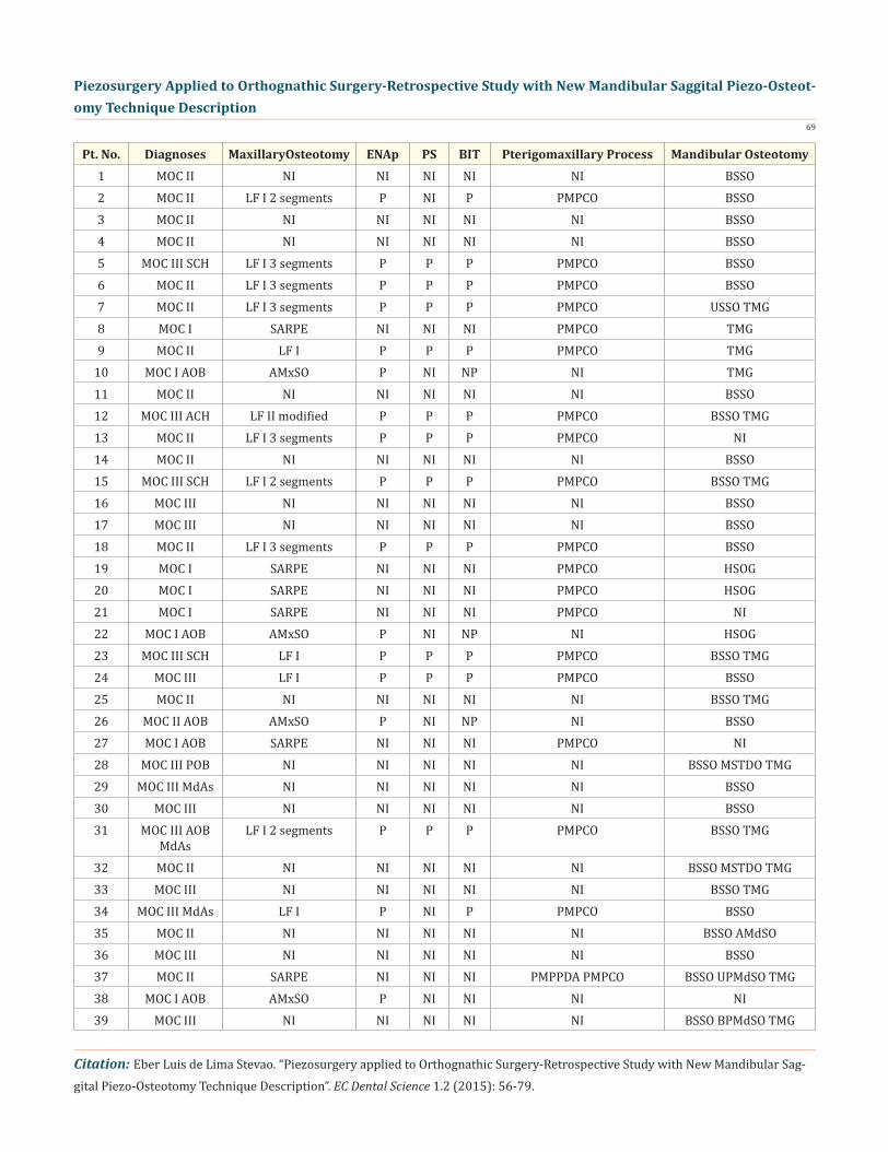
Pt. No. Diagnoses MaxillaryOsteotomy ENAp PS BIT Pterigomaxillary Process Mandibular Osteotomy1 MOC II NI NI NI NI NI BSSO2 MOC II LF I 2 segments P NI P PMPCO BSSO3 MOC II NI NI NI NI NI BSSO4 MOC II NI NI NI NI NI BSSO5 MOC III SCH LF I 3 segments P P P PMPCO BSSO6 MOC II LF I 3 segments P P P PMPCO BSSO7 MOC II LF I 3 segments P P P PMPCO USSO TMG8 MOC I SARPE NI NI NI PMPCO TMG9 MOC II LF I P P P PMPCO TMG
10 MOC I AOB AMxSO P NI NP NI TMG11 MOC II NI NI NI NI NI BSSO12 MOC III ACH LF II modified P P P PMPCO BSSO TMG13 MOC II LF I 3 segments P P P PMPCO NI14 MOC II NI NI NI NI NI BSSO15 MOC III SCH LF I 2 segments P P P PMPCO BSSO TMG16 MOC III NI NI NI NI NI BSSO17 MOC III NI NI NI NI NI BSSO18 MOC II LF I 3 segments P P P PMPCO BSSO19 MOC I SARPE NI NI NI PMPCO HSOG20 MOC I SARPE NI NI NI PMPCO HSOG21 MOC I SARPE NI NI NI PMPCO NI22 MOC I AOB AMxSO P NI NP NI HSOG23 MOC III SCH LF I P P P PMPCO BSSO TMG24 MOC III LF I P P P PMPCO BSSO25 MOC II NI NI NI NI NI BSSO TMG26 MOC II AOB AMxSO P NI NP NI BSSO27 MOC I AOB SARPE NI NI NI PMPCO NI28 MOC III POB NI NI NI NI NI BSSO MSTDO TMG29 MOC III MdAs NI NI NI NI NI BSSO30 MOC III NI NI NI NI NI BSSO31 MOC III AOB
MdAsLF I 2 segments P P P PMPCO BSSO TMG
32 MOC II NI NI NI NI NI BSSO MSTDO TMG33 MOC III NI NI NI NI NI BSSO TMG34 MOC III MdAs LF I P NI P PMPCO BSSO35 MOC II NI NI NI NI NI BSSO AMdSO36 MOC III NI NI NI NI NI BSSO37 MOC II SARPE NI NI NI PMPPDA PMPCO BSSO UPMdSO TMG38 MOC I AOB AMxSO P NI NI NI NI39 MOC III NI NI NI NI NI BSSO BPMdSO TMG
Piezosurgery Applied to Orthognathic Surgery-Retrospective Study with New Mandibular Saggital Piezo-Osteot-omy Technique Description
70
Citation: Eber Luis de Lima Stevao. “Piezosurgery applied to Orthognathic Surgery-Retrospective Study with New Mandibular Sag-gital Piezo-Osteotomy Technique Description”. EC Dental Science 1.2 (2015): 56-79.
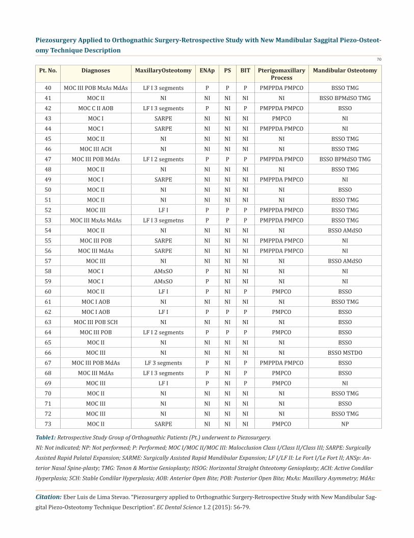
Table1: Retrospective Study Group of Orthognathic Patients (Pt.) underwent to Piezosurgery.
NI: Not indicated; NP: Not performed; P: Performed; MOC I/MOC II/MOC III: Malocclusion Class I/Class II/Class III; SARPE: Surgically
Assisted Rapid Palatal Expansion; SARME: Surgically Assisted Rapid Mandibular Expansion; LF I/LF II: Le Fort I/Le Fort II; ANSp: An-
terior Nasal Spine-plasty; TMG: Tenon & Mortise Genioplasty; HSOG: Horizontal Straight Osteotomy Genioplasty; ACH: Active Condilar
Hyperplasia; SCH: Stable Condilar Hyperplasia; AOB: Anterior Open Bite; POB: Posterior Open Bite; MxAs: Maxillary Asymmetry; MdAs:
Pt. No. Diagnoses MaxillaryOsteotomy ENAp PS BIT Pterigomaxillary Process
Mandibular Osteotomy
40 MOC III POB MxAs MdAs LF I 3 segments P P P PMPPDA PMPCO BSSO TMG41 MOC II NI NI NI NI NI BSSO BPMdSO TMG42 MOC C II AOB LF I 3 segments P NI P PMPPDA PMPCO BSSO43 MOC I SARPE NI NI NI PMPCO NI44 MOC I SARPE NI NI NI PMPPDA PMPCO NI45 MOC II NI NI NI NI NI BSSO TMG46 MOC III ACH NI NI NI NI NI BSSO TMG47 MOC III POB MdAs LF I 2 segments P P P PMPPDA PMPCO BSSO BPMdSO TMG48 MOC II NI NI NI NI NI BSSO TMG49 MOC I SARPE NI NI NI PMPPDA PMPCO NI50 MOC II NI NI NI NI NI BSSO51 MOC II NI NI NI NI NI BSSO TMG52 MOC III LF I P P P PMPPDA PMPCO BSSO TMG53 MOC III MxAs MdAs LF I 3 segmetns P P P PMPPDA PMPCO BSSO TMG54 MOC II NI NI NI NI NI BSSO AMdSO55 MOC III POB SARPE NI NI NI PMPPDA PMPCO NI56 MOC III MdAs SARPE NI NI NI PMPPDA PMPCO NI57 MOC III NI NI NI NI NI BSSO AMdSO58 MOC I AMxSO P NI NI NI NI59 MOC I AMxSO P NI NI NI NI60 MOC II LF I P NI P PMPCO BSSO61 MOC I AOB NI NI NI NI NI BSSO TMG62 MOC I AOB LF I P P P PMPCO BSSO63 MOC III POB SCH NI NI NI NI NI BSSO64 MOC III POB LF I 2 segments P P P PMPCO BSSO65 MOC II NI NI NI NI NI BSSO66 MOC III NI NI NI NI NI BSSO MSTDO67 MOC III POB MdAs LF 3 segments P NI P PMPPDA PMPCO BSSO68 MOC III MdAs LF I 3 segments P NI P PMPCO BSSO69 MOC III LF I P NI P PMPCO NI70 MOC II NI NI NI NI NI BSSO TMG71 MOC III NI NI NI NI NI BSSO72 MOC III NI NI NI NI NI BSSO TMG73 MOC II SARPE NI NI NI PMPCO NP

Piezosurgery Applied to Orthognathic Surgery-Retrospective Study with New Mandibular Saggital Piezo-Osteot-omy Technique Description
71
Citation: Eber Luis de Lima Stevao. “Piezosurgery applied to Orthognathic Surgery-Retrospective Study with New Mandibular Sag-gital Piezo-Osteotomy Technique Description”. EC Dental Science 1.2 (2015): 56-79.
No trans-operative complications or accidents were observed and all saggital splits for proximal and distal segments separation were performed satisfactorily with any undesirable split. For all saggital split osteotomies an inferior border saw (Razek®, São Carlos/SP, Brazil) was used attached to an Aesculap® reciprocating handpiece (Aesculap®, Melsungen, Germany), as a complementary pro-cedure accordingly with the technique described by Wolford and Davis [25].
Basically there are three types of techniques which are performed in bone: osteotomy (bone cut), osteoplasty (surgical remodeling of bone) and ostectomy (superimposition of the first two which is the removal of bone with osteotomy or osteoplasty). Piezosurgery conforms perfectly within these three techniques, exceeding the limits imposed by rotating and oscillatory instruments used in con-ventional techniques for bone cut.
The research carried out by Preti et al. [15] was very important because it showed not only with histomorphological analyses but also through proteins quantification that piezoelectric surgery causes less inflammatory reaction, induces to a higher quantity of BMPs proteins, rouses earlier bone repair and remodeling than the conventional methods of bone perforation.
Bone cut lines were precise and clean without bone debris accumulation. The time for piezo-osteotomy increased from 10 upto 20% for maxillary osteotomies and from 20 to 30% for mandibular osteotomies (genioplasties included) when compared to traditional techniques with burs and reciprocating saw. The highest percentages of time were found in first patients operated during the year of 2007 overlapping with author’s beginning experience with Piezosurgery.
The ultrasonic insert dislocation in cortical direction at the level of entire ascending ramus and its total deepening was very impor-tant for a more homogeneous fracture obtained in the lingual aspect of proximal segment. The formats of these fractures of the lingual segments after saggital osteotomy were very well described for Plooij [27]. For safety reasons the mandibular split was started with chiseling at this level between two cortices (proximal and distal).
The use of the ultrasonic insert EX03 to enlarge the vertical cut at the level of the first molar of the jaw is extremely important, since without him it is not possible to fit in the insert of the separator saggital of Smith.
The advantages of the Obwegeser-Wolford-Stevao technique described are several above but not limited to these ones: a) Greater area in distal segment for bone plating using “L” or “inverted C” formats (Figure 14); b) Maintenance of all cancellous bone in distal seg-ment; c) It improves fracture pattern at lingual aspect of distal segment; d) IAN is always bone confined and is never exposed, resulting in less post-operative neurossensorial disturbances, therefore quicker recovery; e) Dental roots of posterior teeth are more protected when bone plating and screws placement during RFP; f) Precocious sensibility return for inferior lip in the weeks after surgery; g) Less transoperative bleeding due to cavitation phenomenon; h) Less post-operative edema; i) Faster bone repair than with conventional rotating instruments usage; j) Patient returns to estomatognathic system functional activities more quickly, on average the gain is of a month.
The disadvantages of Obwegeser-Wolford-Stevao piezo-osteotomy are limited to: a) Less mandibular body bone vertical height for proximal segment; b) Possibility of undesirable fracture if the technique is not correctly performed because every step is essential; c) Piezo is needed for Piezosurgery and it turns surgery more expensive for surgeons; d) This surgery is highly technical needing a long
Ultrasonic inserts BS01 and BS1S were used for maxillary and mandibular osteotomies.
Mandibular Asymmetry; MxSO/AmdSO: Anterior Maxillary/Mandibular Subapical Osteotomy; BSSO/USSO: Bilateral/Unilateral Saggital
Split Osteotomy; BPMdSO/UPMdSO: Bilateral/Unilateral Posterior Mandibular Subapical Osteotomy; PMPPDA: Pterigomaxillary Process
Piezo-disjuction Attempt; PMPCO: Pterigomaxillary Process Chisel Osteotomy.
Discussion

Piezosurgery Applied to Orthognathic Surgery-Retrospective Study with New Mandibular Saggital Piezo-Osteot-omy Technique Description
72
Citation: Eber Luis de Lima Stevao. “Piezosurgery applied to Orthognathic Surgery-Retrospective Study with New Mandibular Sag-gital Piezo-Osteotomy Technique Description”. EC Dental Science 1.2 (2015): 56-79.
apprenticeship curve; e) Transoperative time is increased; f) Frequent ultrasonic inserts changes during surgical procedures (even though it is the same when using burs); g) Lack or inadequacy of an ultrasonic inserts for bone perforation to accomplish RBFP, for mandible inferior border cutting, and for performing pterigomaxillary process disjunction.
The main difficulty found by the author to execute a complete maxillary piezo-osteotomy was the impossibility of performing pterigomaxillary process piezo-disjunction, due to the anatomical structure depth and deficient ultrasonic insert design for this Le Fort I osteotomy stage. This procedure (pterigomaxillary process disjunction) in all subjects was executed with chisel and hammer, as well as it was a 100% for nasal septum disjunction of maxilla. The anterior nasal spine removal was carried out in a 100% of the cases. Ultrasonic inserts BS01 and BS1S when used for maxillary and mandibular osteotomies had an excellent effectiveness.
As for mandibular piezo-osteotomy the main difficulty found by the author to have it performed completely was the impossibility of carrying out mandible inferior border osteotomy with the inserts BS2L and BS2R.
These ultrasonic inserts present an exaggerated length and not favorable curvatures for a perfect adaption to the mandible inferior border. Even if an intrabuccal incision is over extended to perform the mandibular osteotomy there is no sufficient internal space for retractors, handpiece and ultrasonic insert. Then, entireness of the osteotomy in the reported cases was obtained with conventional inferior border saw following the technique described by Wolford and Davis [25].
Figure 14: Surgical aspect of RFP with W. Lorenz® plate in “C” format (W. Lorenz Surgical, Inc., Florida - USA) perfectly adapted on the design of new mandibular saggital piezo-osteotomy.
Figure 15: Couple of ultrasonic inserts BS2L and BS2R.

Piezosurgery Applied to Orthognathic Surgery-Retrospective Study with New Mandibular Saggital Piezo-Osteot-omy Technique Description
73
Citation:Eber Luis de Lima Stevao. “Piezosurgery applied to Orthognathic Surgery-Retrospective Study with New Mandibular Sag-gital Piezo-Osteotomy Technique Description”. EC Dental Science 1.2 (2015): 56-79.
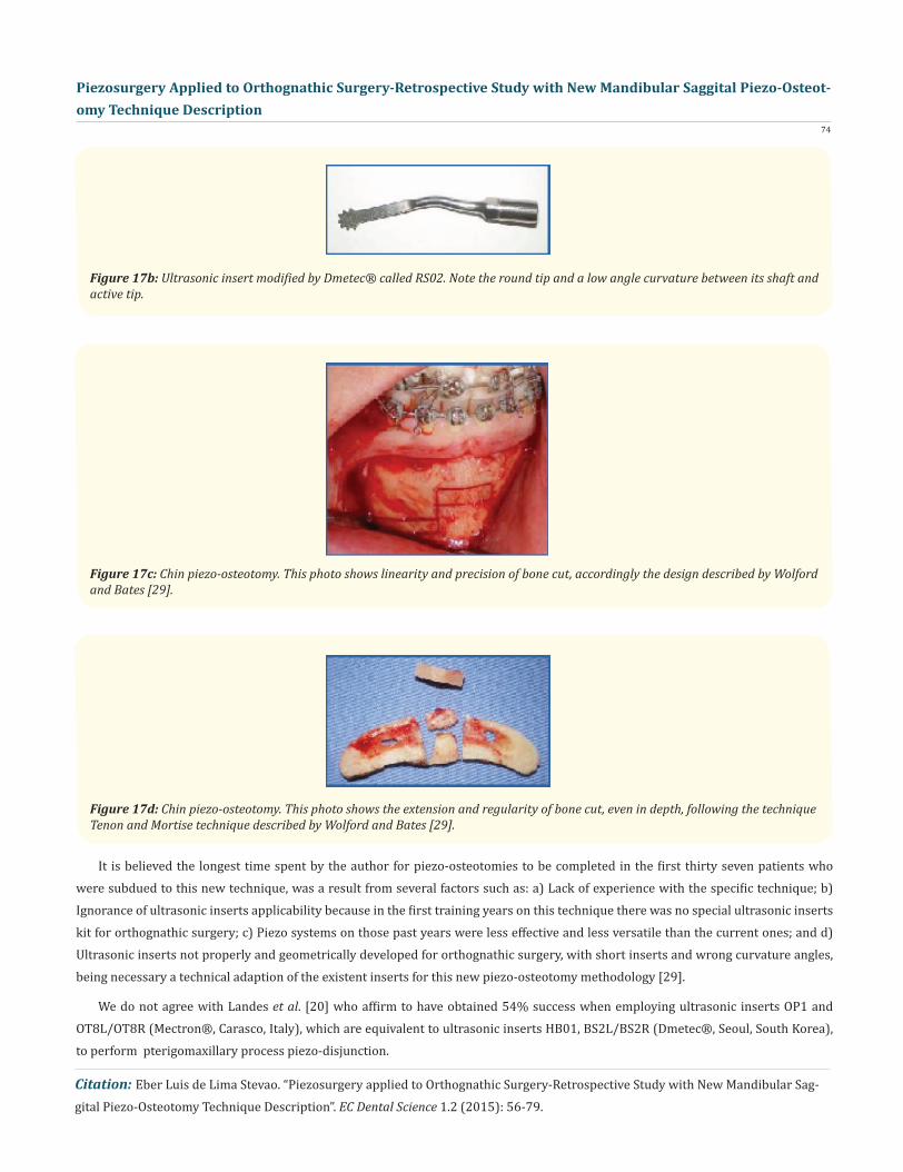
An important ultrasonic insert modification done by Dmetec® was the development of a longer ultrasonic sawing insert (5 mm increase on BS01 length) named RS02, with a lesser curvature angle between its shaft and active tip. Also, instead of a straight sawing tip it was changed for a round one. With this insert is possible to effectively carry out chin piezo-osteotomy without help of any other instrument.
Twenty eight genioplasties were performed. For 11 cases (39.3%) ultrasonic insert BS01 appeared to be very short and it was necessary to have the osteotomy completed with chisel/hammer or reciprocante saw. Other 4 genioplasties (14.3%) were performed from start to end with insert BS01. After ultrasonic insert modification cited (Figures 17a and 17b) it was possible to completely carry out other 13 genioplasties (46.4%) without help of any other instrument [28].
Figure 16a: Schematic photo of ultrasonic insert BS2R to show the impossibility of its usage as a substitute for inferior border saw.
Figure 16b: Schematic photo of ultrasonic insert BS2R. This insert is too long for its destined aim plus it has improper angulations making it impossible to use it trough an intrabuccal incision.
Figure 17a: Ultrasonic insert BS01 in a sawing shape and conventional length.
Piezosurgery Applied to Orthognathic Surgery-Retrospective Study with New Mandibular Saggital Piezo-Osteot-omy Technique Description
74
Citation: Eber Luis de Lima Stevao. “Piezosurgery applied to Orthognathic Surgery-Retrospective Study with New Mandibular Sag-gital Piezo-Osteotomy Technique Description”. EC Dental Science 1.2 (2015): 56-79.
Figure 17b: Ultrasonic insert modified by Dmetec® called RS02. Note the round tip and a low angle curvature between its shaft and active tip.
Figure 17c: Chin piezo-osteotomy. This photo shows linearity and precision of bone cut, accordingly the design described by Wolford and Bates [29].
Figure 17d: Chin piezo-osteotomy. This photo shows the extension and regularity of bone cut, even in depth, following the technique Tenon and Mortise technique described by Wolford and Bates [29].
It is believed the longest time spent by the author for piezo-osteotomies to be completed in the first thirty seven patients who were subdued to this new technique, was a result from several factors such as: a) Lack of experience with the specific technique; b) Ignorance of ultrasonic inserts applicability because in the first training years on this technique there was no special ultrasonic inserts kit for orthognathic surgery; c) Piezo systems on those past years were less effective and less versatile than the current ones; and d) Ultrasonic inserts not properly and geometrically developed for orthognathic surgery, with short inserts and wrong curvature angles, being necessary a technical adaption of the existent inserts for this new piezo-osteotomy methodology [29].
We do not agree with Landes et al. [20] who affirm to have obtained 54% success when employing ultrasonic inserts OP1 and OT8L/OT8R (Mectron®, Carasco, Italy), which are equivalent to ultrasonic inserts HB01, BS2L/BS2R (Dmetec®, Seoul, South Korea), to perform pterigomaxillary process piezo-disjunction.

Piezosurgery Applied to Orthognathic Surgery-Retrospective Study with New Mandibular Saggital Piezo-Osteot-omy Technique Description
75
Citation: Eber Luis de Lima Stevao. “Piezosurgery applied to Orthognathic Surgery-Retrospective Study with New Mandibular Sag-gital Piezo-Osteotomy Technique Description”. EC Dental Science 1.2 (2015): 56-79.
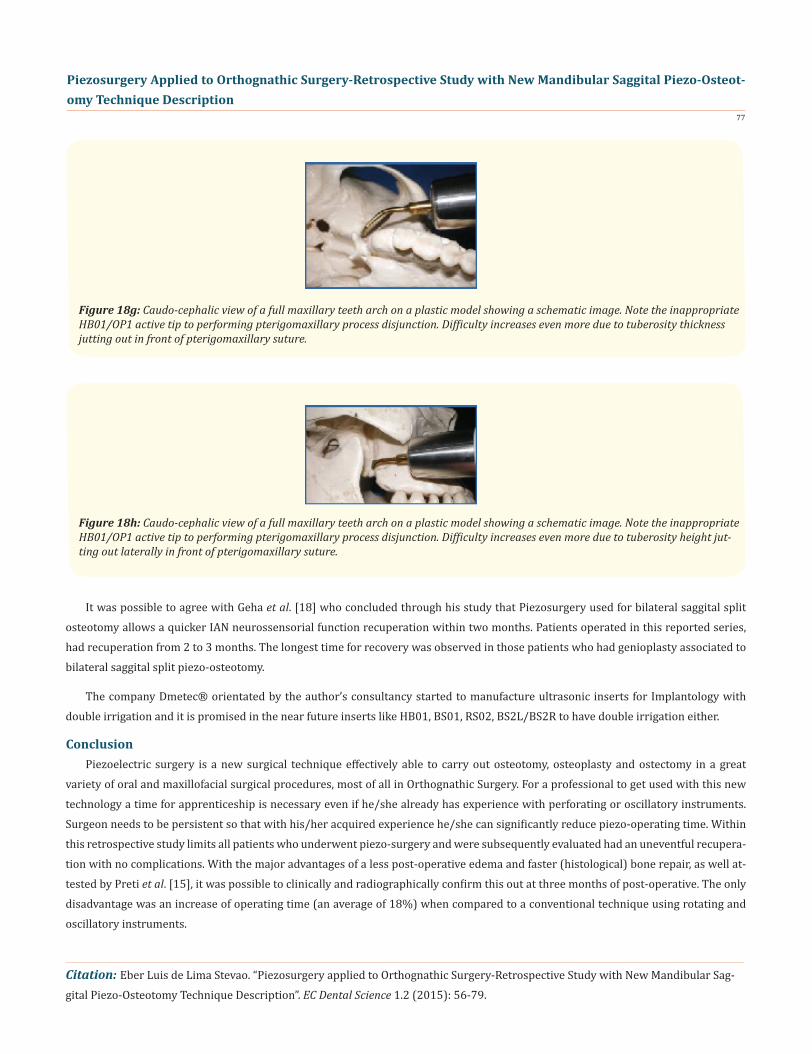
Robiony et al. [13] also reported usage of same ultrasonic curved inserts to perform pterigomaxillary osteotomy in only one case of SARPE without mentioning use of a chisel to complete this disjunction. In this author’s experience it was not possible to utilize ul-trasonic inserts (BS2L/BS2R) in any patient, in spite of several attempts, since their active portion is too short for effectively execution of referred piezo-osteotomy. It is presented below some schematic images to illustrate a great technique difficulty for its execution, not only for a maxillary toothless patient (theoretically it would have to be easier), let alone for a full maxillary teeth arch.
Figure 18a: Caudo-cephalic view of a post-mortem toothless maxilla showing a schematic image. Note the inappropriate BS2R/OT8R active portion to performing pterigomaxillary process disjunction.
Figure 18b: Lateral view of a post-mortem toothless maxilla showing a schematic image. Note the difficulty for direct visualization of the pterigomaxillary suture when insert BS2R/OT8R is employed for piezo-disjunction.
Figure 18c: Lateral view of a post-mortem toothless maxilla showing a schematic image. Note the inappropriate HB01/OP1 active portion to performing pterigomaxillary process disjunction.

Piezosurgery Applied to Orthognathic Surgery-Retrospective Study with New Mandibular Saggital Piezo-Osteot-omy Technique Description
76
Citation: Eber Luis de Lima Stevao. “Piezosurgery applied to Orthognathic Surgery-Retrospective Study with New Mandibular Sag-gital Piezo-Osteotomy Technique Description”. EC Dental Science 1.2 (2015): 56-79.
Figure 18d: Lateral view of a post-mortem toothless maxilla showing a schematic image. Note the difficulty for direct visualization of the pterigomaxillary suture when insert HB01/OP1 is employed for piezo-disjunction.
Figure 18e: Caudo-cephalic view of a full maxillary teeth arch on a plastic model showing a schematic image. Note the inappropriate BS2R/OT8R active portion to performing pterigomaxillary process disjunction. Difficulty increases even more due to tuberosity thick-ness jutting out in front of pterigomaxillary suture.
Figure 18f: Lateral view of a full maxillary teeth arch on a plastic model showing a schematic image. Note the difficulty for direct visualization of the pterigomaxillary suture when insert BS2R/OT8R is employed for piezo-disjunction. Difficulty increases even more due to tuberosity height jutting out in front of pterigomaxillary suture.
In all attempts carried out when suing those inserts the author also did not observe appropriate refrigeration of them. Since the above mentioned inserts do not have double irrigation (internal and external) the refrigerator water jet was always projected against maxillary tuberosity but never reaching insert’s active tip. Concerned for not causing more harms than benefits with the new tech-nique it was opted for traditional pterigomaxillary disjunction using chisel and hammer for a 100% of all cases where disjunction was formally indicated.
Piezosurgery Applied to Orthognathic Surgery-Retrospective Study with New Mandibular Saggital Piezo-Osteot-omy Technique Description
77
Citation: Eber Luis de Lima Stevao. “Piezosurgery applied to Orthognathic Surgery-Retrospective Study with New Mandibular Sag-gital Piezo-Osteotomy Technique Description”. EC Dental Science 1.2 (2015): 56-79.
Conclusion
Figure 18g: Caudo-cephalic view of a full maxillary teeth arch on a plastic model showing a schematic image. Note the inappropriate HB01/OP1 active tip to performing pterigomaxillary process disjunction. Difficulty increases even more due to tuberosity thickness jutting out in front of pterigomaxillary suture.
Figure 18h: Caudo-cephalic view of a full maxillary teeth arch on a plastic model showing a schematic image. Note the inappropriate HB01/OP1 active tip to performing pterigomaxillary process disjunction. Difficulty increases even more due to tuberosity height jut-ting out laterally in front of pterigomaxillary suture.
It was possible to agree with Geha et al. [18] who concluded through his study that Piezosurgery used for bilateral saggital split osteotomy allows a quicker IAN neurossensorial function recuperation within two months. Patients operated in this reported series, had recuperation from 2 to 3 months. The longest time for recovery was observed in those patients who had genioplasty associated to bilateral saggital split piezo-osteotomy.
The company Dmetec® orientated by the author’s consultancy started to manufacture ultrasonic inserts for Implantology with double irrigation and it is promised in the near future inserts like HB01, BS01, RS02, BS2L/BS2R to have double irrigation either.
Piezoelectric surgery is a new surgical technique effectively able to carry out osteotomy, osteoplasty and ostectomy in a great variety of oral and maxillofacial surgical procedures, most of all in Orthognathic Surgery. For a professional to get used with this new technology a time for apprenticeship is necessary even if he/she already has experience with perforating or oscillatory instruments. Surgeon needs to be persistent so that with his/her acquired experience he/she can significantly reduce piezo-operating time. Within this retrospective study limits all patients who underwent piezo-surgery and were subsequently evaluated had an uneventful recupera-tion with no complications. With the major advantages of a less post-operative edema and faster (histological) bone repair, as well at-tested by Preti et al. [15], it was possible to clinically and radiographically confirm this out at three months of post-operative. The only disadvantage was an increase of operating time (an average of 18%) when compared to a conventional technique using rotating and oscillatory instruments.

Piezosurgery Applied to Orthognathic Surgery-Retrospective Study with New Mandibular Saggital Piezo-Osteot-omy Technique Description
78
Citation: Eber Luis de Lima Stevao. “Piezosurgery applied to Orthognathic Surgery-Retrospective Study with New Mandibular Sag-gital Piezo-Osteotomy Technique Description”. EC Dental Science 1.2 (2015): 56-79.
It is the author’s viewpoint that this new kind of bone cut by ultrasound will be the elective method for oral and maxillofacial sur-geons for performing osteotomies. This technique is being quickly spread by several professionals all around the world and every new day it gains new devoted adapts.
Bibliography1. Stubinger S., et al. “Intraoral piezosurgery: preliminary results of a new technique”. Journal of Oral and Maxillofacial Surgery 63.9 (2005): 1283-1287.2. Escoda-Francoli J., et al. “Application of ultrasound in bone surgery: Two case reports”. Medicina Oral Patologia Oral y Cirugia Bucal 15.6 (2010): e902-e905.3. Maintz G. “Animal experiments in the study of the effect of ultrasonic waves on bone regeneration”. Strahlentherapie 82.4(1950): 631-638.4. Vercellotti T. “Piezoelectric Surgery in Implantology: A Case Report - A New Piezoeletric Ridge Expansion Technique”. Interna- tional Journal of Periodontics & Restorative Dentistry 20.4(2000): 358-365. 5. Eggers G., et al. “Piezosurgery: an ultrasound device for cutting bone and its use and limitations in maxillofacial surgery”. British Journal of Oral and Maxillofacial Surgery 42.5 (2004): 451-453.6. Vercellotti T. “Technological characteristics and clinical indications of piezoelectric bone surgery”. Minerva Stomatologica 53.5 (2004): 207-214.7. Marti-Bowen E., et al. “Periapical surgery using the ultrasound technique and silver amalgam retrograde filling. A study of 71 teeth with 100 canals”. Medicina Oral Patologia Oral y Cirugia Bucal 10.Suppl 1 (2005): E67-E73.8. Penarrocha DM., et al. “Evaluation of healing criteria for success after periapical surgery”. Medicina Oral Patologia Oral y Cirugia Bucal 13.2 (2008): E143-E147.9. Politi M., et al. “Piezoelectric surgery: A new method of bone cutting. Preliminary experience in orthogenesis distraction. Ab- stract presented at II International Meeting on Distraction Ontogenesis of the Facial Skeleton, Bologna, Italy; 2002.10. Robiony M., et al. “Piezoelectric bone cutting in multipiece maxillary osteotomies”. Journal of Oral and Maxillofacial Surgery 62.6 (2004): 759-761.11. Gruber RM., et al. “Ultrasonic surgery-an alternative way in orthognathic surgery of the mandible. A pilot study”. International Journal of Oral and Maxillofacial Surgery 34.6 (2005): 590-593.12. Metzger MC., et al. “Inferior alveolar nerve transposition--an in vitro comparison between piezosurgery and conventional bur use”. Journal of Oral Implantology 32.1 (2006): 19-25.13. Robiony M., et al. “Ultrasonic bone cutting for surgically assisted rapid maxillary expansion (SARME) under local anaesthesia”. International Journal of Oral and Maxillofacial Surgery 36.3 (2006): 267-269.14. Schlee M., et al. “Piezosurgery: Basics and possibilities”. Implant Dentistry 15.4 (2006): 334-340.15. Preti G., et al. “Cytokines and Growth Factors Involved in the Osseointegration of Oral Titanium Implants Positioned Using Piezo- electric Bone Surgery Versus a Drill Technique: A Pilot Study in Minipigs”. Journal of Periodontology 78.4 (2007): 716-722.16. Heiland M., et al. “Intraoral osteotomies using piezosurgery for distraction in an infant with Pierre-Robin sequence”. Clinical Oral Investigations 11.3 (2007): 303-306.17. Stubinger S. “Bone Healing After Piezosurgery and its Influence on Clinical Applications”. Journal of Oral and Maxillofacial Sur- gery 65.9 (2007): 39.e7-39.e8.18. Geha HJ., et al. “Sensitivity of the inferior lip and chin following mandibular bilateral saggital split osteotomy using Piezosur- gery”. Plastic and Reconstructive Surgery 118.7 (2006): 1598-1607.19. Schaeren S., et al. “Assessment of Nerve Damage Using a Novel Ultrasonic Device for Bone Cutting”. Journal of Oral and Maxillofa- cial Surgery 66.3 (2007): 593-596.20. Landes CA., et al. “Critical evaluation of piezoelectric osteotomy in orthognathic surgery: operative technique, blood loss, time requirement, nerve and vessel integrity”. Journal of Oral and Maxillofacial Surgery, 66.4 (2007) 657-674.

Piezosurgery Applied to Orthognathic Surgery-Retrospective Study with New Mandibular Saggital Piezo-Osteot-omy Technique Description
79
Citation: Eber Luis de Lima Stevao. “Piezosurgery applied to Orthognathic Surgery-Retrospective Study with New Mandibular Sag-gital Piezo-Osteotomy Technique Description”. EC Dental Science 1.2 (2015): 56-79.
21. Dmetec. Instruction Manual for Surgystar Piezosurgery, 2009. 18 pages.22. Wolford LM and Davis WM. “The mandibular inferior border split: a modification in the saggital split osteotomy”. Journal of Oral and Maxillofacial Surgery 48.1 (1990): 92-94.23. Wolford LM and Epker BN. “The combined anterior and posterior maxillary ostectomy: a new technique”. Journal of Oral and Maxillofacial Surgery 33.11 (1975): 842-851.24. Schuchardt K. “Treatment of oral deformities; progenia, prognathism & open bite”. Langenbecks Arch Klin Chir Ver Dtsch Z Chir Journal 287 (1957): 733-736. 25. Dal Pont G. “L’osteotomia retromolare per la correzione della progeny”. Minerva Chir 1.3-10 (1958).26. Trauner R and Obwegeser H. “The surgical correction of mandibular prognathism and retrognathia with consideration of genio- plasty”. Oral Surg Oral Med Oral Pathology. 10.7 (1957): 677-689.27. Obwegeser H. “The indications for surgical correction of mandibular deformity by the saggital splitting technique”. British Jour- nal of Oral and Maxillofacial Surgery (1964): 157-171.28. Plooij JM., et al. “3D evaluation of the lingual fracture line after a bilateral saggital split osteotomy of the mandible”. Journal of Oral and Maxillofacial Surgery 38.12 (2009): 1244-1249. 29. Wolford LM and Bates JD. “Surgical modification for the correction of chin deformities”. Oral Surg Oral Med Oral Pathology 66.3 (1988): 279-286.
Volume 1 Issue 2 February 2015© All rights are reserved by Eber Luis de Lima Stevao.