crowdsourced estimation of cognitive decline and ...kauwelab.byu.edu/portals/22/docs/2016...
TRANSCRIPT
Alzheimer’s & Dementia - (2016) 1-9
Featured Article
Crowdsourced estimation of cognitive decline and resiliencein Alzheimer’s disease
Genevera I. Allena, Nicola Amorosob,c, Catalina Angheld, Venkat Balagurusamye,Christopher J. Baref, Derek Beatong, Roberto Bellottib,c, David A. Bennetth, Kevin L. Boehmei,
Paul C. Boutrosd,j,k, Laura Caberlottol, Cristian Caloiand, Frederick Campbella,Elias Chaibub Netof, Yu-Chuan Changm, Beibei Chenn, Chien-Yu Cheno, Ting-Ying Chienp,Tim Clarkq,r, Sudeshna Dasq,r, Christos Davatzikoss, Jieyao Dengt,u, Donna Dillenbergere,Richard JB. Dobsonv,w,ddd, Qilin Dongt,u, Jimit Doshis, Denise Dumax, Rosangela Erricoy,Guray Eruss, Evan Everetta, David W. Fardoz,aa, Stephen H. Friendf, Holger Fr€ohlichbb,
Jessica Gana, Peter St George-Hyslopcc, Satrajit S. Ghoshdd,ee, Enrico Glaabff, Robert C. Greengg,Yuanfang Guanhh,ii,jj, Ming-Yi Hongo, Chao Huangkk, Jinseub Hwangll, Joseph Ibrahimkk,
Paolo Inglesemm, Anandhi Iyappanoo,bb, Qijia Jianga, Yuriko Katsumataaa, John S. K. Kauwei,*,Arno Kleinf,**, Dehan Kongkk, Roland Krauseff, Emilie Lalonded, Mario Laurial, Eunjee Leekk,Xihui Lind, Zhandong Liua, Julie Livingstoned, Benjamin A. Logsdonf, Simon Lovestonenn,Tsung-wei Man, Ashutosh Malhotraoo,bb, Lara M. Mangravitef,**, Taylor J. Maxwellpp,
Emily Merrillq, John Nagorskia, Aishwarya Namasivayamff, Manjari Narayana,Mufassra Nazoo,bb, Stephen J. Newhousev,qq, Thea C. Normanf, Ramil N. Nurtdinovrr,
Yen-Jen Oyangm, Yudi Pawitanss, Shengwen Pengt,u, Mette A. Petersf,**, Stephen R. Piccoloi,Paurush Praveenl,bb, Corrado Priamil, Veronica Y. Sabelnykovad, Philipp Sengeroo,Xia Shenss,bbb,ccc, Andrew Simmonsv, Aristeidis Sotirass, Gustavo Stolovitzkyuu,e,
Sabina Tangaroc, Andrea Tateob, Yi-An Tungvv, Nicholas J. Tustisonww, Erdem Varols,George Vradenburgxx, Michael W. Weineryy, Guanghua Xiaon, Lei Xiezz, Yang Xien, Jia Xun,Hojin Yangkk, Xiaowei Zhann, Yunyun Zhoun, Fan Zhuhh, Hongtu Zhukk, Shanfeng Zhut,u,aaa, for
the Alzheimer’s Disease Neuroimaging InitiativeaDepartment of Statistics and Electrical and Computer Engineering, Rice University, Houston, TX, USA
bDipartimento di Fisica “M. Merlin”, Universit�a degli studi di Bari “A. Moro”, Bari, ItalycSezione di Bari, Istituto Nazionale di Fisica Nucleare, Bari, Italy
dOntario Institute for Cancer Research, Informatics and Bio-computing Program, MaRS Centre, Toronto, ON, CanadaeIBM Computational Biology Center, IBM Research, NY, USA
fSage Bionetworks, Seattle, WA, USA
Data used in preparation of this article were obtained from the Alz-
heimer’s Disease Neuroimaging Initiative (ADNI) database (adni.loni.us-
c.edu). As such, the investigators within the ADNI contributed to the
design and implementation of ADNI and/or provided data but did not partic-
ipate in analysis or writing of this report. A complete listing of ADNI
investigators can be found at: http://adni.loni.usc.edu/wp-content/uploads/
how_to_apply/ADNI_Acknowledgement_List.pdf
*Corresponding author. Tel.:11 801 422 2993; Fax:11 801 422 0004.
**Lara M Mangravite: Tel.: 11 206 667 6044; Fax: 11 206 667 2062
Mette A. Peters: Tel.:11 206 667 2113; Fax:11 206 667 2062 Arno Klein:
Tel.: 1 1 917 512 5627
E-mail address: [email protected] (J.S.K.K.), [email protected]
(A.K.), [email protected] (L.M.M.), mette.peters@sagebase.
org (M.A.P.)
http://dx.doi.org/10.1016/j.jalz.2016.02.006
1552-5260/� 2016 The Authors. Published by Elsevier Inc. on behalf of the Alzheimer’s Association. This is an open access article under the CC BY-NC-ND
license (http://creativecommons.org/licenses/by-nc-nd/4.0/).
G.I. Allen et al. / Alzheimer’s & Dementia - (2016) 1-92
gSchool of Behavioral and Brain Sciences, The University of Texas at Dallas, Richardson, TX, USAhRush Alzheimer’s Disease Center, Rush University Medical Center, Chicago, IL, USA
iDepartment of Biology, Brigham Young University, Provo, UT, USAjDepartment of Medical Biophysics, University of Toronto, Toronto, Canada
kDepartment of Pharmacology & Toxicology, University of Toronto, Toronto, CanadalThe Microsoft Research, University of Trento Centre for Computational and Systems Biology (COSBI), Rovereto, Italy
mGraduate Institute of Biomedical Electronics and Bioinformatics, National Taiwan University, Taipei, TaiwannQuantitative Biomedical Research Center, The University of Texas Southwestern Medical Center, Dallas, TX, USA
oDepartment of Bio-Industrial Mechatronics Engineering, National Taiwan University, Taipei, TaiwanpInnovation Center for Big Data and Digital Convergence, Yuan Ze University, Taoyuan, Taiwan
qDepartment of Neurology, Massachusetts General Hospital, Cambridge, MA, USArDepartment of Neurology, Harvard Medical School, Boston, MA, USA
sCenter for Biomedical Image Computing and Analytics, University of Pennsylvania, Philadelphia, PA, USAtSchool of Computer Science, Fudan University, Shanghai, Shanghai, China
uShanghai Key Lab of Intelligent Information Processing, Fudan University, Shanghai, Shanghai, ChinavNIHR Biomedical Research Centre for Mental Health, Kings College London, London, UK
wInstitute of Psychiatry, Psychology and Neuroscience, MRC Social, Genetic and Developmental Psychiatry Centre, Kings College London, London, UKxDepartment of Pediatrics-Neurology, Baylor College of Medicine, Houston, TX, USA
yUniversit�a degli Studi di Genova, Genova, ItalyzSanders-Brown Center on Aging, University of Kentucky, Lexington, KY, USA
aaDepartment of Biostatistics, University of Kentucky, Lexington, KY, USAbbBonn-Aachen International Center for IT, University of Bonn, Bonn, Germany
ccCambridge Institute for Medical Research, University of Cambridge and University of Toronto, Cambridge, CB2, UKddMcGovern Institute for Brain Research, Massachusetts Institute of Technology, Cambridge, MA, USA
eeDepartment of Otology and Laryngology, Harvard Medical School, Boston, MA, USAffLuxembourg Centre for Systems Biomedicine, University of Luxembourg, Esch-sur-Alzette, Luxembourg
ggDivision of Genetics, Department of Medicine, Brigham and Women’s Hospital, Broad Institute and Harvard Medical School, Boston, MA, USAhhDepartment of Computational Medicine and Bioinformatics, University of Michigan, Ann Arbor, MI, USAiiDepartment of Electrical Engineering and Computer Science, University of Michigan, Ann Arbor, MI, USA
jjDepartment of Internal Medicine, University of Michigan, Ann Arbor, MI, USAkkDepartment of Biostatistics, The University of North Carolina at Chapel Hill, Chapel Hill, NC, USA
llDepartment of Computer science and Statistics, Daegu University, Gyeongsan-si, Gyeongsangbuk-do, Republic of KoreammDepartment of Surgery and Cancer, Faculty of Medicine, Imperial College London, London, UK
nnDepartment of Psychiatry, University of Oxford, Warneford Hospital, Oxford, UKooFraunhofer Institute for Algorithms and Scientific Computing (SCAI), Department for Bioinformatics, Schloss Birlinghoven, Sankt Augustin, Germany
ppComputational Biology Institute, The George Washington University, Ashburn, VA, USAqqDepartment of Biostatistics, Kings College London, London, UK
rrDepartment of Neuroimmunology, Foundation Institut de Recerca, Hospital Universitari Vall d’Hebron, Barcelona, SpainssDepartment of Medical Epidemiology and Biostatistics, Karolinska Institutet, Stockholm, Sweden
uuGenetics and Genomics Sciences Department, Icahn School of Medicine at Mount Sinai, New York, NY, USAvvGenome and systems biology degree program, National Taiwan University, Taipei, Taiwan
wwDepartment of Radiology and Medical Imaging, The University of Virginia, Charlottesville, VA, USAxxGlobal CEO Initiative on Alzheimer’s disease, Washington, DC, USA
yyRadiology, Medicine, Psychiatry, and Neurology, UCSF, SFVAMC, San Francisco, CA, USAzzDepartment of Computer Science, Hunter College, The City University of New York, New York, NY, USA
aaaCentre for Computational Systems Biology, Fudan University, Shanghai, ChinabbbUsher Institute of Population Health Sciences and Informatics, University of Edinburgh, Edinburgh, UK
cccMRC Institute of Genetics and Molecular Medicine, University of Edinburgh, Edinburgh, UKdddFarr Institute of Health Informatics Research, UCL Institute of Health Informatics, University College London, London WC1E 6BT, UK
Abstract Identifying accurate biomarkers of cognitive decline is essential for advancing early diagnosis and
prevention therapies in Alzheimer’s disease. The Alzheimer’s disease DREAM Challenge was de-signed as a computational crowdsourced project to benchmark the current state-of-the-art in predict-ing cognitive outcomes in Alzheimer’s disease based on high dimensional, publicly available geneticand structural imaging data. This meta-analysis failed to identify a meaningful predictor developedfrom either data modality, suggesting that alternate approaches should be considered for prediction ofcognitive performance.� 2016 The Authors. Published by Elsevier Inc. on behalf of the Alzheimer’s Association. This is anopen access article under the CC BY-NC-ND license (http://creativecommons.org/licenses/by-nc-nd/4.0/).Keywords: Azheimer’s disease; Biomarkers; Crowdsource; Big data; Bioinformatics; Cognitive decline; Imaging; Genetics
G.I. Allen et al. / Alzheimer’s & Dementia - (2016) 1-9 3
1. Introduction
The Alzheimer’s disease DREAM challenge (http://dx.doi.org/10.7303/syn2290704) was designed to provide an un-biased assessment of current capabilities for estimation ofcognition and prediction of cognitive decline using geneticand imaging data from public data resources using a crowd-sourced approach. The ability to predict rate of cognitivedecline—both before and after diagnosis—is essential toeffective trial design for the development of therapies for Alz-heimer’s disease (AD) prevention and treatment. Majorcollaborative efforts in the field are assessing the associationof genetic loci with ADdiagnosis and the application of struc-tural imaging for development of early biomarkers of diag-nosis, but the utility of these approaches to estimatecognition or predict cognitive decline is not well established.This project was designed under the advisement of a panel ofexperts in the field to evaluate whether these questions couldbe meaningfully addressed with current methods given exist-ing public data sources. To ensure that these questions weretested across a broad spectrum of the latest analyticalapproaches, the study was designed as a crowdsourced,community-based challenge in which participants wereinvited to address one or more of the following threequestions [1]: The prediction of cognitive decline over timebased on genetic data [2]. The prediction of resilience tocognitive decline in individuals with elevated amyloid burdenbased on genetic data [3]. The estimation of cognitive statebased on structural magnetic resonance (MR) imaging data.
2. Results
2.1. Study design and data harmonization
To ensure that predictors were detecting true biologicalvariation rather than study-specific technical variation, thisproject required inclusion of data frommultiple study sources.Although genetic and imaging data have been generatedwithinmany rich longitudinal cohorts across the field, the pro-curement and harmonization of these data sets were anontrivial problem that required solutions to overcome polit-ical, ethical, and technical barriers. For example, the genera-tion of whole genome sequencing data across multiple ADcohorts within theNIH-fundedAD sequencing project has re-sulted in a powerful resource for genetic analysis in the fieldbut longitudinal information on cognitive traits is not readilyavailable in those data sets. Despite limitations on data acces-sibility, multiple relevant data sources were identified andused in this project including the Alzheimer’s Disease Neuro-imaging Initiative (ADNI) [1], the Rush Alzheimer’s DiseaseCenter Religious Orders Study [2], Memory and Aging Proj-ect (ROS/MAP) [3], and the European AddNeuroMed [4]study, which is part of InnoMed, a precursor to the innovativemedicines initiative. Data selection and processing were per-formed based on data availability across these three data sets.As such, cognition was defined using mini mental state exam-ination (MMSE) scores [5], genetic data were provided based
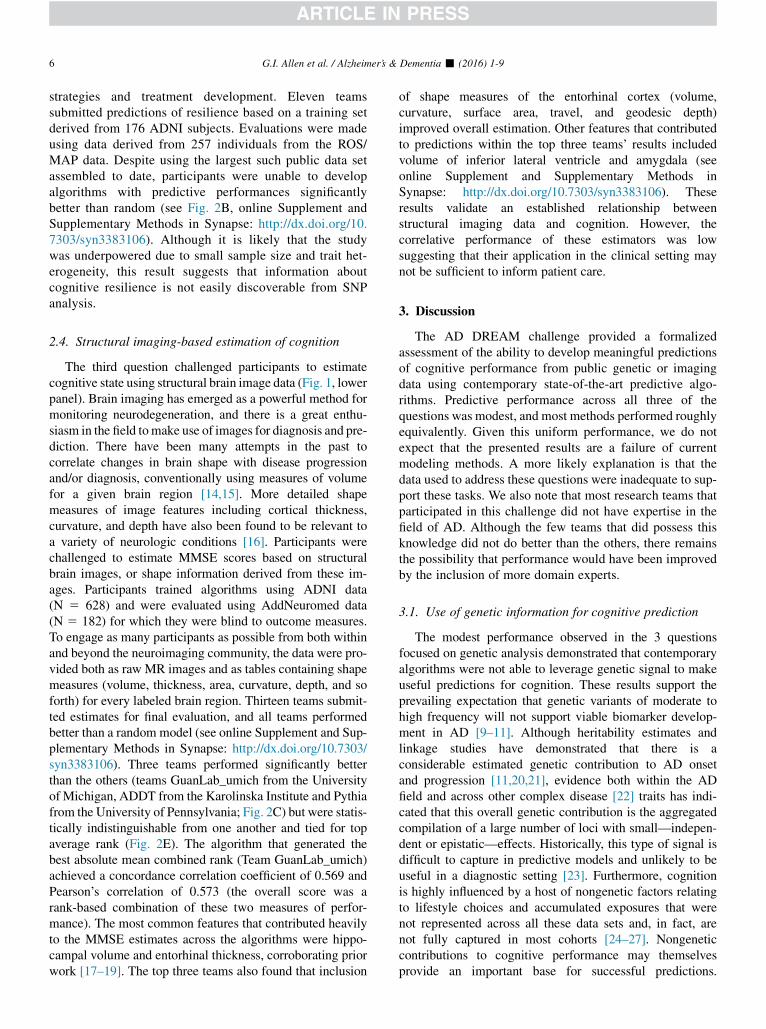
on imputation across array-based genotype data, and struc-tural MR imaging data were reprocessed in each cohort usinga common processing pipeline. Genetic and imaging datawere supplemented with a limited set of covariates includingdiagnosis, initial MMSE score, age at the initial examination,years of education, gender, andAPOE haplotype. Participantswere providedwith data fromADNI to train algorithms over a4-month period and to ensure that participation was notlimited by access to compute resources, they were offereduse of the IBM zEnterprise cloud to perform analyses. Thechallenge generated significant interest with 527 individualsfrom around theworld registered to participate. A leaderboarddisplayed accuracy of submissions throughout the duration ofthe challenge: 1157 submissions were made for question 1,478 submissions for question 2, and 434 submissions forquestion 3. Thirty-two teams submitted final results thatwere scored based on prediction and/or estimation of blindedoutcomes within ROS/MAP for genetic predictions andAddNeuroMed for imaging-based estimations (Fig. 1).
2.2. Genetic prediction of cognitive decline
The first challenge question assessed the ability of currentmethods to predict change in cognitive examination perfor-mance based on genetic data. High prediction accuracywould signal the potential for noninvasive biomarkers ofcognition to have a major clinical impact on early AD diag-nosis and prevention. Previous efforts to develop predictorsof change in cognitive function have not succeeded inproviding robust and replicable models [6–8]. Geneticvariation has been demonstrated to influence AD status:rare genetic mutations at several loci are implicated infamilial forms of early-onset disease [9], whereas commonvariation contributes 33% to variance in sporadic AD, and22 loci have been implicated by large-scale genetic associa-tion analyses [10,11]. However, with the exception of theAPOE ε4 haplotype, there has been little success intransforming these genetic associations into meaningfulclinical predictions of cognitive decline. For this purpose,participants were challenged to predict 2-year changes inMMSE scores based on genotypes imputed from SNP arraydata. Participants trained their algorithms with 767 ADNIsamples, and the algorithms’ predictions were evaluated ona test set of 1175 ROS/MAP samples with blinded outcomemeasures. The algorithm with the best predictive perfor-mance at the midpoint of the challenge did not contain anygenetic features beyond APOE haplotype. As the goal ofthis question was to assess genetic contribution to predictionof cognitive decline, this top-ranked algorithm was openlyshared across teams as an interim baseline on which to incor-porate additional genetic predictors (http://dx.doi.org/10.7303/syn2838779). Eighteen teams submitted final predic-tions. Most methods performed significantly better than apermutation-based random model prediction (Fig. 2A). Acluster of six methods performed significantly better thanthe others (including the interim baseline model) but were

Fig. 1. Challenge overview. The top schematic summarizes the three challenge questions on the left column, the training data in the middle, and the test data on
the right, including numbers of subjects. The symbols represent sources of data (demographic, ROS/MAP genetic, and ADNI or ANM brain images and shape
information). The bottom panel provides example brain image labels and shape information derived from the Mindboggle software (http://mindboggle.info)
provided to the participants for question 3. Anatomic labels for left cortical regions are shown on the left and just a couple of the cortical surface shape measures
are shown on the right (travel depth on top and mean curvature below), for both uninflated and inflated surfaces (top and bottom rows, respectively).
G.I. Allen et al. / Alzheimer’s & Dementia - (2016) 1-94
statistically indistinguishable among themselves (Fig. 2D).Of these, the prediction with the best overall score (teamGuanLab_umich from the University of Michigan) achieveda Pearson correlation of 0.382 and a Spearman correlation of0.433 (the overall score was a rank-based combination ofthese two measures of performance; see online Supplementand Supplementary Methods: http://dx.doi.org/10.7303/syn3383106). However, no significant contribution of ge-netics beyond APOE haplotype to predictive performancewas observed across any of the submissions. Given the smallsample size, no conclusions can be inferred from this analysisregarding the existence of genetic loci associated with cogni-tive decline. Rather, these observations suggest that predic-
tors of cognitive decline developed based on genetic datawill not be useful within the clinical setting.
2.3. Genetic prediction of cognitive resilience
The second question challenged participants to identifygenetic predictors that could distinguish individuals whoexhibit resilience to AD pathology as defined by minimalchange in cognitive function despite evidence of amyloiddeposition [12,13]. Identification of genetic signaturespredictive of cognitive resilience would aid in theelucidation of mechanisms that may confer resilience,providing a powerful tool to help advance AD prevention
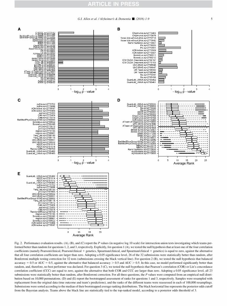
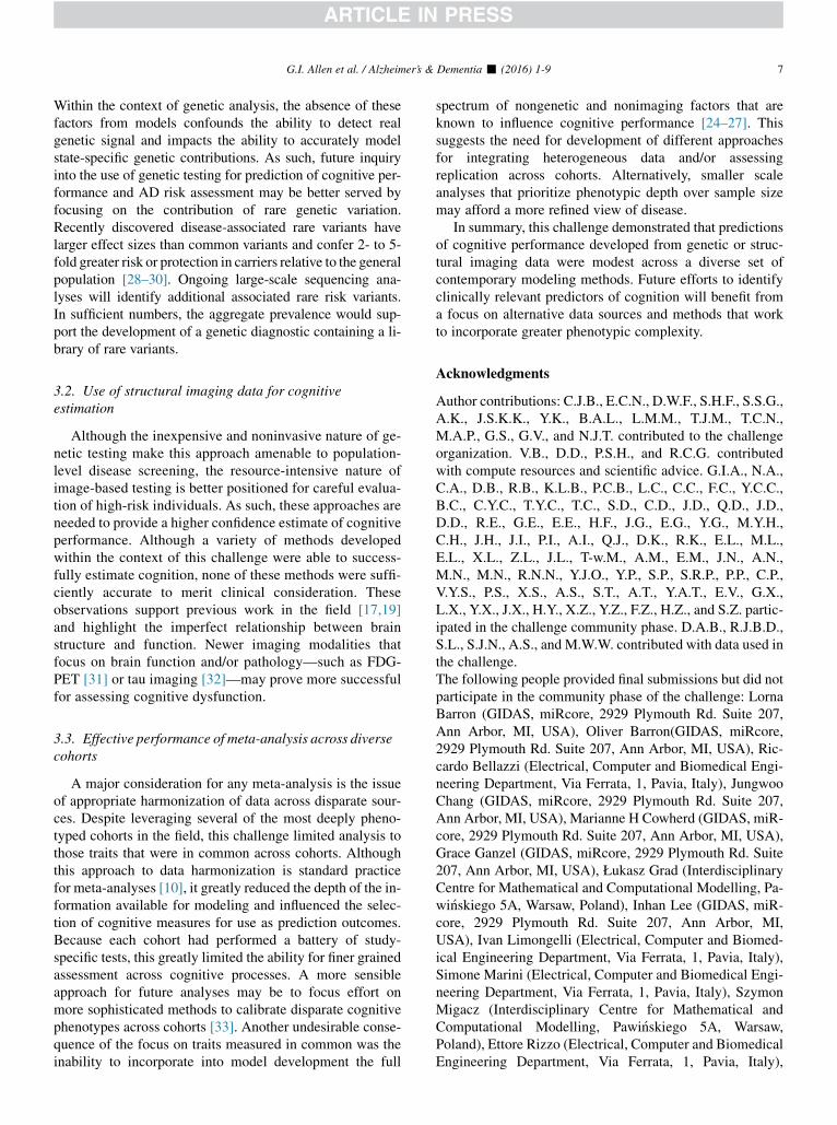
Fig. 2. Performance evaluation results. (A), (B), and (C) report the P values (in negative log 10 scale) for intersection union tests investigating which teams per-
formed better than random for questions 1, 2, and 3, respectively. Explicitly, for question 1 (A), we tested the null hypothesis that at least one of the four correlation
coefficients (namely Pearson/clinical, Pearson/clinical1 genetics, Spearman/clinical, and Spearman/clinical1 genetics) is equal to zero, against the alternative
that all four correlation coefficients are larger than zero. Adopting a 0.05 significance level, 26 of the 32 submissions were statistically better than random, after
Bonferroni multiple testing correction for 32 tests (submissions crossing the black vertical line). For question 2 (B), we tested the null hypothesis that balanced
accuracy 5 0.5 or AUC 5 0.5, against the alternative that balanced accuracy . 0.5 and AUC . 0.5. In this case, no model performed significantly better than
random, and, therefore, no best performer was declared. For question 3 (C), we tested the null hypothesis that Pearson’s correlation (COR) or Lin’s concordance
correlation coefficient (CCC) are equal to zero, against the alternative that both COR and CCC are larger than zero. Adopting a 0.05 significance level, all 23
submissions were statistically better than random, after Bonferroni correction. For all three questions, the P values were computed from an empirical null distri-
bution based on 10,000 permutations. (D) and (E) report the bootstrapped assessment of ranks for questions 1 and 3, respectively. Samples were resampled with
replacement from the original data (true outcome and team’s predictions), and the ranks of the different teams were reassessed in each of 100,000 resamplings.
Submissions were sorted according to themedian of their bootstrapped average ranking distributions. The black horizontal line represents the posterior odds cutoff
from the Bayesian analysis. Teams above the black line are statistically tied to the top-ranked model, according to a posterior odds threshold of 3.
G.I. Allen et al. / Alzheimer’s & Dementia - (2016) 1-9 5
G.I. Allen et al. / Alzheimer’s & Dementia - (2016) 1-96
strategies and treatment development. Eleven teamssubmitted predictions of resilience based on a training setderived from 176 ADNI subjects. Evaluations were madeusing data derived from 257 individuals from the ROS/MAP data. Despite using the largest such public data setassembled to date, participants were unable to developalgorithms with predictive performances significantlybetter than random (see Fig. 2B, online Supplement andSupplementary Methods in Synapse: http://dx.doi.org/10.7303/syn3383106). Although it is likely that the studywas underpowered due to small sample size and trait het-erogeneity, this result suggests that information aboutcognitive resilience is not easily discoverable from SNPanalysis.
2.4. Structural imaging-based estimation of cognition
The third question challenged participants to estimatecognitive state using structural brain image data (Fig. 1, lowerpanel). Brain imaging has emerged as a powerful method formonitoring neurodegeneration, and there is a great enthu-siasm in the field tomake use of images for diagnosis and pre-diction. There have been many attempts in the past tocorrelate changes in brain shape with disease progressionand/or diagnosis, conventionally using measures of volumefor a given brain region [14,15]. More detailed shapemeasures of image features including cortical thickness,curvature, and depth have also been found to be relevant toa variety of neurologic conditions [16]. Participants werechallenged to estimate MMSE scores based on structuralbrain images, or shape information derived from these im-ages. Participants trained algorithms using ADNI data(N 5 628) and were evaluated using AddNeuromed data(N 5 182) for which they were blind to outcome measures.To engage as many participants as possible from both withinand beyond the neuroimaging community, the data were pro-vided both as raw MR images and as tables containing shapemeasures (volume, thickness, area, curvature, depth, and soforth) for every labeled brain region. Thirteen teams submit-ted estimates for final evaluation, and all teams performedbetter than a random model (see online Supplement and Sup-plementary Methods in Synapse: http://dx.doi.org/10.7303/syn3383106). Three teams performed significantly betterthan the others (teams GuanLab_umich from the Universityof Michigan, ADDT from the Karolinska Institute and Pythiafrom the University of Pennsylvania; Fig. 2C) but were statis-tically indistinguishable from one another and tied for topaverage rank (Fig. 2E). The algorithm that generated thebest absolute mean combined rank (Team GuanLab_umich)achieved a concordance correlation coefficient of 0.569 andPearson’s correlation of 0.573 (the overall score was arank-based combination of these two measures of perfor-mance). The most common features that contributed heavilyto the MMSE estimates across the algorithms were hippo-campal volume and entorhinal thickness, corroborating priorwork [17–19]. The top three teams also found that inclusion
of shape measures of the entorhinal cortex (volume,curvature, surface area, travel, and geodesic depth)improved overall estimation. Other features that contributedto predictions within the top three teams’ results includedvolume of inferior lateral ventricle and amygdala (seeonline Supplement and Supplementary Methods inSynapse: http://dx.doi.org/10.7303/syn3383106). Theseresults validate an established relationship betweenstructural imaging data and cognition. However, thecorrelative performance of these estimators was lowsuggesting that their application in the clinical setting maynot be sufficient to inform patient care.
3. Discussion
The AD DREAM challenge provided a formalizedassessment of the ability to develop meaningful predictionsof cognitive performance from public genetic or imagingdata using contemporary state-of-the-art predictive algo-rithms. Predictive performance across all three of thequestions was modest, and most methods performed roughlyequivalently. Given this uniform performance, we do notexpect that the presented results are a failure of currentmodeling methods. A more likely explanation is that thedata used to address these questions were inadequate to sup-port these tasks. We also note that most research teams thatparticipated in this challenge did not have expertise in thefield of AD. Although the few teams that did possess thisknowledge did not do better than the others, there remainsthe possibility that performance would have been improvedby the inclusion of more domain experts.
3.1. Use of genetic information for cognitive prediction
The modest performance observed in the 3 questionsfocused on genetic analysis demonstrated that contemporaryalgorithms were not able to leverage genetic signal to makeuseful predictions for cognition. These results support theprevailing expectation that genetic variants of moderate tohigh frequency will not support viable biomarker develop-ment in AD [9–11]. Although heritability estimates andlinkage studies have demonstrated that there is aconsiderable estimated genetic contribution to AD onsetand progression [11,20,21], evidence both within the ADfield and across other complex disease [22] traits has indi-cated that this overall genetic contribution is the aggregatedcompilation of a large number of loci with small—indepen-dent or epistatic—effects. Historically, this type of signal isdifficult to capture in predictive models and unlikely to beuseful in a diagnostic setting [23]. Furthermore, cognitionis highly influenced by a host of nongenetic factors relatingto lifestyle choices and accumulated exposures that werenot represented across all these data sets and, in fact, arenot fully captured in most cohorts [24–27]. Nongeneticcontributions to cognitive performance may themselvesprovide an important base for successful predictions.
G.I. Allen et al. / Alzheimer’s & Dementia - (2016) 1-9 7
Within the context of genetic analysis, the absence of thesefactors from models confounds the ability to detect realgenetic signal and impacts the ability to accurately modelstate-specific genetic contributions. As such, future inquiryinto the use of genetic testing for prediction of cognitive per-formance and AD risk assessment may be better served byfocusing on the contribution of rare genetic variation.Recently discovered disease-associated rare variants havelarger effect sizes than common variants and confer 2- to 5-fold greater risk or protection in carriers relative to the generalpopulation [28–30]. Ongoing large-scale sequencing ana-lyses will identify additional associated rare risk variants.In sufficient numbers, the aggregate prevalence would sup-port the development of a genetic diagnostic containing a li-brary of rare variants.
3.2. Use of structural imaging data for cognitiveestimation
Although the inexpensive and noninvasive nature of ge-netic testing make this approach amenable to population-level disease screening, the resource-intensive nature ofimage-based testing is better positioned for careful evalua-tion of high-risk individuals. As such, these approaches areneeded to provide a higher confidence estimate of cognitiveperformance. Although a variety of methods developedwithin the context of this challenge were able to success-fully estimate cognition, none of these methods were suffi-ciently accurate to merit clinical consideration. Theseobservations support previous work in the field [17,19]and highlight the imperfect relationship between brainstructure and function. Newer imaging modalities thatfocus on brain function and/or pathology—such as FDG-PET [31] or tau imaging [32]—may prove more successfulfor assessing cognitive dysfunction.
3.3. Effective performance of meta-analysis across diversecohorts
A major consideration for any meta-analysis is the issueof appropriate harmonization of data across disparate sour-ces. Despite leveraging several of the most deeply pheno-typed cohorts in the field, this challenge limited analysis tothose traits that were in common across cohorts. Althoughthis approach to data harmonization is standard practicefor meta-analyses [10], it greatly reduced the depth of the in-formation available for modeling and influenced the selec-tion of cognitive measures for use as prediction outcomes.Because each cohort had performed a battery of study-specific tests, this greatly limited the ability for finer grainedassessment across cognitive processes. A more sensibleapproach for future analyses may be to focus effort onmore sophisticated methods to calibrate disparate cognitivephenotypes across cohorts [33]. Another undesirable conse-quence of the focus on traits measured in common was theinability to incorporate into model development the full
spectrum of nongenetic and nonimaging factors that areknown to influence cognitive performance [24–27]. Thissuggests the need for development of different approachesfor integrating heterogeneous data and/or assessingreplication across cohorts. Alternatively, smaller scaleanalyses that prioritize phenotypic depth over sample sizemay afford a more refined view of disease.
In summary, this challenge demonstrated that predictionsof cognitive performance developed from genetic or struc-tural imaging data were modest across a diverse set ofcontemporary modeling methods. Future efforts to identifyclinically relevant predictors of cognition will benefit froma focus on alternative data sources and methods that workto incorporate greater phenotypic complexity.
Acknowledgments
Author contributions: C.J.B., E.C.N., D.W.F., S.H.F., S.S.G.,A.K., J.S.K.K., Y.K., B.A.L., L.M.M., T.J.M., T.C.N.,M.A.P., G.S., G.V., and N.J.T. contributed to the challengeorganization. V.B., D.D., P.S.H., and R.C.G. contributedwith compute resources and scientific advice. G.I.A., N.A.,C.A., D.B., R.B., K.L.B., P.C.B., L.C., C.C., F.C., Y.C.C.,B.C., C.Y.C., T.Y.C., T.C., S.D., C.D., J.D., Q.D., J.D.,D.D., R.E., G.E., E.E., H.F., J.G., E.G., Y.G., M.Y.H.,C.H., J.H., J.I., P.I., A.I., Q.J., D.K., R.K., E.L., M.L.,E.L., X.L., Z.L., J.L., T-w.M., A.M., E.M., J.N., A.N.,M.N., M.N., R.N.N., Y.J.O., Y.P., S.P., S.R.P., P.P., C.P.,V.Y.S., P.S., X.S., A.S., S.T., A.T., Y.A.T., E.V., G.X.,L.X., Y.X., J.X., H.Y., X.Z., Y.Z., F.Z., H.Z., and S.Z. partic-ipated in the challenge community phase. D.A.B., R.J.B.D.,S.L., S.J.N., A.S., and M.W.W. contributed with data used inthe challenge.The following people provided final submissions but did notparticipate in the community phase of the challenge: LornaBarron (GIDAS, miRcore, 2929 Plymouth Rd. Suite 207,Ann Arbor, MI, USA), Oliver Barron(GIDAS, miRcore,2929 Plymouth Rd. Suite 207, Ann Arbor, MI, USA), Ric-cardo Bellazzi (Electrical, Computer and Biomedical Engi-neering Department, Via Ferrata, 1, Pavia, Italy), JungwooChang (GIDAS, miRcore, 2929 Plymouth Rd. Suite 207,Ann Arbor, MI, USA), Marianne H Cowherd (GIDAS, miR-core, 2929 Plymouth Rd. Suite 207, Ann Arbor, MI, USA),Grace Ganzel (GIDAS, miRcore, 2929 Plymouth Rd. Suite207, Ann Arbor, MI, USA), qukasz Grad (InterdisciplinaryCentre for Mathematical and Computational Modelling, Pa-wi�nskiego 5A, Warsaw, Poland), Inhan Lee (GIDAS, miR-core, 2929 Plymouth Rd. Suite 207, Ann Arbor, MI,USA), Ivan Limongelli (Electrical, Computer and Biomed-ical Engineering Department, Via Ferrata, 1, Pavia, Italy),Simone Marini (Electrical, Computer and Biomedical Engi-neering Department, Via Ferrata, 1, Pavia, Italy), SzymonMigacz (Interdisciplinary Centre for Mathematical andComputational Modelling, Pawi�nskiego 5A, Warsaw,Poland), Ettore Rizzo (Electrical, Computer and BiomedicalEngineering Department, Via Ferrata, 1, Pavia, Italy),

G.I. Allen et al. / Alzheimer’s & Dementia - (2016) 1-98
Witold R Rudnicki (Interdisciplinary Centre for Mathemat-ical and Computational Modelling, Pawi�nskiego 5A, War-saw, Poland; Department of Bioinformatics, University ofBia1ystok, Cio1kowskiego 1M, Bia1ystok, Poland), AndrzejSu1ecki (Interdisciplinary Centre for Mathematical andComputational Modelling, Pawi�nskiego 5A, Warsaw,Poland), Leo Tunkle (GIDAS, miRcore, 2929 PlymouthRd. Suite 207, Ann Arbor, MI, USA), Francesca Vitali (Elec-trical, Computer and Biomedical Engineering Department,Via Ferrata, 1, Pavia, Italy)This study was supported by the following individuals andorganizations: Alan Evans (McGill University), Gaurav Pan-dey (MSSM), Gil Rabinovici (UCSF), Kaj Blennow(G€oteborg University), Kristine Yaffe (UCSF), Maria Isaac(EMA), Nolan Nichols (University of Washington), PaulThompson (UCLA), Reisa Sperling (Harvard), Scott Small(Columbia), Guy Eakin (BrightFocus Foundation), MariaCarillo (Alzheimer’s Association), Neil Buckholz (NIA),Alzheimer’s Research UK, European Medicines Agency,Global CEO Initiative on Alzheimer’s Disease, Pfizer, Inc,Ray and Dagmar Dolby Family Fund, Rosenberg Alz-heimer’s Project, Sanofi S.A, and Takeda PharmaceuticalCompany Ltd, USAgainstAlzheimer’s.Studydatawere providedby the followinggroups: (1)TheAlz-heimer’s Disease Neuroimaging Initiative (ADNI)—ADNI isfunded by the National Institutes of Health (U01AG024904), theNational Institute onAging, theNational Insti-tute of Biomedical Imaging and Bioengineering and throughgenerous contributions from the following: Alzheimer’s Asso-ciation; Alzheimer’s Drug Discovery Foundation; BioClinica,Inc.; Biogen Idec Inc.; Bristol-Myers Squibb Company; EisaiInc.; Elan Pharmaceuticals, Inc.; Eli Lilly and Company; F.Hoffmann-La Roche Ltd and its affiliated company Genen-tech, Inc.; GE Healthcare; Innogenetics, N.V.; IXICO Ltd.;JanssenAlzheimer ImmunotherapyResearch&Development,LLC.; Johnson & Johnson Pharmaceutical Research & Devel-opment LLC.; Medpace, Inc.; Merck & Co., Inc.; Meso ScaleDiagnostics, LLC.;NeuroRxResearch;Novartis Pharmaceuti-cals Corporation; Pfizer Inc.; Piramal Imaging; Servier; SynarcInc.; and Takeda Pharmaceutical Company. The Canadian In-stitutes ofHealth Research is providing funds to supportADNIclinical sites in Canada. Private sector contributions are facili-tated by the Foundation for the National Institutes of Health(www.fnih.org). The grantee organization is the Northern Cal-ifornia Institute for Research and Education, and the study iscoordinated by the Alzheimer’s Disease Cooperative Studyat the University of California, San Diego. ADNI data aredisseminated by the Laboratory for Neuro Imaging at the Uni-versity of Southern California. This research was also sup-ported by NIH grants P30 AG010129 and K01 AG030514.(2) The Rush Alzheimer’s Disease Center, Rush UniversityMedical Center, Chicago—Data collection was supportedthrough funding by NIA grants P30AG10161, R01AG15819,R01AG17917, R01AG30146, R01AG36836, U01AG32984,and U01AG46152, the Illinois Department of Public Health,and the Translational Genomics Research Institute. (3) Euro-
pean AddNeuroMed study—The AddNeuroMed data arefrom a public-private partnership supported by EFPIA com-panies, SMEs and the EU under the FP6 programme. Clinicalleads responsible for data collection are Iwona K1oszewska(Lodz), Simon Lovestone (London), Patrizia Mecocci (Peru-gia), Hilkka Soininen (Kuopio), Magda Tsolaki (Thessalo-niki), and Bruno Vellas (Toulouse).
RESEARCH IN CONTEXT
1. Systematic review: Extensive literature searches us-ing PubMed establish this as the largest study todate using demographic, clinical, imaging, and ge-netic data to predict cognitive decline and the firstmajor instance of crowdsourcing analysis in AD.
2. Interpretation: Over 500 scientists worldwide in theanalytical portion of the challenge, demonstratingthe viability of crowdsourced approaches in ADresearch. Unfortunately, we were unable to detectmeaningful predictors of either cognitive decline orresilience through this effort.
3. Future directions: This experiment in crowdsourcingAD analyses is an invaluable first-of-its-kindcontribution that provides a snapshot of both thestrengths and limitations in big data analytics in ADresearch. The relative inaccessibility and heteroge-neity across data sources severely limits formalizedintegration. Mandates on data sharing, consider-ations of standardized data collection, and mecha-nisms to integrate heterogeneous data are necessaryto address these issues. We anticipate that this workwill initiate those discussions across the community.
References
[1] Mueller SG, Weiner MW, Thal LJ, Petersen RC, Jack C, Jagust W,
et al. The Alzheimer’s disease neuroimaging initiative. Neuroimaging
clinics of North America 2005;15:869–77. xi-xii.
[2] Bennett DA, Schneider JA, Arvanitakis Z, Wilson RS. Overview and
findings from the religious orders study. Current Alzheimer research
2012;9:628–45.
[3] Bennett DA, Schneider JA, Buchman AS, Barnes LL, Boyle PA,
Wilson RS. Overview and findings from the rush Memory and Aging
Project. Current Alzheimer research 2012;9:646–63.
[4] Lovestone S, Francis P, Kloszewska I, Mecocci P, Simmons A,
Soininen H, et al. AddNeuroMed–the European collaboration for the
discovery of novel biomarkers for Alzheimer’s disease. Annals of
the New York Academy of Sciences 2009;1180:36–46.
[5] Folstein MF, Folstein SE, McHugh PR. “Mini-mental state”. A prac-
tical method for grading the cognitive state of patients for the clinician.
Journal of psychiatric research 1975;12:189–98.
G.I. Allen et al. / Alzheimer’s & Dementia - (2016) 1-9 9
[6] Ercoli LM, Siddarth P, Dunkin JJ, Bramen J, Small GW. MMSE items
predict cognitive decline in persons with genetic risk for Alzheimer’s
disease. Journal of geriatric psychiatry and neurology 2003;16:67–73.
[7] Hsiung GY, Alipour S, Jacova C, Grand J, Gauthier S, Black SE, et al.
Transition from cognitively impaired not demented to Alzheimer’s
disease: an analysis of changes in functional abilities in a dementia
clinic cohort. Dementia and geriatric cognitive disorders 2008;
25:483–90.
[8] Vemuri P, Wiste HJ, Weigand SD, Shaw LM, Trojanowski JQ,
Weiner MW, et al. MRI and CSF biomarkers in normal, MCI, and
AD subjects: predicting future clinical change. Neurology 2009;
73:294–301.
[9] Ridge PG, Ebbert MT, Kauwe JS. Genetics of Alzheimer’s disease.
BioMed research international 2013;2013:254954.
[10] Lambert JC, Ibrahim-Verbaas CA, Harold D, Naj AC, Sims R,
Bellenguez C, et al. Meta-analysis of 74,046 individuals identifies
11 new susceptibility loci for Alzheimer’s disease. Nature genetics
2013;45:1452–8.
[11] Ridge PG, Mukherjee S, Crane PK, Kauwe JS, Alzheimer’s Disease
Genetics Consortium. Alzheimer’s disease: analyzing the missing her-
itability. PloS one 2013;8:e79771.
[12] Bennett DA, Schneider JA, Arvanitakis Z, Kelly JF, Aggarwal NT,
Shah RC, et al. Neuropathology of older persons without cognitive
impairment from two community-based studies. Neurology 2006;
66:1837–44.
[13] Price JL, Morris JC. Tangles and plaques in nondemented aging and
“preclinical” Alzheimer’s disease. Annals of neurology 1999;
45:358–68.
[14] Davatzikos C, Xu F, An Y, Fan Y, Resnick SM. Longitudinal progres-
sion of Alzheimer’s-like patterns of atrophy in normal older adults: the
SPARE-AD index. Brain : a journal of neurology 2009;132(Pt
8):2026–35.
[15] Misra C, Fan Y, Davatzikos C. Baseline and longitudinal patterns of
brain atrophy in MCI patients, and their use in prediction of short-
term conversion to AD: results from ADNI. NeuroImage 2009;
44:1415–22.
[16] Im K, Lee JM, Seo SW, Hyung Kim S, Kim SI, Na DL. Sulcal
morphology changes and their relationship with cortical thickness
and gyral white matter volume in mild cognitive impairment and Alz-
heimer’s disease. NeuroImage 2008;43:103–13.
[17] Haight TJ, Jagust WJ, Alzheimer’s Disease Neuroimaging Initiative.
Relative contributions of biomarkers in Alzheimer’s disease. Annals
of epidemiology 2012 Dec;22:868–75.
[18] Nho K, Risacher SL, Crane PK, DeCarli C, Glymour MM, Habeck C,
et al. Voxel and surface-based topography of memory and executive
deficits in mild cognitive impairment and Alzheimer’s disease. Brain
imaging and behavior 2012;6:551–67.
[19] Thung KH, Wee CY, Yap PT, Shen D, Alzheimer’s Disease
Neuroimaging Initiative. Neurodegenerative disease diagnosis using
incomplete multi-modality data via matrix shrinkage and completion.
NeuroImage 2014;91:386–400.
[20] Escott-Price V, Sims R, Bannister C, Harold D, Vronskaya M,
Majounie E, et al. Common polygenic variation enhances risk predic-
tion for Alzheimer’s disease. Brain : a journal of neurology 2015;
138(Pt 12):3673–84.
[21] Lee SH, Harold D, Nyholt DR, ANZGene Consortium, International
Endogene Consortium, Genetic and Environmental Risk for Alz-
heimer’s disease Consortium, et al. Estimation and partitioning of
polygenic variation captured by common SNPs for Alzheimer’s dis-
ease, multiple sclerosis and endometriosis. Hum Mol Genet 2013;
22:832–41.
[22] Chatterjee N, Wheeler B, Sampson J, Hartge P, Chanock SJ, Park JH.
Projecting the performance of risk prediction based on polygenic an-
alyses of genome-wide association studies. Nature genetics 2013;
45:400–5. 5e1–3.
[23] Manolio TA. Bringing genome-wide association findings into clinical
use. Nat Rev Genet 2013;14:549–58.
[24] Scarmeas N, Stern Y, Mayeux R, Luchsinger JA. Mediterranean diet,
Alzheimer disease, and vascular mediation. Arch Neurol 2006;
63:1709–17.
[25] Podewils LJ, Guallar E, Kuller LH, Fried LP, Lopez OL, Carlson M,
et al. Physical activity, APOE genotype, and dementia risk: findings
from the Cardiovascular Health Cognition Study. Am J Epidemiol
2005;161:639–51.
[26] Lindsay J, Laurin D, Verreault R, Hebert R, Helliwell B, Hill GB, et al.
Risk factors for Alzheimer’s disease: a prospective analysis from the
Canadian Study of Health and Aging. Am J Epidemiol 2002;
156:445–53.
[27] Wang HX, Karp A, Winblad B, Fratiglioni L. Late-life engagement in
social and leisure activities is associated with a decreased risk of de-
mentia: a longitudinal study from the Kungsholmen project. Am J Ep-
idemiol 2002;155:1081–7.
[28] Guerreiro R, Wojtas A, Bras J, Carrasquillo M, Rogaeva E,
Majounie E, et al. TREM2 variants in Alzheimer’s disease. N Engl J
Med 2013;368:117–27.
[29] Jonsson T, Stefansson H, Steinberg S, Jonsdottir I, Jonsson PV,
Snaedal J, et al. Variant of TREM2 associated with the risk of Alz-
heimer’s disease. N Engl J Med 2013;368:107–16.
[30] Jonsson T, Atwal JK, Steinberg S, Snaedal J, Jonsson PV, Bjornsson S,
et al. A mutation in APP protects against Alzheimer’s disease and age-
related cognitive decline. Nature 2012;488:96–9.
[31] Gray KR, Wolz R, Heckemann RA, Aljabar P, Hammers A,
Rueckert D, et al. Multi-region analysis of longitudinal FDG-PET
for the classification of Alzheimer’s disease. NeuroImage 2012 Mar;
60:221–9.
[32] James OG, Doraiswamy PM, Borges-Neto S. PET Imaging of Tau Pa-
thology in Alzheimer’s Disease and Tauopathies. Front Neurol 2015;
6:38.
[33] Gross AL, Sherva R, Mukherjee S, Newhouse S, Kauwe JS,
Munsie LM, et al. Calibrating longitudinal cognition in Alzheimer’s
disease across diverse test batteries and datasets. Neuroepidemiology
2014;43:194–205.