crusticorallina gen. nov., a nongeniculate genus …
TRANSCRIPT

CRUSTICORALLINA GEN. NOV., A NONGENICULATE GENUS IN THE SUBFAMILYCORALLINOIDEAE (CORALLINALES, RHODOPHYTA)1
Katharine R. Hind2
Department of Botany and Beaty Biodiversity Research Centre, University of British Columbia, Vancouver, British Columbia,
Canada V6T 1Z4
Paul W. Gabrielson
Biology Department and Herbarium, University of North Carolina, Chapel Hill, Coker Hall CB 3280, Chapel Hill, North Carolina
27599-3280, USA
Cassandra P. Jensen, and Patrick T. Martone
Department of Botany and Beaty Biodiversity Research Centre, University of British Columbia, Vancouver, British Columbia,
Canada V6T 1Z4
Molecular phylogenetic analyses of 18S rDNA(SSU) gene sequences confirm the placement ofCrusticorallina gen. nov. in Corallinoideae, the firstnongeniculate genus in an otherwise geniculatesubfamily. Crusticorallina is distinguished from allother coralline genera by the following suite ofmorpho-anatomical characters: (i) sunken, uniporategametangial and bi/tetrasporangial conceptacles, (ii)cells linked by cell fusions, not secondary pitconnections, (iii) an epithallus of 1 or 2 cell layers,(iv) a hypothallus that occupies 50% or more of thetotal thallus thickness, (v) elongate meristematiccells, and (vi) trichocytes absent. Four species arerecognized based on rbcL, psbA and COI-5Psequences, C. painei sp. nov., the generitype,C. adhaerens sp. nov., C. nootkana sp. nov. andC. muricata comb. nov., previously known asPseudolithophyllum muricatum. Type material ofLithophyllum muricatum, basionym of C. muricata, inTRH comprises at least two taxa, and therefore weaccept the previously designated lectotype specimenin UC that we sequenced to confirm its identity.Crusticorallina species are very difficult todistinguish using morpho-anatomical and/or habitatcharacters, although at specific sites, some speciesmay be distinguished by a combination of morpho-anatomy, habitat and biogeography. The NortheastPacific now boasts six coralline endemic genera, farmore than any other region of the world.
Key index words: COI-5P; crustose coralline algae;cryptic species; genicula; Northeast Pacific endemic;psbA; Pseudolithophyllum; rbcL; sequencing type speci-mens; SSU
Abbreviations: AK, Alaska; BC, British Columbia; BI,Bayesian inference; bp, base pairs; BS, bootstrap;CA, California; COI-5P, cytochrome c oxidase sub-unit 1-five prime; EF2, elongation factor 2; ML,maximum likelihood; PP, posterior probability;psbA, plastid-encoded gene of PSII reaction centerprotein D1; rbcL, plastid-encoded large subunit ofRuBisCO; WA, Washington
Coralline algae (orders Corallinales, Hapalidiales,and Sporolithales) comprise a diverse group of cal-cifying red algae that are present in every ocean onthe planet and play significant roles in marine ecol-ogy and nearshore carbon cycling (Littler 1972,Smith 1972, Foster 1975, Stearn et al. 1977, Paine1984, Steneck 1986). Despite their ecological signifi-cance, basic questions remain about their systemat-ics and biodiversity. Perhaps more than any othergroup of red algae, DNA sequencing is revolutioniz-ing our understanding of coralline taxonomy fromordinal to species ranks (e.g., Bailey and Chapman1998, Le Gall et al. 2010, Gabrielson et al. 2011,Martone et al. 2012, Kato et al. 2013, Nelson et al.2015) and revealing species diversity in all biogeo-graphic provinces that far exceeds what traditionalmorpho-anatomy had proposed (e.g., Hind et al.2014b, 2015, Basso et al. 2015). While a taxon-replete molecular phylogeny is still lacking for thesered algae, numerous taxonomic changes even athigher taxonomic ranks are expected.Two predominant morphological types of coral-
line algae are recognized: geniculate taxa with crus-tose bases and alternating calcified and noncalcifiederect segments (genicula), and nongeniculate taxathat lack segmentation and grow predominately asprostrate crusts or unattached rhodoliths. Beginningin the mid 19th Century, these two morphologicaltypes received formal taxonomic recognition as two
1Received 27 March 2016. Accepted 9 July 2016.2Author for correspondence: e-mail katharine.hind@botany.
ubc.ca.Editorial Responsibility: M. Vis (Associate Editor)
J. Phycol. 52, 929–941 (2016)© 2016 Phycological Society of AmericaDOI: 10.1111/jpy.12449
929

tribes: Corallineae for the geniculates and Melobe-sieae for the nongeniculates (Areschoug 1852). Thisclassification recognized the fundamental impor-tance of the geniculum, which was continued byJohansen (1969) when he recognized three separatelineages of geniculate corallines: subfamilies Coralli-noideae, Amphiroideae, and Metagoniolithoideae.In contrast, Cabioch (1971) emphasized the impor-tance of cellular connections (cell fusions vs sec-ondary pit connections) to group together, for thefirst time, geniculate and nongeniculate taxa in thesame tribes and subfamilies, for example recogniz-ing a geniculate tribe Amphiroae within the other-wise nongeniculate subfamily Lithophylloideae.Johansen (1969; fig. 32) indicated in a diagram thatCorallinoideae evolved from crustose corallines, buthe did not speculate about the specific origins ofAmphiroideae or Metagoniolithoideae and contin-ued to argue the fundamental importance of thepresence of genicula to classify corallines (Adey andJohansen 1972, Johansen 1974, 1981). These diverg-ing views of the evolution, and thus subfamily classi-fication, of geniculate corallines were resolved whenBailey and Chapman (1998) sequenced nuclearsmall subunit (SSU) rDNA gene fragments for 35coralline species from a range of subfamilies. Theydemonstrated, in agreement with Cabioch (1971),that genicula evolved independently in the threesubfamilies recognized by Johansen and that eachgeniculate subfamily likely arose from different non-geniculate ancestors. This conclusion has since beensupported by phylogenetic analyses based on addi-tional markers (Bittner et al. 2011).
Common to both Johansen (1969) and Cabioch(1971) was the singular focus on the evolutionaryprogression from nongeniculate to geniculate coral-lines. Neither researcher ever proposed the reverse,that some nongeniculate corallines could be derivedfrom geniculate ancestors. We now know, however,that dramatic reductions in geniculate fronds haveoccurred during coralline evolution (Martone et al.2012), and in one case this resulted in a completeevolutionary reversal and return to the crustose state.Hind and Saunders (2013) used DNA sequencesfrom three markers, COI-5P, psbA and EF2, to showthat several NE Pacific nongeniculate species thatthey called Pseudolithophyllum muricatum (Foslie) Ste-neck & R.T.Paine were nested within the tribeCorallineae of the subfamily Corallinoideae, a sub-family that, sensu Johansen (1969) but not Cabioch(1971), included only geniculate genera. The typespecies of Pseudolithophyllum Me.Lemoine, however, isP. fuegianum (Heydrich) Me.Lemoine (Silva et al.1996), a species considered to belong in the subfam-ily Lithophylloideae based on morpho-anatomy(Lemoine 1913). Herein, we resolve the identity ofP. muricatum and propose Crusticorallina gen. nov. toaccommodate P. muricatum and three other unde-scribed nongeniculate species that belong in thisgenus, all endemic to the Northeast Pacific.
MATERIALS AND METHODS
Specimens. Specimens for sequencing and morphologicalexamination were field-collected by hand with a hammer andchisel in the low intertidal zone or in the subtidal usingSCUBA and preserved in silica gel. Vouchers were depositedin NCU, UBC, and UNB or were archival specimens fromTRH and UC; herbarium abbreviations follow Thiers (2016).All specimen details, including collection information andherbarium accession numbers, are included in Table S1 inthe Supporting Information.
DNA processing. Field-collected and type specimens pro-cessed by PWG, including preparation, extraction, amplifica-tion, and sequencing of the rbcL gene followed Gabrielsonet al. (2011); amplification of the psbA gene (trimmed to836 bp) followed Broom et al. (2008). Field-collected speci-mens processed by CJ, KRH, and GWS were extracted accord-ing to Saunders (2008). The mitochondrial cytochrome coxidase subunit (COI-5P, 664 bp) was amplified using theprimers GwsFn (Le Gall and Saunders 2010) and GWSRx(Clarkston and Saunders 2012); rbcL was amplified as a singlefragment using primers F57 (Freshwater and Rueness 1994)and rbcLrevNEW (Kucera and Saunders 2012), and weresequenced with additional internal primers TLR1 and TLF5(Wynne and Saunders 2012) to attain full bidirectional data.The psbA locus was amplified using primers psbAF1 andpsbAR2 (Yoon et al. 2002). Additional new primers (forwardpsbA16F 50-GAAAGACGCGAAAGCGCAAG-30, reversepsbA952R 50-GGTTCGCTCTATTTAGGATGTCAG-30) wereused when amplifying and sequencing Crusticorallina adhaerensto avoid contamination issues. The SSU gene (1459 bp) wasamplified as a single fragment using primers G01 and G07,and was sequenced with additional internal primers G04 andG14 to attain full bidirectional data (Harper and Saunders2001). PCR products were sequenced using standard methodson a 3730xl DNA Analyzer (Applied Biosystems, Foster City,CA, USA). All sequence fragments were edited and alignedusing Geneious 7.1.7 (Kearse et al. 2012). Genbank numbersassigned to all DNA sequences are provided in Table S1.
Phylogenetic analyses. SSU rDNA (1,459 bp) and concate-nated COI-5P, psbA, and rbcL (2595 bp) datasets were builtusing previously published sequences and novel sequencesgenerated in this study (Table S2 in the Supporting Informa-tion). Sequence data were aligned using Geneious 7.1.7(Kearse et al. 2012). Following Kato et al. (2011) partial SSUrDNA sequences that could not be aligned due to insertions/deletions were removed from the analyses. Outgroup taxa forthe SSU rDNA analyses were chosen from the order Sporo-lithales (Broom et al. 2008). For the concatenated analyses,outgroup taxa were chosen from the order Hapalidiales(Broom et al. 2008). Maximum likelihood (ML) analyses forboth SSU rDNA and concatenated datasets were conductedusing RAxMLGUI v1.5 (Silvestro and Michalak 2012). ML anal-yses were based on a model of evolution (GTR+I+G) obtainedin jModelTest version 2.1.1 (Darriba et al. 2012) with 1,000bootstrap replicates and partitioned by both gene and codon.Bayesian posterior probabilities (PP) were generated using theprogram MrBayes v3.2 (Huelsenbeck and Ronquist 2001)using the GTR+I+G model and also partitioned by gene andcodon. Both datasets were run for 6 million generations with asampling frequency of 1,000 until convergence of independentchains was reached (average standard deviation of split fre-quencies <0.01). Four Markov chains were used in our analy-ses. Burn-in was performed on the first 25% of trees generated,and were excluded from calculating PP.
Morpho-anatomical assessment. Morpho-anatomical charac-ters informative for genus and species segregation innongeniculate coralline algae were measured following
930 KATHARINE R. HIND ET AL.

Harvey et al. (2005). We use terminology for nongeniculatecoralline algae (e.g., hypothallus, perithallus etc.) as perJohansen (1981). Pieces 5–10 mm long were cut from eachsampled specimen and placed in 0.5 M HCL for decalcifica-tion for 2–24 h depending on the specimen thickness. Acidwas replaced every 30–60 min as needed. Specimens werethen stained in 5% potassium permanganate for 30 min,dehydrated in steps of 25%, 50%, 75%, and 100% ethanolfor 30 min each, and then placed in 100% medium grade LRWhite resin (London Resin Company, Reading, Berkshire,UK) overnight. Decalcified and stained specimens were ori-ented in gelatin capsules filled with LR White resin medium,placed in an oven for 3–4 h at 60°C–65°C until cured thensectioned (7–10 lm thick) with a Sorvall Porter Blum MT-2Ultra-Microtome (DuPont Company, Wilmington, DE, USA).Sections were measured and photographed on an OlympusBX51WI compound microscope (Olympus, Tokyo, Japan)using an Olympus DP21 digital camera.
RESULTS
Molecular results. A phylogram was generatedusing SSU rDNA sequences for 22 representative
coralline genera from each subfamily. ML and Baye-sian inference (BI) analyses were conducted; theirtree topologies were congruent, and bootstrap sup-port (BS) and PP were appended to the ML tree(Fig. 1). Crusticorallina resolved as a monophyleticclade with moderate to strong support (BS=76;PP=0.99) as sister to the lineage containing Johanse-nia K.R.Hind & G.W.Saunders, Corallina Linnaeusand Ellisolandia K.R.Hind & G.W.Saunders, subfam-ily Corallinoideae, family Corallinaceae (Fig. 1).SSU rDNA sequences were not able to differentiateCrusticorallina at the species rank (Appendix S1 inthe Supporting Information).Using DNA sequences from three gene regions
(COI-5P, rbcL, psbA) known to resolve corallines atthe species rank, ML analyses were conducted foreach gene region individually (Figs. S1, S2, S3 inthe Supporting Information) and concatenated(Fig. 2). In each analysis four distinct genetic clus-ters were resolved with strong support (BS=100;
0.04
Neogoniolithon frutescens
Lithothrix aspergillum
Amphiroa tribulus
Lithophyllum kotachyanum
Hydrichia woelkerlingii
Johansenia macmillanii
Corallina officinalis
Crusticorallina nootkana
Pneophyllum conicum
Crusticorallina adhaerens
Sporolithon ptychoides
Clathromorphum compactum
Haliptilon roseum
Hydrolithon reinboldii
Titanoderma pustulatum
Metamastophora flabellata
Jania crassa
Metagoniolithon chara
Mesophyllum lichenoides
Paulsilvella huveorum
Spongites yendoi
Ellisolandia elongata
Crusticorallina painei
Mastophora rosea
Crusticorallina muricatum
9 5/ 1.0
6 5/0.96
7 6/0.99
97/1.0
6 4/0.99
97/0.99
*
64/0.72
*
92/ 1.0
*
8 0/0.99
- /0.76
- /0.96
- /0.89
= Neogoniolithoideae
= Metagoniolithoideae
= Hydrolithoideae
= Mastophoroideae
CO
RA
LLINA
CE
AE
Corallinoideae
HAPALIDIACEAE
SPOROLITHACEAE
Lithophylloideae
*
FIG. 1. Phylogram inferred by maximum likelihood (ML) analysis of SSU sequence data, demonstrating that Crusticorallina species fallwithin the family Corallinaceae, subfamily Corallinoideae. Support values are listed as bootstrap for ML analyses and Bayesian posteriorprobabilities respectively. Asterisks denote nodes that are strongly supported (bootstrap values >98, posterior probabilities = 1.0) in allanalyses. Support values are not indicated for all nodes (i.e., bootstrap values ≤50, posterior probabilities ≤0.65, or between closely relatedspecies). Scale bar refers to substitutions per site. GenBank numbers for SSU sequences are included in Table S2.
CRUSTICORALLINA GEN. NOV. 931

PP=1.0, in the concatenated analysis). Pairwise dis-tance matrices support levels of differentiationindicative of species level divergence (for COI-5P,see Robba et al. 2006, for rbcL see Gabrielson et al.2011, for psbA see Broom et al. 2008)(Appendix S1) and the presence of a barcode gap(maximum intraspecific DNA sequence divergenceis less than the minimum interspecific DNAsequence divergence). The name Crusticorallinamuricata was applied to those specimens thatmatched partial rbcL sequences of lectotype materialof P. muricatum from UC (see below for moredetails). The other three species did not match anytype specimen sequences from the NE Pacific andare newly described below.Taxonomic and morphological results. Crusticorallina
K.R.Hind & P.W.Gabrielson gen. nov.Etymology: crusti refers to the nongeniculate (crus-
tose) morphology; corallina refers to its placementin subfamily Corallinoideae.
Diagnosis: nongeniculate coralline, relatively thick(total crust thickness 360–950 lm) and encrusting;thallus construction monomerous with hypothalluscomprising 50%–80% of total thallus thickness; epi-thallus of 1–2 cell layers; intercalary meristematiccells very elongate (18–38 lm long); cell fusionspresent, secondary pit connections absent; tri-chocytes absent; gametangial and bi/tetrasporangialconceptacles uniporate and sunken (Fig. 3, A–C).Type species: Crusticorallina painei P.W.Gabrielson,
K.R.Hind, G.W.Saunders, Martone & C.P.Jensen sp.nov. We chose C. painei as the generitype of Crustico-rallina to recognize and honor the work of the emi-nent ecologist, Dr. Robert Paine who workedextensively with Crusticorallina in the NE Pacific.Comments: Crusticorallina species have been docu-
mented from central CA, USA through northernBC, Canada (Fig. 4), representing yet anothernortheast Pacific endemic coralline genus. In thefield, specimens often – but not always – appear as
4.0
Crusticorallina painei
Arthrocardia corymbosa
Crusticorallina adhaerens
Chiharaea bodegensis
Lithothamnion glaciale
Mesophyllum lichenoides
Ellisolandia elongata
Calliarthron cheilosporioides
Alatocladia modesta
Jania rubens
Crusticorallina nootkana
Johansenia macmillanii
Corallina officinalis
Haliptilon roseum
Bossiella plumosa
Crusticorallina muricata
Cheilosporum sagittatum
Amphiroa zonata
94 /
94 /
62 / 0.96
73 / 0.98
63 / -
96 /
74 / 0.99
FIG. 2. Phylogram inferred by maximum likelihood (ML) analysis of concatenated CO1-5P, psbA, and rbcL sequence data. Support val-ues are listed as bootstrap for ML analyses and Bayesian posterior probabilities, respectively. Asterisks denote nodes that are strongly sup-ported (bootstrap values >98, posterior probabilities = 1.0) in all analyses. Support values are not indicated for all nodes (i.e., bootstrapvalues ≤50, posterior probabilities ≤0.65, or between closely related species). Scale bar refers to substitutions per site.
932 KATHARINE R. HIND ET AL.

thick crusts sometimes with wide white marginsand/or superficial swirls (Fig. 5) growing in bothsubtidal and low intertidal habitats. Thin sectionsare required to observe the suite of characters thatdefine the genus (Figs. 3 and 6), but rarely are anyof these characters, even in combination, sufficientto differentiate species (Table S3 in the SupportingInformation).
Crusticorallina painei P.W.Gabrielson, K.R.Hind,G.W. Saunders, Martone, & C.P.Jensen sp. nov.
Etymology: Named after Dr. Robert Paine, a pio-neer in intertidal ecology, who studied competitionand other ecological interactions among nongenicu-late coralline algae.
Holotypus: UNB GWS027973.Type Locality: Lost Islands, SE wall, Gwaii Haanas,
BC, Canada. Subtidal, 10 m depth, epilithic. leg.G.W. Saunders.
Description: Thallus encrusting, smooth, sometimes(38%) with scattered white swirls (visible especiallywhen dry, Fig. 5, A and E) to 650 lm thick, whitemargin thin to prominent (0.3–0.55 mm wide,Fig. 5E); epithallial cells 4.0–7.4 lm tall, alwaysflared distally to 6.3–7.6 lm wide Fig. 6I; perithallus118–255 lm thick (Fig. 6E); hypothallus 297–444 lm thick (Fig. 6A); conceptacles flask shapedwith rounded bottoms (Fig. 3B), chambers ~114 lmwide 9 107 lm tall, and elongated canals ~102 lm
long; rbcL, psbA and COI-5P sequences diagnostic(see Table S3, Appendix S1).Habitat and Habit: Only found on bedrock in
exposed habitats; occasionally in the low intertidalzone and common subtidally to 15 m depth.Distribution: Haida Gwaii, BC, Canada south to
Tatoosh Island, WA, USA (Fig. 4).Comments. Biogeography and habitat together
help differentiate Crusticorallina painei from otherCrusticorallina species. Based on our collections,C. painei is a relatively northern species with the lim-ited distribution as stated above (Fig. 4). AlthoughC. painei specimens often morphologically resem-bled the other species (Figs. 5 and 6; Table S3),they were almost always subtidal (90%) and onlyfound on bedrock in wave- or current-exposedhabitats.C. adhaerens K.R.Hind, Martone, C.P.Jensen &
P.W.Gabrielson sp. nov.Etymology: Named for its propensity to adhere and
grow on a wide variety of substrata.Holotypus: UBC A91415.Type Locality: Thrasher Point, Calvert Island, BC.
Subtidal (13 m), growing on coral. leg. K. R. Hind.Description: Thallus thick (to 840 lm), encrusting,
smooth (Fig. 5B, more frequently (57%) with scat-tered white swirls (visible especially when dry,Fig. 5F); white margin absent to prominent (0–
FIG. 3. Reproductive overview of Crusticorallina species, showing (A) patchy distribution of surface conceptacles (C. muricata, UBCA91424), scale = 1 mm; (B) flask-shaped conceptacles with rounded bottoms and elongated canals (C. painei, GWS027973),scale = 0.5 mm; (C) decalcification and plug dislodgement of uniporate, sunken conceptacles (C. muricata, UBC A91424), scale = 500 lm;(D) buried conceptacles from previous years (C. adhaerens, GWS019429), scale = 250 lm; and (E) sunken conceptacles near crust surface(C. adhaerens, GWS010847), scale = 100 lm.
CRUSTICORALLINA GEN. NOV. 933
1.25 mm wide) and rarely nonadherent; epithallialcells 3.6–8.3 lm tall, always flared distally to 5.3–8.0 lm wide (Fig. 6J); perithallus 121–300 lm thick(Fig. 6, B and F); hypothallus 245–644 lm thick(Fig. 6B; conceptacles flask shaped with roundedbottoms (Fig. 3, D and E), chambers 190–236 lmwide 9 200–227 lm tall, and elongated canals 50–96 lm long; rbcL, psbA and COI-5P sequences diag-nostic (See Table S3, Appendix S1).
Habit and Habitat: Common on bedrock, rarely oncobble; epizoic on coral, sponge or mollusc; com-mon subtidally to 20 m depth but also foundintertidally, especially in low intertidal pools, onexposed shores and in more protected waters in theSalish Sea.
Distribution: Haida Gwaii, BC, Canada south to theSalish Sea and with an isolated population at Sober-anes Point, Monterey County, CA, USA (Fig. 4).
Comments: C. adhaerens is often recognizable by itsepizoic habit (once even found growing on glassbottle), but it also occurs epilithically (Fig. 5B) andthen cannot be distinguished from the other Crusti-corallina species. In particular, C. adhaerens is verysimilar in distribution (Fig. 4) and morphology
(Figs. 5, B and F; 6, B, F, J; Table S3) to C. muricata(Figs. 5, C and G; 6, C, G, K; Table S3), and thus itis possible that some of the field-collected materialidentified as P. muricatum in previous studies (e.g.,Steneck and Paine 1986, McCoy 2013, McCoy andPfister 2014) may have been this or another Crustico-rallina species.Crusticorallina muricata (Foslie) P.W.Gabrielson,
Martone, K.R.Hind, & C.P.Jensen comb. nov.Basionym: Phymatolithon muricatum Foslie, Kongelige
Norske Videnskabers Selskabs Skrifter 1906 “1905(10)”:19.Type Locality: Botany Beach, near Port Renfrew,
Vancouver Island, BC, Canada.Lectotypus: UC 341316.Homotypic synonyms:Lithothamnion muricatum (Foslie) De Toni 1924:
622.P. muricatum (Foslie) Steneck & R.T.Paine 1986:
224.Heterotypic synonym:Lithophyllum lichenare L.R.Mason 1953: 339, Pl. 41.Description: Thallus encrusting, loosely attached to
the underlying substratum, both dorsal and ventralsurfaces smooth, frequently undulating, as the crustfollows the contours of the substratum, crust com-monly 350–950 lm thick, frequently (70%) withwhite swirls on the surface (visible especially whendry, Fig. 5G); white margin thin to prominent(0.35–0.7 mm wide, Fig. 5G); epithallial cells 4.1–8.5 lm tall 9 3.7–8.5 lm wide, rarely flared distally(Fig. 6K; perithallus 77–424 lm thick (Fig. 6, C andG); hypothallus 282–646 lm thick (Fig. 6C); con-ceptacles flask shaped with rounded bottoms, cham-bers 68–152 lm wide 9 80–156 lm tall, with canals24–60 lm long; rbcL, psbA and COI-5P sequencesdiagnostic (See Table S3, Appendix S1).Habit and Habitat. Epilithic, common on bedrock
and overgrowing other encrusting corallines or kelpholdfasts. Rarely on cobble or epizoic; common inmid- to low intertidal zones and in pools and subti-dal to 14 m depth on exposed shores and in moreprotected waters in the Salish Sea.Distribution. Haida Gwaii, BC, Canada south to
Malpaso Creek, Monterey County, CA, USA (Fig. 4).Comments. Crusticorallina muricata is distinct from
its congeners by the absence or rare occurrence offlared epithallial cells (Fig. 6K), a feature present inall of the other species. This species typically has athicker perithallus, with no other species producinga perithallus greater than 300 lm thick, and thus athicker total thallus, with no other species growingthicker than 840 lm (Table S3). Biogeography,habit, and habitat did not help distinguish C. muri-cata, as it occurs from Haida Gwaii, British Colum-bia, Canada through central CA, USA (Fig. 4),generally resembles the other species in the field(Fig. 5), and can be found both intertidally and sub-tidally (Table S3). Because of both its morphologi-cal similarity and overlapping distribution with its
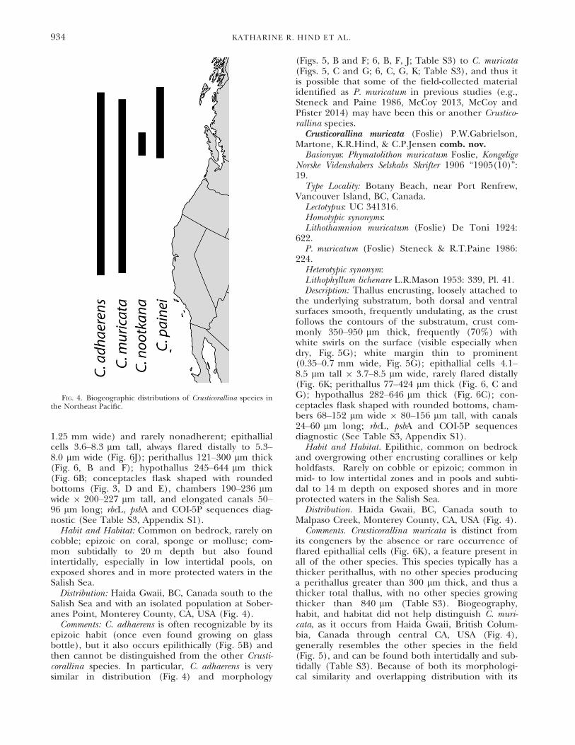
FIG. 4. Biogeographic distributions of Crusticorallina species inthe Northeast Pacific.
934 KATHARINE R. HIND ET AL.

congeners, we based our description only onsequenced specimens. It is possible and even likelythat previous studies on P. muricatum (Steneck andPaine 1986, McCoy 2013, McCoy and Pfister 2014)may have mistakenly included multiple Crusticoral-lina species.
Historical comments. Historically much confusionsurrounds the application of this name because ofthree problems: the difficulty in circumscribing gen-era of nongeniculate corallines; the apparent erro-neous report of multiporate conceptacles in theoriginal description; and that the holotype com-prises at least two heterotypic elements. Foslie(1906) described this species as Phymatolithon murica-tum based on material collected by Yendo at BotanyBeach, near Port Renfrew on Vancouver Island,Canada in June, 1901. Foslie’s original descriptionincluded that the thallus habit was thick, up to4 mm, partly smooth and loosens easily from thesubstratum. Anatomically the species had a well-
developed hypothallus with upswept arcs of cells.Sporangial conceptacles were 200–300 lm diameterwith 12 roof pores. Thalli were reported to occuron rocks and mussel shells. De Toni (1924: 662)transferred the species to Lithothamnion Phillipi.Mason (1953) added information about the epithal-lium (2–3 cell layers with cells 5–6 lm wide and 3–4 lm tall) and cystocarpic conceptacles (40–60 lmdiameter and 30–40 lm tall often embedded in thethallus), and she repeated Foslie’s description ofmultiporate sporangial conceptacles. She pho-tographed some of the specimens from TRH(Mason 1953, Pl. 32a & b) and noted that a frag-ment of the type material was in UC (UC 341316).Adey (1970) examined type material in TRH andreported finding no asexual (bi/tetrasporangial)conceptacles nor conceptacle primordial capscharacteristic of Phymatolithon Foslie, but stated that“. . . the anatomy is peculiar in a number of ways,and some doubt remains.”
FIG. 5. Field photos of (A)C. painei, GWS021033, (B)C. adhaerens, GWS021685, (C)C. muricata, GWS013078, and (D)C. nootkana, UBC A88650; whole-mount thallus photos of (E)C. painei, GWS027973, (F)C. adhaerens, GWS012584, (G)C. muricata, UBC A91424, and(H) C. nootkana, GWS010093.Scale = 1 mm.
CRUSTICORALLINA GEN. NOV. 935

Steneck and Paine (1986) transferred Phyma-tolithon muricatum to Pseudolithophyllum Me. Lemoinefollowing Adey’s concept of Pseudolithophyllum basedon a spurious lectotype (Silva et al. 1996: 269) andnot Lemoine’s (1978) revised concept. Currently
Pseudolithophyllum, with P. fuegianum (Heydrich)M.L.Mendoza & Cabioch as the generitype, is con-sidered a synonym of Lithophyllum Phillipi (Silvaet al. 1996: 269). Steneck and Paine (1986) clearlydescribed and illustrated material that they
FIG. 6. Cross-sections of Crusticorallina species, depicting (A–D) thick hypothalli of C. painei (GWS020983), C. adhaerens (GWS020762),C. muricata (PTM1220), and C. nootkana (UBC A88650); (E–H) elongated meristematic cells of C. painei (GWS020983), C. adhaerens(GWS019429), C. muricata (GWS013078), and C. nootkana (UBC A88650); and (I–L) epithallial cells flared in C. painei (GWS020983),flared in C. adhaerens (GWS021681), unflared in C. muricata (GWS010132), and flared in C. nootkana (GWS010093). Scales = (A–D)100 lm, (E–H) 50 lm, (I–L) 10 lm.
936 KATHARINE R. HIND ET AL.

considered to belong to this species, including itshabit, thick and lobate with distinct white margins,gently undulating surfaces, and common crescent-shaped swirls, its anatomy with flared epithallialcells, very elongate meristematic cells and a thickhypothallus, and its reproduction with buried con-ceptacle primordia and Phymatolithon-shaped con-ceptacles. Taxonomically, they recognized thatLithophyllum lichenare L.R.Mason was conspecific withP. muricatum, and designated one of two specimenfragments in UC as the lectotype, UC 341316 (D)apparently because, “Other Foslie syntypes are refer-able to Pseudolithophyllum whidbeyense and Lithophyl-lum impressum.”
The holotype collection in TRH A3-148 com-prises numerous specimens and fragments, in twoboxes, some thick and epilithic and some thin onMytilus sp. shells. DNA sequencing of some ofthese specimens indicates at least two distinct spe-cies are present, one of which represents an unde-scribed species in another genus (Gabrielson,personal observation). According to Article 9.14 ofthe ICN (McNeill et al. 2012), “When a type. . .contains parts belonging to more than onetaxon. . . the name must remain attached to thepart. . . that corresponds most nearly with the origi-nal description or diagnosis.” It appears that Ste-neck and Paine (1986) recognized that more thanone taxon comprised the holotype (they used theterm syntype, see quote in previous paragraph),and designated the specimen in UC that theyexamined as the lectotype (UC 341316 (D)). Thisspecimen conforms most closely to the originaldescription, being thick and with a strongly devel-oped hypothallus and with conceptacles that fallwithin the range given by Foslie (1906). Like Adey(1970) and Steneck and Paine (1986) we observedno multiporate conceptacles in any of the materialin TRH or UC. We concur with the lectotype desig-nation first made by Steneck and Paine (1986) andin accordance with Article 9.19 of the ICN(McNeill et al. 2012) we follow that designation.The partial rbcL sequence we obtained from UC341316 is identical to field collected material thatwe call C. muricata.
Despite Steneck and Paine’s (1986) clear descrip-tion of C. muricata (as P. muricatum), we are uncer-tain that all of the material that they examinedbelongs to this species due to recognition of threeother species in this genus whose distributions over-lap much of the range of C. muricata and theabsence of distinguishing morpho-anatomical fea-tures among them. Moreover, the specimen fromTorch Bay, AK is over 600 km north of our north-ernmost DNA confirmed site for C. muricata. With-out sequencing type material we would not know towhich of the species of Crusticorallina the lectotypebelonged.
Crusticorallina nootkana G.W.Saunders, Martone,K.R.Hind, C.P.Jensen, & P.W.Gabrielson sp. nov.
Etymology: Named after Nootka Sound on Vancou-ver Island, BC, the center of its known biogeo-graphic distribution.Holotypus: UNB GWS010093.Type Locality: Island south of Clotchman Island,
Spanish Pilot Group, Tahsis, BC, Canada, 23 May2008, on bedrock, 20 m depth, leg. G. W. Saunders.Description: Thallus encrusting, smooth, around
420 lm thick, white margin thin (0.35 mm wide,Fig. 5, D and H), sometimes nonadherent and leafy;white swirls not seen on its surface; epithallial cells4.9–7.0 lm tall, always flared distally to 8.9 lm wide(Fig. 6L); perithallus around 115 lm thick (Fig. 6,D and H); hypothallus around 300 lm thick(Fig. 6D); conceptacles not observed; rbcL, psbA andCOI-5P sequences diagnostic (See Table S3,Appendix S1).Habit and Habitat: On bedrock; low intertidal, par-
ticularly under Phyllospadix spp. (surfgrass) and sub-tidally to 20 m depth on exposed shores.Distribution: Kyuquot, Vancouver Island south to
Botany Beach, Vancouver Island, BC, Canada(Fig. 4).Comments: Crusticorallina nootkana is perhaps the
easiest species to distinguish by its thinner crust(Table S3), wider epithallial cells (Table S3), com-monly nonadherent margin (Fig. 5H), and narrowbiogeographic distribution (Fig. 4). Crusts withthalli thicker than 420 lm are unlikely to beC. nootkana (Table S3). One of our collections ofC. nootkana had a leafy, shelf-like, nonadherent mar-gin (Fig. 5H), but it is difficult to know if this mor-phology is common given the small sample size.
DISCUSSION
Placement of Crusticorallina in subfamily Coralli-noideae. Hind and Saunders (2013) first demon-strated that four different species, all nongeniculatecorallines that they called P. muricatum, belonged inthe tribe Corallineae, a tribe and subfamily (Coralli-noideae) previously considered to include onlygeniculate taxa. In their dataset, Hind and Saunders(2013) did not include Neogoniolithon Setchell &L.R.Mason, hypothesized by Cabioch (1972, 1988)to be sister to Corallinoideae based on morpho-ana-tomical characters and subsequently supported bythe phylogenetic analyses of Bailey et al. (2004) andBittner et al. (2011). Furthermore, they did notascertain which, if any, of the four species that theycalled P. muricatum was indeed that species by com-paring DNA sequences from their collections tosequences from type specimens. Using SSUsequence data from GenBank, we have confirmedthe placement of P. muricatum spp. (as Crusticoral-lina) within the subfamily Corallinoideae (and tribeCorallineae; Fig. 1). Crusticorallina resolved as dis-tinct from the subfamily Neogoniolithoideae, whichcontains Neogoniolithon and at least the generitype ofSpongites K€utzing, S. fruticulosa K€utzing (R€osler et al.
CRUSTICORALLINA GEN. NOV. 937

2016). We also sequenced the lectotype specimen ofPhymatolithon muricatum, basionym of Crusticorallinamuricata, confirming to which of the four speciescalled P. muricatum by Hind and Saunders (2013)that name belongs.
Hind and Saunders (2013) pointed out that thereare no morpho-anatomical characters that distin-guish Neogoniolithoideae from Corallinoideae, andwe concur, even with a more complete characteriza-tion of Crusticorallina, the only nongeniculate taxonof Corallinoideae. Neogoniolithoideae and Coralli-noideae share in common the three reproductivefeatures that were used by Kato et al. (2011) tosegregate subfamilies formerly all grouped in Masto-phoroideae (e.g., Neogoniolithoideae, Porolithoi-deae, Hydrolithoideae), namely: (i) spermatangiaborne on the floor and walls of male conceptacles,(ii) gonimoblast filaments that arise from the dorsaland/or marginal surfaces of the fusion cell, and(iii) tetrasporangial conceptacles formed by fila-ments peripheral to the fertile area (Type 1). Thevegetative characters diagnostic for all species ofCrusticorallina, an encrusting habit, a relatively thickthallus with a hypothallus comprising 50%–80% oftotal thallus thickness, elongate medullary cells andabsence of trichocytes are all characters shared by atleast some species of Neogoniolithon (Penrose 1996,Kato et al. 2013, Mateo-Cid et al. 2014), althoughwe have not found in the literature a single speciesin that genus that exhibits this entire suite of char-acters. Moreover, species concepts in Neogoniolithonare in a state of flux (Kato et al. 2013). Species aredifficult to distinguish by morpho-anatomy (Mateo-Cid et al. 2014), no species in the genus has beenlinked to its type specimen by DNA sequence andnames are applied with little regard for biogeogra-phy (Sissini et al. 2014). Yet in all phylogenetic anal-yses to date, including the most recent andcomprehensive five marker analysis by R€osler et al.(2016), Neogoniolithoideae and Corallinoideaeoccur as distinct, well-supported clades that may ormay not be sister taxa. Thus, we continue to recog-nize Corallinoideae and Neogoniolithoideae as dis-tinct monophyletic subfamilies.Crusticorallina species discrimination. Distinguish-
ing among the four species of Crusticorallina usingecology and/or morpho-anatomy is nearly impossi-ble, in part because they are uncommon and diffi-cult to collect. This is reflected in our low samplesizes (mostly fewer than 10 specimens per species)despite years of collecting in low intertidal habitatsmainly in WA and southern BC (by PWG and PTM)and more recently collecting subtidally by SCUBAin central and northern BC (by GWS and KRH).Because of low sample sizes, the following compar-isons to segregate species should be used with cau-tion. Crusticorallina nootkana is perhaps the easiest todistinguish by its thinner crust, wider epithallialcells, commonly nonadherent margin, and narrowbiogeographic distribution; C. muricata by the
absence or rare occurrence of flared epithallialcells, a feature present in all other species; C. ad-haerens by its epizoic or nonepilithic habit (onceeven found growing on glass bottle), however, C. ad-haerens also occurs epilithically and then cannot bedistinguished from C. painei or possibly the otherspecies. Based on these observations, it is possiblethat some of the field-collected material identifiedas P. muricatum in previous studies (e.g., Steneckand Paine 1986, McCoy 2013, McCoy and Pfister2014) may have been one of these other species.For example, Steneck and Paine (1986) indicatedthat in their samples of P. muricatum, epithallialcells were “somewhat angular (symmetrical trape-zoid) and tapering toward the meristem.” These“flared” epithallial cells (as referred to here) weretypically absent (12%) in our C. muricata speci-mens, but present (100%) in all other species ofCrusticorallina.Failure of morpho-anatomy to delineate coralline tax-
a. No coralline morpho-anatomist ever proposed,or likely ever suspected, that P. muricatum belongedin the subfamily Corallinoideae, perhaps represent-ing the most significant failure of morpho-anatomyto provide characters useful to distinguish corallinesat any taxonomic rank without supporting DNAsequence evidence. This failure of morpho-anatomyto correctly delimit both geniculate and nongenicu-late taxa in the Corallinales and Hapalidiales hasbeen especially evident in the North Pacific Ocean,where in the last 5 years two new genera have beenerected, Johansenia (Hind and Saunders 2013) andCallilithophytum P.W.Gabrielson, W.H.Adey,G.P.Johnson, & Hernandez-Kantun (Adey et al.2015); one genus resurrected, NeopolyporolithonW.H.Adey & H.W.Johansen (Adey et al. 2015); fourgenera placed in synonymy in Corallina Linnaeus–Marginisporum (Yendo) Ganesan, Serraticardia(Yendo) P.C.Silva (Hind and Saunders 2013), Pach-yarthron Manza (Hind et al. 2014a) and YamadaiaSegawa (Martone et al. 2014); and numerous spe-cies transferred to other genera, Calliarthron yessoense(Yendo) Manza to Alatocladia (Yendo) H.W.Johan-sen (Gabrielson et al. 2011), Yamadaia americanaE.Y.Dawson & R.L. Steele to Chiharea H.W.Johansen(Martone et al. 2014); Pseudolithophyllum decipiens(Foslie) Steneck & R.T.Paine to Spongites (Van derMerwe et al. 2015) and Corallina frondescens Postels& Ruprecht to Bossiella P.C.Silva (Hind et al. 2015).Worldwide, all coralline genera and species need tobe reassessed with DNA sequence data to determinewhich morpho-anatomical characters are taxonomi-cally meaningful and at what ranks.Coralline endemism in the Northeast Pacific Ocean. E-
ven with the renewed interest in coralline systemat-ics worldwide driven by DNA sequence data andnew genera and species being recognized, e.g., inthe subarctic (Adey et al. 2015), Europe (Walkeret al. 2009, Pardo et al. 2013, Hernandez-Kantunet al. 2015, Pe~na et al. 2015), Japan (Kato et al.
938 KATHARINE R. HIND ET AL.

2011, Kato et al. 2013), the Red Sea (Basso et al.2015), New Zealand (Broom et al. 2008, Nelsonet al. 2015), and South Africa (Van der Merwe et al.2015), the NE Pacific is proving to be a center ofendemism for both geniculate and nongeniculatecoralline genera. Thus far, four geniculate generaare endemic: Calliarthron Manza (Gabrielson et al.2011), Chiharaea (Martone et al. 2012), Johansenia(Hind and Saunders 2013), and Lithothrix J.E.Gray;and two nongeniculate genera are endemic:Callilithophytum (Adey et al. 2015), the obligateepiphyte on Calliarthron tuberculosum (Postels &Ruprecht) Manza, and, herein, Crusticorallina. Atpresent, no other region worldwide boasts this num-ber of endemic coralline genera, not even New Zeal-and or Australia, both well known for their marinealgal endemics (Womersley 1981, Nelson 2013).The underlying cause for this diversification in cor-alline algal genera in the NE Pacific remainsunknown.Genicula and conceptacle placement. Crusticorallina is
distinct, thus far, from all other taxa in the sub-family Corallinoideae by lacking genicula and bear-ing conceptacles in its crust. There appears to bean evolutionary advantage to having reproductivestructures (conceptacles) in intergenicula, as evenwhen the crustose base is far more extensive inarea than the geniculate axes, as occurs in somespecies of Chiharaea, conceptacles are borne onlyon the erect axes (Martone et al. 2012). This istrue for all geniculate corallines, even thoughgenicula are not all homologous, having arisen atleast three different times from different crustosecoralline ancestors in the subfamilies Coralli-noideae, Lithophylloideae, and Metago-niolithoideae (Bailey and Chapman 1998, Bittneret al. 2011, R€osler et al. 2016). Moreover, our datademonstrate that Crusticorallina species evolvedfrom geniculate coralline ancestors (Fig. 2), repre-senting a complete evolutionary reversal and areturn to a crustose morphology similar to othernongeniculate corallines at the base of the coral-line phylogeny (Bailey and Chapman 1998, Bittneret al. 2011). Given the repeated evolution ofgenicula from crustose coralline ancestors, pastresearchers have speculated that erect, geniculatefronds may be adaptations for increasing spore dis-persal (Johansen 1981), increasing light competi-tion (Johansen 1981), or perhaps decreasingherbivory (Padilla 1984) as documented forbranched crusts (Steneck 1983, 1986). However,adaptive arguments are difficult to maintain whennongeniculate species, such as Crusticorallina spp.,can persist and even radiate despite having lostgeniculate fronds altogether.
We thank Laura Anderson, Bridgette Clarkston, Kyatt Dixon,Aaron Galloway, Sarah Hamsher, Ted Klenk, Dan McDevit,Kevin Miklasz, Kathryn Roy, Kevin Britton-Simmons, JackieSones, H. Stewart, and Scott Toews, for collections. Gary
Saunders provided many collections, including two holotypespecimens; we sincerely appreciate his contributions to thisproject. Collections made by Sandra Lindstrom were sup-ported by funding from Cook Inlet Regional Citizens Advi-sory Council, Hart Crowser, and Natural Sciences andEngineering Research Council (NSERC) and were facilitatedby Amy Deveau, Jochen Halfar, Jonathan Houghton, SusanSaupe, and Nancy Turner. We thank the Makah Tribal Coun-cil for access to Tatoosh Island. We thank Craig Schneiderfor advice regarding taxonomic nomenclature. KRH andPTM thank the Hakai Institute for their contribution of fund-ing and access to outstanding research facilities along thecentral coast of BC, Canada. Wilson Freshwater, DNA AnalysisCore Facility, University of North Carolina, Wilmington pro-vided sequencing support and Todd Vision provided researchspace and equipment to PWG. A portion of this study wasdone while PWG was a visiting professor at the Friday HarborLaboratories, University of Washington. This research wasfunded in part by a NSERC grant to PTM and by a familytrust to PWG.
Adey, W. H. 1970. A revision of the Foslie crustose corallineherbarium. Det. Kong. Norske Vidensk. Selsk. Skr. 1:1–46.
Adey, W. H., Hernandez-Kantun, J. J., Johnson, G. & Gabrielson,P. W. 2015. DNA sequencing, anatomy and calcification pat-terns support a monophyletic, subarctic, carbonate reef-form-ing Clathromorphum (Hapalidiaceae, Corallinales,Rhodophyta). J. Phycol. 51:189–203.
Adey, W. & Johansen, H. W. 1972. Morphology and Taxonomyof Corallinaceae with special reference to Clathromorphum,Mesophyllum and Neopolyporolithon gen. nov. Phycologia11:159–80.
Areschoug, J. E. 1852. Ordo XII. Corallineae. In Agardh, J. G.[Ed.] Species genera et ordines algarum. . . Volumen secundum:algas florideas complectens. C.W.K. Gleerup, Lund, Sweden, pp.506–76.
Bailey, J. C. & Chapman, R. L. 1998. A phylogenetic study of theCorallinales (Rhodophyta) based on nuclear small-subunitrRNA gene sequences. J. Phycol. 34:692–705.
Bailey, J. C., Gabel, J. E. & Freshwater, D. W. 2004. Nuclear 18SrRNA gene sequence analyses indicate that the Mastophoroi-deae (Corallinaceae, Rhodophyta) is a polyphyletic taxon.Phycologia 43:3–12.
Basso, D., Caragnano, A., Le Gall, L. & Rodondi, G. 2015. Thegenus Lithophyllum in the north-western Indian Ocean, withdescription of L. yemenense sp. nov., L. socotraense sp. nov., L.subplicatum comb. et stat. nov., and the resumed L. affine, L.kaiseri and L. subreduncum (Rhodophyta, Corallinales). Phyto-taxa 208:183–200.
Bittner, L., Payri, C., Mandeveldt, G., Couloux, A., Cruaud, C.,Reviers, B. & Le Gall, L. 2011. Evolutionary history of theCorallinales (Corallinophycidae, Rhodophyta) inferred fromnuclear, plastidial and mitochondrial genomes. Mol. Phylo-genet. Evol. 61:697–713.
Broom, J. E. S., Hart, D. R., Farr, T. J., Nelson, W. A., Neill, K. F.,Harvey, A. S. & Woelkerling, W. J. 2008. Utility of psbA andnSSU for phylogenetic reconstruction in the Corallinalesbased on New Zealand taxa. Mol. Phylo. Evol. 46:958–73.
Cabioch, J. 1971. Essai d’une nouvelle classification des Coralli-nac"ees actuelles. Compte Rendu Hebdomadaire des S!eances del’Acad!emie des Sciences. Paris. S!erie D 272:1616–9.
Cabioch, J. 1972. "Etude sur les Corallinac"ees. II - La mor-phogen#ese: cons"equences syst"ematiques et phylog"en"etiques.Cah. Biol. Mar. 13:137–288.
Cabioch, J. 1988. Morphogenesis and generic concepts in coral-line algae. Helgolander Meeresunters 42:493–509.
Clarkston, B. E. & Saunders, G. W. 2012. An examination ofthe red algal genus Pugetia (Kallymeniaceae, Gigartinales)with descriptions of Salishia firma gen. & comb. nov., Puge-tia cryptica sp. nov. and Beringia wynnei sp. nov. Phycologia51:33–61.
CRUSTICORALLINA GEN. NOV. 939

Darriba, D., Taboada, G. L., Doallo, R. & Posada, D. 2012. jMo-delTest 2: more models, new heuristics and parallel comput-ing. Nat. Methods 9:772.
De Toni, G. B. 1924. Sylloge algarum omnium hucusque cognitarum.Vol. IV. Florideae. Sectio V. Additamenta. Sumptibus auc-toris, Patavii [Padua], 767 pp.
Foslie, M. 1906. Den Botaniske Sammlung. Det. Kong. NorskeVidensk. Selsk. Skr. 1905:17–24.
Foster, M. S. 1975. Algal succession in a Macrocystis pyrifera forest.Mar. Biol. 32:313–29.
Freshwater, D. & Rueness, J. 1994. Phylogenetic relationships ofsome European Gelidium (Gelidiales, Rhodophyta) species,based on rbcL nucleotide sequence analysis. Phycologia33:187–94.
Gabrielson, P. W., Miller, K. A. & Martone, P. T. 2011. Morpho-metric and molecular analyses confirm two species of Cal-liarthron (Corallinales, Rhodophyta), a genus endemic to thenortheast Pacific. Phycologia 50:298–316.
Harper, J. T. & Saunders, G. W. 2001. The application of seu-qences of the ribosomal cistron to the systematics and classi-fication of the florideophyte algae (Florideophyceae,Rhodophyta). Cah. Biol. Mar. 42:25–38.
Harvey, A., Woelkerling, W., Farr, T., Neill, K. & Nelson, W. 2005.Coralline algae of central New Zealand: an identificationguide to common ‘crustose’ species. NIWA Information Series57:145.
Hernandez-Kantun, J. J., Rindi, F., Adey, W. H., Heesch, S., Pe~na,V., Le Gall, L. & Gabrielson, P. W. 2015. Sequencing typematerial resolves the identity and distribution of the generit-rype Lithophyllum incrustans, and related European species L.hibernicum and L. bathyporum (Corallinales, Rhodophyta). J.Phycol. 51:791–807.
Hind, K. R., Gabrielson, P. W., Lindstrom, S. C. & Martone, P. T.2014a. Misleading morphologies and the importance ofsequencing type specimens for resolving coralline taxonomy(Corallinales, Rhodophyta): Pachyarthron cretaceum is Corallinaofficinalis. J. Phycol. 50:760–4.
Hind, K. R., Gabrielson, P. W. & Saunders, G. W. 2014b. Molecu-lar-assisted alpha taxonomy reveals pseudocryptic diversityamong species of Bossiella (Corallinales, Rhodophyta) in theeastern Pacific Ocean. Phycologia 53:443–56.
Hind, K. R., Miller, K. A., Young, M., Jensen, C., Gabrielson, P.W. & Martone, P. T. 2015. Resolving cryptic species of Bos-siella (Corallinales, Rhodophyta) using contemporary andhistorical DNA. Am. J. Bot. 102:1–19.
Hind, K. R. & Saunders, G. W. 2013. A molecular phylogeneticstudy of the tribe Corallineae (Corallinales, Rhodophyta)with an assessment of genus-level taxonomic features anddescriptions of novel genera. J. Phycol. 49:103–14.
Huelsenbeck, J. P. & Ronquist, F. 2001. MRBAYES: Bayesian infer-ence of phylogenetic trees. Bioinformatics 17:754–5.
Johansen, H. W. 1969. Morphology and systematics of corallinealgae with special reference to Calliarthron. Univ. Cal. Pub.Bot. 49:1–98.
Johansen, H. W. 1974. Articulated coralline algae. Oceanogr. Mar.Biol. Ann. Rev. 12:77–127.
Johansen, H. W. 1981. Coralline Algae, a First Synthesis. CRC Press,Boca Raton, Florida, 239 pp.
Kato, A., Baba, M. & Suda, S. 2011. Revision of the Mastophoroi-deae (Corallinales, Rhodophyta) and polyphyly in nongenic-ulate species widely distributed on Pacific coral reefs. J.Phycol. 47:662–72.
Kato, A., Baba, M. & Suda, S. 2013. Taxonomic circumscriptionof heterogeneous species Neogoniolithon brassica-florida (Coral-linales, Rhodophyta) in Japan. Phycol. Res. 61:15–26.
Kearse, M., Moir, R., Wilson, A., Stones-Havas, S., Cheung, M.,Sturrock, S., Buxton, S. et al. 2012. Geneious Basic: an inte-grated and extendable desktop software platform for theorganization and analysis of sequence data. Bioinformatics28:1647–9.
Kucera, H. & Saunders, G. W. 2012. A survey of Bangiales (Rho-dophyta) based on multiple molecular markers reveals cryp-tic diversity. J. Phycol. 48:869–82.
Le Gall, L., Payri, C. E., Bittner, L. & Saunders, G. W. 2010.Multigene phylogenetic analyses support recognition of theSporolithales ord. nov. Mol. Phylogenet. Evol. 54:302–5.
Le Gall, L. & Saunders, G. W. 2010. DNA barcoding is a powerfultool to uncover algal diversity: a case study of the Phyl-lophoraceae (Gigartinales, Rhodophyta), in the Canadianflora. J. Phycol. 46:374–89.
Lemoine, M. 1913. M"elob"esi"ees. Revision des M"elob"esi"ees antarc-tiques. In Anon [Ed.] Deuxi"eme Exp!edition AntarctiqueFranc!aise (1908-1910) command!ee par le Dr. Jean Charcot,Sciences Naturelles: Documents Scientifiques, Botanique, Vol. 1.Masson & Cie, Paris, pp. 1–67.
Lemoine, P. 1978. Typification du genre PseudolithophyllumLemoine. Rev. Algol. N. S. 13:117.
Littler, M. M. 1972. The crustose Corallinaceae. Oceanogr. Mar.Biol. Ann. Rev. 10:311–47.
Martone, P. T., Lindstrom, S. C., Miller, K. A. & Gabrielson, P.W. 2012. Chiharaea and Yamadaia (Corallinales, Rhodophyta)represent reduced and recently derived articulated corallinemorphologies. J. Phycol. 48:859–68.
Mason, L. R. 1953. The crustaceous coralline algae of the Pacificcoast of the United States, Canada and Alaska. Univ. Cal.Publ. Bot. 26:313–89.
Mateo-Cid, L. E., Mendoza-Gonzalez, A. C. & Gabrielson, P. W.2014. Neogoniolithon (Corallinales, Rhodophyta) on the Atlan-tic coast of Mexico, including N. siankanensis sp. nov. Phyto-taxa. 190:64–93.
McCoy, S. J. 2013. Morphology of the crustose coralline alga Pseu-dolithophyllum muricatum (Corallinales, Rhodophyta) respondsto 30 years of ocean acidification in the northeast Pacific. J.Phycol. 49:830–7.
McCoy, S. J. & Pfister, C. A. 2014. Historical comparisons revealaltered competitive interactions in a guild of crustose coral-line algae. Ecol. Lett. 17:475–83.
McNeill, J., Barrie, F. R., Buck, W. R., Demoulin, V., Greuter, W.,Hawksworth, D. L., Herendeen, P. S. et al. 2012. InternationalCode of Nomenclature for Algae, Fungi, and Plants (MelbourneCode) adopted by the Eighteenth International Botanical CongressMelbourne, Australia, July 2011. Reg. Veg. 154:1–240.
Nelson, W. A. 2013. New Zealand Seaweeds. An Illustrated Guide. TePapa Press, Wellington, 328 pp.
Nelson, W. A., Sutherland, J. E., Farr, T. J., Hart, D. R., Neill, K.T., Kim, H. J. & Yoon, H. S. 2015. Multi-gene phylogeneticanalyses of New Zealand coralline algae: Corallinapetranovaezelandiae gen. et sp. nov. and recognition of the Hapa-lidiales ord. nov. J. Phycol. 51:454–68.
Padilla, D. K. 1984. The importance of form: differences in com-petitive ability, resistance to consumers and environmentalstress in an assemblage of coralline algae. J. Exp. Mar. Biol.Ecol. 79:105–27.
Paine, R. T. 1984. Ecological determinism in the competition forspace. Ecology 65:1339–48.
Pardo, C., Lopez, L., Pe~na, V., Hern"andez-Kant"un, J., Le Gall, L.,B"arbara, I. & Barreiro, R. 2013. A multilocus species delimi-tation reveals a striking number of species of coralline algaeforming maerl in the OSPAR maritime area. PLoS ONE 9:e104073.
Pe~na, V., De Clerck, O., Afonso-Carrillo, J., Ballesteros, E.,B"arbara, I., Barreiro, R. & Le Gall, L. 2015. An integrativesystematic approach to species diversity and distribution inthe genus Mesophyllum (Corallinales, Rhodophyta) in Atlanticand Mediterranean Europe. Eur. J. Phycol. 50:1–15.
Penrose, D. L. 1996. Genus Neogoniolithon Setchell & Mason1943:90. In Womersley, H. B. S. [Ed.] The Marine BenthicFlora of Southern Australia. Rhodophyta. Part IIIB, Gracilariales,Rhodymeniales, Corallinales, and Bonnemaisoniales. AustralianBiological Resources Study, Canberra, pp. 280–3.
Robba, L., Russell, S. J., Barker, G. L. & Brodie, J. 2006. Assess-ing the use of the mitochondrial COX1 marker for use inDNA barcoding of red algae (Rhodophyta). Am. J. Bot.93:1101–8.
R€osler, A., Perfectti, F., Pe~na, V. & Braga, J. C. 2016. Phylogeneticrelationships of Corallinaceae (Corallinales, Rhodophyta):
940 KATHARINE R. HIND ET AL.

taxonomic implications for reef-building corallines. J. Phycol.52:412–431.
Saunders, G. W. 2008. A DNA barcode examination of the redalgal family Dumontiaceae in Canadian waters reveals sub-stantial cryptic species diversity. 1. The foliose Dilsea - Neodil-sea complex and Weeksia. Botany 86:773–89.
Silva, P. C., Basson, P. W. & Moe, R. L. 1996. Catalogue of thebenthic marine algae of the Indian Ocean. Univ. Cal. Publ.Bot. 79:1–1259.
Silvestro, D. & Michalak, I. 2012. raxmlGUI: a graphical front-endfor RAxML. Org. Divers. Evol. 12:335–7.
Sissini, M. N., Oliveira, M. C., Gabrielson, P. W., Robinson, N. M.,Okolodkov, Y. B., Rodriguez, R. R. & Horta, P. A. 2014. Meso-phyllum erubescens (Corallinales, Rhodophyta)–so many spe-cies in one epithet. Phytotaxa 190:299–319.
Smith, S. V. 1972. Production of calcium carbonate on the main-land shelf of southern California. Limnol. Oceanogr. 17:28–41.
Stearn, C. W., Scoffin, T. P. & Martindale, W. 1977. Calcium car-bonate budget of a fringing reef on the west coast of Barba-dos. Bull. Mar. Sci. 27:479–510.
Steneck, R. S. 1983. Escalating herbivory and resulting adaptivetrends in calcareous algal crusts. Paleobio. 9:44–61.
Steneck, R. S. 1986. The ecology of coralline algal crusts: conver-gent patterns and adaptive strategies. Ann. Rev. Ecol. Syst.17:273–303.
Steneck, R. S. & Paine, R. T. 1986. Ecological and taxonomicstudies of shallow-water encrusting Corallinaceae (Rhodo-phyta) of the boreal northeastern Pacific. Phycologia 25:221–40.
Thiers, B. 2016. (continuously updated) Index Herbariorum: Aglobal directory of public herbaria and associated staff. NewYork Botanical Garden’s Virtual Herbarium. Available athttp://sweetgum.nybg.org/ih/ accessed 9 July, 2016.
Van der Merwe, E., Miklasz, K., Channing, A., Maneveldt, G. W.& Gabrielson, P. W. 2015. DNA sequencing resolves speciesof Spongites (Corallinales, Rhodophyta) in the Northeast Paci-fic and South Africa, including S. agulhensis sp. nov. Phycolo-gia 54:471–90.
Walker, R. H., Brodie, J., Russell, S., Irvine, L. M. & Orfanidis, S.2009. Biodiversity of coralline algae in the northeasternAtlantic Including Corallina caespitosa sp. nov. (Coralli-noideae, Rhodophyta). J. Phycol. 45:287–97.
Womersley, H. B. S. 1981. Biogeography of Australasian marinemacroalgae. In Clayton, M. N. & King, R. J. [Eds.] MarineBotany: An Australasian Perspective. Longman Cheshire, Mel-bourne, pp. 290–307.
Wynne, M. J. & Saunders, G. W. 2012. Taxonomic assessment ofNorth American species of the genera Cumathamnion,
Delesseria, Membranoptera and Pantoneura (Delesseriaceae, Rho-dophyta) using molecular data. Algae 27:155–73.
Yoon, H. S., Hackett, J. D. & Bhattacharya, D. 2002. A single ori-gin of the peridinin and fucoxanthin containing plastids indinoflagellates through tertiary endosymbiosis. Proc. Natl.Acad. Sci. USA 99:11724–9.
Supporting Information
Additional Supporting Information may befound in the online version of this article at thepublisher’s web site:
Figure S1. Phylogram inferred by maximumlikelihood (ML) analysis of COI-5P sequence data(539 bp).
Figure S2. Phylogram inferred by maximumlikelihood (ML) analysis of psbA sequence data(778 bp).
Figure S3. Phylogram inferred by maximumlikelihood (ML) analysis of rbcL sequence data(1,278 bp).
Table S1. List of Crusticorallina specimens,including collection details, herbarium accessionnumbers, and GenBank numbers.
Table S2. List of SSU, CO1-5P, psbA, and rbcLsequences used in the concatenated and SSUrDNA trees.
Table S3. Summary of morphology and habitatassessment of Crusticorallina species examined inthis study.
Appendix S1. Distance matrices indicating per-cent divergence of four genes used to discrimi-nate Crusticorallina species.
CRUSTICORALLINA GEN. NOV. 941