cryopreservation of embryos from model animals and human · model organisms does offer a unique...
TRANSCRIPT

10
Cryopreservation of Embryos from Model Animals and Human
Wai Hung Tsang and King L. Chow Division of Life Science, The Hong Kong University of Science and Technology
Hong Kong
1. Introduction
Diploidic germplasms such as embryos, compared to haploidic gametes, are theoretically a better choice for preservation of an animal species. However, there are significant challenges in cryopreservation of multicellular materials due to their size and physical complexity which affect the permeation of cryoprotectants and water, sensitivity to chilling and toxicity of cryoprotectants. While cryopreservation technologies are well developed and found feasible in embryos/larvae of some species, embryos of other species such as zebrafish failed to be cryopreserved. In addition, cryopreservation in many other emerging model organisms have not been developed at all. Hence, the limited cryopreservation technology has become a bottleneck in the development of various research areas, especially those relying on molecular genetics of emerging model organisms. Thorough understanding of the embryonic development and critical stages tolerant to cryopreservation needs to be identified so as to facilitate expansion of model systems available for specific biological and experimental interrogations.
1.2 Traditional and emerging animal models
Classical animal models, including species that represent major branches of the tree of life, are being used in biological studies. They include Caenorhabditis elegans (a nematode), Drosophila melanogaster (an arthropod), Danio rerio (a teleost fish), Gallus gallus (an avian), and Mus musculus (a mammal). They have been widely used in scientific research, primarily due to the ease of maintenance and specific features that facilitate experimental manipulations, genetic study and observation. As knowledge from these models has accumulated over the years, they offer important insights into the overall organization and functional composition of the general form of life. However, a comprehensive picture of variations of mechanistic innovation in the vast diversity of species in the Animal Kingdom is not available. Greater understanding of these organisms in different branches of the phylogenetic tree is in demand, in order to fill the gaps of existing findings. To meet this demand, more model organisms are emerging to provide unique perspectives of animal development and specific biological functions not yet uncovered in the study of other classical models. Emerging animal models include the brine shrimp Artemia sinica, starlet sea anemone Nematostella vectensis, non-parasitic flatworm Planaria, amphioxus Branchiostoma floridae, sea squirt Ciona intestinalis, sea lamprey Petromyzon marinus, Japanese
www.intechopen.com

Current Frontiers in Cryobiology
260
quail Coturnix japonica, opossum Monodelphis domestica and marmoset Callithrix jacchus, etc. For example, sea lampreys are cyclostomes in a basal group of vertebrates. Comparative studies on lamprey and jawed fish, e.g., zebrafish, reveal key elements guiding jaw evolution. Characteristics shared between lamprey and other vertebrates but absent in non-vertebrate chordates include the presence of neural crest cells and a jaw. Comparative studies can direct us to the origins of these features. Therefore, study of these emerging model organisms does offer a unique approach to understand the relatedness of species in the living world. Another example illustrating the importance of new model systems in studying the evolution of body plan is shown in Figure 1 (Kosik, 2009).
Fig. 1. The identification of miRNAs in different metazoan lineages revealed that the number of microRNAs (in brackets) is generally correlated with the complexity of body plan. Comparative studies involving “non-classical” model organisms further suggested a possible role of new microRNAs in evolutionary innovation. The figure is reproduced from Kosik (2009) with permission from the publisher.
www.intechopen.com

Cryopreservation of Embryos from Model Animals and Human
261
1.3 Needs for cryopreservation
1.3.1 Archives of genetic resources
Modern evolutionary developmental biology and molecular genetic studies on model
organisms largely rely on manipulation of tissues and genomes. The advancement of
technologies for genomic modification of these model organisms, in turn, affects the
popularity of usage of particular model organisms. Eventually, this leads to a rapid increase
in the number of transgenic/mutant strains in each popular species.
For the Caenorhabditis Genetic Center, mutants had been deposited by individual research
groups and various genome wide mutagenesis projects such as the National BioResource
Project for The Nematode and Caenorhabditis elegans Gene Knockout Consortium. In the
National BioResource Project, random mutagenesis was performed for nematodes with UV
in the presence of trimethylpsoralen. Affected genes are identified by screening with a gene-
specific primer set (Gengyo-Ando & Mitani, 2000). As of April 2010, about 4,400 mutants
were available and mutants of some 2800 genes were being screened. Without convenient
procedures for cryopreserving these species, maintenance of these strains is a heavy burden
to these centers, and in other laboratories using these mutants extensively for their studies.
On the other hand, passaging parasitic nematode models in plants and in donor animals,
e.g., Cooperia oncophora in the cattle host (Borgsteede & Hendriks, 1979), is particularly labor
intensive and costly and poses risk of cross contamination.
At Bloomington Drosophila Stock Center, more than 30,000 strains of Drosophila are currently
present. Preparations are in progress to expand the facility to hold up to 70,000 stocks in
order to meet the needs of stocking transgenic strains to be generated for a wide range of
studies, including those made by tissue specific knocking-out of genes for the modeling of
human diseases. In Flybase, 112,278 fly stocks were recorded for 2011 (Flybase FB2011_07
Release Notes).
In The Jackson Laboratory, over 4,000 mouse strains have been deposited and are available
to the public. At the Medical Research Council Harwell and at The European Mouse Mutant
Archive, over 1300 and 2200 mouse strains are stocked respectively (Eppig & Strivens, 1999).
In various mouse stock centers in Japan, including BioResources Center (Riken) and Trans
Genic Inc., about 8,000 mouse strains are stocked. More than 16,000 of the 24,954 protein
coding genes in the mouse genome have been modified by the International Knockout
Mouse Consortium (IKMC), as conditional knockout alleles in embryonic stem cells. So far,
more than 1,000 mutant mice, each containing one of these conditional knockout alleles, are
made available to the community. The ultimate goal is to generate different targeted alleles
in embryonic stem cells (for targeted mice generation) or in targeted mice, to be available to
the research community worldwide (Skarnes et al., 2011). On the other hand, about 1,200
zebrafish lines and about 100 Xenopus lines have been archived in the Zebrafish
International Resource Center and European Xenopus Resource Centers, respectively.
Since keeping live animals is costly in terms of requirements of space, consumables and manpower, strains not being used need to be cryopreserved to reduce the running cost. It is, therefore, a very important technology that keeps various genome-wide knockout consortia affordable to average research laboratories.
www.intechopen.com

Current Frontiers in Cryobiology
262
1.3.2 Genetic stability control
The continuous passing of live animals in a small population may lead animal strains to accumulate spontaneous mutations and undergo genetic drift. In mouse, as an example of a lower mutation rate because of its relatively long life cycle, about 0.4 mutations are accumulated in each genome in each generation (Drake et al., 1998). Using this estimation, and assuming there are four generations per year, about ten mutations are accumulated in each descendent mouse diploid genome every 6.25 years (Tsang & Chow, 2010). After four years of sibling intercrosses, there is a 90% probability that more than one mutation can be fixed in a particular mouse line (Stevens et al., 2007). To circumvent this problem, the Jackson Laboratory (Bar Harbor, Maine, USA) adopted the Genetic Stability Program to refresh some mouse colonies with cryopreserved embryos once every five generations. This strategy aims at wiping out spontaneous mutations accumulated over time to ensure consistency of the mouse genome composition.
1.3.3 Genetic diversity maintenance
Maintaining genetic stability and diversity of wild parasitic nematodes collection is vital for research on parasite-host interactions, drug resistance and their applications. For example, prolonged passage of insecticidal nematodes (i.e. entomopathogenic nematodes) can cause a reduction of traits beneficial to pest control (Shapiro et al., 1996; Stuart & Gaugler, 1996; Wang & Grewal, 2002). Three continuous passages of Galleria mellonella in vitro resulted in a significant reduction in reproductive potential, and attenuated tolerance to heat, UV and desiccation (Wang & Grewal, 2002). These observations suggest that a selective pressure had been exerted on an isolated population that experienced continuous passages. The original genetic diversity in an isolated population will thus be largely reduced if cryopreservation is not practiced immediately after collection, before experimental analysis is performed.
1.3.4 Logistic advancement on assisted reproduction technologies
A traditional human in vitro fertilization cycle involves hormone induced ovarian stimulation for oocyte retrieval, in vitro fertilization, culture of embryos to blastocyst, selection of embryos and transfer of embryos into a recipient in an uninterrupted program. Surplus embryos are discarded. If the first pregnancy failed or another pregnancy is desired, the whole cycle has to be started again. Cryopreserving surplus embryos from the first round of the program can now be a backup for the second or more rounds of pregnancy. It can be done simply by transferring the thawed embryos to the recipients when the endometrial conditions are ready (Parriego et al., 2007).
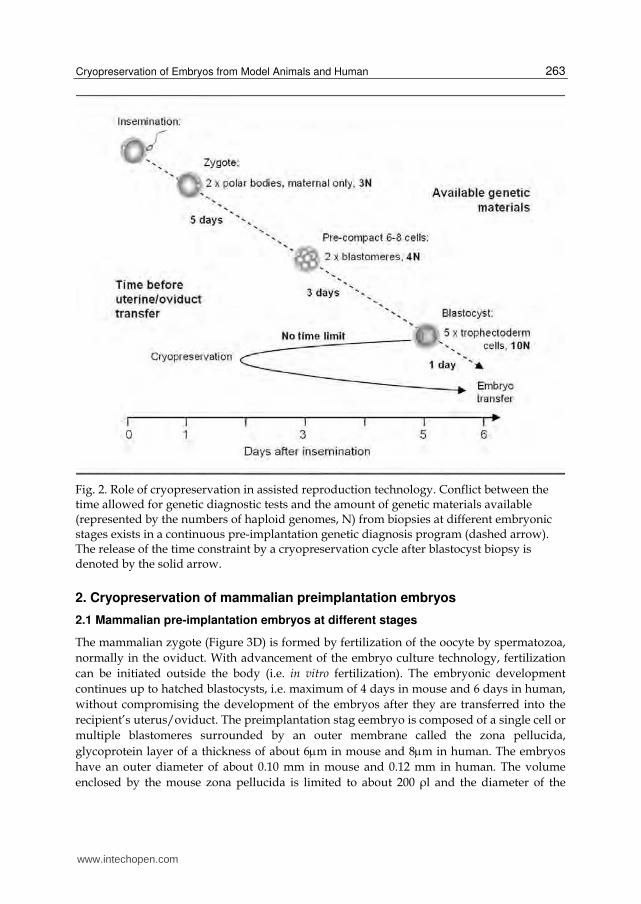
In addition, for preimplantation genetic diagnosis programs, embryos must be transferred back to the mother at or younger than day-6 blastocysts, and so biopsies for prenatal diagnosis must be taken at the latest at day-6. More genetic materials (i.e. blastomeres or trophoblastic cells) can be obtained if the biopsy is performed later. However, accompanied shortcomings will be less time for genetic tests to be performed (Manipalviratn et al., 2009).
Cryopreservation of biopsied embryos can eliminate such conflicts between the quantity of biopsy material and the quality of genetic tests (as a function of available time; Figure 2). It has been demonstrated recently on mouse and then human embryos that biopsied embryos at various stages can survive cryopreservation well (Krzyminska & O'Neill, 1991; Wilton et al., 1989; Liu et al., 1993; Snabes et al., 1993 ; Zhang et al., 2009; Keskintepe et al., 2009 ).
www.intechopen.com
Cryopreservation of Embryos from Model Animals and Human
263
Fig. 2. Role of cryopreservation in assisted reproduction technology. Conflict between the time allowed for genetic diagnostic tests and the amount of genetic materials available (represented by the numbers of haploid genomes, N) from biopsies at different embryonic stages exists in a continuous pre-implantation genetic diagnosis program (dashed arrow). The release of the time constraint by a cryopreservation cycle after blastocyst biopsy is denoted by the solid arrow.
2. Cryopreservation of mammalian preimplantation embryos
2.1 Mammalian pre-implantation embryos at different stages
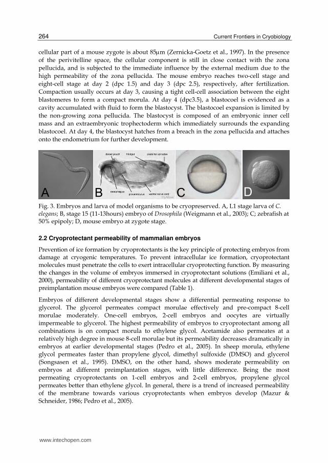
The mammalian zygote (Figure 3D) is formed by fertilization of the oocyte by spermatozoa,
normally in the oviduct. With advancement of the embryo culture technology, fertilization
can be initiated outside the body (i.e. in vitro fertilization). The embryonic development
continues up to hatched blastocysts, i.e. maximum of 4 days in mouse and 6 days in human,
without compromising the development of the embryos after they are transferred into the
recipient’s uterus/oviduct. The preimplantation stag eembryo is composed of a single cell or
multiple blastomeres surrounded by an outer membrane called the zona pellucida,
glycoprotein layer of a thickness of about 6m in mouse and 8m in human. The embryos
have an outer diameter of about 0.10 mm in mouse and 0.12 mm in human. The volume
enclosed by the mouse zona pellucida is limited to about 200 ρl and the diameter of the
www.intechopen.com
Current Frontiers in Cryobiology
264
cellular part of a mouse zygote is about 85m (Zernicka-Goetz et al., 1997). In the presence
of the perivitelline space, the cellular component is still in close contact with the zona
pellucida, and is subjected to the immediate influence by the external medium due to the
high permeability of the zona pellucida. The mouse embryo reaches two-cell stage and
eight-cell stage at day 2 (dpc 1.5) and day 3 (dpc 2.5), respectively, after fertilization.
Compaction usually occurs at day 3, causing a tight cell-cell association between the eight
blastomeres to form a compact morula. At day 4 (dpc3.5), a blastocoel is evidenced as a
cavity accumulated with fluid to form the blastocyst. The blastocoel expansion is limited by
the non-growing zona pellucida. The blastocyst is composed of an embryonic inner cell
mass and an extraembryonic trophectoderm which immediately surrounds the expanding
blastocoel. At day 4, the blastocyst hatches from a breach in the zona pellucida and attaches
onto the endometrium for further development.
Fig. 3. Embryos and larva of model organisms to be cryopreserved. A, L1 stage larva of C. elegans; B, stage 15 (11-13hours) embryo of Drosophila (Weigmann et al., 2003); C; zebrafish at 50% epipoly; D, mouse embryo at zygote stage.
2.2 Cryoprotectant permeability of mammalian embryos
Prevention of ice formation by cryoprotectants is the key principle of protecting embryos from damage at cryogenic temperatures. To prevent intracellular ice formation, cryoprotectant molecules must penetrate the cells to exert intracellular cryoprotecting function. By measuring the changes in the volume of embryos immersed in cryoprotectant solutions (Emiliani et al., 2000), permeability of different cryoprotectant molecules at different developmental stages of preimplantation mouse embryos were compared (Table 1).
Embryos of different developmental stages show a differential permeating response to
glycerol. The glycerol permeates compact morulae effectively and pre-compact 8-cell
morulae moderately. One-cell embryos, 2-cell embryos and oocytes are virtually
impermeable to glycerol. The highest permeability of embryos to cryoprotectant among all
combinations is on compact morula to ethylene glycol. Acetamide also permeates at a
relatively high degree in mouse 8-cell morulae but its permeability decreases dramatically in
embryos at earlier developmental stages (Pedro et al., 2005). In sheep morula, ethylene
glycol permeates faster than propylene glycol, dimethyl sulfoxide (DMSO) and glycerol
(Songsasen et al., 1995). DMSO, on the other hand, shows moderate permeability on
embryos at different preimplantation stages, with little difference. Being the most
permeating cryoprotectants on 1-cell embryos and 2-cell embryos, propylene glycol
permeates better than ethylene glycol. In general, there is a trend of increased permeability
of the membrane towards various cryoprotectants when embryos develop (Mazur &
Schneider, 1986; Pedro et al., 2005).
www.intechopen.com

Cryopreservation of Embryos from Model Animals and Human
265
The findings indicate a dynamic change in permeability of cell membrane to different
cryoprotectants during development. This permeability change does not correlate with
the molecular size of the cryoprotectants. In addition, the dynamic changes in
cryoprotectant permeability do not seem to be caused by the increase in the total surface
area of the embryos. The mouse 8-cell embryos undergo compaction at late day 4, thus
decreasing the total surface area drastically, but it is best penetrated at least by ethylene
glycol, compared with 1-, 2- and pre-compacted 8-cell embryos (Pedro et al., 2005), which
have a higher surface area to volume ratios. Altogether, these findings support the notion
that permeability is a dynamic physiological change related to the cellular differentiation
state, not a simple passive mechanism dictated by the physical size and surface area.
Understanding the changes of permeability of embryos to cryoprotectants at the
molecular level may help further develop the use of cryopreservation technologies on
mammalian embryos and, more importantly, on other organisms that cannot be
cryopreserved yet.
Cryoprotectants Permeability to different mouse embryo
Common name
Molecular property
1-cell 2-cell Pre-compact
morula (8-cell)Compact morula
(8-cell)
Acetamide Amide, MW=59
Moderate Moderate High High
Ethylene glycol
Polyhydric alcohol,
MW=62.1 Moderate Moderate High High
Propylene glycol
Polyhydric alcohol,
MW=76.1 High High High High
DMSO Organosulfur,
MW=78.1 High High High High
Glycerol Polyol, MW=92
Impermeable Impermeable Moderate High
Table 1. Permeability of various common cryoprotectants to mouse preimplantation embryos (Information derived from Pedro et al., 2005)
2.3 Cryoprotectant toxicity to mammalian embryos
The higher the concentration of cryoprotecting agent is in a solution, the lower is the
likelihood water crystals would be formed in the solution in a rapid-cooling process.
However, most cryoprotecting molecules are toxic to embryos with toxicity positively
correlating to their concentrations and the exposure time. When choosing the appropriate
cryoprotectants, toxicity must be considered. Among five common permeating
cryoprotecting agents to be tested, toxicity was determined to be dimethylformamide>
erythritol > DMSO > glycerol and ethylene glycol on mouse morulae (Kasai et al., 1981). By
electron probe microanalysis, Pogorelov et al. (2006 & 2007a) detected a dramatic decrease in
intracellular potassium and sodium content in two-cell embryos treated with procedures
mimicking vitrification in ethylene glycol, demonstrating a potential stress exerted on the
www.intechopen.com

Current Frontiers in Cryobiology
266
embryos. When the mouse morulae were stored in 1.5M ethylene glycol or glycerol for 6
hours, the majority (>75%) of the embryos retained the capacity to develop into expanded
blastocysts (Kasai et al., 1981). To minimize the toxic effect of the cryoprotecting solution to
the embryos while retaining the cryoprotecting function, a mixture of two or more
cryoprotecting agents could be used to decrease the relative concentration of each chemical.
Macromolecules such as polyethylene glycol, ficoll and polyvinylpyrrolidone, which
increase the viscosity of a solution, thus slowing down water molecules associating to form
ice crystals when cooling, can also lower the concentration of cryoprotecting agents to be
used in vitrification.
2.4 Osmotic flows in cryopreserving mammalian embryos
2.4.1 Slow-cooling
In a general slow-cooling procedure, embryos are immersed into permeating cryoprotectants.
Intracellular water leaves the cells by osmosis and re-enters the cells together with the
permeating cryoprotectants by diffusion. A temporal osmotic equilibrium state is acquired at
the end. A 1-2M permeating cryoprotecting agent(s) is often used in slow-cooling. To
cryopreserve the embryos, embryos and the surrounding freezing medium are loaded into a
plastic straw and subjected to cooling to a temperature slightly lower than the freezing
point, i.e. at about -7°C. Controlled ice nucleation is initiated by touching the straw with a
cooler surface (e.g., a pair of forceps) to initiate the growth of ice inside the straw. The
embryos themselves remain unfrozen but supercooled. The removal of water from the
solution by the growing ice crystals increases the solute concentration of the extracellular
medium. By osmosis, the intercellular water leaves the cells, resulting in an increase of
cryoprotectant concentration in the cells. The subsequent slow cooling further dehydrates
the embryos and concentrates the cryoprotectant in the cells to promote intracellular
solidification, without intracellular ice formation, at a sufficiently low temperature. If the
cooling is too fast, it leads to intracellular ice formation because the intracellular solute has
not yet achieved a sufficiently high concentration. Too slow a cooling rate causes cells’ death
due to the prolonged exposure to hypertonic conditions. The cooling rate must be carefully
controlled for each embryonic stage of each species because the permeability of cell
membranes, and thus the hydrodynamics, of different samples, can be different.
2.4.2 Vitrification
In cryopreservation by vitrification (or rapid-cooling), equilibration of cryoprotectants in the embryos and the cryoprotecting medium are not required. Embryos are first permeated by cryoprotecting agents at a low concentration and then immersed in a moderately high concentration (4M or above) of the same cryoprotecting agent, sometimes together with non-permeating cryoprotectants such as 0.5M sucrose. In the presence of the non-permeating cryoprotectants, the embryos shrink osmotically, thus further increasing concentration of intracellular cryoprotectants. The high concentration of cryoprotecting agents in the medium prevents efflux of intracellular cryoprotecting agents by diffusion (Figure 4). The embryos are then loaded into a container and are rapidly cooled to solidify the embryos without the formation of ice crystals (Rall, 1987). Mathematical modelings such as the relativistic permeability approach is able to simulate the osmotic curve in these
www.intechopen.com

Cryopreservation of Embryos from Model Animals and Human
267
processes to facilitate the optimization of vitrification protocols in future (Katkov and Pogorelov, 2007).
2.5 The success of cryopreservation on mammalian embryos
The first mammalian embryo that survived cryopreservation was the mouse embryo 40
years ago by slow-cooling (Whittingham et al., 1972). More than 50% of preimplantation
embryos survived after thawing and about 40% of the surviving embryos developed to
full-term after being transferred to foster females. A similar protocol was applied
successfully on cow embryos (Wilmut & Rowson, 1973), sheep embryos (Willadsen, 1977)
and many other domestic mammals (Saragusty & Arav, 2011), suggesting that slow
cooling is basically applicable to other mammalian embryos, provided the cooling rate can
be optimized and well controlled. By using 1.5M DMSO as a cryoprotectant, sheep
morulae and blastocysts were cooled at a rate of 0.1°C per minute. A survival rate of 80%
can be achieved. With a similar protocol developed from mouse experiments, the first
pregnancy after transfer of a cryopreserved human 8-cell embryo was recorded in 1983
(Trounson & Mohr, 1983). The pregnancy terminated at 24-weeks due to premature
rupture of the membrane and Streptomyces agalactiae infection (Trounson & Mohr, 1983). A
year later, Zeilmaker et al. (1984) described the first live birth after the transfer of
cryopreserved human embryos. More reports of live births of cryopreserved human
embryos were reported in the following year (Cohen et al., 1985; Downing et al., 1985) and
in the years after that.
With the extensive characterization of embryo-cryoprotectant interactions, the feasibility
of embryo cryopreservation by vitrification was demonstrated by Rall and Fahy (1985).
The next challenges were to further lower the concentration of the cryoprotecting agent to
be used and to increase the cooling rate. Increasing the cooling rate not only guarantees
the absence of ice formation but also allows a further decrease in the amount of
cryoprotecting agents being used, thus minimizing the potential toxic effects on the
embryos. It can be illustrated by a recent report describing the use of a highly conductive
micro-capillary to increase the cooling rate up to about 4,000C per second. The
cryoprotectant propylene glycol concentration can be reduced to 1.5M, compared to the
normally used 4M or above (Lee et al., 2010). Over the past decade, a variety of holding
devices have been developed to allow fast transmission of heat (thus a high cooling rate)
from the sample to the coolants (reviewed in detail in Tsang and Chow, 2010). Some
researchers have made use of containers with thinner walls, such as pulled-straw (Vajta et
al., 1998). Others have made use of an open property of the devices to hold the sample on
a surface to allow direct heat transfer between the samples and the coolant. These include
the electronic microscopic metal grid (Martino et al., 1996), cryoloop (Lane et al., 1999),
nylon mesh (Matsumoto et al., 2001), hemi-straw (Vanderzwalmen et al., 2003), cryotop
(Kuwayama et al., 2005), vitrification spatula (Tsang & Chow, 2009) and plastic blade
(Sugiyama et al., 2010). Using most of these open-systems, cryo-survival rates of above
80% are usually obtained. The remaining challenge is to select the right tool by
considering the microbial surveillance requirement (a closed-storage system versus an
open-storage system), the convenience factor and economic considerations in a routine
facility operation.
www.intechopen.com

Current Frontiers in Cryobiology
268
Fig. 4. Morphological changes of mammalian (mouse) preimplantation embryos in response to cryoprotectant treatments (for vitrification) and rehydration. Panel A-E: zygote (1-cell); panel F-J, pre-compact morula (8-cell); panel K-O, compact morula (8-cell); panel P-T, expanded blastocyst. Individual embryos were held by a glass micropipette by a slight suction under physiological isotonic medium (A, F, K and P). Vitrification solutions containing low concentration of permeating cryoprotectants and high concentration of permeating cryoprotectants plus non-permeating cryoprotectants were applied to the surroundings of the embryos, sequentially. About 30 seconds after the application of solution containing low concentration of permeating cryoprotectants, the embryos osmotically shrunk to minimal volumes (panel B, G, L and Q). The embryos (except the blastocyst) later re-expanded to a size closer to the original volumes, after an additional 30 seconds, when cryoprotectants and water re-entered the cells passively (panel C, H, M and R). After addition of the final vitrification solution containing high concentration of permeating cryoprotectants and non-permeating cryoprotectants, the embryos were further dehydrated by osmosis and shrunk without re-expansion. (panel D, I, N and S) High concentration of intracellular permeating cryoprotectants was achieved. Rapid cooling is normally done at this stage to vitrify the embryos but such cooling was not done in this demonstration. Step-wise rehydration of the embryos was done after dehydration to imitate the recovery steps after thawing the embryos from vitrification. After rehydration, the embryos re-expanded to their original size and re-gained normal morphologies (E, J, O and T).
www.intechopen.com

Cryopreservation of Embryos from Model Animals and Human
269
Vitrification is now well accepted as a reliable means for cryopreserving mammalian embryos because of its simplicity; it does not require a controllable cooler. One factor responsible for the acceptance of this technology for cryopreservation of mammalian embryos has been the intensive studies on the interaction between different cryoprotectants and embryos, i.e. permeability and toxicity, at different developmental stages. Luckily, the most permeating cryoprotectants are not very toxic to the embryos. It allows the use of the cryoprotectants at a high concentration, yet below the lethal dose, to promote vitrification in response to a convenient cooling rate in most laboratories.
3. Cryopreservation of larvae of nematodes and platyhelminthes
3.1 Development of the nematode
Nematoda belong to the ecdysozoa, sharing the same clade with arthropoda. The members
range from free living species to parasitic species in plants and animals. The life cycle of the
nematode is generally divided into five morphological stages. Each successive larval stage is
preceded by a molting process to remove the collagenous cuticle from the former larval
stage. At hatching, the first-stage larva (Figure 3 A) consisting of 558 cells is under the
protection by a cuticular layer. The animal grows in size after each hatching. In adulthood, a
reproductive hermaphrodite is about 1mm long, 0.06mm in diameter, containing about
1,000 somatic cells. In comparison, a first-stage larva is about 0.37mm in length and
0.025mm in diameter.
Under favorable conditions, the development of the animal continues through the first- to
the fourth-larval stage and finally to the reproductive adults. In many parasitic species such
as the entomopathogenic species, the third-stage larvae are juveniles that are infective to
their hosts. Under unfavorable conditions, i.e. outside the host body, a second-stage larva
develops into the third-stage infective juvenile but retains the cuticle from the last larval
stage to form a sheath. The entire animal is enclosed in the sheath until a suitable host is
infected.
3.2 Cryopreservation of nematode without cryoprotectant additive
The simplest method for nematode cryopreservation was reported on ruminant nematodes. Infective juveniles were cooled directly in liquid nitrogen vapor after being unsheathed by sodium hypochlorite, and suspended in physiological saline (Campbell and Thomson, 1973; Van Wyk et al., 1977). For example, infective juveniles of sheep nematodes (Haemonchus contortus, Ostertagia circumcincta, Trichostrongylus axei, Trichostrongylus colubriformis, Nematodirus spathiger and Oesophagostomum columbianum) and the bovine nematodes (Haemonchus placei, Ostertagia ostertagi, Nematodirus helvetianus, Oesophagostomum radiatum, Cooperia pectinata and Cooperia punctata) survived these simple cryopreservation procedures.
James (1985) suggested that the presence of natural cryoprotectants plays a role in the cryo-survival of domestic animal parasitic nematodes. Bai et al. (2004) demonstrated in Steinernema carpocapsae and Heterorhabditis bacteriophora that the cryo-survival rates of infective juveniles are positively correlated with worm concentration during the cryoprotectant glycerol incubation step. The survival rates ranged from about 20% to 100%, which was proportional to worm concentration of 120-12,000 per ml (Bai et al., 2004). Infective juveniles indeed produce cryoprotecting molecules such as trehalose and glycerol,
www.intechopen.com

Current Frontiers in Cryobiology
270
in response to thermal and other environmental stresses (Jagdale & Grew, 2003; Qiu & Bedding, 2002). The trehalose content in Steinernema carpocapsae increases from 4% to 8% after being incubated in 22% glycerol for 18 hours, before the animals are further processed for cryopreservation (Popiel & Vasquez, 1991). Production of natural cryoprotectants by the animal itself could, therefore, be the key to good animal survival in this cryopreservation procedure. Exploring an efficient way to induce the production of the natural cryoprotectants can improve cryo-survival. Identifying the molecular pathway responsible for cryoprotectant production may help make cryopreservation of these species simpler.
3.3 Slow freezing with DMSO and glycerol
Storage of live nematodes in liquid nitrogen was first demonstrated by Hwang (1970) who reported survival of animals in the genera Aphelenchoides, Panagrellus, Turbatrix and Caenorhabditis, after slow-cooling in heat-sealed glass ampoules, using DMSO as cryoprotectants. Later, glycerol was applied to slow cooling of Caenorhabditis elegans at the first-larval stage (Brenner, 1974) and since then this cryopreservation procedure has been routinely used for freezing this model organism in laboratories in the past 40 years. Up to 100% of the first-stage larvae survival can be achieved.
On the other hand, a slow-cooling method using DMSO as a cryoprotectant was used for cryopreserving another Caenorhabditis species popular in basic scientific research, i.e. Caenorhabditis briggsae (Haight et al., 1975). The worms were simply suspended in cooled 5%
DMSO for 10 minutes and then slowly cooled at a rate of 0.2C per minute to -100C before storage in liquid nitrogen. About 75% of the animals at the second-larval stage and the third-larval stage, 50% of the forth-stage larvae and 3% of the adult animals survived the freezing/thawing cycle. In contrast to Caenorhabditis elegans, no animals at the first-larval stage survived the cryopreservation (Haight et al., 1975). Obviously, traits favoring the cryopreservation procedures are present in different larval stages in both species.
The dog parasitic nematode Strongyloides stercoralis can be cryopreserved by slow cooling after incubation for up to 60 minutes in a solution containing 10% DMSO. When thawed, third-stage larvae retain infectivity to dogs and the recovered first-stage larvae develop to the third-larval stage and regain infectivity (Nolan et al., 1988). The cryopreserved sheep nematodes Haemonchus contortus, Trichostrongylus colubriformis and Ostertagia circumcincta, at the first-larval stage, retained their infectivity as well as unfrozen worms, when cooled
slowly in 10 % DMSO to -80C before being transferred to liquid nitrogen for storage (Gill and Redwin, 1995).
3.4 Vitrification by an ethylene glycol two-step procedure
Before being used in mouse and fly embryo vitrification, ethylene glycol had been used for vitrifying the human platyhelminth Schistosoma mansoni and the farm animal nematode Onchocerca microfilariae in a similar two-stage procedure. Schistosoma mansoni were pre-
incubated in 10% ethylene glycol at 37C for 10 minutes, then cooled at 0C for 5 minutes
and finally incubated in 35% ethylene glycol for 10 minutes at 0C. Before being rapidly cooled in liquid nitrogen, the worms were spread on a glass sliver (prepared from microscopic coverslips) which acted as an open carrier. About half of the thawed worms survived and remained infective in mice with an efficiency equivalent to half that of
www.intechopen.com

Cryopreservation of Embryos from Model Animals and Human
271
unfrozen worms (James, 1981). On the other hand, 70% of the Onchocerca microfilariae were viable and remained infective after vitrification and thawing (Ham et al., 1981).
A similar vitrification methodology was also applied to the plant parasitic nematode Meloidogyne Graminicola, a rice root-knot nematode (Bridge & Ham 1985). The second-stage
larvae were pre-incubated in 10% ethylene glycol at 37C for 15 minutes and then incubated in 40% ethylene glycol for 30-45 minutes. The worms were then rapidly cooled in liquid nitrogen in the same manner as James (1981) on Schistosoma mansoni. This two-stage vitrification procedure on plant nematodes was later modified by Triantaphyllou & McCabe (1989) who replaced the glass coverslip slivers with a small strip of chromatography paper as a carrier device. A survival rate of up to 90% was obtained. The author reported that the modified two-step method produced satisfactory results on other plant parasitic nematodes also, such as some Meloidogyne and Heterodera species (Triantaphyllou & McCabe, 1989).
3.5 Vitrification by a glycerol/methanol two-step procedure
An entirely different treatment methodology was developed primarily for vitrifying the entomopathogenic nematodes in the genera Steinernema and Heterorhabditis, which are effective as a biological control agent for insect pests in agriculture (Popiel & Vasquez, 1991). Steinernema carpocapsae were pre-incubated in 22% glycerol at room temperature for 24 hours and then in ice cold 70% methanol for 10 minutes. After removal of the majority of
methanol by centrifugation, concentrated worms in methanol (i.e. 20l) were spread on a small strip of filter paper before plunging into liquid nitrogen for rapid cooling. Up to 95% post-thaw survival rate could be obtained. On the other hand, a post-thaw survival rate of about 55% was obtained on Heterorhabditis bacteriophora when the optimal 14% glycerol was used (Popiel & Vasquez, 1991).
Curran et al. (1992) further optimized the protocol by replacing the centrifugation with a filtration step to remove glycerol prior to the methanol incubation. The optimal conditions for glycerol incubation for a number of entomopathogenic nematodes were also determined. Optimal glycerol pre-incubation conditions were determined to be 18% glycerol for 24 hours for Steinernema carpocapsae, 17% glycerol for 72 hours for Heterorhabditis bacteriophora and 13.8% glycerol for 72 hours for Steinernema feltiae and Steinernema glaseri (Curran et al., 1992). Other than that, 167 entomopathogenic nematodes were found to be able to survive the cryopreservation treatments, proving the feasibility of cryobanking of these worms. The mean survival rate of the Steinernema species is 58% (ranging from 25% to 97%) and that of Heterorhabditis species is 51% (ranging from 25% to 87%).
Based on the modification by Curran et al. (1992), Nugent et al. (1996) optimized cryopreservation on seven isolates of Heterorhabditis. Up to 8 days of pre-incubation in 11% or 15% glycerol is optimal for cryopreserving a couple of isolates. Nugent (1996) also found that glycerol can be replaced by DMSO in the pre-incubation step. For example, incubation of isolate HI82 in 8% DMSO for 3 days yielded a survival rate of about 80%, similar to those pre-incubated in 15% DMSO.
To the best of our knowledge, unlike mammals, there have been no studies of interactions between nematodes and cryoprotectants. Conversely, different protocols have been developed independently by different groups for specific worm species. Whether the different protocols are indeed applicable to other groups of nematodes or not requires
www.intechopen.com

Current Frontiers in Cryobiology
272
further investigation. Nonetheless, we can interpret from the protocols that nematodes are generally resistant to cryoprotectant toxicity because most protocols involve a relatively long incubation time in cryoprotecting solutions. Surprisingly, most of the protocols do not involve removal of the cuticle, which is well known for its poor permeability, though successful cryoprotection requires the presence of enough intracellular concentration of cryoprotectant. It is possible that the cryoprotectants themselves can permeabilize the cuticle and then permeate into the cells beneath. Or the cryoprotectant enters the worm via the oral opening and the gut, into the rest of the body. Equally possible is that the "cryoprotectants" did not act as a cryoprotectant per se but acted as inducers to trigger the production of natural cryoprotectant in the worm. Therefore, thorough permeation of the chemicals was not required.
4. Cryopreservation of insect embryos
4.1 Development of insect embryos
Insecta is a class with the greatest interest among the Arthropoda. Pathogenic vectors
associating mosquitoes, flies and bugs are of interest in medical and healthcare related
research. Honeybee, silk moth, beetle and other moths are of economic interest to the
beekeeping industry, silk industry and pest control in agriculture, respectively. The fruit fly
Drosophila melanogaster, a species of the order Diptera, is chosen as an insect model in genetic
studies because of its ease of keeping, handling and observing. It has a short life cycle and is
fertile through out the year. A complete life cycle involves four distinct stages of
development, which takes 8.5 days for a Drosophila egg to reach adulthood at 25C.
A Drosophila melanogaster egg is about 0.18mm in width and 0.5mm in length, occupying a volume of about 9nl. The laid egg is a single cell with about 6,000 nuclei which later migrate to the plasma membrane to form a syncytial blastoderm at around 2 hours. Cell membranes form between the nuclei at around 2.5 hours. As gastrulation occurs, a ventral furrow and a cephalic furrow form. The midgut invaginates followed by a germ band extension (3-7 hours), stomodeal invagination (5-7 hours), germ band shortening, foregut and hindgut deeply invaginate (9-10 hours). The ectoderm closes dorsally and the head involutes at about 10-13 hours (Figure 3B). At around 13 hours, organogenesis has already begun in the 50,000-cell embryos. Organogenesis is completed and the gut regions are joined between 15 and 22 hours (i.e. at hatching) (Markow et al., 2009; Weigmann et al., 2003; Grumbling & Strelets, 2006). The hatched larva experiences three instar larval phases, before the skin of the third-instar larva hardens and encapsulates the animal to form a puparium. It takes four days for the third larva to transform into an adult by metamorphosis. At the end of metamorphosis, the adult of about 2.5mm length emerges from the puparium.
4.2 Permeabilization of insect embryos to cryoprotectants
The outermost layer of the eggshell of an insect embryo is a porous proteinacious chorion
with a thickness ranging from 300-500nm in Drosophila to more than 40m in saturniid moth (Magaritis et al., 1980; Fehrenbach, 1995). Underneath are: (1) the innermost crystalline chorion layer of about 40nm in Drosophila (Papassideri & Margaritis, 1996), (2) the waterproofing wax layer (Beament, 1946; Slifer, 1948) and (3) the vitelline layer (Papassideri et al., 1991). The wax layer is only 5nm in thickness in Drosophila and is intercalating with
www.intechopen.com

Cryopreservation of Embryos from Model Animals and Human
273
the outer crystalline chorion layer and the inner vitelline layer (Papassideri et al., 1991). The wax is mainly composed of n-alkanes and methyl-branched alkanes (Nelson & Leopold, 2003), making the eggshell impermeable even to water, thus protecting the embryos from desiccation. Extracting the dechorionated eggs with a wax removing solvent makes the vitelline membrane permeable to water, cytological stains and antibiotics (Schreuders et al., 1996), (Limbourg & Zalokar, 1973). It supports the notion that the wax component in the vitelline layer is the major factor blocking the cryoprotecting molecule from permeating into the embryos.
To facilitate permeation of cryoprotectants into insect embryos for further cryopreservation
protocol development, attempts were made to permeabilize the eggshell but retain the
viability of the embryos. Removal of the chorion can be done by exposing the eggs to about
2.5% sodium hypochlorite, without compromising the survival of the embryos (Lynch et al.,
1989). Permeabilizing the inner layer of the eggshell with low injury can be done by a 2-step
method. Dechorionated eggs of 12-13 hour embryos were rinsed with isopropanol and
hexane. The embryos in permeabilized eggshell experienced minimal injury with a survival
rate of 75% to 90% in culture medium (Lynch et al., 1989). This procedure resulted in 80%-
95% of the treated eggs being permeabilized to water, ethylene glycol, propylene glycol,
glycerol and DMSO. Mazur et al. (1992b) further optimized the two-step method to allow
permeation of common cryoprotectants such as ethylene glycol and glycerol. The best result
was obtained by exposing the dechorionated 12-14 hour embryos to 0.3%-0.4% 1-butanol in
n-heptane for 90 seconds. At least 90% permeabilization and 80% survival can be obtained.
Older embryos between 14-16 hours are much less sensitive to the above procedures, which
are lethal to 3 hour embryos. Using procedures similar to Mazur (1992b), with decreased
concentration of sodium hypochlorite in dechorionation and replacing isopropanol with air
drying, all mosquito (Anopheles gambiae) embryos at 15-19 hours can be permeabilized in
ethylene glycol with an acceptable survival rate of 30% (Valencia et al., 1996). On the other
hand, heptane treatment seemed to be detrimental to the greater wax moth (Galleria
mellonella) embryos. Replacing the heptane treatment with incubation in 1.25% sodium
hypochlorite with 0.08% Tween-80 for 2 minutes permeabilized the moth embryos with 68%
survival (Cosi et al., 2010). Determination of the optimal permeabilization procedures and
the optimal embryonic stage to be permeabilized in different insecta species has opened the
door for efficient cryopreservation of this largest class of animals on land.
4.3 Chilling sensitivity of insect embryos
Drosophila embryos are highly sensitive to chilling. When 15 hour eggs were incubated at -
15°C, 50% of the embryos died within an hour even in the absence of ice formation. When
younger embryos at 3 hours and 6 hours were cooled to the same temperature, the chilling
injury increased dramatically (Mazur et al., 1992c). All 12 hour embryos died at -25°C when
cooled at 1°C per minute. A similar phenomenon was found in case of honey bee (Apis
mellifera) embryos (Collins & Mazur, 2006). The slow cooling approach that requires time for
efflux of intracellular water osmotically to avoid intracellular ice formation is, therefore,
theoretically impractical for handling insect embryo cryoprotection. It was estimated that
cooling the embryos faster than 300°C per second can circumvent the chilling injury by
shortening the time the embryo stays at such a low temperature. However, lethal
www.intechopen.com
Current Frontiers in Cryobiology
274
intracellular ice forms at such a cooling rate when using the standard concentration of
cryoprotectants (Mazur et al., 1992c). Vitrification is, therefore, the only possible way to
cryopreserve insect embryos.
4.4 The success of insect embryo vitrification
Adopting and modifying the protocol for vitrifying mouse embryos with ethylene glycol
(Rall & Fathy, 1985), Steponkus et al. (1990) first demonstrated successful vitrification of 13-
14 hour Drosophila embryos. Instead of permeabilizing the vitelline layer with the currently
developed method using an alkane, the eggs were “permeabilized” with a medium
containing 2.125M ethylene glycol for 20 minutes. The intracellular concentration of
ethylene glycol was further increased by dehydrating the embryos in 8.5M ethylene at 0°C
for 8 minutes before plunging the embryos into nitrogen slush (-204°C), using a copper
electronic microscopic grid as an open carrier (to achieve a cooling rate of about 400°C per
second). After thawing, 18% of the eggs hatched and 3% developed into fertile adults. On
the contrary, there were no embryos surviving at a lower cooling rate of 15°C per second
when using a polypropylene straw as a carrier. The surrounding cryoprotecting solution
vitrified per se since no crystals were detected by differential scanning calorimetry. The high
lethality is probably due to the suboptimal permeabilization of the vitelline layer by
ethylene glycol, leading to a lower concentration of cryoprotectants in the inner part of the
multicellular/highly differentiated insect embryos. This may result in crystallization of
water in the area with a lower concentration of cryoprotectant which would require a higher
cooling rate to induce vitrification. A higher cooling rate was achieved by using a metal grid
allowing vitrification to occur at such low concentrations of cryoprotectants, thus partially
circumventing this potential permeating defect. Using a similar protocol, other Dipteral
species such as blowfly (Lucilia cuprina, a parasite in sheep) (Leopold & Atkinson, 1999 20),
midge (Culicoides sonorensis) (Nunamaker & Lockwood, 2001) and screw-worm (Leopold et
al., 2001) were reported to be cryopreserved.
On the other hand, Mazur et al. (1992a) made use of the accumulated experience of wax
removal by butanol-heptane based procedures to permeabilize the vitelline membrane prior
to vitrification. In the optimized procedure, dechorionated Drosophila embryos were first
permeabilized in 0.3% 1-butanol in n-heptane for 90 seconds. The embryos were then pre-
incubated in 2M ethylene glycol for 30 minutes and then 8.5M ethylene glycol solution
containing 10% polyvinylpyrrolidone for 5 minutes at 5°C before rapid cooling, using a filter
membrane as an open carrier. The developmental stage of the embryos was also found to be
critical for cryo-survival determination. Vitrifying precisely staged 14.5 hour embryos using
the above mentioned method resulted in 60% of the cryopreserved embryos hatching and
more than 40% of the hatched larvae developed into fertile adults (Mazur et al., 1992a).
The development of cryopreservation on Drosophila embryos suggests that permeabilization
of the sample to cryoprotectant is the key to success even though the embryos are
structurally complex. In nematode larvae, which are also susceptible to being
cryopreserved, the organogenesis is even more advanced. This indicates that the body
complexity brought about by organogenesis is not associated with the susceptibility of an
embryo/larva to be cryopreserved, at least by vitrification. On the other hand, chilling
injury can be circumvented by vitrification practically.
www.intechopen.com

Cryopreservation of Embryos from Model Animals and Human
275
5. Cryopreservation of teleost embryos
5.1 Development of teleost embryos
The zygote of zebrafish (Brachydanio rerio) is about 0.7 mm in diameter when fertilization
occurs. A few minutes later, the chorion swells to increase the diameter to about 1.2 mm,
without much alteration in thickness, generating a significant vitelline space. The cytoplasm
segregates to form the animal pole and the vegetal yolk with an approximate total volume
of 128nl, not including the vitelline space and the chorion (Leung et al., 1998). The first cell
cleavage occurs in the animal pole at about 45 minutes after fertilization. The blastomere
gets divided five more times synchronously, each at about 15 minutes interval, producing a
blastoderm with 64 cells in 2 hours. The daughter cells increase in number with a decrease
in cell size. The blastomeres arrange themselves in a single cell layer before the fifth
cleavage. Afterwards, newly formed daughter cells overlap with each other in the
blastoderm. The multi-cell layered blastoderm spread over the yolk, reaching 30% epiboly at
4.7 hours and 50% epiboly at 5.25 hours (Figure 3C). At the gastrula period, epiboly
continues at 5.3 hours. Two germ layers, i.e. epiblast and hypoblast, are formed by
morphogenetic movement of involution, convergence and extension. Epiboly reaches 90% at
9 hours. At the end of this period, the tail bud and neural plate starts to form. The volume of
the epiboly remains constant from 40% epiboly to 100% epiboly. Entering the segmentation
period at 10 hours, segmentation processes such as formation of neuromeres, somites and
the pharyngeal arch primordia occur. The embryo volume increases to 0.23 mm3 at the six-
somite stage at 12 hours when organogenesis starts (Hagedorn et al., 1997c). At the end of
the 14 hour-period, the yolk largely reduces and tail movement can be seen. Pigment can be
identified after 36 hours. At the third day, the primary organogenesis completes. Cartilage
in the head and pectoral fin develops while hatching occurs anytime in the third day
(Kimmel et al., 1995).
5.2 Complexity of teleost embryos
Fish embryos are composed of several components with distinct physical properties. They
include the highly dynamic cellular part on the animal pole which contributes entirely to
the future animal body, the yolk surrounded by the yolk syncytial layer occupying the
majority of the early stage embryo and the chorion as the outermost mechanical protective
shield. From 40-100% epiboly, the blastoderm and the yolk occupy 18% and 82% of the
dechorionated embryo, respectively, in zebrafish (Brachydanio rerio). At the six-somite
stage, the volume of the blastoderm increases to about 40%, leaving the yolk occupying
60% of the embryo (Hagedorn et al., 1997c). Between the chorion and the embryo is the
perivitelline space filled with liquid with a chemical composition virtually identical to the
surrounding medium (Rawson et al., 2000). The complexity of the teleost embryos is
further increased by the unbalanced partial density of water in different compartments.
At the six-somite stage, the blastoderm occupies about 40% of the dechorionated embryos
but water constitutes 82% of its volume. Conversely, the yolk occupies about 60% of the
volume of the dechorionated embryos but only 42% of this is constituted by water
(Hagedorn et al., 1997b). It was estimated that the osmotically inactive volume in the one-
somite stage and six-somite stage embryos are 72.9% and 82.6%, respectively (Zhang &
Rawson, 1998).
www.intechopen.com
Current Frontiers in Cryobiology
276
5.3 Permeability of teleost embryos
The low permeability of the chorion and the perivitelline is evident from retarded
permeation of radio-labeled DMSO from the external medium into the embryos by several
folds (Harvey et al., 1983). Even after removal of the outermost barrier, the embryos were
poorly permeated by cryoprotectants. By chemical shift selective magnetic resonance
microscopy and magnetic resonance spectroscopy, kinetics of permeation of cryoprotectants
methanol, DMSO and propylene were measured. While methanol can permeate the entire
six-somite zebrafish (Brachydanio rerio) embryos in 15 minutes, DMSO and propylene are
relatively poor in permeating into the embryos when applied to the medium (Hagedorn et
at., 1996). Similar findings were obtained by osmometric measurements of volume changes
in the embryos tested (Hagedorn et al., 1997c). Also, magnetic resonance imaging on the
distribution of cryoprotectants, delivered to the external medium or injected into the yolk, in
the three-somite stage Brachydanio rerio embryo revealed that the yolk is far less permeable
than the blastoderm and the yolk syncytial layer is the major barrier to the cryoprotectants
(Hagedorn et at., 1996; Hagedorn et al., 1997a).
To artificially promote permeation of cryoprotectants into fish embryos, ultrasound of 175V
was used to increase the permeability by methanol to Danio rerio 50% epiboly (Wang et al.,
2008). A high-intensity femtosecond laser was also used to introduce transient pores on
blastomeres and the blastoderm-yolk boundary. Successful delivery of large molecules such
as fluorescein isothiocyanate, streptavidin-conjugated quantum dots and DNA plasmid was
detected on pec-fin stage (Kohli et al., 2007). Whether the physically induced permeation
method can help cryopreservation of the whole teleost embryos requires experimental
verification.
5.4 Chilling sensitivity of teleost embryos
Medaka (Oryzias latipes) embryos at early cleavage stage, i.e. 2-4-cell stage, are very sensitive to cooling at 0°C for 40 minutes. Only 38% of the embryos survived the chilling treatment. However, the same chilling treatment did not affect the survival of embryos in early gastrula stage (Valdez et al., 2005). Similarly, zebrafish (Brachydanio rerio) at cleavage stage are more sensitive to chilling than embryos at epiboly and at three-somite stage (Hagedorn et al., 1997c). This indicates that embryos at later developmental stages are more resistant to chilling. A similar phenomenon was found on other teleost species such as red sea bream, olive flounder and multicolorfin rainbowfish. In the same study, it was found that cleavage stage embryos responded to chilling by obstructing mitotic division and early gastrula stage embryos responded by delayed development at epiboly (Sasaki et al., 1998).
5.5 Cryoprotectant toxicity on teleost embryos
Incubation of the three-somite stage zebrafish (Brachydanio rerio) embryos in 1.5M DMSO and methanol for 30 minutes at room temperature did not adversely affect their survival. While propylene glycol is moderately toxic to the embryos, similar treatment with ethylene glycol or glycerol is lethal to all treated embryos. Of the cryoprotecting agents tested, ethylene glycol solution specifically led to the blastoderm being dissociated from the yolk (Hagedorn et al., 1997c). A similar phenomenon was observed on 14 to 20-somite stage embryos of Danio rerio (Higaki et al., 2010b). Treating the embryos with glycerol at a
www.intechopen.com

Cryopreservation of Embryos from Model Animals and Human
277
concentration of 2M for 30 minutes did not yield any viable cells in the embryos. The rest of the cryoprotectants tested (including methanol, ethylene glycol, DMSO, propylene glycol and 1,3-butylene glycol) gave a survival rate of between 90 to 100%. Treating the embryos with cryoprotectants at a higher concentration revealed that ethylene glycol is the next most toxic cryoprotectant, after glycerol; it kills all cells in the embryos at a concentration of 3M. In comparison, methanol and DMSO are moderately toxic. Propylene glycol and 1,3-butylene glycol are mildly toxic, killing only 58-78% of cells even at a concentration of 5M (Higaki et al., 2010b).
5.6 Attempts in vitrifying teleost embryos
So far, there have been no successful examples of live fish recovery after cryopreservation. The difficulty in controlling the dynamics of cryoprotecting agents and water in the highly structurally complex embryos may be the cause. However, studies have been conducted to assess the degree of protection provided by the cryoprotectant in vitrification. In a study, five-somite stages of turbot and zebrafish embryos were treated for 5 minutes with incremental concentrations of DMSO and then for a total time of 4 minutes in mixtures containing 5M DMSO, 2M methanol and 1M ethylene glycol, before being loaded into plastic straws and plunged into liquid nitrogen for vitrification. Although 50% of the overall glucose-6-phosphate dehydrogenase activity was retained, no embryo hatched after thawing (Robles et al., 2004).
The yolk and the surrounding syncytial layer were suggested to be a major reservoir of osmotically inactive water and a barrier to permeation of cryoprotectant to the blastoderm. After vitrifying yolk-removed zebrafish (Danio rerio) embryos at 14 to 20-somite stage in 20% ethylene glycol, 20% DMSO and 0.5M sucrose, no living embryos were obtained, but 87% of the cells survived after vitrification and up to 90% of the primordial germ cells were viable (Higaki et al., 2010a). Removal of yolk is deleterious to the development of the embryos. Eliminating the solute and water barrier by yolk removal is not the ultimate solution for cryopreserving fish embryos unless an artificial replacement of yolk is made feasible.
5.7 Alternatives for whole embryo cryopreservation
Due to the lack of progress in development of cryopreservation of fish embryos, isolated somatic cells are being explored as a means to preserve diploid genetic materials. The blastomere becomes one of the attractive candidates because of its abundance in embryos and its pluripotent property in the chimeric animals generated by blastomere implantation. The blastomere from genetically pigmented zebrafish embryos at mid-blastula stage were transplanted into an albino recipient embryo of the same developmental stage. In five out of the twenty-eight chimeric fish produced, blastomeres from the donor contributed to the germline, transmitting the pigmented phenotype to the next generation at a frequency of 1% to 40% (Lin et al., 1992). Slowly cooled zebrafish (Danio rerio) blastomeres, isolated from 50% epiboly, were cryopreserved with 1.5M DMSO and 0.1M sucrose in 0.25 ml straws by a programmable freezer. A survival rate of 70% was obtained after thawing (Lin et al., 2009). Combining these technologies, the germline transmission of the cryopreserved genetic materials through blastomeres-embryo chimera seems to be possible. More optimization, e.g., the stages from which the blastomeres are to be isolated, is needed to maximize germline transmission and to minimize operations to be conducted in a recovery procedure.
www.intechopen.com

Current Frontiers in Cryobiology
278
Another alternative diploid material often sought to be cryopreserved is the primordial germ cells. Compared with the blastomere, primordial germ cells are developmentally closer to the cell type to be differentiated in vivo, i.e. the germ cells. The first success in transplantation of primordial germ cells was demonstrated on rainbow trout (Oncorhynchus mykiss), a model with a relatively larger body size. Green fluorescent protein expressing primordial germ cells isolated from the genital ridge of hatchlings were injected into the peritoneal cavities of a wild type hatchling. The maker-labeled primordial germ cells were able to colonize the genital ridge of the recipient animal and transmit the donor characteristic to the next generation through sperm and eggs at a rate of up to about 4% (Takeuchi et al., 2003).
A similar operation in the smaller teleost species such as zebrafish is more challenging. A single primordial germ cell isolated from the pearl Danio (Danio albolineatus) at ten- to fifteen-somite stage was transplanted into the marginal region of each zebrafish (Danio rerio) embryo at the blastula stage and vice versa. The development of host germ cells was prevented in advance by injection of an antisense dead end morpholino oligonucleotide at an earlier embryonic stage (Slanchev et al., 2005). In the host, the transplanted primordial germ cell developed into a single gonad, making the animal regain fertility and transmit the donor genotype to the progenies. This complete germline replacement procedure can be applied to both goldfish (Carassius auratus) and loach (Misgurnus anguillicaudatus) (Saito et al., 2008). The success of these cases suggests that cryopreservation of primordial germ cells is a feasible approach to preserve the diploid germplasm. As the reservoirs of primordial germ cells, genital ridges from Rainbow trout (Oncorhynchus mykiss) embryos were cryopreserved by cooling in dry ice and then liquid nitrogen after treating with 1.8M ethylene glycol. About 51% of primordial germ cells survived. Fifteen to twenty surviving primordial germ cells were transplanted to the peritoneal cavity of each newly hatched animal. Germline transmission of the donor genotype could be found in 7.8% of the hosts and the germline transmission frequency was from 0.1 to 13.5%. (Kobayashi et al., 2007).
Later, Higaki et al. (2010b) vitrified whole zebrafish (Danio rerio) embryos at 14- to 20-somite stage with an optimized vitrification solution to cryopreserve primordial germ cells. With the use of 3M ethylene glycol and 0.5M sucrose, about 4 primordial germ cells, about 40% of all, survived in each embryo, after thawing. To increase cryo-survival, yolk-removed zebrafish (Danio rerio) embryos were vitrified in 20% ethylene glycol, 20% DMSO and 0.5M sucrose. Up to 90% live primordial germ cells were obtained. Half of the primordial germ cells retained pseudopodial movement. After transplanting the motile primordial germ cells into sterilized golden-type zebrafish blastulae, about 2.8% of the recipients developed normally and produced progenies with the donor’s genotype (Higaki et al., 2010a).
Unless there is a breakthrough in cryopreserving and recovering whole fish embryos, cryopreservation of blastomeres or primordial germ cells seem to be the only methods for cryopreserving the fish diploid germplasm. Blastomeres may have advantages over primordial germ cells in generating germline transmitting chimera. Firstly, identification and isolation of primordial germ cells relies on a readily observable transgenic marker (Higaki et al., 2010b; Kobayashi et al., 2007). Breeding of a strain to a marker transgenic strain or freshly injecting DNA constructs is required before cryopreservation procedures, making the procedures more complicated. Removal of the marker from the recovered animals may also be required in some applications. Secondly, for germ-line replacement, the
www.intechopen.com

Cryopreservation of Embryos from Model Animals and Human
279
recipient blastulae have to be sterilized, e.g., by injecting a dead end antisense morpholino (Ciruna et al., 2002). This demands additional procedures in the entire cryopreservation/recovery cycle, making the primordial germ cell-base approach less attractive than the existing sperm cryopreservation. Delivery of antifreezing proteins to directly minimize water crystal formation or aquaporins to increase permeability to cryoprotectants and movement of water (Chauvigne et al., 2011) through transgenesis similarly complicate the cryopreservation procedures.
6. Concluding remarks
Successful cryopreservation relies on a number of conditions and properties of the embryos
or larvae to be fulfilled. The conditions, which may be interdependent on each other, are (1)
the chilling sensitivity of the embryos/larvae; (2) the permeability of the embryos/larvae to
cryoprotectant and water; and (3) the sensitivity of the embryos/larvae to the cryoprotectant
toxicity. The permeability of the embryos/larvae can be a function of size and structural
heterogeneity. The toxicity of the cryoprotectant to the embryos/larvae can be a function of
permeability at a particular developmental stage. Although a cryopreservation protocol can
be as simple as slow freezing Caenorhabditis elegans in 15% glycerol, most of the other
organisms require extensive optimization before being cryopreserved efficiently.
Understanding the behavior of the interacting conditions can help initiate the development
of cryopreservation of other model animals.
Chilling injury We learned from classical model organisms that chilling sensitivity coupled
with a slow cooling procedure could be detrimental and vitrification can be a shortcut or
even a better starting point to achieve the same goal. Vitrification of highly chilling-sensitive
insect embryos is an excellent example. On the other hand, we have to keep in mind that
vitrification requires a relatively high concentration of permeating cryoprotectant(s). If a
new model organism to be cryopreserved is highly sensitive to the cryoprotectant(s) and has
relatively low permeability, vitrification may not be feasible. Slow cooling, which requires a
lower concentration of cryoprotectant, thus also allowing longer time for permeation, may
be considered.
Permeability The permeability of a sample towards cryoprotectants is the major barrier to
cryopreservation of Drosophila melanogaster, Danio rerio and probably some other model
organisms. Understanding the complexity and structural properties of the embryo/larvae
can make cryopreservation possible by developing a corresponding strategy to manage the
flow of cryoprotecting agents and water at will. Although the studies on Danio rerio embryo
complexity and development did not bring about successful cryopreservation of the whole
embryo, they helped development of alternatives for cryopreserving diploidic germplasms.
Cryopreservation of blastomeres and primordial germ cells using the optimized conditions
leads to generation of germline-transmitting chimera after transplantation of cells.
Toxicity of cryoprotectants Knowing the toxicity of cryoprotectants at different developmental
stages of an organism is critical in determining the combination of cryoprotectants with
embryonic/larval stages to be chosen for effective cryopreservation during protocol
development. For example, glycerol and dimethylformamide are very toxic to fish embryos
and mammalian morulae, respectively (Higaki et al., 2010b; Kasai et al., 1981).
www.intechopen.com

Current Frontiers in Cryobiology
280
Model organisms offer a platform to address biological issues of a broad range of interests with ease. An ideal platform must have specific traits allowing convenient manipulations in a manner beneficial to specific fields of study. Use of classical model organisms such as the house mouse Mus musculus, zebrafish Danio rerio, fruit fly Drosophila melanogaster and nematode Caenorhabditis elegans for studying physiology, genetics, genomics, behavior, human diseases and their treatments, etc is well established. These are attractive model organisms from their representative evolutionary position. They are relatively more readily available, tractable, small in body size, rapid in development and have short reproductive cycles. They are still popular models because of the establishment of transgenic technologies related to these animals (Fire, 1986; Gordon et al., 1980; Rubin & Spradling, 1982; Zelenin et al., 1991). Such genome manipulation technologies make reverse genetics possible, allowing studies to be amendable for these model organisms.
Knowledge gained on the above-mentioned classical models and other less popular model organisms has been expanding for the past few decades. Thorough comparative studies in various fields of research will benefit translational research and our understanding of the evolutionary tree of life. More interdisciplinary studies on model organisms representing animals in various branches of the phylogenetic tree will enhance our comparative study. Promising transgenic techniques have been recently established in respect of some of these animals, e.g., planaria Girardia tigrina, brine shrimp Artemia sinica, amphipod crustacean Parhyale hawaiensis, red flour beetle Tribolium castaneum, sea anemone Nematostella vectensis, Mollusk dwarf surfclam Mulinia lateralis, sea squirt Ciona intestinalis, channel catfish Ictalurus punctatus, frog Xenopus laevis and Xenopus tropicalis, chicken Gallus gallus, Japanese quail Coturnix japonica, goat Capra hircus, dog Canis familiaris and marmoset Callithrix jacchus, (Berghammer et al., 1999; Chang et al., 2011; Dunham et al., 2002; Gonzalez-Estevez et al., 2003; Hong et al., 2009; Houdebine, 2009; Huss et al., 2008; Lu et al. 1996; Macha et al., 1997; Mozdziak & Petitte 2004 ; Pavlopoulos and Averof, 2005; Renfer et al., 2010; Sasaki et al., 2009; Sasakura et al., 2007; Wheeler, 2003). The development of genome manipulation techniques for these emerging animal models will open the door to unlimited possibilities of in vivo investigations.
The cost of the knowledge explosion and scientific advancement will be the handling of an enormous number of transgenic strains generated. The cost of managing these invaluable resources can be a substantial burden on research laboratories or institutions world wide, which may impede further development. So far cryopreservation has been developed for embryos/larvae from non-classical model animals, including oyster Crassostrea gigas, hard clam Meretrix lusoria, sea urchin Loxechinus albus, amphioxus Branchiotoma belcheri, brine shrimp Artimia franciscana, euryhaline rotifer Brachionus plicatilis and marmoset Callithrix jacchus, etc. (Barros et al., 1997; Chao et al., 1997; Summers et al., 1987; Sun et al., 2007; Toleda & Kurokura, 1990; Yoshida et al., 2011).
The experience in cryopreserving embryos from such a broad evolutionary range will benefit the development of cryopreservation techniques in other emerging model organisms. The parameters highlighted in this review represent some keys for developing an effective cryopreservation protocol for any organisms for experimental use. The thorough understanding of these parameters in different model systems, the optimization therein, and improved procedures to store transgenic strains will not only release the management stress caused by the need for keeping the live animals but also eliminate the risk of their being affected by disease outbreaks and genetic drifts. It has great practical
www.intechopen.com

Cryopreservation of Embryos from Model Animals and Human
281
value for short term research purposes and daily operations. It is also beneficial for a longer term establishment of these models as alternative platforms for biomedical investigations.
7. Acknowledgement
We would like to thank Miss Mandy Chan and Prof. Andrew L. Miller (HKUST) for contribution to the photograph of a zebrafish embryo in Figure 3C. FlyMove, an internet educational resource (Weigmann 2003), is acknowledged for the permission in reproducing an image of a fly embryo in Figure 3B. WHT is a postdoctoral fellow supported by HKUST postdoctoral fellowship. This work was conducted and supported by Research Grants Council (HKUST 660407 and 660508).
8. References
Bai, C., Shapiro-Ilan, D.I., Gaugler, R. & Yi, S. (2004). Effect of entomopathogenic nematode
concentration on survival during cryopreservation in liquid nitrogen. Journal of
Nematology, Vol. 36, pp. (281-284).
Ballweber, P., Markl, J. & Burmester, T. (2002). Complete hemocyanin subunit sequences of
the hunting spider cupiennius salei: Recent hemocyanin remodeling in entelegyne
spiders. The Journal of Biological Chemistry, Vol. 277, pp. (14451-14457).
Barros, C., Muller, A. & Wood, M.J. (1997). High survival of spermatozoa and pluteus larvae
of sea urchins frozen in Me2SO. Cryobiology, Vol. 35, pp. (341).
Beament, J.W.L. (1946). The waterproofing process in eggs of Rhadnius prolius Stahl.
Proceesdings of the Royal Society B: Biological Science, Vol. 133, pp. (407-418).
Berghammer, A.J., Klingler, M. & Wimmer, E.A. (1999). A universal marker for transgenic
insects. Nature, Vol. 402, pp. (370-371).
Borgsteede, F.H. & Hendriks, J. (1979). Experimental infections with Cooperia oncophora
(Railliet, 1918) in calves. Results of single infections with two graded dose levels of
larvae. Parasitology, Vol. 78, pp. (331-342).
Brenner, S. (1974). The genetics of Caenorhabditis elegans. Genetics, Vol. 77, pp. (71-94).
Bridge, J. & Ham, P.J. (1985). A technique for the cryopreservation of viable juveniles of
Meloidogyne graminicola. Nematologica, Vol. 31, pp. (185-189).
Campbell, W.C. & Thomson, B.M. (1973). Survival of nematode larvae after freezing over
liquid nitrogen. Australian Veterinary Journal, Vol. 49, pp. (110-111).
Chang, S.H., Lee, B.C., Chen, Y.D., Lee, Y.C. & Tsai, H.J. (2011). Development of transgenic
zooplankton Artemia as a bioreactor to produce exogenous protein. Transgenic
Research, (Jan2011). 1573-9368.
Chao, N.H., Lin, T.T., Chen, Y.J., Hsu, W.H. & Liao, I.C. (1997). Cryopreservation of the late
embryos and early larvae in the oyster and hard clam. Aquaculture, Vol. 155, pp.
(31-44).
Chauvigne, F., Lubzens, E. & Cerda, J. (2011). Design and characterization of genetically
engineered zebrafish aquaporin-3 mutants highly permeable to the cryoprotectant
ethylene glycol. BMC Biotechnology, Vol. 11, pp. (34).
Ciruna, B., Weidinger, G., Knaut, H., Thisse, B., Thisse, C., Raz, E. & Schier, A.F. (2002).
Production of maternal-zygotic mutant zebrafish by germ-line replacement.
www.intechopen.com

Current Frontiers in Cryobiology
282
Proceedings of the National Academy of Sciences of the United States of America, Vol. 99,
pp. (14919-14924).
Cohen, J., Simons, R.F., Fehilly, C.B., Fishel, S.B., Edwards, R.G., Hewitt, J., Rowlant, G.F.,
Steptoe, P.C. & Webster, J.M. (1985). Birth after replacement of hatching blastocyst
cryopreserved at expanded blastocyst stage. Lancet, Vol. 1, pp. (647).
Collins, A.M. & Mazur, P. (2006). Chill sensitivity of honey bee, Apis mellifera, embryos.
Cryobiology, Vol. 53, pp. (22-27).
Cosi, E., Abidalla, M.T. & Roversi, P.F. (2010). The effect of Tween 80 on eggshell
permeabilization in Galleria mellonella (L.) (Lepidoptera, pyralidae). CryoLetters, Vol.
31, pp. (291-300).
Curran, J., Gilbert, C. & Butler, K. (1992). Routine cryopreservation of isolates of Steinernema
and Heterorhabditis spp. Journal of Nematology, Vol. 24, pp. (269-270).
Downing, B.G., Mohr, L.R., Trounson, A.O., Freemann, L.E. & Wood, C. (1985). Birth after
transfer of cryopreserved embryos. The Medical Journal of Australia, Vol. 142, pp.
(409-411).
Drake, J.W., Charlesworth, B., Charlesworth, D. & Crow, J.F. (1998). Rates of spontaneous
mutation. Genetics, Vol. 148, pp. (1667-1686).
Dunham, R.A., Warr, G.W., Nichols, A., Duncan, P.L., Argue, B., Middleton, D. & Kucuktas,
H. (2002). Enhanced bacterial disease resistance of transgenic channel catfish
Ictalurus punctatus possessing cecropin genes. Marine Biotechnology, Vol. 4, pp. (338-
344).
Emiliani, S., Van den Bergh, M., Vannin, A.S., Biramane, J. & Englert, Y. (2000). Comparison
of ethylene glycol, 1,2-propanediol and glycerol for cryopreservation of slow-
cooled mouse zygotes, 4-cell embryos and blastocysts. Human Reproduction, Vol. 15,
pp. (905-910).
Eppig, J.T. & Strivens, M. (1999). Finding a mouse: The international mouse strain resource
(IMSR). Trends in Genetics, Vol. 15, pp. (81-82).
Fehrenbach, H. (1995). Egg shells of Lepidoptera - fine structure and phylogenetic
implications. Zoologischer Anzeiger, Vol. 234, pp. (19-41).
Fire, A. (1986). Integrative transformation of Caenorhabditis elegans. The EMBO Journal, Vol. 5,
pp. (2673-2680).
Gengyo-Ando, K. & Mitani, S. (2000). Characterization of mutations induced by ethyl
methanesulfonate, UV, and trimethylpsoralen in the nematode Caenorhabditis
elegans. Biochemical and Biophysical Research Communications, Vol. 269, pp. (64-69).
Gill, J.H. & Redwin, J.M. (1995). Cryopreservation of the first-stage larvae of
trichostrongylid nematode parasites. International Journal for Parasitology, Vol. 25,
pp. (1421-1426).
Gonzalez-Estevez, C., Momose, T., Gehring, W.J. & Salo, E. (2003). Transgenic planarian
lines obtained by electroporation using transposon-derived vectors and an eye-
specific GFP marker. Proceedings of the National Academy of Sciences of the United
States of America, Vol. 100, pp. (14046-14051).
Gordon, J.W., Scangos, G.A., Plotkin, D.J., Barbosa, J.A. & Ruddle, F.H. (1980). Genetic
transformation of mouse embryos by microinjection of purified DNA. Proceedings of
the National Academy of Sciences of the United States of America, Vol. 77, pp. (7380-
7384).
www.intechopen.com

Cryopreservation of Embryos from Model Animals and Human
283
Grumbling, G. & Strelets, V. (2006). FlyBase: Anatomical data, images and queries. Nucleic
Acids Research, Vol. 34, pp. (D484-8).
Hagedorn, M., Hsu, E.W., Pilatus, U., Wildt, D.E., Rall, W.R. & Blackband, S.J. (1996).
Magnetic resonance microscopy and spectroscopy reveal kinetics of cryoprotectant
permeation in a multicompartmental biological system. Proceedings of the National
Academy of Sciences of the United States of America, Vol. 93, pp. (7454-7459).
Hagedorn, M., Hsu, E., Kleinhans, F.W. & Wildt, D.E. (1997a). New approaches for studying
the permeability of fish embryos: Toward successful cryopreservation. Cryobiology,
Vol. 34, pp. (335-347).
Hagedorn, M., Kleinhans, F.W., Freitas, R., Liu, J., Hsu, E.W., Wildt, D.E. & Rall, W.F.
(1997b). Water distribution and permeability of zebrafish embryos, Brachydanio
rerio. The Journal of Experimental Zoology, Vol. 278, pp. (356-371).
Hagedorn, M., Kleinhans, F.W., Wildt, D.E. & Rall, W.F. (1997c). Chill sensitivity and
cryoprotectant permeability of dechorionated zebrafish embryos, Brachydanio rerio.
Cryobiology, Vol. 34, pp. (251-263).
Haight, M., Frim, J., Pasternak, J. & Frey, H. (1975). Freeze-thaw survival of the free-living
nematode Caenorhabditis briggsae. Cryobiology, Vol. 12, pp. (497-505).
Ham, P.J., Townson, S., James, E.R. & Bianco, A.E. (1981). An improved technique for the
cryopreservation of onchocerca microfilariae. Parasitology, Vol. 83, pp. (139-146).
Harvey, B., Kelley, R.N. & Ashwood-Smith, M.J. (1983). Permeability of intact and
dechorionated zebra fish embryos to glycerol and dimethyl sulfoxide. Cryobiology,
Vol. 20, pp. (432-439).
Higaki, S., Eto, Y., Kawakami, Y., Yamaha, E., Kagawa, N., Kuwayama, M., Nagano, M.,
Katagiri, S. & Takahashi, Y. (2010a). Production of fertile zebrafish (Danio rerio)
possessing germ cells (gametes) originated from primordial germ cells recovered
from vitrified embryos. Reproduction, Vol. 139, pp. (733-740).
Higaki, S., Mochizuki, K., Akashi, Y., Yamaha, E., Katagiri, S. & Takahashi, Y. (2010b).
Cryopreservation of primordial germ cells by rapid cooling of whole zebrafish
(Danio rerio) embryos. The Journal of Reproduction and Development, Vol. 56, pp. (212-
218).
Hong, S.G., Kim, M.K., Jang, G., Oh, H.J., Park, J.E., Kang, J.T., Koo, O.J. et al. (2009).
Generation of red fluorescent protein transgenic dogs. Genesis, Vol. 47, pp. (314-
322).
Houdebine, L.M. (2009). Production of pharmaceutical proteins by transgenic animals.
Comparative Immunology, Microbiology and Infectious Diseases, Vol. 32, pp. (107-121).
Huss, D., Poynter, G. & Lansford, R. (2008). Japanese quail (Coturnix japonica) as a laboratory
animal model. Lab Animal, Vol. 37, pp. (513-519).
Hwang, S.W. (1970). Freezing and storage of nematodes in liquid nitrogen. Nematologica,
Vol. 16, pp. (305-308).
Jagdale, G.B. & Grewal, P.S. (2003). Acclimation of entomopathogenic nematodes to novel
temperatures: Trehalose accumulation and the acquisition of thermotolerance.
International Journal for Parasitology, Vol. 33, pp. (145-152).
James, E.R. (1981). Schistosoma mansoni: Cryopreservation of schistosomula by two-step
addition of ethanediol and rapid cooling. Experimental Parasitology, Vol. 52, pp.
(105-116).
www.intechopen.com

Current Frontiers in Cryobiology
284
James, E.R. (1985). Cryopreservation of helminths. Parasitology Today, Vol. 1, pp. (134-139).
Kasai, M., Niwa, K. & Iritani, A. (1981). Effects of various cryoprotective agents on the
survival of unfrozen and frozen mouse embryos. Journal of Reproduction and
Fertility, Vol. 63, pp. (175-180).
Katkov, I.I. & Pogorelov, A.G. (2007). Influence of exposure to vitrification solutions on 2-
cell mouse embryos: II. osmotic effects or chemical toxicity? CryoLetters, Vol. 28, pp.
(409-427).
Keskintepe, L., Sher, G., Machnicka, A., Tortoriello, D., Bayrak, A., Fisch, J. & Agca, Y.
(2009). Vitrification of human embryos subjected to blastomere biopsy for pre-
implantation genetic screening produces higher survival and pregnancy rates than
slow freezing. Journal of Assisted Reproduction and Genetics, Vol. 26, pp. (629-635).
Kimmel, C.B., Ballard, W.W., Kimmel, S.R., Ullmann, B. & Schilling, T.F. (1995). Stages of
embryonic development of the zebrafish. Developmental Dynamics : An Official
Publication of the American Association of Anatomists, Vol. 203, pp. (253-310).
Kobayashi, T., Takeuchi, Y., Takeuchi, T. & Yoshizaki, G. (2007). Generation of viable fish
from cryopreserved primordial germ cells. Molecular Reproduction and Development,
Vol. 74, pp. (207-213).
Kohli, V., Robles, V., Cancela, M.L., Acker, J.P., Waskiewicz, A.J. & Elezzabi, A.Y. (2007). An
alternative method for delivering exogenous material into developing zebrafish
embryos. Biotechnology and Bioengineering, Vol. 98, pp. (1230-1241).
Kosik, K.S. (2009). MicroRNAs tell an evo-devo story. Nature Reviews.Neuroscience, Vol. 10,
No. 10, (Oct), pp. (754-759).
Krzyminska, U. & O'Neill, C. (1991). The effects of cryopreservation and thawing on the
development in vitro and in vivo of biopsied 8-cell mouse embryos. Human
Reproduction, Vol. 6, pp. (832-835).
Kuwayama, M., Vajta, G., Kato, O. & Leibo, S.P. (2005). Highly efficient vitrification method
for cryopreservation of human oocytes. Reproductive Biomedicine Online, Vol. 11, pp.
(300-308).
Lane, M., Schoolcraft, W.B. & Gardner, D.K. (1999). Vitrification of mouse and human
blastocysts using a novel cryoloop container-less technique. Fertility and Sterility,
Vol. 72, pp. (1073-1078).
Lee, H.J., Elmoazzen, H., Wright, D., Biggers, J., Rueda, B.R., Heo, Y.S., Toner, M. & Toth,
T.L. (2010). Ultra-rapid vitrification of mouse oocytes in low cryoprotectant
concentrations. Reproductive Biomedicine Online, Vol. 20, pp. (201-208).
Leung, C.F., Webb, S.E. & Miller, A.L. (1998). Calcium transients accompany ooplasmic
segregation in zebrafish embryos. Development, Growth & Differentiation, Vol. 40, pp.
(313-326).
Limbourg, B. & Zalokar, M. (1973). Permeabilization of Drosophila eggs. Developmental
Biology, Vol. 35, pp. (382-387).
Lin, S., Long, W., Chen, J. & Hopkins, N. (1992). Production of germ-line chimeras in
zebrafish by cell transplants from genetically pigmented to albino embryos.
Proceedings of the National Academy of Sciences of the United States of America, Vol. 89,
pp. (4519-4523).
Lin, C., Zhang, T. & Rawson, D.M. (2009). Cryopreservation of zebrafish (Danio rerio)
blastomeres by controlled slow cooling. CryoLetters, Vol. 30, pp. (132-141).
www.intechopen.com

Cryopreservation of Embryos from Model Animals and Human
285
Liu, J., Van den Abbeel, E. & Van Steirteghem, A. (1993). The in-vitro and in-vivo
developmental potential of frozen and non-frozen biopsied 8-cell mouse embryos.
Human Reproduction, Vol. 8, pp. (1481-1486).
Leopold, R.A. & Atkinson, P.W.(1999). Cryopreservation of sheep blow fly embryos, Lucilia
cuprina (Diptera: Calliphoridae). CryoLetters, Vol. 20, pp. (37-44).
Leopold, R.A., Wang, W.B., Berkebile, D.R. & Freeman T.P.(2001). Cryopreservation of
embryos of the new world screwworm Cochliomyia hominivorax (diptera:
calliphoridae). Annals of the Entomological Society of America, Vol. 94, pp. (695-701).
Lu, J.K., Chen, T.T., Allen, S.K., Matsubara, T. & Burns, J.C. (1996). Production of transgenic
dwarf surfclams, Mulinia lateralis, with pantropic retroviral vectors. Proceedings of
the National Academy of Sciences of the United States of America, Vol. 93, pp. (3482-
3486).
Lynch, D.V., Lin, T.T., Myers, S.P., Leibo, S.P., Macintyre, R.J., Pitt, R.E. & Steponkus, P.L.
(1989). A two-step method for permeabilization of Drosophila eggs. Cryobiology, Vol.
26, pp. (445-452).
Macha, J., Stursova, D., Takac, M., Habrova, V. & Jonak, J. (1997). Uptake of plasmid RSV
DNA by frog and mouse spermatozoa. Folia Biologica, Vol. 43, pp. (123-127).
Manipalviratn, S., DeCherney, A. & Segars, J. (2009). Imprinting disorders and assisted
reproductive technology. Fertility and Sterility, Vol. 91, pp. (305-315).
Margaritis, L.H., Kafatos, F.C. & Petri, W.H. (1980). The eggshell of Drosophila melanogaster. I.
fine structure of the layers and regions of the wild-type eggshell. Journal of Cell
Science, Vol. 43, pp. (1-35).
Markow, T.A., Beall, S. & Matzkin, L.M. (2009). Egg size, embryonic development time and
ovoviviparity in Drosophila species. Journal of Evolutionary Biology, Vol. 22, pp. (430-
434).
Martino, A., Songsasen, N. & Leibo, S.P. (1996). Development into blastocysts of bovine
oocytes cryopreserved by ultra-rapid cooling. Biology of Reproduction, Vol. 54, pp.
(1059-1069).
Matsumoto, H., Jiang, J.Y., Tanaka, T., Sasada, H. & Sato, E. (2001). Vitrification of large
quantities of immature bovine oocytes using nylon mesh. Cryobiology, Vol. 42, pp.
(139-144).
Mazur, P. & Schneider, U. (1986). Osmotic responses of preimplantation mouse and bovine
embryos and their cryobiological implications. Cell Biophysics, Vol. 8, pp. (259-285).
Mazur, P., Cole, K.W., Hall, J.W., Schreuders, P.D. & Mahowald, A.P. (1992a).
Cryobiological preservation of Drosophila embryos. Science (New York, N.Y.), Vol.
258, pp. (1932-1935).
Mazur, P., Cole, K.W. & Mahowald, A.P. (1992b). Critical factors affecting the
permeabilization of Drosophila embryos by alkanes. Cryobiology, Vol. 29, pp. (210-
239).
Mazur, P., Schneider, U. & Mahowald, A.P. (1992c). Characteristics and kinetics of subzero
chilling injury in Drosophila embryos. Cryobiology, Vol. 29, pp. (39-68).
Mozdziak, P.E. & Petitte, J.N. (2004). Status of transgenic chicken models for developmental
biology. Developmental Dynamics, Vol. 229, pp. (414-421).
www.intechopen.com
Current Frontiers in Cryobiology
286
Nelson, D.R. & Leopold, R.A. (2003). Composition of the surface hydrocarbons from the
vitelline membranes of dipteran embryos. Comparative Biochemistry and
Physiology.Part B, Biochemistry & Molecular Biology, Vol. 136, pp. (295-308).
Nolan, T.J., Aikens, L.M. & Schad, G.A. (1988). Cryopreservation of first-stage and infective
third-stage larvae of Strongyloides stercoralis. The Journal of Parasitology, Vol. 74, pp.
(387-391).
Nugent, M.J., O'Leary, S.A. & Burnell, A.M. (1996). Optimised procedures for the
cryopreservation of different species of Heterorhabditis. Fundamental and Applied
Nematology, Vol. 19, pp. (1-6).
Nunamaker, R.A. & Lockwood, J.A. (2001). Cryopreservation of embryos of Culicoides
sonorensis (Diptera: Ceratopogonidae). Journal of Medical Entomology, Vol. 38, pp.
(55-58).
Papassideri, I., Margaritis, L.H. & Gulik-Krzywicki, T. (1991). The egg-shell of Drosophila
melanogaster. VI, structural analysis of the wax layer in laid eggs. Tissue & Cell, Vol.
23, pp. (567-575).
Papassideri, I.S. & Margaritis, L.H. (1996). The eggshell of Drosophila melanogaster: IX.
synthesis and morphogenesis of the innermost chorionic layer. Tissue & Cell, Vol.
28, pp. (401-409).
Parriego, M., Sole, M., Aurell, R., Barri, P.N. & Veiga, A. (2007). Birth after transfer of frozen-
thawed vitrified biopsied blastocysts. Journal of Assisted Reproduction and Genetics,
Vol. 24, pp. (147-149).
Pavlopoulos, A. & Averof, M. (2005). Establishing genetic transformation for comparative
developmental studies in the crustacean Parhyale hawaiensis. Proceedings of the
National Academy of Sciences of the United States of America, Vol. 102, pp. (7888-7893).
Pedro, P.B., Yokoyama, E., Zhu, S.E., Yoshida, N., Valdez, D.M.,Jr, Tanaka, M., Edashige, K.
& Kasai, M. (2005). Permeability of mouse oocytes and embryos at various
developmental stages to five cryoprotectants. The Journal of Reproduction and
Development, Vol. 51, pp. (235-246).
Pogorelov, A.G., Katkov, I.I. & Pogorelova, V.N. (2007). Influence of exposure to vitrification
solutions on 2-cell mouse embryos: I. intracellular potassium and sodium content.
CryoLetters, Vol. 28, pp. (403-408).
Pogorelov, A.G., Katkov, I.I., Smolyaninova, E.I. & Goldshtein, D.V. (2006). Changes in
intracellular potassium and sodium content of 2-cell mouse embryos induced by
exposition to vitrification concentrations of ethylene glycol. CryoLetters, Vol. 27, pp.
(87-98).
Popiel, I. & Vasquez, E.M. (1991). Cryopreservation of Steinernema carpocapsae and
Heterorhabditis bacteriophora. Journal of Nematology, Vol. 23, pp. (432-437).
Qiu, L. & Bedding, R.A. (2002). Characteristics of protectant synthesis of infective juveniles
of Steinernema carpocapsae and importance of glycerol as a protectant for survival of
the nematodes during osmotic dehydration. Comparative Biochemistry and
Physiology.Part B, Biochemistry & Molecular Biology, Vol. 131, pp. (757-765).
Rall, W.F. & Fahy, G.M. (1985). Ice-free cryopreservation of mouse embryos at -196 degrees
C by vitrification. Nature, Vol. 313, pp. (573-575).
Rall, W.F. (1987). Factors affecting the survival of mouse embryos cryopreserved by
vitrification. Cryobiology, Vol. 24, pp. (387-402).
www.intechopen.com

Cryopreservation of Embryos from Model Animals and Human
287
Rawson, D.M., Zhang, T., Kalicharan, D. & Jongebloed, W.L. (2000). FESEM and TEM
studies of the chorion, plasma membrane and syncytial layers of the gastrula stage
embryo of the zebrafish (Brachydanio rerio): A consideration of the structural and
functional relationship with respect to cryoprotectant penetration. Aquaculture
Research, Vol. 31, pp. (325-336).
Renfer, E., Amon-Hassenzahl, A., Steinmetz, P.R. & Technau, U. (2010). A muscle-specific
transgenic reporter line of the sea anemone, Nematostella vectensis. Proceedings of the
National Academy of Sciences of the United States of America, Vol. 107, pp. (104-108).
Robles, V., Cabrita, E., de Paz, P., Cunado, S., Anel, L. & Herraez, M.P. (2004). Effect of a
vitrification protocol on the lactate dehydrogenase and glucose-6-phosphate
dehydrogenase activities and the hatching rates of zebrafish (Danio rerio) and turbot
(Scophthalmus maximus) embryos. Theriogenology, Vol. 61, pp. (1367-1379).
Rubin, G.M. & Spradling, A.C. (1982). Genetic transformation of Drosophila with
transposable element vectors. Science, Vol. 218, pp. (348-353).
Saito, T., Goto-Kazeto, R., Arai, K. & Yamaha, E. (2008). Xenogenesis in teleost fish through
generation of germ-line chimeras by single primordial germ cell transplantation.
Biology of Reproduction, Vol. 78, pp. (159-166).
Saragusty, J. & Arav, A. (2011). Current progress in oocyte and embryo cryopreservation by
slow freezing and vitrification. Reproduction, Vol. 141, pp. (1-19).
Sasaki, K., Kurokura, H. & Kasahara, S. (1988). Changes in low temperature tolerance of the
eggs of certain marine fish during embryonic development. Comparative
Biochemistry and Physiology, Vol. 91A, pp. (183-187).
Sasaki, E., Suemizu, H., Shimada, A., Hanazawa, K., Oiwa, R., Kamioka, M., Tomioka, I. et
al. (2009). Generation of transgenic non-human primates with germline
transmission. Nature, Vol. 459, pp. (523-527).
Sasakura, Y., Oogai, Y., Matsuoka, T., Satoh, N. & Awazu, S. (2007). Transposon mediated
transgenesis in a marine invertebrate chordate: Ciona intestinalis. Genome Biology,
Vol. 8 Suppl 1, pp. (S3).
Scavarda, N.J. & Hartl, D.L. (1984). Interspecific DNA transformation in Drosophila.
Proceedings of the National Academy of Sciences of the United States of America, Vol. 81,
pp. (7515-7519).
Schreuders, P.D., Kassis, J., Cole, K.W., Schneider, U., Mahowald, A.P. & Mazur, P. (1996).
Kinetics of embryo drying in Drosophila melanogaster as a function of the steps of
permeabilization: Experimental. Journal of Insect Physiology, Vol. 42, pp. (501-516).
Shapiro, D.I., Glazer, I. & Segal, D. (1996). Trait stability and fitness of the heat tolerant
entomopathogenic nematode Heterorhabditis bacteriophora IS5 strain. Biological
Control, Vol. 6, pp. (238-244).
Skarnes, W.C., Rosen, B., West, A.P., Koutsourakis, M., Bushell, W., Iyer, V., Mujica, A.O. et
al. (2011). A conditional knockout resource for the genome-wide study of mouse
gene function. Nature, Vol. 474, pp. (337-342).
Slanchev, K., Stebler, J., de la Cueva-Mendez, G. & Raz, E. (2005). Development without
germ cells: The role of the germ line in zebrafish sex differentiation. Proceedings of
the National Academy of Sciences of the United States of America, Vol. 102, pp. (4074-
4079).
www.intechopen.com

Current Frontiers in Cryobiology
288
Slifer, E.H. (1948). Isolation of a wax-like material from the shell of the grasshopper egg.
Discussions of the Faraday Society, Vol. 3, pp. (182-187).
Snabes, M.C., Cota, J. & Hughes, M.R. (1993). Cryopreserved mouse embryos can
successfully survive biopsy and refreezing. Journal of Assisted Reproduction and
Genetics, Vol. 10, pp. (513-516).
Songsasen, N., Buckrell, B.C., Plante, C. & Leibo, S.P. (1995). In vitro and in vivo survival of
cryopreserved sheep embryos. Cryobiology, Vol. 32, No. 1, (Feb), pp. (78-91).
Steponkus, P.L., Myers, S.P., Lynch, D.V., Gardner, L., Bronshteyn, V., Leibo, S.P., Rall, W.F.,
Pitt, R.E., Lin, T.T. & MacIntyre, R.J. (1990). Cryopreservation of Drosophila
melanogaster embryos. Nature, Vol. 345, pp. (170-172).
Stevens, J.C., Banks, G.T., Festing, M.F. & Fisher, E.M. (2007). Quiet mutations in inbred
strains of mice. Trends in Molecular Medicine, Vol. 13, pp. (512-519).
Stuart, R.J. & Gaugler, R. (1996). Genetic adaptation and founder effect in laboratory
populations of the entomopathogenic nematode Steinernema glaseri. Canadian
Journal of Zoology, Vol. 74, pp. (164-170).
Sugiyama, R., Nakagawa, K., Shirai, A., Sugiyama, R., Nishi, Y., Kuribayashi, Y. & Inoue, M.
(2010). Clinical outcomes resulting from the transfer of vitrified human embryos
using a new device for cryopreservation (plastic blade). Journal of Assisted
Reproduction and Genetics, Vol. 27, pp. (161-167).
Summers, P.M., Shephard, A.M., Taylor, C.T. & Hearn, J.P. (1987). The effects of
cryopreservation and transfer on embryonic development in the common
marmoset monkey, Callithrix jacchus. Journal of Reproduction and Fertility, Vol. 79, pp.
(241-250).
Sun, Y., Zhang, Q.J. & Wang, Y.Q. (2007). Programmed cryopreservation of the amphioxus
Branchiotoma belcheri embryos. Current Zoology, Vol. 53, pp. (524-530).
Takeuchi, Y., Yoshizaki, G. & Takeuchi, T. (2003). Generation of live fry from
intraperitoneally transplanted primordial germ cells in rainbow trout. Biology of
Reproduction, Vol. 69, pp. (1142-1149).
Toledo, J.D. & Kurokura, H. (1990). Cryopreservation of the euryhaline rotifer Brachionus
plicatilis. Aquaculture, Vol. 91, pp. (385-394).
Triantaphyllou, A.C. & McCabe, E. (1989). Efficient preservation of root-knot and cyst
nematodes in liquid nitrogen. Journal of Nematology, Vol. 21, pp. (423-426).
Trounson, A. & Mohr, L. (1983). Human pregnancy following cryopreservation, thawing
and transfer of an eight-cell embryo. Nature, Vol. 305, pp. (707-709).
Tsang, W.H. & Chow, K.L. (2009). Mouse embryo cryopreservation utilizing a novel high-
capacity vitrification spatula. BioTechniques, Vol. 46, pp. (550-552).
Tsang, W.H. & Chow, K.L. (2010). Cryopreservation of mammalian embryos: Advancement
of putting life on hold. Birth Defects Research.Part C, Embryo Today : Reviews, Vol. 90,
, pp. (163-175).
Vajta, G., Holm, P., Kuwayama, M., Booth, P.J., Jacobsen, H., Greve, T. & Callesen, H. (1998).
Open pulled straw (OPS) vitrification: A new way to reduce cryoinjuries of bovine
ova and embryos. Molecular Reproduction and Development, Vol. 51, pp. (53-58).
Valdez, D.M.,Jr, Miyamoto, A., Hara, T., Edashige, K. & Kasai, M. (2005). Sensitivity to
chilling of medaka (Oryzias latipes) embryos at various developmental stages.
Theriogenology, Vol. 64, pp. (112-122).
www.intechopen.com

Cryopreservation of Embryos from Model Animals and Human
289
Valencia, M.D., Miller, L.H. & Mazur, P. (1996). Permeabilization of eggs of the malaria
mosquito Anopheles gambiae. Cryobiology, Vol. 33, pp. (149-162).
Van Wyk, J.A., Gerber, H.M. & Van Aardt, W.P. (1977). Cryopreservation of the infective
larvae of the common nematodes of ruminants. The Onderstepoort Journal of
Veterinary Research, Vol. 44, pp. (173-194).
Vanderzwalmen, P., Bertin, G., Debauche, C., Standaert, V., Bollen, N., van Roosendaal, E.,
Vandervorst, M., Schoysman, R. & Zech, N. (2003). Vitrification of human
blastocysts with the hemi-straw carrier: Application of assisted hatching after
thawing. Human Reproduction, Vol. 18, pp. (1504-1511).
Wang, X. & Grewal, P.S. (2002). Rapid genetic deterioration of environmental tolerance and
reproductive potential of an entomopathogenic nematode during laboratory
maintenance. Biological Control, Vol. 23, pp. (78).
Wang, R.Y., Guan, M., Rawson, D.M. & Zhang, T. (2008). Ultrasound enhanced methanol
penetration of zebrafish (Danio rerio) embryos measured by permittivity changes
using impedance spectroscopy. European Biophysics Journal, Vol. 37, pp. (1039-1044).
Weigmann, K., Klapper, R., Strasser, T., Rickert, C., Technau, G., Jackle, H., Janning, W. &
Klambt, C. (2003). FlyMove--a new way to look at development of Drosophila.
Trends in Genetics,, Vol. 19, pp. (310-311).
Wheeler, M.B. (2003). Production of transgenic livestock: Promise fulfilled. Journal of Animal
Science, Vol. 81 Suppl 3, pp. (32-37).
Whittingham, D.G., Leibo, S.P. & Mazur, P. (1972). Survival of mouse embryos frozen to -
196 degrees and -269 degrees C. Science, Vol. 178, pp. (411-414).
Willadsen, S.M. (1977). Factors affecting the survival of sheep embryos during-freezing and
thawing. Ciba Foundation Symposium, Vol. (52), pp. (175-201).
Wilmut, I. & Rowson, L.E. (1973). Experiments on the low-temperature preservation of cow
embryos. The Veterinary Record, Vol. 92, pp. (686-690).
Wilton, L.J., Shaw, J.M. & Trounson, A.O. (1989). Successful single-cell biopsy and
cryopreservation of preimplantation mouse embryos. Fertility and Sterility, Vol. 51,
pp. (513-517).
Yoshida, T., Arii, Y., Hino, K., Sawatani, I., Tanaka, M., Takahashi, R., Bando, T., Mukai, K.
& Fukuo, K. (2011). High hatching rates after cryopreservation of hydrated cysts of
the brine shrimp A. franciscana. CryoLetters, Vol. 32, pp. (206-215).
Zeilmaker, G.H., Alberda, A.T., van Gent, I., Rijkmans, C.M. & Drogendijk, A.C. (1984). Two
pregnancies following transfer of intact frozen-thawed embryos. Fertility and
Sterility, Vol. 42, pp. (293-296).
Zelenin, A.V., Alimov, A.A., Barmintzev, V.A., Beniumov, A.O., Zelenina, I.A., Krasnov,
A.M. & Kolesnikov, V.A. (1991). The delivery of foreign genes into fertilized fish
eggs using high-velocity microprojectiles. FEBS Letters, Vol. 287, pp. (118-120).
Zernicka-Goetz, M., Pines, J., McLean Hunter, S., Dixon, J.P., Siemering, K.R., Haseloff, J. &
Evans, M.J. (1997). Following cell fate in the living mouse embryo. Development,
Vol. 124, pp. (1133-1137).
Zhang, T. & Rawson, D.M. (1998). Permeability of dechorionated one-cell and six-somite
stage zebrafish (Brachydanio rerio) embryos to water and methanol. Cryobiology, Vol.
37, pp. (13-21).
www.intechopen.com

Current Frontiers in Cryobiology
290
Zhang, X., Trokoudes, K.M. & Pavlides, C. (2009). Vitrification of biopsied embryos at
cleavage, morula and blastocyst stage. Reproductive Biomedicine Online, Vol. 19, pp.
(526-531).
www.intechopen.com

Current Frontiers in CryobiologyEdited by Prof. Igor Katkov
ISBN 978-953-51-0191-8Hard cover, 574 pagesPublisher InTechPublished online 09, March, 2012Published in print edition March, 2012
InTech EuropeUniversity Campus STeP Ri Slavka Krautzeka 83/A 51000 Rijeka, Croatia Phone: +385 (51) 770 447 Fax: +385 (51) 686 166www.intechopen.com
InTech ChinaUnit 405, Office Block, Hotel Equatorial Shanghai No.65, Yan An Road (West), Shanghai, 200040, China
Phone: +86-21-62489820 Fax: +86-21-62489821
Almost a decade has passed since the last textbook on the science of cryobiology, Life in the Frozen State,was published. Recently, there have been some serious tectonic shifts in cryobiology which were perhaps notseen on the surface but will have a profound effect on both the future of cryobiology and the development ofnew cryopreservation methods. We feel that it is time to revise the previous paradigms and dogmas, discussthe conceptually new cryobiological ideas, and introduce the recently emerged practical protocols forcryopreservation. The present books, "Current Frontiers in Cryobiology" and "Current Frontiers inCryopreservation" will serve the purpose. This is a global effort by scientists from 27 countries from allcontinents and we hope it will be interesting to a wide audience.
How to referenceIn order to correctly reference this scholarly work, feel free to copy and paste the following:
Wai Hung Tsang and King L. Chow (2012). Cryopreservation of Embryos from Model Animals and Human,Current Frontiers in Cryobiology, Prof. Igor Katkov (Ed.), ISBN: 978-953-51-0191-8, InTech, Available from:http://www.intechopen.com/books/current-frontiers-in-cryobiology/cryopreservation-of-embryos-of-model-animals-and-human