crystal structure of a novel conformational state of the
TRANSCRIPT
JOURNAL OF VIROLOGY, Dec. 2009, p. 12895–12906 Vol. 83, No. 240022-538X/09/$12.00 doi:10.1128/JVI.00942-09Copyright © 2009, American Society for Microbiology. All Rights Reserved.
Crystal Structure of a Novel Conformational State of the FlavivirusNS3 Protein: Implications for Polyprotein Processing
and Viral Replication�
Rene Assenberg,1 Eloise Mastrangelo,2,3 Thomas S. Walter,1 Anil Verma,1 Mario Milani,2,3
Raymond J. Owens,1 David I. Stuart,1 Jonathan M. Grimes,1 and Erika J. Mancini1*Division of Structural Biology and Oxford Protein Production Facility, Henry Wellcome Building for Genomic Medicine,
Oxford University, Roosevelt Drive, Oxford OX3 7BN, United Kingdom1; Department of Biomolecular Sciences andBiotechnology, University of Milano, I-20133 Milano, Italy2; and CNR-INFM S3, National Research Center on
Nanostructure and BioSystems at Surfaces, 41100 Modena, Italy3
Received 12 May 2009/Accepted 22 September 2009
The flavivirus genome comprises a single strand of positive-sense RNA, which is translated into a polypro-tein and cleaved by a combination of viral and host proteases to yield functional proteins. One of these,nonstructural protein 3 (NS3), is an enzyme with both serine protease and NTPase/helicase activities. NS3plays a central role in the flavivirus life cycle: the NS3 N-terminal serine protease together with its essentialcofactor NS2B is involved in the processing of the polyprotein, whereas the NS3 C-terminal NTPase/helicaseis responsible for ATP-dependent RNA strand separation during replication. An unresolved question remainsregarding why NS3 appears to encode two apparently disconnected functionalities within one protein. Here wereport the 2.75-Å-resolution crystal structure of full-length Murray Valley encephalitis virus NS3 fused withthe protease activation peptide of NS2B. The biochemical characterization of this construct suggests thatthe protease has little influence on the helicase activity and vice versa. This finding is in agreement with thestructural data, revealing a single protein with two essentially segregated globular domains. Comparison of thestructure with that of dengue virus type 4 NS2B-NS3 reveals a relative orientation of the two domains that isradically different between the two structures. Our analysis suggests that the relative domain-domain orien-tation in NS3 is highly variable and dictated by a flexible interdomain linker. The possible implications of thisconformational flexibility for the function of NS3 are discussed.
Flaviviruses such as dengue virus (DENV), yellow fever vi-rus (YFV), West Nile virus (WNV), and Japanese encephalitisvirus (JEV) belong to the family Flaviviridae and are the caus-ative agents of a range of serious human diseases includinghemorrhagic fever, meningitis, and encephalitis (37). They re-main a global health priority, as many viruses are endemic inlarge parts of the Americas, Africa, Australia, and Asia, andvaccines remain unavailable for most members (31, 46, 57).
Flaviviruses have a positive-sense single-stranded RNA(ssRNA) genome (approximately 11 kb) that encodes onelarge open reading frame containing a 5� type 1 cap and con-served RNA structures at both the 5� and 3� untranslatedregions that are important for viral genome translation andreplication. The genomic RNA is translated into a singlepolyprotein precursor (11) consisting of three structural (C[capsid], prM [membrane], and E [envelope]) and seven non-structural (NS1, NS2a, NS2b, NS3, NS4a, NS4b, and NS5)proteins arranged in the order C-prM-E-NS1-NS2a-NS2b-NS3-NS4a-NS4b-NS5 (reviewed in reference 33) (Fig. 1). Onlythe structural proteins become part of the mature, infectiousvirion, whereas the nonstructural proteins are involved in
polyprotein processing, viral RNA synthesis, and virus mor-phogenesis (33, 43). The precursor protein is directed by signalsequences into the host endoplasmic reticulum (ER), whereNS1 and the exogenous domains of prM and E face the lumen,while C, NS3, and NS5 are cytoplasmic. NS2A, NS2B, NS4A,and NS4B are largely hydrophobic transmembrane proteinswith small hydrophilic segments (Fig. 1). The post- and co-translational cleavage of the polyprotein is performed by NS3in the cytoplasm and by host proteases in the ER lumen toyield the mature proteins (Fig. 1) (33, 43). Of the nonstructuralproteins, NS3 and NS5 are the best characterized, and both areessential for viral replication (23, 27, 41). Both proteins aremultifunctional. The N-terminal one-third of NS3 contains theviral protease (NS3pro), which requires a portion of NS2B forits activity, while the remaining portion codes for the RNAhelicase/NTPase/RTPase domain (NS3hel) (21, 22, 32, 55).NS5, however, contains both an N-terminal methyltransferaseand a C-terminal RNA-dependent RNA polymerase (16, 51).The functions of NS1, NS2A, NS4A, and NS4B are not wellunderstood, but they appear to play important roles in rep-lication and virus assembly/maturation and have been foundto bind to NS3 and NS5, possibly modulating their activity(33, 36).
Because of its enzymatic activities and its critical role inviral replication and polyprotein processing, NS3 constitutes apromising drug target for antiviral therapy (31). NS3pro (res-idues 1 to 169) is a trypsin-like serine protease with the char-acteristic catalytic triad (Asp-His-Ser) and a highly specific
* Corresponding author. Mailing address: Division of Structural Bi-ology, the Henry Wellcome Building for Genomic Medicine, OxfordUniversity, Roosevelt Drive, Oxford OX3 7BN, United Kingdom.Phone: 44 1865 287560. Fax: 44 1865 56 287547. E-mail: [email protected].
� Published ahead of print on 30 September 2009.
12895
Dow
nloa
ded
from
http
s://j
ourn
als.
asm
.org
/jour
nal/j
vi o
n 25
Nov
embe
r 20
21 b
y 59
.158
.22.
155.

substrate recognition sequence, conserved in all flaviviruses,consisting of two basic residues in P2 and P1 followed by asmall unbranched amino acid in P1� (11). NS3pro has an ab-errant fold compared to the canonical trypsin structure, and itsfolding and protease activity are dependent on a noncovalentassociation with a central 47-amino-acid hydrophilic domain ofNS2B (19, 21). The remainder of NS2B contains three trans-membrane helices involved in membrane associations. NS3mediates cleavages at the C-terminal side of the highly con-served dibasic residue located at the coding junctions NS2A/NS2B, NS2B/NS3, NS3/NS4A, and NS4B/NS5 and also be-tween the C terminus of C and NS4A (11, 33) (Fig. 1).
The C-terminal portion of NS3 (NS3hel, residues 170 to619) performs several catalytically related activities, namely,RNA strand separation and (poly)nucleotide hydrolysis (5, 22,32, 55) at a common, RecA-like NTPase catalytic center thatcouples the energy released from the hydrolysis of the triphos-phate moieties of nucleotides to RNA unwinding. Althoughthe precise role of NS3 in replication has not been established,its helicase activity is thought to separate nascent RNA strandsfrom the template strands and to assist replication initiation byunwinding RNA secondary structure in the 3� untranslatedregion (11, 13, 15, 33). NS3 is a member of the DEAH/D boxfamily within helicase superfamily 2 (SF2) and is characterizedby seven conserved sequence motifs involved in nucleic acidbinding and hydrolysis (45). In addition, its RNA triphos-phatase activity is thought to be involved in the capping of theviral RNA. In the process of replication, NS3 interacts, most
likely via its C-terminal domain, with NS5 (13, 15, 24, 26, 58,62). The NS3 5� triphosphatase and NS5 methyltransferaseactivities probably cooperate in cap formation by removingthe terminal �-phosphate and performing sequential N7 and2� O methylations, respectively (16, 28, 46, 56). The guany-lyltransferase activity required for cap formation remainselusive at present, although recent evidence suggests that itmay be present in NS5 (8, 17). In addition, the interactionbetween NS3 and NS5 can stimulate NS3 helicase/NTPaseactivity (15, 62).
The atomic structures of NS3pro in the presence andabsence of ligands and/or the NS2B activating domain (2,19, 47) and NS3hel (35, 38, 39, 49, 58–60) are known, andrecently, the structure of full-length DENV4 (one of fourdengue virus serotypes) NS3 fused to an 18-residue NS2Bcofactor (NS2B18NS3) was reported (34). This structure re-vealed an elongated conformation, with the protease domaininterfacing with the NTP binding pocket and being separatedfrom NS3hel by a relatively flexible linker, which suggestedthat the protease domain may have a positive effect on theactivity of the NTPase/helicase domain. However, other reportssuggested that NS3pro has no or a very limited effect on theactivity of NS3hel (32, 62). In addition, since current evidencesuggests that NS2B is not part of the replication complex (Fig.1) (36), and it is known that in the absence of the NS2Bcofactor, NS3pro is unfolded and inactive, it becomes hard toenvisage what effect the NS3 protease domain may have on thehelicase domain in a biologically relevant context. Equally, it is
FIG. 1. Schematic diagram of flavivirus polyprotein organization and processing. (Top) Linear organization of the structural and nonstructuralproteins within the polyprotein. (Middle) Putative membrane topology of the polyprotein predicted from biochemical and cellular analyses, whichis then processed by cellular and viral proteases (indicated by arrows). (Bottom) Different complexes that are thought to arise in different cellularcompartments during and following polyprotein processing.
12896 ASSENBERG ET AL. J. VIROL.
Dow
nloa
ded
from
http
s://j
ourn
als.
asm
.org
/jour
nal/j
vi o
n 25
Nov
embe
r 20
21 b
y 59
.158
.22.
155.

still not clear what role the helicase domain plays duringpolyprotein processing by NS3pro and, in general, why thesetwo apparently distinct and unrelated catalytic activities areharbored within a single polypeptide.
In order to gain further insights into these questions, wereport the biochemical analysis and crystallographic structureat a 2.75-Å resolution of full-length NS3 from Murray Valleyencephalitis virus (MVEV), a member of the JEV group offlaviviruses, fused to the entire protease activation peptide ofthe NS2B cofactor (NS2B45NS3). The structure reveals theprotease and helicase domains to be structurally independentand differs dramatically from the structure observed forDENV4 NS2B18NS3. We discuss the implications of this un-expectedly different configuration of the NS3 protein and ar-gue that the structural flexibility observed is likely to be crucialfor its multifunctional nature.
MATERIALS AND METHODS
Construction of the NS2B-NS3 protein. Primers used were NS2Bfwd (5�-AAGTTCTGTTTCAGGGCCCGGCCACAGACATGTGGCTTGAAAGAGCAGCAGATGTGTCATGG-3�), NS2Brev (5�-GACTTTCATCTGCTCAATGACCCTGGAGTTGGTGGCGGAGGCAGTGGAGGAGGGGGT-3�), NS3fwd (5�-GGTGGCGGAGGCAGTGGAGGAGGGGGTGGGGGTGTTTTTTGGGATACGCCTTCC-3�), and NS3rev (5�-ATGGTCTAGAAAGCTTTACCGCTTCCCAGCTGCAAAGTCCTTG-3�).
PCR was performed by using KOD HotStart polymerase (Merck Biosciences)according to the manufacturer’s recommendations. The MVEV NS2B fragment(amino acids 49 to 93) was amplified using NS2Bfwd and NS2Brev (10 pmoleach), and the MVEV NS3 open reading frame was amplified with NS3fwd andNS3rev (10 pmol each). Primers NS2Brev and NS3fwd added an overlappingsequence to allow seamless gene fusion through PCR. Following PCR and PCRclean-up (Qiagen PCR clean-up kit), 1-ng aliquots of each fragment were com-bined in a fresh PCR mixture with primers NS2Bfwd and NS3rev (10 pmol each).This produced the NS2B-NS3 PCR fusion product with the NS2B fragmentseparated from NS3 by a sequence encoding a 9-amino-acid linker (GGGGSGGGG). The PCR product contained terminal 15-bp tags that allowed in-fusioncloning into pOPINF (7), adding an N-terminal His6-3C protease site tag to theconstruct. Cloning of the spin-column-purified PCR product was performed asdetailed previously (7).
Expression and purification. Methods used for expression and purificationwere essentially identical to a method for the purification of the MVEV NS5methyltransferase domain (4) except that for size exclusion chromatography, abuffer containing 25 mM HEPES (pH 8.0), 300 mM NaCl, 0.5% glycerol, and 0.5mM Tris(2-carboxyethyl)phosphine was used. The peak fractions were pooled,and 200 �g of 3C protease was added to remove the His tag, followed by gentlerocking at room temperature for 2 h. Imidazole was added to 40 mM, followedby the addition of 2 ml of Ni-Sepharose (GE Healthcare), and the suspensionwas incubated for 30 min at room temperature before passage over a disposablecolumn. The flowthrough was collected, concentrated, and applied onto aHiLoad Superdex 200 column (GE Healthcare) equilibrated at 4°C in gel filtrationbuffer as described above. The peak fractions were pooled and concentrated to5.5 mg/ml. The protein was characterized by liquid chromatography-electro-spray ionization mass spectrometry and sodium dodecyl sulfate (SDS)-poly-acrylamide gel electrophoresis analysis.
Crystallization, data collection, and processing. Crystallization trials of theconcentrated protein were set using the automated pipeline in the Oxford Pro-tein Production Facility as described elsewhere previously (53). Thin, blade-likecrystals formed within 2 days in a solution containing 100 mM MES (morpho-lineethanesulfonic acid) (pH 6.0) and 20% (wt/vol) polyethylene glycol 6000 witheither 200 mM magnesium chloride or 200 mM calcium chloride (PACTpremierscreen; Molecular Dimensions) or in a solution containing 100 mM MES (pH6.0), 20% (wt/vol) polyethylene glycol 8000, and 200 mM calcium chloride (Em-erald Wizard II screen; Emerald Biosystems). The crystals were often stackedand formed tight clusters, but isolated single crystals were obtained by usingmicroseeding as described previously for NS3hel (38, 54). For data collection,crystals were briefly immersed in mother liquor containing 30% glycerol andflash-frozen in liquid nitrogen. A total of 360° of data were collected at theEuropean Synchrotron Radiation Facility beam-line ID29 (Grenoble, France)
as a series of 1.0° oscillations. Data were acquired by use of a Q315Rcharge-coupled-device detector (ADSC) with an exposure time of 2 s perframe and processed using XDS (25) and SCALA (20) to a resolution of 2.75Å. The crystal parameters and data collection statistics are summarized inTable 1.
Structure solution and refinement. The structure was solved by molecularreplacement using the program PHASER (ccp4i suite [14]), with the MVEVNS3178-618 helicase (Protein Data Bank [PDB] accession number 2v8o) (38) andthe WNV NS2B-NS3 protease (PDB accession number 2ijo) (2) as search mod-els. One copy of each search model was located in the asymmetric unit with asolvent content of 46% (based on a Matthews coefficient Vm value of 2.26). Themodel was subjected to rigid body refinement and simulated annealing usingCNS (10), manual model building with Coot (18), and refinement with PHENIX(1) and REFMAC (14). Buried surface areas and contacts between domainswere calculated by using PISA (29). Superposition of structures was carried outby using the program SHP (50). Figures were prepared by using the programPymol (http://www.pymol.org).
ATPase assay. ATPase activity was measured bye using a malachite green-based ATPase assay according to the manufacturer’s instructions (Innova Bio-sciences) with 5 nM of NS2B-NS3 or NS3hel and various amounts of ATP in asolution containing 25 mM HEPES (pH 7.5) and 5 mM MgCl2. The reaction wasallowed to proceed for 10 min at 28°C, after which the reaction was terminatedand the absorbance at 630 nm was recorded. Reaction velocities were deter-mined by the inclusion of phosphate standards. Time course experiments showedthat the absorbance increase was linear at the ATP concentrations tested (datanot shown). Kinetic parameters were calculated with GraphPad (GraphPadSoftware Inc.).
Helicase activity assay. The substrate of the partially double-stranded helicaseassay was obtained by annealing RNA synthetic oligonucleotides to produce a16-bp duplex with a 14-nucleotide 3� overhang (Primm, Milan, Italy) (58). Toform the partial double-stranded substrate, the 5�-CACCUCUCUAGAGUCGACCUGCAGGCAUCG-3� strand was labeled with [�-32P]ATP at its 5� end byusing T4 polynucleotide kinase and annealed with complementary primer 5�-CGACUCUAGAGAGGUG-3�. The annealed duplex was purified by high-per-formance liquid chromatography (Sephadex G25 columns; GE Healthcare).
The assay was performed using 20-�l reaction mixture volumes containing 25mM HEPES (pH 7.5), 1 mM MgCl2, 2 mM dithiothreitol, 2 mM ATP, 5%glycerol, 5 U RNasin, and 10 fmol of RNA substrate. The reaction was initiatedby adding the recombinant protein at various concentrations between 50 and1,000 nM, or an equivalent volume of the buffer (negative control), and stoppedafter 30 min at 37°C by adding 6 �l of loading dye (50% EDTA, 0.5% SDS, 50%glycerol, 0.01% bromophenol blue) to the mixture. The helicase assay mixtureswere resolved by electrophoresis using nondenaturing 17% polyacrylamide gelsthat were dried and analyzed by using a phosphorimager (Typhoon; GE Health-care). The percentage of duplex unwinding was calculated by comparing theintensities of the two bands (ImageQuant software; Amersham Bioscience).
Protease activity assay. The protease activity of NS2B-NS3 was measured byusing an AMC (7-amino-4-methylcoumarin) fluorophore-based protease assay(SensoLyte AMC WNV protease assay kit; Anaspec). The peptide provided inthe kit (Pyr-RTKR-AMC) generates free AMC upon NS3 protease cleavage,which can be detected by excitation at 354 nm and emission at 442 nm (Spec-traFluorPlus; Tecan). For kinetic analysis, cleavage was allowed to proceed for45 min at room temperature, after which fluorescence was measured. Timecourse experiments showed a linear increase in fluorescence emission over thisperiod. For inhibitor studies, 150 pmol of inhibitor (undeca-D-Arg-NH2, includedin the kit) was mixed for 5 min with 150 or 1,500 pmol of NS2B-NS3, and theincrease in fluorescence was measured over time and compared to the fluores-cence of reaction mixtures lacking an inhibitor.
Protein structure accession number. Coordinates and structure factors havebeen deposited in the PDB under accession number 2WV9.
RESULTS AND DISCUSSION
Expression and crystallization of NS2B-NS3 and NS3hel.The association of a central 47-amino-acid hydrophilic peptideof NS2B promotes the productive folding and activity ofNS3pro. While the first 18 N-terminal residues of this peptideof NS2B are sufficient to express NS3 protease in a solubleform, this construct is catalytically inactive. To express soluble,catalytically competent full-length NS3, a fusion to NS2B res-idues 49 to 93 (NS2B45) was prepared by overlap PCR and
VOL. 83, 2009 STRUCTURE OF THE OF NS3 PROTEASE-HELICASE FROM MVEV 12897
Dow
nloa
ded
from
http
s://j
ourn
als.
asm
.org
/jour
nal/j
vi o
n 25
Nov
embe
r 20
21 b
y 59
.158
.22.
155.

in-fusion cloning to generate a construct encoding full-lengthNS3 coupled N terminally to NS2B45 via a nonapeptide linker(G4SG4) (see Materials and Methods and Fig. 3a). This con-struct is hereafter named NS2B45NS3. This method, analogousto that used previously to express and crystallize the NS2B-bound NS3pro of WNV and DENV (19), leads to high levels ofsoluble protein (�10 mg/liter of culture) using Escherichia coliexpression. The recombinant NS2B45NS3 possesses full pro-tease and helicase activities (see below) and appears to be, atleast within the time frame of the experiments, insensitive toproteolytic degradation (data not shown). Finally, we were ableto show that full-length NS2B45NS3 was the molecular speciescontained in the crystals that led to structure determination(data not shown).
The NS3hel (NS3 residues 178 to 619) construct was clonedand purified as described previously (38).
ATPase and RNA-unwinding activities. The ATPase activityof the NS2B45NS3 protein was compared with that of the NS3helicase domain construct previously used for structural anal-ysis (residues 178 to 619) (38). As observed previously, thereaction velocities followed Michaelis-Menten kinetics withrespect to the ATP concentration. The results, shown in Fig.2a, indicate that differences between the two protein constructsare small, although the helicase domain appears to exhibit aslightly higher affinity for ATP (kcat value of 6.0 s�1 and Km
value of 0.29 mM � 0.04 mM for NS2B45NS3, and kcat value of5.5 s�1 and Km value of 0.19 mM � 0.03 mM for NS3hel). Theturnover value and affinity for ATP of NS2B45NS3 are remark-ably similar to those observed previously for full-length NS3 ofDENV2 (Km � 0.30 mM; kcat � 5.8 s�1) (59).
RNA-unwinding activity was assessed, as described previ-ously for the YFV NS3 protein (58), by using a partiallydouble-stranded RNA molecule consisting of a 14-base 3�single-stranded tail followed by a 16-bp double-strandedRNA region. A comparison of NS2B45NS3 and NS3hel, asshown in Fig. 2b, suggests that both are fully active helicaseswithout an apparent difference between the two proteins underthe conditions tested.
Taken together, the results suggest that although the cofac-tor-bound protease might have a small effect on ATP binding(1.5-fold), the overall effect of its presence on MVEV NS3NTPase and helicase activities is small.
This is in apparent contrast to the observations describedpreviously by Xu et al. (59) for DENV2, where full-length NS3showed a 10-fold-lower ATP affinity than NS3hel (residues 171to 618) while displaying a 30-fold-higher RNA-unwinding ac-tivity. A similar increase in the rate of RNA unwinding wasfound previously for Kunjin virus (KUNV) full-length NS3(39). More recently, using fluorescence correlation spectros-copy, Luo et al. (34) showed that DENV4 NS2B18NS3 displaysa 10-fold-higher ATP affinity than NS3hel (residues 177 to 618)(no attempt was made at measuring its ATPase or helicaseactivity). While these results appear to be irreconcilable, notethat several studies have now shown that the choice of theN-terminal boundary of NS3hel can greatly affect the activityof this domain, indicating that comparisons between full-lengthand truncated NS3 molecules need to be regarded with cau-tion. The helicase and the protease domains are connected viaa flexible interdomain linker (residues 169 to 181). Generally,NS3hel proteins containing longer linker regions showed a
similar ATPase activity compared to but a higher RNA heli-case activity than those with short linkers (32, 39, 58), but thereason remains unclear; it may have a functional role (40) orcause structural artifacts, as observed previously for KUNVNS3hel (39). Remember, however, that there is no evidence todate for an in vivo role of the isolated NS3 helicase and that,therefore, a choice of boundaries for this domain is arbitrary.
Protease activity. The protease activity of NS2B45NS3 wasassessed by using the SensoLyte 440 WNV protease assay kitby using an AMC-coupled peptide: Pyr-RTKR-AMC. Kineticanalysis showed that the MVEV NS2B45NS3 protease hadnearly identical activity compared to that of WNV NS2B-NS3pro (48), with a kcat of 1.0 s�1, and a slightly lower affinity,with a Km of 134 �M, compared to 43 �M for WNV (Fig. 2c).Furthermore, the MVEV NS2B45NS3 protease was inhibitedby the undeca-D-Arg-NH2 peptide (data not shown), devel-oped previously as an inhibitor for WNV NS2B-NS3pro (48).
Together, these results indicate that the protease domain inMVEV NS2B-NS3 was fully active and possessed propertiescomparable to those of the WNV NS2B-NS3 protease. Thelack of an apparent effect due to the presence of the helicasedomain, in line with recent observations for hepatitis C virus(HCV) NS3 (52) and WNV NS3 (12), suggests that the heli-case domain has little influence on the protease activity insolution.
Structure determination. Initial analyses suggested that thecrystals belong to space group P21, with the following cellparameters: a equals 41.8 Å, b equals 105.2 Å, c equals 79.9 Å,and � equals 97.4°. Based on this assumption, the crystallo-graphic asymmetric unit contains one monomer, and the struc-ture could be solved and refined to a 2.75-Å resolution with afinal R factor of 27% (Rfree � 30%), with good stereochemistry(more than 96% of residues in the most favored region of theRamachandran plot [30]). However, careful inspection of thediffraction patterns reveals an alternative indexing solution,where the a axis is doubled but the crystal symmetry and othercell parameters remain the same. In this cell, there are twomolecules of NS2B45NS3 related by the translation of half aunit cell along a. This results in half the reflections (h � 2n 1) being systematically extremely weak, particularly at a lowresolution. The additional data available by integrating theimages in the larger cell are therefore limited, and a doublingof the degrees of freedom with refinement in the larger cellwould result in an overfitting of the X-ray data. Rigid bodyrefinement in the large-cell data of the two molecules in thesmall-cell positions, performed with the program XPLOR (10)without solvent correction between a 5- and 3.5-Å resolution,reduced the R factor for the h � 2n reflections only from 38.1to 35.0%, while the R factor for the h � 2n 1 reflections fellfrom 100% to 46.8%. Given the limitations of the currentsoftware, we therefore chose to refine the model for thesmaller cell on the basis that the relationship between the twomolecules in the larger cell is very close to being crystallo-graphic (�0.35 Å from the position required for a true “smallcell”). This allowed a robust refinement of the model (althoughthe resultant R factors are poor), and all the results below arebased on this refinement (Table 1).
Regarding electron density, a total number of 580 out of 618residues of full-length NS3 are well defined, together with 21residues out of 45 of NS2B45. The 9 residues of the nonapep-
12898 ASSENBERG ET AL. J. VIROL.
Dow
nloa
ded
from
http
s://j
ourn
als.
asm
.org
/jour
nal/j
vi o
n 25
Nov
embe
r 20
21 b
y 59
.158
.22.
155.

tide linker, 24 residues at the C terminus of the NS2B cofactor,18 residues at the N terminus of the protease domain, and 7residues in the linker region between protease and helicase aremissing from the refined model.
Overall structure. The structure of MVEV NS2B45NS3comprises two separate globular folds, representing the pro-
tease and helicase/NTPase, respectively, coupled via a short,stretched, and partially disordered linker region (Fig. 3c andd). Despite the absence of visible density for residues 169 to175 of the interdomain linker, there is an unequivocal choicefor the protease/helicase pair forming the biologically relevantunit (18 Å separating residue 169 from residue 175 in the
FIG. 2. Catalytic activity studies of MVEV NS2B45NS3. (a) Comparison of the ATPase activities of MVEV NS2B45NS3 (light gray) and MVEVNS3171-618 (dark gray). The ATPase assay was carried out with 5 nM of enzyme in the presence of the indicated concentrations of ATP. The amountof inorganic phosphate released during catalysis was measured with malachite green. (b) Helicase activities of MVEV NS2B45NS3 (dark gray) andMVEV NS3171-618 (light gray). Unwinding activity was measured by using a radiolabeled double-stranded RNA substrate. Control lanes areincluded (positive control [heat denatured duplex] and negative control [in the absence of enzyme]). Enzyme concentrations are indicated. Thevalues represent average data from three experiments. (c) Assay of the protease activity of NS2B45NS3 was carried out with 5 nM of enzyme inthe presence of the indicated concentrations of peptide (see Materials and Methods). The amount of AMC released during proteolysis wasdetected by excitation at 354 nm and emission at 442 nm using a SpectraFluorPlus reader (Tecan).
VOL. 83, 2009 STRUCTURE OF THE OF NS3 PROTEASE-HELICASE FROM MVEV 12899
Dow
nloa
ded
from
http
s://j
ourn
als.
asm
.org
/jour
nal/j
vi o
n 25
Nov
embe
r 20
21 b
y 59
.158
.22.
155.
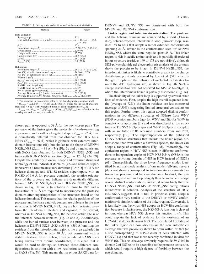
chosen pair as opposed to 38 Å for the next closest pair). Thepresence of the linker gives the molecule a beads-on-a-stringappearance and a rather elongated shape (Zmax � 97 Å) thatis dramatically different from that observed for HCV NS3-NS4A (Zmax � 66 Å), which is globular, with extensive inter-domain interactions (61), but similar to the shape of DENV4NS2B18NS3 (Zmax � 96 Å) (34) (Fig. 3c and d) and consistentwith SAXS data obtained for both DENV4 NS2B18NS3 andfull-length KUNV NS3 in solution (Dmax � 120 Å) (34, 39).Despite the similarity in overall shape and extensive structuralhomology of the individual domains (422/618 residues super-impose with a root mean square deviation [RMSD] of 1.5 Å forhelicase domains, and 151/152 residues superimpose with anRMSD of 1.6 Å for protease domains), the relative orienta-tions of the protease and helicase are dramatically differentbetween MVEV NS2B45NS3 and DENV4 NS2B18NS3, asshown in Fig. 3b and c (a rotation of close to 180° and atranslation of 17 Å are required to superimpose the proteasedomains after superimposition of the structures based on thehelicase domains). This means that the relative positions of theprotease and helicase catalytic centers are different in the twostructures: in MVEV NS2B45NS3, they are both oriented awayfrom the interdomain interface and exposed to the solvent,whereas in DENV4 NS2B18NS3, the helicase active site is atthe interface between domains (Fig. 3c and d). Additionally,while the buried surface area between the protease and thehelicase in DENV4 NS2B18NS3 is 568 Å2 (380 Å2 omitting theresidues from the interdomain region), the area excluded inMVEV NS2B45NS3 is only 30 Å2, not consistent with astable interface. Nevertheless, from simulated SAXS scat-tering curves from atomic coordinates, it is clear that itwould be hard to distinguish between these different con-figurations in solution with a low-resolution technique suchas SAXS (Fig. 3b). This means that previous SAXS data for
DENV4 and KUNV NS3 are consistent with both theMVEV and DENV4 conformations.
Linker region and interdomain orientation. The proteaseand the helicase domains are connected by a short (13-resi-due), solvent-exposed, interdomain loop (“linker” loop, resi-dues 169 to 181) that adopts a rather extended conformationspanning 24 Å, similar to the conformation seen for DENV4NS2B18NS3, where the same peptide spans 25 Å. This linkerregion is rich in acidic amino acids and is partially disorderedin our structure (residues 169 to 175 are not visible), althoughSDS-polyacrylamide gel electrophoresis analysis of the crystalsshows the protein to be intact. In DENV4 NS2B18NS3, theinterdomain linker is likely to contribute greatly to the chargedistribution previously observed by Luo et al. (34), which isthought to optimize the diffusion of nucleotide substrates to-ward the ATP hydrolysis site, as shown in Fig. 4b. Such acharge distribution was not observed for MVEV NS2B45NS3,where the interdomain linker is partially disordered (Fig. 4a).
The flexibility of the linker loop is supported by several otherlines of evidence. First, despite the high overall sequence iden-tity (average of 72%), the linker residues are less conserved(average of 50%), suggesting limited structural constraints onthis region. Furthermore, this region adopted distinct confor-mations in two different structures of NS3pro from WNV(PDB accession numbers 2ggv for WNV and 2ijo for WNV incomplex with aprotinin [2]) and was disordered in the struc-tures of DENV2 NS3pro apo and WNV NS3pro in complexwith an inhibitor (PDB accession numbers 2fom and 2fp7,respectively [19]). The superimposition of the publishedDENV helicase structures that include the linker region fur-ther shows that even within a flavivirus species, the linker canadopt a range of conformations (Fig. 4d). Interestingly, theequivalent region in HCV NS3 is seen in different conforma-tions in independent copies of HCV NS3-NS4A (NS4A is theprotease activating domain of NS3 in HCV instead of NS2B)(61). Unsurprisingly, the three lowest-frequency modes iden-tified by normal-mode analysis of our model (elNemo server)(data not shown) correspond to interdomain movements be-tween the protease and helicase domains. In short, the evi-dence suggests that this loop is highly flexible and able to adoptseveral distinct conformations; indeed, it seems likely that theDENV4 NS2B18NS3 and MVEV NS2B45NS3 configurationsinterconvert in solution. Analysis of the structure of HCVNS3-NS4A suggests that it too, in principle, could adopt aconformation very similar to the DENV4 or MVEV confor-mations via simple rotations of the linker region. Conversely, itis less likely that flavivirus NS3 adopts an HCV-like conforma-tion because in flaviviruses, the NS3-NS4A junction is cleavedin trans, whereas HCV NS3 cleaves this junction in cis. Thiscould explain the lack of evidence for the existence of anHCV-like state for flavivirus NS3. The postulated flexibility ofthe linker region can now also explain the slow autolytic ciscleavage that was previously shown to occur within NS3hel (ata site corresponding to R459-G460) in cells infected withDENV2 (3) and that was more recently confirmed in vitro forWNV (6). This cis cleavage obviously requires R459-G460 indomain 2 of NS3hel to be accessible to the protease active site,which would require a high degree of flexibility between thetwo domains.
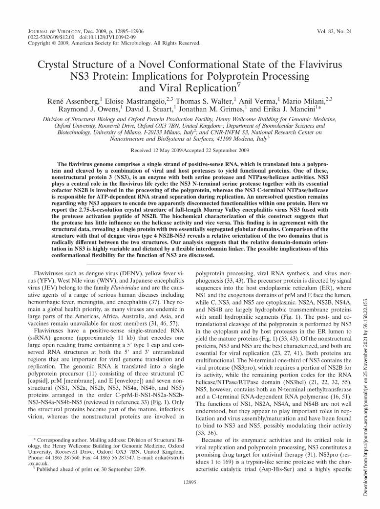
TABLE 1. X-ray data collection and refinement statistics
Statistic Valuea
Data collectionSpace group ........................................................................P21Unit cell dimensions a, b, c (Å).......................................a � 41.8, b � 105.2,
c � 79.9� (°)......................................................................................97.4Resolution range (Å) ........................................................39.60–2.75 (2.82–2.75)Observations .......................................................................129,498Unique reflections..............................................................17,578Completeness (%) .............................................................98.6 (98.2)I/ (I)...................................................................................19.2 (3.2)Rmerge
b (%).........................................................................8.7 (72.8)
RefinementResolution range (Å) ........................................................30.0–2.75 (2.82–2.75)No. (%) of reflections in working set .............................16,866 (1,226)No. (%) of reflections in test set .....................................884 (66)Wilson B (Å2).....................................................................6.2Rfactor
c working set (%) ....................................................26.8 (32.4)Rfree
c (%) ............................................................................30.0 (31.1)RMSD bond length (Å)....................................................0.006RMSD bond angle (°) .......................................................0.899No. of atoms (protein/water)............................................601 (91)Average B factors (Å2) (main chain/water) ...................51.3 (36.7)Average B factors (Å2) NS3hel, NS3pro, NS2B............52.2, 51.1, 50.0
a The numbers in parentheses refer to the last (highest) resolution shell.b Rmerge � �h�i�Ii(h) � �I(h) �/�h�i�Ii(h) , where Ii(h) is the ith measure-
ment and �I(h) is the weighted mean of all measurements of Ii(h).c Rfactor and Rfree � �h�F(h)obs� � �F(h)calc�/�h�F(h)obs� for reflection in the
working set and test set, respectively.
12900 ASSENBERG ET AL. J. VIROL.
Dow
nloa
ded
from
http
s://j
ourn
als.
asm
.org
/jour
nal/j
vi o
n 25
Nov
embe
r 20
21 b
y 59
.158
.22.
155.

FIG. 3. Overall structure of MVEV NS2B45NS. (a) Diagram of the MVEV NS2B and NS3 protein organization and of the NS2B45NS3synthetic construct used for crystallization. (b) Superimposition of the scattering from an ab initio model of MVEV NS2B45NS3 and DENV4NS2B18NS3 computed by use of the program CRYSOL (50). As a result of within-experiment errors, the two curves are indistinguishable. Rg isthe radius of gyration. (c) Comparison of the structures of MVEV (MVE) NS2B45NS3, DENV4 NS2B18NS3, and HCVNS314NS4A. A cartoondiagram (main) and surface representation (inset) of equivalent views of the three synthetic constructs are shown. Color coding is the same as thatin panel a: NS2B (and NS4A) is blue, the NS3pro domain is red, NS3hel is yellow, the NS3pro catalytic pocket is green, the NS3hel catalytic pocketis cyan, and the interdomain linker loop is magenta. (d) Orthogonal view of panel c.
12901
Dow
nloa
ded
from
http
s://j
ourn
als.
asm
.org
/jour
nal/j
vi o
n 25
Nov
embe
r 20
21 b
y 59
.158
.22.
155.

The protease domain. The structure of the protease domainof MVEV NS2B45NS3 is very similar to that of the proteasedomain of WNV (NS2B40NS3pro) in both the inactive (apo)and active (inhibitor bound) conformations, with RMSDs of1.6 Å for 153 atoms and 1.3 Å for 146 atoms, respectively (Fig.
5) (19). This, combined with the protease kinetics reportedabove, suggests that MVEV NS2B45NS3 is a fully active pro-tease, with its activity and structure comparable to that of theisolated WNV NS2B40NS3pro.
The NS3 N-terminal protease has been solved in the pres-
FIG. 4. Structural analysis of protease-helicase interdomain interactions. (a) Electrostatic surface view of MVEV NS2B45NS3 and ribbondiagram of the protease (magenta)-helicase (yellow) interdomain region, with residues at the interacting interface represented as sticks (inset).Residues involved in hydrogen bonds and salt bridges are shown in boldface type, and interatomic distances are indicated. (b) DENV4 NS2B18NS3.(c) Structural superimposition of MVEV NS2B45NS3 (this study) (yellow), DENV4 NS2B18NS3 (PDB accession number 2vbc) (magenta), andDENV4NS3helADPssRNA (PDB accession number 2jlz) (green). In DENV4, protease domain residue S68 contacts the catalytically relevantN329 in the helicase domain (which is blocked in a catalytically unfavorable conformation). E66 of the protease domain in particular is likely toblock access to the ATP base, as its side chain sits directly in front of the binding pocket and appears to orientate R418 and K201 via long-rangeelectrostatic interactions. In addition, D175 and E177 of the linker region contact R202 of the P loop, and D175 further contacts G80 of theprotease domain. Indeed, superimposition of helicase domain 1 of DENV4 NS2B-NS3 and the ADP and ssRNA-bound helicase domain ofDENV4 (PDB accession number 1jlv [34]), which represents the product-bound form, shows that domain 2 movement would cause a significantclash with the protease domain. (d) Analysis of the linker orientations in different flavivirus NS3 crystal structures. Shown is a ribbon diagram andtransparent surface representation of the structure of MVEV NS2B45NS3. Shown is the superimposition of the interdomain linker region(residues 169 to 181) of MVEV NS2B45NS3 (magenta) (this study) with those of the MVEV NS3hel structure (cyan) (PDB accession number1v80), DENV4 NS2B18NS3 (green) (PDB accession number 2vbc), and a number of DENV4 NS3hel structures solved in the presence andabsence of cofactors (DENV4 NS3hel [blue] [PDB accession number 2jlq], DENV4 NS3helADP [yellow] [PDB accession number 2jls], andDENV4NS3helADPssRNA [red] [PDB accession number 2jlz]).
FIG. 5. Structural comparison of cofactor-bound Flaviviridae NS3 protease domains. (a) Cartoon representation of MVEV NS2B45NS3pro(this study). (b) WNV NS2B45NS3pro (PDB accession number 2ggv). (c) WNV NS2B45NS3pro bound to Naph-KKR-H (PDB accession number3e90). (d) HCV NS3pro18NS4A (PDB accession number 1cu1). Blue, NS2B and NS4A cofactors; red, NS3pro; green, stick representation of theNS3pro catalytic triad (His-Asp-Ser); yellow, hydrophobic residues in its membrane-inserted region. Putative transmembrane helices of thecofactors NS2B and NS4A are shown as cylinders, together with their distance to the nearest visualized residue in the atomic structure.
12902 ASSENBERG ET AL. J. VIROL.
Dow
nloa
ded
from
http
s://j
ourn
als.
asm
.org
/jour
nal/j
vi o
n 25
Nov
embe
r 20
21 b
y 59
.158
.22.
155.

ence and absence of the cofactor domain of NS2B for DENVand WNV (2, 19, 47). The protease has a trypsin-like fold,containing two �-barrels with the Asp-His-Ser catalytic triadlocated in a cleft situated between the two barrels. In theabsence of the NS2B cofactor, the protease is virtually inactiveand less stable. Stabilization by NS2B is achieved through theinsertion of a �-strand of NS2B into the N-terminal �-barrel ofNS3pro, with minor additional interactions with the C-terminal�-barrel. As observed for the DENV NS2B-NS3pro structure,in the absence of a substrate, the MVEV NS2B region beyondresidue 69 is disordered. Upon the addition of a substrate, theC-terminal part of NS2B undergoes a dramatic conformationalchange, wrapping around the C-terminal barrel and forming astabilizing � hairpin that inserts into the active site (2, 19, 47).
These features of the flavivirus NS3 protease are similar tothose observed for the more distantly related HCV NS3 pro-tease (Fig. 5). The overall fold of the HCV protease is similarto that of the flavivirus NS3 proteases (Fig. 5). Like flavivi-ruses, HCV requires a cofactor for activity, but this is found inNS4A rather than NS2B. Crystallographic and nuclear mag-netic resonance analyses showed a more subtle involvement ofNS4A: the binding of NS4A induces stabilizing conformationalchanges in the NS3 protease without actually engaging with theactive site (42, 61). Moreover, the activating region withinNS4A was found to be much shorter (comprising a single �sheet) (Fig. 5).
Based on the recently reported crystal structure of WNVNS2B-NS3pro bound to the inhibitor Naph-KKR-H (47) andthe predicted membrane domains of NS2B, the distances ofthe termini of the NS2B activating region to the ER membraneare rather short, 3 residues from the cofactor N terminus and1 residue from the C terminus, placing the activated NS3protease domain in a very tight membrane-anchored NS2Bsling (Fig. 5b and c). As recently suggested (9), the NS3 mem-brane association is likely to be further stabilized by the mem-brane insertion of a � hairpin with conserved hydrophobicresidues (MVEV NS3pro residues 27 to 34), analogous to theproposed membrane inserted amphipathic helix �0 in HCVNS3 (Fig. 5).
The NTPase/helicase domain. The flavivirus NS3 C-terminalNTPase/helicase has been solved for YFV (58), DENV (35,59), MVEV (38), KUNV (39), JEV (60), and Kokobera virus(49). It is a DEAH/D box helicase belonging to the SF2 su-perfamily of helicases containing the characteristic Walker Aand B motifs responsible for NTP and Mg2 binding andconsisting of three domains of approximately equal size,forming a large positively charged cleft at their juncture,which is the RNA binding site. The structure is closelyrelated to HCV NS3 helicase with respect to domains 1 and2, but domain 3 shows a unique conformation typical offlaviviruses. Differences between the YFV, DENV, KUNV,Kokobera virus, JEV, and MVEV structures were found to beconfined primarily to the orientation of domain 2 relative todomains 1 and 3, suggesting that the movement of domain 2affects nucleic acid translocation in an ATP-dependent manneraccording to the inchworm model, which was recently con-firmed by a detailed study of the catalytic cycle of the ATPaseactivity of a DENV4 helicase (35).
In the DENV4 NS2B-NS3 structure (34), the protease andlinker regions directly contact the helicase NTP binding
pocket, forming a network of hydrogen bonds and salt bridges(Fig. 4b). These interactions hold the NS3hel catalytic loop (Ploop, or Walker A motif), critical for driving the conforma-tional changes that occur upon NTP hydrolysis, in a confor-mation very similar to that of the apo form observed previouslyfor the DENV2 helicase domain structure (35), limiting accessto the nucleotide binding pocket (Fig. 4c). Another strikingfeature of the DENV4 conformation is that the loop betweenR461 and D471 (in domain 2) is moved outward compared tothose of MVEV NS2B-NS3 and all other published helicasedomain structures, where the loop is structurally much moreconserved (Fig. 4c). The conformation of this loop in DENV4NS2B18NS3 is likely to restrict the conformational freedom ofhelicase domain 2 and may therefore adversely affect NS3helicase activity. In contrast, for the MVEV structure, theseinteractions are not present, leading to a more exposed NTPbinding site and a lack of constraints on the R461-D471 loop(Fig. 4a).
Possible biological significance of the MVEV and DENV4NS2B-NS3 conformations. Our study has revealed a novel con-formational state of a flavivirus NS3 protein where the pro-tease and NTPase/helicase domains are spatially separated. Inline with this, biochemical analyses of the helicase, ATPase,and protease activities suggest that the domains do not exerta significant effect on each other’s activities. The individualMVEV helicase and protease domains are structurally similarto other flavivirus NS3 helicase and protease domains and tothose of DENV4 NS2B-NS3. The crucial difference betweenthe DENV4 and MVEV NS2B-NS3 structures is a largechange in the relative orientations of the two domains. It seemslikely that the flexible nature of the linker region and the smallprotease/helicase interfaces in both configurations allow theirinterconversion in solution. In other words, our analysisstrongly suggests that the flavivirus NS3 protease and helicasedomains are loosely tethered.
This does not resolve the question of what is the functionalrelevance, if any, of the yoking of two uncoupled activitieswithin a single polypeptide chain. NS3 plays a major role inpolyprotein processing, genome replication, and viral packing(44). Polyprotein processing and replication are thought tooccur in adjacent but distinct compartments: convoluted mem-branes and vesicle packets, respectively (for a review, see ref-erence 36). In both these processes, it is likely that the activitiesare performed in the vicinity of membranes. As outlined above,NS3 and NS2B associate with membranes yielding a ratherrigidly membrane-bound NS3pro domain, especially in thefully activated state of NS3pro (Fig. 5). Figure 6 shows acartoon representation of the MVEV and DENV4 NS2B-NS3crystal structures with their protease domains superimposedand bound to the membrane, where the membrane positioningis based on the anchoring illustrated in Fig. 5c. This modelfor a possible structural organization of the NS2B-NS3 mol-ecule at the cellular membrane suggests that the DENV4NS2B18NS3 helicase is positioned very near the membrane,with the active site oriented toward the membrane and withvery little space to accommodate RNA, while in the MVEVstructure, the helicase domain is positioned away from themembrane, with the active site facing the cytoplasm. In bothcases the protease active site is freely accessible. This mightargue that the DENV4 conformation is not physiologically
VOL. 83, 2009 STRUCTURE OF THE OF NS3 PROTEASE-HELICASE FROM MVEV 12903
Dow
nloa
ded
from
http
s://j
ourn
als.
asm
.org
/jour
nal/j
vi o
n 25
Nov
embe
r 20
21 b
y 59
.158
.22.
155.

relevant (because the conformational flexibility of the NS3linker may prevent its adoption in vivo); however, it is impor-tant that NS3 is also membrane anchored at its C terminus viaits coupling to the membrane-bound NS4A (prior to the cleav-age of the NS3-NS4A junction). This implies that additional invivo constraints that probably restrict the conformational free-
dom of the NS3hel domain are present. Since the conforma-tion of NS4A is not known, we cannot exclude the possibilitythat the DENV4 conformation may occur in vivo. Indeed, NS3might adopt a DENV4-like conformation during polyproteinprocessing, where the activity of the C-terminal helicase is notneeded. Indirect support for this possibility comes from the
FIG. 6. Model for the structural organization of MVEV NS2B45NS3 and DENV4 NS2B18NS3 at the cellular membrane. Positioning on themembrane is based upon the tight anchoring of NS2B-NS3 at three points, at the NS2B N- and C-terminal transmembrane helices and at theNS3pro hydrophobic helix, as predicted by the structure of WNV NS2B45NS3pro-Naph-KKR-H and as shown in Fig. 5c. After positioning of WNVNS2B45NS3pro-Naph-KKR-H on the membrane, MVEV NS2B45NS3 and DENV4 NS2B18NS3 were superimposed based on the protease domainalone. A model for the membrane is shown as van der Waals balls and, atomic structures are shown in a cartoon representation and color codedaccording to the following convention: NS2B (blue), NS3pro (red), and NS3hel by subdomains (domain 1, magenta; domain 2, white; domain 3,yellow). NS4A (shown schematically in orange) was positioned at the NS3 C terminus (domain 3), and the RNA (shown schematically in pink) ispositioned in the ssRNA binding groove (shown to be bound in DENV4NS3helADPssRNA [PDB accession number 2jlz]). The NS3pro (green)and NS3hel catalytic pockets are shown in a ball representation. The position of hinge axis is shown. aa, amino acids.
12904 ASSENBERG ET AL. J. VIROL.
Dow
nloa
ded
from
http
s://j
ourn
als.
asm
.org
/jour
nal/j
vi o
n 25
Nov
embe
r 20
21 b
y 59
.158
.22.
155.
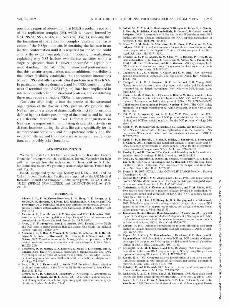
previously reported observation that NS2B is probably not partof the replication complex (36), which is instead formed byNS1, NS2A, NS3, NS4A, and NS5 (36) (Fig. 1), implying thatthe formation of the replication complex results in the inacti-vation of the NS3pro domain. Maintaining the helicase in aninactive conformation until it is required for replication couldcontrol the switch from polyprotein processing to replication,explaining why NS3 harbors two distinct activities within asingle polypeptide chain. However, the significant gaps in ourunderstanding of the role of the nonstructural proteins meansthat this remains a speculation, and an additional possibility isthat linker flexibility establishes the appropriate interactionsbetween NS3 and other nonstructural proteins as well as RNA.In particular, helicase domains 2 and 3 of NS3, constituting themost-C-terminal part of NS3 (Fig. 4c), have been implicated ininteractions with other nonstructural proteins, and establishingthese may require a flexible NS3hel arrangement.
Our data offer insights into the puzzle of the structuralorganization of the flavivirus NS3 protein. We propose thatNS3 can assume a range of dramatically distinct conformationsdefined by the relative positioning of the protease and helicasevia a flexible interdomain linker. Different configurations inNS3 may be important for modulating its catalytic activities indistinct locations during the virus life cycle, specifically for itsmembrane-anchored cis- and trans-protease activity and theswitch to helicase and triphosphatase activities during replica-tion, and possibly other functions.
ACKNOWLEDGMENTS
We thank the staff at ID29 European Synchrotron Radiation FacilityGrenoble for support with data collection, Joanne Nettleship for helpwith the mass spectrometry analysis, and R. Hurrelbrink and S. Fullerfor useful discussions. We gratefully acknowledge Roman Tuma’s helpwith Fig. 3b.
E.J.M. is supported by the Royal Society, and D.I.S., J.M.G., and theOxford Protein Production Facility are supported by the UK MedicalResearch Council and European Commission grants LSHGCT-2006-031220 (SPINE2 COMPLEXES) and LSHG-CT-2004-511960 (VI-ZIER).
REFERENCES
1. Adams, P. D., R. W. Grosse-Kunstleve, L. W. Hung, T. R. Ioerger, A. J.McCoy, N. W. Moriarty, R. J. Read, J. C. Sacchettini, N. K. Sauter, and T. C.Terwilliger. 2002. PHENIX: building new software for automated crystallo-graphic structure determination. Acta Crystallogr. D Biol. Crystallogr. 58:1948–1954.
2. Aleshin, A. E., S. A. Shiryaev, A. Y. Strongin, and R. C. Liddington. 2007.Structural evidence for regulation and specificity of flaviviral proteases andevolution of the Flaviviridae fold. Protein Sci. 16:795–806.
3. Arias, C. F., F. Preugschat, and J. H. Strauss. 1993. Dengue 2 virus NS2Band NS3 form a stable complex that can cleave NS3 within the helicasedomain. Virology 193:888–899.
4. Assenberg, R., J. Ren, A. Verma, T. S. Walter, D. Alderton, R. J. Hurrel-brink, S. D. Fuller, S. Bressanelli, R. J. Owens, D. I. Stuart, and J. M.Grimes. 2007. Crystal structure of the Murray Valley encephalitis virus NS5methyltransferase domain in complex with cap analogues. J. Gen. Virol.88:2228–2236.
5. Benarroch, D., B. Selisko, G. A. Locatelli, G. Maga, J. L. Romette, and B.Canard. 2004. The RNA helicase, nucleotide 5�-triphosphatase, and RNA5�-triphosphatase activities of dengue virus protein NS3 are Mg2-depen-dent and require a functional Walker B motif in the helicase catalytic core.Virology 328:208–218.
6. Bera, A. K., R. J. Kuhn, and J. L. Smith. 2007. Functional characterizationof cis and trans activity of the flavivirus NS2B-NS3 protease. J. Biol. Chem.282:12883–12892.
7. Berrow, N. S., D. Alderton, S. Sainsbury, J. Nettleship, R. Assenberg, N.Rahman, D. I. Stuart, and R. J. Owens. 2007. A versatile ligation-indepen-dent cloning method suitable for high-throughput expression screening ap-plications. Nucleic Acids Res. 35:e45.
8. Bollati, M., M. Milani, E. Mastrangelo, S. Ricagno, G. Tedeschi, S. Nonnis,E. Decroly, B. Selisko, X. de Lamballerie, B. Coutard, B. Canard, and M.Bolognesi. 2009. Recognition of RNA cap in the Wesselsbron virus NS5methyltransferase domain: implications for RNA-capping mechanisms inflavivirus. J. Mol. Biol. 385:140–152.
9. Brass, V., J. M. Berke, R. Montserret, H. E. Blum, F. Penin, and D. Mo-radpour. 2008. Structural determinants for membrane association and dy-namic organization of the hepatitis C virus NS3-4A complex. Proc. Natl.Acad. Sci. USA 105:14545–14550.
10. Brunger, A. T., P. D. Adams, G. M. Clore, W. L. DeLano, P. Gros, R. W.Grosse-Kunstleve, J. S. Jiang, J. Kuszewski, M. Nilges, N. S. Pannu, R. J.Read, L. M. Rice, T. Simonson, and G. L. Warren. 1998. Crystallography &NMR system: a new software suite for macromolecular structure determi-nation. Acta Crystallogr. D 54:905–921.
11. Chambers, T. J., C. S. Hahn, R. Galler, and C. M. Rice. 1990. Flavivirusgenome organization, expression, and replication. Annu. Rev. Microbiol.44:649–688.
12. Chappell, K. J., M. J. Stoermer, D. P. Fairlie, and P. R. Young. 2007.Generation and characterization of proteolytically active and highly stabletruncated and full-length recombinant West Nile virus NS3. Protein Expr.Purif. 53:87–96.
13. Chen, C. J., M. D. Kuo, L. J. Chien, S. L. Hsu, Y. M. Wang, and J. H. Lin.1997. RNA-protein interactions: involvement of NS3, NS5, and 3� noncodingregions of Japanese encephalitis virus genomic RNA. J. Virol. 71:3466–3473.
14. Collaborative Computational Project, Number 4. 1994. The CCP4 suite:programs for protein crystallography. Acta Crystallogr. D Biol. Crystallogr.50:760–763.
15. Cui, T., R. J. Sugrue, Q. Xu, A. K. Lee, Y. C. Chan, and J. Fu. 1998.Recombinant dengue virus type 1 NS3 protein exhibits specific viral RNAbinding and NTPase activity regulated by the NS5 protein. Virology 246:409–417.
16. Egloff, M. P., D. Benarroch, B. Selisko, J. L. Romette, and B. Canard. 2002.An RNA cap (nucleoside-2�-O-)-methyltransferase in the flavivirus RNApolymerase NS5: crystal structure and functional characterization. EMBO J.21:2757–2768.
17. Egloff, M. P., E. Decroly, H. Malet, B. Selisko, D. Benarroch, F. Ferron, andB. Canard. 2007. Structural and functional analysis of methylation and 5�-RNA sequence requirements of short capped RNAs by the methyltrans-ferase domain of dengue virus NS5. J. Mol. Biol. 372:723–736.
18. Emsley, P., and K. Cowtan. 2004. Coot: model-building tools for moleculargraphics. Acta Crystallogr. D Biol. Crystallogr. 60:2126–2132.
19. Erbel, P., N. Schiering, A. D’Arcy, M. Renatus, M. Kroemer, S. P. Lim, Z.Yin, T. H. Keller, S. G. Vasudevan, and U. Hommel. 2006. Structural basisfor the activation of flaviviral NS3 proteases from dengue and West Nilevirus. Nat. Struct. Mol. Biol. 13:372–373.
20. Evans, P. R. 1997. SCALA. Joint CCP4ESF-EAMCB Newslett. ProteinCrystallogr. 1997:33.
21. Falgout, B., M. Pethel, Y. M. Zhang, and C. J. Lai. 1991. Both nonstructuralproteins NS2B and NS3 are required for the proteolytic processing of denguevirus nonstructural proteins. J. Virol. 65:2467–2475.
22. Gorbalenya, A. E., E. V. Koonin, A. P. Donchenko, and V. M. Blinov. 1989.Two related superfamilies of putative helicases involved in replication, re-combination, repair and expression of DNA and RNA genomes. NucleicAcids Res. 17:4713–4730.
23. Hanley, K. A., J. J. Lee, J. E. Blaney, Jr., B. R. Murphy, and S. S. Whitehead.2002. Paired charge-to-alanine mutagenesis of dengue virus type 4 NS5generates mutants with temperature-sensitive, host range, and mouse atten-uation phenotypes. J. Virol. 76:525–531.
24. Johansson, M., A. J. Brooks, D. A. Jans, and S. G. Vasudevan. 2001. A smallregion of the dengue virus-encoded RNA-dependent RNA polymerase, NS5,confers interaction with both the nuclear transport receptor importin-betaand the viral helicase, NS3. J. Gen. Virol. 82:735–745.
25. Kabsch, W. 1993. Automatic processing of rotation diffraction data fromcrystals of initially unknown symmetry and cell constants. J. Appl. Crystal-logr. 26:795–800.
26. Kapoor, M., L. Zhang, M. Ramachandra, J. Kusukawa, K. E. Ebner, and R.Padmanabhan. 1995. Association between NS3 and NS5 proteins of denguevirus type 2 in the putative RNA replicase is linked to differential phosphor-ylation of NS5. J. Biol. Chem. 270:19100–19106.
27. Khromykh, A. A., M. T. Kenney, and E. G. Westaway. 1998. trans-Comple-mentation of flavivirus RNA polymerase gene NS5 by using Kunjin virusreplicon-expressing BHK cells. J. Virol. 72:7270–7279.
28. Koonin, E. V. 1993. Computer-assisted identification of a putative methyl-transferase domain in NS5 protein of flaviviruses and lambda 2 protein ofreovirus. J. Gen. Virol. 74(Pt. 4):733–740.
29. Krissinel, E., and K. Henrick. 2007. Inference of macromolecular assembliesfrom crystalline state. J. Mol. Biol. 372:774–797.
30. Laskowski, R. A., D. S. Moss, and J. M. Thornton. 1993. Main-chain bondlengths and bond angles in protein structures. J. Mol. Biol. 231:1049–1067.
31. Lescar, J., D. Luo, T. Xu, A. Sampath, S. P. Lim, B. Canard, and S. G.Vasudevan. 2008. Towards the design of antiviral inhibitors against flavivi-
VOL. 83, 2009 STRUCTURE OF THE OF NS3 PROTEASE-HELICASE FROM MVEV 12905
Dow
nloa
ded
from
http
s://j
ourn
als.
asm
.org
/jour
nal/j
vi o
n 25
Nov
embe
r 20
21 b
y 59
.158
.22.
155.

ruses: the case for the multifunctional NS3 protein from dengue virus as atarget. Antivir. Res. 80:94–101.
32. Li, H., S. Clum, S. You, K. E. Ebner, and R. Padmanabhan. 1999. The serineprotease and RNA-stimulated nucleoside triphosphatase and RNA helicasefunctional domains of dengue virus type 2 NS3 converge within a region of20 amino acids. J. Virol. 73:3108–3116.
33. Lindenbach, B., H. Thiel, and C. Rice. 2007. Flaviviridae: the viruses andtheir replication, p. 1101–1152. In D. M. Knipe, P. M. Howley, D. E. Griffin,R. A. Lamb, M. A. Martin, B. Roizman, and S. E. Straus (ed.), Fieldsvirology, 5th ed. Lippincott Williams & Wilkins, Philadelphia, PA.
34. Luo, D., T. Xu, C. Hunke, G. Gruber, S. G. Vasudevan, and J. Lescar. 2008.Crystal structure of the NS3 protease-helicase from dengue virus. J. Virol.82:173–183.
35. Luo, D., T. Xu, R. P. Watson, D. Scherer-Becker, A. Sampath, W. Jahnke,S. S. Yeong, C. H. Wang, S. P. Lim, A. Strongin, S. G. Vasudevan, and J.Lescar. 2008. Insights into RNA unwinding and ATP hydrolysis by theflavivirus NS3 protein. EMBO J. 27:3209–3219.
36. Mackenzie, J. 2005. Wrapping things up about virus RNA replication. Traffic6:967–977.
37. Mackenzie, J. S., D. J. Gubler, and L. R. Petersen. 2004. Emerging flavivi-ruses: the spread and resurgence of Japanese encephalitis, West Nile anddengue viruses. Nat. Med. 10:S98–S109.
38. Mancini, E. J., R. Assenberg, A. Verma, T. S. Walter, R. Tuma, J. M. Grimes,R. J. Owens, and D. I. Stuart. 2007. Structure of the Murray Valley enceph-alitis virus RNA helicase at 1.9 Angstrom resolution. Protein Sci. 16:2294–2300.
39. Mastrangelo, E., M. Milani, M. Bollati, B. Selisko, F. Peyrane, V. Pandini,G. Sorrentino, B. Canard, P. V. Konarev, D. I. Svergun, X. de Lamballerie,B. Coutard, A. A. Khromykh, and M. Bolognesi. 2007. Crystal structure andactivity of Kunjin virus NS3 helicase; protease and helicase domain assemblyin the full length NS3 protein. J. Mol. Biol. 372:444–455.
40. Matusan, A. E., P. G. Kelley, M. J. Pryor, J. C. Whisstock, A. D. Davidson,and P. J. Wright. 2001. Mutagenesis of the dengue virus type 2 NS3 pro-teinase and the production of growth-restricted virus. J. Gen. Virol. 82:1647–1656.
41. Matusan, A. E., M. J. Pryor, A. D. Davidson, and P. J. Wright. 2001.Mutagenesis of the dengue virus type 2 NS3 protein within and outsidehelicase motifs: effects on enzyme activity and virus replication. J. Virol.75:9633–9643.
42. McCoy, M. A., M. M. Senior, J. J. Gesell, L. Ramanathan, and D. F. Wyss.2001. Solution structure and dynamics of the single-chain hepatitis C virusNS3 protease NS4A cofactor complex. J. Mol. Biol. 305:1099–1110.
43. Murray, C. L., C. T. Jones, and C. M. Rice. 2008. Architects of assembly:roles of Flaviviridae non-structural proteins in virion morphogenesis. Nat.Rev. Microbiol. 6:699–708.
44. Patkar, C. G., and R. J. Kuhn. 2008. Yellow fever virus NS3 plays anessential role in virus assembly independent of its known enzymatic func-tions. J. Virol. 82:3342–3352.
45. Pyle, A. M. 2008. Translocation and unwinding mechanisms of RNA andDNA helicases. Annu. Rev. Biophys. 37:317–336.
46. Ray, D., and P. Y. Shi. 2006. Recent advances in flavivirus antiviral drugdiscovery and vaccine development. Recent Pat. Anti-Infect. Drug Discov.1:45–55.
47. Robin, G., K. Chappell, M. J. Stoermer, S. H. Hu, P. R. Young, D. P. Fairlie,and J. L. Martin. 2009. Structure of West Nile virus NS3 protease: ligandstabilization of the catalytic conformation. J. Mol. Biol. 385:1568–1577.
48. Shiryaev, S. A., B. I. Ratnikov, A. V. Chekanov, S. Sikora, D. V. Rozanov, A.
Godzik, J. Wang, J. W. Smith, Z. Huang, I. Lindberg, M. A. Samuel, M. S.Diamond, and A. Y. Strongin. 2006. Cleavage targets and the D-arginine-based inhibitors of the West Nile virus NS3 processing proteinase. Biochem.J. 393:503–511.
49. Speroni, S., L. De Colibus, E. Mastrangelo, E. Gould, B. Coutard, N. L.Forrester, S. Blanc, B. Canard, and A. Mattevi. 2008. Structure and bio-chemical analysis of Kokobera virus helicase. Proteins 70:1120–1123.
50. Stuart, D. I., M. Levine, H. Muirhead, and D. K. Stammers. 1979. Crystalstructure of cat muscle pyruvate kinase at a resolution of 2.6 A. J. Mol. Biol.134:109–142.
51. Tan, B. H., J. Fu, R. J. Sugrue, E. H. Yap, Y. C. Chan, and Y. H. Tan. 1996.Recombinant dengue type 1 virus NS5 protein expressed in Escherichia coliexhibits RNA-dependent RNA polymerase activity. Virology 216:317–325.
52. Thibeault, D., M. J. Massariol, S. Zhao, E. Welchner, N. Goudreau, R.Gingras, M. Llinas-Brunet, and P. W. White. 2009. Use of the fused NS4Apeptide-NS3 protease domain to study the importance of the helicase do-main for protease inhibitor binding to hepatitis C virus NS3-NS4A. Bio-chemistry 48:744–753.
53. Walter, T. S., J. M. Diprose, C. J. Mayo, C. Siebold, M. G. Pickford, L.Carter, G. C. Sutton, N. S. Berrow, J. Brown, I. M. Berry, G. B. Stewart-Jones, J. M. Grimes, D. K. Stammers, R. M. Esnouf, E. Y. Jones, R. J.Owens, D. I. Stuart, and K. Harlos. 2005. A procedure for setting uphigh-throughput nanolitre crystallization experiments. Crystallization work-flow for initial screening, automated storage, imaging and optimization. ActaCrystallogr. D Biol. Crystallogr. 61:651–657.
54. Walter, T. S., E. J. Mancini, J. Kadlec, S. C. Graham, R. Assenberg, J. Ren,S. Sainsbury, R. J. Owens, D. I. Stuart, J. M. Grimes, and K. Harlos. 2008.Semi-automated microseeding of nanolitre crystallization experiments. ActaCrystallogr. F Struct. Biol. Cryst. Commun. 64:14–18.
55. Wengler, G. 1991. The carboxy-terminal part of the NS 3 protein of the WestNile flavivirus can be isolated as a soluble protein after proteolytic cleavageand represents an RNA-stimulated NTPase. Virology 184:707–715.
56. Wengler, G. 1993. The NS 3 nonstructural protein of flaviviruses contains anRNA triphosphatase activity. Virology 197:265–273.
57. Whitehead, S. S., J. E. Blaney, A. P. Durbin, and B. R. Murphy. 2007.Prospects for a dengue virus vaccine. Nat. Rev. Microbiol. 5:518–528.
58. Wu, J., A. K. Bera, R. J. Kuhn, and J. L. Smith. 2005. Structure of theflavivirus helicase: implications for catalytic activity, protein interactions, andproteolytic processing. J. Virol. 79:10268–10277.
59. Xu, T., A. Sampath, A. Chao, D. Wen, M. Nanao, P. Chene, S. G. Vasudevan,and J. Lescar. 2005. Structure of the dengue virus helicase/nucleosidetriphosphatase catalytic domain at a resolution of 2.4 Å. J. Virol. 79:10278–10288.
60. Yamashita, T., H. Unno, Y. Mori, H. Tani, K. Moriishi, A. Takamizawa, M.Agoh, T. Tsukihara, and Y. Matsuura. 2008. Crystal structure of the catalyticdomain of Japanese encephalitis virus NS3 helicase/nucleoside triphos-phatase at a resolution of 1.8 A. Virology 373:426–436.
61. Yao, N., P. Reichert, S. S. Taremi, W. W. Prosise, and P. C. Weber. 1999.Molecular views of viral polyprotein processing revealed by the crystal struc-ture of the hepatitis C virus bifunctional protease-helicase. Structure 7:1353–1363.
62. Yon, C., T. Teramoto, N. Mueller, J. Phelan, V. K. Ganesh, K. H. Murthy,and R. Padmanabhan. 2005. Modulation of the nucleoside triphosphatase/RNA helicase and 5�-RNA triphosphatase activities of dengue virus type 2nonstructural protein 3 (NS3) by interaction with NS5, the RNA-dependentRNA polymerase. J. Biol. Chem. 280:27412–27419.
12906 ASSENBERG ET AL. J. VIROL.
Dow
nloa
ded
from
http
s://j
ourn
als.
asm
.org
/jour
nal/j
vi o
n 25
Nov
embe
r 20
21 b
y 59
.158
.22.
155.