crystal structure of glft2 1 tetrameric structure of glft2 reveals a
TRANSCRIPT

Crystal Structure of GlfT2
1
Tetrameric structure of GlfT2 reveals a scaffold for the assembly of mycobacterial arabinogalactan
Robert W. Wheatley,1,2 Ruixiang Blake Zheng,1,3 Michele R. Richards,1,3 Todd L. Lowary*,1,3 and Kenneth K. S. Ng*,1,2
1From the Alberta Glycomics Centre
2Department of Biological Sciences University of Calgary, Calgary AB, T2N 1N4, Canada 3Department of Chemistry, Gunning-Lemieux Chemistry Centre University of Alberta,
Edmonton AB, T6G 2G2, Canada
To whom correspondence should be addressed: Kenneth K.S. Ng, Department of Biological Sciences University of Calgary, Calgary AB, T2N 1N4, Canada, Tel.: (403-220-4320; Fax: (403) 289-9311; E-mail: [email protected] and Todd L. Lowary, Department of Chemistry, Gunning-Lemieux Chemistry Centre University of Alberta, Edmonton AB, T6G 2G2, Canada; Email: [email protected]
Keywords: Glycosyltransferase; Mycobacterium tuberculosis; X-ray crystallography _____________________________________________________________________________________ Background: GlfT2 is a bifunctional galactofuranosyltransferase essential for mycobacterial cell wall biosynthesis. Results: Crystal structures of free and UDP-bound GlfT2, along with mutagenesis and kinetic studies, reveal novel details underlying substrate-binding and catalysis. Conclusion: The homotetrameric architecture, distinctive nucleotide binding site, and unprecedented structural features underlying the bifunctional polymerase activity of GlfT2 are revealed. Significance: Novel insights into polymerizing glycosyltransferases and the design of anti-mycobacterial therapeutics are obtained. Biosynthesis of the mycobacterial cell wall relies on the activities of many enzymes, including several glycosyltransferases (GTs). The polymerizing galactofuranosyltransferase GlfT2 (Rv3808c) synthesizes the bulk of the galactan portion of the mycolyl–arabinogalactan complex, which is the largest component of the mycobacterial cell wall. We used X-ray crystallography to determine the 2.45 Å resolution crystal structure of GlfT2, revealing an unprecendented multidomain structure in which an N-terminal β -barrel domain and two primarily α -helical C-terminal domains flank a
central GT-A domain. The kidney-shaped protomers assemble into a C4-symmetric homotetramer with an open central core and a surface containing exposed hydrophobic and positively charged residues likely involved with membrane binding. The structure of a 3.1 Å resolution complex of GlfT2 with UDP reveals a distinctive mode of nucleotide recognition. In addition, models for the binding of UDP-Galf and acceptor substrates in combination with site-directed mutagenesis and kinetic studies suggest a mechanism that explains the unique ability of GlfT2 to generate alternating β-(1→5) and β -(1→6) glycosidic linkages using a single active site. The topology imposed by docking a tetrameric assembly onto a membrane bilayer also provides novel insights into aspects of processivity and chain length regulation in this, and possibly other, polymerizing GTs.
Tuberculosis (TB), a disease that remains a significant worldwide health threat, is caused by infection with Mycobacterium tuberculosis. In common with all mycobacteria, M. tuberculosis possesses a unique, glycolipid-rich cell wall structure that provides substantial protection from the environment, including preventing the passage of antibiotics into the organism (1). The standard TB drug regimen (2) therefore involves multiple
http://www.jbc.org/cgi/doi/10.1074/jbc.M112.347484The latest version is at JBC Papers in Press. Published on June 15, 2012 as Manuscript M112.347484
Copyright 2012 by The American Society for Biochemistry and Molecular Biology, Inc.
by guest on February 11, 2018http://w
ww
.jbc.org/D
ownloaded from

Crystal Structure of GlfT2
2
antibiotics, including some that target cell wall biosynthesis and others that have intracellular targets (3). Concerns about drug resistance (4) have led to heightened interest in identifying novel therapeutic agents and developing a more detailed understanding of mycobacterial biochemical pathways to facilitate this task. In this regard, cell wall assembly has received particular attention (1).
The largest component of the mycobacterial cell wall is the mycolyl–arabinogalactan (mAG) complex. This glycolipid has at its core a galactan domain composed of 30–35 galactofuranose (Galf) residues attached in alternating β-(1→5) and β-(1→6) linkages, which, in turn, is covalently bound to cell wall peptidoglycan through a linker consisting of rhamnose and N-acetyl-glucosamine phosphate (1). The galactan serves as the attachment site for three arabinan domains, each containing ~30 α-(1→5), α-(1→3), and β-(1→2)-linked arabinofuranose residues. Esterified to the non-reducing termini of the arabinan are mycolic acids, branched long-chain (C60–C90) lipids that are characteristic to mycobacteria and related organisms such as nocardia and corynebacteria. Two clinically used anti-TB drugs target enzymes involved in the assembly of the mAG (1). Ethambutol inhibits at least one of the arabinosyltransferases responsible for arabinan formation (5) while isoniazid is a prodrug that targets an enoyl–acyl carrier protein reductase required for mycolic acid biosynthesis (6). Although none of the drugs in use or in development for the treatment of TB are known to target enzymes responsible for galactan biosynthesis, these enzymes are likely suitable targets, as the galactan forms the foundation upon which both the arabinan and mycolate moieties are incorporated. This proposal is further supported by gene knock-out studies demonstrating that galactan formation is essential for mycobacterial viability (7). Because a clearer, more detailed picture of galactan biosynthesis is a necessary prerequisite for identifying novel therapeutics, this area is now receiving increasing attention (8).
Previous work has revealed that mycobacterial galactan biosynthesis requires two bifunctional
GTs, GlfT1 and GlfT2, which use UDP-α-D-galactofuranose (UDP-Galf) as the donor and a growing polyprenol-bound oligosaccharide as the acceptor (9,10). The α-stereochemistry in UDP-Galf and the β-stereochemistry of the newly synthesized glycosidic linkages in the products generated by GlfT1 and GlfT2 indicate that catalysis follows an inverting mechanism (9,11). GlfT1 adds the first two Galf moieties, whereas GlfT2, a polymerizing glycosyltransferase that produces both β-(1→5) and β-(1→6) linkages between Galf residues (10-12), introduces the remaining ~30 monosaccharides (Fig. 1). GlfT2 has been more extensively studied than GlfT1. Of particular note are saturation-transfer difference (STD) NMR studies suggesting that GlfT2 synthesizes both the β-(1→5) and β-(1→6) linkages using a single active site (13), and mass spectrometry studies indicating that the enzyme is processive (14). A very recent study examining GlfT2 mutants provides additional support for the presence of a single active site that carries out both glycosylations (15).
In addition to the importance of understanding how GlfT2 works as a key enzyme in mycobacterial cell wall synthesis, GlfT2 is a member of the Carbohydrate-Active enZyme (CAZy) GT-2 family (16,17), which also contains other polymerizing GTs, including cellulose synthase (18) and chondroitin polymerase (19). GlfT2 has recently become of interest as a model for studying processivity and chain length control in a wide range of polymerizing GTs (14,20). EXPERIMENTAL PROCEDURES
Protein expression and purification For crystallographic studies, GlfT2 was produced from E. coli C41(DE3) transformed with pET-15b/Rv3808c (9). Cells stored in glycerol stocks were grown overnight on MDG-agar (21). Single colonies were added to MDG (100 mL) and grown for 36 h at 25 °C. These starter cultures were added to Overnight Express Instant TB medium (Novagen, 1 L) and grown for 24 h at 25 °C. Cells were harvested by centrifugation (5000 X
by guest on February 11, 2018http://w
ww
.jbc.org/D
ownloaded from

Crystal Structure of GlfT2
3
g, 20 min) and stored at –70 °C. All purification steps were performed at 4 °C. Thawed cells (~16 g) were suspended in 50 mL disruption buffer (50 mM NaH2PO4, pH 7.6, 400 mM NaCl, 15% (v/v) glycerol, 20 mM imidazole, 5 mM β -ME, and 0.2% (v/v) Triton X-100), disrupted by 2 passes through a Model 110 Microfluidizer (Microfluidics) and clarified by centrifugation (60 min, 100 000 X g). The supernatant was loaded onto Ni-Sepharose HP (7 mL, GE Healthcare) equilibrated with buffer A (50 mM NaH2PO4, pH 7.6, 400 mM NaCl, 15% (v/v) glycerol, 5 mM β-ME) plus 20 mM imidazole. The column was washed with 30 mL buffer A plus 50 mM imidazole, and eluted with a 90 mL gradient of buffer A plus 50-400 mM imidazole. Fractions containing GlfT2 were identified by SDS-PAGE and dialyzed overnight against 2 L buffer B (50 mM MOPS, pH 7.6, 15 % (v/v) glycerol, 5 mM β-ME). The dialysate was centrifuged (12 000 X g, 15 min) and the supernatant concentrated to ~3.5 mg/mL (Bradford Assay, Biorad) using Ultrafree-15 centrifugal filters (10 000 MWCO, Millipore). Aliquots were flash frozen in liquid nitrogen and stored at –80 °C.
Gel filtration chromatography was performed using a Sephacryl S-200 HR 16/60 column (GE Healthcare) equilibrated with buffer C (50 mM MOPS, pH 7.6, 100 mM NaCl, 10 mM MgCl2, 5 mM β -ME). GlfT2 (1 mL X 8 mg/mL) or a mixture of standard proteins including Blue Dextran 2000 was eluted at 0.5 mL/min and monitored by absorbance at 280 nm.
Crystallization Crystals were grown by hanging-drop vapor diffusion at room temperature by mixing GlfT2 (2 µL X 2.2 mg/mL, buffer B plus 0.2% (v/v) Triton X-100) with precipitant A (2 µL, 5–7% (w/v) PEG 4000, 100 mM sodium cacodylate pH 6.5, 100 mM sodium acetate). Crystals (~350 x 70 x 70 µm) grew to full size in 7–14 days. Crystals were cryoprotected by transferring to precipitant A plus 1% (w/v) PEG 4000 and successively 5, 10, 15, and 20 % (v/v) glycerol. Crystals were equilibrated at least 15 min between transfers and flash-cooled under nitrogen gas at 100 K.
A lutetium derivative was grown by mixing GlfT2 (1.8 µL X 2.2 mg/mL, buffer C plus 1.5 mM LuCl3, 0.2 % (v/v) Triton X-100, 5 mM TCEP and no β -ME) with precipitant B (1.8 µL, 3–5 % (wt/vol) PEG 4000, 100 mM sodium acetate, 5 mM LuCl3, and 5 mM TCEP). Crystals (~200 x 100 x 25 µm) grew to full size in 7–10 days, and were adapted to precipitant B plus 1% (w/v) PEG 4000 and 20 % (v/v) glycerol, as described for native crystals.
A complex of GlfT2 and UDP was prepared by mixing GlfT2 (2 µL X 2.2 mg/mL, buffer B plus 5 mM TCEP, 0.2% (v/v) Triton X-100) with precipitant C (2 µL, 6–7% (w/v) PEG 4000, 100 mM sodium acetate, 10 % (v/v) glycerol, 5 mM TCEP). Crystals (~500 x 50 x 50 µm) grew to full size in 3–7 days. Crystals were adapted to precipitant C plus 10 mM MgCl2, 1% (w/v) PEG 4000, and successively 12, 16, and 20 % (v/v) glycerol. Crystals were allowed to equilibrate at least 15 min between transfers. Immediately before flash cooling, crystals were soaked for 10 min in the final cryoprotectant solution plus 5 mM UDP.
Structure determination and refinement. Diffraction intensities were measured at 100 K at Stanford Synchrotron Research Laboratory (SSRL), beamline 9-2, using a MAR 325 CCD detector. Data were processed and scaled using HKL-2000 (22). All crystals belonged to space group P4212 with 2 protein molecules per asymmetric unit. Data collection and refinement statistics are presented in Table 1. The data were somewhat anisotropic, which is reflected by the higher value of Rsym in the high-resolution shell, and the high resolution cutoff was chosen as a compromise that indicates the average diffraction limit in all directions without sacrificing significant data in the direction of stronger, high resolution diffraction. Three lutetium sites were located, and multiple-wavelength anomalous dispersion phases were determined to 3.1 Å using SOLVE (23). The initial figure of merit was only 0.37, but nearly the entire model could be built using PHENIX (24) after density modification, including two-fold non-crystallographic averaging and phase extension to 2.45 Å using amplitudes
by guest on February 11, 2018http://w
ww
.jbc.org/D
ownloaded from

Crystal Structure of GlfT2
4
from a higher resolution native data set. The final model of the native structure was refined by iterative cycles of model building with COOT (25) and reciprocal-space refinement with REFMAC (26). Non-crystallographic restraints were not applied during refinement. Structural comparisons were performed using DALI (27), and structural alignments with LSQMAN (28). PyMOL (Schrödinger, LLC) was used for graphical structural analysis and to generate figures.
Mutagenesis and Kinetics. GlfT2 mutants were expressed from a chemically synthesized gene with codon usage optimized for E. coli (Genscript). Mutagenesis was performed using the QuikChange II XL kit (Stratagene) with the oligonucleotide primer sequences given in Supplementary Table 2. GlfT2 mutants were expressed and purified as described previously (29). Enzyme activity was measured using a coupled spectrophotometric assay (29) and a radiochemical assay (9) as previously described.
The spectrophotometric assay was performed in a 384-well microtiter plate in with an incubation mixture containing 50 mM MOPS, pH 7.6, 50 mM KCl, 20 mM MgCl2, 1.1 mM NADH, 3.5 mM PEP, 7.5 U pyruvate kinase (PK, EC 2.7.1.40), 16.8 U lactate dehydrogenase (LDH, EC 1.1.1.27), acceptor 1 (Fig. 1B), and UDP-Galf. Each reaction was initiated by the addition of 15 µg GlfT2 protein or the related mutant to the assay mixture. Assays were performed at 37 °C in a total volume of 20 µL and continuously monitored at 37 °C using a Spectra Max 340PC microplate reader in the kinetics read mode. Each reaction was monitored at 12–15 s intervals for up to 15 min at 340 nm. The rate of NADH oxidation was converted to pmoles by using a pathlength of 0.37 cm and an extinction co-efficient of 6300 (M·cm)-1.
The radiochemical assay mixtures consisted of 50 mM MOPS, pH 7.6, 5 mM β-mercaptoethanol, 10 mM MgCl2, 62.5 µM ATP, 10 mM NADH, 5µM UDP-Galp, 4µL purified UDP-Galp mutase (4 mg/mL), 6 µL purified E371S or E372S mutant (0.5 mg/mL), and uridine diphosphate galactose, [galactose-6-3H] (American
Radiolabeled Chemicals, Inc., 20 Ci/mmol, 0.1µL) in a total reaction volume of 20 µL. The reaction mixtures were incubated at 37 °C for 1 h and 16 h, respectively. Radiolabeled products were isolated by reverse-phase chromatography on SepPak C18 cartridges (Waters) and eluted with methanol (3.5 mL). The reaction products in the eluants were quantified by liquid scintillation counting on a Beckman LS6500 Scintillation Counter using 10 mL of Ecolite cocktail.
Molecular modeling of donor and acceptor substrate conformations. The starting point for the modeling of donor and acceptor molecules was to generate conformations consistent with the preferences expected in solution. Low energy conformations of methyl β -D-galactofuranoside from DFT calculations [B3LYP/6-31+G** (30-33)] in the gas phase (Lowary, unpublished) indicated a preference for the 4E ring pucker. As a result, two conformers of 1 and 2 were generated by restraining the galactofuranose rings to 4E in minimizations with the MM2 force field (34).
Comparisons between the structure of GlfT2 and related GT enzymes containing bound acceptor molecules suggested a way to place 1 and 2 into the putative acceptor-binding site of GlfT2. LSQMAN (28) was used to superimpose the GT domains of related enzymes and to superimpose parts of 1 and 2 onto acceptor molecules bound to these related enzymes. The torsion angles for the glycosidic linkages of 1 and 2 were adjusted to avoid steric clashes within the glycan and between the glycan and the protein. The hydroxyl group expected to react with the anomeric carbon of UDP-Galf was positioned near the predicted general base catalyst (Asp372) and the anomeric carbon of UDP-Galf.
The conformation adopted by UDP-Galf was modeled by adding a galactofuranose ring onto the end of the UDP molecule seen in the product complex crystal structure. The conformation of the pyrophosphate moiety and general position of the Galf ring were modeled to be consistent with crystal structures determined at high resolution, as well as NMR and molecular mechanics calculations (35). The conformation adopted by
by guest on February 11, 2018http://w
ww
.jbc.org/D
ownloaded from

Crystal Structure of GlfT2
5
the galactofuranose ring was modeled in the 2T1 conformation at the energy minimum from molecular mechanics calculations and near the broad minimum consistent with NMR and molecular dynamics simulations of α-D-galactofuranosyl phosphate and methyl α-D-galactofuranoside (unpublished, Richards and Lowary). The torsion angle of the glycosidic bond was manually adjusted to minimize steric clashes with the protein, and the position and torsion angles of the exocyclic moiety at C-4 were rotated to minimize steric clashes and to form favourable hydrogen bonds with the protein. RESULTS
Structure of the GlfT2 protomer. Crystal structures were determined for the unliganded form of GlfT2 as well as for the enzyme in complex with the product UDP. Crystals of GlfT2 contain two protein chains per asymmetric unit. Apart from the N-terminal histidine tag and the final eight (native) or nine (UDP complex) residues, the entire polypeptide backbone of both chains is clearly defined by electron density. The structures of the two chains in both the unliganded and product-bound enzyme are nearly identical (r.m.s.d. 0.18 and 0.29 Å, respectively), even though non-crystallographic symmetry restraints were not imposed during refinement. Each protein chain forms a kidney-shaped structure with four domains (Fig. 2A). A central GT family A (GT-A) (36) domain is preceded by an N-terminal β-sandwich domain and followed by an α -helical domain and a C-terminal mixed α+β domain (Fig. 2A and Supplementary Fig. 1).
The N-terminal domain contains two short helices preceding a 10-stranded β -sandwich with jelly-roll topology. Structural alignments using DALI (37) indicate similarities with carbohydrate-binding modules (CBM) from a variety of glycosidases, with the CBM from ManA (38) scoring highest at 9.9, despite sharing only ~11% sequence identity. Although the arrangement of a CBM domain N-terminal to a GT-A domain has not yet been described, enzymes from the GT family 27 (GT-27) have trefoil-fold CBMs
appended C-terminal to GT-A domains (39). By binding to peptide-linked glycans, the CBMs in some GT-27 enzymes direct the GT domain to specific sites of glycosylation (40). In contrast, the N-terminal β -sandwich domain in GlfT2 has not yet been shown to act as a CBM. Indeed, the most commonly conserved binding site for carbohydrates in CBMs, formed by one of the faces of the β -sandwich (41), is part of the interface between protomers in the GlfT2 tetramer. A second, less commonly observed binding site in CBMs is formed by long loops at one of the edges of the β -sandwich (42). In GlfT2, the analogous loops are short, lack homology with known carbohydrate-binding sites, and form the N-terminal face of the protein, which is distant from the active site.
The GT-A domain in GlfT2 is most similar to enzymes in CAZy families 2, 27 and 78, and less similar to those from families 13 and 64. Although sequence identity is less than 15% to other GTs with known three-dimensional structures, the GT-A fold is conserved. Common features of the GT-A domain that are found in GlfT2 include the 3214657 topology of the parallel β -sheet core and a “DxD” (here DDD) motif following β4 (43,44). GT-A domain proteins commonly contain a less highly conserved “variable region” following β5. In GlfT2, this region consists of a β-hairpin followed by a long loop and a short α -helix. Although this region differs from other GT-2 proteins, a similar β-hairpin structure is also found in GT-27 (39) and GT-64 proteins. The β-hairpin extends away from the GT-A core to contact a helix–loop–helix motif at the beginning of domain 3. In the GT-27 ppGaNTases, this structural motif forms one of the walls of the substrate-binding channel for the extended peptide acceptor and it may play a similar function for an extended carbohydrate acceptor in GlfT2, as described below. The loop and helix following the β-hairpin also forms a key interface with domain 1. As a result, this entire variable region forms key interactions between domain 2 and both domains 1 and 3.
by guest on February 11, 2018http://w
ww
.jbc.org/D
ownloaded from

Crystal Structure of GlfT2
6
Immediately following the GT-A domain is a long loop (Loop 1, residues 397–407), which, in various GT-A enzymes, can adopt alternate conformations to cover and uncover the active site (45). Although Loop 1 is often poorly ordered in GT-A structures, the complete chain can be traced in GlfT2 and it adopts a similar conformation in both the presence and absence of UDP. Loop 1 is proposed to have an important role in the binding of acceptor substrates (below).
The third domain of GlfT2 consists of eight α-helices, five of which immediately follow the GT domain and form the central portion of domain 3. Three helices at the C-terminus of the polypeptide chain wrap around these core helices to complete the domain. The first helix in domain 3 anchors Loop 1 and is conserved in many GT-A proteins (43). However, for the rest of the domain, structural comparisons using DALI indicate only minor similarities [Z < 5.3] to proteins unrelated in overall structure and function. The fourth and last domain consists of a three-stranded β -sheet surrounded by three α-helices. The DALI algorithm fails to find any structures with significant similarity to this fold.
The accessory domains flanking the central GT domain in GlfT2 appear to be conserved in a small family of closely related proteins, but differ significantly from all other GT-A proteins of known structure. Sequence comparisons indicate that the flanking domains and many hydrophobic residues found at the interface between domains 2 and 3 are conserved in >100 proteins from mycobacteria, related Actinobacteria and even more distantly related species, thus suggesting conserved structure and function relationships (Supplementary Fig. 2 and 3). Outside this family, however, there appear to be less closely related enzymes, such as the non-polymerizing mycobacterial GT GlfT1, which contain a homologous GT-A domain but no flanking regions (10).
Structure of the GlfT2 homotetramer. Each GlfT2 protomer is 90 x 50 x 50 Å in size, and each of the two protomers in the asymmetric unit forms a separate C4-symmetric homotetramer in the
crystal (Fig. 3). This 295 kDa oligomer is consistent with the elution of the protein in gel filtration chromatography. Approximately 10.4% of the accessible surface area (2740 Å2 out of 26,400 Å2) for each protomer is buried in the tetramer. The amount and percentage of total buried surface area are both lower than the average (3900 Å2) seen in homodimeric proteins (46), and the average percentage of buried surface area seen in multimeric proteins (18%) (47). However, each of the two independent protomers in the asymmetric unit forms a separate but essentially identical tetramer, thus arguing that the tetramer is likely the form seen in solution and not a crystallographic artifact.
The dimensions of the tetramer are 100 x 100 x 75 Å. A hollow, funnel-shaped pore with a diameter of less than 10 Å at the face formed primarily by the N-terminal portion of each protomer (N-face, Fig. 3A) expands to over 40 Å at the opposing face (C-face, Fig. 3B). The C-face contains large hydrophobic patches (Fig. 3D) and the charged residues exposed are predominantly positive (Fig. 3E). Notably, the hydrophobic and positively charged residues on the C-face of GlfT2 are quite highly conserved in GlfT2 homologues predicted to have flanking C-terminal domains. For example, W555 is perfectly conserved in the 110 most closely related sequences in Genbank, whereas R554 and K445 are positively charged in nearly all of the ~80 most closely related sequences. Many surface-exposed leucine, alanine, isoleucine and tryptophan residues are also generally conserved in the ~80 most closely related sequences. These features suggest that the C-face associates with the hydrophobic acyl chains and negatively charged head groups of membrane phospholipids. This inference is consistent with the observation that GlfT2 is localized in the membrane fractions of mycobacteria and heterologous hosts (11,12). Assuming this orientation of the tetramer relative to the cell membrane, the N-face contains a small pore at its centre, as well as grooves between adjacent subunits (Fig. 3C). Each of these grooves provides an opening towards the active site, which allows
by guest on February 11, 2018http://w
ww
.jbc.org/D
ownloaded from

Crystal Structure of GlfT2
7
for the entry of UDP-Galf and the release of UDP after glycosyl transfer.
Substrate binding and catalysis. Similar to other GT-A enzymes, the active site of GlfT2 contains a divalent metal ion (Fig. 4A). Anomalous difference Fourier electron density maps indicate that Mn2+ is bound to the active site, even though Mg2+ and not Mn2+ was present in purification and crystallization solutions. The Mn2+-binding site is formed by Asp256 and Asp258 from the “DXD” motif found in the loop following β4, as well as His396 at the C-terminal end of the GT domain. This arrangement of coordinating residues resembles that seen in many GT-A enzymes, especially those from families 2, 27 and 78, although the details of metal-ion coordination vary substantially amongst these enzymes.
The structure of the GlfT2–UDP complex also reveals a mode of UDP recognition with similarities to other GT-A enzymes, as well as some unique features (Fig. 4A). The side chain –NH2 and =O groups of Asn229 donate and accept hydrogen bonds from the O4 and N3 atoms of the uracil base, and the side chain –NH2 group of Gln200 donates a hydrogen bond to the O2 atom. This hydrogen-bonding arrangement resembles the interaction between the side chain of Asn130 and the uracil base in the human GT-64 enzyme EXTL2 (48), and these two amino acids occupy a similar position in the loop preceding α3 in the GT-A fold. However, this hydrogen-bonding arrangement differs from the one most commonly seen in enzymes from the GT-2, 13 and 27 families, where Asp takes the place of Gln200 and accepts a hydrogen bond from the uracil ring nitrogen. The aromatic rings of Phe173 and Phe367 flank the two faces of the uracil ring, forming face-to-face and edge-to-face stacking interactions, respectively. The former aromatic residue is highly conserved in other GTs, whereas the latter is more variable. In addition to these contacts, the uracil base as well as the ribose C1 and O4 atoms also contact the main chain of Gly231 and Gly232. The ribose moiety only forms a single hydrogen bond with the protein: the ring
oxygen accepts an H-bond from the backbone –NH group of Gly232. These Gly residues lie at the N-terminal end of α3 in the GT domain and are part of a highly conserved motif found only in the family of Actinobacterial GTs to which GlfT2 and GlfT1 belong. The lack of side chains at these key positions provides a larger space for accommodating the ribose moiety than seen in most other GTs of known three-dimensional structure. In most other GTs, such as SpsA and EXTL2 for example, the other side of the ribose interacts with Asp residues equivalent to Asp257, the middle ‘X’ residue in the ‘DXD’ motif. Although Asp257 in GlfT2 is positioned similarly to the corresponding residue in these other GTs, the ribose moiety is too distant to interact (over 5.5 Å). STD-NMR studies confirm the distinctive binding mode seen in the GlfT2–UDP complex, as the UDP protons making the most intimate contacts with the protein are attached to the C1 atom of the ribose and the uracil base (49). Each phosphate group in the diphosphate moiety coordinates to Mn2+ through a single oxygen atom, as seen for UDP in solution and when bound to enzymes (35).
The conformation of the diphosphate group allows for the construction of a model for UDP-Galf by placing a furanose ring adopting a low-energy 2T1 conformer in a “folded-back” conformation relative to the diphosphate group (Fig. 4B). This conformation allows hydroxyl groups on the Galf residue to form hydrogen bonds with the side chains of His396, Asp371 and Asp372. In this model, the plane of the Galf ring is roughly perpendicular to the axis formed by the diphosphate moiety. A similar conformation is seen in nearly all structures of GT–donor complexes (36,45). In other inverting GTs, a carboxylate group in the active site is proposed to act as a general base to activate an oxygen atom of the acceptor sugar for reaction with the anomeric carbon of the sugar nucleotide (36). In GlfT2, Asp372 is positioned appropriately for this function (Fig. 4B). To evaluate the role of the carboxylate side chain of Asp372, we replaced this residue by Ser using site-directed mutagenesis.
by guest on February 11, 2018http://w
ww
.jbc.org/D
ownloaded from

Crystal Structure of GlfT2
8
Using both a coupled spectrophotometric assay (29) and a more sensitive radiochemical assay (9), the D372S mutant showed no detectable activity, even following an overnight incubation (Table 3). In comparison, replacing the nearby Asp371 residue with Ser also leads a dramatic loss of activity. However, a very small amount of residual activity can be detected by overnight incubation of this enzyme in the radiochemical assay. Recently reported mutagenesis results using a mass spectrometric method that is less sensitive than the radiochemical assay are also consistent with these observations (15). The enzyme–substrate interactions inferred from the structure and mutagenesis experiments are also consistent with the findings from a recent study in which a panel of methyl and deoxy UDP-Galf analogs were evaluated against the enzyme. In particular, UDP-Galf derivatives methylated at O2, O5 or O6 were all inactive as substrates, while those deoxygenated at C5 or C6 were very weak substrates (50).
Based on homology with other GT-A structures, the structure of GlfT2 suggests that the acceptor binding site is located in a region containing an intriguing “ring” of mostly aromatic side chains from Trp309, Lys369, Trp370, Trp399 and His413 (Fig. 5A,B). Trp399 and adjacent residues are part of Loop 1, which is likely flexible and may close more tightly over the active site during catalysis as seen in related GTs (45). Supporting the importance of Trp399 in acceptor binding, the apparent Km for a trisaccharide acceptor 1 (Fig. 1B) increases several-fold, and kcat decreases by more than 1000-fold when Trp399 is replaced by Ser. Similar effects on Km and kcat are also seen when His413 is replaced by Ser. This putative binding site allows the hydroxyl group that reacts with the sugar nucleotide to be positioned next to the carboxylate side chain of Asp372, the catalytic base. Nearby, His296, Glu300 and Tyr344 comprise a highly conserved block of residues that form a binding pocket for either the 5-OH or the 6-OH group of the terminal Galf residue in the nascent polysaccharide chain; this binding pocket is proposed to bind the other
terminal hydroxyl group not involved in glycosyl transfer (Fig. 4A,B). Consistent with this model, replacing Glu300 with Ser again increases the apparent Km for the acceptor by several fold, while decreasing kcat by over 1000-fold. The importance of Glu300 is further supported by the observation that acceptor substrates deoxygenated at these positions are inactive as substrates (50). This mode of binding also positions the growing polysaccharide into the central hollow core of the homotetrameric complex. These initial mutagenesis results and models for acceptor substrates support the involvement of this region of the protein in positioning the acceptor substrate for glycosyl transfer. However, it is clear that additional experimentally determined structural information will be needed to establish the details of acceptor recognition and the positioning of reactive groups to promote catalysis. DISCUSSION
One of the key puzzles central to the biological function of GlfT2 is its ability to generate alternating β-(1→5) and β -(1→6) glycosidic linkages through a single active site. Although the structures of GlfT2 bound to acceptor substrate molecules have not yet been determined, the structure of the GlfT2-UDP complex reveals a narrow channel near Loop 1, which can accommodate low energy conformers of trisaccharide substrates (Fig. 1), as well as the non-reducing ends of longer galactan substrates. The structure of this channel, which may adopt a different conformation in the presence of acceptor suggests a simple explanation for the formation of alternating β -(1→5) and β -(1→6) linkages by GlfT2. The hydrophobic ring of residues can accommodate both β -(1→5) and β -(1→6) linkages, but the difference in length of the two types of linkages leads to differing positions of the terminal residue at the non-reducing end of the growing chain. The more extended β-(1→6) linkage positions the terminal residue deeper into the active site, which promotes reaction with UDP-Galf with the 5-OH. In contrast, the less extended β-(1→5) linkage positions the terminal
by guest on February 11, 2018http://w
ww
.jbc.org/D
ownloaded from

Crystal Structure of GlfT2
9
residue less deep in the active site, thus promoting reaction with the 6-OH (Fig. 5A,B). This mechanism is consistent with the mutagenesis results described above, the presence of a single GT-A domain and single active site per GT protomer, as well as STD-NMR studies (13).
Studies with a series of deoxygenated acceptor oligosaccharide analogues are also consistent with these binding interactions (Lowary, unpublished). Up to eight-fold higher activity (kcat/Km) for some of these analogues when compared to the parent acceptors may reflect the improved binding of specific deoxy analogues with the primarily hydrophobic and aromatic residues in the acceptor “ring” binding site.
The structure of GlfT2 also suggests how interactions with the lipid bilayer membrane may affect how the lipid-linked acceptor is presented to the enzyme active site. As mentioned above, the C-face of the GlfT2 tetramer contains an abundance of exposed hydrophobic and positively charged residues (Figs. 3D,E and 6; Supplementary Fig. 3). The patch of exposed hydrophobic residues contributed by the α2 and α6 helices from domain 3, and the α1 helix from domain 4 likely generate a binding site for the hydrophobic acyl chains of membrane phospholipids or possibly the hydrophobic decaprenol group of the acceptor substrate. This structural feature provides support for an earlier proposal for the direct binding of GlfT2 to hydrophobic aglycones of synthetic acceptor substrates in vitro, in the absence of a phospholipid bilayer (20). In vivo, because the decaprenol group of the acceptor substrate is likely buried in the hydrophobic membrane bilayer, it is more plausible that the exposed hydrophobic residues of GlfT2 would interact with the acyl groups of the membrane phospholipids. However, it is also possible that some interactions with the decaprenol group could help guide the acceptor into the active site. If the hydrophobic residues in the C-face of GlfT2 interact with the membrane bilayer, the positively charged lysine and arginine side chains located between this hydrophobic patch and Loop 1 are positioned appropriately for interacting with
the negatively charged phosphate groups of membrane phospholipids.
The role of GlfT2 in galactan biosynthesis is to add ~30 Galf residues to a β-D-Galf-(1→5)-β-D-Galf-(1→4)-α-L-Rhap-(1→3)-α-D-GlcpNAc-decaprenyl-pyrophosphate acceptor substrate (10). Models of the lipid-linked tetrasaccharide substrate and longer nascent glycan chains indicate that the growing polymer would be located in the hollow core of the GlfT2 tetramer, anchored to the membrane by the decaprenol group (Figs. 3 and 6; Supplementary Fig. 4). The structure of GlfT2 indicates a distance of ~30 Å from the location of the hydrophobic residues in the C-face to the active site. The pyrophosphate group and carbohydrate residues of the nascent glycan chain span most of this distance. Some distortions in the plasma membrane from protein binding or transient interactions between the acceptor decaprenyl-pyrophosphate moiety and the protein may also be present. Specific interactions between the carbohydrate residues of the acceptor and the protein were not detected in an earlier study (20), which is consistent with the apparent lack of a well-formed carbohydrate-binding site with highly conserved residues in the hollow core.
The molecular mechanisms underlying the polymerase activity of GlfT2 have been the subject of recent investigations (20) (15). Considered in aggregate, the model outlined above is generally consistent with some features of the tethering model proposed by Kiessling and coworkers (20). However, the structure of GlfT2 suggests that instead of a direct interaction between the decaprenol group of the natural acceptor substrate and the enzyme, the lipid-linked acceptor may diffuse freely in the portion of the membrane bilayer underlying the hollow central core of GlfT2 while still being confined by the surrounding protein. This topology suggests that GlfT2 acts to promote polymerization processivity through a mechanism akin to metabolic channeling.
In addition, the geometric restrictions of the central core on the growing polysaccharide may assist in limiting the extent of polymerization to the lengths of glycans observed in vivo (~30 Galf
by guest on February 11, 2018http://w
ww
.jbc.org/D
ownloaded from

Crystal Structure of GlfT2
10
residues). The volume of the central cavity depends on assumptions about the location of the tetramer relative to the membrane bilayer, something that is not well-defined at present. However, calculations using VOIDOO (51) estimate a volume of ~60 000 Å3, which is sufficient to accomodate at least 100–150 residues of Galf. Thus, the dimensions of the internal cavity appear to provide only sufficient space to accomodate four nascent chains of ~30 Galf residues each. Further studies are clearly needed to define the specific mechanisms underlying the polymerization activity of GlfT2 and possibly other GTs using membrane-embedded, lipid-linked acceptor substrates. Notably, the structure of GlfT2 indicates for the first time that the topology imposed by docking a tetrameric assembly onto a membrane bilayer is likely important for the function of the enzyme as a polymerase in vivo.
The efficacy of ethambutol, an arabinosyltransferase inhibitor, for the treatment of TB has prompted the search for other inhibitors of polysaccharide biosynthetic GTs as therapeutics for mycobacterial diseases (16). Knockout studies also indicate that GlfT2 is essential for growth, (7) and the structure of the enzyme reported here suggests at least three novel approaches for designing inhibitors that may be suitable lead compounds for therapeutic development. First, the recognition of uracil by Asn229 in GlfT2 is unusual, as a negatively charged Asp residue
occupies this position in most other GTs using UDP-donors, with the exception of GT64 enzyme EXTL2, which has Gln (48). This feature could be combined with modifications of the uridine base to take advantage of a potential binding site provided by surface-exposed hydrophobic residues (e.g., Trp408 from Loop 1, which packs against Leu480 in domain three) adjacent to C5 in the uridine base to generate specificity and affinity (Fig. 3B). Related to this idea, modifications of the uridine base at C5 were recently shown to produce novel GT inhibitors with allosteric effects (52). Second, the larger binding pocket surrounding the ribose moiety of the UDP donor may be a distinctive feature of GlfT2 that could be exploited for inhibitor design. Increased specificity for GlfT2 could possibly be generated by incorporating a bulky functional group that would occupy this distinctive binding pocket in GlfT2 but lead to steric clashes with the smaller pocket found in many other GTs (45). Finally, the structure suggests that patches of surface-exposed hydrophobic and positively charged residues may also provide a target for inhibitor design. If these regions on the C-face of the GlfT2 tetramer are important for membrane attachment and confining the acceptor substrate within the central cavity, an inhibitor that bound to these regions with high affinity could prevent GlfT2 from forming some of the critical binding interactions required for galactan synthesis.
by guest on February 11, 2018http://w
ww
.jbc.org/D
ownloaded from

Crystal Structure of GlfT2
11
REFERENCES 1. Kaur, D., Guerin, M. E., Skovierova, H., Brennan, P. J., and Jackson, M. (2009) Adv Appl
Microbiol 69, 23-78
2. Bass, J. B., Jr., Farer, L. S., Hopewell, P. C., O'Brien, R., Jacobs, R. F., Ruben, F., Snider, D. E.,
Jr., and Thornton, G. (1994) Am J Respir Crit Care Med 149, 1359-1374
3. Dover, L. G., and Coxon, G. D. (2011) J. Med. Chem. 54, 6157-6165
4. Nachega, J. B., and Chaisson, R. E. (2003) Clin Infect Dis 36, S24-30
5. Belanger, A. E., Besra, G. S., Ford, M. E., Mikusova, K., Belisle, J. T., Brennan, P. J., and
Inamine, J. M. (1996) Proc Natl Acad Sci USA 93, 11919-11924
6. Banerjee, A., Dubnau, E., Quemard, A., Balasubramanian, V., Um, K. S., Wilson, T., Collins, D.,
de Lisle, G., and Jacobs, W. R., Jr. (1994) Science 263, 227-230
7. Pan, F., Jackson, M., Ma, Y., and McNeil, M. (2001) J Bacteriol 183, 3991-3998
8. Richards, M. R., and Lowary, T. L. (2009) Chembiochem 10, 1920-1938
9. Rose, N. L., Completo, G. C., Lin, S. J., McNeil, M., Palcic, M. M., and Lowary, T. L. (2006) J
Am Chem Soc 128, 6721-6729
10. Belanova, M., Dianiskova, P., Brennan, P. J., Completo, G. C., Rose, N. L., Lowary, T. L., and
Mikusova, K. (2008) J Bacteriol 190, 1141-1145
11. Kremer, L., Dover, L. G., Morehouse, C., Hitchin, P., Everett, M., Morris, H. R., Dell, A.,
Brennan, P. J., McNeil, M. R., Flaherty, C., Duncan, K., and Besra, G. S. (2001) J Biol Chem 276,
26430-26440
12. Mikusova, K., Yagi, T., Stern, R., McNeil, M. R., Besra, G. S., Crick, D. C., and Brennan, P. J.
(2000) J Biol Chem 275, 33890-33897
13. Szczepina, M. G., Zheng, R. B., Completo, G. C., Lowary, T. L., and Pinto, B. M. (2009)
Chembiochem 10, 2052-2059
14. Levengood, M. R., Splain, R. A., and Kiessling, L. L. (2011) J Am Chem Soc 133, 12758-12766
15. May, J. F., Levengood, M. R., Splain, R. A., Brown, C. D., and Kiessling, L. L. (2012)
Biochemistry 51, 1148-1159
16. Berg, S., Kaur, D., Jackson, M., and Brennan, P. J. (2007) Glycobiology 17, 35-56R
17. Cantarel, B. L., Coutinho, P. M., Rancurel, C., Bernard, T., Lombard, V., and Henrissat, B. (2009)
Nucleic Acids Res 37, D233-238
18. Somerville, C. (2006) Annu Rev Cell Dev Biol 22, 53-78
19. Osawa, T., Sugiura, N., Shimada, H., Hirooka, R., Tsuji, A., Shirakawa, T., Fukuyama, K.,
Kimura, M., Kimata, K., and Kakuta, Y. (2009) Biochem Biophys Res Commun 378, 10-14
20. May, J. F., Splain, R. A., Brotschi, C., and Kiessling, L. L. (2009) Proc Natl Acad Sci USA 106,
11851-11856
by guest on February 11, 2018http://w
ww
.jbc.org/D
ownloaded from

Crystal Structure of GlfT2
12
21. Studier, F. W. (2005) Protein Expr Purif 41, 207-234
22. Otwinowski, Z., and Minor, W. (1997) Methods Enzymol. 276, 307-326
23. Terwilliger, T. C., and Berendzen, J. (1999) Acta Crystallogr D Biol Crystallogr 55, 849-861
24. Adams, P. D., Afonine, P. V., Bunkoczi, G., Chen, V. B., Davis, I. W., Echols, N., Headd, J. J.,
Hung, L. W., Kapral, G. J., Grosse-Kunstleve, R. W., McCoy, A. J., Moriarty, N. W., Oeffner, R.,
Read, R. J., Richardson, D. C., Richardson, J. S., Terwilliger, T. C., and Zwart, P. H. (2010) Acta
Crystallogr D Biol Crystallogr 66, 213-221
25. Emsley, P., Lohkamp, B., Scott, W. G., and Cowtan, K. (2010) Acta Crystallogr D Biol
Crystallogr 66, 486-501
26. Murshudov, G. N., Vagin, A. A., and Dodson, E. J. (1997) Acta Crystallogr D Biol Crystallogr
53, 240-255
27. Holm, L., and Park, J. (2000) Bioinformatics 16, 566-567
28. Kleywegt, G. J., and Jones, T. A. (1997) Methods Enzymol 277, 525-545
29. Rose, N. L., Zheng, R. B., Pearcey, J., Zhou, R., Completo, G. C., and Lowary, T. L. (2008)
Carbohydr Res 343, 2130-2139
30. Becke, A. D. (1993) J Chem Phys 98, 5648-5652
31. Rassolov, V. A., Ratner, M. A., Pople, J. A., Redfern, P. C., and Curtiss, L. A. (2001) J Comput
Chem 22, 976-984
32. Clark, T., Chandrasekhar, J., Spitznagel, G. W., and Schleyer, P. V. (1983) J Comput Chem 4,
294-301
33. Frisch, M. J., Pople, J. A., and Binkley, J. S. (1984) J. Chem. Phys. 80, 3265-3269
34. Burkert, U., and Allinger, N. L. (1982) Molecular Mechanics, American Chemical Society,
Washington, D.C.
35. Petrova, P., Monteiro, C., Herve du Penhoat, C., Koca, J., and Imberty, A. (2001) Biopolymers 58,
617-635
36. Lairson, L. L., Henrissat, B., Davies, G. J., and Withers, S. G. (2008) Annu Rev Biochem 77,
521-555
37. Holm, L., Kaariainen, S., Rosenstrom, P., and Schenkel, A. (2008) Bioinformatics 24, 2780-2781
38. Bae, B., Ohene-Adjei, S., Kocherginskaya, S., Mackie, R. I., Spies, M. A., Cann, I. K., and Nair,
S. K. (2008) J Biol Chem 283, 12415-12425
39. Fritz, T. A., Hurley, J. H., Trinh, L. B., Shiloach, J., and Tabak, L. A. (2004) Proc Natl Acad Sci
U S A 101, 15307-15312
40. Raman, J., Fritz, T. A., Gerken, T. A., Jamison, O., Live, D., Liu, M., and Tabak, L. A. (2008) J
Biol Chem 283, 22942-22951
41. Sunna, A., Gibbs, M. D., and Bergquist, P. L. (2001) Biochem J 356, 791-798
by guest on February 11, 2018http://w
ww
.jbc.org/D
ownloaded from

Crystal Structure of GlfT2
13
42. Czjzek, M., Bolam, D. N., Mosbah, A., Allouch, J., Fontes, C. M., Ferreira, L. M., Bornet, O.,
Zamboni, V., Darbon, H., Smith, N. L., Black, G. W., Henrissat, B., and Gilbert, H. J. (2001) J
Biol Chem 276, 48580-48587
43. Liu, J., and Mushegian, A. (2003) Protein Sci 12, 1418-1431
44. Breton, C., Snajdrova, L., Jeanneau, C., Koca, J., and Imberty, A. (2006) Glycobiology 16,
29R-37R
45. Qasba, P. K., Ramakrishnan, B., and Boeggeman, E. (2005) Trends Biochem Sci 30, 53-62
46. Janin, J., Bahadur, R. P., and Chakrabarti, P. (2008) Q Rev Biophys 41, 133-180
47. Ponstingl, H., Kabir, T., Gorse, D., and Thornton, J. M. (2005) Prog Biophys Mol Biol 89, 9-35
48. Pedersen, L. C., Dong, J., Taniguchi, F., Kitagawa, H., Krahn, J. M., Pedersen, L. G., Sugahara,
K., and Negishi, M. (2003) J Biol Chem 278, 14420-14428
49. Szczepina, M. G., Zheng, R. B., Completo, G. C., Lowary, T. L., and Pinto, B. M. (2010) Bioorg
Med Chem 18, 5123-5128
50. Poulin, M. B., Zhou, R., and Lowary, T. L. (2012) Org Biomol Chem
51. Kleywegt, G. J., and Jones, T. A. (1994) Acta crystallographica. Section D, Biological
crystallography 50, 178-185
52. Pesnot, T., Jorgensen, R., Palcic, M. M., and Wagner, G. K. (2010) Nat Chem Biol 6, 321-323
53. Eisenberg, D., Schwarz, E., Komaromy, M., and Wall, R. (1984) J Mol Biol 179, 125-142
54. Lomize, M. A., Pogozheva, I. D., Joo, H., Mosberg, H. I., and Lomize, A. L. (2012) Nucleic Acids
Res. 40, D370-376
by guest on February 11, 2018http://w
ww
.jbc.org/D
ownloaded from

Crystal Structure of GlfT2
14
Acknowledgments We acknowledge the help of the staff at the Stanford Synchrotron Radiation Laboratory and the Canadian Light Source for providing access to the synchrotron radiation beamlines used for crystal screening and data collection at various stages of this project. Portions of this research were carried out at the Stanford Synchrotron Radiation Lightsource, a Directorate of SLAC National Accelerator Laboratory and an Office of Science User Facility operated for the U.S. Department of Energy Office of Science by Stanford University. The SSRL Structural Molecular Biology Program is supported by the DOE Office of Biological and Environmental Research, and by the National Institutes of Health, National Institute of General Medical Sciences (including P41GM103393) and the National Center for Research Resources (P41RR001209). The contents of this publication are solely the responsibility of the authors and do not necessarily represent the official views of NIGMS, NCRR or NIH. Portions of the research described in this paper was performed using beamline 08ID-1 at the Canadian Light Source, which is supported by the Natural Sciences and Engineering Research Council of Canada, the National Research Council Canada, the Canadian Institutes of Health Research, the Province of Saskatchewan, Western Economic Diversification Canada, and the University of Saskatchewan. FOOTNOTES 1This work was supported by funding from the Alberta Glycomics Centre, the Natural Sciences and Engineering Research Council (Discovery Grants to TLL and KKSN, and the Alberta Heritage Foundation for Medical Research (Senior Scholar Award to KKSN). 2The abbreviations used are: CAZy, Carbohydrate-Active enzyme; Galf, galactofuranose; GlcpNAc, 2-acetamido-2-deoxy-glucopyranose; GT, glycosyltransferase; Rhap, rhamnopyranose; 3The atomic coordinates and structure factors for the GlfT2:Mn2+ complex and the GlfT2:Mn2+:UDP complex (codes 4FIX and 4FIY) have been deposited in the Protein Data Bank, Research Collaboratory for Structural Bioinformatics, Rutgers University, New Brunswick, NJ.
by guest on February 11, 2018http://w
ww
.jbc.org/D
ownloaded from
Crystal Structure of GlfT2
15
FIGURE LEGENDS FIGURE 1. (A) Alternating glycosylation reactions catalyzed by GlfT2. The β-(1→5)-transferase activity of the enzyme first glycosylates the galactofuranosyl residue at the nonreducing terminus of the polyprenol-bound tetrasaccharide acceptor. The resulting product is then a substrate for the β-(1→6)-transferase activity of GlfT2. Repetition of these two alternating glycosylations generates the elongated product. (B) Chemical structures of synthetic acceptor 1 and 2 used in enzyme kinetics and modeling studies. FIGURE 2. GlfT2 bound to UDP. (A) Ribbon diagram of a single protomer, with domains 1–4 drawn in different colors, Mn2+ drawn as a gray sphere and UDP drawn in stick representation. FIGURE 3. (A) N-face view of the GlfT2 tetramer, with each protomer colored as in Fig. 1, and a single protomer drawn with semi-transparent surface representation superimposed. (B) C-face view of the tetramer. (C) View rotated 90° from the views in (A) and (B), showing a side view of the tetramer. (D-E) Stereoscopic views of the C-face view of the unliganded GlfT2 tetramer, showing the extent of the hollow central core of the enzyme. (D) Electrostatic surface potential representation (red = negatively charged, white = neutral, blue = positively charged). (E) Hydrophobic surface potential representation (red = hydrophobic, white = polar) (53).
FIGURE 4. Binding site for UDP-Galf donor. (A) Structure of UDP bound to GlfT2, showing interactions with highly conserved residues and Mn2+ (gray sphere), as well as the coordinating side chains. The difference electron density map (contoured at 3σ) was calculated with phases after the model of UDP was removed, and the structure of GlfT2 was subjected to twenty cycles of positional and temperature factor refinement. (B) Model of the structure of UDP-Galf (magenta) and a Galf acceptor residue (gray) showing possible hydrogen bonding interactions (dashed red lines) with highly conserved residues. In this model, the 6-OH of the acceptor sugar is poised for interaction with the side chain of Asp-372 (general base catalysis) and the anomeric carbon of the donor sugar-nucleotide. FIGURE 5. Model of (A) acceptor 1 (β-D-Galf-(1→5)-β-D-Galf-(1→6)-β-D-Galf-octyl) (gray) and (B) acceptor 2 (β-D-Galf-(1→6)-β-D-Galf-(1→5)-β-D-Galf-octyl) (white) bound to GlfT2, Mn2+ (gray sphere) and UDP-Galf (magenta). Loop 1 is colored yellow. (C) Chemical structures of acceptor 1 and 2. FIGURE 6. (A) Model showing the location of UDP-Galf (green) and β-D-Galf-(1→5)-β-D-Galf-(1→4)-α-L-Rhap-(1→3)-α-D-GlcpNAc-decaprenyl-pyrophosphate located inside the central cavity of the GlfT2 tetramer during glycan synthesis. Different parts of the lipid-linked acceptor are colored as follows: Galf residues magenta, Rhap cyan and GlcpNAc cyan, pyrophosphate orange and decaprenol black. The approximate location of the junction between the hydrophilic and hydrophobic portions of the membrane is denoted by yellow spheres, as calculated by the PPM server (54). Semi-transparent electrostatic surface potential representation of GlfT2 (red = negatively charged, white = neutral, blue = positively charged). (B) The grey pentagon overlaid onto GlfT2 shows the approximate extent of the hollow core inside the tetramer. The locations of different parts of the lipid-linked acceptor and UDP-Galf are shown schematically and colored as in (A). (C) A schematic diagram illustrates how two nascent chains (each eight Galf residues long in this example), are expected to form a loop extending into the central cavity formed by the tetramer and the lipid bilayer.
by guest on February 11, 2018http://w
ww
.jbc.org/D
ownloaded from

Crystal Structure of GlfT2
16
TABLES
TABLE 1. Crystallographic statistics Lu3+ apo-enzyme UDP complex
Data collection Unit cell dimensions
a=b, c (Å) 150.2, 145.2 150.8, 148.0 150.4, 147.4 wavelength (Å) 1.34046 1.34085 0.91837 1.0442 0.97911 Resolution (Å)a 40-3.0 (3.11-3.0) 40.0–2.45 (2.54– 2.45) 40.0 – 3.1
(3.21-3.1) Rsymb 0.147
(0.837)
0.144
(0.897)
0.153
(0.888)
0.104 (0.942) 0.125 (1.00) I/σI 18.2 (1.2) 16.7 (1.0) 13.6 (1.8) 27.2 (2.0) 22.0 (1.9)
Completeness (%) 98.0 (82.3) 96.9 (76.1) 99.9 (99.7) 99.9 (99.4) 99.8 (99.2) Redundancy 6.7 (3.1) 6.7 (2.8) 6.2 (4.9) 11.9 (9.4) 8.8 (7.0)
Refinement
Resolution (Å) 40.0 – 2.45 40.0 – 3.1 Unique reflections 59863 30138
Rworkc/Rfree
d 0.186/0.222 0.204/0.279 No. of atoms 10410 10032
Protein 9966 9956
Glycerol 30 6 Mn2+ 4 2 UDP 0 50
Water atoms 414
14
Avg. temp. factors Protein 41.3 49.9
Glycerol 72.6 66.1 Mn2+ 74.1 108.4 UDP 113.9 Water 38.7 31.5
r.m.s.d. from ideal
geometry
Bond lengths (Ǻ) 0.008 0.008 Bond angles (°) 1.06 1.08
a Values from the outermost resolution shell are given in parentheses. b Rsym = ∑i | Ii - <I> | / ∑i Ii where Ii is the ith integrated intensity of a given reflection and <I> is the weighted mean of all measurements of I. c Rwork = ∑||Fo| - |Fc|| / ∑|Fo| for 95% of reflection data used in refinement. d Rfree = ∑||Fo| - |Fc|| / ∑|Fo| for 5% of reflection data excluded from refinement.
by guest on February 11, 2018http://w
ww
.jbc.org/D
ownloaded from
Crystal Structure of GlfT2
17
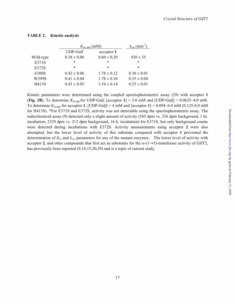
TABLE 2. Kinetic analysis
Km, app (mM) kcat (min-1) UDP-Galf acceptor 1
Wild-type 0.38 ± 0.06 0.60 ± 0.20 430 ± 35 E371S * * * E372S * * * E300S 0.42 ± 0.06 1.78 ± 0.12 0.30 ± 0.01 W399S 0.41 ± 0.04 1.78 ± 0.10 0.35 ± 0.04 H413S 0.43 ± 0.05 1.54 ± 0.14 0.25 ± 0.01
Kinetic parameters were determined using the coupled spectrophotometric assay (29) with acceptor 1 (Fig. 1B). To determine Km,app for UDP-Galf, [acceptor 1] = 3.0 mM and [UDP-Galf] = 0.0625–4.0 mM. To determine Km,app for acceptor 1, [UDP-Galf] = 4 mM and [acceptor 1] = 0.094–6.0 mM (0.125-8.0 mM for H413S). *For E371S and E372S, activity was not detectable using the spectrophotometric assay. The radiochemical assay (9) detected only a slight amount of activity (565 dpm vs. 236 dpm background, 1 hr. incubation; 2329 dpm vs. 212 dpm background, 16 h. incubation) for E371S, but only background counts were detected during incubations with E372S. Activity measurements using acceptor 2 were also attempted, but the lower level of activity of this substrate compared with acceptor 1 prevented the determination of Km and kcat parameters for any of the mutant enzymes. The lower level of activity with acceptor 2, and other compounds that first act as substrates for the α-(1→5)-transferase activity of GlfT2, has previously been reported (9,14,15,20,29) and is a topic of current study.
by guest on February 11, 2018http://w
ww
.jbc.org/D
ownloaded from

O-R
OO
HO
OHOH
HOO
HO
OHHO
O-R
OO
HO
OHOH
HOO
HO
OHHO
HOO
HO
OHO
Full-length galactan
UDP-Galf, GlfT2
-(1 6)-Transfer
O-R
OO
HO
OHOH
OO
HO
OHOH
HOO
HO
OHO
HOO
HO
OHHO
UDP-Galf, GlfT2-(1 5)-Transfer
P OO
OPO
OO O
HO OH
NNH
O
OOHO
OHO
HOHO
UDP-Galf
R = 4)-Rhap-(1 3)-GlcpNAc-PP-Polyprenol UDP-Galf, GlfT2Repeat
A
B O(CH2)7CH3
OO
HO
OHOH
HOO
HO
OHO
HOO
HO
OHHO
O(CH2)7CH3
OO
HO
OHOH
HOO
HO
OHHO
HOO
HO
OHO
1 2
Figure 1
18
by guest on February 11, 2018http://w
ww
.jbc.org/D
ownloaded from

N
C
D1
D2 D3
D4
Figure 2
D1 D4D2 D3bD3a
1 489151 580396 637
Loop 1
β−sandwich α/βGT-A α−helical α−helical
19
by guest on February 11, 2018http://w
ww
.jbc.org/D
ownloaded from

Figure 3
BA C
D
20
E
by guest on February 11, 2018http://w
ww
.jbc.org/D
ownloaded from

Q200
N229
F367
W408
D258
D256
R171
F169
H396
Figure 4
D372
D371
D258
D256
H396
E300
Y344
H413
6-OH
2-OH
3-OH
6-OH
5-OH2-OH
B
A
5-OH
G232
21
by guest on February 11, 2018http://w
ww
.jbc.org/D
ownloaded from

A
B
Figure 5
UDP-Galf
UDP-Galf
β-D-Galf-(1→5)- β-D-Galf-(1→6)- β-D-Galf
β-D-Galf-(1→6)- β-D-Galf-(1→5)- β-D-Galf
6-OH
5-OH
β-(1→5) linkage
β-(1→6) linkage
W399
K369
H413
W309
W399 K369
H413
W309
H296
E300Y344
W348
D372
H396 D258D256
H296
E300Y344
W348
D372
H396 D258D256
22
by guest on February 11, 2018http://w
ww
.jbc.org/D
ownloaded from

Figure 6
23
Centralcavity
Nascent glycanchains
Membrane
A B C
by guest on February 11, 2018http://w
ww
.jbc.org/D
ownloaded from

Kenneth K.S. NgRobert W. Wheatley, Ruixiang Blake Zheng, Michele R. Richards, Todd L. Lowary and
arabinogalactanTetrameric structure of GlfT2 reveals a scaffold for the assembly of mycobacterial
published online June 15, 2012J. Biol. Chem.
10.1074/jbc.M112.347484Access the most updated version of this article at doi:
Alerts:
When a correction for this article is posted•
When this article is cited•
to choose from all of JBC's e-mail alertsClick here
Supplemental material:
http://www.jbc.org/content/suppl/2012/06/15/M112.347484.DC1
by guest on February 11, 2018http://w
ww
.jbc.org/D
ownloaded from