crystallization of the recombinant endonuclease … of the recombinant endonuclease endog from...
TRANSCRIPT
Patiño-Márquez et al. Actual Biol Volumen 37 / Número 102, 2014
Crystallization of the recombinant endonuclease EndoG from Leishmania (Viannia) panamensis
Cristalización de la endonucleasa EndoG recombinante de Leishmania (Viannia) panamensis
Isabel A. Patiño-Márquez1, 3, 4, Juan F. Alzate 2, 5, Edwin Patiño-González1, 3, 6
AbstractBackground and objectives: Endonuclease G (EndoG) is an enzyme that specifically cleaves double stranded DNA at the dG and dC positions and has been shown to participate in chromatin degradation during apoptosis in Leishmania. The main goal of this work was to purify and crystallize EndoG in preparation for future structural studies that will permit a detailed understanding of the function of this enzyme. Materials and methods: EndoG protein was purified using Ni-affinity chromatography under denaturing conditions, then refolded in vitro and crystallized by the hanging-drop vapor diffusion method. Results and conclusion: The endonuclease G protein from Leishmania (viannia) panamensis was overexpressed, refolded, purified and demonstrated to be enzymatically active. Here, we reports the first successful crystallization of the EndoG protein in this group of protozoan parasites. The protein was crystallized by the hanging-drop vapor diffusion method. High quality EndoG crystals were obtained that perhaps will permit determination of the three-dimensional structure of EndoG using X-ray diffraction.
Key words: crystallization, EndoG, hanging-drop, Leishmania, refolding
ResumenAntecedentes y objetivos: La endonucleasa G (EndoG) es una enzima que escinde específicamente en las posiciones dG y dC del ADN de cadena doble y se ha demostrado que participa en la degradación de la cromatina durante el proceso de apoptosis en Leishmania. El objetivo principal de este trabajo fue la purificación y cristalización de EndoG como preámbulo para los estudios estructurales futuros que permitan entender detalladamente el funcionamiento de esta enzima. Materiales y métodos: La proteína EndoG fue purificada en condiciones desnaturalizantes usando cromatografía de Ni, luego fue renaturalizada in vitro y cristalizada por el método de difusión de vapor por gota colgante. Resultados y conclusión: La proteína EndoG de Leishmania (viannia) panamensis fue sobreexpresada, renaturalizada, purificada y demostró estar enzimáticamente activa. Aquí, se registra la primera cristalización exitosa de la proteína EndoG de este grupo de parásitos protozoarios. La proteína fue cristalizada por el método de difusión de vapor por gota colgante. Se obtuvieron cristales de alta calidad de EndoG que posiblemente nos permitirán determinar la estructura tridimensional de EndoG usando difracción de rayos-X.
Palabras clave: cristalización, EndoG, gota colgante, Leishmania, renaturalización
INTRODUCTION
Leishmania is a genus of obligate intracellular protozoa that parasitizes humans and whose infection is revealed in wide spectrum of clinical manifestations generally known as leishmaniasis. The parasite survives inside of
macrophages using a strategy that involve apoptosis of some parasites after the mosquito bite (Kobets et al. 2012, Naderer and McConville 2011). Disease exacerbation has been found to occur upon the injection of a mixture of live and apoptotic parasites, whereas parasites have been found to not establish a successful infection after the depletion
267
Recibido: junio 2013; aceptado: mayo 2014.1 Grupo Interdisciplinario de Estudios Moleculares (GIEM), Universidad de Antioquia, Medellín (Antioquia) Colombia.2 Grupo Parasitología, Facultad de Medicina, Universidad de Antioquia, Medellín (Antioquia) Colombia.3 Grupo de Bioquímica Estructural de Macromoléculas, Universidad de Antioquia, Medellín (Antioquia) Colombia. Correos electrónicos: 4 <[email protected]>; 5 <[email protected]>; 6 <[email protected]>.
Actual Biol Volumen 37 / Número 102, 2014 Patiño-Márquez et al.
of apoptotic promastigotes (Van Zandbergen et al. 2006). Apoptosis is a type of programmed cell death, which is associated with the defense mechanism of cells against pathogens (Estaquier et al. 2012). However, apoptosis in Leishmania parasites is utilized as a mechanism of infection instead of defense. Apoptotic parasites suppressed the immune response through the release of anti-inflammatory cytokines such as IL-10 and TGF-β (Van Zandbergen et al. 2006). Apoptosis in Leishmania is triggered by a caspase-independent pathway (Dolai et al. 2011, Gannavaram et al. 2008, Kumar et al. 2010, Saha et al. 2009). During apoptosis the mitochondrial endonuclease G (EndoG) is released from the mitochondrion and translocated to the nucleus where it degrades DNA (Gannavaram et al. 2008, Rico et al. 2009, Vařecha et al. 2012). Endonuclease G is an unspecific DNA/RNA hydrolase that is named due to the fact that it preferentially cleaves at guanine nucleotides. Mg2+ is necessary for its activity, however EndoG can function using other cofactors such as Mn2+ (Ruiz-Carrillo and Renaud 1987).
This paper reports the expression, purification refolding, and crystallization of Leishmania (viannia) panamensis EndoG. It was also demonstrated that the enzyme was catalytically active and preliminary crystallization procedure is presented. Needle and spherule crystals forms were obtained at low protein concentration. Larger crystals in roller, rhombs and rectangular forms were obtained with increased protein concentration. A preliminary analysis of crystal quality using polarized light reveals the change in crystal birefringence, and indicates that the EndoG crystals have a regular arrangement of protein molecules.
MATERIALS AND METHODS
Expression of EndoG. The cDNA for 401 amino acids of EndoG protein was amplified by PCR from a template obtained from promastigotes. Two primers were made based on the nucleotide sequences found at the GenBank (DNA sequence: GQ119624). The primers were designed to amplify the EndoG CDS but excluding the first amino acids that were previously described as signal peptide. Recombinant construct of EndoG flanked with His-Tag, and one T7-Tag at the amino terminal of the vector was obtained by insertion of the amplified cDNA into pET28a using the restriction sites BamHI y XhoI. Cloned cDNA sequence of EndoG was verified by Sanger sequencing (data not show). The plasmid pET28a-EndoG was transformed into BL 21 Escherichia coli cells (Novagen). A single colony of transformed E. coli cells was inoculated
into 80 ml E. coli culture medium containing 50 μg/ml kanamycin, and shaken at 37 °C and 130 rpm overnight. A 20 ml aliquot was then used to inoculate Erlenmeyer flasks containing 400 mL fresh Luria-Bertani (LB) medium (1:20 dilution). The flasks were incubated at 37 °C with shaking until an optical density of 0.6 at 600 nm was reached. At this point, IPTG was added to a final concentration of 1 mM to induce protein expression. Incubation was then continued for 3 hours under the same conditions. Cells were harvested by centrifugation at 5,000 RCF for 20 min at 4 °C. The cell pellet was resuspended in 10-15 volumes of lysis buffer TBSE (10 mM Tris-HCl, pH 8.0, 1.0 mM EDTA, 150 mM NaCl, and 1% β-ME).
Escherichia coli cell lysis. The cells were lysed by sonication (204 W for 5min at 4 °C). The mixture was centrifuged at 8,500 RCF for 30 min at 4 °C. The pellet was washed twice with 30 volumes (v/w) of TBSE buffer and, each washed was followed by centrifugation at 8,500 RCF for 30 min. The EndoG protein was then extracted by resuspension of the pellet into 10 mM Tris pH 8.0, 1 mM EDTA, and 8 M GuHCl buffer. The suspension was centrifuged at 9,500 RCF for 30 min at 4 °C. The protein concentration was determined by optical density (OD) spectrum at 280 nm.
Refolding and purification of EndoG. An average of 65 mg of recombinant protein were purified by Ni-Affinity Chromatography under denaturing conditions using 50 mM sodium acetate pH 5.0, 6.0 M GuHCl, 10 mM Imidazole buffer and 3 ml of Profinity IMAC resin (Biorad). The bound EndoG was eluted with the same buffer plus 250 mM Imidazole. Two milliliters protein fractions were collected, and the protein concentration was adjusted to 20 mg/ml. For refolding, 50 mg of the purified protein were diluted 500 fold in 50 mM Tris-HCl, 100 mM NaCl, 0.4 mM KCl, 2.0 M Urea, 0.05% Triton X-100, 0.1 mM GSH y 0.01 mM GSSH, at pH 8.0 and stirred at 4 °C overnight. The urea concentration was then reduced in a stepwise dialysis procedure using 50 mM Tris pH 8.0. After dialysis, the protein solution was concentrated by ultrafiltration and subjected to preparative affinity chromatography using a new Ni-column pre-equilibrated with 100 mM NaCl, 50 mM Tris-HCl, 0.4 mM KCl, 0.05% Triton X-100, at pH 8.0. The bound EndoG was eluted as described above. One-milliliter protein fractions were collected and pooled. The purity of EndoG was analyzed by SDS-PAGE.
Screening buffer to enhance concentration of refolding EndoG. To identify the conditions that maintain the protein soluble while it was concentrated, we evaluated different
268
Patiño-Márquez et al. Actual Biol Volumen 37 / Número 102, 2014
buffers. Briefly, the protein was diluted during refolding at an initial concentration of 100 µg/ml, and dialyzed into a series of buffer conditions: 1) 100 mM Tris-HCl pH: 8.0, 0.1 mM GSH y 0.01 mM GSSH; 2) 50 mM Tris-HCl pH 8.0, 100 mM NaCl, 0.1 mM GSH y 0.01 mM GSSH; 3) 50 mM Tris-HCl pH: 8.0, 0.4 mM KCl, 0.1 mM GSH y 0.01 mM GSSH; and 4) 50 mM Tris-HCl pH: 8.0, 0.4 mM KCl, 0.05% Triton X-100, 0.1 mM GSH y 0.01 mM GSSH. The diluted protein was incubated as previously described (24 h at 4 ºC). Afterwards, the protein was concentrated using an Amicon system (Millipore), with a filter membrane with a molecular weight cut-off of 3.0 kDa resulting in a final protein concentration of 3.3 mg/ml.
Crystallization of EndoG. Crystallization screening kits from Hampton research were used in the initial trials. Hanging-drop vapor diffusion using 24 well plates was employed for initial setup (Cudney et al. 1994). Each crystallization reaction was prepared by mixing 0.5 μl of EndoG protein solution (3.3 mg ml-1) and 1 μl of the crystallization solution. The second and third crystallization reactions were prepared by mixing 1 or 2 μl of EndoG protein solution (3.3 mg ml-1) and 1 μl the crystallization solution. The crystallization drop was placed over 500 μl of reservoir solution. All crystallization trials were performed at room temperature.
Endonuclease activity. The biological activity of EndoG was evaluated with the digestion of human DNA. The enzymatic reaction mixture consisted of 33 mM Tris-acetate pH 7.9, 10 mM magnesium acetate, 66 mM potassium acetate, 0.1 mg/ml BSA (buffer Tango from Fermentas), 1 μg human DNA and 0.5-1.5 μg EndoG. Inhibited endonuclease activity assays were carried out with the same buffer supplemented with 150 or 200 mM KCl and 2 μg EndoG. Assays were performed at 37 ºC for 60 min. Samples were electrophoresed in 1.5% agarose gel for approximately 120 min at 120 V in 1X TBE buffer.
RESULTS
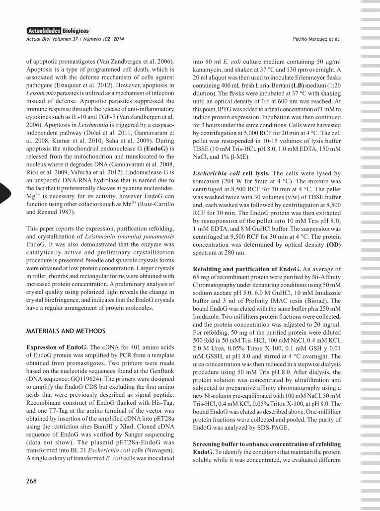
Purification and refolding of the EndoG of Leishmania (Viannia) panamensis. For the induction and accumulation of EndoG, conditions were varied: IPTG concentration (up to 1 mM), time of IPTG treatment (up to 3 h) and cell density. The level of recombinant protein induction was evaluated using SDS-PAGE. Under optimized conditions, EndoG was accumulated until it becomes the most abundant cellular protein (figure 1, lane 2) and also the main protein recovered from the inclusion bodies
(IB) after sonication (figure 1, lane 3). Protein in the IB was solubilized with 8 M GuHCL (figure 1, lane 4) and Ni-Affinity chromatography was performed to purify His-tagged EndoG as a denatured protein (figure 1, lanes 5 and 6). SDS-PAGE in figure 1 shows the analysis of whole cell EndoG before and after solubilization of the inclusion bodies. The electrophoresis analysis also showed that the expected 48 kDa band correlated well with the cDNA cloned into pET-28a between BamHI and XhoI restriction sites (figure 1). After Ni-chromatography, a single EndoG protein band was observed in the SDS-PAGE electrophoresis, showing a successful purification process (figure 1). Subsequent refolding procedure was applied to the purified endonuclease. For this purpose, GuHCl was exchanged for urea, and renaturation of EndoG was performed using a stepwise dialysis approach that consisted of a decrease in urea to a final zero concentration. After refolding, renatured EndoG was purified by Ni-chromatography under native conditions and SDS-PAGE analysis using coomassie blue staining showed only one detectable protein band in the gel that corresponds to EndoG (figure 1, lanes 5 and 6).
Figure 1. Overexpression and purification of endonuclease G (EndoG). Total cell lysates of Escherichia coli without or with IPTG induction state on each lane: lane M, protein molecular weight marker (kDa); lane 1, E. coli uninduced; lane 2, 1 mM IPTG induction; lane 3, insoluble fraction; lane 4, solubilized inclusion bodies; lane 5, EndoG purified after refolding experiment 1; lane 6, EndoG purified after refolding experiment 2
Concentration of EndoG. Buffer conditions were screened to get the maximum soluble concentration of EndoG after refolding. Buffer additives were limited to salt and detergent. A maximum concentration of 1.0 mg/ml was reached in buffer containing 50 mM Tris-HCl pH 8.0 and 100 mM NaCl. The same concentration was also achieved when 0.4 mM KCl or 0.05% Triton X-100 was added. Interestingly, while neither 0.4 mM KCl nor 0.05% Triton X-100 alone could allow the enzyme to reach higher
269
Actual Biol Volumen 37 / Número 102, 2014 Patiño-Márquez et al.
concentrations in a soluble condition, a combination of both additives render the EndoG stably soluble at concentrations up to 3.3 mg/ml.
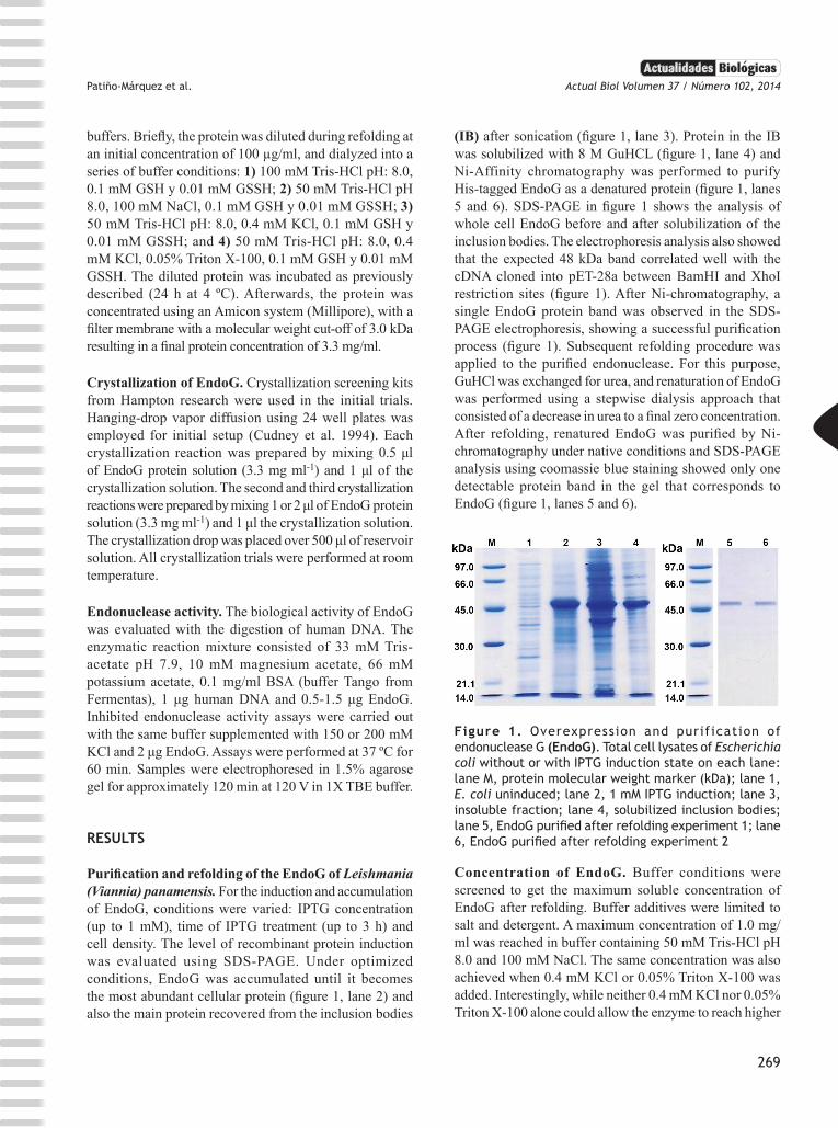
Enzymatic activity of the EndoG of Leishmania (Viannia) panamensis. Genomic human DNA was used as the substrate for refolded EndoG. Figure 2 shows the result of a typical digestion assay by unspecific endonuclease (figure 2, lanes 3, 4 and 5). Potassium is known to inhibit the activity of EndoG, and Mg2+ was found to be an important cofactor for DNA digestion (Toro et al. 2011). The results showed that potassium totally inhibited the digestion of DNA (figure 2, lanes 6 and 7), and that denatured EndoG (in 8 M GuHCl) did not show any endonuclease activity (figure 2, lane 2).
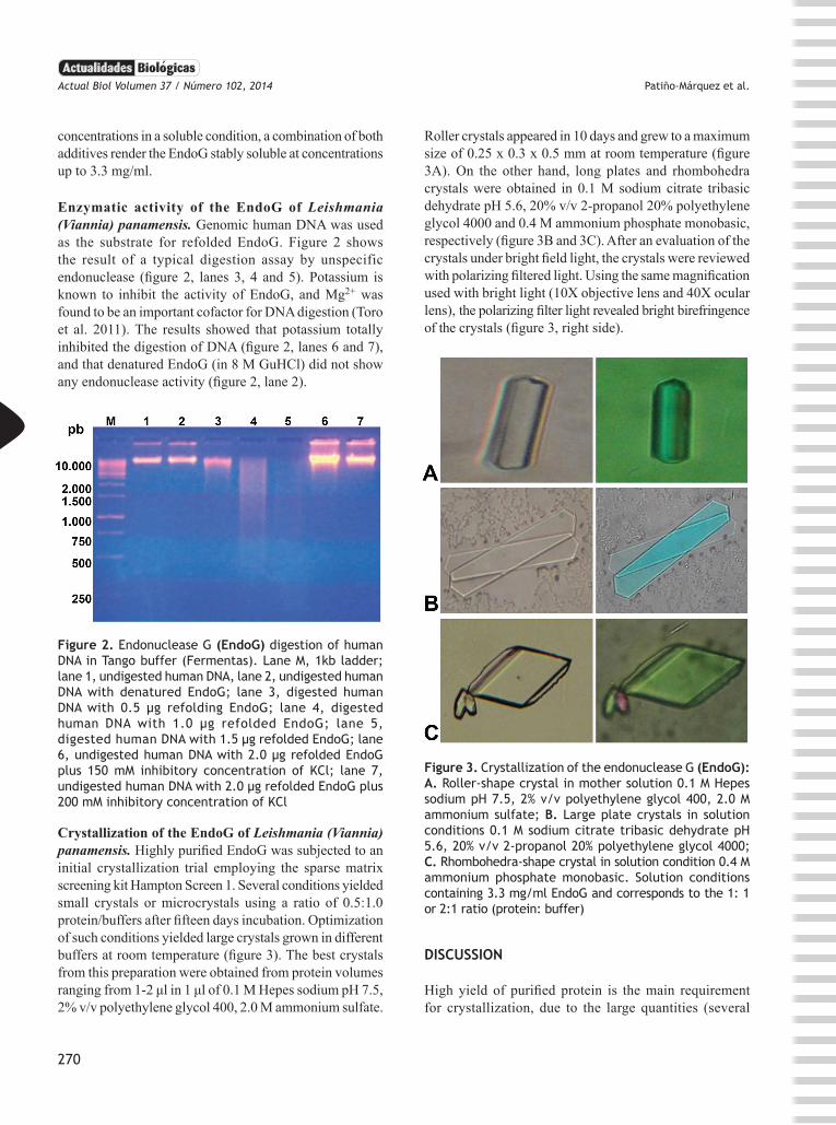
Roller crystals appeared in 10 days and grew to a maximum size of 0.25 x 0.3 x 0.5 mm at room temperature (figure 3A). On the other hand, long plates and rhombohedra crystals were obtained in 0.1 M sodium citrate tribasic dehydrate pH 5.6, 20% v/v 2-propanol 20% polyethylene glycol 4000 and 0.4 M ammonium phosphate monobasic, respectively (figure 3B and 3C). After an evaluation of the crystals under bright field light, the crystals were reviewed with polarizing filtered light. Using the same magnification used with bright light (10X objective lens and 40X ocular lens), the polarizing filter light revealed bright birefringence of the crystals (figure 3, right side).
Figure 2. Endonuclease G (EndoG) digestion of human DNA in Tango buffer (Fermentas). Lane M, 1kb ladder; lane 1, undigested human DNA, lane 2, undigested human DNA with denatured EndoG; lane 3, digested human DNA with 0.5 µg refolding EndoG; lane 4, digested human DNA with 1.0 µg refolded EndoG; lane 5, digested human DNA with 1.5 µg refolded EndoG; lane 6, undigested human DNA with 2.0 µg refolded EndoG plus 150 mM inhibitory concentration of KCl; lane 7, undigested human DNA with 2.0 µg refolded EndoG plus 200 mM inhibitory concentration of KCl
Crystallization of the EndoG of Leishmania (Viannia) panamensis. Highly purified EndoG was subjected to an initial crystallization trial employing the sparse matrix screening kit Hampton Screen 1. Several conditions yielded small crystals or microcrystals using a ratio of 0.5:1.0 protein/buffers after fifteen days incubation. Optimization of such conditions yielded large crystals grown in different buffers at room temperature (figure 3). The best crystals from this preparation were obtained from protein volumes ranging from 1-2 μl in 1 μl of 0.1 M Hepes sodium pH 7.5, 2% v/v polyethylene glycol 400, 2.0 M ammonium sulfate.
Figure 3. Crystallization of the endonuclease G (EndoG): A. Roller-shape crystal in mother solution 0.1 M Hepes sodium pH 7.5, 2% v/v polyethylene glycol 400, 2.0 M ammonium sulfate; B. Large plate crystals in solution conditions 0.1 M sodium citrate tribasic dehydrate pH 5.6, 20% v/v 2-propanol 20% polyethylene glycol 4000; C. Rhombohedra-shape crystal in solution condition 0.4 M ammonium phosphate monobasic. Solution conditions containing 3.3 mg/ml EndoG and corresponds to the 1: 1 or 2:1 ratio (protein: buffer)
DISCUSSION
High yield of purified protein is the main requirement for crystallization, due to the large quantities (several
270
Patiño-Márquez et al. Actual Biol Volumen 37 / Número 102, 2014
milligrams) required for the optimization of crystallization conditions. In this work, we were able to achieve high levels of expression of EndoG in E. coli in LB broth. Such recombinant expression levels rendered the protein insoluble, as previously reported in by Toro et al. (2011). Overexpression of EndoG was checked by SDS-PAGE electrophoresis of the supernatant and pellet after bacterial cells disruption by sonication (figure 1). A band corresponding to 48 kDa was observed in the total lysate and inclusion bodies fraction, and was absent in the equivalent control lysate of E. coli without IPTG induction. The 48 kDa band corresponds to the recombinant EndoG that lacks the native signal peptide of the enzyme but includes two His-Tag sequences at both ends of the enzyme. Furthermore the enzyme also includes a T7-Tag sequence after the amino-terminal His-Tag. The extra amino acids of the expressed recombinant EndoG did not affect the refolding, enzyme activity or crystallization of the enzyme (figure 2 y 3). Ni-chromatography under denaturing condition allowed the removal of the majority of the protein contaminants achieving high levels of enzyme purity. Further refolding of the purified recombinant enzyme restores the endonuclease activity to its native state. A second step of Ni-affinity chromatography under native conditions was successfully performed in order to ensure protein homogeneity. Previous studies reported the purification and use of recombinant EndoG at low concentrations of only 100 ng/ml (Toro et al. 2011). However, our purified EndoG was successfully concentrated to 3.3 mg/ml without affecting its solubility. It represents more than a 1000 fold improvement of the reported concentration (Toro et al. 2011). Our success with the high concentration tolerance of EndoG can be explained with at least with 3 modifications, in comparison with the Toro et al. (2011) report: 1) we used Tris buffer pH 8.0 instead of phosphate buffer due to the phosphate capacity to sequester divalent cations necessary for the enzymes stability. Moreover, Tris pKa is 8.0 so it will have a better buffer capacity at pH 8.0. 2) EndoG was refolded in presence of low concentration of salts such as NaCl and KCl. These salts increase the electrostatic interactions and weaken the attractive forces between monomers; and 3) the detergent Triton X-100 was used to minimized protein aggregation and avoids precipitation of the monomers. Refolded EndoG was evaluated for endonuclease activity, and crystallization trials were initiated by the hanging-drop vapor diffusion method. The nuclease activity of the enzyme was successfully recovered after refolding, a finding previously reported by Toro et al (2011). Furthermore, the endonuclease activity of the purified EndoG depends on its native conformation because the use
GuHCl renders the enzyme inactive. In these crystallization trials, after fifteen days of equilibration at room temperature multiple, tiny and needle-shaped crystals were grown in drops with a ratio of 0.5:1 protein/buffer (data not show). Crystals improved when the ratio was increased to 1:1 and 2:1 protein/buffer. Under these conditions, rhombohedra, roller, and large plate-shape crystals were found to have grown in Crystal Screen I solutions N.º 3, 39, and 40, respectively. We were able to reproduce these crystal types after 10 days if plates were not moved at all, and remained uninspected during the incubation period. The crystallization of EndoG was the first step required to determine the structure of the protein. To know the EndoG structure is crucial to facilitate the rational design of new drugs with improved anti-Leihsmanial activity.
CONCLUSION
We were able to improve the purification efficacy of EndoG to enable its crystallization analysis. Our findings also show that the EndoG recovers its catalytic activity after refolding and that it can be crystallized when the protein is stabilized with supplemented Tris buffer that allow high concentration of the enzyme in soluble conditions.
ACKNOWLEDGEMENTS
The authors thank Diego Duchi Llumigusín (Department of Physics, University of Oxford) for his valuable collaboration. This work was supported by University of Antioquia (CODI) as part of project: Determinación de las Condiciones de Cristalización de la Proteína Lack de Leishmania. Estrategia de sostenibilidad 2011-2012.
REFERENCES
Cudney R, Patel S, Weisgraber K, Newhouse Y, McPherson A. 1994. Screening and optimization strategies for macromolecular crystal growth. Acta Crystallographica Section D, 50 (4): 414-423.
Dolai S, Pal S, Yadav RK, Adak S. 2011. Endoplasmic reticulum stress-induced apoptosis in Leishmania through Ca2+-dependent and caspase-independent mechanism. Journal of Biological Chemistry, 286 (15): 13638-13646.
Estaquier J, Vallette F, Vayssiere JL, Mignotte B. 2012. The mitochondrial pathways of apoptosis. Advances in Experimental Medicine and Biology, 942:157-183.
Gannavaram S, Vedvyas C, Debrabant A. 2008. Conservation of the pro-apoptotic nuclease activity of endonuclease G in unicellular trypanosomatid parasites. Journal of Cell Science, 121 (1): 99-109.
271
Actual Biol Volumen 37 / Número 102, 2014 Patiño-Márquez et al.
Kobets T, Grekov I, Lipoldova M. 2012. Leishmaniasis: prevention, parasite detection and treatment. Current Medicinal Chemistry, 19 (10): 1443-1474.
Kumar P, Lodge R, Trudel N, Ouellet M, Ouellette M, Tremblay MJ. 2010. Nelfinavir, an HIV-1 protease inhibitor, induces oxidative stress-mediated, caspase-independent apoptosis in Leishmania amastigotes. PLoS Negleted Tropical Diseases, 4 (3): e642.
Naderer T, McConville MJ. 2011. Intracellular growth and pathogenesis of Leishmania parasites. Essays in Biochemistry, 51: 81-95.
Rico E, Alzate JF, Arias AA, Moreno D, Clos J, Gago F, Moreno I, Dominguez M, Jimenez-Ruiz A. 2009. Leishmania infantum expresses a mitochondrial nuclease homologous to EndoG that migrates to the nucleus in response to an apoptotic stimulus. Molecular and Biochemical Parasitology, 163 (1): 28-38.
Ruiz-Carrillo A, Renaud J. 1987. Endonuclease G: a (dG)n X (dC)n-specific DNase from higher eukaryotes. The EMBO Journal, 6 (2): 401-407.
Saha P, Sen R, Hariharan C, Kumar D, Das P, Chatterjee M. 2009. Berberine chloride causes a caspase-independent, apoptotic-like death in Leishmania donovani promastigotes. Free Radical Research, 43 (11): 1101-1110.
Toro L, Patiño E, Robledo S, Jiménez RA, Alzate JF. 2011. Leishmania (Viannia) panamensis expresses a nuclease with molecular andbiochemical features similar to the Endonuclease G of higher eukaryotes. Colombia Médica, 41(2): 154-165.
Van Zandbergen G, Bollinger A, Wenzel A, Kamhawi S, Voll R, Klinger M, Müller A, Hölscher C, Herrmann M, Sacks D, Solbach W, Laskay T. 2006. Leishmania disease development depends on the presence of apoptotic promastigotes in the virulent inoculum. Proceedings of the National Academy of Sciences of the United States of America, 103 (37): 13837-13842.
Vařecha M, Potěšilová M, Matula P, Kozubek M. 2012. Endonuclease G interacts with histone H2B and DNA topoisomerase II alpha during apoptosis. Molecular and Cellular Biochemistry, 363 (1-2): 301-307.
272