crystallographic structure of porcine adenovirus...
TRANSCRIPT
JOURNAL OF VIROLOGY, Oct. 2010, p. 10558–10568 Vol. 84, No. 200022-538X/10/$12.00 doi:10.1128/JVI.00997-10Copyright © 2010, American Society for Microbiology. All Rights Reserved.
Crystallographic Structure of Porcine Adenovirus Type 4 Fiber Headand Galectin Domains�
Pablo Guardado-Calvo,1† Eva M. Munoz,2 Antonio L. Llamas-Saiz,3 Gavin C. Fox,4‡ Richard Kahn,4David T. Curiel,5 Joel N. Glasgow,5,6* and Mark J. van Raaij1,7*
Departamento de Bioquímica y Biología Molecular, Facultad de Farmacia,1 Departamento de Química Organica, Facultad deQuímica,2 and Unidad de Rayos X, Laboratorio Integral de Dinamica y Estructura de Biomoleculas Jose R. Carracido,3
Universidad de Santiago de Compostela, E-15782 Santiago de Compostela, Spain; Laboratoire de Proteines Membranaires,Institut de Biologie Structurale J.P. Ebel, F-38027 Grenoble, France4; Division of Human Gene Therapy,
Departments of Medicine, Obstetrics and Gynecology, Pathology, Surgery, the Gene Therapy Center,5 andDivision of Cardiology,6 University of Alabama at Birmingham, Birmingham, Alabama; and
Departamento de Biología Estructural, Instituto de Biología Molecular deBarcelona (CSIC), E-02808 Barcelona, Spain7
Received 7 May 2010/Accepted 21 July 2010
Adenovirus isolate NADC-1, a strain of porcine adenovirus type 4, has a fiber containing an N-terminal virusattachment region, shaft and head domains, and a C-terminal galectin domain connected to the head by anRGD-containing sequence. The crystal structure of the head domain is similar to previously solved adenovirusfiber head domains, but specific residues for binding the coxsackievirus and adenovirus receptor (CAR), CD46,or sialic acid are not conserved. The structure of the galectin domain reveals an interaction interface betweenits two carbohydrate recognition domains, locating both sugar binding sites face to face. Sequence evidencesuggests other tandem-repeat galectins have the same arrangement. We show that the galectin domain bindscarbohydrates containing lactose and N-acetyl-lactosamine units, and we present structures of the galectindomain with lactose, N-acetyl-lactosamine, 3-aminopropyl-lacto-N-neotetraose, and 2-aminoethyl-tri(N-acetyl-lactosamine), confirming the domain as a bona fide galectin domain.
Adenoviridae are nonenveloped viruses with a linear double-stranded DNA genome that can infect all five major vertebrateclasses. They may be used as vectors for gene or cancer therapyand as vaccination agents (1, 2, 45). Adenoviruses have anicosahedral (T�25) capsid consisting of the trimeric hexon,forming the facets of the particle, the pentameric penton base,which forms the vertices, and the trimeric fiber protein, whichextends from the penton base at the vertex positions (38). Thedistal tip of each fiber is composed of a globular head domain,which serves as the major viral attachment site for a variety ofcellular receptors, such as coxsackievirus and adenovirus re-ceptor (CAR) (42), CD46 (15), and sialic acid (6). The struc-tural characterization of different fiber proteins from humanand nonhuman adenoviruses has been critical for understand-ing adenovirus tropism and developing new vectors with mod-ified tropisms. Animal adenoviruses are of particular interest,as they may be less immunogenic to humans and may havenovel receptor-binding properties. Recently, canine and fowl
adenovirus fiber heads have been crystallized and their struc-tures determined (44, 17, 18, 13).
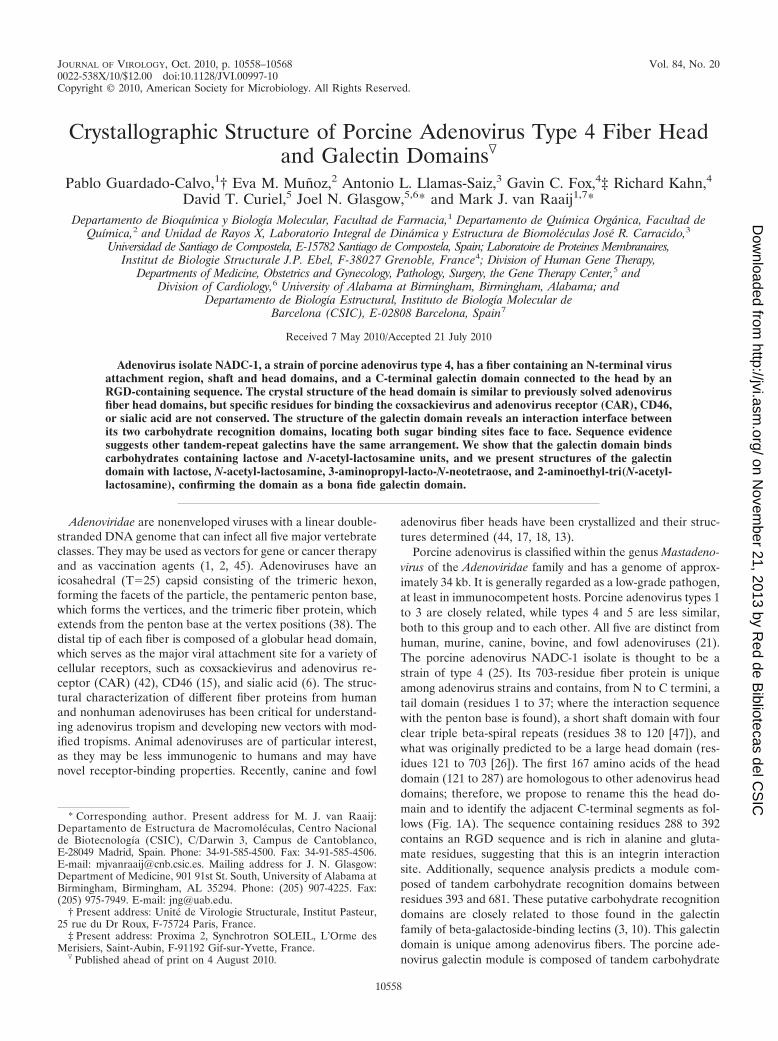
Porcine adenovirus is classified within the genus Mastadeno-virus of the Adenoviridae family and has a genome of approx-imately 34 kb. It is generally regarded as a low-grade pathogen,at least in immunocompetent hosts. Porcine adenovirus types 1to 3 are closely related, while types 4 and 5 are less similar,both to this group and to each other. All five are distinct fromhuman, murine, canine, bovine, and fowl adenoviruses (21).The porcine adenovirus NADC-1 isolate is thought to be astrain of type 4 (25). Its 703-residue fiber protein is uniqueamong adenovirus strains and contains, from N to C termini, atail domain (residues 1 to 37; where the interaction sequencewith the penton base is found), a short shaft domain with fourclear triple beta-spiral repeats (residues 38 to 120 [47]), andwhat was originally predicted to be a large head domain (res-idues 121 to 703 [26]). The first 167 amino acids of the headdomain (121 to 287) are homologous to other adenovirus headdomains; therefore, we propose to rename this the head do-main and to identify the adjacent C-terminal segments as fol-lows (Fig. 1A). The sequence containing residues 288 to 392contains an RGD sequence and is rich in alanine and gluta-mate residues, suggesting that this is an integrin interactionsite. Additionally, sequence analysis predicts a module com-posed of tandem carbohydrate recognition domains betweenresidues 393 and 681. These putative carbohydrate recognitiondomains are closely related to those found in the galectinfamily of beta-galactoside-binding lectins (3, 10). This galectindomain is unique among adenovirus fibers. The porcine ade-novirus galectin module is composed of tandem carbohydrate
* Corresponding author. Present address for M. J. van Raaij:Departamento de Estructura de Macromoleculas, Centro Nacionalde Biotecnología (CSIC), C/Darwin 3, Campus de Cantoblanco,E-28049 Madrid, Spain. Phone: 34-91-585-4500. Fax: 34-91-585-4506.E-mail: [email protected]. Mailing address for J. N. Glasgow:Department of Medicine, 901 91st St. South, University of Alabama atBirmingham, Birmingham, AL 35294. Phone: (205) 907-4225. Fax:(205) 975-7949. E-mail: [email protected].
† Present address: Unite de Virologie Structurale, Institut Pasteur,25 rue du Dr Roux, F-75724 Paris, France.
‡ Present address: Proxima 2, Synchrotron SOLEIL, L’Orme desMerisiers, Saint-Aubin, F-91192 Gif-sur-Yvette, France.
� Published ahead of print on 4 August 2010.
10558
on Novem
ber 21, 2013 by Red de B
ibliotecas del CS
IChttp://jvi.asm
.org/D
ownloaded from
recognition domains linked by a 23-residue sequence rich inprolines.
Lectins of the galectin family specifically bind beta-galac-toside sugars (10). Their carbohydrate recognition domainsconsist of two beta-sheets with a jelly roll topology. Strandsare designated F1 to F5 and S1 to S6, in which the S sheetfaces the carbohydrate. Galectins have been assigned toprototype, chimera-type, or tandem-repeat-type subfamilies(22). Prototype galectins consist of a single-carbohydraterecognition domain and usually form homodimers with thetwo carbohydrate recognition domains in a back-to-backorientation and sugar binding sites placed at opposite ends(31). Tandem-repeat-type galectins contain two nonidenti-cal carbohydrate recognition domains separated by a linkerpeptide. The organization of the carbohydrate recognitiondomains in tandem-repeat galectins has not been deter-mined; only structures of single carbohydrate recognitiondomains are known (37). However, by analogy to the pro-totype galectins, a back-to-back orientation, and thus biva-lent binding, is generally accepted. Eight highly conservedresidues, localized within a pocket formed by three adjacentbeta strands (S4, S5, and S6), are involved in carbohydratebinding. Other, less-conserved residues, situated in betastrands S2 and S3, are implicated in the recognition ofspecific sugars.
Here, we report the structures of the porcine adenovirustype 4 NADC1 isolate fiber head domain and of the galectindomain, the first tandem-repeat-type galectin in which bothcarbohydrate recognition domains are simultaneously ob-served, revealing a face-to-face orientation of the carbohy-drate-binding domains. Furthermore, we demonstrate that the
galectin domain binds N-acetyl-lactosamine-containing carbo-hydrates.
MATERIALS AND METHODS
Adenoviruses. Ad5Luc1 is a replication-defective E1-deleted adenovirus vec-tor containing a firefly luciferase reporter gene driven by a cytomegaloviruspromoter, and it encodes the native adenovirus type 5 fiber protein (27). TheAd5Luc1-PK vector is isogenic to Ad5Luc1, except that the adenovirus type 5fiber head domain (residues 404 to 581) is replaced with the head, RGD, andgalectin domains (residues 124 to 703) of the fiber protein from the NADC-1isolate of porcine adenovirus type 4 (40). Adenovirus vectors were propagated in293 cells and purified by equilibrium centrifugation in cesium chloride gradientsusing a standard protocol (16). Viral particle concentration was determined at260 nm by the method of Maizel et al. (34) using a conversion factor of 1.1 � 1012
viral particles per absorbance unit.Glycan array. Glycan screening was performed using a high-throughput glycan
array developed by cores D and H of the Consortium for Functional Glycomics(a National Institutes of Health, National Institute of General Medical Sciencesinitiative, Emory University School of Medicine, Atlanta, GA). The printedglycan array (version 3.2) contained 406 different natural and synthetic glycansprinted on glass slides that contain six individual addresses per glycan or glyco-conjugate, as described previously (5). A printed slide was incubated with a 70-�lvolume of Ad5Luc1 or Ad5Luc1-PK virions at a concentration of 5.5 � 106 viralparticles/�l in sample buffer comprised of 20 mM Tris-HCl, pH 7.4, 150 mMsodium chloride, 2 mM calcium chloride, 2 mM magnesium chloride, 0.05%(vol/vol) Tween 20, and 1% (wt/vol) bovine serum albumin. Following the bind-ing of the virions to the slide, a fluorescein-conjugated anti-hexon primary mono-clonal antibody was overlaid on the bound virions. The fluorescence intensity wasdetected using a ScanArray 5000 confocal scanner (Perkin-Elmer, Waltham,MA). The image was analyzed using the IMAGENE image analysis software(BioDiscovery, El Segundo, CA).
Surface plasmon resonance measurements. Sensor chips were purchased fromXantec Bioanalytics (Muenster, Germany), and neutravidin was from PierceProtein Research Products (Rockford, IL). Surface plasmon resonance bindingassays were carried out using an SR7000DC optical biosensor spectrometer(Reichert Analytical Systems, Depew, NY). All experiments were performed at
FIG. 1. (A) Schematic drawing of the domain organization of porcine adenovirus type 4 NADC-1 strain fiber. The predicted virus-bindingtail is shown in black, the shaft domain in white, the head domain in light gray, the RGD-containing domain in white, and the tandem-repeatgalectin domain in dark gray. The putative integrin-binding RGD sequence is indicated with an asterisk. Electron microscopy images of thefiber indicate flexibility between the head and C-terminal domains; therefore, the C-terminal domains are drawn in different orientations.(B) Sequence alignment of tandem-repeat galectins. Human galectin 4 (HGal-4; UNIPROT code P56470), human galectin 8 (H-Gal8;O00214), and human galectin 9 (HGal-9; O00182) are shown. Secondary structural elements of the porcine adenovirus 4 galectin domainare indicated.
VOL. 84, 2010 PORCINE ADENOVIRUS 4 FIBER HEAD AND GALECTIN 10559
on Novem
ber 21, 2013 by Red de B
ibliotecas del CS
IChttp://jvi.asm
.org/D
ownloaded from
25°C using 10 mM N-2-hydroxyethylpiperazine-N-2-ethanesulphonic acid-NaOH, pH 7.4, 150 mM sodium chloride, 3 mM ethylenediaminetetra-aceticacid, and 1 mM dithiothreitol as the running buffer. Data were processed withSCRUBBER 2.0b software (BioLogic Software, Campbell, Australia). Lactosewas obtained from Sigma-Aldrich Quimica (Madrid, Spain); N-acetyl-lac-tosamine and lacto-N-neotetraose were from Dextra Laboratories (Reading,United Kingdom); and 3-aminopropyl-lacto-N-neotetraose, 2-azidoethyl-di(N-acetyl-lactosamine), and 2-aminoethyl-tri(N-acetyl-lactosamine) were from theGlycan Array Synthesis Core D of the Consortium for Functional Glycomics(The Scripps Research Institute, Department of Molecular Biology, La Jolla,CA). The porcine adenovirus 4 galectin domain was covalently immobilized ona flow cell of a polycarboxylated hydrogel-coated gold surface (HC1000 sensorchip) at a flow rate of 0.01 ml/min. Surface activation was performed with 0.05 MN-hydroxysuccinimide and 0.2 M N-ethyl-N�-(3-dimethyl-aminopropyl)-carbodi-imide hydrochloride (10 min), and a 1.3-mg/ml solution of porcine adenovirus 4galectin domain in 10 mM sodium acetate, pH 5, was injected (10 min); unre-acted N-hydroxysuccinimide esters were deactivated with 1 M ethanolamine, pH8.5 (10 min). The control flow cell was coated with neutravidin using the samecoupling protocol. Two sensor chips were prepared with galectin/neutravidinimmobilization levels of 12 � 103 �RiU/14 � 103 �RiU (sensor chip 1) and10.5 � 103 �RiU/6.4 � 103 �RiU (sensor chip 2); 1 �RiU corresponds to 0.73ng/mm2 coverage in terms of mass (RiU stands for refractive index unit). Forbinding assays, solutions of each oligosaccharide were prepared in running buffer[1.4 to 45 mM for lactose, 0.12 to 2.6 mM for N-acetyl-lactosamine, 0.030 to 1.3mM for lacto-N-neotetraose, 0.039 to 1.5 mM for 3-aminopropyl-lacto-N-neote-traose, 0.040 to 2.0 mM for 2-azidoethyl-di(N-acetyl-lactosamine), and 0.010 to1.9 mM for 2-aminoethyl-tri(N-acetyl-lactosamine)]. The samples were injectedover the galectin-coated and control surfaces at 0.05 ml/min for 120 s in dupli-cate. As analytes fully dissociated after injection, regeneration steps were notnecessary. Experimental data were corrected for instrumental and bulk artifactsby double referencing to the control sensor chip surface and buffer injections. Inall cases, sensorgrams showed on and off binding profiles that were too fast forkinetic analysis, so the equilibrium analysis of the sensorgrams was performed toobtain the dissociation constants of the complexes.
Crystallization. The porcine adenovirus type 4 isolate NADC-1 head domainand ligand-free galectin domain were independently expressed, purified, andcrystallized as described previously (19). Sitting drop vapor diffusion cocrystal-lization methods also were employed to yield crystals of the complexes. Lactosewas obtained from Sigma-Aldrich Quimica (Madrid, Spain), N-acetyl-lac-
tosamine was from Dextra Laboratories (Reading, United Kingdom), and3-aminopropyl-lacto-N-neotetraose and 2-aminoethyl-tri(N-acetyl-lactosamine)were from the Glycan Array Synthesis Core D of the Consortium for FunctionalGlycomics (The Scripps Research Institute, Department of Molecular Biology,La Jolla, CA). Crystals of the lactose complex were obtained using 28% (wt/vol)polyethylene glycol 3350, 100 mM sodium nitrate, 5 mM dithiothreitol as thereservoir solution, and 70 mM lactose in the protein solution. Crystals of theN-acetyl-lactosamine complex were obtained using 35% (wt/vol) polyethyleneglycol 3350, 500 mM sodium nitrate, 5 mM dithiothreitol as the reservoir solu-tion, and 40 mM N-acetyl-lactosamine in the protein solution. Crystals of thelacto-N-neotetraose complex were obtained using 27% (wt/vol) polyethyleneglycol 3350, 200 mM lithium nitrate, 5 mM dithiothreitol as the reservoir solu-tion, and 5 mM lacto-N-neotetraose in the protein solution. Crystals of the2-aminoethyl-tri(N-acetyl-lactosamine) complex were obtained using 28% (wt/vol) polyethylene glycol 3350, 300 mM lithium nitrate, 5 mM dithiothreitol as thereservoir solution, and 5 mM 2-aminoethyl-tri(N-acetyl-lactosamine) in the pro-tein solution. Crystallization experiments were performed using the sitting dropvapor diffusion method at 18°C. All crystals appeared after 1 to 2 weeks and werecryoprotected by raising the polyethylene glycol 3350 concentration to 35%(wt/vol) if necessary.
Structure solution and analysis. Data collection and processing for the headand ligand-free galectin domains were performed as described previously (19);data for the galectin domain cocrystallized with carbohydrates was collected andprocessed similarly. Crystallographic models were examined and corrected man-ually using the program COOT (14). Restrained refinement was performed usingthe program REFMAC (36). The program MOLPROBITY (8) was used toevaluate the stereochemistry. Figures were prepared with Pymol (DeLano Sci-entific, Palo Alto, CA). Multiple-sequence alignments were performed withCLUSTALW (9). Interfaces were analyzed using PISA (28). The structure so-lution of the head domain was achieved by molecular replacement using theadenovirus 5 head trimer (Protein Data Bank [PDB] code 1KNB) as a searchmodel with the program MOLREP (46); a clear solution consisting of twotrimers in the asymmetric unit was obtained. Despite the moderate resolution(3.2 Å), the high solvent content and the 6-fold noncrystallographic symmetrycontribute to generate electron density maps of high quality. Refinement wasperformed with noncrystallographic symmetry restraints throughout. Toward theend, TLS (translation/libration/screw) refinement was included (48), definingeach protein chain as a TLS group. The use of a model refined against high-resolution data in molecular replacement, the aforementioned high solvent con-
TABLE 1. Crystallographic data, refinement, and validation statisticsa
Statistic Head domain
Galectin domain
Ligand free Lactose N-acetyl-lactosamine
3-Aminopropyl-lacto-N-
neotetraose
2-Aminoethyl-tri(N-acetyl-lactosamine)
DataSpace group P212121 C2 C2 C2 C2 C2Cell parameters a, b, c (Å) 125.7, 145.4, 147.6 167.6, 77.3, 94.1 114.6, 38.8, 70.2 108.5, 43.7, 63.9 95.4, 38.8, 84.4 176.1, 38.3, 86.5Cell parameters �, �, � (°) 90.0, 90.0, 90.0 90.0, 101.5, 90.0 90.0, 93.5, 90.0 90.0, 105.8, 90.0 90.0, 102.2, 90.0 90.0, 92.1, 90.0No. of unique observations 45,035 (4,367) 89,027 (12,129) 21,094 (3,019) 22,025 (3,229) 23,751 (3,155) 20,441 (2,926)Resolution range (Å) 40.0–3.2 (3.32–3.2) 40.0–1.9 (2.0–1.9) 40.0–2.0 (2.1–2.0) 35.0–1.9 (2.0–1.9) 40.0–1.9 (2.0–1.9) 40.0–2.5 (2.6–2.5)Multiplicity 3.8 (3.8) 4.1 (3.8) 3.5 (3.5) 2.7 (2.7) 3.4 (2.7) 3.6 (3.6)Completeness (%) 98.9 (97.5) 96.1 (90.1) 99.6 (99.3) 98.0 (98.5) 98.5 (91.7) 99.9 (99.8)Mean I/�(I) 17.5 (2.9) 10.0 (3.7) 10.4 (3.3) 5.9 (2.7) 12.6 (3.2) 8.8 (3.1)Rsymm
b (%) 7.2 (30.7) 7.5 (35.2) 6.9 (32.2) 9.8 (31.3) 5.8 (30.3) 9.5 (43.2)
RefinementResolution range 29.2–3.2 (3.37–3.20) 35–1.9 (2.00–1.90) 40–2.0 (2.11–2.00) 35–1.91 (2.01–1.91) 40–1.9 (2.00–1.90) 40–2.5 (2.63–2.50)No. of protein molecules/
asymmetric unit6 4 1 1 1 2
R factorc 0.173 (0.264) 0.206 (0.298) 0.183 (0.244) 0.182 (0.270) 0.187 (0.275) 0.199 (0.324)Rfree 0.201 (0.299) 0.259 (0.327) 0.228 (0.296) 0.238 (0.337) 0.223 (0.310) 0.268 (0.355)
Final modelRMSD bond lengths (Å) 0.009 0.010 0.009 0.012 0.011 0.009RMSD bond angles (°) 1.3 1.3 1.3 1.3 1.4 1.3Ramachandran plotd (%) 93.9/6.0/0.1 97.0/2.4/0.6 96.6/3.4/0.0 97.7/2.0/0.3 98.0/2.0/0.0 97.5/2.5/0.0
PDB accession code 2WST 2WSU 2WSV 2WT0 2WT1 2WT2
a One crystal was used per structure; values in parentheses are for the highest-resolution shell.b Rsymm � ¥h¥i�IhiIh��/¥h¥i�Ihi�, where Ihi is the intensity of the ith measurement of the same reflection, and Ih� is the mean observed intensity for that reflection.c R factor � ���Fobs(hkl)� �Fcalc(hkl)��/��Fobs(hkl)�. Fobs(hkl) and Fcalc(hkl) are observed and calculated structure factors at indices hkl, respectively.d The percentages are of residues in the favored, allowed, and outlier regions, respectively.
10560 GUARDADO-CALVO ET AL. J. VIROL.
on Novem
ber 21, 2013 by Red de B
ibliotecas del CS
IChttp://jvi.asm
.org/D
ownloaded from
tent and 6-fold noncrystallographic symmetry, and the use of TLS refinement ledto unusually low R factors for a 3.2-Å resolution structure (Table 1).
The structure of the ligand-free galectin domain was solved by molecularreplacement using the galectin-3 carbohydrate recognition domain (PDB code1A3K) as a search model with the program PHASER (35). A clear solutioncomposed of eight carbohydrate recognition domains, forming four molecules inthe asymmetric unit, was obtained. Refinement without noncrystallographic re-straints was carried out. Four TLS groups were defined for each molecule,representing the vestige of the purification tag (GGQQGRI; numbered 386 to391), the N-terminal carbohydrate recognition domain (amino acids 392 to 525),the linker that connects them (residues 526 to 543), and the C-terminal carbo-hydrate recognition domain (residues 544 to 685). In chains B and C the C-terminal tail (residues 686 to 691) was defined as a fifth group. There are sevenoutliers in the Ramachandran plot: Ala539 in all four chains and Pro540 in chainA, which are in the linker region, and Pro429 of chains A and C, which are in asurface loop. The structures of the galectin module cocrystallized in the presenceof sugars were solved by molecular replacement using chain A of the ligand-freestructure as a search model. Refinement was carried out as described for theligand-free form. In the model with lactose, lactose bound to the N-terminalcarbohydrate recognition domain has clear density. Lactose bound to the C-terminal carbohydrate recognition domain has density for the galactose ring, andsome disperse clouds of positive density for the glucose moiety; it was refinedwith occupancy at 0.5. In the model of the galectin domain in complex withN-acetyl-lactosamine, Pro540, which is placed within the rather disordered re-gion 539 to 542, is the unique outlier in the Ramachandran plot.
RESULTS
To obtain structural information on the porcine adenovirustype 4 isolate NADC-1 fiber protein, we constructed expres-sion vectors for amino acids 40 to 703, encoding shaft, head,RGD-containing, and galectin domains; amino acids 116 to703, encoding head, RGD-containing, and galectin domains;amino acids 40 to 392, encoding shaft, head, and RGD-containing domains; amino acids 40 to 291, encoding shaft-
plus-head domains; amino acids 116 to 291, encoding thehead domain; amino acids 292 to 392, encoding the RGD-containing domain; and amino acids 392 to 703, encodingthe galectin domain. All contained N-terminal six-histidinepurification tags. The first two constructs expressed only smallamounts of protein, while for the others milligram amounts ofprotein could be obtained for extensive crystallization trials.From these, crystals were obtained for the shaft-plus-headdomain construct (residues 40 to 291), the head domain alone,and the galectin domain (19). However, only the structures ofthe head and galectin domains could be determined by molec-ular replacement and refined (Table 1). The shaft domain wasdisordered in the crystals of the shaft-plus-head domain.Transmission electron microscopy images of negatively stainedspecimens of the almost-full-length fiber containing residues40 to 703 (data not shown) suggest that the galectin domainsdo not adopt a fixed orientation relative to the head and shaftdomains, rather the linker consisting of residues 292 to 392appears to allow flexibility.
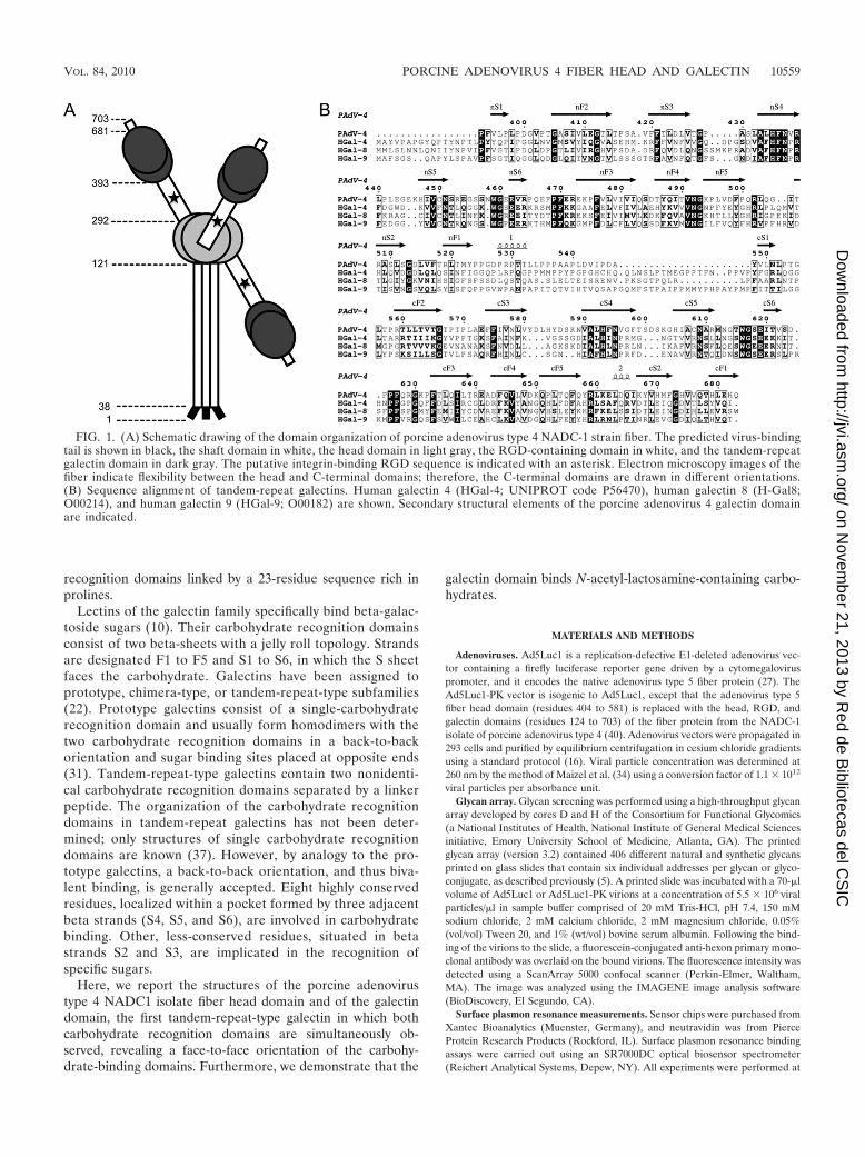
Structure of the head domain. The head domain of eachmonomer forms an antiparallel beta sandwich of eight betastrands (Fig. 2A), which associate to form a beta propellertrimer (Fig. 2B). When the structure is compared to that of thefiber head of human adenovirus type 5 (50), the most promi-nent secondary structural variations occur in the region of theCD and IJ loops, which are located at the top of the trimer andare notably shorter in porcine adenovirus type 4; the HI loopat the side of the trimer also is shorter in porcine adenovirustype 4. Many human adenoviruses bind CAR as a primaryattachment receptor (42). Structural analyses of domain D1 of
FIG. 2. Structure of the porcine adenovirus 4 (NADC-1 strain) fiber head. (A) The monomer is depicted with beta strands colored yellow,helices red, and loops green. Beta strands are labeled with letters, and alpha helices are numbered. (B) A view from the top along the 3-fold axisof the trimer. Monomers are colored red, green, and yellow. (C) Comparison of the porcine adenovirus type 4 fiber head structure (yellow) to thatof the human adenovirus type 12 fiber head (blue) bound to the CAR D1 domain (red). (D) Comparison of the porcine adenovirus type 4 fiberhead structure (yellow) to that of the human adenovirus type 11 fiber head (blue) bound to the CD46 short consensus repeats 1 and 2 (red). TheDG, HI, and IJ loops of the adenovirus fiber head are shown and labeled; residues of the human adenovirus type 11 fiber head involved in theinteraction also are labeled. (E and F) Electrostatic potential surfaces (blue, positive; red, negative) of human adenovirus type 37 fiber head incomplex with sialyl-lactose (E; shown as sticks) and the porcine adenovirus type 4 fiber head (F).
VOL. 84, 2010 PORCINE ADENOVIRUS 4 FIBER HEAD AND GALECTIN 10561
on Novem
ber 21, 2013 by Red de B
ibliotecas del CS
IChttp://jvi.asm
.org/D
ownloaded from
CAR in complex with human adenovirus type 12 fiber head (4)have revealed the interaction residues, which are conserved inthe fibers of types that use CAR as a receptor. It also has beensuggested that the fiber heads that bind CAR have a specificconformation of the AB loop (43). The AB loop of porcineadenovirus type 4 (yellow) structurally resembles AB loopsfound in human adenovirus type 12 fiber and other CAR bind-ing fibers (Fig. 2C). However, none of the CAR binding resi-dues are conserved in porcine adenovirus type 4 fiber, apartfrom Pro-222. Additionally, we could not detect any interac-tion between the CAR D1 domain and porcine adenovirus 4head domain in size-exclusion chromatography or surface plas-mon resonance experiments; the CAR binding adenovirus type5 fiber head also did not block the infection of adenoviruscontaining NADC-1 fiber (data not shown). CD46 is used as areceptor by human adenoviruses of species B as well as someof species D (15, 49). Interaction regions involve the DG, HI,and IJ loops of the adenovirus head domain (41). Structuralsuperposition shows that the conformation of loops DG andHI are different, and the IJ loop is shorter in porcine adeno-virus type 4 (Fig. 2D); furthermore, binding residues Asn-283,Asp-284, Arg-280, and Gln-305 are not conserved in porcineadenovirus 4 fiber head. Finally, cell surface glycoconjugatescontaining alpha-(2,3)- or alpha-(2,6)-linked sialic acid resi-dues have been reported to serve as receptors for humanadenovirus types 37 and 19p (6), which is consistent with apositively charged patch on their surfaces (Fig. 2E). The rela-tive absence of positive charge on the porcine adenovirus type4 head domain (Fig. 2F) suggests that porcine adenovirus type4 head domain does not bind sialic acid. Taken together, thesefindings suggest that the fiber head domain does not bindcommon adenovirus receptors.
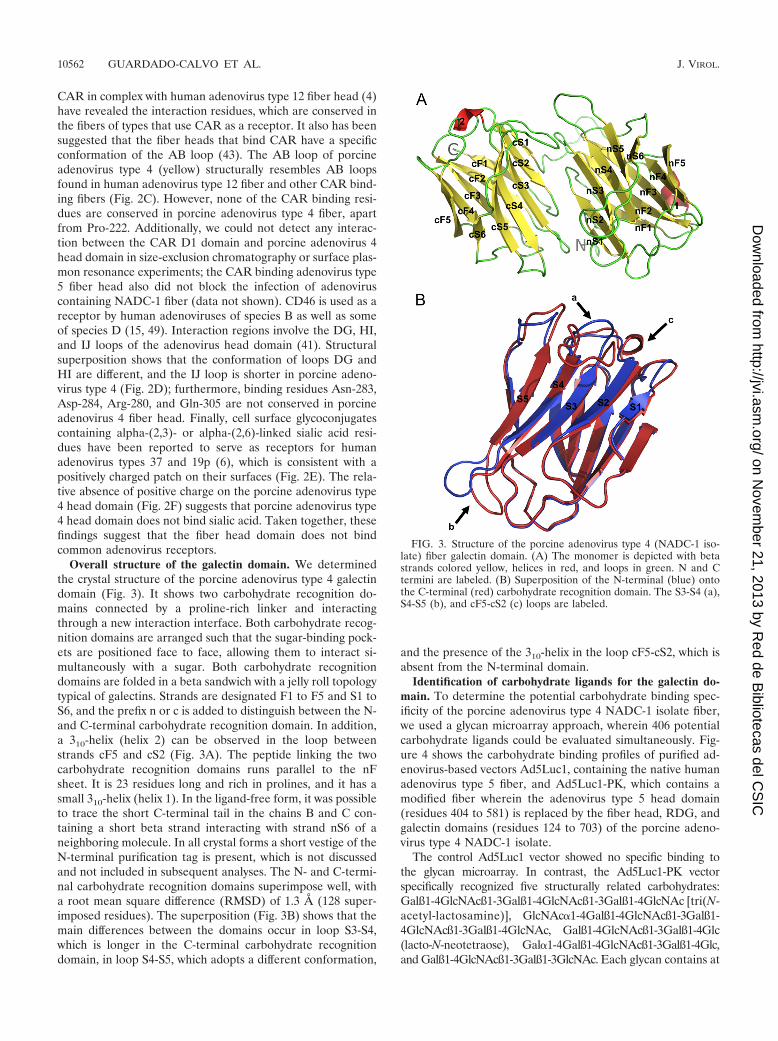
Overall structure of the galectin domain. We determinedthe crystal structure of the porcine adenovirus type 4 galectindomain (Fig. 3). It shows two carbohydrate recognition do-mains connected by a proline-rich linker and interactingthrough a new interaction interface. Both carbohydrate recog-nition domains are arranged such that the sugar-binding pock-ets are positioned face to face, allowing them to interact si-multaneously with a sugar. Both carbohydrate recognitiondomains are folded in a beta sandwich with a jelly roll topologytypical of galectins. Strands are designated F1 to F5 and S1 toS6, and the prefix n or c is added to distinguish between the N-and C-terminal carbohydrate recognition domain. In addition,a 310-helix (helix 2) can be observed in the loop betweenstrands cF5 and cS2 (Fig. 3A). The peptide linking the twocarbohydrate recognition domains runs parallel to the nFsheet. It is 23 residues long and rich in prolines, and it has asmall 310-helix (helix 1). In the ligand-free form, it was possibleto trace the short C-terminal tail in the chains B and C con-taining a short beta strand interacting with strand nS6 of aneighboring molecule. In all crystal forms a short vestige of theN-terminal purification tag is present, which is not discussedand not included in subsequent analyses. The N- and C-termi-nal carbohydrate recognition domains superimpose well, witha root mean square difference (RMSD) of 1.3 Å (128 super-imposed residues). The superposition (Fig. 3B) shows that themain differences between the domains occur in loop S3-S4,which is longer in the C-terminal carbohydrate recognitiondomain, in loop S4-S5, which adopts a different conformation,
and the presence of the 310-helix in the loop cF5-cS2, which isabsent from the N-terminal domain.
Identification of carbohydrate ligands for the galectin do-main. To determine the potential carbohydrate binding spec-ificity of the porcine adenovirus type 4 NADC-1 isolate fiber,we used a glycan microarray approach, wherein 406 potentialcarbohydrate ligands could be evaluated simultaneously. Fig-ure 4 shows the carbohydrate binding profiles of purified ad-enovirus-based vectors Ad5Luc1, containing the native humanadenovirus type 5 fiber, and Ad5Luc1-PK, which contains amodified fiber wherein the adenovirus type 5 head domain(residues 404 to 581) is replaced by the fiber head, RDG, andgalectin domains (residues 124 to 703) of the porcine adeno-virus type 4 NADC-1 isolate.
The control Ad5Luc1 vector showed no specific binding tothe glycan microarray. In contrast, the Ad5Luc1-PK vectorspecifically recognized five structurally related carbohydrates:Galß1-4GlcNAcß1-3Galß1-4GlcNAcß1-3Galß1-4GlcNAc [tri(N-acetyl-lactosamine)], GlcNAc�1-4Galß1-4GlcNAcß1-3Galß1-4GlcNAcß1-3Galß1-4GlcNAc, Galß1-4GlcNAcß1-3Galß1-4Glc(lacto-N-neotetraose), Gal�1-4Galß1-4GlcNAcß1-3Galß1-4Glc,and Galß1-4GlcNAcß1-3Galß1-3GlcNAc. Each glycan contains at
FIG. 3. Structure of the porcine adenovirus type 4 (NADC-1 iso-late) fiber galectin domain. (A) The monomer is depicted with betastrands colored yellow, helices in red, and loops in green. N and Ctermini are labeled. (B) Superposition of the N-terminal (blue) ontothe C-terminal (red) carbohydrate recognition domain. The S3-S4 (a),S4-S5 (b), and cF5-cS2 (c) loops are labeled.
10562 GUARDADO-CALVO ET AL. J. VIROL.
on Novem
ber 21, 2013 by Red de B
ibliotecas del CS
IChttp://jvi.asm
.org/D
ownloaded from
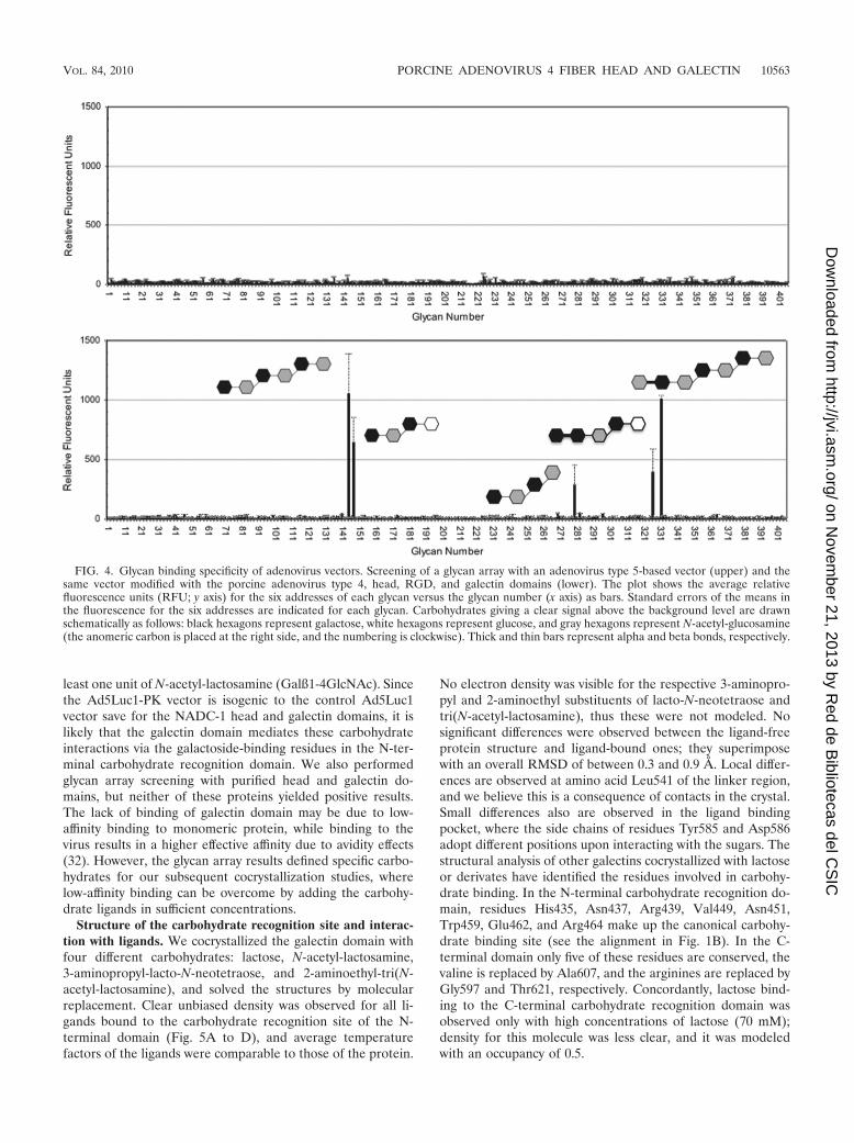
least one unit of N-acetyl-lactosamine (Galß1-4GlcNAc). Sincethe Ad5Luc1-PK vector is isogenic to the control Ad5Luc1vector save for the NADC-1 head and galectin domains, it islikely that the galectin domain mediates these carbohydrateinteractions via the galactoside-binding residues in the N-ter-minal carbohydrate recognition domain. We also performedglycan array screening with purified head and galectin do-mains, but neither of these proteins yielded positive results.The lack of binding of galectin domain may be due to low-affinity binding to monomeric protein, while binding to thevirus results in a higher effective affinity due to avidity effects(32). However, the glycan array results defined specific carbo-hydrates for our subsequent cocrystallization studies, wherelow-affinity binding can be overcome by adding the carbohy-drate ligands in sufficient concentrations.
Structure of the carbohydrate recognition site and interac-tion with ligands. We cocrystallized the galectin domain withfour different carbohydrates: lactose, N-acetyl-lactosamine,3-aminopropyl-lacto-N-neotetraose, and 2-aminoethyl-tri(N-acetyl-lactosamine), and solved the structures by molecularreplacement. Clear unbiased density was observed for all li-gands bound to the carbohydrate recognition site of the N-terminal domain (Fig. 5A to D), and average temperaturefactors of the ligands were comparable to those of the protein.
No electron density was visible for the respective 3-aminopro-pyl and 2-aminoethyl substituents of lacto-N-neotetraose andtri(N-acetyl-lactosamine), thus these were not modeled. Nosignificant differences were observed between the ligand-freeprotein structure and ligand-bound ones; they superimposewith an overall RMSD of between 0.3 and 0.9 Å. Local differ-ences are observed at amino acid Leu541 of the linker region,and we believe this is a consequence of contacts in the crystal.Small differences also are observed in the ligand bindingpocket, where the side chains of residues Tyr585 and Asp586adopt different positions upon interacting with the sugars. Thestructural analysis of other galectins cocrystallized with lactoseor derivates have identified the residues involved in carbohy-drate binding. In the N-terminal carbohydrate recognition do-main, residues His435, Asn437, Arg439, Val449, Asn451,Trp459, Glu462, and Arg464 make up the canonical carbohy-drate binding site (see the alignment in Fig. 1B). In the C-terminal domain only five of these residues are conserved, thevaline is replaced by Ala607, and the arginines are replaced byGly597 and Thr621, respectively. Concordantly, lactose bind-ing to the C-terminal carbohydrate recognition domain wasobserved only with high concentrations of lactose (70 mM);density for this molecule was less clear, and it was modeledwith an occupancy of 0.5.
FIG. 4. Glycan binding specificity of adenovirus vectors. Screening of a glycan array with an adenovirus type 5-based vector (upper) and thesame vector modified with the porcine adenovirus type 4, head, RGD, and galectin domains (lower). The plot shows the average relativefluorescence units (RFU; y axis) for the six addresses of each glycan versus the glycan number (x axis) as bars. Standard errors of the means inthe fluorescence for the six addresses are indicated for each glycan. Carbohydrates giving a clear signal above the background level are drawnschematically as follows: black hexagons represent galactose, white hexagons represent glucose, and gray hexagons represent N-acetyl-glucosamine(the anomeric carbon is placed at the right side, and the numbering is clockwise). Thick and thin bars represent alpha and beta bonds, respectively.
VOL. 84, 2010 PORCINE ADENOVIRUS 4 FIBER HEAD AND GALECTIN 10563
on Novem
ber 21, 2013 by Red de B
ibliotecas del CS
IChttp://jvi.asm
.org/D
ownloaded from
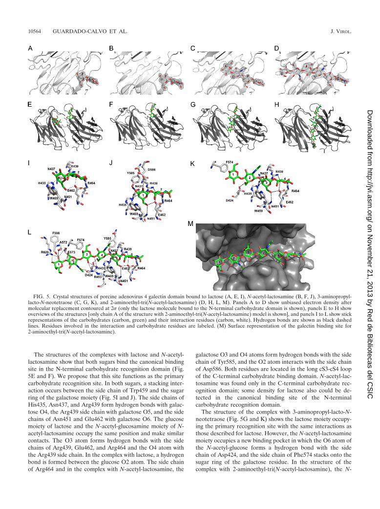
The structures of the complexes with lactose and N-acetyl-lactosamine show that both sugars bind the canonical bindingsite in the N-terminal carbohydrate recognition domain (Fig.5E and F). We propose that this site functions as the primarycarbohydrate recognition site. In both sugars, a stacking inter-action occurs between the side chain of Trp459 and the sugarring of the galactose moiety (Fig. 5I and J). The side chains ofHis435, Asn437, and Arg439 form hydrogen bonds with galac-tose O4, the Arg439 side chain with galactose O5, and the sidechains of Asn451 and Glu462 with galactose O6. The glucosemoiety of lactose and the N-acetyl-glucosamine moiety of N-acetyl-lactosamine occupy the same position and make similarcontacts. The O3 atom forms hydrogen bonds with the sidechains of Arg439, Glu462, and Arg464 and the O4 atom withthe Arg439 side chain. In the complex with lactose, a hydrogenbond is formed between the glucose O2 atom. The side chainof Arg464 and in the complex with N-acetyl-lactosamine, the
galactose O3 and O4 atoms form hydrogen bonds with the sidechain of Tyr585, and the O2 atom interacts with the side chainof Asp586. Both residues are located in the long cS3-cS4 loopof the C-terminal carbohydrate binding domain. N-acetyl-lac-tosamine was found only in the C-terminal carbohydrate rec-ognition domain; some density for lactose also could be de-tected in the canonical binding site of the N-terminalcarbohydrate recognition domain.
The structure of the complex with 3-aminopropyl-lacto-N-neotetraose (Fig. 5G and K) shows the lactose moiety occupy-ing the primary recognition site with the same interactions asthose described for lactose. However, the N-acetyl-lactosaminemoiety occupies a new binding pocket in which the O6 atom ofthe N-acetyl-glucose forms a hydrogen bond with the sidechain of Asp424, and the side chain of Phe574 stacks onto thesugar ring of the galactose residue. In the structure of thecomplex with 2-aminoethyl-tri(N-acetyl-lactosamine), the N-
FIG. 5. Crystal structures of porcine adenovirus 4 galectin domain bound to lactose (A, E, I), N-acetyl-lactosamine (B, F, J), 3-aminopropyl-lacto-N-neotetraose (C, G, K), and 2-aminoethyl-tri(N-acetyl-lactosamine) (D, H, L, M). Panels A to D show unbiased electron density aftermolecular replacement contoured at 2� (only the lactose molecule bound to the N-terminal carbohydrate domain is shown), panels E to H showoverviews of the structures [only chain A of the structure with 2-aminoethyl-tri(N-acetyl-lactosamine) model is shown], and panels I to L show stickrepresentations of the carbohydrates (carbon, green) and their interaction residues (carbon, white). Hydrogen bonds are shown as black dashedlines. Residues involved in the interaction and carbohydrate residues are labeled. (M) Surface representation of the galectin binding site for2-aminoethyl-tri(N-acetyl-lactosamine).
10564 GUARDADO-CALVO ET AL. J. VIROL.
on Novem
ber 21, 2013 by Red de B
ibliotecas del CS
IChttp://jvi.asm
.org/D
ownloaded from
acetyl-lactosamine at the reducing end (i.e., residues 5 and 6 inthe hexasaccharide) is located at the primary recognition site(Fig. 5H and L) and, with the exception of the interaction withthe side chain of Asp586, we observed the same interactions asthose seen with N-acetyl-lactosamine alone (Fig. 5J). The cen-tral N-acetyl-lactosamine unit (residues 3 and 4) binds in thesame place as the N-acetyl-lactosamine unit of 3-aminopropyl-lacto-N-neotetraose, and, besides the interactions with Phe574and Asp424 described above, the N-acetyl-glucose O4 andgalactose O3 atoms form hydrogen bonds with the side chainof Arg453. Finally, the N-acetyl-lactosamine unit at the non-reducing end (residues 1 and 2) is located in a binding sitemade up of residues of the C-terminal carbohydrate recogni-tion domain, where the galactose O2 and O3 atom hydrogenbonds interact with the side chain of Arg629, and the carbonyloxygen of Ala572, Glu573, and Phe598 interacts with the ga-lactose O2 atom.
The tri(N-acetyl-lactosamine) ligand fills the groove betweenboth carbohydrate recognition domains (Fig. 5M). The loweraffinity of the C-terminal carbohydrate recognition domain aswell as steric hindrance from the long loop cS3-cS4 prevent thisinteraction from occurring with the sugar in the opposite ori-entation. Our structures suggest that the porcine adenovirustype 4 galectin domain would not make additional interactionswith longer N-acetyl-lactosamine polymers, therefore an en-hanced affinity for longer poly-N-acetyl-lactosamine polymersmay be explained by avidity contributions. However, branchedsugars may facilitate additional interactions with the protein,possibly involving the C-terminal carbohydrate recognition do-main (one might even speculate that the second lactose-bind-ing site is mimicking such an interaction).
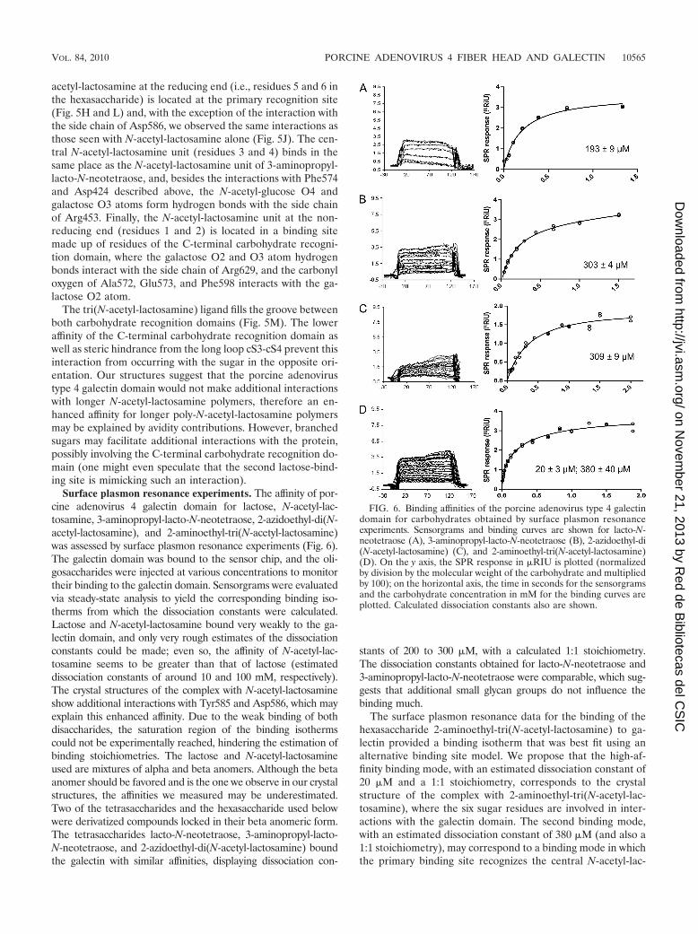
Surface plasmon resonance experiments. The affinity of por-cine adenovirus 4 galectin domain for lactose, N-acetyl-lac-tosamine, 3-aminopropyl-lacto-N-neotetraose, 2-azidoethyl-di(N-acetyl-lactosamine), and 2-aminoethyl-tri(N-acetyl-lactosamine)was assessed by surface plasmon resonance experiments (Fig. 6).The galectin domain was bound to the sensor chip, and the oli-gosaccharides were injected at various concentrations to monitortheir binding to the galectin domain. Sensorgrams were evaluatedvia steady-state analysis to yield the corresponding binding iso-therms from which the dissociation constants were calculated.Lactose and N-acetyl-lactosamine bound very weakly to the ga-lectin domain, and only very rough estimates of the dissociationconstants could be made; even so, the affinity of N-acetyl-lac-tosamine seems to be greater than that of lactose (estimateddissociation constants of around 10 and 100 mM, respectively).The crystal structures of the complex with N-acetyl-lactosamineshow additional interactions with Tyr585 and Asp586, which mayexplain this enhanced affinity. Due to the weak binding of bothdisaccharides, the saturation region of the binding isothermscould not be experimentally reached, hindering the estimation ofbinding stoichiometries. The lactose and N-acetyl-lactosamineused are mixtures of alpha and beta anomers. Although the betaanomer should be favored and is the one we observe in our crystalstructures, the affinities we measured may be underestimated.Two of the tetrasaccharides and the hexasaccharide used belowwere derivatized compounds locked in their beta anomeric form.The tetrasaccharides lacto-N-neotetraose, 3-aminopropyl-lacto-N-neotetraose, and 2-azidoethyl-di(N-acetyl-lactosamine) boundthe galectin with similar affinities, displaying dissociation con-
stants of 200 to 300 �M, with a calculated 1:1 stoichiometry.The dissociation constants obtained for lacto-N-neotetraose and3-aminopropyl-lacto-N-neotetraose were comparable, which sug-gests that additional small glycan groups do not influence thebinding much.
The surface plasmon resonance data for the binding of thehexasaccharide 2-aminoethyl-tri(N-acetyl-lactosamine) to ga-lectin provided a binding isotherm that was best fit using analternative binding site model. We propose that the high-af-finity binding mode, with an estimated dissociation constant of20 �M and a 1:1 stoichiometry, corresponds to the crystalstructure of the complex with 2-aminoethyl-tri(N-acetyl-lac-tosamine), where the six sugar residues are involved in inter-actions with the galectin domain. The second binding mode,with an estimated dissociation constant of 380 �M (and also a1:1 stoichiometry), may correspond to a binding mode in whichthe primary binding site recognizes the central N-acetyl-lac-
FIG. 6. Binding affinities of the porcine adenovirus type 4 galectindomain for carbohydrates obtained by surface plasmon resonanceexperiments. Sensorgrams and binding curves are shown for lacto-N-neotetraose (A), 3-aminopropyl-lacto-N-neotetraose (B), 2-azidoethyl-di(N-acetyl-lactosamine) (C), and 2-aminoethyl-tri(N-acetyl-lactosamine)(D). On the y axis, the SPR response in �RIU is plotted (normalizedby division by the molecular weight of the carbohydrate and multipliedby 100); on the horizontal axis, the time in seconds for the sensorgramsand the carbohydrate concentration in mM for the binding curves areplotted. Calculated dissociation constants also are shown.
VOL. 84, 2010 PORCINE ADENOVIRUS 4 FIBER HEAD AND GALECTIN 10565
on Novem
ber 21, 2013 by Red de B
ibliotecas del CS
IChttp://jvi.asm
.org/D
ownloaded from
tosamine residue, and only the first four sugar residues fromthe nonreducing end are involved in interactions with the pro-tein. A similar binding mode has been reported for the N-terminal domain of human tandem-repeat-type galectin 9 (37).Although the measured affinities toward the monomeric galec-tin are moderate, one has to remember that adenovirus fiber istrimeric and thus contains three independent galectin do-mains. Furthermore, each viral particle contains 12 trimericfibers. This multimeric arrangement would enable the estab-lishment of multivalent interactions between the viral particlesand target surface-bound oligosaccharides on cell surfaces,resulting in higher functional affinities (32). Furthermore, oli-gosaccharides of sufficient length may bind more than onegalectin domain (33). Another thing that should be kept inmind is that more-complex, branched sugars not present in theglycan array (and not used in our binding or cocrystallizationstudies) may make even more contacts with galectin and bindwith higher affinity than the carbohydrates mentioned.
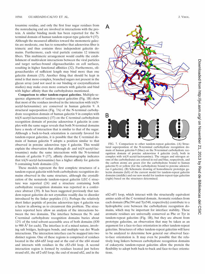
Comparison to other tandem-repeat galectins. Multiple-se-quence alignments of tandem-repeat galectins (Fig. 1B) showthat most of the residues involved in the interaction with tri(N-acetyl-lactosamine) are conserved in human galectin 9. Astructural superposition (Fig. 7A) of the N-terminal carbohy-drate recognition domain of human galectin 9 in complex withtri(N-acetyl-lactosamine) (37) on the C-terminal carbohydraterecognition domain of porcine adenovirus 4 galectin in com-plex with the same sugar reveals that both N-terminal domainshave a mode of interaction that is similar to that of the sugar.Although a back-to-back orientation is currently favored fortandem-repeat galectins, it is possible that the C-terminal do-main of human galectin 9 adopts a position similar to thatobserved in porcine adenovirus type 4 galectin. This wouldexplain the observation that although di- and tri(N-acetyl-lac-tosamine) make the same interactions with the N-terminaldomain of galectin 9 (37), affinity chromatography indicatesthat tri(N-acetyl-lactosamine) has a higher affinity for galectin9 containing both domains (23).
These models represent the first complete structures of atandem-repeat galectin with both carbohydrate recognition do-mains observed in the same structure, although the crystalli-zation of the nematode tandem-repeat galectin LEC-1 struc-ture was reported (24) and a structure containing bothcarbohydrate recognition domains was reported in a confer-ence abstract (39). It has been suggested previously that tan-dem-repeat galectins do not crystallize readily due to disorderintroduced by the linker peptides (11). Perhaps the relativelyshort linker peptide of porcine adenovirus type 4 galectin wasa factor in allowing us to circumvent this problem. The struc-tures reported here allow an analysis of the interactions be-tween the two domains. The interface between the N- andC-terminal carbohydrate recognition domains buries about12% of the total solvent-accessible area of both domains (830to 900 Å2 for each). The interface is of a mixed nature, involv-ing salt bridges, hydrogen bonds, and multiple van der Waalsinteractions. The interaction interface can be mapped into twodistinct regions. One of these regions is comprised of residueslocated in the nS4-nS5 loop and at the end of the nS4 strandand interacts with residues in the cS3-cS4 loop. A secondinteraction region is formed by residues located just beforestrand nS1, the nF2-nS3 loop, the end of strand nS2, and in the
nS2-nF1 loop, which interact with the structurally equivalentamino acids of the C-terminal domain. Aromatic residues fromeach domain (Phe394 and Tyr548, respectively) contribute to ahydrophobic core between the carbohydrate recognition do-mains, which may be important for interface stability. Thesearomatic residues are universally conserved as Phe or Tyr intandem-repeat galectins (Fig. 1B), but they are absent fromprototype galectins, an observation that may be taken as anargument for a face-to-face orientation in other tandem-repeatgalectins. Structures of other tandem-repeat galectins will haveto be analyzed to determine how general our observed face-to-face orientation is. It may even be possible that the rela-tively long linkers between carbohydrate recognition domainsof eukaryotic tandem-repeat galectins allow the protein theflexibility to adopt both back-to-back and face-to-face orienta-tions.
FIG. 7. Comparison to other tandem-repeat galectins. (A) Struc-tural superposition of the N-terminal carbohydrate recognition do-main of human galectin-9 (blue) on the N-terminal carbohydrate rec-ognition domain of porcine adenovirus 4 galectin (red), both incomplex with tri(N-acetyl-lactosamine). The oxygen and nitrogen at-oms of the carbohydrates are colored in red and blue, respectively, andthe carbon atoms are green (for the carbohydrate bound to humangalectin-9) or yellow (for the carbohydrate bound to porcine adenovi-rus 4 galectin). (B) Schematic drawing of homodimeric prototype ga-lectin domains (left) of the current model for tandem-repeat galectindomains (middle) and our new model for tandem-repeat-type galectins(right) based on the structures determined.
10566 GUARDADO-CALVO ET AL. J. VIROL.
on Novem
ber 21, 2013 by Red de B
ibliotecas del CS
IChttp://jvi.asm
.org/D
ownloaded from
DISCUSSION
The natural receptor of porcine adenovirus type 4 has notbeen identified but does not appear to be CAR, CD46, or sialicacid (40 and this paper). The analysis of the sequence C ter-minal to the head domain revealed an RGD-containing se-quence and a galectin-like sequence consisting of two putativecarbohydrate recognition domains. In many adenoviruses, theRGD-containing sequence is located in the penton base pro-tein, the capsid protein into which the fiber is anchored. There,it has been shown to be important for interaction with integrins(30). It is plausible that porcine adenovirus type 4 has theability to interact with integrins via the RGD sequence of itsfiber (the sequence of the penton protein is not known). Theresults presented in this paper demonstrate the ability of theporcine adenovirus 4 fiber C-terminal galectin domain to bindsugars containing N-acetyl-lactosamine units, thus confirmingit as a bona fide galectin. Three linearly connected N-acetyl-lactosamine units may bind at the same time. The location ofthe galectin domain at the C-terminal end of the NADC-1 fibersuggests that this domain projects away from the virus, makingit probable that sugars containing repeating N-acetyl-lac-tosamine units are natural receptors for this virus. However,the relative importance of N-acetyl-lactosamine-containingcarbohydrates and integrins as receptors for porcine adenovi-rus type 4 remains to be determined in detail, although pre-liminary experiments (not shown) indicate that the isolatedgalectin domain partially blocks gene transfer by the vectorAd5Luc1-PK, containing the porcine adenovirus type 4 fiberC-terminal domains.
Structural studies have revealed the structural basis of thedimeric organization in some prototype galectins, in whichcarbohydrate-binding sites are projected away from eachother, and it is unlikely that the two sites cooperatively bindone carbohydrate molecule (3) (Fig. 7B, left). The dimericprototype galectin structure has been used to build models oftandem-repeat galectins (51) (Fig. 7B, middle), although ex-perimental evidence suggests cooperative interactions betweenthe two domains and the necessity of the linker peptide forproper functioning (29). Furthermore, glycan array experi-ments carried out with other tandem-repeat galectins point tothe necessity of both domains for the recognition of specificsugars, and studies with human galectin 8 have shown thatalthough the specificity of both domains is additive in solution,they bind synergistically to carbohydrates (7). Here, we reportthe first structure of a tandem-repeat galectin in which bothcarbohydrate recognition domains are present and organizedin a way not resembling the prototype galectin dimer. Thedomains are arranged such that they can interact cooperativelywith a single glycan (Fig. 7B, right); the cocrystal structure with2-aminoethyl-tri(N-acetyl-lactosamine) reported here confirmsthey do. This structure may represent a new paradigm in theorganization of tandem-repeat galectins and explains the im-portance of the linker peptide as an organizer of both domains.The face-to-face orientations of the carbohydrate recognitiondomains allow residues of both domains to interact with thesame oligosaccharide and, in principle, may allow for the rec-ognition of more complex sugars than that of one domainalone. Future structures of other tandem-repeat galectins willreveal how general the face-to-face orientation we observe is.
Porcine adenovirus type 4 has been associated with enceph-alitis (12). The results reported here will be of use in futureefforts focused on elucidating the early steps of the porcineadenovirus 4 replication cycle and may lead to therapeuticapplications for swine diseases. Incorporating the galectin do-main in human gene therapy vectors may allow the targeting ofthese vectors to specific cells. The cocrystal structures pre-sented here identify the interactions of the galectin domainwith the N-acetyl-lactosamine repeats, opening up the possi-bility of generating site-directed mutants to change the speci-ficity of the galectin domain and the targeting of adenovirus-based vectors to specific disease-related carbohydrates (20).
ACKNOWLEDGMENTS
We thank Jose M. Otero, Sergio Galan-Bartual, and Bruno Da-cunha-Marinho for help with data collection and Patricia FerracesCasais for technical assistance. We gratefully acknowledge David F.Smith and Jamie Heimburg-Molinaro of the Protein-Glycan Interac-tion Core Facility (Core H) of the Consortium for Functional Glyco-mics, funded by NIGMS GM62116, Emory University School of Med-icine (Atlanta, GA) for performing the glycan array analysis, and thestaff of the ESRF beam lines ID14-2 and BM30A and EMBL-DESYbeam line X11 for help with data collection.
This research was sponsored by research grants BFU2005-02974,BFU2005-24982-E, and BFU2008-01588 and by a predoctoral FPUfellowship to P.G.-C. from the Spanish Ministry of Education andScience. This work also was supported by funds from the EuropeanCommission under contract NMP4-CT-2006-033256 (BeNatural-coor-dinated project), by NIH grant 1R01HL092941 to D.T.C., and by theXunta de Galicia via an Isidro Parga Pondal fellowship to E.M.M.
REFERENCES
1. Arnberg, N. 2009. Adenovirus receptors: implications for tropism, treatmentand targeting. Rev. Med. Virol. 19:165–178.
2. Bachtarzi, H., M. Stevenson, and K. Fisher. 2008. Cancer gene therapy withtargeted adenoviruses. Expert Opin. Drug Deliv. 5:1231–1240.
3. Barondes, S. H., D. N. Cooper, M. A. Gitt, and H. Leffler. 1994. Galectins.Structure and function of a large family of animal lectins. J. Biol. Chem.269:20807–20810.
4. Bewley, M. C., K. Springer, Y. B. Zhang, P. Freimuth, and J. M. Flanagan.1999. Structural analysis of the mechanism of adenovirus binding to itshuman cellular receptor, CAR. Science 286:1579–1583.
5. Blixt, O., S. Head, T. Mondala, C. Scanlan, M. E. Huflejt, R. Alvarez, M. C.Bryan, F. Fazio, D. Calarese, J. Stevens, N. Razi, D. J. Stevens, J. J. Skehel,I. van Die, D. R. Burton, I. A. Wilson, R. Cummings, N. Bovin, C. H. Wong,and J. C. Paulson. 2004. Printed covalent glycan array for ligand profiling ofdiverse glycan binding proteins. Proc. Natl. Acad. Sci. U. S. A. 101:17033–17038.
6. Burmeister, W. P., D. Guilligay, S. Cusack, G. Wadell, and N. Arnberg. 2004.Crystal structure of species D adenovirus fiber knobs and their sialic acidbinding sites. J. Virol. 78:7727–7736.
7. Carlsson, S., C. T. Oberg, M. C. Carlsson, A. Sundin, U. J. Nilsson, D.Smith, R. D. Cummings, J. Almkvist, A. Karlsson, and H. Leffler. 2007.Affinity of galectin-8 and its carbohydrate recognition domains for ligands insolution and at the cell surface. Glycobiology 17:663–676.
8. Chen, V. B., W. B. Arendall, J. J. Headd, D. A. Keedy, R. M. Immormino,G. J. Kapral, L. W. Murray, J. S. Richardson, and D. C. Richardson. 2010.MolProbity: all-atom structure validation for macromolecular crystallogra-phy. Acta Crystallogr. D 66:12–21.
9. Chenna, R., H. Sugawara, T. Koike, R. Lopez, T. J. Gibson, D. G. Higgins,and J. D. Thompson. 2003. Multiple sequence alignment with the Clustalseries of programs. Nucleic Acids Res. 31:3497–3500.
10. Cooper, D. N. 2002. Galectinomics: finding themes in complexity. Biochim.Biophys. Acta 1572:209–231.
11. Cummings, R. D., and F. Liu. 2009. S-type lectins (galectins), p. 475–479. InA. Varki, R. D. Cummings, J. D. Esko, H. H. Freeze, G. W. Hart, and J.Marth (ed.), Essentials of glycobiology, 2nd ed. Cold Spring Harbor Labo-ratory Press, Woodbury, NY.
12. Edington, N., L. Kasza, and G. J. Christofinis. 1972. Meningo-encephalitis ingnotobiotic pigs inoculated intransally and orally with porcine adenovirus 4.Res. Vet. Sci. 13:289–291.
13. El Bakkouri, M., E. Seiradake, S. Cusack, R. W. Ruigrok, and G. Schoehn.2008. Structure of the C-terminal head domain of the fowl adenovirus type1 short fibre. Virology 378:169–176.
VOL. 84, 2010 PORCINE ADENOVIRUS 4 FIBER HEAD AND GALECTIN 10567
on Novem
ber 21, 2013 by Red de B
ibliotecas del CS
IChttp://jvi.asm
.org/D
ownloaded from
14. Emsley, P., and K. Cowtan. 2004. Coot: model-building tools for moleculargraphics. Acta Crystallogr. D 60:2126–2132.
15. Gaggar, A., D. M. Shayakhmetov, and A. Lieber. 2003. CD46 is a cellularreceptor for group B adenoviruses. Nat. Med. 9:1408–1412.
16. Graham, F. L., and L. Prevec. 1995. Methods for construction of adenovirusvectors. Mol. Biotechnol. 3:207–220.
17. Guardado Calvo, P., A. L. Llamas-Saiz, P. Langlois, and M. J. van Raaij.2006. Crystallization of the C-terminal head domain of the avian adenovirusCELO long fibre. Acta Crystallogr. F 62:449–452.
18. Guardado-Calvo, P., A. L. Llamas-Saiz, G. C. Fox, P. Langlois, and M. J. vanRaaij. 2007. Structure of the C-terminal head domain of the fowl adenovirustype 1 long fiber. J. Gen. Virol. 88:2407–2416.
19. Guardado-Calvo, P., A. L. Llamas-Saiz, G. C. Fox, J. N. Glasgow, and M. J.van Raaij. 2009. Crystallization of the head and galectin-like domains ofporcine adenovirus isolate NADC-1 fibre. Acta Crystallogr. F 65:1149–1152.
20. Hakomori, S. 1984. Tumor-associated carbohydrate antigens. Annu. Rev.Immunol. 2:103–126.
21. Hammond, J. M., and M. A. Johnson. 2005. Porcine adenovirus as a deliverysystem for swine vaccines and immunotherapeutics. Vet. J. 169:17–27.
22. Hirabayashi, J., and K. Kasai. 1993. The family of metazoan metal-inde-pendent beta-galactoside-binding lectins: structure, function and molecularevolution. Glycobiology 3:297–304.
23. Hirabayashi, J., T. Hashidate, Y. Arata, N. Nishi, T. Nakamura, M.Hirashima, T. Urashima, T. Oka, M. Futai, W. E. Muller, F. Yagi, and K.Kasai. 2002. Oligosaccharide specificity of galectins: a search by frontalaffinity chromatography. Biochim. Biophys. Acta 1572:232–254.
24. Itagaki, T., S. Nishizaki, K. Sekihashi, H. Kobayashi, S. Kidokoro, Y. Ke-zuka, Y. Arata, J. Hirabayashi, K. Kasai, and T. Nonaka. 2008. Crystalliza-tion and preliminary X-ray crystallographic analysis of galectin LEC-1 fromCaenorhabditis elegans. Protein Pept. Lett. 15:419–422.
25. Kleiboeker, S. B., B. S. Sealm, and W. L. Mengeling. 1993. Genomic cloningand restriction site mapping of a porcine adenovirus isolate: demonstrationof genomic stability in porcine adenovirus. Arch. Virol. 133:357–368.
26. Kleiboeker, S. B. 1995. Sequence analysis of the fiber genomic region of aporcine adenovirus predicts a novel fiber protein. Virus Res. 39:299–309.
27. Krasnykh, V., N. Belousova, N. Korokhov, G. Mikheeva, and D. T. Curiel.2001. Genetic targeting of an adenovirus vector via replacement of the fiberprotein with the phage T4 fibritin. J. Virol. 75:4176–4183.
28. Krissinel, E., and K. Henrick. 2007. Inference of macromolecular assembliesfrom crystalline state. J. Mol. Biol. 372:774–797.
29. Levy, Y., S. Auslender, M. Eisenstein, R. R. Vidavski, D. Ronen, A. D.Bershadsky, and Y. Zick. 2006. It depends on the hinge: a structure-func-tional analysis of galectin-8, a tandem-repeat type lectin. Glycobiology 16:463–476.
30. Lindert, S., M. Silvertry, T. M. Mullen, G. Nemerow, and P. L. Stewart. 2009.Cryo-electron microscopy structure of an adenovirus-integrin complex indi-cates conformational changes in both penton base and integrin. J. Virol.83:11491–11501.
31. Lobsanov, Y. D., M. A. Gitt, H. Leffler, S. H. Barondes, and J. M. Rini. 1993.X-ray crystal structure of the human dimeric S-Lac lectin L-14-II, in complexwith lactose at 2.9 Å resolution. J. Biol. Chem. 268:27034–27038.
32. Lortat-Jacob, H., E. Chouin, S. Cusack, and M. J. van Raaij. 2001. Kineticanalysis of adenovirus fiber binding to its receptor reveals an avidity mech-anism for trimeric receptor-ligand interactions. J. Biol. Chem. 276:9009–9015.
33. Lundquist, J. J., and E. J. Toone. 2002. The cluster glycoside effect. Chem.Rev. 102:555–578.
34. Maizel, J. V., Jr., D. O. White, and M. D. Scharff. 1968. The polypeptides of
adenovirus. I. Evidence for multiple protein components in the virion and acomparison of types 2, 7A, and 12. Virology 36:115–125.
35. McCoy, A. J., R. W. Grosse-Kunstleve, P. D. Adams, M. D. Winn, L. C.Storoni, and R. J. Read. 2007. Phaser crystallographic software. J. Appl.Crystallogr. 40:658–674.
36. Murshudov, G. N., A. A. Vagin, and E. J. Dodson. 1997. Refinement ofmacromolecular structures by the maximum-likelihood method. Acta Crys-tallogr. D 53:240–255.
37. Nagae, M., N. Nishi, T. Murata, T. Usui, T. Nakamura, S. Wakatsuki, andR. Kato. 2009. Structural analysis of the recognition mechanism of poly-N-acetyllactosamine by the human galectin-9 N-terminal carbohydrate recog-nition domain. Glycobiology 19:112–117.
38. Nemerow, G. R., L. Pache, V. Reddy, and P. L. Stewart. 2009. Insights intoadenovirus host cell interactions from structural studies. Virology 384:380–388.
39. Nonaka, T., K. Sekihashi, S. Nishizaki, Y. Arata, J. Hirabayashi, K. Kasai,and Y. Mitsui. 1999. Crystal structure of the tandem-repeat galectin of thenematode Caenorhabditis elegans. Glycoconj. J. 16:S119.
40. Paul, C. P., M. Everts, J. N. Glasgow, P. Dent, P. B. Fisher, I. V. Ulasov,M. S. Lesniak, M. A. Stoff-Khalili, J. C. Roth, M. A. Preuss, C. M. Dirven,M. L. Lamfers, G. P. Siegal, Z. B. Zhu, and D. T. Curiel. 2008. Character-ization of infectivity of knob-modified adenoviral vectors in glioma. CancerBiol. Ther. 7:786–793.
41. Persson, B. D., D. M. Reiter, M. Marttila, Y. F. Mei, J. M. Casasnovas, N.Arnberg, and T. Stehle. 2007. Adenovirus type 11 binding alters the confor-mation of its receptor CD46. Nat. Struct. Mol. Biol. 14:164–166.
42. Roelvink, P. W., A. Lizonova, J. G. Lee, Y. Li, J. M. Bergelson, R. W. Finberg,D. E. Brough, I. Kovesdi, and T. J. Wickham. 1998. The coxsackievirus-adeno-virus receptor protein can function as a cellular attachment protein for adeno-virus serotypes from subgroups A, C, D, E, and F. J. Virol. 72:7909–7915.
43. Seiradake, E., and S. Cusack. 2005. Crystal structure of enteric adenovirusserotype 41 short fiber head. J. Virol. 79:14088–14094.
44. Seiradake, E., H. Lortat-Jacob, O. Billet, E. J. Kremer, and S. Cusack. 2006.Structural and mutational analysis of human Ad37 and canine adenovirus 2fiber heads in complex with the D1 domain of coxsackie and adenovirusreceptor. J. Biol. Chem. 281:33704–33716.
45. Thacker, E. E., L. Timares, and Q. L. Matthews. 2009. Strategies to over-come host immunity to adenovirus vectors in vaccine development. ExpertRev. Vaccines 8:761–777.
46. Vagin, A., and A. Teplyakov. 2010. Molecular replacement with MOLREP.Acta Crystallogr. D 66:22–25.
47. van Raaij, M. J., A. Mitraki, G. Lavigne, and S. Cusack. 1999. A triplebeta-spiral in the adenovirus fibre shaft reveals a new structural motif for afibrous protein. Nature 401:935–938.
48. Winn, M. D., M. N. Isupov, and G. N. Murshudov. 2001. Use of TLSparameters to model anisotropic displacements in macromolecular refine-ment. Acta Crystallogr. D 57:122–133.
49. Wu, E., S. A. Trauger, L. Pache, T. M. Mullen, D. J. von Seggern, G. Siuzdak,and G. R. Nemerow. 2004. Membrane cofactor protein is a receptor foradenoviruses associated with epidemic keratoconjunctivitis. J. Virol.78:3897–3905.
50. Xia, D., L. J. Henry, R. D. Gerard, and J. Deisenhofer. 1994. Crystal struc-ture of the receptor-binding domain of adenovirus type 5 fiber protein at 1.7Å resolution. Structure 2:1259–1270.
51. Zick, Y., M. Eisenstein, R. A. Goren, Y. R. Hadari, Y. Levy, and D. Ronen.2004. Role of galectin-8 as a modulator of cell adhesion and cell growth.Glycoconj. J. 19:517–526.
10568 GUARDADO-CALVO ET AL. J. VIROL.
on Novem
ber 21, 2013 by Red de B
ibliotecas del CS
IChttp://jvi.asm
.org/D
ownloaded from