csf c – reactive protein estimation for the bed side
TRANSCRIPT
CSF C ndash REACTIVE PROTEIN ESTIMATION FOR THE BED SIDE DIAGNOSIS OF
PYOGENIC MENINGITIS
Dissertation Submitted for
MD DEGREE EXAMINATION BRANCH VII - PAEDIATRIC MEDICINE
INSTITUTE OF CHILD HEALTH AND HOSPITALFOR CHILDREN
MADRAS MEDICAL COLLEGETHE TAMIL NADU DR MGR MEDICAL
UNIVERSITY CHENNAI
MARCH 2008
CERTIFICATE
Certified that this dissertation entitled CSF C ndash REACTIVE PROTEIN
ESTIMATION FOR THE BED SIDE DIAGNOSIS OF PYOGENIC
MENINGITIS is a bonafide work done by DrSKALPANA MD Postgraduate
student of Paediatric Medicine Institute of Child Health and Hospital for Children
Egmore Chennai ndash8 attached to Madras Medical College during the academic year
2005-2008
Prof DR S BHAGAVATHY MD DCHAdditional Professor of PaediatricsInstitute Of Child Health and Hospital for ChildrenMadras Medical College
Prof Dr SARADHA SURESH MD PhD FRCP (Glas) Director and Superintendent (IC)Institute of Child Health and Hospital for Children
Madras Medical College ChennaiProf Dr T P KALANITI MDDeanMadras Medical College Chennai
SPECIAL ACKNOWLEDGEMENT
My sincere thanks to Prof Dr TP KALANITI MD the Dean of Madras
Medical College for allowing me to do this dissertation and to utilize the facilities of the
institution
ACKNOWLEDGEMENTS
The satisfaction and elation that accompanies the successful completion of any
task would be incomplete without the mention of the people who have made it possible
It is my privilege to express my gratitude and respect to all those who have guided me
and inspired me during the course of my dissertation
I would like to express my sincere gratitude to
Prof Dr SARADHA SURESH MD PhD FRCP (Glas) Professor and Head
of the Department of Pediatrics and Director and Superintendent (IC) of Institute of
Child Health and Hospital for Children for permitting me to undertake this study
I am extremely thankful to my unit Chief
Prof Dr SBHAGAVATHY MD DCH for her invaluable help guidance
encouragement and support throughout the study
I am also extremely thankful to the Professor of Microbiology DrMEERAN
MOHAMMED MD and Assistant professor DrUMADEVI MD for guiding me in
my study
I thank the assistant professors of my unit Dr S PARIVATHINI MD
DrBSATHYAMOORTHY MD DrHEMACHITHRAJ MD for their guidance
and support
I extend my sincere thanks to the Registrar DrPRAMACHANDRAN MD
DCH and Dr NEDUNCHEZIAN MD for their valuable suggestions in doing this
work
I also thank Mrs Basilea Watson for all her help in statistics through out the
study
I am also highly indebted to all the innocent children who were part my study
without whom this would not have been possible
Above all I am thankful to God the Almighty and my parents and my friends for
their invaluable help in pursuing my dissertation
CONTENTS
SLNO
TITLE PAGE NO
I INTRODUCTION 1
II REVIEW OF LITERATURE 22
III STUDY JUSTIFICATION 34
IV OBJECTIVES OF THE STUDY 35
V MATERIALS AND METHODS 36
VI RESULTS 40
VII DISCUSSION 56
VIII CONCLUSION 61
BIBLIOGRAPHY
ANNEXURE
INTRODUCTION
Infections of the central nervous system are fairly common in pediatric practice
The clinical profile is protean A high index of suspicion of the treating physician is
essential to make an early diagnosis The need for early diagnosis is imperative Potent
antibiotics have reduced mortality but do not prevent sequelae especially if therapy is
delayed The newer rapid diagnostic tests and imaging modalities have improved the
holistic management of children with CNS infections Pretreatment with antibiotics of
patients with purulent meningitis can modify the clinical picture and CSF findings So
distinctive etiological diagnosis becomes difficult Gramrsquo staining of CSF can provide a
rapid preliminary identification of infective organism but is liable to misinterpretation
especially in inexperienced hands
Culture of CSF takes 24 to 48 hours for isolating the causative organism Further
more culture may not always be positive in children who have received antibiotics prior
to hospitalization
Bacterial polysaccharide antigen of microorganisms can be detected by newer
immunological tests like Counter immuno electrophoresis Latex agglutination tests etc
Since antigen may be present in the CSF even after lysis of bacteria these
immunological tests could prove useful even in partially treated patients The present
study was therefore designed to evaluate the utility of CRP in CSF in diagnosing cases
of pyogenic meningitis to study the spectrum of bacterial pathogens causing acute
bacterial meningitis their sensitivity pattern to antibiotics and to analyse the clinical
profile of bacterial meningitis
OVERVIEW OF PYOGENIC MENINGITIS IN CHILDREN
Infections of central nervous system can be broadly classified into
1) Meningitis ndash acute subacute chronic
2) Encephalitis and infective encephalopathy
3) Cerebral abcess granuloma parasitic infestations
Meningitis may be
i Acute- bacterial viral
ii Subacute or chronic- TB fungal parasitic neoplastic or chemical
iii Partially treated
Acute bacterial meningitis which is a major cause of mortality and morbidity
among children occurs both in epidemic and sporadic pattern It may follow septicemia
apparently trivial illness like upper respiratory tract infections otitis media pyoderma
and minor head trauma Patients with diminished host resistance as in diabetes mellitus
malignancies and patients on immunosuppresive drugs are more susceptible to develop
meningitis
PATHOPHYSIOLOGY OF BACTERIAL MENINGITIS
Nasopharyngeal colonization Paranasal sinusitis Otitis media Orbital cellulitis
+ - Viral infn Cranial or vertebral osteomyelitis Local invasion
Endocarditis Bacteremia Pneumonia
thromophlebitis Severe burns Indwelling catheters Contaminated infusion Systemic hypotension equipment
Meningeal invasion Penetrating cranial trauma Meningomyelocele Dermal sinus tractsLoss of auto Regulation Ventricular amp Subarachnoid space infln
Vasculitis
Vasospasm CSF outflow resistance cytotoxic BBB permeability thrombosis edema
Transient Hydrocephalus
CerebralIschemia infarction Interstitial edema Vasogenic edema
RAISED ICT
Common etiological pathogens1
AGE GROUP COMMON BACTERIAL PATHOGENS
NEWBORN Escherichia coli
Klebseilla pneumoniae
Listeria monocytogenes
Enterococcus Sp
Salmonella Sp
4 WEEKS TO 12
WEEKS
Hemophilus influenza
Streptococcus pneumoniae
Gp B streptococcus
Listeria monocytogenes
MORE THAN 12
WEEKS
Hemophilus influenza
Streptococcus pneumoniae
Nisseriea miningitidis
Epidemiology of acute bacterial meningitis is primarily a reflection of epidemics of
bacteremia
Incidence of pyogenic meningitis
In developed countries 15 100000
In developing countries 20 100000
The case fatality rate in early childhood is 10
Incidence in ICH
YEAR
2003 2004 2005 2006
NO OF CASES 126 120 84 55
PREDISPOSING FACTORS
Acute otitis media
Chronic sinusitis
Pneumonia
Endocarditis
Head injury
Recent neurosurgery
Neurosurgical devices
Altered immune status
CSF leak
FACTORS CONTRIBUTING TO INFECTION LEADING TO MENINGITIS
Host factors
Age
Sex Male infants have higher incidence of Gram neg organisms
Female infants are more susceptible to Lmonocytogenes
Group B streptococcus affects both sexes equally
Race blacks have a higher incidence rate
Immunity level of host
Genetic predisposition presence of HLA B12
Presence of HLA BW 40 ndash H influenza
Presence of HLA B27 - N meningitidis
Environmental factors incidence more in
Winter season
Overcrowding
Poor socioeconomic conditions
HOST DEFENSE MECHANISMS IN SUBARACHNOID SPACE
The host response to infection is by way of cerebral edema which causes the
multitude of features in meningitis
i Complement mediated
Complement levels are low absent in normal CSF Complement levels
increases in infection but is insufficient
Low CSF complement is due to
Variable permeability of BBB
Variable degrees of sub arachnoid space inflammation
Enhanced clearance from subarachnoid space
Low production rates in CNS
Degradation at site of infection
Leukocyte protease degrades functional complement C3b with formation of non-
opsonic break down product C3d Hence opsonic and bacteriocidal activity is absent
ii Humoral
Normal CSF ndash IgM absent
IgG levels low
Immunoglobulins increase in acute bacterial meningitis but are insufficient
iii Cell Mediated
Hall mark ndash Neutrophilic phagocytosis
C5a acts as chemotactic factor for neutrophils
Meningeal amp perivascular macrophages also play a role
SPREAD
Hematogenous ndash most common
Direct
Contiguous
CLINICAL FEATURES4
Neonate
Usually nonspecific with few signs referable to the CNS
Bulging fontanelle
Seizures (40)
Temperature instability
Irritability high pitched cry
Lethargy poor feeding vomiting
Apnea cyanosis
Infants
Mild irritability reluctance to flex the neck
Projectile vomiting vacant stare convulsions high pitched cry
Tense and bulging fontanelle
Temperature instability
Older children
Headache
Projectile vomiting
Lethargy mental confusion
90 to 95 have fever
Anorexia arthralgia myalgia
Photophobia
Papilloedema
Untreated illness progresses to convulsions coma and death
Signs of meningeal irritation43
Nuchal rigidity
Kernigrsquo sign
Brudzinskirsquo neck and leg sign
Signs of meningeal irritation may be absent in
Neonates
Young infants
Comatose
Paralyzed children
Pretreatment with antibiotics
Unduly sedated child
Cutaneous manifestations of bacterial meningitis
Purpura in meningococcal meningitis
Ecthyma gangrenosum in pseudomonas meningitis
Tache cerebrale due to autonomic disturbances
Petichiae and purpura in DIVC
Others
Waterhouse Friedrichson syndrome in meningococcal meningitis
Other signs of raised ICP like Charcotrsquos triad
Focal neurological signs
DIAGNOSIS
Complete blood count
Differential count
Blood culture
Gold standard is CSF culture
CSF analysis
Cell count
CSF protein
CSF sugar
Gramrsquo stain
Chest X ray
Others blood sugar blood gases and serum electrolytes must be monitored
regularly to rule out hypo or hyperglycemia acidosis SIADH
CSF analysis is corner stone for the diagnosis of meningitis 5
CSF NORMAL BACTERIAL
MENINGITIS
TB MENINGITIS
VIRAL MENINGITIS
PROTEIN(mgdl) 20 - 45 100- 500 100 ndash 3000 50 - 200
GLUCOSE(mgdl) gt 50 lt 40 lt 50 N
CELLS(mm3) lt 5 100-10000
Polymorphs
10-500
lymphocytes
UPTO 1000
CSF glucose is not a reliable indicator and is the least informative measurement to
confirm the diagnosis of bacterial meningitis Bacteria have also been isolated from CSF
that did not have abnormal protein or abnormal number of cells
TREATMENT6
Stabilise airway breathing and circulation
Maintain fluid and electrolyte balance
By prescribing the correct type and volume of fluid the risk of development of
brain edema can be minimized The child should receive fluids sufficient to maintain
systolic blood pressure at around 80 mm Hg urinary output of 500 mLm2d and
adequate tissue perfusion While care to avoid SIADH is important underhydrating the
patient and risk of decreased cerebral perfusion are equally concerning as well
Dopamine and other inotropic agents may be necessary to maintain blood pressure
and adequate circulation
Management of ICP elevation
Seizure control
Fever control
Treatment of predisposing factors
Nursing care
Immediate Lumbar puncture if not contraindicated
Dexamethasone administration
Animal models of bacterial meningitis have shown decreased inflammation
reduction in cerebral edema and intracranial pressure and lessening brain damage with
use of dexamethasone
Adjunctive dexamethasone is recommended in cases of
H influenzae type b meningitisit has to be initiated 10-20 minutes prior to or at least
concomitant with the first antimicrobial dose at 015 mgkg q6h for 2-4 days It
decreases in the incidence of neurologic and audiologic sequelae with evidence of
clinical benefit being greatest for overall hearing impairment
Data from pediatric patients so far does not demonstrate a clear clinical benefit
with dexamethasone use in patients with S pneumoniae meningitis Given the lack of
clear benefit favoring dexamethasone use in this setting and the concerns about
decreased antibiotic penetration in the CSF with its use decision to use this agent is
considered on a case-by-case basis after weighing the potential risks and benefits
Empirical parenteral antibiotics Prior to its isolation a combination of antibiotics
covering both Gram negative and Gram positive organism is given
AGE STANDARD ALTERNATIVE DOSEKg DAY
Birth to 2
months
Cefotaxime
+
Ampicillin
Ampicillin
+
Gentamycin
2 months
to 12
years
Cefatoxime
Chloramphenicol
+
Ampicillin
Cefatoxime ndash 200 mg in
4 divided doses
Ceftrioxone 100 mg
once
Ampicilllin 200 mg in 4
divided doses
Gentamycin 5 mg to
75mgKg in 2 to 3
divided doses
TREATMENT
The choice of antibiotic depends on the isolation of the organism and its
sensitivity pattern
ORGANISM ANTIBIOTIC DOSE
H influenza type B Cefotaxime
or ceftrioxone
200 mgkgday in 4 div
doses
100mg kgday in 2 div
doses
Streptococcus
pneumoniae
If allergic to beta
lactam antibiotics
Vancomycin
+
cefotaxime
or ceftrioxone
chloramphenicol
60 mg kg day in 4 div
doses
dose same as for HiB
100 mg kg day in 4
div doses
Listeria
monocytogenes
Alternative is
Ampicillin
Cotrimoxozole IV
200mg kg day in 4
div doses
Gram negative enteric
bacilli
If Pseudomonas
Cefotaxime
+
aminoglycoside
ceftazidime 150mgkgday
Duration of antimicrobial therapy
bull N meningitidis - 7 days
bull H influenzae - 7 days
bull S pneumoniae - 10-14 days
bull S agalactiae - 14-21 days
bull Aerobic gram-negative bacilli - 21 days or 2 weeks beyond first sterile culture
(whichever is longer)
bull L monocytogenes - gt21 days
Indications for repeat lumbar puncture include lack of clinical improvement or
meningitis caused by resistant S pneumoniae strains or by gram-negative enteric bacilli
In neonates with gram-negative bacillary meningitis examination of CSF during
treatment is necessary to verify that cultures are sterile Reexamination of CSF for
chemistries and culture should be performed 48-72 hours after treatment initiation
further specimens are obtained based upon demonstrating lack of sterilization or lack of
apparent clinical response
PREVENTION5
CHEMOPROPHYLAXIS
Chemoprophylaxis is recommended for household contact which means one who
lives in the residence of the index case or who has spent a minimum of 4 hours with the
index case for at least 5 of 7 days preceding the patientsrsquo hospitalization In case of
Hinfluenzae type b infection the patient and the household contact should receive
Rifampicin 20mgkgdose every day for 4 days
In case of meningococcal infection the close contacts should receive rifampicin
10 mgkgdose every 12 hours for two days Ceftriaxone is an effective alternative for
pregnant womenCiprofloxacin can also be given in a single dose of 500 mg
PRIMARY PREVENTION
Immunisation with H influenza type B vaccine ndash HIB OC ( 3 doses
intramuscularly at 2 4 6 months and a booster 15 months ) or PRP ndash OMP ( 2 doses
intramuscularly at 2 4 months and booster at 12 months) causes an impressive decline
in incidence of meningitis due to HIB57 Pnuemoccocal 7 valent conjugate vaccine
( PCV 7) which contains commonly prevalent serotypes can be used 3 doses PCV 7 are
recommended to be given at 2 4 and 6 months of age and a 4 th dose is given at 12 to 15
months
Quadrivalent meningococcal vaccine for meningoccocus AC Y or W 135 is
given in selected groups
COMPLICATIONS7
During treatment the common complications are seizures syndrome of
inappropriate secretion of antidiuretic hormone (SIADH) and subdural effusion
Symptomatic subdural effusion should be treated by aspiration Persistent fever may
complicate the management and may be due to subdural effusion drug reaction
nosocomial infection phlebitis pneumonia pericarditis or arthritis Disseminated
intravascular coagulation (DIC) is most often common in patients with shock and
purpura
The neurological sequelae are recurrent seizures sensorineuronal hearing
losshydrocephalus hemiparesis and mental retardation
Recrudescence is the reappearance of infection during therapy with appropriate
antibiotics and is usually due to development of bacterial resistant Relapse occurs
between 3 days and 3 weeks after therapy and indicates persistent bacterial infection in
the CNS (subdural empyema ventriculitis cerebral abscess or mastoiditis cranial
osteomyelitis orbital infection)
Recurrence is a new episode of meningitis due to reinfection with the same or
another pyogenic pathogen and suggests the presence of an anatomic communication or
immunocompromised states
CRP estimation is a simple and rapid test8 and does not require expertise It is less
expensive and results will be available within a short time It is an acute phase reactant
belonging to a group of serum proteins called PENTAXTRINS It is normally present
in trace amounts in blood of healthy individuals Its level increases within hours of acute
injury or onset of inflammation and reaches a peak level within 24 to 48 hours9 In
serum it is found in association with the very low density lipoprotein fraction Its
activity resembles those of an antibiotic CRP is elevated in all bacterial infections10 in
acute stage The exact mechanism by which CRP enters the CSF is not known Probably
it reaches the CSF by passive diffusion through the highly inflamed meninges
BIOCHEMICAL PROPERTIES11
It has a molecular weight of approximately 110000 daltons CRP consists of five
apparently identical noncovalently bound polypeptide units Each subunit has a
molecular weight of 21500 daltons and is composed of 189 aminoacids residues with
one disulphide bond In electron microscopy the subunit appears spherical and the entire
CRP molecule has the shape of a pentagon Studies of the binding specificities have
indicated that CRP has reactivity with (a)phosphocholine and phosphate esters and
hence with lipids widely distributed in mammalian and microbial cells And (b) with
multiple widely distributed polycations including those derived from leucocyte
granules Interaction with either of these ligands has been shown to alter CRP in such a
way that it can bring about activation if the complement system with generation of all
known C dependant reactivities including complement consumption adherence
phagocytosis and cytolysis12
Experimentally CRP synthesis and the production of other acute phase reactants
have been shown to be induced by a factor released from macrophages Since this factor
has been independently discovered by many investigators it has many names
Interleukin I
Lymphocyte activating factor
Lymphocyte endogenous mediator
Endogenouspyrogen
REVIEW OF LITERATURE
The meningitis syndrome has been recognized for many centuries Meningitis
may have been described in the Middle Ages but it was first accurately identified by a
scientific-literary association during an outbreak in Geneva Switzerland in
1805Meningoccoci was first isolated by Anton Weichselbaum in Veinna
Quincke introduced lumbar puncture in 1891
Ajay Gaur Seshan S V et al 13 had studied one hundred children suffering from
meningitis and other neurological disorders admitted over a period of one year in a
hospital in India The patients admitted with suspicion of meningitis that later proved to
be either tubercular or pyogenic meningitis were included in the study The control
group consisted of patients with febrile convulsions acute respiratory tract infection
with meningismus and acute flaccid paralysis The C-reactive protein test was able to
detect 80 of cases of pyogenic meningitis and was negative in the control group The
positive predictive value of the test for pyogenic and tubercular meningitis was 100
Similarly negative cerebrospinal fluid C-reactive protein test was 100 specific for
absence of pyogenic and tubercular meningitis It indicates that estimates of C-reactive
protein in cerebrospinal fluid is a valuable and rapid bedside diagnostic test for pyogenic
meningitis with reasonably good sensitivity 100 specificity and positive predictive
value
Ali W Ahmad et al 14 in their study C - reactive protein (CRP) levels were
studied 20 cases served as control 38 had pyogenic meningitis 4 had Tubercular
meningitis 3 had Aseptic meningitis and 5 had Encephalitis Mean value of Serum and
CSF CRP level in pyogenic meningitis was 325 mll and 205 mgl respectively Mean
value of serum CRP in Tubercular meningitis was 9 mgl while as CSF CRP in
tubercular meningitis was normal Serum and CSF CRP levels in case of Aseptic
meningitis and Encephalitis were normal Serial estimation of Serum and CSF CRP
levels revealed that CRP levels rapidly returned to normal with clinical recovery and
response to therapy However these patients who showed no response to therapy and
who developed sequelae continued to have raised Serum and CSF CRP levels
Measurement of CRP is therefore an excellent parameter in diagnosis differentiation
and prognosis of CNS infections
Nandita Chinchankar et al 15 in their study conducted in a hospital in Pune done
on patients with a provisional diagnosis of acute bacterial meningitis had concluded that
etiological diagnosis can be enhanced by LAT and good culture media H influenzae b
and S pneumoniae account for more than 60 of acute bacterial meningitis in early
childhood In that study more than two-third patients were positive for LAT whereas
cultures were positive in only one-half However the LAT kits are expensive and
available only for common organisms and hence not suitable as the lone diagnostic
technique in ABM Besides the kits cannot provide information on antibiotics
sensitivity and hence both techniques (LAT and culture sensitivity) should be used
together In their series 10 patients were negative on culture and LAT but the final
diagnosis was made on CSF biochemistry and clinical features In such cases it is
difficult to rule out tuberculous and viral meningitis in the initial stages
Zvezdana et al 16 compared CRP concentrations in the blood and CSF along with
CSF nitric oxide (NO) protein glucose and leukocyte count in 17 consecutive patients
(age range 2 months to 47years) with suspected bacterial meningitis and in
noninfected controls(P lt 0001) CRP in CSF was significantly higher in patients with
gram negative bacterial meningitis as compared to patients with gram-positive bacterial
meningitis
The usefulness of CSF C-reactive protein in differentiating bacterial and non-
bacterial meningitis was studied by Vaidya AK et al 17 who concluded that serum CRP
levels can been used to monitor the infections of central nervous system and also to
differentiate between bacterial and viral meningitis since the CRP levels have been
found to be significantly lower in cases of viral meningitisCSF- CRP values appeared to
be more sensitive in differentiating bacterial and non-bacterial meningitis than the usual
parameters measured in CSF like cell count protein sugar and Gram stain Their study
demonstrated that CSF-CRP levels are also useful in diagnosing partially treated cases
of meningitis CRP detected by latex agglutination was a helpful screening test to
differentiate bacterial and non-bacterial meningitis at the bedside and CRP detected
patients should be considered to have bacterial meningitis until proved otherwise
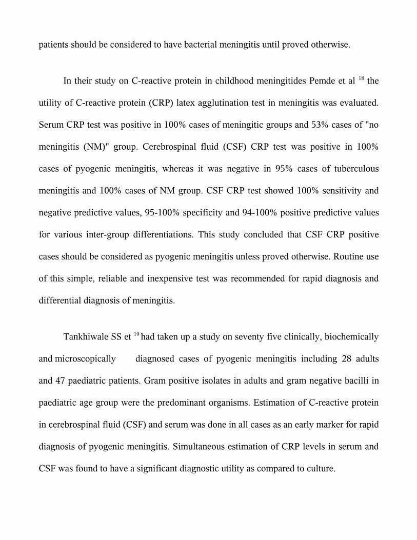
In their study on C-reactive protein in childhood meningitides Pemde et al 18 the
utility of C-reactive protein (CRP) latex agglutination test in meningitis was evaluated
Serum CRP test was positive in 100 cases of meningitic groups and 53 cases of no
meningitis (NM) group Cerebrospinal fluid (CSF) CRP test was positive in 100
cases of pyogenic meningitis whereas it was negative in 95 cases of tuberculous
meningitis and 100 cases of NM group CSF CRP test showed 100 sensitivity and
negative predictive values 95-100 specificity and 94-100 positive predictive values
for various inter-group differentiations This study concluded that CSF CRP positive
cases should be considered as pyogenic meningitis unless proved otherwise Routine use
of this simple reliable and inexpensive test was recommended for rapid diagnosis and
differential diagnosis of meningitis
Tankhiwale SS et 19 had taken up a study on seventy five clinically biochemically
and microscopically diagnosed cases of pyogenic meningitis including 28 adults
and 47 paediatric patients Gram positive isolates in adults and gram negative bacilli in
paediatric age group were the predominant organisms Estimation of C-reactive protein
in cerebrospinal fluid (CSF) and serum was done in all cases as an early marker for rapid
diagnosis of pyogenic meningitis Simultaneous estimation of CRP levels in serum and
CSF was found to have a significant diagnostic utility as compared to culture
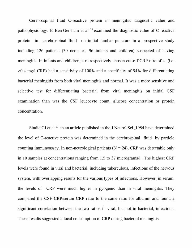
Cerebrospinal fluid C-reactive protein in meningitis diagnostic value and
pathophysiology E Ben Gersham et al 20 examined the diagnostic value of C-reactive
protein in cerebrospinal fluid on initial lumbar puncture in a prospective study
including 126 patients (30 neonates 96 infants and children) suspected of having
meningitis In infants and children a retrospectively chosen cut-off CRP titre of 4 (ie
gt04 mgl CRP) had a sensitivity of 100 and a specificity of 94 for differentiating
bacterial meningitis from both viral meningitis and normal It was a more sensitive and
selective test for differentiating bacterial from viral meningitis on initial CSF
examination than was the CSF leucocyte count glucose concentration or protein
concentration
Sindic CJ et al 21 in an article published in the J Neurol Sci 1984 have determined
the level of C-reactive protein was determined in the cerebrospinal fluid by particle
counting immunoassay In non-neurological patients (N = 24) CRP was detectable only
in 10 samples at concentrations ranging from 15 to 37 microgramsl The highest CRP
levels were found in viral and bacterial including tuberculous infections of the nervous
system with overlapping results for the various types of infections However in serum
the levels of CRP were much higher in pyogenic than in viral meningitis They
compared the CSF CRPserum CRP ratio to the same ratio for albumin and found a
significant correlation between the two ratios in viral but not in bacterial infections
These results suggested a local consumption of CRP during bacterial meningitis
Rajmani et al 22 who estimated of C - reactive protein in Serum and CSF for
diagnosis of various meningitis have said that CSF-CRP sensitivity was found in
8333 cases of pyogenic and none from TBM or viral meningitis CSF-CRP levels in
pyogenic meningitis were very high (104plusmn9021mgdL) but within normal range in
TBMviral meningitis and controls (lt6 mgL)Thus CSF-CRP levels above normal (6
mgL) indicates diagnosis of pyogenic meningitis They concluded that latex
agglutination for serum and CSF is a probable rapid simple economic test and could be
performed at bedside and very much helpful for laboratories of developing countries
even in rural areas
John M Raj IS et al 23 estimated the utility of cerebrospinal fluid C-reactive
protein measurement--a bedside test in the rapid diagnosis of bacterial meningitis C-
reactive protein determinations were performed by the Latex agglutination method on
the cerebrospinal fluid samples of 212 patients with clinical features suggestive of
meningitis Their conclusion was that C-RP was a better indicator of bacterial meningitis
(sensitivity 91 per cent) than the Grams stain (sensitivity 46 per cent)In their study C-
RP determination in CSF proved to be a useful indicator of bacterial meningitis and
served to distinguish it from viral encephalitis tuberculous meningitisfebrile
convulsions and other central nervous system disorders
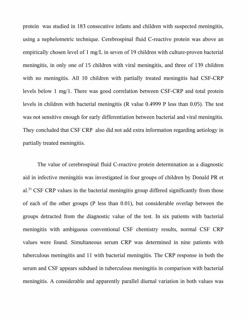
Shaltout A et al 24 had done a study on the Evaluation of cerebrospinal fluid C-
reactive protein in the diagnosis of suspected meningitis Cerebrospinal fluid C-reactive
protein was studied in 183 consecutive infants and children with suspected meningitis
using a nephelometric technique Cerebrospinal fluid C-reactive protein was above an
empirically chosen level of 1 mgL in seven of 19 children with culture-proven bacterial
meningitis in only one of 15 children with viral meningitis and three of 139 children
with no meningitis All 10 children with partially treated meningitis had CSF-CRP
levels below 1 mg1 There was good correlation between CSF-CRP and total protein
levels in children with bacterial meningitis (R value 04999 P less than 005) The test
was not sensitive enough for early differentiation between bacterial and viral meningitis
They concluded that CSF CRP also did not add extra information regarding aetiology in
partially treated meningitis
The value of cerebrospinal fluid C-reactive protein determination as a diagnostic
aid in infective meningitis was investigated in four groups of children by Donald PR et
al25 CSF CRP values in the bacterial meningitis group differed significantly from those
of each of the other groups (P less than 001) but considerable overlap between the
groups detracted from the diagnostic value of the test In six patients with bacterial
meningitis with ambiguous conventional CSF chemistry results normal CSF CRP
values were found Simultaneous serum CRP was determined in nine patients with
tuberculous meningitis and 11 with bacterial meningitis The CRP response in both the
serum and CSF appears subdued in tuberculous meningitis in comparison with bacterial
meningitis A considerable and apparently parallel diurnal variation in both values was
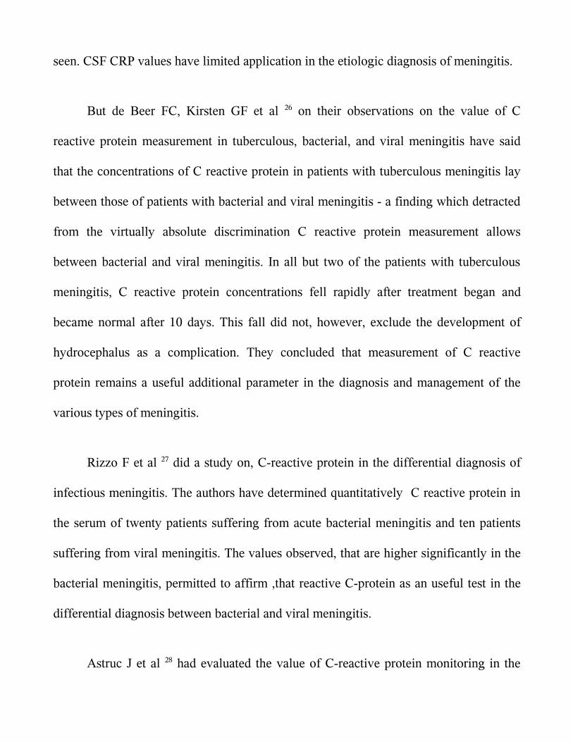
seen CSF CRP values have limited application in the etiologic diagnosis of meningitis
But de Beer FC Kirsten GF et al 26 on their observations on the value of C
reactive protein measurement in tuberculous bacterial and viral meningitis have said
that the concentrations of C reactive protein in patients with tuberculous meningitis lay
between those of patients with bacterial and viral meningitis - a finding which detracted
from the virtually absolute discrimination C reactive protein measurement allows
between bacterial and viral meningitis In all but two of the patients with tuberculous
meningitis C reactive protein concentrations fell rapidly after treatment began and
became normal after 10 days This fall did not however exclude the development of
hydrocephalus as a complication They concluded that measurement of C reactive
protein remains a useful additional parameter in the diagnosis and management of the
various types of meningitis
Rizzo F et al 27 did a study on C-reactive protein in the differential diagnosis of
infectious meningitis The authors have determined quantitatively C reactive protein in
the serum of twenty patients suffering from acute bacterial meningitis and ten patients
suffering from viral meningitis The values observed that are higher significantly in the
bacterial meningitis permitted to affirm that reactive C-protein as an useful test in the
differential diagnosis between bacterial and viral meningitis
Astruc J et al 28 had evaluated the value of C-reactive protein monitoring in the
reduction of antibiotic treatment of bacterial meningitis in children21 of 24
meningococcal meningitis were treated for 4 or 5 days 16 of 22 Haemophilus influenzae
and 4 of 6 pneumococcal meningitis were treated for 7 days without increase in
neurologic sequelae A return of blood CRP levels to normal values was observed in all
these patients simultaneously Thus CRP was a good biological parameter for assesing
treatment discontinuation Furthermore in some complications such as subdural
effusion a new increase of CRP levels was observed after the 5th day A sequential
follow-up of CRP levels at days J0 5 7 10 seemed to be a very useful tool for
management of bacterial meningitis
Ramos Lizana J et al 29 had given a score for the differential diagnosis of bacterial
and viral meningitis The purpose of the study was to verify the statistical validity of the
score proposed by Thomeacute et al for the differential diagnosis between bacterial and viral
meningitis and to study the utility of two new parameters (CRP and the patients age)
They concluded that the score was a useful instrument in the differential diagnosis
between bacterial and viral meningitis Furthermore the introduction of CRP and the
patients age improved the diagnostic value of the test
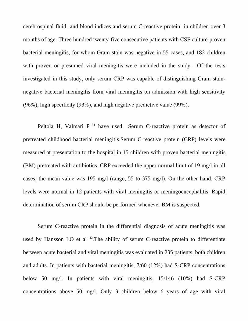
Another study by Sormunen P et al 30 have endorsed the view that C-reactive
protein is useful in distinguishing Gram stain-negative bacterial meningitis from viral
meningitis in childrenThe objective was to clarify to what extent Gram stain-negative
bacterial meningitis can be distinguished from viral meningitis by assessment of
cerebrospinal fluid and blood indices and serum C-reactive protein in children over 3
months of age Three hundred twenty-five consecutive patients with CSF culture-proven
bacterial meningitis for whom Gram stain was negative in 55 cases and 182 children
with proven or presumed viral meningitis were included in the study Of the tests
investigated in this study only serum CRP was capable of distinguishing Gram stain-
negative bacterial meningitis from viral meningitis on admission with high sensitivity
(96) high specificity (93) and high negative predictive value (99)
Peltola H Valmari P 31 have used Serum C-reactive protein as detector of
pretreated childhood bacterial meningitisSerum C-reactive protein (CRP) levels were
measured at presentation to the hospital in 15 children with proven bacterial meningitis
(BM) pretreated with antibiotics CRP exceeded the upper normal limit of 19 mgl in all
cases the mean value was 195 mgl (range 55 to 375 mgl) On the other hand CRP
levels were normal in 12 patients with viral meningitis or meningoencephalitis Rapid
determination of serum CRP should be performed whenever BM is suspected
Serum C-reactive protein in the differential diagnosis of acute meningitis was
used by Hansson LO et al 32The ability of serum C-reactive protein to differentiate
between acute bacterial and viral meningitis was evaluated in 235 patients both children
and adults In patients with bacterial meningitis 760 (12) had S-CRP concentrations
below 50 mgl In patients with viral meningitis 15146 (10) had S-CRP
concentrations above 50 mgl Only 3 children below 6 years of age with viral
meningitis had S-CRP concentration above 20 mgl but none exceeded 50 mgl An S-
CRP value above 50 mgl in patients with CSF pleocytosis usually indicates bacterial
etiology However S-CRP values above 50 mgl may occasionally be seen in viral
meningitis
Another study by Cuevas LE et al 33 on C-reactive protein and bacterial meningitis
concluded that CSF CRP should not be used as a useful discriminatory test in areas
where malaria and TB meningitis are common
STUDY JUSTIFICATION
Various indices have been initiated to screen for bacterial meningitis majority
being neither highly sensitive nor specific34 The value of certain rapid and easy
screening tests to detect the presence of infection has been studied by various authors
with varying conclusions Though various tests are available to detect bacterial
infections they are expensive and they need expertise to be done and to be interpreted
C-reactive protein an acute phase reactant has been used to diagnose and follow
the course of infection
The present study was undertaken to establish the utility of C-RP in CSF in early
diagnosis of bacterial meningitis in children This not only enables us to start antibiotics
in meningitic doses in cases of pyogenic meningitis but also avoid unnecessary
antibiotics in the rest
Its advantages include
Rapid diagnosis can be done within few minutes
Can be performed at hospitals where full fledged microbiology lab is unavailable
Low cost
OBJECTIVES OF THE STUDY
(a) To analyse the diagnostic utility of C reactive protein as a bedside test to
diagnose pyogenic meningitis
(b) To analyse the common organisms being isolated from CSF in cases of
pyogenic meningitis in our hospital as well as antibiotic susceptibility
patterns
MATERIALS AND METHODS
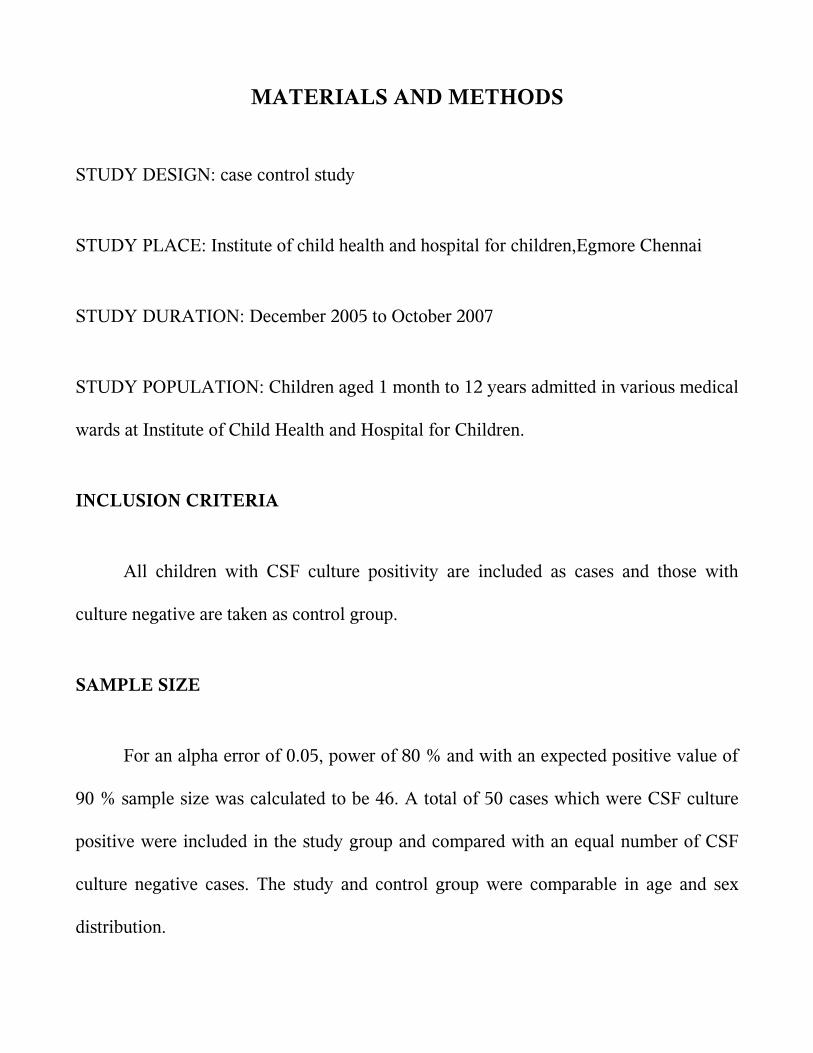
STUDY DESIGN case control study
STUDY PLACE Institute of child health and hospital for childrenEgmore Chennai
STUDY DURATION December 2005 to October 2007
STUDY POPULATION Children aged 1 month to 12 years admitted in various medical
wards at Institute of Child Health and Hospital for Children
INCLUSION CRITERIA
All children with CSF culture positivity are included as cases and those with
culture negative are taken as control group
SAMPLE SIZE
For an alpha error of 005 power of 80 and with an expected positive value of
90 sample size was calculated to be 46 A total of 50 cases which were CSF culture
positive were included in the study group and compared with an equal number of CSF
culture negative cases The study and control group were comparable in age and sex
distribution
CONTROL GROUP
Included all cases for which CSF analysis was done for reasons other than
pyogenic meningitis They consisted of patients with febrile convulsions acute
respiratory tract infection with meningismus and acute flaccid paralysis
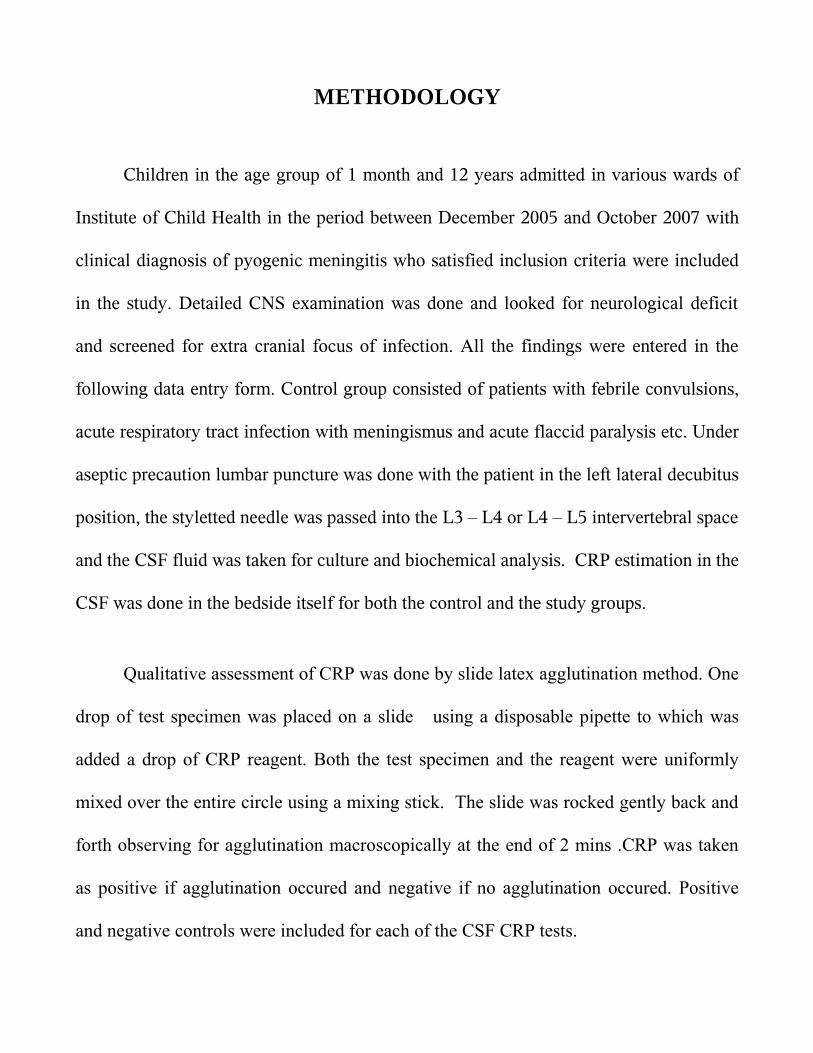
METHODOLOGY
Children in the age group of 1 month and 12 years admitted in various wards of
Institute of Child Health in the period between December 2005 and October 2007 with
clinical diagnosis of pyogenic meningitis who satisfied inclusion criteria were included
in the study Detailed CNS examination was done and looked for neurological deficit
and screened for extra cranial focus of infection All the findings were entered in the
following data entry form Control group consisted of patients with febrile convulsions
acute respiratory tract infection with meningismus and acute flaccid paralysis etc Under
aseptic precaution lumbar puncture was done with the patient in the left lateral decubitus
position the styletted needle was passed into the L3 ndash L4 or L4 ndash L5 intervertebral space
and the CSF fluid was taken for culture and biochemical analysis CRP estimation in the
CSF was done in the bedside itself for both the control and the study groups
Qualitative assessment of CRP was done by slide latex agglutination method One
drop of test specimen was placed on a slide using a disposable pipette to which was
added a drop of CRP reagent Both the test specimen and the reagent were uniformly
mixed over the entire circle using a mixing stick The slide was rocked gently back and
forth observing for agglutination macroscopically at the end of 2 mins CRP was taken
as positive if agglutination occured and negative if no agglutination occured Positive
and negative controls were included for each of the CSF CRP tests
In the microbiology lab CSF was cultured in enriched media like blood agar and
chocolate agar for the organisms like pneumococci beta hemolytic streptococci and H
influenza It was also inoculated in Mac Conky agar for gram negative lactose
fermenting and non lactose fermenting organisms like E coli klebsiella and salmonella
respectively The plates were incubated in a incubator of 37 0C overnight The next day
colony morphology was read and with help of biochemical reactions the organisms were
differentiated and confirmed with special tests The antibiotic sensitivity with the drugs
were done in Mueller Hunten agar
Culture was taken as gold standard 50 cases of culture positive CSF was taken as
the study group of pyogenic meningitis 50 cases which were culture negative in CSF
study were included in the control group CSF analysis for cells CSF protein sugar and
CRP were simultaneously estimated in the control group samples also
RESULTS
A total of 100 children were included in the study Children in the age group of
one month to 12 years with a suspicion of meningitis who were later found to have
growth in their CSF were included in the study group (n=50) 50 cases of children who
did not have any CSF growth and were subsequently diagnosed as non pyogenic causes
of encephalopathy were included in the control group Data was analyzed using SSPS
windows statistical software program in the computer The results were tabulated and
the clinical profile was analysed using simple statistical proportions Sensitivity
specificity positive predictive value and negative predictive value of CRP against the
gold standard of culture in diagnosing bacterial meningitis was calculated
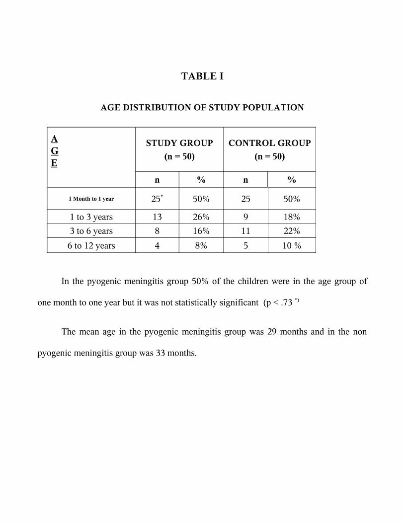
TABLE I
AGE DISTRIBUTION OF STUDY POPULATION
AGE
STUDY GROUP
(n = 50)
CONTROL GROUP
(n = 50)
n n
1 Month to 1 year 25 50 25 50
1 to 3 years 13 26 9 18
3 to 6 years 8 16 11 22
6 to 12 years 4 8 5 10
In the pyogenic meningitis group 50 of the children were in the age group of
one month to one year but it was not statistically significant (p lt 73 )
The mean age in the pyogenic meningitis group was 29 months and in the non
pyogenic meningitis group was 33 months
0
5
10
15
20
25
30
PYOGENICMENINGITIS
NONPYOGENICMENINGITIS
1 Month to 1 year
1 to 3 years
3 to 6 years
6 to 12 years
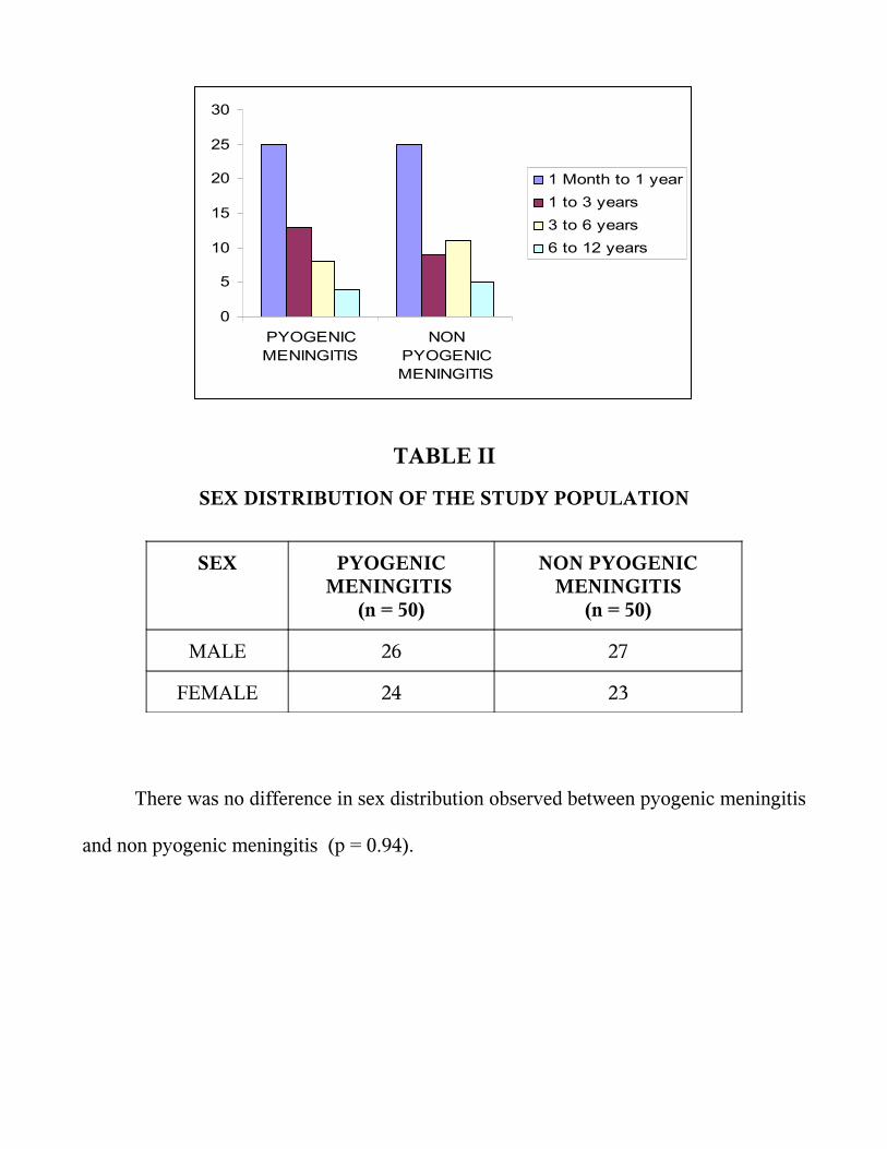
TABLE II
SEX DISTRIBUTION OF THE STUDY POPULATION
SEX PYOGENIC MENINGITIS
(n = 50)
NON PYOGENIC MENINGITIS
(n = 50)
MALE 26 27
FEMALE 24 23
There was no difference in sex distribution observed between pyogenic meningitis
and non pyogenic meningitis (p = 094)
21
22
23
24
25
26
27
28
PYOGENICMENINGITIS
NON PYOGENICMENINGITIS
MALE
FEMALE
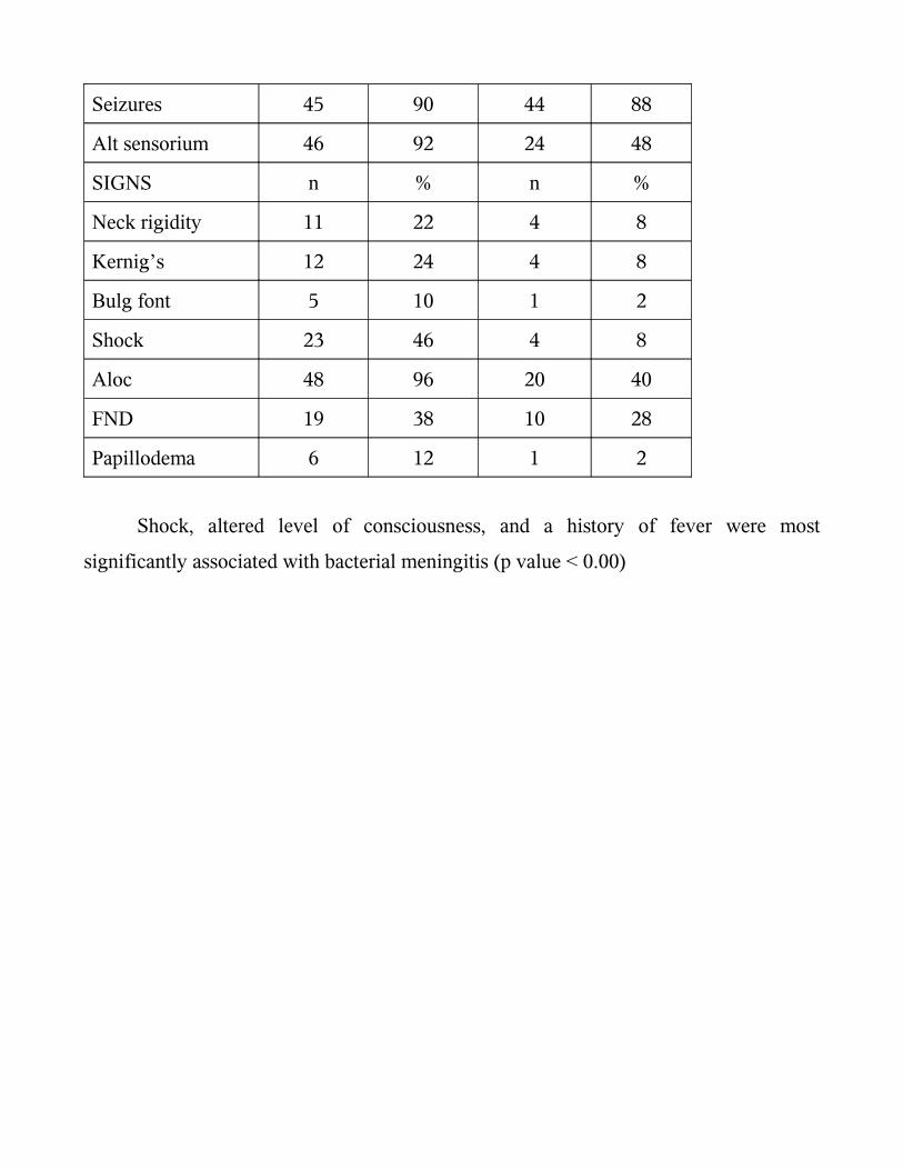
TABLE III
CLINICAL FEATURES OF CHILDREN PRESENTING AS ACUTE CNS INFECTION
CLINCAL
FEATURES
PYOGENIC MENINGITIS
(n = 50)
NON PYOGENIC MENINGITIS
(n = 50)
SYMPTOMS n n
Fever 44 88 27 54
Headache 9 18 3 6
Vomitting 17 34 5 10
Seizures 45 90 44 88
Alt sensorium 46 92 24 48
SIGNS n n
Neck rigidity 11 22 4 8
Kernigrsquos 12 24 4 8
Bulg font 5 10 1 2
Shock 23 46 4 8
Aloc 48 96 20 40
FND 19 38 10 28
Papillodema 6 12 1 2
Shock altered level of consciousness and a history of fever were most
significantly associated with bacterial meningitis (p value lt 000)
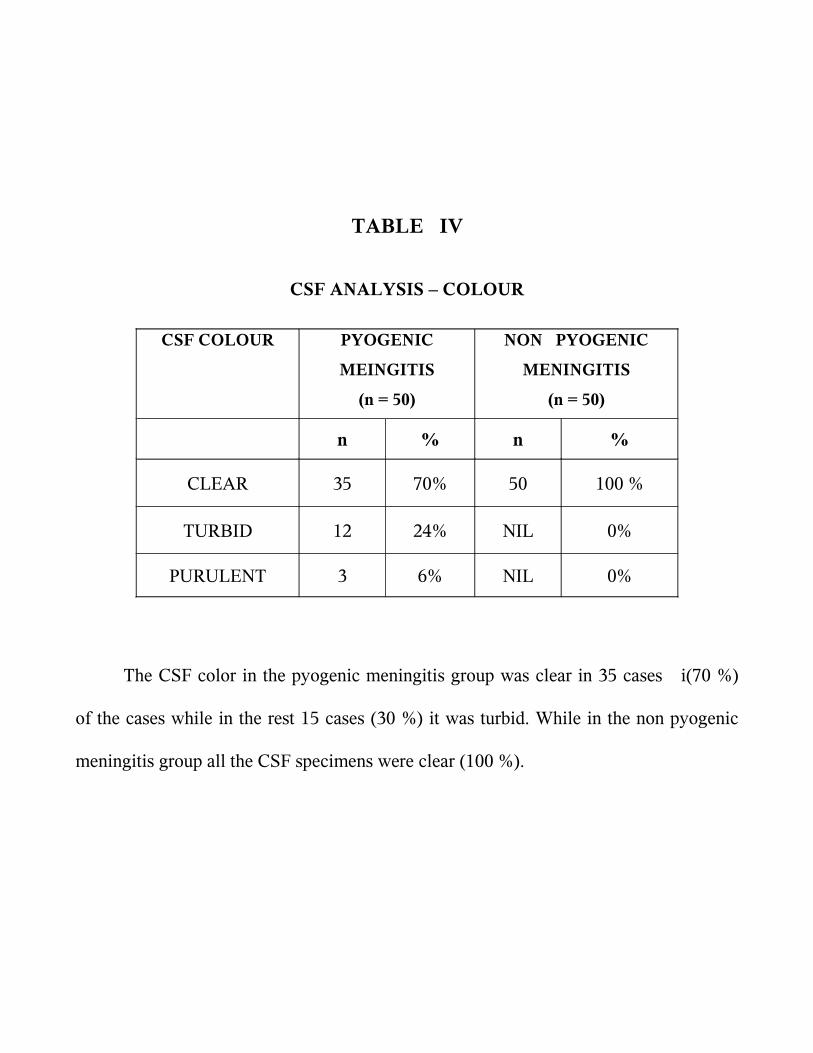
TABLE IV
CSF ANALYSIS ndash COLOUR
CSF COLOUR PYOGENIC
MEINGITIS
(n = 50)
NON PYOGENIC
MENINGITIS
(n = 50)
n n
CLEAR 35 70 50 100
TURBID 12 24 NIL 0
PURULENT 3 6 NIL 0
The CSF color in the pyogenic meningitis group was clear in 35 cases i(70 )
of the cases while in the rest 15 cases (30 ) it was turbid While in the non pyogenic
meningitis group all the CSF specimens were clear (100 )
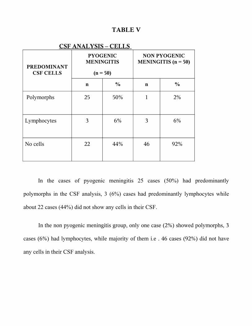
TABLE V
CSF ANALYSIS ndash CELLS
PREDOMINANT CSF CELLS
PYOGENIC MENINGITIS
(n = 50)
NON PYOGENIC MENINGITIS (n = 50)
n n
Polymorphs 25 50 1 2
Lymphocytes 3 6 3 6
No cells 22 44 46 92
In the cases of pyogenic meningitis 25 cases (50) had predominantly
polymorphs in the CSF analysis 3 (6) cases had predominantly lymphocytes while
about 22 cases (44) did not show any cells in their CSF
In the non pyogenic meningitis group only one case (2) showed polymorphs 3
cases (6) had lymphocytes while majority of them ie 46 cases (92) did not have
any cells in their CSF analysis
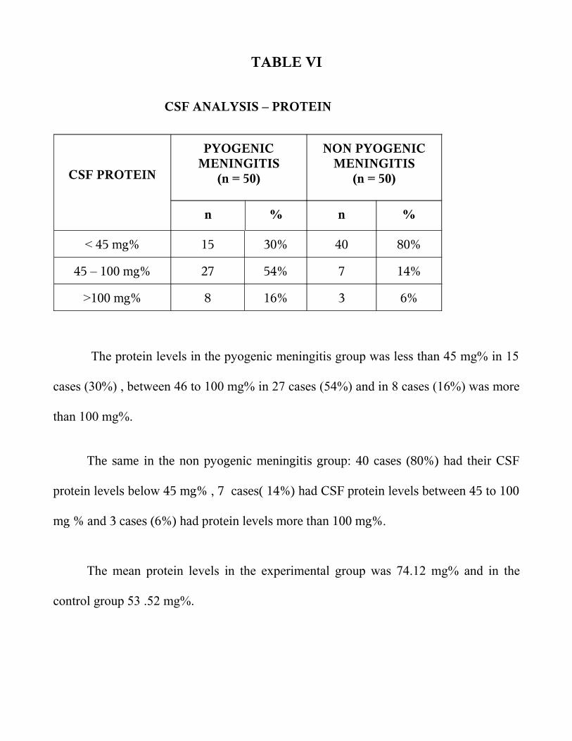
TABLE VI
CSF ANALYSIS ndash PROTEIN
CSF PROTEIN
PYOGENIC MENINGITIS
(n = 50)
NON PYOGENIC MENINGITIS
(n = 50)
n n
lt 45 mg 15 30 40 80
45 ndash 100 mg 27 54 7 14
gt100 mg 8 16 3 6
The protein levels in the pyogenic meningitis group was less than 45 mg in 15
cases (30) between 46 to 100 mg in 27 cases (54) and in 8 cases (16) was more
than 100 mg
The same in the non pyogenic meningitis group 40 cases (80) had their CSF
protein levels below 45 mg 7 cases( 14) had CSF protein levels between 45 to 100
mg and 3 cases (6) had protein levels more than 100 mg
The mean protein levels in the experimental group was 7412 mg and in the
control group 53 52 mg
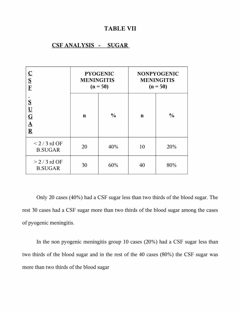
TABLE VII
CSF ANALYSIS - SUGAR
CSF SUGAR
PYOGENIC MENINGITIS
(n = 50)
NONPYOGENIC MENINGITIS
(n = 50)
n n
lt 2 3 rd OF BSUGAR
20 40 10 20
gt 2 3 rd OF BSUGAR
30 60 40 80
Only 20 cases (40) had a CSF sugar less than two thirds of the blood sugar The
rest 30 cases had a CSF sugar more than two thirds of the blood sugar among the cases
of pyogenic meningitis
In the non pyogenic meningitis group 10 cases (20) had a CSF sugar less than
two thirds of the blood sugar and in the rest of the 40 cases (80) the CSF sugar was
more than two thirds of the blood sugar
The mean CSF sugar in the study group was 582 mg and in the control group it
was 7514 mg
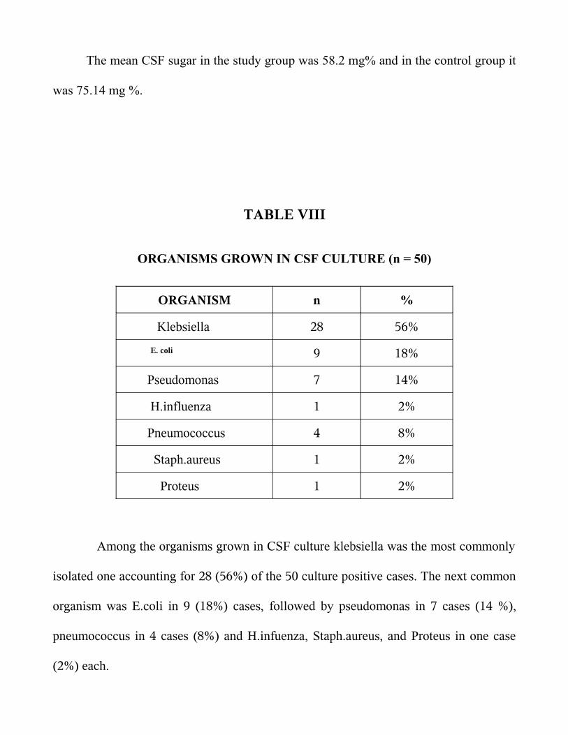
TABLE VIII
ORGANISMS GROWN IN CSF CULTURE (n = 50)
ORGANISM n
Klebsiella 28 56
E coli 9 18
Pseudomonas 7 14
Hinfluenza 1 2
Pneumococcus 4 8
Staphaureus 1 2
Proteus 1 2
Among the organisms grown in CSF culture klebsiella was the most commonly
isolated one accounting for 28 (56) of the 50 culture positive cases The next common
organism was Ecoli in 9 (18) cases followed by pseudomonas in 7 cases (14 )
pneumococcus in 4 cases (8) and Hinfuenza Staphaureus and Proteus in one case
(2) each
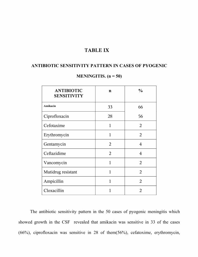
TABLE IX
ANTIBIOTIC SENSITIVITY PATTERN IN CASES OF PYOGENIC
MENINGITIS (n = 50)
ANTIBIOTIC SENSITIVITY
n
Amikacin 33 66
Ciprofloxacin 28 56
Cefotaxime 1 2
Erythromycin 1 2
Gentamycin 2 4
Ceftazidime 2 4
Vancomycin 1 2
Mutidrug resistant 1 2
Ampicillin 1 2
Cloxacillin 1 2
The antibiotic sensitivity pattern in the 50 cases of pyogenic meningitis which
showed growth in the CSF revealed that amikacin was sensitive in 33 of the cases
(66) ciprofloxacin was sensitive in 28 of them(56) cefatoxime erythromycin
vancomycin ampicillin and cloxacilin were sensitive in in one cases each (2 each)
Gentamycin and
ceftazidime were sensitive in 2 cases each (4 each) One case was multidrug
resisistant
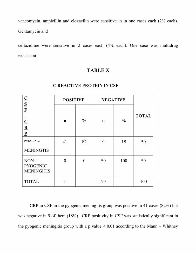
TABLE X
C REACTIVE PROTEIN IN CSF
CSF CRP
POSITIVE NEGATIVE
n n TOTAL
PYOGENIC
MENINGTIS
41 82 9 18 50
NON PYOGENIC MENINGITIS
0 0 50 100 50
TOTAL 41 59 100
CRP in CSF in the pyogenic meningitis group was positive in 41 cases (82) but
was negative in 9 of them (18) CRP positivity in CSF was statistically significant in
the pyogenic meningitis group with a p value lt 001 according to the Mann ndash Whitney
test
In the non pyogenic meningitis group CRP done on CSF was negative in all the
cases
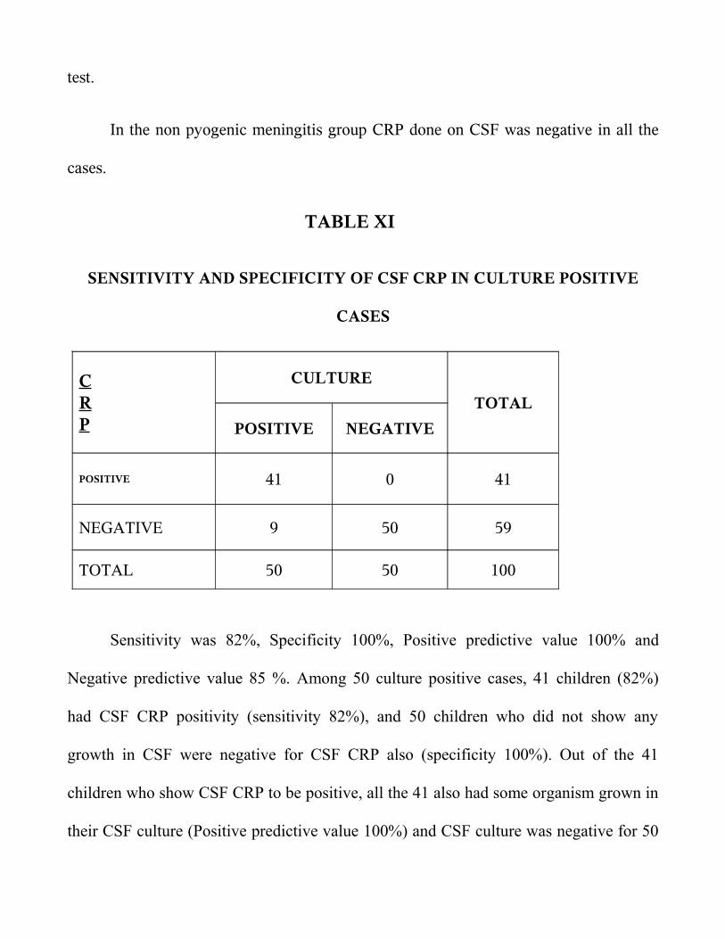
TABLE XI
SENSITIVITY AND SPECIFICITY OF CSF CRP IN CULTURE POSITIVE
CASES
CRP
CULTURE
POSITIVE NEGATIVE
TOTAL
POSITIVE 41 0 41
NEGATIVE 9 50 59
TOTAL 50 50 100
Sensitivity was 82 Specificity 100 Positive predictive value 100 and
Negative predictive value 85 Among 50 culture positive cases 41 children (82)
had CSF CRP positivity (sensitivity 82) and 50 children who did not show any
growth in CSF were negative for CSF CRP also (specificity 100) Out of the 41
children who show CSF CRP to be positive all the 41 also had some organism grown in
their CSF culture (Positive predictive value 100) and CSF culture was negative for 50
children among 59 children who tested to be CSF CRP negative (Negative predictive
value 85)
Overall accuracy was 91 (91 100)
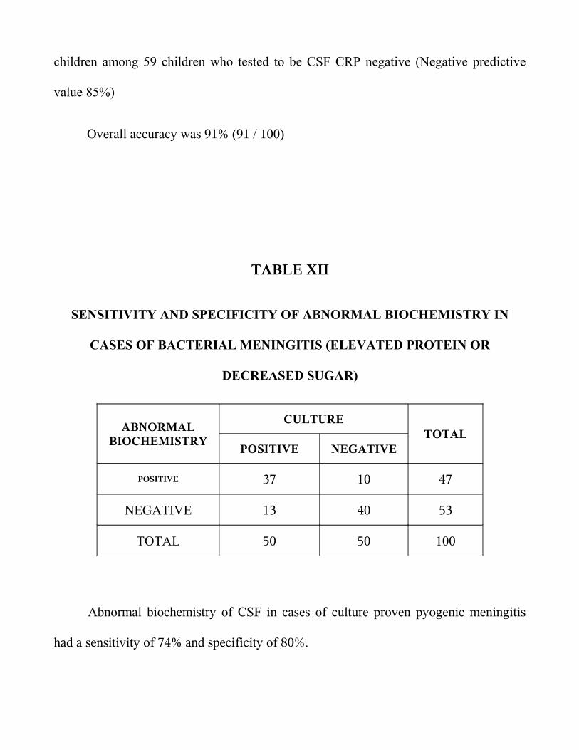
TABLE XII
SENSITIVITY AND SPECIFICITY OF ABNORMAL BIOCHEMISTRY IN
CASES OF BACTERIAL MENINGITIS (ELEVATED PROTEIN OR
DECREASED SUGAR)
ABNORMAL BIOCHEMISTRY
CULTURE
POSITIVE NEGATIVETOTAL
POSITIVE 37 10 47
NEGATIVE 13 40 53
TOTAL 50 50 100
Abnormal biochemistry of CSF in cases of culture proven pyogenic meningitis
had a sensitivity of 74 and specificity of 80
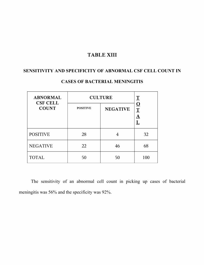
TABLE XIII
SENSITIVITY AND SPECIFICITY OF ABNORMAL CSF CELL COUNT IN
CASES OF BACTERIAL MENINGITIS
ABNORMAL CSF CELL
COUNT
CULTURE
POSITIVE NEGATIVE
TOTAL
POSITIVE 28 4 32
NEGATIVE 22 46 68
TOTAL 50 50 100
The sensitivity of an abnormal cell count in picking up cases of bacterial
meningitis was 56 and the specificity was 92
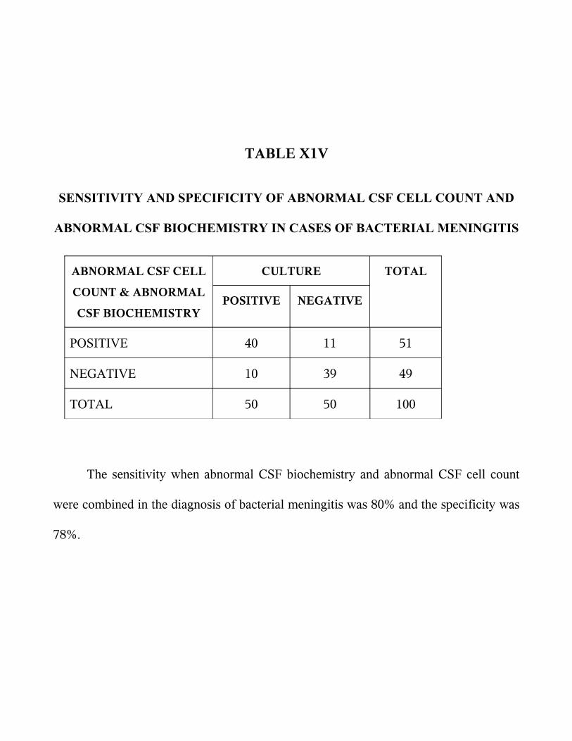
TABLE X1V
SENSITIVITY AND SPECIFICITY OF ABNORMAL CSF CELL COUNT AND
ABNORMAL CSF BIOCHEMISTRY IN CASES OF BACTERIAL MENINGITIS
ABNORMAL CSF CELL
COUNT amp ABNORMAL
CSF BIOCHEMISTRY
CULTURE
POSITIVE NEGATIVE
TOTAL
POSITIVE 40 11 51
NEGATIVE 10 39 49
TOTAL 50 50 100
The sensitivity when abnormal CSF biochemistry and abnormal CSF cell count
were combined in the diagnosis of bacterial meningitis was 80 and the specificity was
78
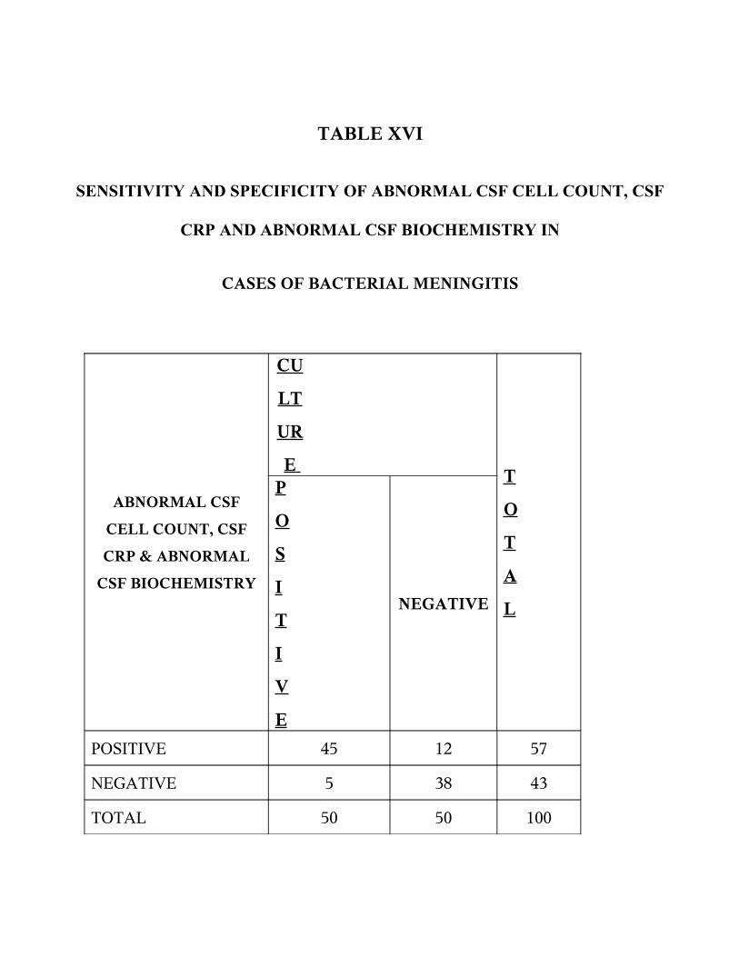
TABLE XVI
SENSITIVITY AND SPECIFICITY OF ABNORMAL CSF CELL COUNT CSF
CRP AND ABNORMAL CSF BIOCHEMISTRY IN
CASES OF BACTERIAL MENINGITIS
ABNORMAL CSF
CELL COUNT CSF
CRP amp ABNORMAL
CSF BIOCHEMISTRY
CU
LT
UR
E P
O
S
I
T
I
V
E
NEGATIVE
T
O
T
A
L
POSITIVE 45 12 57
NEGATIVE 5 38 43
TOTAL 50 50 100
The sensitivity when all three parameters ie CSF CRP abnormal CSF
biochemistry and cell count were combined for the diagnosis of bacterial meningitis was
90
DISCUSSION
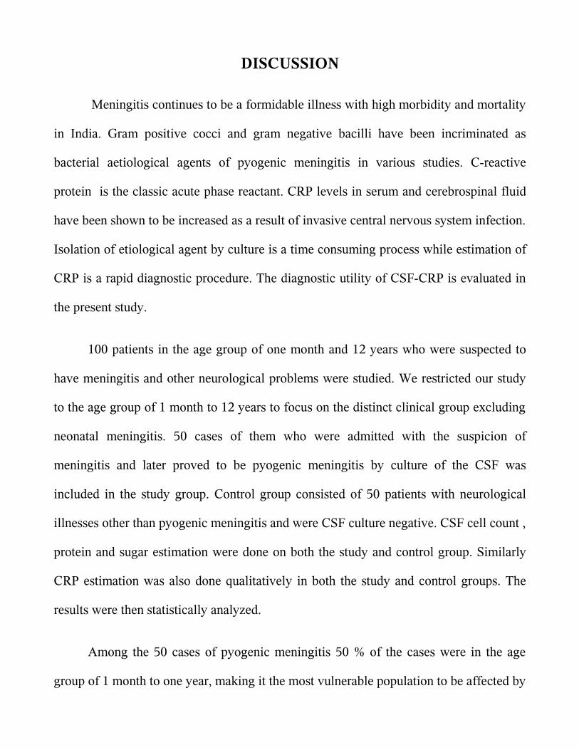
Meningitis continues to be a formidable illness with high morbidity and mortality
in India Gram positive cocci and gram negative bacilli have been incriminated as
bacterial aetiological agents of pyogenic meningitis in various studies C-reactive
protein is the classic acute phase reactant CRP levels in serum and cerebrospinal fluid
have been shown to be increased as a result of invasive central nervous system infection
Isolation of etiological agent by culture is a time consuming process while estimation of
CRP is a rapid diagnostic procedure The diagnostic utility of CSF-CRP is evaluated in
the present study
100 patients in the age group of one month and 12 years who were suspected to
have meningitis and other neurological problems were studied We restricted our study
to the age group of 1 month to 12 years to focus on the distinct clinical group excluding
neonatal meningitis 50 cases of them who were admitted with the suspicion of
meningitis and later proved to be pyogenic meningitis by culture of the CSF was
included in the study group Control group consisted of 50 patients with neurological
illnesses other than pyogenic meningitis and were CSF culture negative CSF cell count
protein and sugar estimation were done on both the study and control group Similarly
CRP estimation was also done qualitatively in both the study and control groups The
results were then statistically analyzed
Among the 50 cases of pyogenic meningitis 50 of the cases were in the age
group of 1 month to one year making it the most vulnerable population to be affected by
bacterial meningitis
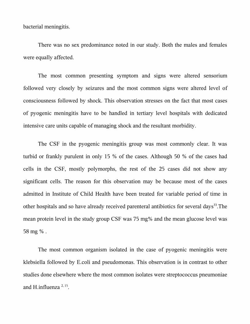
There was no sex predominance noted in our study Both the males and females
were equally affected
The most common presenting symptom and signs were altered sensorium
followed very closely by seizures and the most common signs were altered level of
consciousness followed by shock This observation stresses on the fact that most cases
of pyogenic meningitis have to be handled in tertiary level hospitals with dedicated
intensive care units capable of managing shock and the resultant morbidity
The CSF in the pyogenic meningitis group was most commonly clear It was
turbid or frankly purulent in only 15 of the cases Although 50 of the cases had
cells in the CSF mostly polymorphs the rest of the 25 cases did not show any
significant cells The reason for this observation may be because most of the cases
admitted in Institute of Child Health have been treated for variable period of time in
other hospitals and so have already received parenteral antibiotics for several days33The
mean protein level in the study group CSF was 75 mg and the mean glucose level was
58 mg
The most common organism isolated in the case of pyogenic meningitis were
klebsiella followed by Ecoli and pseudomonas This observation is in contrast to other
studies done elsewhere where the most common isolates were streptococcus pneumoniae
and Hinfluenza 2 15
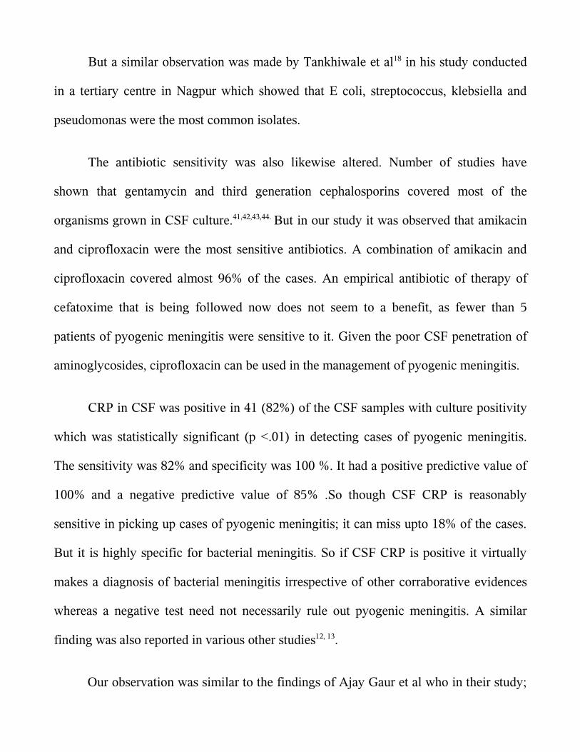
But a similar observation was made by Tankhiwale et al18 in his study conducted
in a tertiary centre in Nagpur which showed that E coli streptococcus klebsiella and
pseudomonas were the most common isolates
The antibiotic sensitivity was also likewise altered Number of studies have
shown that gentamycin and third generation cephalosporins covered most of the
organisms grown in CSF culture41424344 But in our study it was observed that amikacin
and ciprofloxacin were the most sensitive antibiotics A combination of amikacin and
ciprofloxacin covered almost 96 of the cases An empirical antibiotic of therapy of
cefatoxime that is being followed now does not seem to a benefit as fewer than 5
patients of pyogenic meningitis were sensitive to it Given the poor CSF penetration of
aminoglycosides ciprofloxacin can be used in the management of pyogenic meningitis
CRP in CSF was positive in 41 (82) of the CSF samples with culture positivity
which was statistically significant (p lt01) in detecting cases of pyogenic meningitis
The sensitivity was 82 and specificity was 100 It had a positive predictive value of
100 and a negative predictive value of 85 So though CSF CRP is reasonably
sensitive in picking up cases of pyogenic meningitis it can miss upto 18 of the cases
But it is highly specific for bacterial meningitis So if CSF CRP is positive it virtually
makes a diagnosis of bacterial meningitis irrespective of other corraborative evidences
whereas a negative test need not necessarily rule out pyogenic meningitis A similar
finding was also reported in various other studies12 13
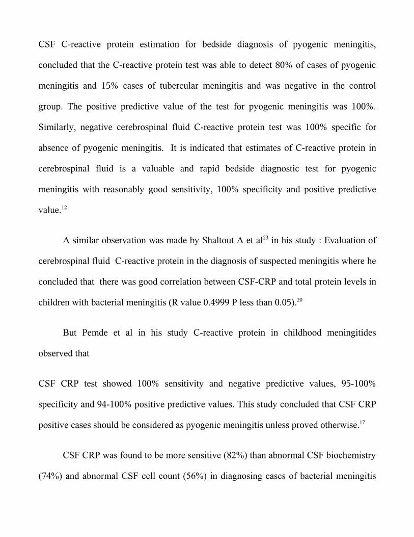
Our observation was similar to the findings of Ajay Gaur et al who in their study
CSF C-reactive protein estimation for bedside diagnosis of pyogenic meningitis
concluded that the C-reactive protein test was able to detect 80 of cases of pyogenic
meningitis and 15 cases of tubercular meningitis and was negative in the control
group The positive predictive value of the test for pyogenic meningitis was 100
Similarly negative cerebrospinal fluid C-reactive protein test was 100 specific for
absence of pyogenic meningitis It is indicated that estimates of C-reactive protein in
cerebrospinal fluid is a valuable and rapid bedside diagnostic test for pyogenic
meningitis with reasonably good sensitivity 100 specificity and positive predictive
value12
A similar observation was made by Shaltout A et al23 in his study Evaluation of
cerebrospinal fluid C-reactive protein in the diagnosis of suspected meningitis where he
concluded that there was good correlation between CSF-CRP and total protein levels in
children with bacterial meningitis (R value 04999 P less than 005)20
But Pemde et al in his study C-reactive protein in childhood meningitides
observed that
CSF CRP test showed 100 sensitivity and negative predictive values 95-100
specificity and 94-100 positive predictive values This study concluded that CSF CRP
positive cases should be considered as pyogenic meningitis unless proved otherwise17
CSF CRP was found to be more sensitive (82) than abnormal CSF biochemistry
(74) and abnormal CSF cell count (56) in diagnosing cases of bacterial meningitis
The sensitivity when both abnormal CSF biochemistry and cell count were combined
was 80 but it was observed that when all three parameters were included the
sensitivity rose to 90 So it can be deduced that CSF CRP is a very useful test in
diagnosing cases of bacterial meningitis in combination with other factors especially
when facilities for culture are not available
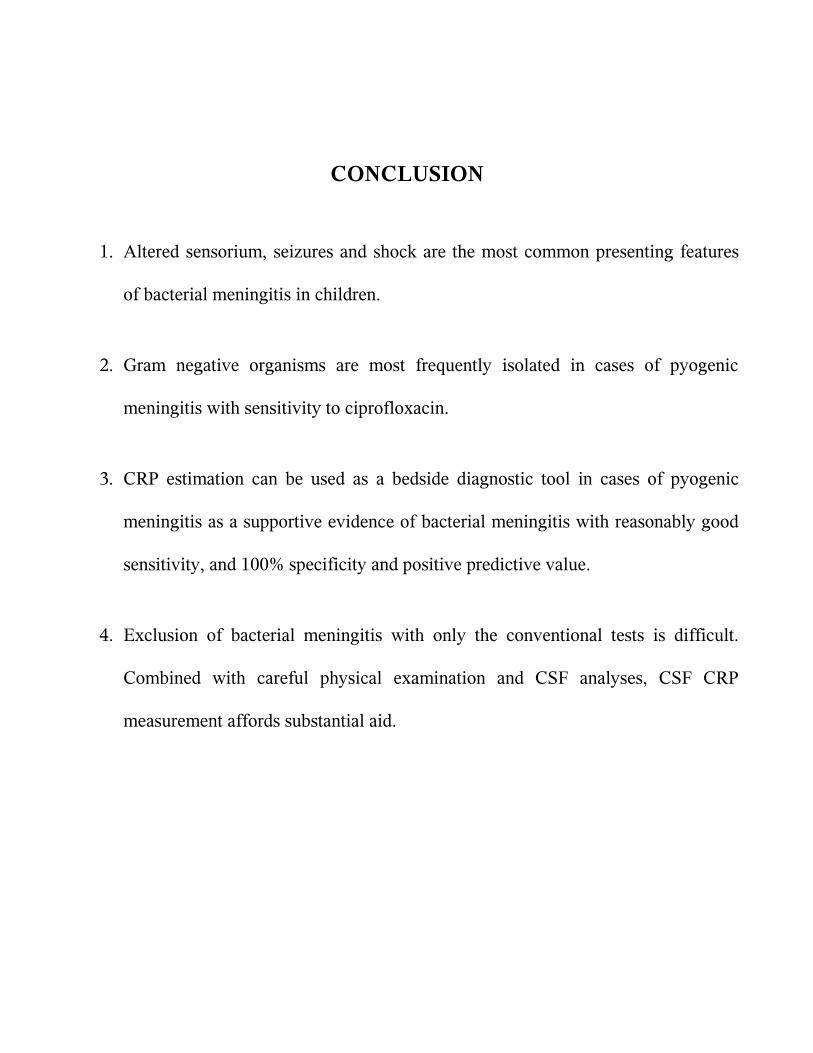
CONCLUSION
1 Altered sensorium seizures and shock are the most common presenting features
of bacterial meningitis in children
2 Gram negative organisms are most frequently isolated in cases of pyogenic
meningitis with sensitivity to ciprofloxacin
3 CRP estimation can be used as a bedside diagnostic tool in cases of pyogenic
meningitis as a supportive evidence of bacterial meningitis with reasonably good
sensitivity and 100 specificity and positive predictive value
4 Exclusion of bacterial meningitis with only the conventional tests is difficult
Combined with careful physical examination and CSF analyses CSF CRP
measurement affords substantial aid
BIBLIOGRAPHY
1 Meningitis in childhood Annales nestle 1997 55 103 ndash 110
2 B Robbins et al Surveillance for Bacterial Meningitis Clinical Infectious Diseases
2005 4026ndash27
3 Quagliavello V and Scheld VMbacterial meningitispathogens pathophysiology and
progress N Engl J Med 1992 327 864
4 Swartz MN Bodge PR bacterial meningitis ndash A review of selected aspects General
clinical features special problems and unusual meningeal rections mimicking
bacterial meningitis N Engl J Med 1962 272725
5 Charles G Prober Acute bacterial meningitis beyond the neonatal periodNelsonrsquo s
textbook of paediatrics17th editionp2038-44
6 Harrisonrsquos textbook of medicine
7 Baraff LJLee SI Schiger DLoutcome of bacterial meningitis in children a
metaanalysis Pediatr infectDis J121993389- 394
8 Whibrittle HC Jugwell Pet al rapid bacteriological diagnosis of pyogenic
meningitis by latex agglutination lancet 216191976
9 Gewvrz HMold CSiegelJFiedel c reactive protein in bacterial infections
AdvIntMed198227345 -372
10Nudelman RBenjamin M Kagan CRP InpediatrAdv in Pediatr198030517 -547
11Peppy MB CRP fifty years on Lancet 19873653 -656
12Abamsom JSHampton DK Babu S et althe use of C reactve protein from CSF for
differentiating meningitis from other CNS diseases Pediatr infectDis J 1985
151854
13CSF C-reactive protein estimation for bedside diagnosis of pyogenic meningitis
Ajay Gaur Seshan S V Indian Pediatr 2004 41 10
14C-reactive protein in CNS infections in children Jan M Ali W Ahmad M Sethi AS
Department of Paediatrics SKIMS Soura Srinagar India JK Practitioner 1998 Oct-
Dec 5(4) 283
15Nandita Chinchankar et al Diagnosis and outcome of acute bacterial meningitis in
children Indian pediatr 2002 39
16Zvezdana et al TO assess the C-Reactive Protein Concentrations in Cerebral Spinal
Fluid in ability of CSF CRP to differentiate gram-positive from gram negative
meningitis clinical chemistry 2002 591 -592
17Vaidya AK Wagle NM et al Use of CSF C-reactive protein in differentiating
bacterial and non-bacterial meningitis journal of Postgrad Med 19873358-60
18 Pemde HK et alC-reactive protein in childhood meningitides Indian J Pediatr 1996
Jan-Feb 63 (1)73-7
19Tankhiwale SS et al Bacteriological study of pyogenic meningitis with special
reference to C-reactive protein Indian Journal of Medical Microbiology 2001 19 (3)
159-160
20E Ben GershAtildeacutem et al Cerebrospinal fluid C-reactive protein in meningitis
diagnostic value and pathophysiology European Journal of Pediatrics
101007BF00439393
21 Sindic CJ et al C-reactive protein in serum and cerebrospinal fluid in various
neurological disorders Apparent local consumption during bacterial meningitis J
Neurol Sci 1984 Mar 63(3)339-44
22Rajmani Gupta Ajay Gupta Bharat et al Estimation of C-Reactive Protein in Serum
and CSF for Diagnosis of Various Meningitis
23John M et al Cerebrospinal fluid C-reactive protein measurement - a bedside test in
the rapid diagnosis of bacterial meningitis J Trop Pediatr 1990 36213-7
24 Shaltout Aet al Evaluation of cerebrospinal fluid (CSF) C-reactive protein in the
diagnosis of suspected meningitis Ann Trop Paediatr 1986 Mar6(1)31-5
25 Donald PR et al Cerebrospinal fluid C-reactive protein in infective meningitis in
childhood J Lab Clin Med 1985 Oct106 (4)424-7
26 De Beer Fc et alValue of C reactive protein measurement in tuberculous bacterial
and viral meningitis Arch Dis Child 1984 Jul59(7)653-6
27 Rizzo Fet al C-reactive protein in the differential diagnosis of infectious meningitis
Quad Sclavo Diagn 1987 Mar 23(1)100-8
28 Astruc J et al Reduction of antibiotic treatment of bacterial meningitis in children
Value of C-reactive protein monitoring] Arch Fr Pediatr 1990 Nov47(9)637-40
29 Ramos Lizana J et al A score for the differential diagnosis of bacterial and viral
meningitis An Esp Pediatr 1996 Jan 44 (1)
35-9
30 Sormunen P et al C-reactive protein is useful in distinguishing Gram stain-negative
bacterial meningitis from viral meningitis in children
31 Peltola H et al Serum C-reactive protein as detector of pretreated childhood bacterial
meningitis I P Neurology 1985 Feb35 (2)251-3
32 Hansson LO et al Serum C-reactive protein in the differential diagnosis of acute
meningitis Scand J Infect Dis 1993 25(5)625-30
33 Cuevas LEet al C-reactive protein and bacterial meningitis Ann Trop Paediatr
1988 Dec8 (4)230-3
34Dalton HPAlleson MJmodification of laboratory results by partial treatment of
bacterial meningitis Am J Clin Patho 196849410
35Swartz M Acute bacterial meningitis Infectious diseases 2nd ed
36Kawamura M Nishida HThe usefulness of serial C-reactive protein measurement in
managing neonatal infectionActa Paediatr 19958410-3
37Ram Y Meningitis In Jenson HB Baltimore RS editorsPrinciples and practice of
Paediatric Infectious diseases 2nd edPhiladelphia WB Saunders 2002630-50
38Tatara R Imai H Serum C-reactive protein in the differential diagnosis of childhood
meningitis Pediatr Int 2000 42(5)541-6
39Corrall CJ Pepple JM Moxan ER Hughes WT C-reactive protein in spinal fluid in
children with meningitis J Pediatr198199365-9
40Gerdes LU et al C-reactive protein and bacterial meningitis a meta analysis Scand J
Clin Inves
41 Sabel KG et al The clinical usefulness of C-reactive protein (CRP) determinations
in bacterial meningitis and septicemia in infancy Acta Paediatr Scand 1974
May63(3)381-8
42 Bandaru Narasinga Rao et al Etiology and occurrence of acute bacterial meningitis in
children in Benghazi Libyan Arab Jamahiriya 4(1)1998 50 - 57
43 Panjarathinam R Shah RK Pyogenic meningitis in Ahmedabad Indian journal of
pediatrics 1993 60669-73
44 Osoba AO et al Susceptibility of common bacterial isolates to ceftriaxone Saudi
medical journal 1990 11187-90
45 Wafaa M et al Acute bacterial meningitis in neonates and infants in Benghazi
Garyounis medical journal 1980 355-9
46Leboulleux et al Clinical features and prognostic factors in children with bacterial
meningitis N Engl J Med 351 (18) 1849-59
47Provan Drew Andrew Krentz (2005) Oxford Handbook of clinical and laboratory
investigation Oxford Oxford university press
48 Nigrovic LE Kuppermann N et al Clinical prediction rule for identifying children
with cerebrospinal fluid pleocytosis at very low risk of bacterial meningitis JAMA
2972007 (1) 52-60
49Ryan KJ Ray CG (editors) Sherris Medical Microbiology 4th ed 2004876ndash9
50Vasallo G T R Martland (Jan 2004) Neurological complications of pneumococcal
meningitis Developmental Medicine and Child Neurology Vol 46 pg 11
51Richardson MP Reid A Tarlow MJ Rudd PT (1997) Hearing loss during bacterial
meningitis Arch Dis Child 76 (2) 134-8
52Peltola H (2000) Worldwide Haemophilus influenzae type b disease at the
beginning of the 21st century global analysis of the disease burden 25 years after the
use of the polysaccharide vaccine and a decade after the advent of conjugates Clin
Microbiol Rev 13 (2) 302-17
53 Fraser A Gafter-Gvili A Paul M Leibovici L (2006) Antibiotics for preventing
meningococcal infections Cochrane database of systematic reviews (Online) (4)
CD004785
PROFORMA
NAME
AGE
SEX
IP NO
WARD
SYMPTOMS
FEVER
HEADACHE
VOMITING
SEIZURES
ALTERED SENSORIUM
SIGNS
NECK RIGIDITY
BULGING FONTANELLE
SHOCK
ALOC
FOCAL NEUROLOGICAL DEFICIT
PAPILLEDEMA
BLOOD SUGAR
CSF ANALYSIS
COLOUR
CELLS
BIOCHEMICAL ANALYSIS
PROTEIN
SUGAR
CULTURE
SENSITIVITY PATTERN
C REACTIVE PROTEIN
FINAL DIAGNOSIS
- Altered immune status
- CSF leak
- Age
- HOST DEFENSE MECHANISMS IN SUBARACHNOID SPACE
- Chest X ray
-
- Stabilise airway breathing and circulation
- Immediate Lumbar puncture if not contraindicated
-
- INCLUSION CRITERIA
- SAMPLE SIZE
- CONTROL GROUP
-
- 1 Month to 1 year
-
- CSF ANALYSIS ndash PROTEIN
-
- E coli
- Amikacin
-
- C REACTIVE PROTEIN IN CSF
-
- PYOGENIC
- POSITIVE
- POSITIVE
- POSITIVE
-
- BIBLIOGRAPHY
-
CERTIFICATE
Certified that this dissertation entitled CSF C ndash REACTIVE PROTEIN
ESTIMATION FOR THE BED SIDE DIAGNOSIS OF PYOGENIC
MENINGITIS is a bonafide work done by DrSKALPANA MD Postgraduate
student of Paediatric Medicine Institute of Child Health and Hospital for Children
Egmore Chennai ndash8 attached to Madras Medical College during the academic year
2005-2008
Prof DR S BHAGAVATHY MD DCHAdditional Professor of PaediatricsInstitute Of Child Health and Hospital for ChildrenMadras Medical College
Prof Dr SARADHA SURESH MD PhD FRCP (Glas) Director and Superintendent (IC)Institute of Child Health and Hospital for Children
Madras Medical College ChennaiProf Dr T P KALANITI MDDeanMadras Medical College Chennai
SPECIAL ACKNOWLEDGEMENT
My sincere thanks to Prof Dr TP KALANITI MD the Dean of Madras
Medical College for allowing me to do this dissertation and to utilize the facilities of the
institution
ACKNOWLEDGEMENTS
The satisfaction and elation that accompanies the successful completion of any
task would be incomplete without the mention of the people who have made it possible
It is my privilege to express my gratitude and respect to all those who have guided me
and inspired me during the course of my dissertation
I would like to express my sincere gratitude to
Prof Dr SARADHA SURESH MD PhD FRCP (Glas) Professor and Head
of the Department of Pediatrics and Director and Superintendent (IC) of Institute of
Child Health and Hospital for Children for permitting me to undertake this study
I am extremely thankful to my unit Chief
Prof Dr SBHAGAVATHY MD DCH for her invaluable help guidance
encouragement and support throughout the study
I am also extremely thankful to the Professor of Microbiology DrMEERAN
MOHAMMED MD and Assistant professor DrUMADEVI MD for guiding me in
my study
I thank the assistant professors of my unit Dr S PARIVATHINI MD
DrBSATHYAMOORTHY MD DrHEMACHITHRAJ MD for their guidance
and support
I extend my sincere thanks to the Registrar DrPRAMACHANDRAN MD
DCH and Dr NEDUNCHEZIAN MD for their valuable suggestions in doing this
work
I also thank Mrs Basilea Watson for all her help in statistics through out the
study
I am also highly indebted to all the innocent children who were part my study
without whom this would not have been possible
Above all I am thankful to God the Almighty and my parents and my friends for
their invaluable help in pursuing my dissertation
CONTENTS
SLNO
TITLE PAGE NO
I INTRODUCTION 1
II REVIEW OF LITERATURE 22
III STUDY JUSTIFICATION 34
IV OBJECTIVES OF THE STUDY 35
V MATERIALS AND METHODS 36
VI RESULTS 40
VII DISCUSSION 56
VIII CONCLUSION 61
BIBLIOGRAPHY
ANNEXURE
INTRODUCTION
Infections of the central nervous system are fairly common in pediatric practice
The clinical profile is protean A high index of suspicion of the treating physician is
essential to make an early diagnosis The need for early diagnosis is imperative Potent
antibiotics have reduced mortality but do not prevent sequelae especially if therapy is
delayed The newer rapid diagnostic tests and imaging modalities have improved the
holistic management of children with CNS infections Pretreatment with antibiotics of
patients with purulent meningitis can modify the clinical picture and CSF findings So
distinctive etiological diagnosis becomes difficult Gramrsquo staining of CSF can provide a
rapid preliminary identification of infective organism but is liable to misinterpretation
especially in inexperienced hands
Culture of CSF takes 24 to 48 hours for isolating the causative organism Further
more culture may not always be positive in children who have received antibiotics prior
to hospitalization
Bacterial polysaccharide antigen of microorganisms can be detected by newer
immunological tests like Counter immuno electrophoresis Latex agglutination tests etc
Since antigen may be present in the CSF even after lysis of bacteria these
immunological tests could prove useful even in partially treated patients The present
study was therefore designed to evaluate the utility of CRP in CSF in diagnosing cases
of pyogenic meningitis to study the spectrum of bacterial pathogens causing acute
bacterial meningitis their sensitivity pattern to antibiotics and to analyse the clinical
profile of bacterial meningitis
OVERVIEW OF PYOGENIC MENINGITIS IN CHILDREN
Infections of central nervous system can be broadly classified into
1) Meningitis ndash acute subacute chronic
2) Encephalitis and infective encephalopathy
3) Cerebral abcess granuloma parasitic infestations
Meningitis may be
i Acute- bacterial viral
ii Subacute or chronic- TB fungal parasitic neoplastic or chemical
iii Partially treated
Acute bacterial meningitis which is a major cause of mortality and morbidity
among children occurs both in epidemic and sporadic pattern It may follow septicemia
apparently trivial illness like upper respiratory tract infections otitis media pyoderma
and minor head trauma Patients with diminished host resistance as in diabetes mellitus
malignancies and patients on immunosuppresive drugs are more susceptible to develop
meningitis
PATHOPHYSIOLOGY OF BACTERIAL MENINGITIS
Nasopharyngeal colonization Paranasal sinusitis Otitis media Orbital cellulitis
+ - Viral infn Cranial or vertebral osteomyelitis Local invasion
Endocarditis Bacteremia Pneumonia
thromophlebitis Severe burns Indwelling catheters Contaminated infusion Systemic hypotension equipment
Meningeal invasion Penetrating cranial trauma Meningomyelocele Dermal sinus tractsLoss of auto Regulation Ventricular amp Subarachnoid space infln
Vasculitis
Vasospasm CSF outflow resistance cytotoxic BBB permeability thrombosis edema
Transient Hydrocephalus
CerebralIschemia infarction Interstitial edema Vasogenic edema
RAISED ICT
Common etiological pathogens1
AGE GROUP COMMON BACTERIAL PATHOGENS
NEWBORN Escherichia coli
Klebseilla pneumoniae
Listeria monocytogenes
Enterococcus Sp
Salmonella Sp
4 WEEKS TO 12
WEEKS
Hemophilus influenza
Streptococcus pneumoniae
Gp B streptococcus
Listeria monocytogenes
MORE THAN 12
WEEKS
Hemophilus influenza
Streptococcus pneumoniae
Nisseriea miningitidis
Epidemiology of acute bacterial meningitis is primarily a reflection of epidemics of
bacteremia
Incidence of pyogenic meningitis
In developed countries 15 100000
In developing countries 20 100000
The case fatality rate in early childhood is 10
Incidence in ICH
YEAR
2003 2004 2005 2006
NO OF CASES 126 120 84 55
PREDISPOSING FACTORS
Acute otitis media
Chronic sinusitis
Pneumonia
Endocarditis
Head injury
Recent neurosurgery
Neurosurgical devices
Altered immune status
CSF leak
FACTORS CONTRIBUTING TO INFECTION LEADING TO MENINGITIS
Host factors
Age
Sex Male infants have higher incidence of Gram neg organisms
Female infants are more susceptible to Lmonocytogenes
Group B streptococcus affects both sexes equally
Race blacks have a higher incidence rate
Immunity level of host
Genetic predisposition presence of HLA B12
Presence of HLA BW 40 ndash H influenza
Presence of HLA B27 - N meningitidis
Environmental factors incidence more in
Winter season
Overcrowding
Poor socioeconomic conditions
HOST DEFENSE MECHANISMS IN SUBARACHNOID SPACE
The host response to infection is by way of cerebral edema which causes the
multitude of features in meningitis
i Complement mediated
Complement levels are low absent in normal CSF Complement levels
increases in infection but is insufficient
Low CSF complement is due to
Variable permeability of BBB
Variable degrees of sub arachnoid space inflammation
Enhanced clearance from subarachnoid space
Low production rates in CNS
Degradation at site of infection
Leukocyte protease degrades functional complement C3b with formation of non-
opsonic break down product C3d Hence opsonic and bacteriocidal activity is absent
ii Humoral
Normal CSF ndash IgM absent
IgG levels low
Immunoglobulins increase in acute bacterial meningitis but are insufficient
iii Cell Mediated
Hall mark ndash Neutrophilic phagocytosis
C5a acts as chemotactic factor for neutrophils
Meningeal amp perivascular macrophages also play a role
SPREAD
Hematogenous ndash most common
Direct
Contiguous
CLINICAL FEATURES4
Neonate
Usually nonspecific with few signs referable to the CNS
Bulging fontanelle
Seizures (40)
Temperature instability
Irritability high pitched cry
Lethargy poor feeding vomiting
Apnea cyanosis
Infants
Mild irritability reluctance to flex the neck
Projectile vomiting vacant stare convulsions high pitched cry
Tense and bulging fontanelle
Temperature instability
Older children
Headache
Projectile vomiting
Lethargy mental confusion
90 to 95 have fever
Anorexia arthralgia myalgia
Photophobia
Papilloedema
Untreated illness progresses to convulsions coma and death
Signs of meningeal irritation43
Nuchal rigidity
Kernigrsquo sign
Brudzinskirsquo neck and leg sign
Signs of meningeal irritation may be absent in
Neonates
Young infants
Comatose
Paralyzed children
Pretreatment with antibiotics
Unduly sedated child
Cutaneous manifestations of bacterial meningitis
Purpura in meningococcal meningitis
Ecthyma gangrenosum in pseudomonas meningitis
Tache cerebrale due to autonomic disturbances
Petichiae and purpura in DIVC
Others
Waterhouse Friedrichson syndrome in meningococcal meningitis
Other signs of raised ICP like Charcotrsquos triad
Focal neurological signs
DIAGNOSIS
Complete blood count
Differential count
Blood culture
Gold standard is CSF culture
CSF analysis
Cell count
CSF protein
CSF sugar
Gramrsquo stain
Chest X ray
Others blood sugar blood gases and serum electrolytes must be monitored
regularly to rule out hypo or hyperglycemia acidosis SIADH
CSF analysis is corner stone for the diagnosis of meningitis 5
CSF NORMAL BACTERIAL
MENINGITIS
TB MENINGITIS
VIRAL MENINGITIS
PROTEIN(mgdl) 20 - 45 100- 500 100 ndash 3000 50 - 200
GLUCOSE(mgdl) gt 50 lt 40 lt 50 N
CELLS(mm3) lt 5 100-10000
Polymorphs
10-500
lymphocytes
UPTO 1000
CSF glucose is not a reliable indicator and is the least informative measurement to
confirm the diagnosis of bacterial meningitis Bacteria have also been isolated from CSF
that did not have abnormal protein or abnormal number of cells
TREATMENT6
Stabilise airway breathing and circulation
Maintain fluid and electrolyte balance
By prescribing the correct type and volume of fluid the risk of development of
brain edema can be minimized The child should receive fluids sufficient to maintain
systolic blood pressure at around 80 mm Hg urinary output of 500 mLm2d and
adequate tissue perfusion While care to avoid SIADH is important underhydrating the
patient and risk of decreased cerebral perfusion are equally concerning as well
Dopamine and other inotropic agents may be necessary to maintain blood pressure
and adequate circulation
Management of ICP elevation
Seizure control
Fever control
Treatment of predisposing factors
Nursing care
Immediate Lumbar puncture if not contraindicated
Dexamethasone administration
Animal models of bacterial meningitis have shown decreased inflammation
reduction in cerebral edema and intracranial pressure and lessening brain damage with
use of dexamethasone
Adjunctive dexamethasone is recommended in cases of
H influenzae type b meningitisit has to be initiated 10-20 minutes prior to or at least
concomitant with the first antimicrobial dose at 015 mgkg q6h for 2-4 days It
decreases in the incidence of neurologic and audiologic sequelae with evidence of
clinical benefit being greatest for overall hearing impairment
Data from pediatric patients so far does not demonstrate a clear clinical benefit
with dexamethasone use in patients with S pneumoniae meningitis Given the lack of
clear benefit favoring dexamethasone use in this setting and the concerns about
decreased antibiotic penetration in the CSF with its use decision to use this agent is
considered on a case-by-case basis after weighing the potential risks and benefits
Empirical parenteral antibiotics Prior to its isolation a combination of antibiotics
covering both Gram negative and Gram positive organism is given
AGE STANDARD ALTERNATIVE DOSEKg DAY
Birth to 2
months
Cefotaxime
+
Ampicillin
Ampicillin
+
Gentamycin
2 months
to 12
years
Cefatoxime
Chloramphenicol
+
Ampicillin
Cefatoxime ndash 200 mg in
4 divided doses
Ceftrioxone 100 mg
once
Ampicilllin 200 mg in 4
divided doses
Gentamycin 5 mg to
75mgKg in 2 to 3
divided doses
TREATMENT
The choice of antibiotic depends on the isolation of the organism and its
sensitivity pattern
ORGANISM ANTIBIOTIC DOSE
H influenza type B Cefotaxime
or ceftrioxone
200 mgkgday in 4 div
doses
100mg kgday in 2 div
doses
Streptococcus
pneumoniae
If allergic to beta
lactam antibiotics
Vancomycin
+
cefotaxime
or ceftrioxone
chloramphenicol
60 mg kg day in 4 div
doses
dose same as for HiB
100 mg kg day in 4
div doses
Listeria
monocytogenes
Alternative is
Ampicillin
Cotrimoxozole IV
200mg kg day in 4
div doses
Gram negative enteric
bacilli
If Pseudomonas
Cefotaxime
+
aminoglycoside
ceftazidime 150mgkgday
Duration of antimicrobial therapy
bull N meningitidis - 7 days
bull H influenzae - 7 days
bull S pneumoniae - 10-14 days
bull S agalactiae - 14-21 days
bull Aerobic gram-negative bacilli - 21 days or 2 weeks beyond first sterile culture
(whichever is longer)
bull L monocytogenes - gt21 days
Indications for repeat lumbar puncture include lack of clinical improvement or
meningitis caused by resistant S pneumoniae strains or by gram-negative enteric bacilli
In neonates with gram-negative bacillary meningitis examination of CSF during
treatment is necessary to verify that cultures are sterile Reexamination of CSF for
chemistries and culture should be performed 48-72 hours after treatment initiation
further specimens are obtained based upon demonstrating lack of sterilization or lack of
apparent clinical response
PREVENTION5
CHEMOPROPHYLAXIS
Chemoprophylaxis is recommended for household contact which means one who
lives in the residence of the index case or who has spent a minimum of 4 hours with the
index case for at least 5 of 7 days preceding the patientsrsquo hospitalization In case of
Hinfluenzae type b infection the patient and the household contact should receive
Rifampicin 20mgkgdose every day for 4 days
In case of meningococcal infection the close contacts should receive rifampicin
10 mgkgdose every 12 hours for two days Ceftriaxone is an effective alternative for
pregnant womenCiprofloxacin can also be given in a single dose of 500 mg
PRIMARY PREVENTION
Immunisation with H influenza type B vaccine ndash HIB OC ( 3 doses
intramuscularly at 2 4 6 months and a booster 15 months ) or PRP ndash OMP ( 2 doses
intramuscularly at 2 4 months and booster at 12 months) causes an impressive decline
in incidence of meningitis due to HIB57 Pnuemoccocal 7 valent conjugate vaccine
( PCV 7) which contains commonly prevalent serotypes can be used 3 doses PCV 7 are
recommended to be given at 2 4 and 6 months of age and a 4 th dose is given at 12 to 15
months
Quadrivalent meningococcal vaccine for meningoccocus AC Y or W 135 is
given in selected groups
COMPLICATIONS7
During treatment the common complications are seizures syndrome of
inappropriate secretion of antidiuretic hormone (SIADH) and subdural effusion
Symptomatic subdural effusion should be treated by aspiration Persistent fever may
complicate the management and may be due to subdural effusion drug reaction
nosocomial infection phlebitis pneumonia pericarditis or arthritis Disseminated
intravascular coagulation (DIC) is most often common in patients with shock and
purpura
The neurological sequelae are recurrent seizures sensorineuronal hearing
losshydrocephalus hemiparesis and mental retardation
Recrudescence is the reappearance of infection during therapy with appropriate
antibiotics and is usually due to development of bacterial resistant Relapse occurs
between 3 days and 3 weeks after therapy and indicates persistent bacterial infection in
the CNS (subdural empyema ventriculitis cerebral abscess or mastoiditis cranial
osteomyelitis orbital infection)
Recurrence is a new episode of meningitis due to reinfection with the same or
another pyogenic pathogen and suggests the presence of an anatomic communication or
immunocompromised states
CRP estimation is a simple and rapid test8 and does not require expertise It is less
expensive and results will be available within a short time It is an acute phase reactant
belonging to a group of serum proteins called PENTAXTRINS It is normally present
in trace amounts in blood of healthy individuals Its level increases within hours of acute
injury or onset of inflammation and reaches a peak level within 24 to 48 hours9 In
serum it is found in association with the very low density lipoprotein fraction Its
activity resembles those of an antibiotic CRP is elevated in all bacterial infections10 in
acute stage The exact mechanism by which CRP enters the CSF is not known Probably
it reaches the CSF by passive diffusion through the highly inflamed meninges
BIOCHEMICAL PROPERTIES11
It has a molecular weight of approximately 110000 daltons CRP consists of five
apparently identical noncovalently bound polypeptide units Each subunit has a
molecular weight of 21500 daltons and is composed of 189 aminoacids residues with
one disulphide bond In electron microscopy the subunit appears spherical and the entire
CRP molecule has the shape of a pentagon Studies of the binding specificities have
indicated that CRP has reactivity with (a)phosphocholine and phosphate esters and
hence with lipids widely distributed in mammalian and microbial cells And (b) with
multiple widely distributed polycations including those derived from leucocyte
granules Interaction with either of these ligands has been shown to alter CRP in such a
way that it can bring about activation if the complement system with generation of all
known C dependant reactivities including complement consumption adherence
phagocytosis and cytolysis12
Experimentally CRP synthesis and the production of other acute phase reactants
have been shown to be induced by a factor released from macrophages Since this factor
has been independently discovered by many investigators it has many names
Interleukin I
Lymphocyte activating factor
Lymphocyte endogenous mediator
Endogenouspyrogen
REVIEW OF LITERATURE
The meningitis syndrome has been recognized for many centuries Meningitis
may have been described in the Middle Ages but it was first accurately identified by a
scientific-literary association during an outbreak in Geneva Switzerland in
1805Meningoccoci was first isolated by Anton Weichselbaum in Veinna
Quincke introduced lumbar puncture in 1891
Ajay Gaur Seshan S V et al 13 had studied one hundred children suffering from
meningitis and other neurological disorders admitted over a period of one year in a
hospital in India The patients admitted with suspicion of meningitis that later proved to
be either tubercular or pyogenic meningitis were included in the study The control
group consisted of patients with febrile convulsions acute respiratory tract infection
with meningismus and acute flaccid paralysis The C-reactive protein test was able to
detect 80 of cases of pyogenic meningitis and was negative in the control group The
positive predictive value of the test for pyogenic and tubercular meningitis was 100
Similarly negative cerebrospinal fluid C-reactive protein test was 100 specific for
absence of pyogenic and tubercular meningitis It indicates that estimates of C-reactive
protein in cerebrospinal fluid is a valuable and rapid bedside diagnostic test for pyogenic
meningitis with reasonably good sensitivity 100 specificity and positive predictive
value
Ali W Ahmad et al 14 in their study C - reactive protein (CRP) levels were
studied 20 cases served as control 38 had pyogenic meningitis 4 had Tubercular
meningitis 3 had Aseptic meningitis and 5 had Encephalitis Mean value of Serum and
CSF CRP level in pyogenic meningitis was 325 mll and 205 mgl respectively Mean
value of serum CRP in Tubercular meningitis was 9 mgl while as CSF CRP in
tubercular meningitis was normal Serum and CSF CRP levels in case of Aseptic
meningitis and Encephalitis were normal Serial estimation of Serum and CSF CRP
levels revealed that CRP levels rapidly returned to normal with clinical recovery and
response to therapy However these patients who showed no response to therapy and
who developed sequelae continued to have raised Serum and CSF CRP levels
Measurement of CRP is therefore an excellent parameter in diagnosis differentiation
and prognosis of CNS infections
Nandita Chinchankar et al 15 in their study conducted in a hospital in Pune done
on patients with a provisional diagnosis of acute bacterial meningitis had concluded that
etiological diagnosis can be enhanced by LAT and good culture media H influenzae b
and S pneumoniae account for more than 60 of acute bacterial meningitis in early
childhood In that study more than two-third patients were positive for LAT whereas
cultures were positive in only one-half However the LAT kits are expensive and
available only for common organisms and hence not suitable as the lone diagnostic
technique in ABM Besides the kits cannot provide information on antibiotics
sensitivity and hence both techniques (LAT and culture sensitivity) should be used
together In their series 10 patients were negative on culture and LAT but the final
diagnosis was made on CSF biochemistry and clinical features In such cases it is
difficult to rule out tuberculous and viral meningitis in the initial stages
Zvezdana et al 16 compared CRP concentrations in the blood and CSF along with
CSF nitric oxide (NO) protein glucose and leukocyte count in 17 consecutive patients
(age range 2 months to 47years) with suspected bacterial meningitis and in
noninfected controls(P lt 0001) CRP in CSF was significantly higher in patients with
gram negative bacterial meningitis as compared to patients with gram-positive bacterial
meningitis
The usefulness of CSF C-reactive protein in differentiating bacterial and non-
bacterial meningitis was studied by Vaidya AK et al 17 who concluded that serum CRP
levels can been used to monitor the infections of central nervous system and also to
differentiate between bacterial and viral meningitis since the CRP levels have been
found to be significantly lower in cases of viral meningitisCSF- CRP values appeared to
be more sensitive in differentiating bacterial and non-bacterial meningitis than the usual
parameters measured in CSF like cell count protein sugar and Gram stain Their study
demonstrated that CSF-CRP levels are also useful in diagnosing partially treated cases
of meningitis CRP detected by latex agglutination was a helpful screening test to
differentiate bacterial and non-bacterial meningitis at the bedside and CRP detected
patients should be considered to have bacterial meningitis until proved otherwise
In their study on C-reactive protein in childhood meningitides Pemde et al 18 the
utility of C-reactive protein (CRP) latex agglutination test in meningitis was evaluated
Serum CRP test was positive in 100 cases of meningitic groups and 53 cases of no
meningitis (NM) group Cerebrospinal fluid (CSF) CRP test was positive in 100
cases of pyogenic meningitis whereas it was negative in 95 cases of tuberculous
meningitis and 100 cases of NM group CSF CRP test showed 100 sensitivity and
negative predictive values 95-100 specificity and 94-100 positive predictive values
for various inter-group differentiations This study concluded that CSF CRP positive
cases should be considered as pyogenic meningitis unless proved otherwise Routine use
of this simple reliable and inexpensive test was recommended for rapid diagnosis and
differential diagnosis of meningitis
Tankhiwale SS et 19 had taken up a study on seventy five clinically biochemically
and microscopically diagnosed cases of pyogenic meningitis including 28 adults
and 47 paediatric patients Gram positive isolates in adults and gram negative bacilli in
paediatric age group were the predominant organisms Estimation of C-reactive protein
in cerebrospinal fluid (CSF) and serum was done in all cases as an early marker for rapid
diagnosis of pyogenic meningitis Simultaneous estimation of CRP levels in serum and
CSF was found to have a significant diagnostic utility as compared to culture
Cerebrospinal fluid C-reactive protein in meningitis diagnostic value and
pathophysiology E Ben Gersham et al 20 examined the diagnostic value of C-reactive
protein in cerebrospinal fluid on initial lumbar puncture in a prospective study
including 126 patients (30 neonates 96 infants and children) suspected of having
meningitis In infants and children a retrospectively chosen cut-off CRP titre of 4 (ie
gt04 mgl CRP) had a sensitivity of 100 and a specificity of 94 for differentiating
bacterial meningitis from both viral meningitis and normal It was a more sensitive and
selective test for differentiating bacterial from viral meningitis on initial CSF
examination than was the CSF leucocyte count glucose concentration or protein
concentration
Sindic CJ et al 21 in an article published in the J Neurol Sci 1984 have determined
the level of C-reactive protein was determined in the cerebrospinal fluid by particle
counting immunoassay In non-neurological patients (N = 24) CRP was detectable only
in 10 samples at concentrations ranging from 15 to 37 microgramsl The highest CRP
levels were found in viral and bacterial including tuberculous infections of the nervous
system with overlapping results for the various types of infections However in serum
the levels of CRP were much higher in pyogenic than in viral meningitis They
compared the CSF CRPserum CRP ratio to the same ratio for albumin and found a
significant correlation between the two ratios in viral but not in bacterial infections
These results suggested a local consumption of CRP during bacterial meningitis
Rajmani et al 22 who estimated of C - reactive protein in Serum and CSF for
diagnosis of various meningitis have said that CSF-CRP sensitivity was found in
8333 cases of pyogenic and none from TBM or viral meningitis CSF-CRP levels in
pyogenic meningitis were very high (104plusmn9021mgdL) but within normal range in
TBMviral meningitis and controls (lt6 mgL)Thus CSF-CRP levels above normal (6
mgL) indicates diagnosis of pyogenic meningitis They concluded that latex
agglutination for serum and CSF is a probable rapid simple economic test and could be
performed at bedside and very much helpful for laboratories of developing countries
even in rural areas
John M Raj IS et al 23 estimated the utility of cerebrospinal fluid C-reactive
protein measurement--a bedside test in the rapid diagnosis of bacterial meningitis C-
reactive protein determinations were performed by the Latex agglutination method on
the cerebrospinal fluid samples of 212 patients with clinical features suggestive of
meningitis Their conclusion was that C-RP was a better indicator of bacterial meningitis
(sensitivity 91 per cent) than the Grams stain (sensitivity 46 per cent)In their study C-
RP determination in CSF proved to be a useful indicator of bacterial meningitis and
served to distinguish it from viral encephalitis tuberculous meningitisfebrile
convulsions and other central nervous system disorders
Shaltout A et al 24 had done a study on the Evaluation of cerebrospinal fluid C-
reactive protein in the diagnosis of suspected meningitis Cerebrospinal fluid C-reactive
protein was studied in 183 consecutive infants and children with suspected meningitis
using a nephelometric technique Cerebrospinal fluid C-reactive protein was above an
empirically chosen level of 1 mgL in seven of 19 children with culture-proven bacterial
meningitis in only one of 15 children with viral meningitis and three of 139 children
with no meningitis All 10 children with partially treated meningitis had CSF-CRP
levels below 1 mg1 There was good correlation between CSF-CRP and total protein
levels in children with bacterial meningitis (R value 04999 P less than 005) The test
was not sensitive enough for early differentiation between bacterial and viral meningitis
They concluded that CSF CRP also did not add extra information regarding aetiology in
partially treated meningitis
The value of cerebrospinal fluid C-reactive protein determination as a diagnostic
aid in infective meningitis was investigated in four groups of children by Donald PR et
al25 CSF CRP values in the bacterial meningitis group differed significantly from those
of each of the other groups (P less than 001) but considerable overlap between the
groups detracted from the diagnostic value of the test In six patients with bacterial
meningitis with ambiguous conventional CSF chemistry results normal CSF CRP
values were found Simultaneous serum CRP was determined in nine patients with
tuberculous meningitis and 11 with bacterial meningitis The CRP response in both the
serum and CSF appears subdued in tuberculous meningitis in comparison with bacterial
meningitis A considerable and apparently parallel diurnal variation in both values was
seen CSF CRP values have limited application in the etiologic diagnosis of meningitis
But de Beer FC Kirsten GF et al 26 on their observations on the value of C
reactive protein measurement in tuberculous bacterial and viral meningitis have said
that the concentrations of C reactive protein in patients with tuberculous meningitis lay
between those of patients with bacterial and viral meningitis - a finding which detracted
from the virtually absolute discrimination C reactive protein measurement allows
between bacterial and viral meningitis In all but two of the patients with tuberculous
meningitis C reactive protein concentrations fell rapidly after treatment began and
became normal after 10 days This fall did not however exclude the development of
hydrocephalus as a complication They concluded that measurement of C reactive
protein remains a useful additional parameter in the diagnosis and management of the
various types of meningitis
Rizzo F et al 27 did a study on C-reactive protein in the differential diagnosis of
infectious meningitis The authors have determined quantitatively C reactive protein in
the serum of twenty patients suffering from acute bacterial meningitis and ten patients
suffering from viral meningitis The values observed that are higher significantly in the
bacterial meningitis permitted to affirm that reactive C-protein as an useful test in the
differential diagnosis between bacterial and viral meningitis
Astruc J et al 28 had evaluated the value of C-reactive protein monitoring in the
reduction of antibiotic treatment of bacterial meningitis in children21 of 24
meningococcal meningitis were treated for 4 or 5 days 16 of 22 Haemophilus influenzae
and 4 of 6 pneumococcal meningitis were treated for 7 days without increase in
neurologic sequelae A return of blood CRP levels to normal values was observed in all
these patients simultaneously Thus CRP was a good biological parameter for assesing
treatment discontinuation Furthermore in some complications such as subdural
effusion a new increase of CRP levels was observed after the 5th day A sequential
follow-up of CRP levels at days J0 5 7 10 seemed to be a very useful tool for
management of bacterial meningitis
Ramos Lizana J et al 29 had given a score for the differential diagnosis of bacterial
and viral meningitis The purpose of the study was to verify the statistical validity of the
score proposed by Thomeacute et al for the differential diagnosis between bacterial and viral
meningitis and to study the utility of two new parameters (CRP and the patients age)
They concluded that the score was a useful instrument in the differential diagnosis
between bacterial and viral meningitis Furthermore the introduction of CRP and the
patients age improved the diagnostic value of the test
Another study by Sormunen P et al 30 have endorsed the view that C-reactive
protein is useful in distinguishing Gram stain-negative bacterial meningitis from viral
meningitis in childrenThe objective was to clarify to what extent Gram stain-negative
bacterial meningitis can be distinguished from viral meningitis by assessment of
cerebrospinal fluid and blood indices and serum C-reactive protein in children over 3
months of age Three hundred twenty-five consecutive patients with CSF culture-proven
bacterial meningitis for whom Gram stain was negative in 55 cases and 182 children
with proven or presumed viral meningitis were included in the study Of the tests
investigated in this study only serum CRP was capable of distinguishing Gram stain-
negative bacterial meningitis from viral meningitis on admission with high sensitivity
(96) high specificity (93) and high negative predictive value (99)
Peltola H Valmari P 31 have used Serum C-reactive protein as detector of
pretreated childhood bacterial meningitisSerum C-reactive protein (CRP) levels were
measured at presentation to the hospital in 15 children with proven bacterial meningitis
(BM) pretreated with antibiotics CRP exceeded the upper normal limit of 19 mgl in all
cases the mean value was 195 mgl (range 55 to 375 mgl) On the other hand CRP
levels were normal in 12 patients with viral meningitis or meningoencephalitis Rapid
determination of serum CRP should be performed whenever BM is suspected
Serum C-reactive protein in the differential diagnosis of acute meningitis was
used by Hansson LO et al 32The ability of serum C-reactive protein to differentiate
between acute bacterial and viral meningitis was evaluated in 235 patients both children
and adults In patients with bacterial meningitis 760 (12) had S-CRP concentrations
below 50 mgl In patients with viral meningitis 15146 (10) had S-CRP
concentrations above 50 mgl Only 3 children below 6 years of age with viral
meningitis had S-CRP concentration above 20 mgl but none exceeded 50 mgl An S-
CRP value above 50 mgl in patients with CSF pleocytosis usually indicates bacterial
etiology However S-CRP values above 50 mgl may occasionally be seen in viral
meningitis
Another study by Cuevas LE et al 33 on C-reactive protein and bacterial meningitis
concluded that CSF CRP should not be used as a useful discriminatory test in areas
where malaria and TB meningitis are common
STUDY JUSTIFICATION
Various indices have been initiated to screen for bacterial meningitis majority
being neither highly sensitive nor specific34 The value of certain rapid and easy
screening tests to detect the presence of infection has been studied by various authors
with varying conclusions Though various tests are available to detect bacterial
infections they are expensive and they need expertise to be done and to be interpreted
C-reactive protein an acute phase reactant has been used to diagnose and follow
the course of infection
The present study was undertaken to establish the utility of C-RP in CSF in early
diagnosis of bacterial meningitis in children This not only enables us to start antibiotics
in meningitic doses in cases of pyogenic meningitis but also avoid unnecessary
antibiotics in the rest
Its advantages include
Rapid diagnosis can be done within few minutes
Can be performed at hospitals where full fledged microbiology lab is unavailable
Low cost
OBJECTIVES OF THE STUDY
(a) To analyse the diagnostic utility of C reactive protein as a bedside test to
diagnose pyogenic meningitis
(b) To analyse the common organisms being isolated from CSF in cases of
pyogenic meningitis in our hospital as well as antibiotic susceptibility
patterns
MATERIALS AND METHODS
STUDY DESIGN case control study
STUDY PLACE Institute of child health and hospital for childrenEgmore Chennai
STUDY DURATION December 2005 to October 2007
STUDY POPULATION Children aged 1 month to 12 years admitted in various medical
wards at Institute of Child Health and Hospital for Children
INCLUSION CRITERIA
All children with CSF culture positivity are included as cases and those with
culture negative are taken as control group
SAMPLE SIZE
For an alpha error of 005 power of 80 and with an expected positive value of
90 sample size was calculated to be 46 A total of 50 cases which were CSF culture
positive were included in the study group and compared with an equal number of CSF
culture negative cases The study and control group were comparable in age and sex
distribution
CONTROL GROUP
Included all cases for which CSF analysis was done for reasons other than
pyogenic meningitis They consisted of patients with febrile convulsions acute
respiratory tract infection with meningismus and acute flaccid paralysis
METHODOLOGY
Children in the age group of 1 month and 12 years admitted in various wards of
Institute of Child Health in the period between December 2005 and October 2007 with
clinical diagnosis of pyogenic meningitis who satisfied inclusion criteria were included
in the study Detailed CNS examination was done and looked for neurological deficit
and screened for extra cranial focus of infection All the findings were entered in the
following data entry form Control group consisted of patients with febrile convulsions
acute respiratory tract infection with meningismus and acute flaccid paralysis etc Under
aseptic precaution lumbar puncture was done with the patient in the left lateral decubitus
position the styletted needle was passed into the L3 ndash L4 or L4 ndash L5 intervertebral space
and the CSF fluid was taken for culture and biochemical analysis CRP estimation in the
CSF was done in the bedside itself for both the control and the study groups
Qualitative assessment of CRP was done by slide latex agglutination method One
drop of test specimen was placed on a slide using a disposable pipette to which was
added a drop of CRP reagent Both the test specimen and the reagent were uniformly
mixed over the entire circle using a mixing stick The slide was rocked gently back and
forth observing for agglutination macroscopically at the end of 2 mins CRP was taken
as positive if agglutination occured and negative if no agglutination occured Positive
and negative controls were included for each of the CSF CRP tests
In the microbiology lab CSF was cultured in enriched media like blood agar and
chocolate agar for the organisms like pneumococci beta hemolytic streptococci and H
influenza It was also inoculated in Mac Conky agar for gram negative lactose
fermenting and non lactose fermenting organisms like E coli klebsiella and salmonella
respectively The plates were incubated in a incubator of 37 0C overnight The next day
colony morphology was read and with help of biochemical reactions the organisms were
differentiated and confirmed with special tests The antibiotic sensitivity with the drugs
were done in Mueller Hunten agar
Culture was taken as gold standard 50 cases of culture positive CSF was taken as
the study group of pyogenic meningitis 50 cases which were culture negative in CSF
study were included in the control group CSF analysis for cells CSF protein sugar and
CRP were simultaneously estimated in the control group samples also
RESULTS
A total of 100 children were included in the study Children in the age group of
one month to 12 years with a suspicion of meningitis who were later found to have
growth in their CSF were included in the study group (n=50) 50 cases of children who
did not have any CSF growth and were subsequently diagnosed as non pyogenic causes
of encephalopathy were included in the control group Data was analyzed using SSPS
windows statistical software program in the computer The results were tabulated and
the clinical profile was analysed using simple statistical proportions Sensitivity
specificity positive predictive value and negative predictive value of CRP against the
gold standard of culture in diagnosing bacterial meningitis was calculated
TABLE I
AGE DISTRIBUTION OF STUDY POPULATION
AGE
STUDY GROUP
(n = 50)
CONTROL GROUP
(n = 50)
n n
1 Month to 1 year 25 50 25 50
1 to 3 years 13 26 9 18
3 to 6 years 8 16 11 22
6 to 12 years 4 8 5 10
In the pyogenic meningitis group 50 of the children were in the age group of
one month to one year but it was not statistically significant (p lt 73 )
The mean age in the pyogenic meningitis group was 29 months and in the non
pyogenic meningitis group was 33 months
0
5
10
15
20
25
30
PYOGENICMENINGITIS
NONPYOGENICMENINGITIS
1 Month to 1 year
1 to 3 years
3 to 6 years
6 to 12 years
TABLE II
SEX DISTRIBUTION OF THE STUDY POPULATION
SEX PYOGENIC MENINGITIS
(n = 50)
NON PYOGENIC MENINGITIS
(n = 50)
MALE 26 27
FEMALE 24 23
There was no difference in sex distribution observed between pyogenic meningitis
and non pyogenic meningitis (p = 094)
21
22
23
24
25
26
27
28
PYOGENICMENINGITIS
NON PYOGENICMENINGITIS
MALE
FEMALE
TABLE III
CLINICAL FEATURES OF CHILDREN PRESENTING AS ACUTE CNS INFECTION
CLINCAL
FEATURES
PYOGENIC MENINGITIS
(n = 50)
NON PYOGENIC MENINGITIS
(n = 50)
SYMPTOMS n n
Fever 44 88 27 54
Headache 9 18 3 6
Vomitting 17 34 5 10
Seizures 45 90 44 88
Alt sensorium 46 92 24 48
SIGNS n n
Neck rigidity 11 22 4 8
Kernigrsquos 12 24 4 8
Bulg font 5 10 1 2
Shock 23 46 4 8
Aloc 48 96 20 40
FND 19 38 10 28
Papillodema 6 12 1 2
Shock altered level of consciousness and a history of fever were most
significantly associated with bacterial meningitis (p value lt 000)
TABLE IV
CSF ANALYSIS ndash COLOUR
CSF COLOUR PYOGENIC
MEINGITIS
(n = 50)
NON PYOGENIC
MENINGITIS
(n = 50)
n n
CLEAR 35 70 50 100
TURBID 12 24 NIL 0
PURULENT 3 6 NIL 0
The CSF color in the pyogenic meningitis group was clear in 35 cases i(70 )
of the cases while in the rest 15 cases (30 ) it was turbid While in the non pyogenic
meningitis group all the CSF specimens were clear (100 )
TABLE V
CSF ANALYSIS ndash CELLS
PREDOMINANT CSF CELLS
PYOGENIC MENINGITIS
(n = 50)
NON PYOGENIC MENINGITIS (n = 50)
n n
Polymorphs 25 50 1 2
Lymphocytes 3 6 3 6
No cells 22 44 46 92
In the cases of pyogenic meningitis 25 cases (50) had predominantly
polymorphs in the CSF analysis 3 (6) cases had predominantly lymphocytes while
about 22 cases (44) did not show any cells in their CSF
In the non pyogenic meningitis group only one case (2) showed polymorphs 3
cases (6) had lymphocytes while majority of them ie 46 cases (92) did not have
any cells in their CSF analysis
TABLE VI
CSF ANALYSIS ndash PROTEIN
CSF PROTEIN
PYOGENIC MENINGITIS
(n = 50)
NON PYOGENIC MENINGITIS
(n = 50)
n n
lt 45 mg 15 30 40 80
45 ndash 100 mg 27 54 7 14
gt100 mg 8 16 3 6
The protein levels in the pyogenic meningitis group was less than 45 mg in 15
cases (30) between 46 to 100 mg in 27 cases (54) and in 8 cases (16) was more
than 100 mg
The same in the non pyogenic meningitis group 40 cases (80) had their CSF
protein levels below 45 mg 7 cases( 14) had CSF protein levels between 45 to 100
mg and 3 cases (6) had protein levels more than 100 mg
The mean protein levels in the experimental group was 7412 mg and in the
control group 53 52 mg
TABLE VII
CSF ANALYSIS - SUGAR
CSF SUGAR
PYOGENIC MENINGITIS
(n = 50)
NONPYOGENIC MENINGITIS
(n = 50)
n n
lt 2 3 rd OF BSUGAR
20 40 10 20
gt 2 3 rd OF BSUGAR
30 60 40 80
Only 20 cases (40) had a CSF sugar less than two thirds of the blood sugar The
rest 30 cases had a CSF sugar more than two thirds of the blood sugar among the cases
of pyogenic meningitis
In the non pyogenic meningitis group 10 cases (20) had a CSF sugar less than
two thirds of the blood sugar and in the rest of the 40 cases (80) the CSF sugar was
more than two thirds of the blood sugar
The mean CSF sugar in the study group was 582 mg and in the control group it
was 7514 mg
TABLE VIII
ORGANISMS GROWN IN CSF CULTURE (n = 50)
ORGANISM n
Klebsiella 28 56
E coli 9 18
Pseudomonas 7 14
Hinfluenza 1 2
Pneumococcus 4 8
Staphaureus 1 2
Proteus 1 2
Among the organisms grown in CSF culture klebsiella was the most commonly
isolated one accounting for 28 (56) of the 50 culture positive cases The next common
organism was Ecoli in 9 (18) cases followed by pseudomonas in 7 cases (14 )
pneumococcus in 4 cases (8) and Hinfuenza Staphaureus and Proteus in one case
(2) each
TABLE IX
ANTIBIOTIC SENSITIVITY PATTERN IN CASES OF PYOGENIC
MENINGITIS (n = 50)
ANTIBIOTIC SENSITIVITY
n
Amikacin 33 66
Ciprofloxacin 28 56
Cefotaxime 1 2
Erythromycin 1 2
Gentamycin 2 4
Ceftazidime 2 4
Vancomycin 1 2
Mutidrug resistant 1 2
Ampicillin 1 2
Cloxacillin 1 2
The antibiotic sensitivity pattern in the 50 cases of pyogenic meningitis which
showed growth in the CSF revealed that amikacin was sensitive in 33 of the cases
(66) ciprofloxacin was sensitive in 28 of them(56) cefatoxime erythromycin
vancomycin ampicillin and cloxacilin were sensitive in in one cases each (2 each)
Gentamycin and
ceftazidime were sensitive in 2 cases each (4 each) One case was multidrug
resisistant
TABLE X
C REACTIVE PROTEIN IN CSF
CSF CRP
POSITIVE NEGATIVE
n n TOTAL
PYOGENIC
MENINGTIS
41 82 9 18 50
NON PYOGENIC MENINGITIS
0 0 50 100 50
TOTAL 41 59 100
CRP in CSF in the pyogenic meningitis group was positive in 41 cases (82) but
was negative in 9 of them (18) CRP positivity in CSF was statistically significant in
the pyogenic meningitis group with a p value lt 001 according to the Mann ndash Whitney
test
In the non pyogenic meningitis group CRP done on CSF was negative in all the
cases
TABLE XI
SENSITIVITY AND SPECIFICITY OF CSF CRP IN CULTURE POSITIVE
CASES
CRP
CULTURE
POSITIVE NEGATIVE
TOTAL
POSITIVE 41 0 41
NEGATIVE 9 50 59
TOTAL 50 50 100
Sensitivity was 82 Specificity 100 Positive predictive value 100 and
Negative predictive value 85 Among 50 culture positive cases 41 children (82)
had CSF CRP positivity (sensitivity 82) and 50 children who did not show any
growth in CSF were negative for CSF CRP also (specificity 100) Out of the 41
children who show CSF CRP to be positive all the 41 also had some organism grown in
their CSF culture (Positive predictive value 100) and CSF culture was negative for 50
children among 59 children who tested to be CSF CRP negative (Negative predictive
value 85)
Overall accuracy was 91 (91 100)
TABLE XII
SENSITIVITY AND SPECIFICITY OF ABNORMAL BIOCHEMISTRY IN
CASES OF BACTERIAL MENINGITIS (ELEVATED PROTEIN OR
DECREASED SUGAR)
ABNORMAL BIOCHEMISTRY
CULTURE
POSITIVE NEGATIVETOTAL
POSITIVE 37 10 47
NEGATIVE 13 40 53
TOTAL 50 50 100
Abnormal biochemistry of CSF in cases of culture proven pyogenic meningitis
had a sensitivity of 74 and specificity of 80
TABLE XIII
SENSITIVITY AND SPECIFICITY OF ABNORMAL CSF CELL COUNT IN
CASES OF BACTERIAL MENINGITIS
ABNORMAL CSF CELL
COUNT
CULTURE
POSITIVE NEGATIVE
TOTAL
POSITIVE 28 4 32
NEGATIVE 22 46 68
TOTAL 50 50 100
The sensitivity of an abnormal cell count in picking up cases of bacterial
meningitis was 56 and the specificity was 92
TABLE X1V
SENSITIVITY AND SPECIFICITY OF ABNORMAL CSF CELL COUNT AND
ABNORMAL CSF BIOCHEMISTRY IN CASES OF BACTERIAL MENINGITIS
ABNORMAL CSF CELL
COUNT amp ABNORMAL
CSF BIOCHEMISTRY
CULTURE
POSITIVE NEGATIVE
TOTAL
POSITIVE 40 11 51
NEGATIVE 10 39 49
TOTAL 50 50 100
The sensitivity when abnormal CSF biochemistry and abnormal CSF cell count
were combined in the diagnosis of bacterial meningitis was 80 and the specificity was
78
TABLE XVI
SENSITIVITY AND SPECIFICITY OF ABNORMAL CSF CELL COUNT CSF
CRP AND ABNORMAL CSF BIOCHEMISTRY IN
CASES OF BACTERIAL MENINGITIS
ABNORMAL CSF
CELL COUNT CSF
CRP amp ABNORMAL
CSF BIOCHEMISTRY
CU
LT
UR
E P
O
S
I
T
I
V
E
NEGATIVE
T
O
T
A
L
POSITIVE 45 12 57
NEGATIVE 5 38 43
TOTAL 50 50 100
The sensitivity when all three parameters ie CSF CRP abnormal CSF
biochemistry and cell count were combined for the diagnosis of bacterial meningitis was
90
DISCUSSION
Meningitis continues to be a formidable illness with high morbidity and mortality
in India Gram positive cocci and gram negative bacilli have been incriminated as
bacterial aetiological agents of pyogenic meningitis in various studies C-reactive
protein is the classic acute phase reactant CRP levels in serum and cerebrospinal fluid
have been shown to be increased as a result of invasive central nervous system infection
Isolation of etiological agent by culture is a time consuming process while estimation of
CRP is a rapid diagnostic procedure The diagnostic utility of CSF-CRP is evaluated in
the present study
100 patients in the age group of one month and 12 years who were suspected to
have meningitis and other neurological problems were studied We restricted our study
to the age group of 1 month to 12 years to focus on the distinct clinical group excluding
neonatal meningitis 50 cases of them who were admitted with the suspicion of
meningitis and later proved to be pyogenic meningitis by culture of the CSF was
included in the study group Control group consisted of 50 patients with neurological
illnesses other than pyogenic meningitis and were CSF culture negative CSF cell count
protein and sugar estimation were done on both the study and control group Similarly
CRP estimation was also done qualitatively in both the study and control groups The
results were then statistically analyzed
Among the 50 cases of pyogenic meningitis 50 of the cases were in the age
group of 1 month to one year making it the most vulnerable population to be affected by
bacterial meningitis
There was no sex predominance noted in our study Both the males and females
were equally affected
The most common presenting symptom and signs were altered sensorium
followed very closely by seizures and the most common signs were altered level of
consciousness followed by shock This observation stresses on the fact that most cases
of pyogenic meningitis have to be handled in tertiary level hospitals with dedicated
intensive care units capable of managing shock and the resultant morbidity
The CSF in the pyogenic meningitis group was most commonly clear It was
turbid or frankly purulent in only 15 of the cases Although 50 of the cases had
cells in the CSF mostly polymorphs the rest of the 25 cases did not show any
significant cells The reason for this observation may be because most of the cases
admitted in Institute of Child Health have been treated for variable period of time in
other hospitals and so have already received parenteral antibiotics for several days33The
mean protein level in the study group CSF was 75 mg and the mean glucose level was
58 mg
The most common organism isolated in the case of pyogenic meningitis were
klebsiella followed by Ecoli and pseudomonas This observation is in contrast to other
studies done elsewhere where the most common isolates were streptococcus pneumoniae
and Hinfluenza 2 15
But a similar observation was made by Tankhiwale et al18 in his study conducted
in a tertiary centre in Nagpur which showed that E coli streptococcus klebsiella and
pseudomonas were the most common isolates
The antibiotic sensitivity was also likewise altered Number of studies have
shown that gentamycin and third generation cephalosporins covered most of the
organisms grown in CSF culture41424344 But in our study it was observed that amikacin
and ciprofloxacin were the most sensitive antibiotics A combination of amikacin and
ciprofloxacin covered almost 96 of the cases An empirical antibiotic of therapy of
cefatoxime that is being followed now does not seem to a benefit as fewer than 5
patients of pyogenic meningitis were sensitive to it Given the poor CSF penetration of
aminoglycosides ciprofloxacin can be used in the management of pyogenic meningitis
CRP in CSF was positive in 41 (82) of the CSF samples with culture positivity
which was statistically significant (p lt01) in detecting cases of pyogenic meningitis
The sensitivity was 82 and specificity was 100 It had a positive predictive value of
100 and a negative predictive value of 85 So though CSF CRP is reasonably
sensitive in picking up cases of pyogenic meningitis it can miss upto 18 of the cases
But it is highly specific for bacterial meningitis So if CSF CRP is positive it virtually
makes a diagnosis of bacterial meningitis irrespective of other corraborative evidences
whereas a negative test need not necessarily rule out pyogenic meningitis A similar
finding was also reported in various other studies12 13
Our observation was similar to the findings of Ajay Gaur et al who in their study
CSF C-reactive protein estimation for bedside diagnosis of pyogenic meningitis
concluded that the C-reactive protein test was able to detect 80 of cases of pyogenic
meningitis and 15 cases of tubercular meningitis and was negative in the control
group The positive predictive value of the test for pyogenic meningitis was 100
Similarly negative cerebrospinal fluid C-reactive protein test was 100 specific for
absence of pyogenic meningitis It is indicated that estimates of C-reactive protein in
cerebrospinal fluid is a valuable and rapid bedside diagnostic test for pyogenic
meningitis with reasonably good sensitivity 100 specificity and positive predictive
value12
A similar observation was made by Shaltout A et al23 in his study Evaluation of
cerebrospinal fluid C-reactive protein in the diagnosis of suspected meningitis where he
concluded that there was good correlation between CSF-CRP and total protein levels in
children with bacterial meningitis (R value 04999 P less than 005)20
But Pemde et al in his study C-reactive protein in childhood meningitides
observed that
CSF CRP test showed 100 sensitivity and negative predictive values 95-100
specificity and 94-100 positive predictive values This study concluded that CSF CRP
positive cases should be considered as pyogenic meningitis unless proved otherwise17
CSF CRP was found to be more sensitive (82) than abnormal CSF biochemistry
(74) and abnormal CSF cell count (56) in diagnosing cases of bacterial meningitis
The sensitivity when both abnormal CSF biochemistry and cell count were combined
was 80 but it was observed that when all three parameters were included the
sensitivity rose to 90 So it can be deduced that CSF CRP is a very useful test in
diagnosing cases of bacterial meningitis in combination with other factors especially
when facilities for culture are not available
CONCLUSION
1 Altered sensorium seizures and shock are the most common presenting features
of bacterial meningitis in children
2 Gram negative organisms are most frequently isolated in cases of pyogenic
meningitis with sensitivity to ciprofloxacin
3 CRP estimation can be used as a bedside diagnostic tool in cases of pyogenic
meningitis as a supportive evidence of bacterial meningitis with reasonably good
sensitivity and 100 specificity and positive predictive value
4 Exclusion of bacterial meningitis with only the conventional tests is difficult
Combined with careful physical examination and CSF analyses CSF CRP
measurement affords substantial aid
BIBLIOGRAPHY
1 Meningitis in childhood Annales nestle 1997 55 103 ndash 110
2 B Robbins et al Surveillance for Bacterial Meningitis Clinical Infectious Diseases
2005 4026ndash27
3 Quagliavello V and Scheld VMbacterial meningitispathogens pathophysiology and
progress N Engl J Med 1992 327 864
4 Swartz MN Bodge PR bacterial meningitis ndash A review of selected aspects General
clinical features special problems and unusual meningeal rections mimicking
bacterial meningitis N Engl J Med 1962 272725
5 Charles G Prober Acute bacterial meningitis beyond the neonatal periodNelsonrsquo s
textbook of paediatrics17th editionp2038-44
6 Harrisonrsquos textbook of medicine
7 Baraff LJLee SI Schiger DLoutcome of bacterial meningitis in children a
metaanalysis Pediatr infectDis J121993389- 394
8 Whibrittle HC Jugwell Pet al rapid bacteriological diagnosis of pyogenic
meningitis by latex agglutination lancet 216191976
9 Gewvrz HMold CSiegelJFiedel c reactive protein in bacterial infections
AdvIntMed198227345 -372
10Nudelman RBenjamin M Kagan CRP InpediatrAdv in Pediatr198030517 -547
11Peppy MB CRP fifty years on Lancet 19873653 -656
12Abamsom JSHampton DK Babu S et althe use of C reactve protein from CSF for
differentiating meningitis from other CNS diseases Pediatr infectDis J 1985
151854
13CSF C-reactive protein estimation for bedside diagnosis of pyogenic meningitis
Ajay Gaur Seshan S V Indian Pediatr 2004 41 10
14C-reactive protein in CNS infections in children Jan M Ali W Ahmad M Sethi AS
Department of Paediatrics SKIMS Soura Srinagar India JK Practitioner 1998 Oct-
Dec 5(4) 283
15Nandita Chinchankar et al Diagnosis and outcome of acute bacterial meningitis in
children Indian pediatr 2002 39
16Zvezdana et al TO assess the C-Reactive Protein Concentrations in Cerebral Spinal
Fluid in ability of CSF CRP to differentiate gram-positive from gram negative
meningitis clinical chemistry 2002 591 -592
17Vaidya AK Wagle NM et al Use of CSF C-reactive protein in differentiating
bacterial and non-bacterial meningitis journal of Postgrad Med 19873358-60
18 Pemde HK et alC-reactive protein in childhood meningitides Indian J Pediatr 1996
Jan-Feb 63 (1)73-7
19Tankhiwale SS et al Bacteriological study of pyogenic meningitis with special
reference to C-reactive protein Indian Journal of Medical Microbiology 2001 19 (3)
159-160
20E Ben GershAtildeacutem et al Cerebrospinal fluid C-reactive protein in meningitis
diagnostic value and pathophysiology European Journal of Pediatrics
101007BF00439393
21 Sindic CJ et al C-reactive protein in serum and cerebrospinal fluid in various
neurological disorders Apparent local consumption during bacterial meningitis J
Neurol Sci 1984 Mar 63(3)339-44
22Rajmani Gupta Ajay Gupta Bharat et al Estimation of C-Reactive Protein in Serum
and CSF for Diagnosis of Various Meningitis
23John M et al Cerebrospinal fluid C-reactive protein measurement - a bedside test in
the rapid diagnosis of bacterial meningitis J Trop Pediatr 1990 36213-7
24 Shaltout Aet al Evaluation of cerebrospinal fluid (CSF) C-reactive protein in the
diagnosis of suspected meningitis Ann Trop Paediatr 1986 Mar6(1)31-5
25 Donald PR et al Cerebrospinal fluid C-reactive protein in infective meningitis in
childhood J Lab Clin Med 1985 Oct106 (4)424-7
26 De Beer Fc et alValue of C reactive protein measurement in tuberculous bacterial
and viral meningitis Arch Dis Child 1984 Jul59(7)653-6
27 Rizzo Fet al C-reactive protein in the differential diagnosis of infectious meningitis
Quad Sclavo Diagn 1987 Mar 23(1)100-8
28 Astruc J et al Reduction of antibiotic treatment of bacterial meningitis in children
Value of C-reactive protein monitoring] Arch Fr Pediatr 1990 Nov47(9)637-40
29 Ramos Lizana J et al A score for the differential diagnosis of bacterial and viral
meningitis An Esp Pediatr 1996 Jan 44 (1)
35-9
30 Sormunen P et al C-reactive protein is useful in distinguishing Gram stain-negative
bacterial meningitis from viral meningitis in children
31 Peltola H et al Serum C-reactive protein as detector of pretreated childhood bacterial
meningitis I P Neurology 1985 Feb35 (2)251-3
32 Hansson LO et al Serum C-reactive protein in the differential diagnosis of acute
meningitis Scand J Infect Dis 1993 25(5)625-30
33 Cuevas LEet al C-reactive protein and bacterial meningitis Ann Trop Paediatr
1988 Dec8 (4)230-3
34Dalton HPAlleson MJmodification of laboratory results by partial treatment of
bacterial meningitis Am J Clin Patho 196849410
35Swartz M Acute bacterial meningitis Infectious diseases 2nd ed
36Kawamura M Nishida HThe usefulness of serial C-reactive protein measurement in
managing neonatal infectionActa Paediatr 19958410-3
37Ram Y Meningitis In Jenson HB Baltimore RS editorsPrinciples and practice of
Paediatric Infectious diseases 2nd edPhiladelphia WB Saunders 2002630-50
38Tatara R Imai H Serum C-reactive protein in the differential diagnosis of childhood
meningitis Pediatr Int 2000 42(5)541-6
39Corrall CJ Pepple JM Moxan ER Hughes WT C-reactive protein in spinal fluid in
children with meningitis J Pediatr198199365-9
40Gerdes LU et al C-reactive protein and bacterial meningitis a meta analysis Scand J
Clin Inves
41 Sabel KG et al The clinical usefulness of C-reactive protein (CRP) determinations
in bacterial meningitis and septicemia in infancy Acta Paediatr Scand 1974
May63(3)381-8
42 Bandaru Narasinga Rao et al Etiology and occurrence of acute bacterial meningitis in
children in Benghazi Libyan Arab Jamahiriya 4(1)1998 50 - 57
43 Panjarathinam R Shah RK Pyogenic meningitis in Ahmedabad Indian journal of
pediatrics 1993 60669-73
44 Osoba AO et al Susceptibility of common bacterial isolates to ceftriaxone Saudi
medical journal 1990 11187-90
45 Wafaa M et al Acute bacterial meningitis in neonates and infants in Benghazi
Garyounis medical journal 1980 355-9
46Leboulleux et al Clinical features and prognostic factors in children with bacterial
meningitis N Engl J Med 351 (18) 1849-59
47Provan Drew Andrew Krentz (2005) Oxford Handbook of clinical and laboratory
investigation Oxford Oxford university press
48 Nigrovic LE Kuppermann N et al Clinical prediction rule for identifying children
with cerebrospinal fluid pleocytosis at very low risk of bacterial meningitis JAMA
2972007 (1) 52-60
49Ryan KJ Ray CG (editors) Sherris Medical Microbiology 4th ed 2004876ndash9
50Vasallo G T R Martland (Jan 2004) Neurological complications of pneumococcal
meningitis Developmental Medicine and Child Neurology Vol 46 pg 11
51Richardson MP Reid A Tarlow MJ Rudd PT (1997) Hearing loss during bacterial
meningitis Arch Dis Child 76 (2) 134-8
52Peltola H (2000) Worldwide Haemophilus influenzae type b disease at the
beginning of the 21st century global analysis of the disease burden 25 years after the
use of the polysaccharide vaccine and a decade after the advent of conjugates Clin
Microbiol Rev 13 (2) 302-17
53 Fraser A Gafter-Gvili A Paul M Leibovici L (2006) Antibiotics for preventing
meningococcal infections Cochrane database of systematic reviews (Online) (4)
CD004785
PROFORMA
NAME
AGE
SEX
IP NO
WARD
SYMPTOMS
FEVER
HEADACHE
VOMITING
SEIZURES
ALTERED SENSORIUM
SIGNS
NECK RIGIDITY
BULGING FONTANELLE
SHOCK
ALOC
FOCAL NEUROLOGICAL DEFICIT
PAPILLEDEMA
BLOOD SUGAR
CSF ANALYSIS
COLOUR
CELLS
BIOCHEMICAL ANALYSIS
PROTEIN
SUGAR
CULTURE
SENSITIVITY PATTERN
C REACTIVE PROTEIN
FINAL DIAGNOSIS
- Altered immune status
- CSF leak
- Age
- HOST DEFENSE MECHANISMS IN SUBARACHNOID SPACE
- Chest X ray
-
- Stabilise airway breathing and circulation
- Immediate Lumbar puncture if not contraindicated
-
- INCLUSION CRITERIA
- SAMPLE SIZE
- CONTROL GROUP
-
- 1 Month to 1 year
-
- CSF ANALYSIS ndash PROTEIN
-
- E coli
- Amikacin
-
- C REACTIVE PROTEIN IN CSF
-
- PYOGENIC
- POSITIVE
- POSITIVE
- POSITIVE
-
- BIBLIOGRAPHY
-
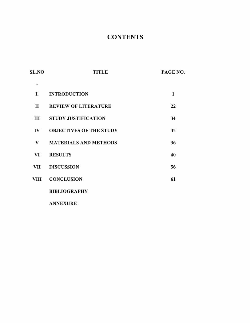
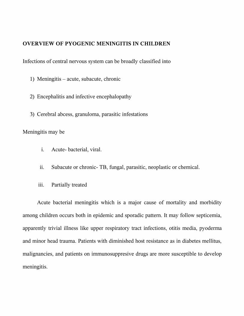
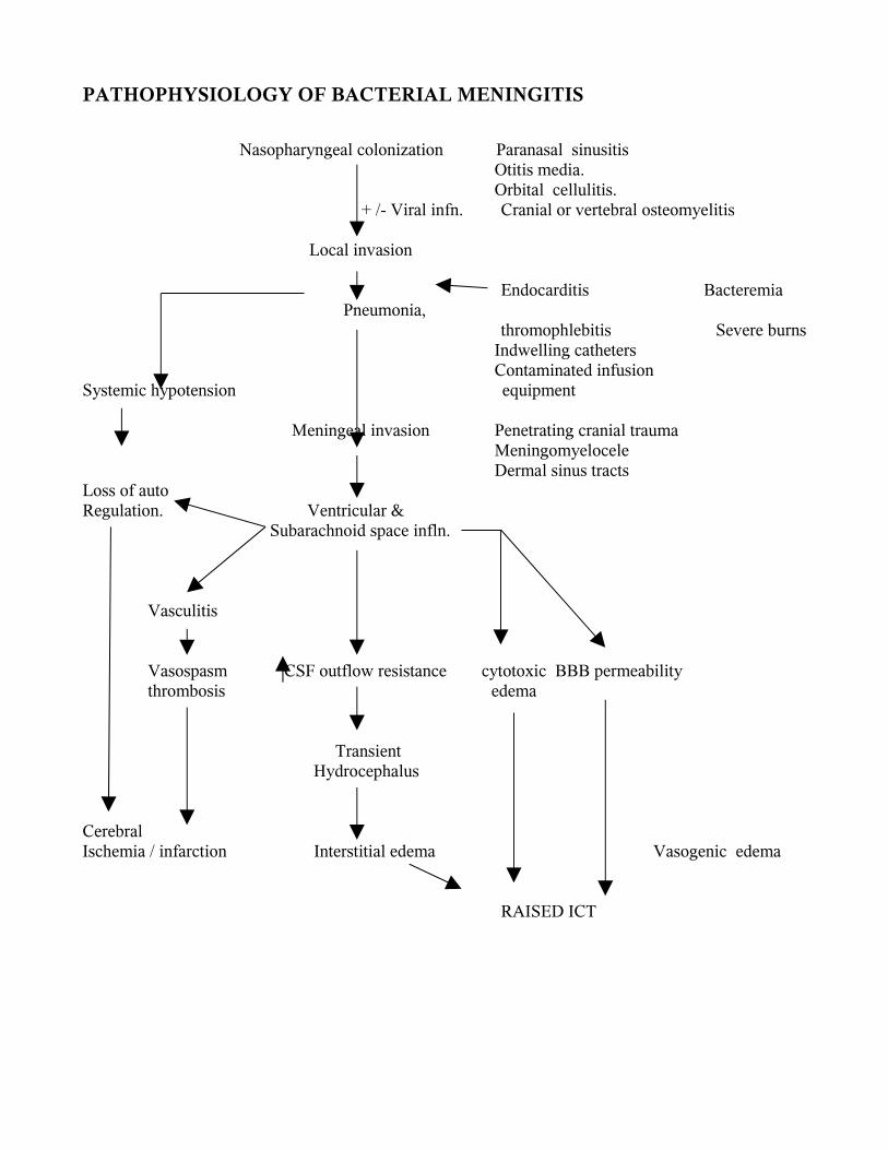
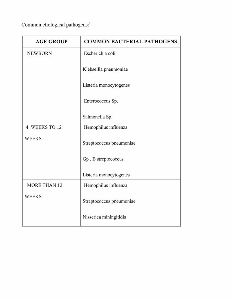
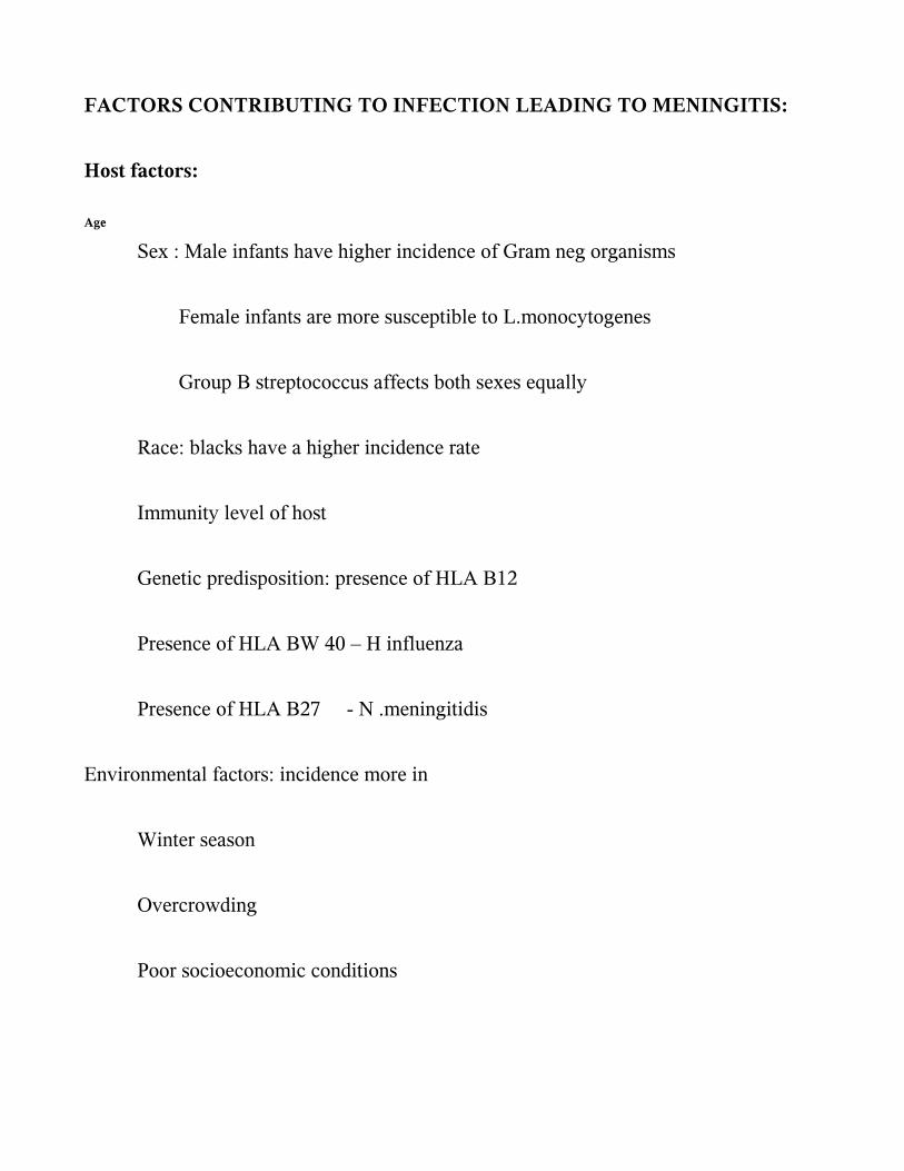
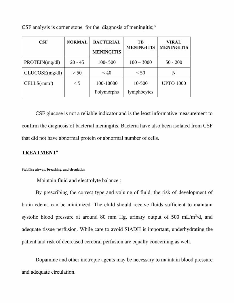
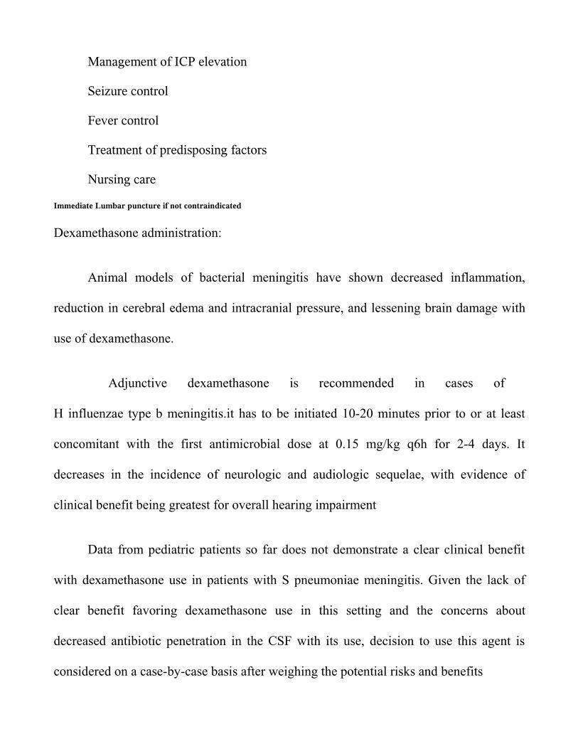
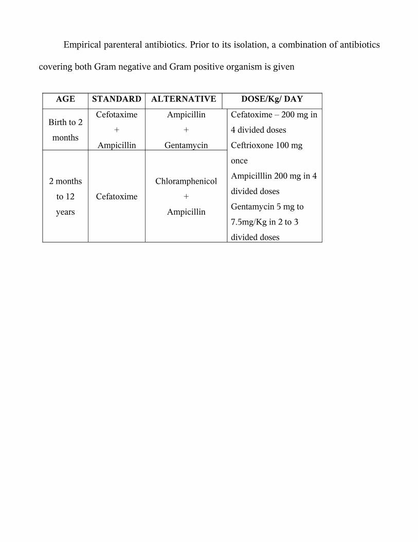
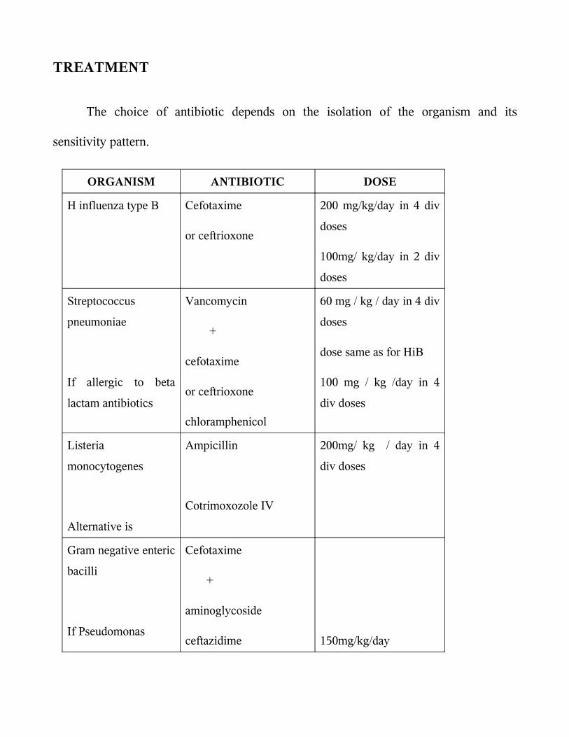