ct angiography in extremity acute vascular trauma · introduction •prompt diagnosis of vascular...
TRANSCRIPT

CT Angiography in Extremity Acute Vascular Trauma
Agnieszka Solberg, MD Noemi Brunner, MD
Department of Radiology TTUHCS El Paso UMC Hospital
Learning Objectives
• Name main indications for performing CT angiography in suspected vascular trauma.
• Describe CT angiography findings of vascular trauma.
• Describe potential pitfalls of CTA
• Demonstrate ability to proceed with therapy without time delay.

Introduction
• Prompt diagnosis of vascular injury is imperative – Injuries can be associated with considerable morbidity
and mortality
• CT is the preferred imaging modality in patients with significant trauma – Adding CTA to the trauma protocol when indicated
should not cause a significant delay
• CTA is usually sufficient to determine the need for and type of treatment in patients with vascular trauma.

Advantages of CTA
• Short acquisition time
• Simultaneous visualization of adjacent structures
– Bones, muscles, etc.
• 3D visualization of anatomic relationships of vasculature and bones beneficial for surgical planning
• With MDCT - ability to evaluate small arteries

Efficacy of CTA
• Evaluation of arterial injury in proximal extremities (Single detector CTA)1
– Sensitivity 95%
– Specificity 99%
• Evaluation of arterial injury in proximal and distal extremities2
– Sensitivity 95%
– Specificity 87%
1. Soto et al. Radiology. 1999, 23, 188.
2. Gakhal & Sartip. AJR. 2009, 193, W49.

Potential Pitfalls of CTA
• False positive – vasospasm
• Lack of opacification of distal vessels due to CTA bolus timing
• Venous injuries may be missed
– single phase protocol
• Use of MIP images alone – failure to detect subtle injury
– Must review axial thin cuts – time intensive
• Surgical hardware/foreign bodies artifact

Artifact – Metallic Foreign Body
• Limited Evaluation at level of foreign body

Clinical Findings
• Hard findings of vascular injury – Pulsatile bleeding
– Loss of distal pulses
– Expanding or pulsatile hematoma
– Pallor, cyanosis, decreased temperature
– Audible bruit or palpable thrill (AVF)
– Massive distention of superficial veins (AVF)
• More than 90% of patients with hard findings have arterial injury at time of surgery – Immediate surgical intervention or DSA

Clinical Findings
• Soft Findings of Vascular Injury – Palpable but diminished pulse
– Isolated peripheral nerve injury
– History of severe hemorrhage in the field
– Unexplained hypotension
– Large non-pulsatile hematoma
– Prolonged capillary refill (controversial)
• Up to 35% of patients with soft findings have positive angiographic studies – A portion of these require emergency repair

Clinical Findings
• High Risk Injuries
– Proximity of penetrating would to vascular structure
– Trajectory crosses vascular bundle
– Examples
• Bites from large dogs
• Shotgun wounds
• Severely displaced fractures
• Crush injury
• Major joint dislocation (especially knee)
– Missed occult vascular injury in 6-hour “warm ischemia” window = delayed complications

Self-inflicted stab wound to thigh
PFA pseudoaneurysm
CTA MIP, axial
CTA MIP, coronal
CTA MIP, sagittal

Profunda Femoris Artery Pseudoaneurysm and AV Fistula
Sequential DSA of RLE: - PFA pseudoaneurysm - Simultaneous arterial and venous
opacification - Enlarged draining vein
DSA of RLE: - Status post
coil embolization – 3 coils
AFV suspected: - Palpable thrill or
audible bruit - Distention of
superficial draining veins
- Early venous enhancement during arterial phase

Pedestrian struck by motor vehicle
Peroneal artery pseudoaneurysm
CTA, axial
CTA MIP, sagittal
Left peroneal artery pseudo-aneurysm was identified on
CTA

Peroneal Artery Pseudoaneurysm • Left peroneal artery
pseudoaneurysm was confirmed on DSA
• Successfully treated with coil embolization
DSA, before and after coil embolization
• Pseudoaneurysm can present as a pulsatile mass

Peroneal Artery Pseudoaneurysm • Left peroneal artery
pseudoaneurysm was confirmed on DSA
• Successfully treated with coil embolization
• Pseudoaneurysm can present as a pulsatile mass
• On CTA it is an organized, localized extravascular contrast filled sac connected to the artery in question

Ulnar Artery Pseudoaneurysm
3D Soft Tissue CTA MIP sagittal CTA MIP coronal 3D Bone and
Vascular
• The patient presented for suture removal s/p laceration of the volar aspect of the hand. Suture removal was complicated by pulsatile bleeding.
• CTA demonstrated a large pseudoaneurysm of the distal ulnar artery

Filling Defect of Radial Artery
• Patient presented with a stab wound to the arm.
• A non-occlusive dissection of the right radial artery was suspected on CTA.
• Patient was taken to the OR due to compartment syndrome.
CTA MIP sagittal

Narrowing of Radial Artery
• Radial artery was normal at surgery – Possibly spasm
– Pitfall of CTA
• Reduction of vessel caliber : – Traumatic
dissection
– Spasm
– External compression
– Thrombus
CTA MIP sagittal
3D Vascular and Soft Tissue
3D Vascular

MVA – Ejected with amnesia Occlusion of the Subclavian Artery
• Comminuted fracture of the scapula with short
segment occlusion of the subclavian artery, likely
secondary to dissection
Oblique MPR
Axial MIP

MVA – Ejected with amnesia Occlusion of the Subclavian Artery
• DSA demonstrated reconstitution however
attempts to cross the occluded segment failed.
• Collaterals are sometimes
difficult to identify given
their small caliber in the
acute setting
• In the chronic setting, the
collaterals are more
established.
DSA

Patient punches window
Occlusion – Transected Radial Artery
• Occlusion of radial artery identified on CTA
• Vessel disruption
–Diminished peripheral pulses
–Pale, cold extremity
• Arterial spasm - may prevent visualization of active contrast extravasation in the setting of artery transection.
• Transection necessitates surgical repair.
CTA MIP oblique

Pt fell in the woods – piece of wood embedded in medial thigh Active Contrast Extravasation
• Hematoma with active contrast extravasation
• Small superficial artery ligated in OR
CTA MIP coronal CTA axial
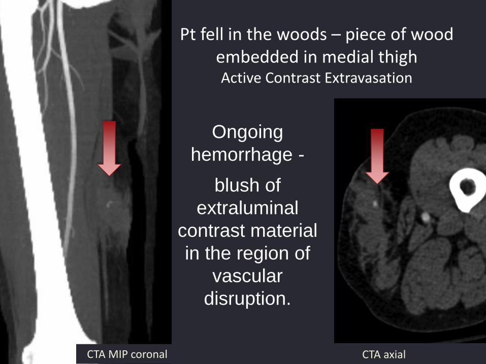
Pt fell in the woods – piece of wood embedded in medial thigh Active Contrast Extravasation
Ongoing
hemorrhage -
blush of
extraluminal
contrast material
in the region of
vascular
disruption.
CTA MIP coronal CTA axial

Cut forearm with circular saw Active contrast extravasation
• Contrast Extravasation from distal ulnar artery and possible pseudoaneurysm
• Transected ulnar artery identified in Operating Room
CTA MIP sagittal CTA MIP coronal

Conclusion
• Indications for CTA in vascular trauma – Soft findings of vascular injury
• Describe CT angiography findings of vascular trauma. – Pseudoaneurysm, AVF, Interruption, Contrast Extravasation
• Potential pitfalls of CTA – Artifacts, Technique/contrast timing, vasospasm
• Demonstrate ability to proceed with therapy without time delay – Avoid missing injury in 6-hour “warm ischemia” window,
therefore decreasing morbidity

References
• Peng et al. Am Surg. 2008, 74, 103.
• Gakhal & Sartip. AJR. 2009, 193, W49.
• Miller-Thomas et al. RadioGraphics.
2005, 25, S133.
• Fishman et al. RadioGraphics. 2008, 28,
653.
• Uyeda et al. Radiol Clin NA. 2010, 48,
423.
• Soto et al. Radiology. 1999, 23, 188.