ct imaging of blunt chest trauma - springer · keywords blunt trauma.lungs.ct introduction chest...
TRANSCRIPT

PICTORIAL REVIEW
CT imaging of blunt chest trauma
Anastasia Oikonomou & Panos Prassopoulos
Received: 6 August 2010 /Revised: 28 November 2010 /Accepted: 27 January 2011 /Published online: 11 February 2011# European Society of Radiology 2011
AbstractBackground Thoracic injury overall is the third mostcommon cause of trauma following injury to the head andextremities. Thoracic trauma has a high morbidity andmortality, accounting for approximately 25% of trauma-related deaths, second only to head trauma. More than 70%of cases of blunt thoracic trauma are due to motor vehiclecollisions, with the remainder caused by falls or blows fromblunt objects.Methods The mechanisms of injury, spectrum of abnormal-ities and radiological findings encountered in blunt thoracictrauma are categorised into injuries of the pleural space(pneumothorax, hemothorax), the lungs (pulmonary contu-sion, laceration and herniation), the airways (tracheobronchiallacerations, Macklin effect), the oesophagus, the heart, theaorta, the diaphragm and the chest wall (rib, scapular, sternalfractures and sternoclavicular dislocations). The possiblecoexistence of multiple types of injury in a single patient isstressed, and therefore systematic exclusion after thoroughinvestigation of all types of injury is warranted.Results The superiority of CT over chest radiography indiagnosing chest trauma is well documented. Moreover,with the advent of MDCT the imaging time for traumapatients has been significantly reduced to several seconds,allowing more time for appropriate post-diagnosis care.Conclusion High-quality multiplanar and volumetric refor-matted CT images greatly improve the detection of injuries
and enhance the understanding of mechanisms of trauma-related abnormalities.
Keywords Blunt trauma . Lungs . CT
Introduction
Chest trauma is classified as blunt or penetrating, with blunttrauma being the cause of most thoracic injuries (90%). Themain difference lies in the presence of an opening to theinner thorax in penetrating trauma, created by stabbing orgunshot wounds, which is absent in blunt chest trauma [1].Blunt thoracic injuries are the third most common injury inpolytrauma patients following head and extremities injuries[2]. Although half of thoracic injuries are minor, 33%require hospital admission [3]. Overall, blunt chest traumais directly responsible for 25% of all trauma deaths [3] andis a major contributor in another 50% of trauma-relateddeaths. Moreover, chest trauma is the second most commoncause of death, following only head trauma, and is by farthe most common cause of death in the young age groupbetween 15 and 44 years old [4]. Most blunt thoracicinjuries are caused by motor vehicle crashes (MVC; 63–78%), with the remainder (10–17%) caused by falls fromheights and a minority from blows from blunt objects orexplosive devices [5].
Portable chest radiography is the initial imaging methodused at the emergency workup of the polytrauma patient,and it is useful for detecting serious life-threateningconditions, such as a tension pneumothorax or haemo-thorax, mediastinal haematoma, flail chest or malpositionedtubes. However, the superiority of CT over chest radiogra-phy has been documented in the literature; CT detectssignificant disease in patients with normal initial radio-
A. Oikonomou (*) : P. PrassopoulosDepartment of Radiology, University Hospitalof Alexandroupolis, Democritus University of Thrace,Dragana, 68100 Alexandroupolis, Thrace, Greecee-mail: [email protected]
P. Prassopoulose-mail: [email protected]
Insights Imaging (2011) 2:281–295DOI 10.1007/s13244-011-0072-9

graphs and in 20% will reveal more extensive injuriescompared with the abnormal initial radiographs, necessitat-ing a change of management [6]. CT is far more effectivethan chest radiography in detecting pulmonary contusion,thoracic aortic injury and osseous trauma, especially at thecervicorthoracic spine. MDCT has dramatically decreasedimaging times and offers readily available multiplanarreformatted images or more sophisticated volume-renderedand MIP images. Therefore, it has been established as thegold standard for the imaging evaluation of chest traumaand trauma in general [7].
This review focusses mainly on the typical CT findingsas well as the pitfalls associated with the wide spectrum oftypes of injury in the thorax, including injury of the pleura(haemothorax, pneumothorax), the lung parenchyma (con-tusion, laceration, lung herniation and blast lung), thetrachea and airways, the aorta, the heart and pericardium,the oesophagus, the diaphragm and the thoracic wall. Thepossible coexistence of multiple types of injury is stressed.
Biomechanics of injury/trauma
Four main mechanisms of injury are responsible for chesttrauma: direct impact to the chest, thoracic compression,rapid acceleration/deceleration and blast injury.
Injuries from a direct impact are usually less dangerousand affect mainly the soft tissues of the chest wall(haematomas, rubbings). Occasionally, a localised injuryto the osseous part of the chest wall can occur (rib fracture,sternal fracture and sternoclavicular dislocation) or, rarely,direct impact forces may be transmitted through the chestwall to the deeper organs, causing serious injury to theheart, lung or large mediastinal vessels.
In thoracic compression injuries intrathoracic structuresstrike a fixed anatomical structure—such as the chest or thespine—causing organ contusion or rupture. Thoraciccompression may cause contusion or laceration of the lungparenchyma, pneumothorax or haemothorax, tracheobron-chial fractures as well as rupture of the diaphragm.
In decelaration injuries the production of shearing forcescauses direct compression against fixed points. This type isthe most common and potentially lethal injury, and maycause major tracheobronchial disruption, cardiac contu-sions, aortic and diaphragmatic rupture [3].
Finally, with the increasing use of improvised explosivedevices in terrorist attacks, blast injuries are occurring at anincreasing rate. Explosion results from the instantaneousconversion of a solid or liquid material into gas afterdetonation of an explosive material. The blast pressurewave that is created exerts forces and pressure differentialsmainly at air-tissue interfaces within the body, mostlyaffecting the pulmonary, gastrointestinal and auditorysystems (primary blast injury). Secondary blast injuriesresult from objects propelled by the explosion, impactingthe individual, while tertiary injuries follow when theindividual is being propelled by the explosion [8, 9].
CT protocols
According to the type of CT available, a collimation of1.25 mm (4-slice and 16-slice) or 0.6 mm (64-slice) isrecommended. Use of 120 Kv and 300 mA is acceptable[10], although attempts to reduce the radiation dose shouldbe constantly pursued, especially if Automatic ExposureControl (AEC) can be applied wherever available [11].Intravenous administration of contrast medium is impera-tive for imaging polytrauma patients, and as there is usuallyno luxury of time, only post-enhanced imaging is per-formed so as not to miss any injury of the major mediastinalvessels and the heart; optimal opacification may beobtained with injection of 100–140 ml of iodinated contrastmedium at a flow rate of 3–4 ml/s and a delay of 25–40 s. IfCT of the thorax is part of a whole body trauma CT, then acompromise can be made with a 75-s delay for the wholebody. When active bleeding is suspected, a delayedacquisition at 5 min is highly recommended, provided thatthe patient’s haemodynamic stability allows for it (Table 1)[12, 13]. Use of ECG gating for thoracic trauma is quitecontroversial, as although it may offer a higher diagnosticquality for any possible aortic, coronary or cardiac injury, itmay on the other hand reduce the quality of bone and lunginjury [14]. Given the fact that retrospective ECG gatingcompared with prospective ECG gating increases theradiation dose significantly, and that polytrauma patientsmay have an unstable heart rate higher than 80 beats/min,one should weigh the use of ECG gating carefully so as notto lose valuable time [14, 15]. The axial thin slices can beused to create axial, coronal and sagittal reformations at 2–
Table 1 CT protocols
Contrast medium Collimation kVp mAs Flow rate Delay Additional image
Thoracic CT only 120–140 ml 4 × 1.25 16 × 1.2564 × 0.6
80–120 300–400 3–4 ml/s 25–40 s –
Thorax: part of wholebody CT
120–140 ml » » » » 70–75 s –
Suspected extravasation 120–140 ml » » » » 25–40 or 75 s 5 min
282 Insights Imaging (2011) 2:281–295

2.5 mm for immediate viewing and exclusion of life-threatening injuries. In the case of positive findings theimages are transferred to a workstation for a moresophisticated imaging process and construction of maxi-mum intensity projections (MIPs) or 3D aortogram-arteriogram CT, 3D reformation of fractures or dislocations,and volume-rendered images for lung parenchyma orairway abnormalities [12, 13]. All reformatted imagesshould be routinely viewed at soft-tissue, lung and bonewindows.
Pleura
Pneumothorax
Trauma-related pneumothorax occurs in 30–40% of cases,and it is most commonly associated with rib fractures thatlacerate the lung. Less commonly, pneumothorax may becaused by a disruption of closed airway spaces, such as thealveoli, due to a sudden increase in intrathoracic pressure orto a direct impact or deceleration force to the chest wall.Tracheobronchial injuries are also always associated withpneumothorax [5, 7, 16, 17]. CT is more sensitive indetecting pneumothoraces (Fig. 1), as 78% of them arenowadays believed to be missed on chest radiograph (occultpneumothoraces) [18, 19]. Pneumothorax in supine poly-trauma patients tends to accumulate at the anterior and
medial aspect of the lung, rendering it difficult to recogniseon a supine chest radiograph, although it might be visibleon an upright chest radiograph. Radiographic signs thatmay be present in the case of an occult pneumothoraxinclude:
(1) Increased lucency at the affected hemidiaphragm,(2) An abnormally deep costophrenic sulcus sign,(3) A sharply defined radiolucent border of the mediasti-
num or heart, and(4) The “double diaphragm sign” caused by the presence
of air outlining the dome and insertion of thediaphragm [7, 20].
It is crucial to detect even a small pneumothorax in thetrauma patient, as this can significantly enlarge underpositive mechanical ventilation in the ICU or duringgeneral anaesthesia and endotracheal tube placement.Consequently, a prophylactic chest tube placement isconsidered [18] in small asymptomatic pneumothoraces(<20%), although controversies exist about this practice inthe literature [21]. However, there is growing evidence thatoccult pneumothorax can be safely treated without thor-acostomy in non-ventilated patients [19].
Tension pneumothorax is an urgent clinical diagnosiswhere progressive accumulation of air—due to the one-valve mechanism—increases the intrathoracic pressure ofthe hemithorax involved, causing a contralateral shift of themediastinum, compression of the superior vena cava andloss of venous return to the heart with resultant haemody-namic impairment. Chest radiography and CT will bothshow a contralateral shift in the mediastinum, hyper-expansion and hyperlucency of the ipsilateral lung withlung collapse towards the hilum, and inversion of theipsilateral diaphragm (Fig. 2). Sudden evacuation of a largepneumothorax with tube drainage can be complicated by re-expansion pulmonary oedema, presenting the correspondingradiological signs. The complication is more common inyounger patients (20–50 years of age) and occurs more oftenthan was previously believed, and although it may beentirely asymptomatic, it has a reported variable mortalityrate reaching 20% [22].
Haemothorax
Haemothorax occurs in 50% of chest trauma cases, withblood pooling into the pleural space from variable sources:the lung parenchyma, the chest wall, the great vessels, theheart or even the liver and spleen through diaphragmaticrupture [20]. Arterial bleeding (more commonly from theintercostal arteries, and the subclavian and internal mam-mary arteries) causes a more significant progressiveincrease in volume and mass effect compared with a
Fig. 1 Occult pneumothorax. Axial CT at lung window shows a smalloccult pneumothorax (missed on chest radiography) at the anterome-dial part of the left upper lobe (white arrow). There are also bilateralground-glass opacities and areas of consolidation in both upper lobesconsistent with lung contusions. Note the presence of numerous smallconfluent pneumatoceles (Swiss cheese appearance) in the anteriorsegment of the left upper lobe, consistent with lung laceration (blackarrow). Note also the presence of small pneumomediastinum (dottedwhite arrow)
Insights Imaging (2011) 2:281–295 283

venous origin of haemorrhage [23]. Massive haemothoraxoccurs when the accumulation of blood in the pleural spaceexceeds 1 l and is accompanied by haemodynamicimpairment (Fig. 3) [2]. CT is very sensitive in detectingeven a small haemothorax and can further characterise it bymeasuring accurately the Hounsfield (HU) units attenuationvalues of the pleural fluid. A reactive pleural effusion willhave values not higher than 15 HU, while liquid blood will
measure 30 to 45 HU, and the clotted blood shouldmeasure around 50–90 HU units [20, 24]. Occasionally a“haematocrit effect” is caused by the layering of differentages and statuses of coagulation of pleural blood products.In the case of active bleeding the fresh extravasated bloodin contrast-enhanced CT may have attenuation valuessimilar to the adjacent enhanced thoracic vessels (±10 HU)[17].
Lung parenchyma
Pulmonary contusion
Lung contusion is a focal parenchymal injury caused bydisruption of the capillaries of the alveolar walls and septa,and leakage of blood into the alveolar spaces andinterstitium [25]. It is the most common type of lung injuryin blunt chest trauma with a reported prevalence of 17–70%[26]. The main mechanism is compression and tearing ofthe lung parenchyma at the site of impact (it may also occurcontralaterally “contre-coup”) against osseous structures,rib fractures or pre-existing pleural adhesions [27]. Lungcontusion occurs at the time of injury, but it may beundetectable on chest radiography for the first 6 h aftertrauma. The pooling of haemorrhage and oedema willblossom at 24 h, rendering the contusion radiographicallymore evident, although CT may readily reveal it from theinitial imaging [28]. The appearance of consolidation onchest radiography after the first 24 h should raise suspicionof other pathological conditions such as aspiration, pneu-monia and fat embolism [2]. Contusions appear asgeographic, non-segmental areas of ground-glass or nodularopacities or consolidation on CT that do not respect thelobar boundaries and may manifest air bronchograms if thebronchioles are not filled with blood (Fig. 4) [17].Subpleural sparing of 1–2 mm may be seen, especially inchildren (Fig. 4b) [29]. Clearance of an uncomplicatedcontusion begins at 24 to 48 h with complete resolutionafter 3 to 14 days [9]. Lack of resolution within theexpected time frame should raise the suspicion of compli-cations such as pneumonia, abscess or ARDS. Pulmonarycontusion—despite the advances in prompt diagnosis withimaging and supportive management with critical caremedicine—remains a predictor of ARDS and has a highmortality rate (10–25%) [30].
Pulmonary laceration
Pulmonary laceration occurs in major chest trauma whendisruption and tearing of the lung parenchyma followsshearing forces, caused by direct impact, compression orinertial deceleration [27]. Lung lacerations have been
Fig. 3 Tension haemopneumothorax. Axial contrast-enhanced CT atmediastinal window shows a right tension haemopneumothorax withheterogeneous increased density due to presence of blood clots and asignificant shift of the mediastinum contralaterally
Fig. 2 Tension pneumothorax. Sagittal reformatted CT image at lungwindow showing tension pneumothorax with significantly collapsedlung at the posterior part of the hemithorax associated with ipsilateralpleural effusion
284 Insights Imaging (2011) 2:281–295

classified into the following four types according to themechanism of injury [27]:
Type 1 Compression rupture injury (the most commontype) is centrally located, can become very largeand is produced by compression of the lungagainst the tracheobronchial tree.
Type 2 Compression shear injury is produced when thelower lobes are suddenly squeezed against the
spine. It is located paraspinally and may betubular in morphology (Fig. 5).
Type 3 Rib penetration tear is peripherally located, issmall and round and is usually associated withpneumothorax (Fig. 6).
Type 4 The adhesion tear is seen adjacent to a previouspleuropulmonary adhesion and is almost alwaysseen at surgery or at autopsy. Lung tissuesurrounding a laceration retracts—because of the
Fig. 4 Lung contusion. Axial(a, b) and coronal (c) CT imagesat lung window show nodularopacities of ground-glassopacity that do not respect thelung boundaries of the rightupper lobe (white arrows) (a),diffuse areas of ground-glassopacity in the upper lobesbilaterally with subpleuralsparing (white arrows) (b) andmultiple areas of consolidationwith air bronchograms (whitearrows) and small lacerations(black arrows) in both lungsconsistent with lung contusions.Note small bilateralpneumothorax in both lungapices (black dotted arrows) andcardiophrenic angles (blackdotted arrows) (c)
Fig. 5 Lung laceration, type II.Coronal reformatted CT imageat lung window (a) shows alobulated paraspinal pneumato-cele (arrow) surrounded byground-glass opacity (contusion)in the right lung consistent withlung laceration (type II?). Onmediastinal window lung lacer-ation is seen to have beencomplicated by acute pulmonaryembolism (dotted arrow)
Insights Imaging (2011) 2:281–295 285

lung elastic recoil—leaving a round or oval cavitythat may be filled with air (pneumatocele), blood(haematocele or haematoma) or both, creating anair-fluid level (haematopneumatocele). A lacera-tion, although it may be filled with air, is usuallysurrounded by lung contusion and therefore ishidden on a chest radiograph during the first 2–3 days, until the contusion begins to resolve. CT,on the other hand, is significantly superior to chestradiography in detecting even a small lacerationand in revealing the overall extent of the lacer-ations [27]. Lacerations (Fig. 1) may range from asolitary lesion to multiple confluent small onespresenting a “Swiss cheese appearance” [20].Lacerations resolve more slowly than contusions,and clearance may take weeks or even months,and they may end in residual scarring [13].Uncommonly, lacerations may be complicated bya pulmonary abscess, enlarge through a ball-valvemechanism or form a bronchopleural fistula [17],or it may be associated with acute pulmonaryembolism (Fig. 5).
Lung herniation
Herniation of the lung parenchyma is an uncommonmanifestation of blunt chest trauma, and it can occurthrough a congenital or a traumatic chest wall defect such
as multiple rib fractures or sternoclavicular or costochon-dral dislocations. Surgical repair is indicated when thepatient is symptomatic or if the patient needs intubation andgeneral anaesthesia as herniation may increase withpositive-pressure ventilation [24, 31].
Blast injury
Blast lung is the most common fatal injury among initialsurvivors of explosions; 17–47% of people who die fromexplosions have had primary blast lung injury [8, 9].However, the in-hospital mortality rate for these patientsranges from 3.4 to 25% because of prompt diagnosis andaggressive treatment. The blast wave causes thoracicacceleration and propagates through lung parenchyma withsubsequent severe disruption at the capillary-alveolar
Fig. 6 Lung laceration, type IV. Axial CT image of the left lung atlung window shows a small peripheral laceration (white arrow)beneath a rib fracture (black arrow) surrounded by ground-glassopacity (lung contusion) and associated with a small ipsilateralpneumothorax
Fig. 7 Blast lung injury. Twenty-two-year-old patient who experi-enced the explosion of a grenade in his hands. Coronal CT reformattedcontrast-enhanced CT image at mediastinal window shows bilateralperilar consolidations mimicking a butterfly or bat-wing appearance,consistent with blast lung. The left lung is almost completelycollapsed, and there are bilateral haemothoraces
Fig. 8 Bronchial transection. A 22-year-old man involved in a caraccident. Volume-rendered image of the tracheobronchial tree showingcomplete transection of the right intermediate bronchus (two-wayarrow). (Courtesy of Dr Montserrat Bret, University Hospital La Paz,Madrid)
286 Insights Imaging (2011) 2:281–295

interface. This results in parenchymal haemorrhage andcontusions, pulmonary oedema, pneumothorax, barotraumaand air embolism from arteriovenous fistulas, causingsubstantial immediate and delayed injury. Chest radiogra-phy and CT (Fig. 7) will reveal the “butterfly or batwing”pattern, representing central bilateral perihilar air spaceconsolidation and ground-glass opacities that may containair bronchograms [32].
Airways
Trachea-bronchi
Tracheobronchial injuries are rare, occurring in 0.2–8% ofall cases of chest trauma. It is anticipated that theprevalence is higher, as 50% of patients die at the traumascene within the first 2 h from associated injuries and
respiratory insufficiency [2, 33]. They have a mortality rateof 30%, and in two thirds of cases the diagnosis is delayedwith subsequent serious complications, such as pneumonia,abscess, empyema, mediastinitis, sepsis, airway obstructionor atelectasis. Bronchial injuries occur more commonlythan tracheal, usually on the right side and within 2.5 cmfrom the carina [5, 24], while 85% of tracheal lacerationsoccur 2 cm above the carina. Bronchial lacerations areusually parallel to the cartilage rings as opposed to trachealones that are vertical to the cartilage rings. A direct CTfinding of tracheobronchial injuries is the cutoff of thetracheal and bronchial wall with extraluminal air surround-ing the airway (Fig. 8). Indirect findings are the “fallenlung” sign, corresponding to the collapsed lung restingaway from the hilum towards the dependent portion of thehemithorax [34], persistent pneumothorax after chest tubeplacement and herniation or overdistention of an endotra-cheal balloon if this is placed at the same level as thetracheal laceration [33]. Tracheal lacerations are usuallyassociated with cervical subcutaneous emphysema. Tra-cheobronchial injuries in general are accompanied bypneumothorax and pneumomediastinum.
Fig. 9 Pneumomediastinum.Axial CT images at wide lungwindow show pneumomediasti-num with the presence of septaewithin the air in the anteriormediastinum (black arrows) (a),and in the middle and posteriormediastinum (black arrows) (b).Note also a right pneumothorax,bilateral lower lobe atelectasesand subcutaneous emphysema(a) and a right haemopneu-mothorax and left pneumothorax(b)
Fig. 10 Haemopericardium. Axial contrast-enhanced CT of the lowerthorax at mediastinal window shows haemopericardium that mayrepresent an indirect sign of pericardial or heart injury in apolytraumatised patient after a motor vehicle accident. There is alsoa small right haemothorax
Fig. 11 Pneumopericardium. Axial CT image at lung window showsextensive pneumopericardium (white arrow), pneumomediastinum(black arrows), haemopneumothorax (black dotted arrows), collapsedleft lung with ipsilateral shift of the mediastinum and collapse of theright lower lobe
Insights Imaging (2011) 2:281–295 287

Mediastinal structures
Pneumomediastinum, the Macklin effect
Pneumomediastinum occurs in 10% of patients with bluntchest trauma, with less than 2% caused by blunt tracheo-bronchial injuries. Other sources of air originate from lungparenchymal injury, oesophageal injury, chest wall, neckand retroperitoneal injury. In a number of patients pneumo-mediastinum is attributed to the Macklin effect caused byalveolar ruptures that lead to air dissecting along broncho-
vascular bundles and spreading of the pulmonary interstitialemphysema into the mediastinum. Streaks of air surround-ing and paralleling the bronchovascular bundles associatedwith pneumomediastinum may be observed on CT [35].Pneumomediastinum may be mistaken for pneumothorax,but the presence of septae within it—delineated on widelung window—may help in differentiating the two findings,especially if they coexist (Fig. 9).
Heart and pericardium
Cardiac injuries are the most lethal in chest trauma patients.They are more common in penetrating trauma, but they canoccasionly occur in motor vehicle accidents and from
Fig. 12 Traumatic oesophageal rupture. A 12-year-old boy trauma-tised during a fall from a tree. Oesophagogram with per osadministration of water-soluble contrast medium (a) shows leakageof contrast medium into both pleural spaces. Axial CT image of thethorax at the level of the lung bases (b) verifies the leakage of the
contrast medium into the left and right pleural spaces. (Imagereproduced from: Arora A, Puri SK, Upreti L, et al (2010).Oesophageal rupture: a rare complication of blunt trauma, {Online}.URL: http://www.eurorad.org/case.php?id=8447)
Fig. 13 Traumatic aortic pseudoaneurysm. Sagittal reformattedcontrast-enhanced CT image of the thoracic aorta reveals a pseudoa-neurysm of the greater curve of the mid-descending thoracic aorta(black arrow)
Fig. 14 Traumatic aortic pseudoaneurysm. Three-dimensional recon-structed CT image of the thoracic aorta shows a pseudoaneurysm ofthe inferior curve of the thoracic aorta immediately distal to theisthmus (arrow)
288 Insights Imaging (2011) 2:281–295
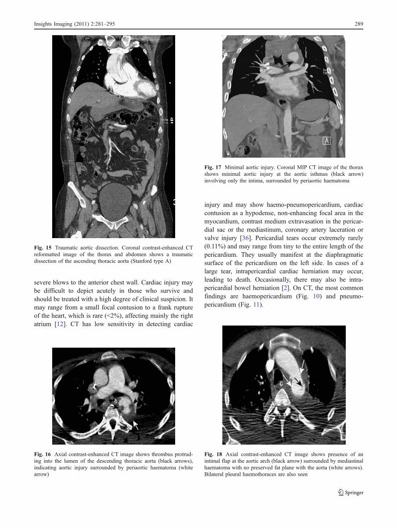
severe blows to the anterior chest wall. Cardiac injury maybe difficult to depict acutely in those who survive andshould be treated with a high degree of clinical suspicion. Itmay range from a small focal contusion to a frank ruptureof the heart, which is rare (<2%), affecting mainly the rightatrium [12]. CT has low sensitivity in detecting cardiac
injury and may show haemo-pneumopericardium, cardiaccontusion as a hypodense, non-enhancing focal area in themyocardium, contrast medium extravasation in the pericar-dial sac or the mediastinum, coronary artery laceration orvalve injury [36]. Pericardial tears occur extremely rarely(0.11%) and may range from tiny to the entire length of thepericardium. They usually manifest at the diaphragmaticsurface of the pericardium on the left side. In cases of alarge tear, intrapericardial cardiac herniation may occur,leading to death. Occasionally, there may also be intra-pericardial bowel herniation [2]. On CT, the most commonfindings are haemopericardium (Fig. 10) and pneumo-pericardium (Fig. 11).
Fig. 15 Traumatic aortic dissection. Coronal contrast-enhanced CTreformatted image of the thorax and abdomen shows a traumaticdissection of the ascending thoracic aorta (Stanford type A)
Fig. 16 Axial contrast-enhanced CT image shows thrombus protrud-ing into the lumen of the descending thoracic aorta (black arrows),indicating aortic injury surrounded by periaortic haematoma (whitearrow)
Fig. 17 Minimal aortic injury. Coronal MIP CT image of the thoraxshows minimal aortic injury at the aortic isthmus (black arrow)involving only the intima, surrounded by periaortic haematoma
Fig. 18 Axial contrast-enhanced CT image shows presence of anintimal flap at the aortic arch (black arrow) surrounded by mediastinalhaematoma with no preserved fat plane with the aorta (white arrows).Bilateral pleural haemothoraces are also seen
Insights Imaging (2011) 2:281–295 289

Oesophagus
Oesophageal injury is far more common in penetrating oriatrogenic injury and occurs only in 1% of cases of bluntchest trauma. The main mechanisms are a direct blow to theneck mainly affecting the cervical oesophagus, burst-typeforce or hyperextension injury affecting the distal oesoph-agus, or rupture by a vertebral body fracture [12]. On CTthere will be mainly indirect findings of oesophagealrupture such as pneumomediastinum and peri-oesophagealair or abnormal mediastinal contour secondary to leakage offluid, haematoma or mediastinitis. Hydropneumothorax isusually seen on the left side. Diagnosis can be confirmed(Fig. 12) by water-soluble contrast oesophagographyshowing leakage of contrast medium into the mediastinalor pleural space [37].
Aorta, great vessels
Aortic injury occurs in 0.5%–2% of all non-lethal MVAsand is responsible for 10–20% of deaths in MVAs. Aorticinjuries have a high morbidity and mortality; 90% of thepatients die at the trauma scene, while 90% of initialsurvivors die within 4 months if the injury is undetectedand untreated [38]. In the remaining 20% of cases, aorticinjury is caused by falls and pedestrian injuries. Mecha-nisms of injury are variable and may overlap, includingrapid deceleration, shearing forces, increased intravascularpressure caused by compression exceeding 2,000 mmHg(the water-hammer effect) or the “osseous pinch”, whichrepresents direct compression of the aorta between theanterior chest wall and the spine [38]. The most commonsite of injury is the isthmus, corresponding to 90–95% of
Fig. 19 Dependent viscera sign. Axial contrast-enhanced CT at thelevel of the lower lobes, at mediastinal window, shows intrathoracicpresence of the stomach abutting the left posterior thoracic wallwithout intervening in the left hemidiaphragm (black arrows)
Fig. 20 The “hourglass” or “collar” sign of diaphragmatic rupture.Reformatted coronal contrast-enhanced CT image of the thorax showswaist-like stricture of the herniated left colon intrathoracically throughthe small defect of the left hemidiaphragm (black arrows)
Fig. 21 Reformatted coronal contrast-enhanced CT image of thethorax shows intrathoracic herniation of intraperitoneal fat through thelarge defect of the left hemidiaphragm (white arrows)
Fig. 22 Coronal MIP CT image showing multiple contiguous left ribfractures (arrows)
290 Insights Imaging (2011) 2:281–295

cases. Uncommon sites include the aortic root-ascendingaorta, the aortic arch-branch vessels and the mid-distaldescending aorta. The injury may be partial (65%),involving only the intima and media, or transmural (35%),also affecting the adventitia, which is lethal in almost allcases. The injury may be circumferential (45% of cases) orsegmental (in 55% of cases), involving either the greater(Fig. 13) or the inferior curve (Fig. 14). CT may showdirect and indirect signs. Direct signs include activecontrast medium extravasation, dissection (Fig. 15), pseu-doaneurysm (Figs. 13 and 14), intimal tear/flap (Fig. 16),thrombus protruding into the lumen (Fig. 17), and abruptchange in calibre (pseudocoarctation). Indirect CT signs are
indistinctness of mediastinal flat planes, periaortic haema-toma and mediastinal haematoma (Figs. 16–18). Mediasti-nal haematoma is less than 20% predictive of aortic injury.In the absence of aortic injury, mediastinal haematoma mayoriginate from venous injuries. In such cases the fat planewith the aorta is preserved, contrary to thoracic aorticinjury, where haematoma develops in close contact with theaortic wall (Fig. 16–18). Minimal aortic injuries affect onlythe intima. They are diagnosed with increasing frequency—because of improved MDCT technology—and constitute adiagnostic dilemma [39], as most of them remain stable orresolve on follow-up (Fig. 18). MDCT has very highsensitivity and specificity, reaching 98% and 100% accord-ingly for diagnosing aortic trauma. However, in studieswhere the presence of mediastinal haematoma (an indirectsign of aortic injury) was considered as a positive criterionfor aortic injury, a significant number of false-positive
Fig. 23 Coronal (a) and sagittal(b) reconstructed CT imagesshow fractures of threecontiguous right ribs (arrows)that were associated withparadox motion of the chestduring respiration. Flail chestwas suspected clinically andverified on imaging
Fig. 24 Sternal fracture. Sagittal reconstructed CT image showsmultiple fractures of the manubrium and the body of the sternum(white arrows) accompanied by extensive retrosternal haematoma(black ball arrows). Note also fracture of a thoracic vertebra (blackarrow)
Fig. 25 Sternal fracture. Axial CT image at mediastinal windowshows sternal fracture associated with retrosternal haematoma (blackarrow). Note the preserved fat plane with the aorta, excluding thepresence of aortic injury (white arrows)
Insights Imaging (2011) 2:281–295 291
diagnoses occurred, and the specificity for aortic injurydropped significantly by up to 62%. Nevertheless, MDCThas become the gold standard for ruling out aortic injury,and in those patients with unequivocal evidence of aorticinjury, no further imaging is required [40]. No furtherworkup is indicated if there is no direct evidence of aorticinjury and no mediastinal haematoma on CT [38].
Diaphragm
Diaphragmatic injury occurs in 0.16% to 5% of blunttrauma cases, and it is more common in abdominal than inchest trauma. It is three times more common on the left sidethan on the right side, and the main mechanism is thoughtto be the sudden increase in intra-abdominal-thoracic
Fig. 26 Anterior sternoclavicular dislocation. Axial CT image showsclavicular fracture and anterior sternoclavicular dislocation (dottedarrows)
Fig. 27 Posterior sternoclavicular dislocation. Axial CT image showsposterior sternoclavicular dislocation (black arrow) associated withcompression of the left innominate vein (black dotted arrow)
Fig. 28 Scapular fracture. Sagittal reconstructed CT image showsmultiple fractures of the left scapula (arrows)
Fig. 29 Thoracic spine fracture. Coronal (a) and sagittal (b) CTreconstructed images of two different patients show fractures of theupper thoracic vertebrae with great detail
292 Insights Imaging (2011) 2:281–295

pressure against a fixed diaphragm. The most common siteof rupture is at the posterolateral surface, at the site ofembryonic diaphragmatic fusion. Through the diaphrag-matic defect, depending on its size, there may beintrathoracic herniation of intra-abdominal visceral organs,which may be incarcerated, strangulated or perforated. CTfindings of diaphragmatic rupture include [41] the dia-phragmatic discontinuation and defect, the “collar sign” or“hourglass sign” formed by the waist-like stricture of partialintrathoracic herniation of the stomach or bowel (Fig. 19),
and the “dependent viscera sign”, which is formed by theposterior fall of the viscera towards and abutting theposterior dependent thoracic wall (with the patient supine)without the support of the intervening diaphragm (Fig. 20).Contrast-enhanced CT may reveal contrast material extrav-asation at the site of diaphragmatic rupture. Occasionally,there may be only peritoneal fat intrathoracically herniatedthrough the defect (Fig. 21). Diaphragmatic injury isusually a delayed diagnosis because of other associatedserious injuries that may mask the clinical symptoms. It hasa high mortality rate (30%) if it remains unrecognised [42].The high-resolution coronal and sagittal reformationsroutinely produced with MDCT—compared with single-spiral CT—allow detection with high sensitivity, even of asmall diaphragmatic defect.
Thoracic wall
Soft tissue haematoma
Soft tissue haematomas may occur during direct compres-sion trauma when rib fractures cause laceration of veins orarteries. Soft tissue haematoma may become life-threatening if the patient is under anticoagulant therapy. Ifit is arterial in origin, embolisation is indicated. Breasthaematomas can be serious in direct impact or compressioninjuries [24].
Ribs
Rib fractures are the most common injury in blunt chesttrauma, occurring in 50% of cases. A single rib fracture isusually not clinically significant, whereas multiple ribfractures indicate severe injury. Fractures of the first threeribs imply high-energy trauma that may be associated withinjury of the brachial plexus or subclavian vessels.Fractures of the fourth up to the eighth ribs are the mostcommon, while fractures of the last four ribs are usuallyassociated with intra-abdominal injury. Reconstructed MIP
Fig. 30 Thoracic spine fracture and compressive myelopathy. SagittalT2-weighted MRI of the cervicothoracic spine undertaken 1 weekafter a motor vehicle accident verifies the presence of extensivecompressive myelopathy (between the two white arrows with blackoutline) due to fractures of the second and the third thoracic vertebrae(white arrows)
Table 2 Associated injuries
Sternal fracture Heart injury
Rib fracture Pulmonary contusion, laceration
Upper rib fracture (first three ribs) Brachial plexus, subclavian vessels
Lower rib fractures (last four ribs) Intra-abdominal injury
Subcutaneous emphysema Airway injury, oesophageal injury
Pneumomediastinum Airway injury, lung injury, oesophageal injury
Sternoclavicular fracture (posterior sternoclavicular dislocation) Mediastinal vessels, tracheal injury, oesophageal injury
Scapular fracture Haemopneumothorax, lung injury, spine and clavicle fracture, subclavianvessels, brachial plexus
Insights Imaging (2011) 2:281–295 293

and volume-rendered CT images depict with great detail thenumber and sites of rib fractures (Fig. 22). Flail chest is amarker of significant intrathoracic injury with increasedmorbidity, in which three or more contiguous ribs arefractured in two or more sites (Fig. 23). The diagnosis isclinical based on the paradoxical motion during respiration,which may result in ventilatory compromise. More than50% of cases require surgical treatment and prolongedmechanical ventilation [9].
Sternum
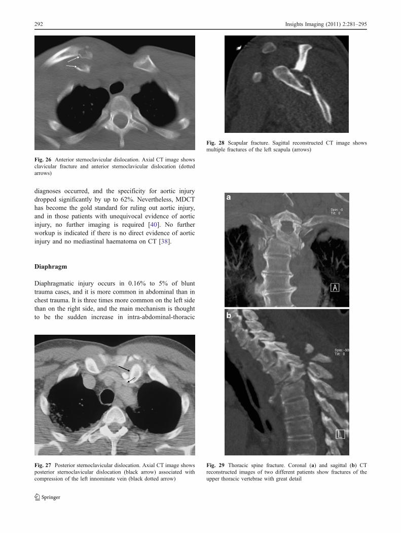
Sternal fractures have a prevalence of 3–8% in blunt chesttrauma. The main mechanism is deceleration injury or adirect blow to the anterior chest wall. It is considered amarker of cardiac contusion (1.5%–6%). Sternal fracturesare difficult to detect on lateral chest radiographs and evenon axial CT images, as opposed to sagittal and coronalMDCT reformats, which have significant superiority(Fig. 24). It is almost always accompanied by anteriormediastinal haemorrhage, which has a preserved fat planewith the aorta (Fig. 25), as opposed to an anteriormediastinal haemorrhage secondary to aortic injury, whichwill present with a lost fat plane with the aorta [17].
Sternoclavicular dislocation is rare and occurs in 1–3%of all types of dislocation. Anterior sternoclaviculardislocation is more common and easily detectable, as it ispalpable (Fig. 26). It usually has a benign course, but itimplies a high-energy trauma and may be associated withhaemopneumothorax, rib fractures or pulmonary contusion[43]. Posterior sternoclavicular dislocation is clinically andradiographically silent and carries serious morbidity, as it isassociated with injuries of the mediastinal vessels, nerves,trachea and oesophagus (Fig. 27).
Scapula
Scapular fracture is uncommon, occurring in 3.7% of casesof blunt chest trauma. It is easily detected on initialradiographs and may be masked clinically by otherassociated serious injuries (Fig. 28). It indicates a high-energy force trauma with a direct blow to the scapula orforce transmitted through the humerus. Associated injuriesare pneumothorax, haemothorax, clavicular fracture andinjuries of the lung parenchyma, subclavian vessels,brachial plexus or spine [44].
Spine
Thoracic spine fractures account for up to 30% of all spinefractures. Sixty-two percent of spine fractures will result inneurological deficits. The most vulnerable site is betweenthe ninth and twelfth vertebra. The main mechanism is
hyperflexion and axial loading. Plain radiographs may missfractures of the spine and therefore may be unnecessary inthose patients scheduled for CT [45]. Sagittal and coronalMDCT reformats (Fig. 29) readily reveal even small spinalfractures, whereas volume-rendered images are not helpful[12]. MDCT of the spine is highly indicated for spinalsurvey for possible fractures. However, in the case ofsuspected compressive myelopathy, MRI is the method ofchoice (Fig. 30).
Coexisting and associated injuries
It is important to remember that multiple types of injury ina single patient may coexist, and radiologists should not bedisorientated by depicting one type of trauma and neglectother coexisting or associated types of injury (Table 2).Therefore, systematic exclusion after thorough investigationof all sites of possible injury in the thorax is warranted.
Acknowledgements The authors would like to thank the followingradiologists for their valuable contributions: Aristi Kouri (Nicosia,Cyprus), Christoforos Schizas (Nicosia, Cyprus), Jean Seely (Ottawa,Canada), Rennae Thiessen (Vancouver, Canada), Argiro Voloudaki(Heraklion, Greece).
References
1. Shanmuganathan K, Matsumoto J (2006) Imaging of penetratingchest trauma. Radiol Clin North Am 44:225–238, Review
2. Kaewlai R, Avery LL, Asrani AV, Novelline RA (2008) Multi-detector CT of blunt thoracic trauma. Radiographics 28:1555–1570
3. Scaglione M, Pinto A, Pedrosa I, Sparano A, Romano L (2008)Multi-detector row computed tomography and blunt chest trauma.Eur J Radiol 65:377–388
4. The American College of Surgeons Committee on TraumaLeadership (2007) In: Clark DE, Fantus RJ (eds) National TraumaData Bank (NTDB) Annual Report 2007. American College ofSurgeons, Chicago, IL, pp 1–64
5. Mayberry JC (2000) Imaging in thoracic trauma: the traumasurgeon's perspective. J Thorac Imaging 15:76–86
6. Exadaktylos AK, Sclabas G, Schmid SW, Schaller B, ZimmermannH (2001) Do we really need routine computed tomographicscanning in the primary evaluation of blunt chest trauma in patientswith “normal” chest radiograph? J Trauma 51:1173–1176
7. Peters S, Nicolas V, Heyer CM (2010) Multidetector computedtomography-spectrum of blunt chest wall and lung injuries inpolytraumatized patients. Clin Radiol 65:333–338, Review
8. Wolf SJ, Bebarta VS, Bonnett CJ, Pons PT, Cantrill SV (2009)Blast injuries. Lancet 374:405–415
9. Wanek S, Mayberry JC (2004) Blunt thoracic trauma: flail chest,pulmonary contusion, and blast injury. Crit Care Clin 20:71–81
10. Fanucci E, Fiaschetti V, Rotili A, Floris R, Simonetti G (2007)Whole body 16-row multislice CT in emergency room: effects ofdifferent protocols on scanning time, image quality and radiationexposure. Emerg Radiol 13:251–257
11. McCollough CH, Bruesewitz MR, Kofler JM Jr (2006) CT dosereduction and dose management tools: overview of availableoptions. Radiographics 26:503–512, Review
294 Insights Imaging (2011) 2:281–295

12. Rivas LA, Fishman JE, Múnera F, Bajayo DE (2003) MultisliceCT in thoracic trauma. Radiol Clin North Am 41:599–616,Review
13. Novelline RA (2007) Imaging chest trauma. In: Diseases of theHeart, Chest & Breast. Part 1. Springer, Milan
14. Schertler T, Glücker T, Wildermuth S, Jungius KP, Marincek B,Boehm T (2005) Comparison of retrospectively ECG-gated andnongated MDCT of the chest in an emergency setting regardingworkflow, image quality, and diagnostic certainty. Emerg Radiol12:19–29
15. Bruzzi JF, Rémy-Jardin M, Delhaye D, Teisseire A, Khalil C,Rémy J (2006) When, why, and how to examine the heart duringthoracic CT: Part 1, basic principles. AJR Am J Roentgenol186:324–332, Review
16. Lomoschitz FM, Eisenhuber E, Linnau KF, Peloschek P, Schoder M,Bankier AA (2003) Imaging of chest trauma: radiological patterns ofinjury and diagnostic algorithms. Eur J Radiol 48:61–70
17. Miller LA (2006) Chest wall, lung, and pleural space trauma.Radiol Clin North Am 44:213–224
18. McGillicuddy D (2007) Diagnostic dilemmas and current contro-versies in blunt chest trauma. Emerg Med Clin North Am 25:695–711
19. Ball CG, Kirkpatrick AW, Laupland KB, Fox DI, Nicolaou S,Anderson IB, Hameed SM, Kortbeek JB, Mulloy RR, LitvinchukS, Boulanger BR (2005) Incidence, risk factors, and outcomes foroccult pneumothoraces in victims of major trauma. J Trauma59:917–924
20. Mirvis SE (2005) Imaging of acute thoracic injury: the advent ofMDCT screening. Semin Ultrasound CT MR 26:305–331
21. Brasel KJ, Stafford RE, Weigelt JA, Tenquist JE, Borgstrom DC(1999) Treatment of occult pneumothoraces from blunt trauma. JTrauma 46:987–990
22. Kim YK, Kim H, Lee CC, Choi HJ, Lee KH, Hwang SO, Oh JH,Lee YH, Singer AJ (2009) New classification and clinicalcharacteristics of reexpansion pulmonary edema after treatmentof spontaneous pneumothorax. Am J Emerg Med 27:961–967
23. Shanmugathan K, Mirvis SE (1999) Imaging diagnosis ofnonaortic thoracic injury. Radiol Clin North Am 37:533–551
24. Sangster GP, González-Beicos A, Carbo AI, Heldmann MG,Ibrahim H, Carrascosa P, Nazar M, D'Agostino HB (2007) Blunttraumatic injuries of the lung parenchyma, pleura, thoracic wall,and intrathoracic airways: multidetector computer tomographyimaging findings. Emerg Radiol 14:297–310
25. Wicky S, Wintermark M, Schnyder P, Capasso P, Denys A (2000)Imaging of blunt chest trauma. Eur Radiol 10:1524–1538, Review
26. Gavelli G, Canini R, Bertaccini P, Battista G, Bna C, Fattori R(2002) Traumatic injuries: imaging of thoracic injuries. Eur Radiol12:1273–1294
27. Wagner RB, Crawford WO Jr, Schimpf PP (1988) Classificationof parenchymal injuries to the lung. Radiology 167:77–82
28. Schild HH, Strunk H, Weber W, Stoerkel S, Doll G, Hein K,Weitz M (1989) Pulmonary contusion: CT vs plain radiograms. JComput Assist Tomogr 13:417–420
29. Donnelly LF, Klosterman LA (1997) Subpleural sparing: a CTfinding of lung contusion in children. Radiology 204:385–387
30. Miller PR, Croce MA, Bee TK, Qaisi WG, Smith CP, Collins GL,Fabian TC (2001) ARDS after pulmonary contusion: accuratemeasurement of contusion volume identifies high-risk patients. JTrauma 51:223–230
31. Clark AJ, Hughes N, Chisti F (2009) Traumatic extrathoracic lungherniation. Br J Radiol 82:e82–e84
32. Avidan V, Hersch M, Armon Y, Spira R, Aharoni D, Reissman P,Schecter WP (2005) Blast lung injury: Clinical manifestations,treatment and outcome. Am J Surg 190:927–931
33. Scaglione M, Romano S, Pinto A, Sparano A, Scialpi M, RotondoA (2006) Acute tracheobronchial injuries: Impact of imaging ondiagnosis and management implications. Eur J Radiol 59:336–343
34. Tack D, Defrance P, Delcour C, Gevenois PA (2000) The CTfallen-lung sign. Eur Radiol 10:719–721
35. Wintermark M, Schnyder P (2001) The Macklin effect: a frequentetiology for pneumomediastinum in severe blunt chest trauma.Chest 120:543–547
36. Sliker CW, Mirvis SE, Shanmuganathan K, Meyer CA (2000)Blunt cardiac rupture: value of contrast-enhanced spiral CT. ClinRadiol 55:805–808
37. de Lutio di Castelguidone E, Merola S, Pinto A, Raissaki M,Gagliardi N, Romano L (2006) Esophageal injuries: spectrum ofmultidetector row CT findings. Eur J Radiol 59:344–348
38. Steenburg SD, Ravenel JG, Ikonomidis JS, Schönholz C, ReevesS (2008) Acute traumatic aortic injury: imaging evaluation andmanagement. Radiology 248:748–762, Review
39. Malhotra AK, Fabian TC, Croce MA, Weiman DS, Gavant ML,Pate JW (2001) Minimal aortic injury: a lesion associated withadvancing diagnostic techniques. J Trauma 51:1042–1048
40. Mirvis SE (2006) Thoracic vascular injury. Radiol Clin North Am44:181–197
41. Bergin D, Ennis R, Keogh C, Fenlon HM, Murray JG (2001) The“dependent viscera” sign in CT diagnosis of blunt traumaticdiaphragmatic rupture. AJR Am J Roentgenol 177:1137–1140
42. Mirvis SE, Shanmuganagthan K (2007) Imaging hemidiaphrag-matic injury. Eur Radiol 17:1411–1421
43. Cope R (1993) Dislocations of the sternoclavicular joint. SkeletalRadiol 22:233–238
44. Weening B, Walton C, Cole PA, Alanezi K, Hanson BP, BhandariM (2005) Lower mortality in patients with scapular fractures. JTrauma 59:1477–1481
45. Rhea JT, Sheridan RL, Mullins ME, Nonelline RA (2001) Canchest and abdominal trauma CT eliminate the need for plain filmsof the spine? - Experience with 329 multiple trauma patients.Emerg Radiol 8:99–104
Insights Imaging (2011) 2:281–295 295