cta for acute chest pain in the emergency … for acute chest...cta for acute chest pain in the...
TRANSCRIPT
CTA FOR ACUTE CHEST PAIN IN THE
EMERGENCY ROOM
Charles White MD Director of Thoracic Imaging
Department of Radiology
University of Maryland
No Disclosures

OBJECTIVES
Clinical background of ED chest pain
Conventional imaging options for ED CP
ED CTA » Technique
» Practice
» Literature
» Future

BACKGROUND/EPIDEMIOLOGY
Diagnosis of chest pain in the ED remains challenging
ACS (acute coronary syndrome) vs. other cardiac, non-cardiac causes
ACS definition
» Transmural MI
» Subendocardial MI
» Unstable angina

EPIDEMIOLOGY
6+ million ED visits yearly for CP in US
Work-up often >12 hours
Only 10-15% have ACS
50% of patients admitted, many with normal biomarkers/ECG
~30% “gap”
>$8B spent per year
Goal: Speed work-up, improve accuracy

BACKGROUND
ACS Diagnosis Triad
» History
» ECG
» Cardiac enzymes (CK, troponin)
Troponin-C

TRIAGE / ROLE OF NON –INVASIVE IMAGING
Group 1: Clear ACS (acute ECG, elevated biomarkers) – immediate cath
Group 2: Minimal risk – early D/C
Group 3: Equivocal – Non-invasive imaging useful
» Atypical history
» Normal or nonspecific ECG
» Normal early troponins

FURTHER OPTIONS FOR ED TRIAGE
Standard
ETT (non-imaging)
Radionuclide Perfusion
Echocardiography
Potential Rb-PET
CT scan (for cardiac causes)

RADIONUCLIDE IMAGING
Myocardial Perfusion Study
Image 45-60 minutes after injection of 99mTc-Sestamibi
Can do stress/rest
Sensitivity – 92% for ED chest pain
NPV – 99% (for ED pts.)
Disadvantage: Typically requires movement
of patient out of ED suite

99mTc-sestamibi
RADIONUCLIDE PERFUSION IMAGING
RCA occlusion
Baseline
ED
Courtesy: V. Dilsizian

ED CARDIAC CTA
JUSTIFICATIONS
Rapidly improving scanner technology
CT proximity to ED
Widely used for non-cardiac ED indications
» Pulmonary embolism
» Aortic dissection
» Trauma, headache, abd pain
Track record for reducing ED cost
» Appendicitis

ED CARDIAC CTA - TECHNICAL
Two approaches
Cardiac-focused
One-stop shop or “triple rule-out”
(Cardiac and Non-cardiac)
» CAD/ACS
» Aortic dissection
» Pulmonary embolism

DEDICATED CTA vs TRIPLE RULE-OUT
Dedicated
Coronaries only
Better spatial res. of
cor arteries
Less radiation
Less contrast (3-ph)
8 secs
Craniocaudal
Triple R/O
CAD, PE, Ao diss.
Lesser spatial res. of
cor arteries
More radiation
More contrast (2-ph)
15 secs
?Caudalcranial with
64D

ED CT CP PROTOCOLS
COR CTA
TRIPLE R/O
Recon 10 cardiac phases Recon 10 cardiac phases +
Full field-of-view
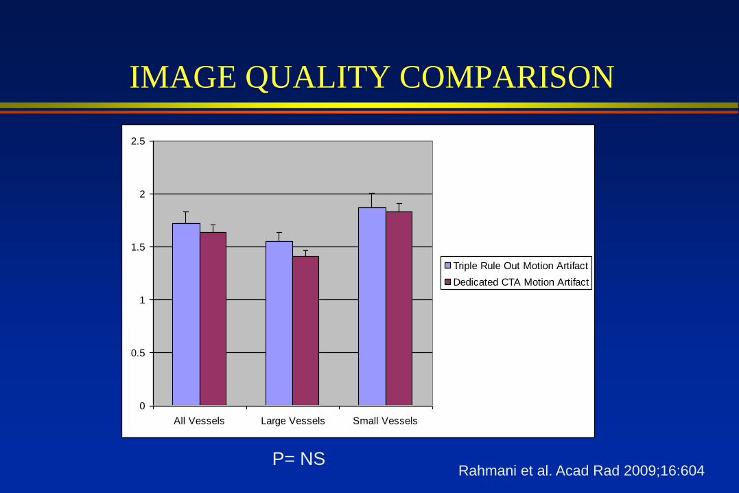
IMAGE QUALITY COMPARISON
0
0.5
1
1.5
2
2.5
All Vessels Large Vessels Small Vessels
Triple Rule Out Motion Artifact
Dedicated CTA Motion Artifact
Rahmani et al. Acad Rad 2009;16:604 P= NS

CONTRAST INJECTION
Ao,CA PA
HU
Time
Cor CTA
Triple R/O
CONTRAST ADMINISTATION
COR CTA
• Test injection
» 20 ml @ 6 cc/sec
Injection protocol
» 80 ml (100%) @ 6 cc/sec
» 40 ml (50/50) @ 5 cc/sec
» 50 ml (saline) @ 5 cc/sec
Bolus tracking
100 CC
TRIPLE R/O
• Test injection
» 20 ml @ 6 cc/sec
Injection protocol
» 80 ml (100%) @ 6 cc/sec
» 50 ml (100%) @ 2 cc/sec
» 50 ml (saline) @ 5 cc/sec
Bolus tracking
130 CC
Courtesy: MedRad

CONTRAST ADMINISTATION
COR CTA
TRIPLE R/O

DEDICATED CTA vs TRIPLE RULE-OUT

DEDICATED CTA vs TRIPLE RULE-OUT
?Frequency of PE miss on dedicated CTA
96 patients (46 with PE) Dx’d on chest CT
Scans masked to mimic dedicated cor CTA
Two blinded readers assessed cor CTA for
PE
37/46 (80%) of PE diagnosed
20% of PE missed
Huard et al. STR 2007

PE MISS

ER CHEST PAIN ON MDCT
MARYLAND CLINICAL PROTOCOL
Available 7A-5P weekdays
Prelim reading at night
Low-intermediate risk non-admitted pts
ED physician orders ECG-gated study
» Triple R/O vs Cardiac only
» Patient given 100 mg po metoprolol immediately
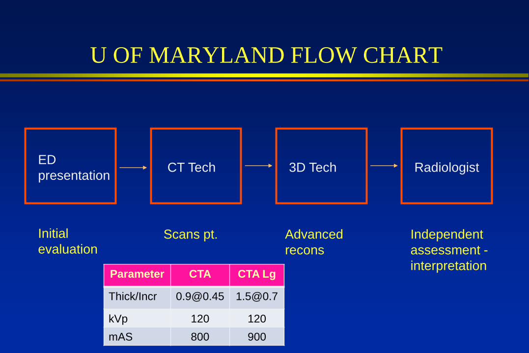
U OF MARYLAND FLOW CHART
ED
presentation
Initial
evaluation
CT Tech 3D Tech
Scans pt. Advanced
recons
Radiologist
Independent
assessment -
interpretation Parameter CTA CTA Lg
Thick/Incr [email protected] [email protected]
kVp 120 120
mAS 800 900

TRIAGE
CASE EXAMPLES

CTA-CATH (46 yo F)

CTA-CATH (58 yo M)

CTA-CATH (49 yo M)

CTA-PERFUSION (COCAINE)

CTA-PERFUSION (55 yoM)

ED CP LITERATURE
DEDICATED CORONARY CTA
64-slice CT (ROMICAT1)
368 admitted patients (39%F, mean –
53 yrs)
Prospective with 6 mo F/U for MACE
Caregivers and patients blinded
Low-intermediate risk (neg biomarkers)
Significant stenosis ≥ 50%
Hoffmann JACC 2009;53:1652

ED CP LITERATURE
DEDICATED CORONARY CTA
RESULTS
8% ACS
50% no plaque, 41% nonobs/indet plaque, 9% stenosis
Sens = 100%,NPV=100% (no plaque)
Increasing plaque→ increased ACS
Conclusion: May dec unnecessary admissions

AGE AND GENDER
ROMICAT1 population
ACS occurrence increased with age
Looked at decrease in risk category
CTA most effective (decrease in risk
category
» Men<55 years
» Women<65 years
Bamberg AJC 2009;104:1165

ED CP - DEDICATED CORONARY CTA
Hoffmann Circ 2006

ED CP LITERATURE
TRIPLE R/O CTA
64-slice CT
197 patients completed protocol
Low to moderate risk**
22/197 (11%) had >50% stenosis
NPV - 99.4% at 30 day F/U
Non-CAD clinically important findings
explained CP in 22/197 (11%) patients
Takakuwa Rad 2008:248:438

ED CP – TRIPLE R/O CTA
Alternative findings explaining CP Takakuwa Rad 2008:248:438

CHALLENGES
Technical/Labor
Radiation dose
Economics

TECHNICAL/LABOR
Need minimum 64-slice CT for triple R/O
3D-tech ?supportable with 3D codes
Off-hours options
» residents
» in-house 24/7 staff
» remote portal, client-server/nighthawk
» prelim reads (dual-mode) – wait until AM for final disposition if pos

LABOR ISSUES
Restricted to low-intermediate risk pts

OFF-HOURS
The dual mode triage option
Hold
(50%)
NEG
(50%)
1
2

RADIATION EXPOSURE
Dose (mSv)
Background - Yearly 3.6
Chest radiograph 0.05
Sesta/Thal – rest/stress 1.5-5/6-25
Cardiac Cath 3-15
Chest CT (conventional) 5
Gated CT 9-15
Gated CT – dose modulated 6-9
Triple Rule-out 20-30

Fig 1(c). Prospectively-gated Step & Shoot scans
Tube output ON Tube output OFF
PROSPECTIVE GATING
Tube output (x-ray) ON
Spiral Acquisition
Retrospective ECG
Tagging

256 SLICES – PROSPECTIVE CORONARY

256 SLICE TRIPLE RULE-OUT
PROSPECTIVE-GATING

PROSPECTIVE GATING – TRIPLE R/O
Shuman AJR 2009;192:1662
Retrospective: 31.8 ±5.1 mSv (range, 27.3–40.5 mSv).
Prospective: 9.2 ± 2.2 mSv (range, 7.2–11.6 mSv)
= 71% dose savings

RADIATION EXPOSURE
Dose (mSv)
Background - Yearly 3.6
Sestamibi – rest/stress 1.5-5/6-25
Cardiac Cath 3-15
Chest CT (conventional) 5
Gated CT 10-15
Gated CT – dose modulated 6-9
Gated CT- prospective axial 3-4
Triple R/O 20-30
Triple R/O-prospective axial 7-10

ECONOMIC CONSIDERATIONS
Cost (USD)*
Chest radiograph $35
Sestamibi – rest/stress $500-$700
Stress Echo $300-$500
Cardiac Cath $2000 and up
Cardiac (Chest) CT $500-$600
*Based on 2007 Medicare
rates

MULTI- SITE TRIAL (CT STAT)
700 pts, 15 centers (50:50 – CTA:SOC)
CTA Triage: <25% D/C, 26%-70% MPI, >70%
Cath
Stenosis: None – 82%, Mid – 9%, Sig. – 8%
Cath rate: CTA – 5.1%, SOC 4.6% p=ns
ACS rate: CTA – 3.2%, SOC 3% p=ns
Time to Dx: CTA- 3 hrs, SOC 6.2 hrs p<.01
Cost of care: CTA-$2,000, SOC ~$3,500
AHA 2009

FINAL THOUGHTS
Suggestions to avoid overuse, NEED GATEKEEPER
» ? Chest pain unit criteria
» Within that, only low-intermediate risk
Dedicated vs Triple R/O
» Depends on PE suspicion
Larger studies
» CT-STAT, ROMICAT2, ACRIN

CARDIAC ANTENNAS

THANK YOU