curcumin suppresses growth of mesothelioma cells in vitro and in vivo, in part, by stimulating...
DESCRIPTION
Ying WangTRANSCRIPT

Curcumin suppresses growth of mesothelioma cells in vitroand in vivo, in part, by stimulating apoptosis
Ying Wang • Arun K. Rishi • Wenjuan Wu •
Lisa Polin • Sunita Sharma • Edi Levi •
Steven Albelda • Harvey I. Pass • Anil Wali
Received: 31 January 2011 / Accepted: 6 May 2011 / Published online: 19 May 2011
� Springer Science+Business Media, LLC. 2011
Abstract Malignant pleural mesothelioma (MPM) is an
aggressive, asbestos-related malignancy of the thoracic
pleura. Although, platinum-based agents are the first line of
therapy, there is an urgent need for second-line therapies to
treat the drug-resistant MPM. Cell cycle as well as apop-
tosis pathways are frequently altered in MPM and thus
remain attractive targets for intervention strategies. Cur-
cumin, the major component in the spice turmeric, alone or
in combination with other chemotherapeutics has been
under investigation for a number of cancers. In this study,
we investigated the biological and molecular responses of
MPM cells to curcumin treatments and the mechanisms
involved. Flow-cytometric analyses coupled with western
immunoblotting and gene-array analyses were conducted
to determine mechanisms of curcumin-dependent growth
suppression of human (H2373, H2452, H2461, and H226)
and murine (AB12) MPM cells. Curcumin inhibited MPM
cell growth in a dose- and time-dependent manner while
pretreatment of MPM cells with curcumin enhanced cis-
platin efficacy. Curcumin activated the stress-activated p38
kinase, caspases 9 and 3, caused elevated levels of proa-
poptotic proteins Bax, stimulated PARP cleavage, and
apoptosis. In addition, curcumin treatments stimulated
expression of novel transducers of cell growth suppression
Y. Wang
John D. Dingell VA Medical Center, Karmanos Cancer Institute,
Wayne State University, VAMC, 4646 John R., Detroit,
MI 48201, USA
A. K. Rishi (&)
John D. Dingell VA Medical Center, Departments of Oncology
and Internal Medicine, Karmanos Cancer Institute, Wayne State
University, Room B4334, VAMC, 4646 John R., Detroit,
MI 48201, USA
e-mail: [email protected]
W. Wu
John D. Dingell VA Medical Center, Karmanos Cancer Institute,
Wayne State University, Room B4325, VAMC, 4646 John R.,
Detroit, MI 48201, USA
L. Polin
Departments of Oncology and Internal Medicine, Karmanos
Cancer Institute, Wayne State University, 110 E. Warren Street,
Detroit, MI 48201, USA
S. Sharma
John D. Dingell VA Medical Center, Department of Surgery,
Karmanos Cancer Institute, Wayne State University, VAMC,
4646 John R., Detroit, MI 48201, USA
E. Levi
John D. Dingell VA Medical Center, Departments of Pathology
and Oncology, Wayne State University, VAMC, 4646 John R.,
Detroit, MI 48201, USA
S. Albelda
Pulmonary, Allergy & Critical Care Division, Abramson
Research center, University of Pennsylvania Medical Center,
Philadelphia, PA, USA
H. I. Pass
Division of Cardiothoracic Surgery, New York University
Cancer Center, New York, USA
A. Wali
John D. Dingell VA Medical Center, Department of Surgery,
Karmanos Cancer Institute, Wayne State University, Room
B4325, VAMC, 4646 John R., Detroit, MI 48201, USA
Present Address:A. Wali
Center to Reduce Cancer Health Disparities, National Cancer
Institute, National Institutes of Health, 6116 Executive
Boulevard, Suite 602, Rockville, MD 20852, USA
123
Mol Cell Biochem (2011) 357:83–94
DOI 10.1007/s11010-011-0878-2
such as CARP-1, XAF1, and SULF1 proteins. Oral
administration of curcumin inhibited growth of murine
MPM cell-derived tumors in vivo in part by stimulating
apoptosis. Thus, curcumin targets cell cycle and promotes
apoptosis to suppress MPM growth in vitro and in vivo.
Our studies provide a proof-of-principle rationale for fur-
ther in-depth analysis of MPM growth suppression mech-
anisms and their future exploitation in effective
management of resistant MPM.
Keywords Malignant pleural mesothelioma �Curcumin � Apoptosis � Gene expression
Introduction
Malignant Pleural Mesothelioma (MPM) are the tumors of
mesothelial or mesenchymal cells that line the thoracic
cavity, peritoneum, or pericardium. MPM is an aggressive,
locally recurrent cancer with no effective curative options
[1, 2]. The median survival for patients diagnosed with
MPM is between 8 and 18 months, despite aggressive
multimodality strategies involving surgery, chemotherapy,
and radiation [3–5]. The platinum-based agents are cur-
rently used either as monotherapy or in combination with
other agents as first line therapeutics for MPM. However,
the response rates remain unacceptably low.
MPM is characterized by a number of molecular events
including chromosome 3p and neurofibromatosis 2 gene
abnormalities [6–8], Wilms tumor [9], VEGF [10],
thrombospondin [11], IGF receptor amplification [12], and
inactivation of p53 and Rb by SV40 T antigen [13]. Simian
virus 40 (SV40)-dependent Akt signaling has been shown
to drive mesothelial cell transformation after asbestos
exposure and knockdown of receptor tyrosine kinase
EphB4 was found to reduce phosphorylation of Akt in
murine model of mesothelioma [14]. In the absence of an
effective standard second-line of treatment options, strat-
egies to exploit aberrant growth and apoptosis signaling
have been proposed for identification of new and more
efficacious means to combat this disease [15].
A number of studies suggest that agents derived from
dietary fruits and vegetables are helpful in either inhibiting
or reversing the development of cancer [16, 17]. Curcumin,
a phenolic compound isolated from the dietary plant Cur-
cumina longa, has been demonstrated to possess anti-
inflammatory, anti-oxidant, and anti-cancer properties.
Curcumin has been shown to suppress growth of cancer
cells in vitro, while in animals, it interferes with tumor
initiation as well as tumor promotion [18–22]. Although,
the precise mechanisms of action of curcumin are yet to be
elucidated, available evidence thus far indicates that it
suppresses cancer cell growth in large part by down-
regulating pathways of cell proliferation and survival. In
this context, curcumin inhibition of the NF-jB as well as
the PI3K-Akt pathways of cell survival has been well
documented. Since inhibition of the NF-jB and the PI3K-
Akt signaling is often associated with increased apoptotic
index in many cell types, the anti-cancer effects of curcu-
min also involve activation of the extrinsic and/or intrinsic
pathways of apoptosis [21, 22].
In this study, we investigated effects of curcumin on
growth of MPM cells in vitro. Consistent with observations
in other models, curcumin treatments caused cell growth
inhibition that involved elevated apoptosis. A combination of
curcumin and cisplatin were superior in suppressing growth
of the MPM cells in vitro. Although curcumin stimulated
expression of pro-apoptotic Bax and activated p38 SAPK and
caspase-9, our gene-array-based analysis revealed that cur-
cumin induced expression of novel transducers of apoptosis
signaling such as XIAP-associated factor1 (XAF1) and
CARP-1/CCAR1 [23, 24]. Curcumin treatments also stim-
ulated expression of a putative tumor suppressor SULF1
protein [25]. Further oral administration of curcumin sup-
pressed growth of murine mesothelioma allografts in part by
enhancing apoptosis. Our proof-of-concept studies reveal,
for the first time, MPM inhibitory properties of curcumin and
are expected to facilitate utilization of this agent or its potent
derivatives as potential adjuvants for treatment and perhaps
chemoprevention of MPM.
Materials and methods
Cells and reagents
Three MPM patient-derived cell lines [H2373, H2452, and
H2461] established in our laboratory and characterized in
detail [26] were cultured in RPMI 1640 (Mediatech Inc.,
Herndon, VA) supplemented with 100 units/ml of penicil-
lin, 100 lg/ml streptomycin, 4 mM L-glutamine, and 10%
fetal calf serum. H226 MPM cells were obtained from
ATCC (Manassas, VA) and maintained following vendor’s
guidelines. The AB12 murine malignant mesothelioma cell
line was derived from BALB/c mice and was shown to form
subcutaneous tumors when implanted in mice [27, 28]. This
cell line was cultured and maintained in high-glucose
DMEM supplemented with 10% fetal bovine serum,
100 units/ml penicillin, 100 lg/ml streptomycin. Cells were
incubated at 37�C in a humidified atmosphere of 5% CO2 in
air and were passaged weekly. Curcumin was obtained from
Sigma-Aldrich (St. Louis, MO). Mouse monoclonal anti-
body against human poly (ADP-ribose) polymerase (PARP)
was from BIOMOL International LP (Plymouth Meeting,
PA). Anti-XAF1 antibodies were obtained from Abcam Inc.,
(Cambridge, MA). Anti-Bax, anti-caspase-3, anti-caspase-9,
84 Mol Cell Biochem (2011) 357:83–94
123

anti-Akt, anti-p-p38 antibodies were purchased from
Cell Signaling Technology (Beverly, MA). Anti-IkB-a and
anti-Bcl2 antibodies were obtained from Santa Cruz
Biotechnology Inc. (Santa Cruz, CA), while anti-b actin
antibody was purchased from Sigma-Aldrich (St. Louis,
MO). Anti-HSulf-1 rabbit polyclonal antibodies were pur-
chased from Abcam. Generation and characterization of the
anti-CARP-1/CCAR1 rabbit polyclonal antibodies have
been described before [24]. 3-(4,5-dimethyltiazol-2-yl)-2,
5-diphenyl-tetrazolium bromide (MTT) were purchased
from Sigma-Aldrich (St. Louis, MO).
Cell growth inhibition studies by MTT assay
MPM (H2373, H2452, H2461, H226 and AB12) cells
(5 9 103) were seeded in a 96-well culture plate and
subsequently treated with indicated agents at different
concentrations for noted times. Control cells were treated
with 0.1% dimethyl sulfoxide (DMSO) in culture medium.
After treatment, the cells were incubated with 1 mg/ml of
MTT reagent at 37�C for 4 h and then MTT was removed
and 100 ll of DMSO was added, followed by colorimetric
analysis using a multilabel plate reader at 560 nm (Victor3;
PerkinElmer, Wellesley, MA, USA). Results were plotted
as the mean from triplicate experiments.
Western blot analysis
Cells were harvested and lysed in RIPA buffer (50 mM
Tris–HCI, pH 8.0, 150 mM sodium chloride, 1.0% NP-40,
0.5% sodium deoxycholate, 0.1% sodium dodecyl sulfate,
and 0.1% of protease inhibitor cocktail) for 20 min at 4�C.
The lysates were centrifuged at 14,000 rpm at 4�C for
15 min to remove debris. Protein concentrations of whole
cell lysates were determined using the Protein Assay Kit.
Supernatant proteins, 50 lg from each sample, were sep-
arated by SDS-10% polyacrylamide gel electrophoresis
(SDS-PAGE) and transferred to polyvinylidene difluoride
(PVDF) membrane (Bio-rad, Hercules, CA) by standard
procedures. The membranes were hybridized with primary
antibodies followed by incubation with appropriate sec-
ondary antibodies. The antibody-bound proteins were
visualized by treatment with the chemiluminescence
detection reagent (Pierce) according to manufacturer’s
instructions, followed by exposure to film (Kodak
X-Omat). The same membrane was reprobed with the anti-
b actin antibody, which was used as an internal control for
protein loading.
Flow cytometry and cell cycle analysis
The cell cycle was analyzed by flow cytometry. In brief,
1 9 106 cells were untreated or treated with cisplatin,
curcumin, or a combination of both, and harvested and
washed in PBS, then fixed in 70% alcohol for 30 min at
4�C. After washing in cold PBS thrice, cells were resus-
pended in 1 ml of PBS solution with 50 lg of propidium
iodide and 100 lg of RNaseA for 30 min at 37�C. Samples
were then analyzed for their DNA content by FACSCalibur
(Becton–Dickinson, Mountain View, CA).
Isolation of RNA and microarray analysis
Total RNA was extracted from untreated or curcumin-
treated H2373 and H2461 MPM cells. At the end of
treatments, the untreated and treated cells were harvested
and total RNA were isolated, and purified using the
RNeasy Mini kit and RNase-free DNase Set (Qiagen,
Valencia, CA) according to the manufacturer’s protocols.
Cucumin-dependent changes in gene expression in
MPM cells were performed at the Genomic Core Facility,
Karmanos Cancer Institute utilizing Illumina BeadChip�Arrays essentially according to manufacturer’s instruction
(Illumina). In brief, 0.5 lg total RNA was biotin-labeled
and hybridized with BeadChips. The signal was detected
with streptovadin-Cy3 according to manufacturer’s
instruction (Illumina). The imaging of the BeadChips was
conducted using a Bead Array Reader in conjunction with
Bead Studio software (Illumina). Normalization of the data
was carried out using a quantile-based approach which
transforms the raw data so that the resulting normalized
expression values of each sample have the same distribu-
tion [29]. An unsupervised cluster analysis was performed
to detect similarities among samples based on gene
expression profiles. The genes retained to perform the
clustering were those varying the most regardless their
group membership as described elsewhere [30]. Signifi-
cance of the differentially expressed genes among various
groups was tested using a moderated t-test to allow for
P-value computation for the significance of gene changes.
The P-values were then adjusted using the False Discovery
Rate method [30] to derive corrected P-values. The
P-values of\0.5 were considered significant provided that
the fold change in expression was also equal to or larger
than 2.
Murine mesothelioma allograft experiments
Female BALB/c mice aged 5 weeks were purchased from
Taconic Research Animal Services and housed in accor-
dance with protocols approved by the Institutional Labo-
ratory Animal Care and Use Committee of Wayne State
University. On day 1, AB12 murine mesothelioma cells
(0.5 9 106) were suspended in 0.1 ml serum-free DMEM
medium and inoculated subcutaneously (s.c.) in the right
flank of each mouse (n = 5 each group). When the tumors
Mol Cell Biochem (2011) 357:83–94 85
123

became palpable (on day 10 after inoculation), the mice
were randomly assigned into two groups and given various
treatments by gavage. The control group received the
vehicle (DMSO) while the test group received 500 mg/kg
curcumin daily by oral gavage. Tumor sizes were measured
daily using calipers and their volumes calculated using a
standard formula: width2 9 length/2. Body weight was
measured weekly. The mice were sacrificed after 16 day-
treatment when control tumors reached to *1,000 mm3.
H&E staining confirmed the presence of tumor.
Immunohistochemical analysis
Apoptosis in tumor tissues was determined by TUNEL
assay using in situ cell Death Detection kit from Roche
Applied Science (Indianapolis, IN) according to the man-
ufacturer’s instruction. The formalin-fixed tumor xenograft
biopsies from untreated or curcumin-treated animals were
paraffin embedded and processed essentially following our
previously described procedures [31, 32]. The tumor tissue
slides were stained for presence of CARP-1, cyclin-
dependent kinase inhibitor p27, HSULF1, or p-p38 by
utilizing respective antibodies, and were then photographed
under different magnifications using Zeiss microscope with
a 35-mm camera attached for recording the photomicro-
graphs. H&E counter-staining of tumor tissues was per-
formed following our previously described methods [32].
Results
Curcumin inhibits MPM cell growth
A number of studies have revealed chemo-preventive and
anti-cancer properties of curcumin that are mainly due to
its ability to cause cell cycle arrest and promote apoptosis
in various cancer cells of diverse histological origins [21].
In this study, we utilized a number of MPM cells to
investigate their growth inhibition by curcumin. As a first
step, we determined the effects of curcumin on the growth
of human MPM (H2373, H2452, H2461, and H226) as well
as AB12 murine MPM cells. Each of the cells was sepa-
rately treated with medium containing various doses of
curcumin for periods of 24, 48, and 72 h followed by
measurements of their viabilities by conducting MTT
assays as detailed in ‘‘Materials and methods’’ section.
Curcumin inhibited growth of all the MPM cells in a dose-
dependent manner. Although, curcumin was cytotoxic to
the MPM cells, the IC50 of *20–25 lM were noted for the
MPM cells (Fig. 1). Curcumin treatments also inhibited
growth of the murine mesothelioma cells with an IC50 of
*25 lM (Fig. 2). Thus, curcumin inhibits growth of MPM
cells in a dose-dependent manner.
Curcumin enhances cisplatin-induced MPM cell
growth inhibition
We have previously performed survival studies to establish
a baseline cytotoxicity profile for MPM cell lines exposed
to cisplatin alone. The MPM cells were relatively resistant
to cisplatin with the IC50 values around 80 lM [33]. These
IC50 values were approximately three-to-four fold higher
than those for curcumin noted in Figs. 1 and 2. Since 20 lM
dose of curcumin for 48-h treatment period caused *60%,
*60%, *50%, and *30% inhibition of H2373, H2452,
H2461, and H226 MPM cell growth, respectively, (Fig. 1),
Fig. 1 Antiproliferative effect of curcumin on human MPM cells.
Cells were treated with vehicle (Control, denoted as NT) or indicated
doses of curcumin for noted times. Determination of viable/live cells
was carried out by MTT assay. Columns in each of the histograms
represent means of 3–4 independent experiments; bars, S.E. *, #, &,
and @ significantly different from NT, P = \0.01, 0.015, 0.017 and
0.006, respectively
86 Mol Cell Biochem (2011) 357:83–94
123
we utilized sub-optimal doses of this agent to examine its
potential to enhance activity of cisplatin. MPM cells were
pretreated with either 10 lM curcumin for 24 h followed by
treatments with 5 or 10 lM cisplatin for additional 72 h. Of
note is the fact that all the MPM cells were treated with
different doses of cisplatin alone for 24-h periods. MPM
cell viabilities were measured by MTT assay as above. As
shown in Fig. 3, curcumin pre-treatments enhanced the
growth inhibitory activity of cisplatin in H2373 and H2452
MPM cells when compared with their untreated, curcumin-
or cisplatin-treated counterparts. A combination of the
10 lM doses of curcumin and cisplatin each, however,
elicited modest growth inhibition of H2461 cells.
To investigate the growth inhibitory mechanisms uti-
lized by curcumin, we conducted cell cycle analyses of
untreated, curcumin-, cisplatin-, and curcumin-plus-cis-
platin-treated H2373 MPM cells using propidium iodide
staining and flow cytometry. Consistent with our earlier
studies [34], 10 lM cisplatin treatment caused significant
accumulation of cells in S and G2 M phases (Fig. 4a).
Treatments with curcumin alone, however, failed to alter
accumulation of these cells in any of the cell cycle phases
when compared with the cell cycle distribution of their
untreated counterparts. Moreover, MPM cells treated with
a combination of curcumin and cisplatin had significant
accumulation in the apoptotic (sub G0) fractions while
undergoing a drastic reduction in cell numbers in the G0/
G1 phase (Fig. 4a). Together, these data support targeting
of cell cycle progression in MPM cells by cisplatin alone or
in combination with curcumin that involve their accumu-
lation in S and G2M phases. We next determined whether
growth inhibition of MPM cells by these agents involved
induction of apoptotic cell death. In the first instance, we
measured cellular apoptosis-associated morphological
changes (such as nuclear condensation and fragmentation)
in cells treated with curcumin, cisplatin, or a combination
of both the agents. Cellular morphology changes (i.e.,
spherical and detached changes) were visualized by phase-
contrast microscopy. The apoptotic cellular changes
(Fig. 4b) were observed in the H2373 cells treated with
curcumin, cisplatin, or a combination of both, but not in the
cells that were treated with DMSO.
In an attempt to elucidate the mechanism of enhanced
apoptosis of the MPM cells by curcumin, we assessed the
levels of PARP, caspase 3, caspase 9, Bax, and Bcl2 pro-
teins, which are known to be involved in diverse apoptosis-
signaling pathways. H2373 cells were either untreated or
treated with 20 lM curcumin for 24, 48, and 72 h, fol-
lowed by western blot analysis of the cell lysates in con-
junction with respective antibodies for the above noted
proteins. Treatment with curcumin caused significant
increase in expression of pro-apoptotic protein Bax,
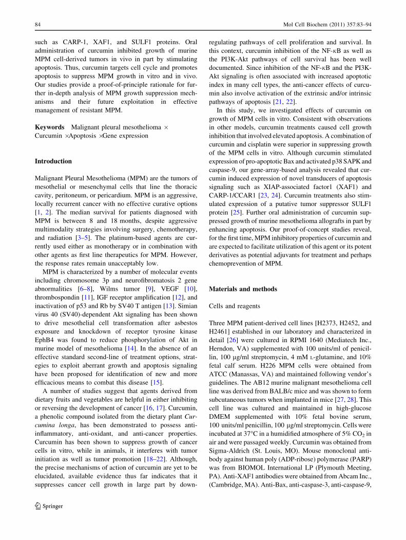
Fig. 2 Antiproliferative effect of curcumin on murine MPM cells.
Cells were treated with vehicle (Control, denoted as 0) or indicated
doses of curcumin for noted times. Determination of viable/live cells
was carried out by MTT assay. Columns in each of the histograms
represent means of three independent experiments; bars, S.E. *, #, $,
@, and & significantly different from untreated controls noted as 0,
P = 0.002, 0.0005, 0.0005, 0.008, and 0.0009, respectively
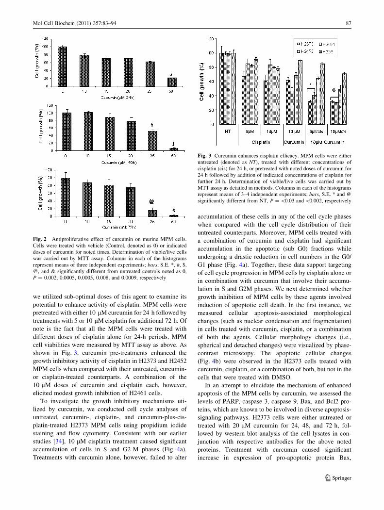
Fig. 3 Curcumin enhances cisplatin efficacy. MPM cells were either
untreated (denoted as NT), treated with different concentrations of
cisplatin (cis) for 24 h, or pretreated with noted doses of curcumin for
24 h followed by addition of indicated concentrations of cisplatin for
further 24 h. Determination of viable/live cells was carried out by
MTT assay as detailed in methods. Columns in each of the histograms
represent means of 3–4 independent experiments; bars, S.E. * and @
significantly different from NT, P = \0.03 and \0.002, respectively
Mol Cell Biochem (2011) 357:83–94 87
123
cleaved caspase 3, caspase 9, and PARP proteins (Fig. 5a).
In addition, the H2373 human MPM and AB12 murine
MPM cells were treated with various doses of curcumin for
72 h and levels of the apoptosis-signaling associated pro-
teins p38 SAPK, PARP, caspase 3, and Bcl2 were deter-
mined by western blot analysis. In the human H2373 MPM
cells, a 10 lM dose of curcumin induced activation of p38,
caspase 3, and cleavage of PARP (Fig. 5b). Since curcumin
also inhibited growth of the murine MPM cells, the western
blot analysis revealed activation of caspase 3, cleavage of
PARP, and loss of Bcl2 in the cells that were exposed to
50 lM dose of curcumin (Fig. 5c). Of note is the fact that
48-h treatments with 20 lM curcumin elicited *60 and
*20% inhibition of H2373 and AB12 cells (Figs. 1, 2),
respectively, suggest that H2373 cells are likely more
sensitive to inhibition by curcumin when compared with
the murine AB12 cells. This is also consistent with the
western blot data in Fig. 5 where 10 lM dose of curcumin
activated apoptosis signaling in H2373 cells, while a
higher, 50 l dose of curcumin was found to elicit activa-
tion of apoptosis in AB12 cells. Taken together, our in vitro
data thus far suggest that curcumin suppresses growth of
the MPM cells in part by promoting apoptosis.
Curcumin targets novel apoptosis transducers
The molecular complexity of cancers and therapy-associ-
ated side effects often limit effectiveness of many anti-
cancer modalities, and necessitate identification of novel
cancer cell growth inhibitory targets/pathways for potential
exploitation in devising efficacious therapeutic strategies.
To further investigate mechanisms of MPM cell growth
regulation by curcumin, a gene-array-based strategy was
employed. H2373 and H2461 MPM cells were either
untreated or separately treated with two doses of curcumin
as described in methods. The RNAs from each group were
hybridized with gene-array chips, and the data computed to
identify genes that had a significant two-fold or higher
altered expression following curcumin treatments of both
the MPM cells. Next, a subset of genes that had similar
curcumin-dependent altered expression in both the MPM
cells were derived from this data and a select subset of these
genes are indicated in Table 1. Of note is the fact that
curcumin treatment caused down-regulation of several
growth-promoting genes. These included receptor type
protein tyrosine phosphatase R (PTPRR), RAS oncogene
family member RAB8A, inhibitor of DNA binding (ID) 1,
Curcumin (10µM )
Cisplatin (µµM)
– – – + + +
– 5 10 – 5 10
H2373
B
NT 10µM Curcumin 10µM Cisplatin 10µM Cur +10µM Cis
H2373
A
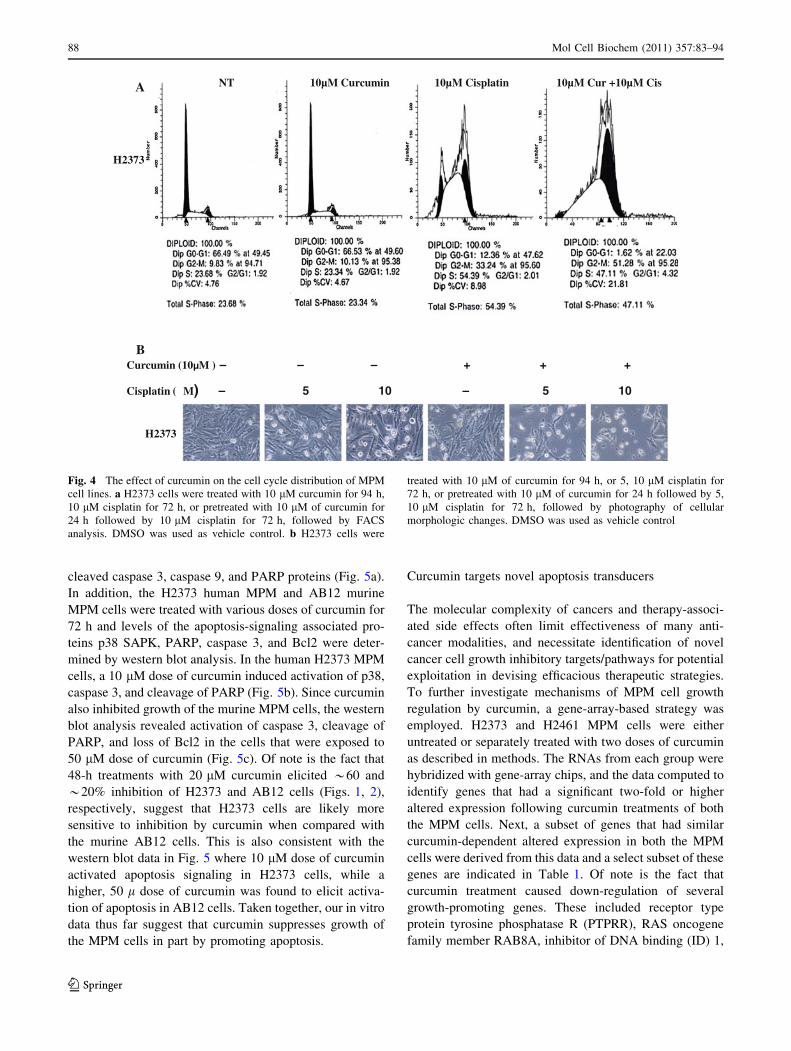
Fig. 4 The effect of curcumin on the cell cycle distribution of MPM
cell lines. a H2373 cells were treated with 10 lM curcumin for 94 h,
10 lM cisplatin for 72 h, or pretreated with 10 lM of curcumin for
24 h followed by 10 lM cisplatin for 72 h, followed by FACS
analysis. DMSO was used as vehicle control. b H2373 cells were
treated with 10 lM of curcumin for 94 h, or 5, 10 lM cisplatin for
72 h, or pretreated with 10 lM of curcumin for 24 h followed by 5,
10 lM cisplatin for 72 h, followed by photography of cellular
morphologic changes. DMSO was used as vehicle control
88 Mol Cell Biochem (2011) 357:83–94
123

2, 3, and regulator of G-protein signaling (RGS) 4. On the
other hand, curcumin caused elevated expression of several
growth inhibitory genes. These group of genes included
XIAP-associated factor 1 (XAF1), osteoblast specific factor
periostin, decorin, myosin light chain kinase, sulfatase
(SULF) 1, damage-regulated autophagy modulator, filamin
A interacting protein 1-like protein, growth arrest-specific
(GAS) 6 protein, insulin-like growth factor binding proteins
(IGFBP) 3, 7, Sparc/osteonectin, and interleukin (IL) 6b.
Our western blot analyses further demonstrate that treat-
ments of the 2373 MPM cells with curcumin results in
elevated expression of XAF1 and SULF1 proteins (Fig. 5a).
CARP-1/CCAR1, a novel signaling transducer, was
previously identified as a target of select chemotherapy
such as adriamycin in the human breast cancer and lym-
phoma cells [23, 31, 32]. We further observed that treat-
ments of MPM cells with proteasome inhibitor velcade also
caused elevated levels of CARP-1 [33]. In this study, we
investigated whether curcumin-dependent MPM cell
growth inhibition involved elevated CARP-1 expression.
H2373 Cells were treated with 20 lM curcumin for various
time periods, and the cell lysates were analyzed by western
blotting for CARP-1 expression. Our data in Fig. 5a show
that curcumin treatments caused increased expression of
CARP-1 in MPM cells. Together, data in Fig. 5 and
Table 1 strongly suggest that curcumin-dependent inhibi-
tion of MPM cancer cells is accomplished, in part, by
stimulating novel transducers of apoptosis signaling such
as CARP-1/CCAR1 and XAF1.
Curcumin inhibits MPM cell-derived xenograft growth
The Balb/c mice in each group were injected with AB12
murine MPM cells as in methods. After the tumors
Caspase 9
Akt
IkB-a
PARP
Actin
Caspase 3
35 kDa
19 kDa17 kDa
47 kDa37 kDa
17 kDa
35 kDa
Bax
NT 1d 2d 3d
H2373 (20µM )
A
CARP-1
XAF1p-p38
Actin
116 kDa
85 kDa
SULF1
B0 5 10 15 20 25 40 µM
p-p38
Cleaved Caspase 3
PARP 116 kDa85 kDa
SULF1
C0 10 25 50 µM
Bcl2
Cleaved Caspase 3
Total Caspase 3
Actin
PARP 116 kDa
85 kDa
SULF1
Fig. 5 Curcumin induces apoptosis in MPM cells. H2373 cells were
treated with 20 lM of curcumin for 1–3 days in (a), or with different
concentrations of curcumin for 72 h in (b). In panel c, AB12 cells
were treated with different concentrations of curcumin for 72 h. Cell
lysates were prepared from DMSO-treated vehicle control and
curcumin-treated cells in each panel, and analyzed by SDS-PAGE,
followed by western immunoblotting of the membranes with
antibodies for indicated proteins as described in methods. All the
membranes were probed with anti-actin antibodies for actin protein as
a loading control
Mol Cell Biochem (2011) 357:83–94 89
123

became palpable, the mice were randomly assigned to
control (untreated) or treated groups. The mice were
gavage treated with curcumin as detailed in methods. The
animals were observed throughout the treatment period
and did not show any visible toxicity including symptoms
of diarrhea, dehydration, weight loss, hair loss, or any
other discomfort. Mice were sacrificed on day 18, and
tumor size and appearance from one flank for three mice
each under the control and treatment condition is shown
in Fig. 6a. Subcutaneous tumor volume in each flank was
measured on regular intervals, and tumor progression
under each treatment condition is shown in Fig. 6b. In
addition, the bar histogram in Fig. 6b shows tumor
weight from control and treated animals following cal-
culations using the formula indicated in methods. As is
evident from Fig. 6a, b, curcumin administration caused
profound suppression of the MPM cell-derived xenograft
growth in vivo. Whether curcumin treatments suppressed
murine MPM xenograft growth by stimulating apoptosis
was determined next by conducting immuno-histochemi-
cal analyses of the xenografted tumor biopsies from the
control and treated animals for levels of apoptosis.
Consistent with our data in Figs. 4 and 5, immuno-
staining showed elevated levels of pro-apoptotic phos-
phorylated p38 MAPK and CARP-1 proteins in the
xenograft biopsies from the curcumin-treated animals
when compared with their untreated control counterparts
(Fig. 6c). Moreover, levels of cyclin-dependent kinase
inhibitor p27 protein, SULF1, and the number of apop-
totic, TUNEL-positive cells were elevated in xenograft
Table 1 List of select curcumin regulated genes in MPM cells
ID Adj.P.val Fold change Direction SYMBOL Name ENTREZ
5220451 0.999951362 4.216387218 UP POSTN Periostin, osteoblast specific factor 10631
6980296 0.999951362 3.893999579 UP DCN Decorin 1634
6350608 0.999951362 2.931475288 UP MYLK Myosin light chain kinase 4638
2640519 0.999951362 2.495608056 UP COL4A1 Collagen, type IV, alpha 1 1282
7050703 0.999951362 3.756081427 UP SULF1 Sulfatase 1 23213
3460278 0.999951362 2.051210568 DOWN PTPRR Protein tyrosine phosphatase, receptor type, R 5801
840537 0.999951362 2.728935376 UP PDGFRB Platelet-derived growth factor receptor, beta polypeptide 5159
5810471 0.999951362 2.289118934 UP DRAM Damage-regulated autophagy modulator 55332
4570377 0.999951362 2.956642534 UP ABCG1 ATP-binding cassette, sub-family G (WHITE), member 1 9619
940220 0.999951362 9.987995292 DOWN RAB8A RAB8A, member RAS oncogene family 4218
7380300 0.999951362 2.204011716 UP FILIP1L Filamin A interacting protein 1-like 11259
3310368 0.999951362 2.407088299 DOWN ID3 Inhibitor of DNA binding 3, dominant negative helixloophelix protein 3399
4780008 0.999951362 2.523453734 UP IGFBP7 Insulin-like growth factor binding protein 7 3490
1070403 0.999951362 2.489312792 UP GAS6 Growth arrest-specific 6 2621
4670092 0.999951362 2.086346517 DOWN ID2 Inhibitor of DNA binding 2, dominant negative helixloop–helix protein 3398
5570538 0.999951362 2.125908257 DOWN ID1 Inhibitor of DNA binding 1, dominant negative helixloop-helix protein 3397
7050682 0.999951362 2.616469369 UP CXCR7 Chemokine (C-X-C motif) receptor 7 57007
1740341 0.999951362 2.139974705 UP XAF1 XIAP-associated factor 1 54739
3440500 0.999951362 2.705678862 UP IL6 Interleukin 6 (interferon, beta 2) 3569
1400709 0.999951362 2.198712979 DOWN RGS4 Regulator of G-protein signaling 4 5999
2480450 0.999951362 2.064923366 UP SPOCK1 Sparc/osteonectin, cwcv and kazal-like domains proteoglycan
(testican) 1
6695
4540551 0.999951362 2.044121375 UP IGFBP3 Insulin-like growth factor binding protein 3 3486
Genes that are down regulated by curcumin are made italic
Fig. 6 Curcumin inhibits growth of MPM xenografts. The efficacy
studies were carried out as in methods, and data were analyzed
essentially as described before [33]. Animals were either untreated or
treated with curcumin as in methods. a Tumor volumes were
monitored every other day in each group and tumor weights were
determined by the end of the experiment. b Points, mean tumor
volume in each experimental group containing four mice; columns,
mean tumor weights in vehicle or curcumin-treated group. Bars, SD.
In panel c, the formalin-fixed tumor xenograft biopsies from panel
(a) were paraffin embedded, processed, and subsequently subjected to
immunohistochemical staining as detailed in methods. Representative
photomicrographs (2009 magnification) in the first row show
hematoxylin and eosin (H&E) staining of the tissue sections.
Representative photomicrographs (2009 magnification) are also
presented demonstrating apoptosis in xenografted tumors following
their staining using TUNEL assay (second row), with anti-CARP-1
(third row), anti-phospho-p38 (fourth row), anti-p27 antibodies (fifthrow), or anti sulfatase1 antibodies (sixth row)
c
90 Mol Cell Biochem (2011) 357:83–94
123
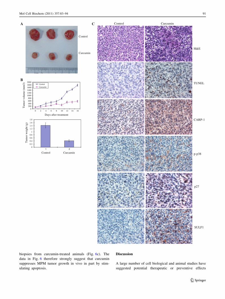
biopsies from curcumin-treated animals (Fig. 6c). The
data in Fig. 6 therefore strongly suggest that curcumin
suppresses MPM tumor growth in vivo in part by stim-
ulating apoptosis.
Discussion
A large number of cell biological and animal studies have
suggested potential therapeutic or preventive effects
Control
Curcumin
A
B
0200400600800
100012001400160018002000
0 2 4 6 8 10 12 14 16
Days after treatment
Tum
or v
olum
e (m
m3) Control
Curcumin
0
0.2
0.4
0.6
0.81
1.2
1.4
1.6
1.8
1 2
Control Curcumin
Tum
or w
eigh
t (g)
p27
H&E
TUNEL
C Control Curcumin
CARP-1
p-p38
SULF1
Mol Cell Biochem (2011) 357:83–94 91
123
associated with curcumin in a variety of malignancies
including cancer. It is thought to have antitumor, antioxi-
dant, antiarthritic, anti-amyloid, anti-ischemic, and anti-
inflammatory properties [34–36]. With regard to the human
pathologies, recent and current clinical trials have further
elaborated potential utility of curcumin for multiple mye-
loma, pancreatic cancer, myelodysplastic syndromes, colon
cancer, psoriasis, and Alzheimer’s disease. The anticancer
effects of curcumin however are due largely to its ability to
inhibit the NF-jB- and Akt-dependent survival pathways as
well as induce apoptosis in cancer cells with minimal
cytotoxicity toward the healthy cells [21]. Moreover, syn-
ergistic, pro-apoptotic effects of curcumin and various
chemotherapies have also been described [22]. Despite a
wealth of knowledge with regard to anti-cancer and other
beneficial properties of curcumin, the molecular mecha-
nisms of its action are yet to be fully elucidated. In this
investigation, we conducted in vitro and in vivo studies by
utilizing human and murine MPM cells to investigate anti-
MPM effects of curcumin and the molecular mechanisms
involved. Consistent with observations in other models, we
found that curcumin suppressed MPM growth in vitro and in
vivo and enhanced efficacy of the first line anti-MPM ther-
apeutic cisplatin.
Targeting of cancer cell growth and survival pathways
remains an attractive anti-cancer strategy. Our current
studies indicating involvement of apoptosis in curcumin-
dependent inhibition of MPM growth highlights anti-MPM
potential for this safe and non-toxic agent. In this context,
prior micro-array analyses of the curcumin-treated human
breast cancer cells have revealed a subset of apoptosis-
associated genes that were altered by curcumin treatment
[22]. Because alterations in apoptosis-signaling pathways
often contribute to MPM growth and survival [38], we
undertook an in vitro and gene-array-based profiling to
identify novel transducers of apoptosis signaling that may
be activated/induced by curcumin. Our studies revealed
that while curcumin suppressed levels of several growth-
promoting genes, it also caused elevated expression of
multiple novel cell growth inhibitory and apoptosis trans-
ducers such as CCAR1/CARP-1, XAF1, and SULF1.
Our current data and several previous studies [21, 22]
amply demonstrate that curcumin treatments induced
apoptosis in a variety of cancer cells types, including
MPM. Although, a robust cleavage of PARP, caspase 3,
and induction of Bax was noted in H2373 MPM cells that
were treated with 10–20 lM curcumin, a somewhat higher
dose of curcumin was required for robust activation of
apoptosis transducers in murine MPM cells (Figs. 5b, c).
Whether this difference in the IC50 of curcumin is due to
additional cell-selective signaling leading to increased
sensitivity of human MPM cells or is a consequence of
differences in the cellular uptake and metabolism of
curcumin by the MPM cells remains to be clarified. These
data nonetheless suggest that curcumin inhibits MPM
growth by inducing growth arrest and apoptosis, and its
effects are likely transduced in part by targeting specific
mediator(s). Our gene-array-based analyses (Table 1)
together with western blot data in Figs. 5 and 6 convinc-
ingly reveal a new group of apoptosis signal-transducing
genes that are activated by curcumin in MPM cells in vitro
and in vivo. Since, CCAR1/CARP-1 is an emerging and
novel target of diverse cell growth and apoptosis-signaling
pathways [23, 32, 33, 38–40] and is not involved in cis-
platin-dependent HBC growth inhibition [23], identifica-
tion of CCAR1/CARP-1 as a downstream effector of
curcumin signaling is anticipated to allow for design of
effective strategies for optimizing curcumin potential as
suppressor of MPM and its drug (Cisplatin)-resistant phe-
notype. Moreover, curcumin also stimulated XAF1
expression (Fig. 5; Table 1). XAF1 is an inhibitor of XIAP,
a transducer of cell survival signaling. Together with our
earlier studies demonstrating increased levels of XAF1 in
MPM and breast cancer cells in the presence of velcade
[33], loss of XIAP expression in velcade-treated MPM
cells further underscores an important role for XIAP-
dependent cell survival pathway by velcade and curcumin.
The gene-array data also revealed a number of interest-
ing genes that may be involved in regulating MPM cell
growth and survival in the presence of curcumin. Of note is
the down-regulation of three members of the inhibitor of
DNA binding (ID) protein family (ID1, 2, 3; Table 1). ID
proteins are a group of helix-loop-helix (HLH) proteins that
lack a basic DNA-binding domain but are able to form
hetero-dimers with other HLH proteins, thereby inhibiting
DNA binding and transcription functions. ID proteins that
are thought to be necessary for G1 progression as well as
transduction of p53-dependent DNA damage response [41,
42]. Whether and to what extent curcumin interferes with
cell cycle progression of the MPM cells in part by dimin-
ishing levels of ID proteins remains to be clarified. Curcu-
min treatments however resulted in up-regulation of several
proteins that are known to function as inhibitors of cell
growth. In addition to XAF1, curcumin interestingly stim-
ulated expression of SULF1 (Table 1; Figs. 5, 6). Recent
studies have highlighted a potential tumor suppressor
function of human SULF1 in carcinogenesis. SULF1
inhibits the co-receptor function of cellular heparin sulfate
proteoglycans (HSPGs) and thus impacts multiple receptor
tyrosine kinase (RTK)-signaling pathways regulated by the
heparin-binding growth factors such as FGF, HB-EGF,
VEGF, PDGF, and HGF [25]. SULF1 is down-regulated by
epigenetic mechanisms in many cancers and its forced
expression interferes with cancer cell proliferation, migra-
tion, and invasion processes. Although, curcumin induces
apoptosis in MPM cells, increased levels of SULF1 in the
92 Mol Cell Biochem (2011) 357:83–94
123
presence of curcumin likely attenuates RTK-dependent
MPM growth and survival signaling. If proven correct,
SULF-1-dependent signaling in the curcumin-treated MPM
cells will not only support a novel mechanism of action of
this agent, but will underscore a broader utility for this
compound as a future anti-cancer and perhaps chemo-pre-
ventive agent for MPM and possibly other cancers.
Acknowledgments This study was supported by the department of
Veterans Affairs Merit Review grants to AKR and AW, Susan G
Komen for the Cure grant to AKR, and MARF support to AW.
References
1. Belli C, Fennell D, Giovannini M, Gaudino G, Mutti L (2009)
Malignant pleural mesothelioma: current treatments and emerg-
ing drugs. Expert Opin Emerg Drugs 14:423–437
2. Fennell DA, Gaudino G, O’Byrne KJ, Mutti L, van Meerbeeck J
(2008) Advances in the systemic therapy of malignant pleural
mesothelioma. Nat Clin Pract Oncol 5:136–147
3. Janne PA (2003) Chemotherapy for malignant pleural mesothe-
lioma. Clin Lung Cancer 5:98–106
4. Goudar RK (2005) New therapeutic options for mesothelioma.
Curr Oncol Rep 7:260–265
5. Krug LM (2005) An overview of chemotherapy for mesotheli-
oma. Hematol Oncol Clin North Am 19:1117–1136
6. De Rienzo A, Balsara BR, Apostolou S, Jhanwar SC, Testa JR
(2001) Loss of heterozygosity analysis defines a 3-cM region of
15q commonly deleted in human malignant mesothelioma.
Oncogene 20:6245–6249
7. Pylkkanen L, Sainio M, Ollikainen T, Mattson K, Nordling S,
Carpen O, Linnainmaa K, Husgafvel-Pursiainen K (2002) Con-
current LOH at multiple loci in human malignant mesothelioma
with preferential loss of NF2 gene region. Oncol Rep 9:955–959
8. Musti M, Kettunen E, Dragonieri S, Lindholm P, Cavone D,
Serio G, Knuutila S (2006) Cytogenetic and molecular genetic
changes in malignant mesothelioma. Cancer Genet Cytogenet
170:9–15
9. Langerak AW, Williamson KA, Miyagawa K, Hagemeijer A,
Versnel MA, Hastie ND (1995) Expression of the Wilm’s tumor
gene WT1 in human malignant mesothelioma cell lines and
relationship to platelet-derived growth factor A and insulin-like
growth factor 2 expression. Genes Chromosom Cancer 12:87–96
10. Adachi Y, Yoshio-Hoshino N, Aoki C, Nishimoto N (2010)
VEGF targeting in mesotheliomas using an interleukin-6 signal
inhibitor based on adenovirus gene delivery. Anticancer Res
30:1947–1952
11. Ohta Y, Shridhar V, Kalemkerian GP, Bright RK, Watanabe Y,
Pass HI (1999) Thrombospondin-1 expression and clinical
implications in malignant pleural mesothelioma. Cancer
85:2570–2576
12. Whitson BA, Jacobson BA, Frizelle S, Patel MR, Yee D, Mad-
daus MA, Kratzke RA (2006) Effects of insulin-like growth
factor-1 receptor inhibition in mesothelioma. Ann Thorac Surg
82:996–1002
13. Pass HI, Bocchetta M, carbone M (2004) Evidence of an
important role for SV40 in mesothelioma. Thorac Surg Clin
14:489–495
14. Xia G, Kumar SR, Masood R, Koss M, Templeman C, Quinn D,
Zhu S, Reddy R, Krasnoperov V, Gill PS (2005) Up-regulation of
EphB4 in mesothelioma and its biological significance. Clin
Cancer Res 11:4305–4315
15. Fennell DA, Rudd RM (2004) Defective core-apoptosis signal-
ling in diffuse malignant pleural mesothelioma: opportunities for
effective drug development. Lancet Oncol 5:354–362
16. Surh YJ (2003) Cancer chemoprevention with dietary phyto-
chemicals. Nat Rev Cancer 3:768–780
17. Scott EN, Gescher AJ, Steward WP, Brown K (2009) Develop-
ment of dietary phytochemical chemopreventive agents: bio-
markers and choice of dose for early clinical trials. Cancer Prev
Res 2:525–530
18. Huang MT, Wang ZY, Georgiadis CA, Laskin JD, Conney AH
(1992) Inhibitory effects of curcumin on tumor initiation by
benzo[a]pyrene and 7,12-dimethylbenz[a]anthracene. Carcino-
genesis 13:2183–2186
19. Conney AH, Lysz T, Ferraro T, Abidi TF, Manchand PS, La-
skin JD, Huang MT (1991) Inhibitory effect of curcumin and
some related dietary compounds on tumor promotion and ara-
chidonic acid metabolism in mouse skin. Adv Enzyme Regul
31:385–396
20. Huang MT, Smart RC, Wong CQ, Conney AH (1988) Inhibitory
effect of curcumin, chlorogenic acid, caffeic acid, and ferulic
acid on tumor promotion in mouse skin by 12-O tetra-
decanoylphorbol-13-acetate. Cancer Res 48:5941–5946
21. Reuter S, Eifes S, Dicato M, Aggarwal BB, Diederich M (2008)
Modulation of anti-apoptotic and survival pathways by curcumin
as a strategy to induce apoptosis in cancer cells. Biochem Phar-
macol 76:1340–1351
22. Ramachandran C, Rodriguez S, Ramachandran R, Raveendran
Nair PK, Fonseca H, Khatib Z, Escalon E, Melnick SJ (2005)
Expression profiles of apoptotic genes induced by curcumin in
human breast cancer and mammary epithelial cell lines. Anti-
cancer Res 25:3293–3302
23. Rishi AK, Zhang L, Boyanapalli M, Wali A, Mohammad RM, Yu
Y, Fontana JA, Hatfield JS, Dawson MI, Majumdar APN, Reic-
hert U (2003) Identification and characterization of a cell-cycle
and apoptosis regulatory protein (CARP)-1 as a novel mediator of
apoptosis signaling by retinoid CD437. J Biol Chem 278:
33422–33435
24. Liston P, Fong WG, Kelly NL, Toji S, Miyazaki T, Conte D,
Tamai K, Craig CG, McBurney MW, Korneluk RG (2001)
Identification of XAF1 as an antagonist of XIAP anti-caspase
activity. Nat Cell Biol 3:128–133
25. Lai J-P, Sandhu DS, Shire AM, Roberts LR (2008) The tumor
suppressor function of human sulfatase 1 (SULF1) in carcino-
genesis. J Gastrointest Cancer 39:149–158
26. Pass HI, Lott D, Lonardo F, Harbut M, Liu Z, Tang N, Carbone
M, Webb C, Wali A (2005) Asbestos exposure, pleural meso-
thelioma, and serum osteopontin levels. N Engl J Med 353:
1564–1573
27. Suzuki E, Kim S, Cheung HK, Corbley MJ, Zhang X, Sun L,
Shan F, Singh J, Lee WC, Albelda SM, Ling LE (2007) A novel
small-molecule inhibitor of transforming growth factor beta type
I receptor kinase (SM16) inhibits murine mesothelioma tumor
growth in vivo and prevents tumor recurrence after surgical
resection. Cancer Res 67:2351–2359
28. Odaka M, Sterman D, Wiewrodt R, Zhang Y, Kiefer M, Amin
KM, Gao G-P, Wilson JM, Barsoum J, Kaiser LR, Albelda SM
(2001) Eradication of intraperitoneal and distant tumor by ade-
novirus-mediated interferon-b gene therapy due to induction of
systemic immunity. Cancer Res 61:6201–6212
29. Smyth GK, Limma L (2005) Liner models for microarray data.
In: Gentleman R, Carey V, Duoit S, Irizarry R, Huber W (eds)
Bioinformatics and computational biology solutions using R and
bioconductor. Springer, New York, pp 397–420
30. Tarca AL, Carey VJ, Chen XW, Romero R, Draghici S (2007)
Machine learning and its applications to biology. PLoS Comput
Biol 3:e116
Mol Cell Biochem (2011) 357:83–94 93
123

31. Zhang L, Levi E, Majumder P et al (2007) Transactivator of
transcription tagged cell cycle and apoptosis regulatory protein-1
peptides suppress growth of human breast cancer cells in vitro
and in vivo. Mol Cancer Ther 6:1661–1672
32. Levi E, Zhang L, Aboukameel A, Rishi S, Mohammad RM, Polin
L, Hatfield JS, and Rishi AK (2010) Cell cycle and apoptosis
regulatory protein (CARP)-1 is a novel, adriamycin-inducible,
diffuse large B-cell lymphoma (DLBL) growth suppressor.
Cancer Chemother Pharmacol (in press)
33. Wang Y, Rishi AK, Puliyappadamba VT, Sharma S, Yang H,
Tarca A, Dou QP, Lonardo F, Ruckdeschel JC, Pass HI, Wali A
(2010) Targeted proteasome inhibition by velcade induces
apoptosis in human mesothelioma and breast cancer cell lines.
Cancer Chemother Pharmacol 66:455–466
34. Hatcher H, Planalp R, Cho J, Torti FM, Torti SV (2008) Cur-
cumin: from ancient medicine to current clinical trials. Cell Mol
Life Sci 65:1631–1652
35. Aggarwal BB, Shishodia S (2006) Molecular targets of dietary
agents for prevention and therapy of cancer. Biochem Pharmacol
71:1397–1421
36. Shukla PK, Khanna VK, Ali MM, Khan MY, Srimal RC (2008)
Anti-ischemic effect of curcumin in rat brain. Neurochem Res
33:1036–1043
37. Gordon GJ, Mani M, Mukhopadhyay L, Dong L, Yeap BY,
Sugarbaker DJ, Bueno R (2007) Inhibitor of apoptosis proteins
are regulated by tumour necrosis factor-alpha in malignant
pleural mesothelioma. J Pathol 211:439–446
38. Rishi AK, Zhang L, Yu Y, Jiang Y, Nautiyal J, Wali A, Fontana
JA, Levi E, Majumdar APN (2006) Cell cycle and apoptosis
regulatory protein (CARP)-1 is involved in apoptosis signaling by
epidermal growth factor receptor. J Biol Chem 281:13188–13198
39. Kim JH, Yang CK, Heo K, Roeder RG, An W, Stallcup MR
(2008) CCAR1, a key regulator of mediator complex recruitment
to nuclear receptor transcription complexes. Mol Cell 31:
510–519
40. Ou CY, Kim JH, Yang CK, Stallcup MR (2009) Requirement of
cell cycle and apoptosis regulator 1 for target gene activation by
Wnt and {beta}-catenin and for anchorage-independent growth of
human colon carcinoma cells. J Biol Chem 284:20629–20637
41. Qian Y, Chen X (2008) ID1, inhibitor of differentiation/DNA
binding, is an effector of the p53-dependent DNA damage
response pathway. J Biol Chem 283:22410–22416
42. Hara E, Yamaguchi T, Nojima H, Ide T, Campisi J, Okayama H,
Oda K (1994) Id-related genes encoding helix-loop-helix proteins
are required for G1 progression and are repressed in senescent
human fibroblasts. J Biol Chem 269:2139–2145
94 Mol Cell Biochem (2011) 357:83–94
123