current protocols in cell biology || use of in vivo biotinylation for chromatin immunoprecipitation
TRANSCRIPT
UNIT 17.12Use of In Vivo Biotinylation forChromatin Immunoprecipitation
Arman Kulyyassov,1,2 Muhammad Shoaib,1 and Vasily Ogryzko1
1CNRS, Universite Paris, Villejuif, France2National Center for Biotechnology of the Republic of Kazakhstan, Astana, Republic ofKazakhstan
ABSTRACT
This unit describes a system for expression of biotinylated proteins in mammalian cellsin vivo, and its application to chromatin immunoprecipitation (ChIP). The system is basedon co-expression of the target protein fused to a short biotin acceptor domain, togetherwith the biotinylating enzyme BirA from Escherichia coli. The superior strength ofthe biotin-avidin interaction in the modified ChIP protocol presented here allows oneto employ more stringent washing conditions, resulting in a better signal/noise ratio.Methods for interpreting the data obtained from ChIP samples analyzed by qPCR, andmethods for testing the efficiency of biotinylation using a streptavidin gel-shift are alsopresented. In addition, a complementary method, based on isothermal multiple stranddisplacement amplification (IMDA) of circular concatemers generated from the DNAfragments obtained after ChIP, is described. This method helps to decrease bias in DNAamplification and is useful for the analysis of complex mixtures of DNA fragmentstypically generated in miniscale ChIP experiments. Curr. Protoc. Cell Biol. 51:17.12.1-17.12.22. C© 2011 by John Wiley & Sons, Inc.
Keywords: chromatin immunoprecipitation � biotinylation in vivo � BirA � IMDA �
amplification bias
INTRODUCTION
Chromatin immunoprecipitation (ChIP) is a method for analyzing protein-DNA interac-tions directly in cells. The principle of this procedure is illustrated in Figure 17.12.1A.Given that the in vivo binding between DNA and proteins is labile (with the exception ofcore histones, the typical rate of dissociation of proteins bound to DNA in vivo is only afew minutes; Phair and Misteli, 2000; Hager et al., 2002), intact cells are first subjectedto cross-linking (typically by formaldehyde), in order to fix the chromatin proteins onthe DNA. Cellular lysates are prepared from the cross-linked cells, which are then soni-cated to shear chromatin into fragments of the size that allow accurate determination ofthe protein position, without compromising the efficiency of DNA detection afterwards.The chromatin fragments containing the protein of interest are purified from the cellularlysates by affinity chromatography.
In the original ChIP procedure, a specific antibody against the protein of interest wasused for such purification. The method described in Basic Protocol 1 employs proteinsbiotinylated in vivo, which permits the use of immobilized streptavidin (avidin) insteadof antibodies, allowing for use of more stringent washing conditions and resulting in abetter signal/noise ratio. After reversal of the cross-links, the selectively enriched DNAis further analyzed, usually by PCR, DNA array methodology (Negre et al., 2006),or high-throughput sequencing (Park, 2009). Support Protocol 1 provides methods forinterpreting the data obtained from ChIP samples analyzed by quantitative real-timePCR (qPCR), while Support Protocol 2 describes a procedure for testing the efficiency
Current Protocols in Cell Biology 17.12.1-17.12.22, June 2011Published online June 2011 in Wiley Online Library (wileyonlinelibrary.com).DOI: 10.1002/0471143030.cb1712s51Copyright C© 2011 John Wiley & Sons, Inc.
MacromolecularInteractions inCells
17.12.1
Supplement 51
Use of In VivoBiotinylation
for ChromatinImmuno-
precipitation
17.12.2
Supplement 51 Current Protocols in Cell Biology
A B
+cross-linking
fragmentationof chromatin
precipitationwith specificantibody
reversecrosslinking
detection
PCRmicroarraycloningsequencing
BAP-X
b-BAP-X
biotin , ATPM A G L D I F E A Q K I E W H E
BirA
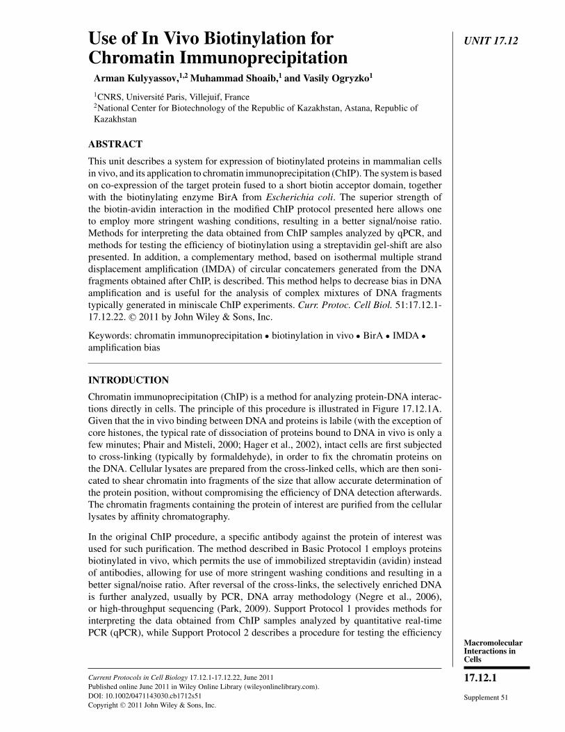
Figure 17.12.1 General principles of chromatin immunoprecipitation (ChIP) and in vivo biotiny-lation. (A) General scheme of ChIP. The proteins are cross-linked to DNA in vivo, the chromatinis fragmented, and the chromatin fragments with the protein of interest are affinity-purified. Thecross-links are reversed, and the DNA is usually analyzed by PCR, DNA arrays, or high-throughputsequencing. (B) Epitope tagging by in vivo biotinylation. Two recombinant constructs are co-expressed in the cells: BAP-X, comprising the protein of interest X fused to a minimal biotinacceptor peptide (BAP, sequence shown), and bacterial biotin ligase BirA, specifically transferringthe biotin moiety on the lysine residue of BAP. The reaction requires the presence of ATP andbiotin in the cells.
of biotinylation using a streptavidin gel-shift. In addition, a complementary method,which helps to decrease bias in DNA amplification, is presented in Basic Protocol 2. Itis based on isothermal multiple strand displacement amplification (IMDA) of circularconcatemers generated from the DNA fragments obtained after ChIP. This method isuseful for the analysis of complex mixtures of DNA fragments typically generated inminiscale ChIP experiments.
STRATEGIC PLANNING
Choice of Expression System
The principle of in vivo biotinylation is based on co-expression of the protein of interestfused to a biotin acceptor domain (BAD), or a shorter biotin acceptor peptide (BAP),together with the bacterial biotin ligase BirA. These two proteins can be expressedfrom different plasmids (see trans design in Fig. 17.12.2; de Boer et al., 2003; Kimet al., 2009), although this method is somewhat cumbersome. It can also decrease thebiotinylation efficiency due to promoter competition and the need for the molecules ofsubstrate and enzyme to meet in the confines of a large mammalian cell, in order forthe biotinylation event to occur. Alternatively, a single bicistronic vector that expressesone mRNA encoding both BirA and the target BAP-fused protein can be used (seecis design in Fig 17.12.2). Such bicistronic design increases the local concentration ofthe enzyme in the vicinity of the target, leading to more efficient biotinylation (Vienset al., 2004). Although such design has been shown to work well for protein detectionby immunoblotting or microscopy (electron or fluorescence; Viens et al., 2008), in ourexperience, the expression levels of the biotinylated proteins can sometimes be low.Therefore the choice of system depends on the protein of interest—its abundance and
MacromolecularInteractions inCells
17.12.3
Current Protocols in Cell Biology Supplement 51
UT – +cis trans
shiftedGFP
GFP
NS
1 2 3 4 5 6
W.B α-GFP
UT – +
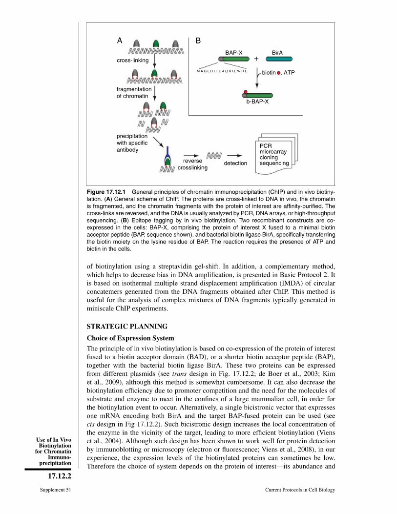
Figure 17.12.2 Streptavidin gel-shift. Immunoblot analysis with anti-GFP antibody of extractsfrom the HEK 293 cells expressing N-terminal BAP fusion of GFP. Streptavidin was added beforeSDS-PAGE in lanes 3 and 6 (+). Left panel: both BAP-GFP and BirA were expressed from thesame mRNA (cis-biotinylation). Right panel: BAP-GFP and BirA were expressed from two differentplasmids that were cotransfected into HEK 293 cells (trans-biotinylation). The positions of GFPand its streptavidin-shifted version are indicated by arrows. Abbreviations: GFP, green-fluorescentprotein; NS, nonspecific signal corresponding to endogenously biotinylated proteins; UT, extractfrom untransfected or untransduced cells.
other characteristics. If expression from two separate plasmids is chosen, the use ofhuman-optimized BirA helps to achieve a more efficient biotinylation of the targetprotein (Mechold et al., 2005).
Transient Transfection Versus Stable Cell Lines
Generation of cell lines that express the biotinylated protein of interest is time consumingand can become a limiting factor, especially if the two genes must be introduced separatelyinto the cell line. Transient transfection typically yields less material; however, the useof unbiased DNA amplification after ChIP makes it a viable alternative. Accordingly,we provide a method for amplifying, without bias, DNA obtained in ChIP experiments(Basic Protocol 2).
Testing Biotinylation Efficiency with the Streptavidin Gel-Shift
If the antibodies against the protein of interest are available, two useful tests combinedin one preliminary experiment can be performed. Their aims are (1) comparison of thelevels of expression of the BAP-tagged protein and those of its endogenous counterpart,and (2) testing the efficiency of biotinylation of the BAP-fused protein. Both aims can beaccomplished by using a streptavidin gel-shift (Viens et al., 2004; van Werven and Tim-mers, 2006; see Support Protocol 2 and Fig. 17.12.2). Given that the interaction betweenstreptavidin and biotin is sufficiently strong to survive the conditions of SDS-PAGEseparation, the addition of streptavidin to the sample before loading leads to an easilydetectable decrease in the mobility of the biotinylated protein (∼50 kDa). Comparing theintensities of the immunoblot signals corresponding to the shifted (streptavidin-bound)and nonshifted (unbound, and hence, unbiotinylated) forms of the protein provides anestimate of what percent of the total BAP-fusion protein was biotinylated. If the antibod-ies against native protein are not available, or if knowledge about the relative expressionlevels of the BAP-fusion and the endogenous protein are not essential for the experiment,antibodies for an additional tag, usually present in the expression vector (e.g., 6×His,Viens et al., 2004; FLAG, Kim et al., 2009) can be used for the immunoblot analysis.
Use of In VivoBiotinylation
for ChromatinImmuno-
precipitation
17.12.4
Supplement 51 Current Protocols in Cell Biology
formaldehyde biotin
treatment duration (min)
biotinylated BSA
streptavidin peroxidase detection
A
?
?
O
HN
HN
SO
CH H
OOH
0 10 20 30 40 50 60NT
B
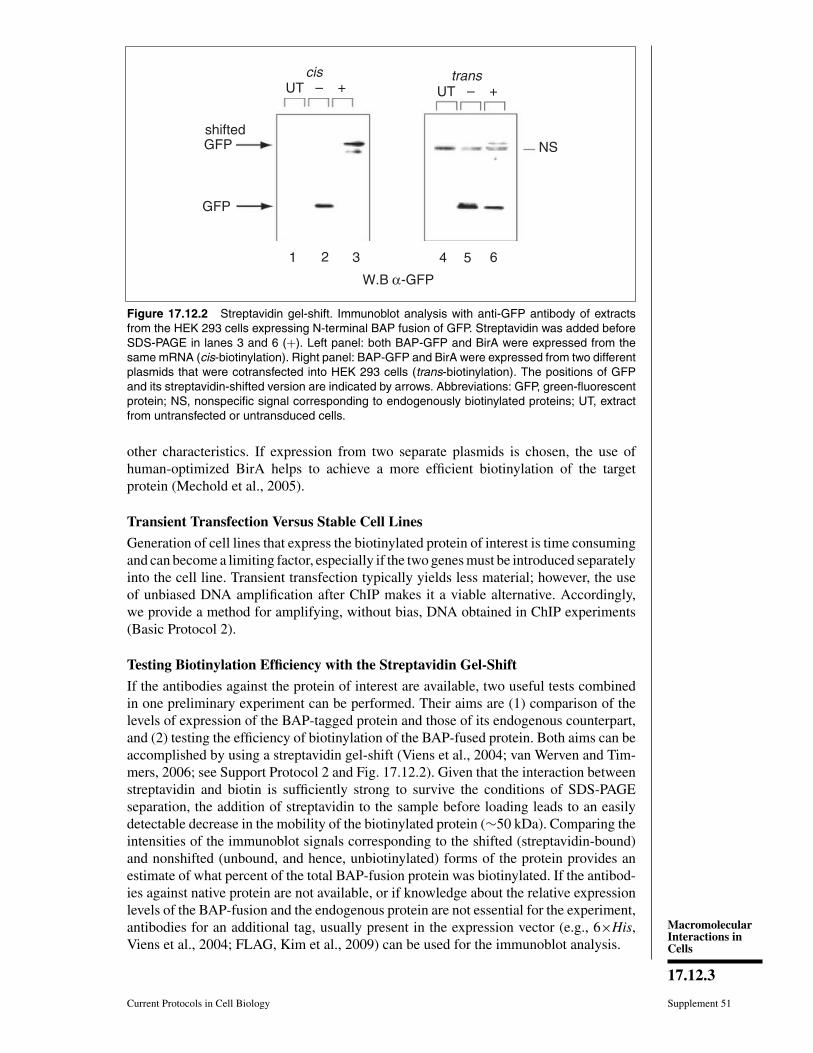
Figure 17.12.3 Use of biotinylated proteins is compatible with cross-linking by formaldehyde.Since biotin possesses two amino groups, it can react with formaldehyde, thus losing its affinityto streptavidin. To test whether formaldehyde treatment impairs the biotin-streptavidin interaction,biotinylated bovine serum albumin (BSA) was incubated in cell culture medium and treated with 1%formaldehyde for 10 to 60 min. The effect of the treatment on the ability of streptavidin to recognizebiotinylated BSA was monitored by immunoblotting using streptavidin-conjugated peroxidase asa detection reagent. No effect of the formaldehyde treatment on the biotin-streptavidin interactionwas observed, based on the intensity of the chemiluminescent signal, even for long incubations.Thus, the biotin tag is compatible with formaldehyde cross-linking. Abbreviation: NT, not treated.
Choice of Cross-Linking Method
The methods used to covalently link protein to DNA in vivo include ultraviolet (UV)(Gilmour and Lis, 1984; Pashev et al., 1991) and chemical cross-linking, most commonlyby formaldehyde. Formaldehyde produces both protein-nucleic acid and protein-proteincross-links in vivo. A key feature of formaldehyde-induced cross-links is their reversibil-ity, which allows one to purify the DNA for subsequent analyses (Orlando, 2000). Asa result, formaldehyde has been the reagent of choice for cross-linking in ChIP appli-cations. While it is possible that the use of formaldehyde can negatively affect epitoperecognition. (e.g., biotin’s nitrogen atoms could participate in a nucleophilic reactionwith formaldehyde), we have shown that for the typical reaction times of formaldehydetreatment used in a ChIP protocol, the biotin-streptavidin affinity is not significantlyaffected (Fig. 17.12.3).
Another potential problem with formaldehyde is that it cross-links macromoleculeswithin ∼2
◦A of each other; thus, it becomes less efficient when examining proteins
that indirectly associate with DNA. To improve detection of such proteins, longer-rangebifunctional cross-linkers have been used in addition to formaldehyde (Zeng et al., 2006).However, before using such a cross-linker in the biotin-ChIP protocol, we recommendfirst testing how the particular reagent affects the biotin moiety.
Finally, in contrast to cross-link ChIP (XChIP), native ChIP (NChIP) omits cross-linking(O’Neill and Turner, 2003). It is well suited for the analysis of histones because oftheir strong binding to DNA. Compared to biotin-ChIP, NChIP does not take advantageof stringent washing conditions (e.g., 2% SDS) because of the potential to disrupt thehistone-DNA interactions. Nonetheless, biotin-NChIP has been used for histones (Ooiet al., 2010), as the high biotin-streptavidin affinity allows one to use smaller amountsof affinity resin. As a result, using regular washing conditions, one can achieve lessnonspecific background compared to regular XChIP.
MacromolecularInteractions inCells
17.12.5
Current Protocols in Cell Biology Supplement 51
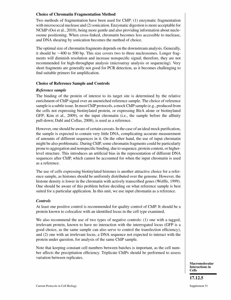
Choice of Chromatin Fragmentation Method
Two methods of fragmentation have been used for ChIP: (1) enzymatic fragmentationwith micrococcal nuclease and (2) sonication. Enzymatic digestion is more acceptable forNChIP (Ooi et al., 2010), being more gentle and also providing information about nucle-osome positioning. When cross-linked, chromatin becomes less accessible to nuclease,and DNA shearing by sonication becomes the method of choice.
The optimal size of chromatin fragments depends on the downstream analysis. Generally,it should be ∼400 to 500 bp. This size covers two to three nucleosomes. Longer frag-ments will diminish resolution and increase nonspecific signal; therefore, they are notrecommended for high-throughput analysis (microarray analysis or sequencing). Veryshort fragments are generally not good for PCR detection, as it becomes challenging tofind suitable primers for amplification.
Choice of Reference Sample and Controls
Reference sampleThe binding of the protein of interest to its target site is determined by the relativeenrichment of ChIP signal over an unenriched reference sample. The choice of referencesample is a subtle issue. In most ChIP protocols, a mock ChIP sample (e.g., produced fromthe cells not expressing biotinylated protein, or expressing BirA alone or biotinylatedGFP; Kim et al., 2009), or the input chromatin (i.e., the sample before the affinitypull-down; Dahl and Collas, 2008), is used as a reference.
However, one should be aware of certain caveats. In the case of an ideal mock purification,the sample is expected to contain very little DNA, complicating accurate measurementof amounts of different sequences in it. On the other hand, the use of input chromatinmight be also problematic. During ChIP, some chromatin fragments could be particularlyprone to aggregation and nonspecific binding, due to sequence, protein content, or higher-level structure. This introduces an artificial bias in the representation of different DNAsequences after ChIP, which cannot be accounted for when the input chromatin is usedas a reference.
The use of cells expressing biotinylated histones is another attractive choice for a refer-ence sample, as histones should be uniformly distributed over the genome. However, thehistone density is lower in the chromatin with actively transcribed genes (Wolffe, 1999).One should be aware of this problem before deciding on what reference sample is bestsuited for a particular application. In this unit, we use input chromatin as a reference.
ControlsAt least one positive control is recommended for quality control of ChIP. It should be aprotein known to colocalize with an identified locus in the cell type examined.
We also recommend the use of two types of negative controls: (1) one with a tagged,irrelevant protein, known to have no interaction with the interrogated locus (GFP is agood choice, as the same sample can also serve to control the transfection efficiency),and (2) one with an irrelevant locus, a DNA sequence not expected to interact with theprotein under question, for analysis of the same ChIP sample.
Note that keeping constant cell numbers between batches is important, as the cell num-ber affects the precipitation efficiency. Triplicate ChIPs should be performed to assessvariation between replicates.
Use of In VivoBiotinylation
for ChromatinImmuno-
precipitation
17.12.6
Supplement 51 Current Protocols in Cell Biology
BASICPROTOCOL 1
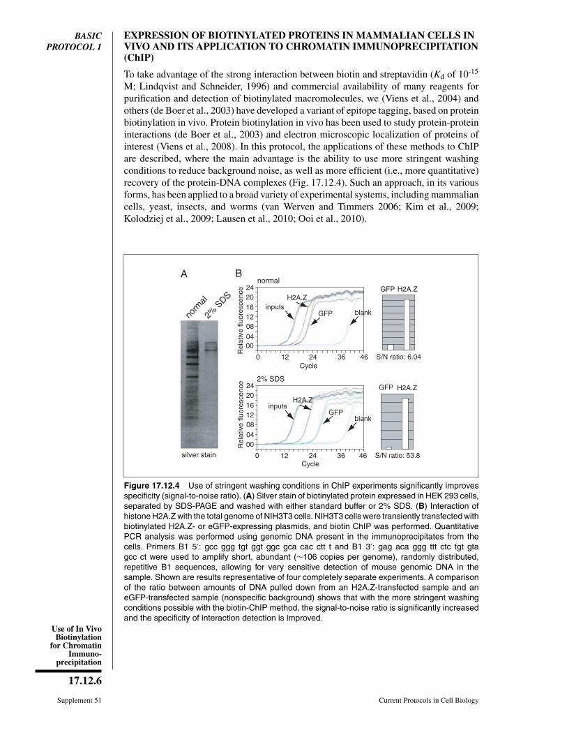
EXPRESSION OF BIOTINYLATED PROTEINS IN MAMMALIAN CELLS INVIVO AND ITS APPLICATION TO CHROMATIN IMMUNOPRECIPITATION(ChIP)
To take advantage of the strong interaction between biotin and streptavidin (Kd of 10-15
M; Lindqvist and Schneider, 1996) and commercial availability of many reagents forpurification and detection of biotinylated macromolecules, we (Viens et al., 2004) andothers (de Boer et al., 2003) have developed a variant of epitope tagging, based on proteinbiotinylation in vivo. Protein biotinylation in vivo has been used to study protein-proteininteractions (de Boer et al., 2003) and electron microscopic localization of proteins ofinterest (Viens et al., 2008). In this protocol, the applications of these methods to ChIPare described, where the main advantage is the ability to use more stringent washingconditions to reduce background noise, as well as more efficient (i.e., more quantitative)recovery of the protein-DNA complexes (Fig. 17.12.4). Such an approach, in its variousforms, has been applied to a broad variety of experimental systems, including mammaliancells, yeast, insects, and worms (van Werven and Timmers 2006; Kim et al., 2009;Kolodziej et al., 2009; Lausen et al., 2010; Ooi et al., 2010).
silver stain
A
norm
al
2% S
DSGFP H2A.Z
S/N ratio: 6.04
S/N ratio: 53.8
GFP H2A.Z
460
H2A.Zinputs
GFP
12 24 36Cycle
460
Rel
ativ
e flu
ores
cenc
e
00040812162024
H2A.Zinputs
GFPblank
12 24 36Cycle
normal
2% SDS
B
Rel
ativ
e flu
ores
cenc
e
00040812162024
Figure 17.12.4 Use of stringent washing conditions in ChIP experiments significantly improvesspecificity (signal-to-noise ratio). (A) Silver stain of biotinylated protein expressed in HEK 293 cells,separated by SDS-PAGE and washed with either standard buffer or 2% SDS. (B) Interaction ofhistone H2A.Z with the total genome of NIH3T3 cells. NIH3T3 cells were transiently transfected withbiotinylated H2A.Z- or eGFP-expressing plasmids, and biotin ChIP was performed. QuantitativePCR analysis was performed using genomic DNA present in the immunoprecipitates from thecells. Primers B1 5′: gcc ggg tgt ggt ggc gca cac ctt t and B1 3′: gag aca ggg ttt ctc tgt gtagcc ct were used to amplify short, abundant (∼106 copies per genome), randomly distributed,repetitive B1 sequences, allowing for very sensitive detection of mouse genomic DNA in thesample. Shown are results representative of four completely separate experiments. A comparisonof the ratio between amounts of DNA pulled down from an H2A.Z-transfected sample and aneGFP-transfected sample (nonspecific background) shows that with the more stringent washingconditions possible with the biotin-ChIP method, the signal-to-noise ratio is significantly increasedand the specificity of interaction detection is improved.
MacromolecularInteractions inCells
17.12.7
Current Protocols in Cell Biology Supplement 51
Materials
Human embryonic kidney (HEK) 293 cells (ATCC)Dulbecco’s modified Eagle’s medium (APPENDIX 2A)/10% (v/v) fetal bovine serum
(DMEM-10)2 M CaCl2 (store up to 6 months At –20◦C)Plasmid DNA with expression system for target protein (see Strategic Planning)
and controls (plasmid expressing BAP-GFP; Viens et al., 2004), checked forbiotinylation efficiency (Support Protocol 2)
2×HeBS (APPENDIX 2A)Biotin (Sigma cat. no. B4639)37% (v/v) formaldehyde stock solution (Electron Microscopy Services)1.25 M glycine stock (store up to 3 months at room temperature)Phosphate-buffered saline (PBS, APPENDIX 2A), ice-cold25× protease inhibitor stock (see recipe)PBS + protease inhibitors, ice cold: dilute 25× protease inhibitor stock (see recipe)
1:1000 with PBS just before use1% (w/v) agarose gel (see Voytas, 2001)ChIP Buffer (see recipe)70% (v/v) ethanol5 M and 300 mM NaCl10 mg/ml RNase A (e.g., Invitrogen)Streptavidin-coupled magnetic beads (Dynabeads M-280 Streptavidin; Invitrogen)Washing buffer 1: 2% (w/v) SDS; store up to 2 months at room temperatureWashing buffer 2: 10 mM Tris·Cl, pH 8 (see APPENDIX 2A)/1 mM EDTA (see
APPENDIX 2A)/0.25 mM LiCl/1% (w/v) Nonidet P-40/1% (v/v)/sodiumdeoxycholic acid; store up to several months at 4◦C
Washing buffer 3: 20 mM Tris·Cl, pH 7.6 (see APPENDIX 2A)/50 mM NaCl/1 mMEDTA (see APPENDIX 2A); store up to 2 months at room temperature
20 mg/ml proteinase K (Roche Applied Science) in 10 mM Tris·Cl, pH 7.5 (seeAPPENDIX 2A)/20 mM calcium chloride/5% (v/v) glycerol
5× proteinase K buffer: 50 mM Tris·Cl, pH 7.5 (see APPENDIX 2A)/25 mM EDTA(see APPENDIX 2A)/1.25% (w/v) SDS
3 M sodium acetate (NaOAc), pH 5 to 5.2DNA purification kit for PCR (e.g., Qiagen): includes MiniElute columns, PBI
buffer, PE buffer, and EB buffer
1.5-ml microcentrifuge tubes14-ml round-bottom tube (Falcon 2059, or equivalent).15-ml conical polystyrene tubes (Falcon)Sonicator, with appropriate tube holder and accessories (e.g., Diagenode)Magnetic separator for 1.5-ml tubes (e.g., Dynal MPC, Invitrogen)Test-tube rotators, at room temperature and in a cold room1.5-ml screw-cap tubes (VWR)67◦C heating block or thermal cycler
Additional reagents and equipment for culturing mammalian cells (UNIT 1.1)performing agarose gel electrophoresis (Voytas, 2001)
Transfect cells to generate biotinylated protein1. Plate HEK 293 cells at the density of 6 × 10⁶ per 100-mm tissue culture dish in
10 ml of DMEM-10, and grow overnight at 5% CO2, 37◦C (e.g., see UNIT 1.1).
Transient transfection procedures (reagents, DNA amounts) depend on the cell line used.Here we describe transfection of HEK 293 with calcium phosphate.
Use of In VivoBiotinylation
for ChromatinImmuno-
precipitation
17.12.8
Supplement 51 Current Protocols in Cell Biology
2. Dilute 24.8 μl of 2 M CaCl2 with water to a total volume of 200 μl in a 1.5-mlmicrocentrifuge tube, and add 5 μg of the plasmid DNA to be transfected.
3. Place 200 μl of 2× HeBS in a 14-ml round-bottom tube. Add the DNA/calciummixture (from step 2) into the 2× HeBS dropwise, mixing after each addition.
The pH of the HeBS buffer is crucial. The precipitate should be barely visible undermicroscope.
For control of transfection efficiency, we recommend transfecting a separate cell well withBAP-GFP (Viens et al., 2004) or any other fluorescent protein. This sample can also belater used as a negative control in ChIP.
4. Add the transfection mixture from step 3 dropwise to the packaging cells in the dish(from step 1), stirring well after each addition, and incubate for 12 hr at 5% CO2,37◦C.
5. Replace the medium with fresh DMEM-10 with 1 mg/ml biotin added. Incubateovernight.
Perform formaldehyde cross-linking6. In a fume hood, add 270 μl of 37% formaldehyde stock to a final concentration of
1% (v/v) directly to the culture plate without changing the medium.
Make sure that the reagent is mixed well with the medium.
7. Keep the plate at room temperature for 10 min, shaking occasionally.
Time of cross-linking may vary, depending on the cells and the protein to be immunopre-cipitated. For optimization, several samples with different cross-linking times should betested. In most cases, 8 to 10 min for cross-linking is sufficient for ChIP with mammaliancells.
8. Add 1 ml of 1.25 M glycine stock (125 mM final concentration) to the culture plateto quench the formaldehyde, mix well, and incubate for 5 min at room temperature.
9. Remove the culture medium completely by aspiration, and rinse the cells with5 ml ice-cold PBS + protease inhibitors. Remove the PBS + protease inhibitors byaspiration. Avoid drying the plates.
10. Scrape the adherent cells from the culture dish into a 15-ml conical polystyrene tube.Rinse the culture dish with 5 ml PBS + protease inhibitors, add the rinse to the same15-ml tube, and mix.
Trypsin can be used to detach the cells from the culture plate; however we have foundthat for cross-linked HEK 293 cells, a scraper works well.
11. Gently pipet up and down to break up cell aggregates. Centrifuge 5 min at 2,400 ×g, 4◦C.
To prevent cell lysis during pipetting of cells, use a 1000- or 200-ml pipet tip cut off toincrease the diameter of the opening.
12. Remove the supernatant by careful aspiration to avoid losing the cell pellet.
13. Add 10 ml cold PBS + protease inhibitors, wash the cells by gently pipetting, andcentrifuge 5 min at 2,400 × g, 4◦C.
14. Remove the supernatant by careful aspiration to avoid losing the cell pellet. Repeatsteps 12 and 13.
15. Without adding any more solution, centrifuge the tube one more time at 5 min at2,400 × g, 4◦C.
MacromolecularInteractions inCells
17.12.9
Current Protocols in Cell Biology Supplement 51
16. Remove all of the supernatant by careful pipetting.
The sample medium contains biotin, which can compete for the binding to streptavidinwith the biotinylated chromatin in the downstream pull-down step. Thorough washing ofthe medium from the cells is therefore critical.
The sample pellet can be snap frozen in liquid nitrogen and stored indefinitely in liquidnitrogen, or up to several months at −80◦C.
Lyse cells and shear chromatinTypically (Weinmann and Farnham, 2002), nuclei are prepared from the cross-linkedcells before chromatin shearing. We found that, due to the strong affinity between biotinand streptavidin and our stringent washing conditions, this step is not necessary in ourprotocol.
17. Resuspend the pellet (kept cold on ice) in 500 μl of ice-cold ChIP buffer.
Applying more ChIP buffer helps to shear the chromatin; however, too much dilutionincreases the volume of the reaction, which may require increased volume of streptavidinbeads used for binding.
18. Sonicate the sample using a sonicator kept in cold room, as follows (for the Diagenodesonicator):
a. Transfer the samples to appropriate tubes for sonication.
The best choice is 15-ml polystyrene tubes.
b. Remove the tube holder, and check that water level is at the blue mark.
c. Use the appropriate tube holder and accessories for samples, and rinse with ethanolbefore use.
d. Balance the tubes in sonicator.
e. Input sonication settings of 5 pulses for 30 sec each (red needle), with 1-min restintervals between pulses (green needle). Set the LMH dial to high.
This results in chromatin fragments of 300 to 800 bp in length.
The pulse duration, intensity, and number will vary, depending on the extent of cross-linking and cell type. You must optimize conditions for your experiment. Ideally, the leastamount of input energy that gives satisfactory fragmentation should be used.
f. Rinse the used tube holder and accessories with water, and then with 70% ethanol.
Check sonication efficiency and fragment size19. Reverse the cross-link of a 10-μl aliquot, as follows:
a. Add 40 μl 300 nM NaCl to 10 μl sample.
b. Add 2 μl of 5 M NaCl (0.2 M NaCl final concentration).
c. Boil for 15 min.
d. Cool down to room temperature.
e. Add 1 μl of 10 mg/ml RNase A.
20. Electrophorese the sample on a 1% agarose gel and visualize by staining (e.g., seeVoytas, 2001).
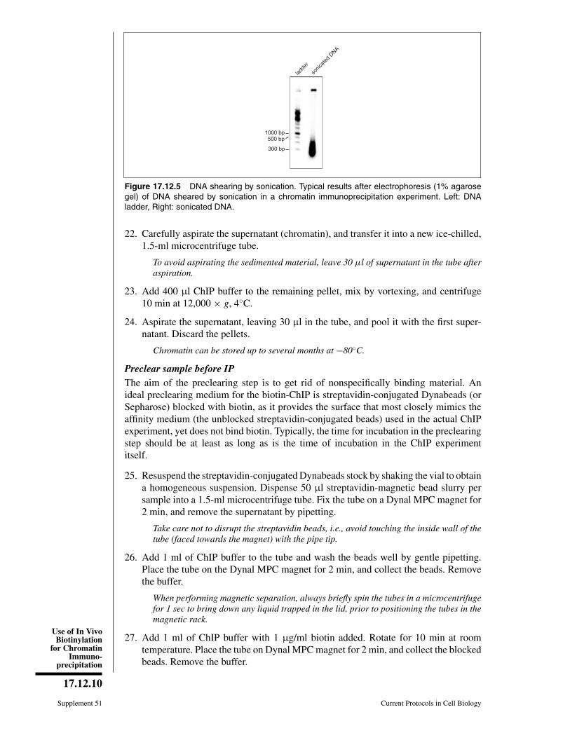
The average size of the sheared DNA fragments should be in the range 0.2 to 0.5 kb(Fig. 17.12.5). If the average size of sheared DNA fragments is too large, continuesonication as in step 18, and repeat steps 19 and 20 to check the results.
Isolate chromatin samples21. Transfer the sonicated material to an ice-chilled, 1.5-ml microcentrifuge tube, and
centrifuge 10 min at 12,000 × g, 4◦C.
Use of In VivoBiotinylation
for ChromatinImmuno-
precipitation
17.12.10
Supplement 51 Current Protocols in Cell Biology
ladde
r
sonic
ated
DNA
1000 bp500 bp
300 bp
Figure 17.12.5 DNA shearing by sonication. Typical results after electrophoresis (1% agarosegel) of DNA sheared by sonication in a chromatin immunoprecipitation experiment. Left: DNAladder, Right: sonicated DNA.
22. Carefully aspirate the supernatant (chromatin), and transfer it into a new ice-chilled,1.5-ml microcentrifuge tube.
To avoid aspirating the sedimented material, leave 30 μl of supernatant in the tube afteraspiration.
23. Add 400 μl ChIP buffer to the remaining pellet, mix by vortexing, and centrifuge10 min at 12,000 × g, 4◦C.
24. Aspirate the supernatant, leaving 30 μl in the tube, and pool it with the first super-natant. Discard the pellets.
Chromatin can be stored up to several months at −80◦C.
Preclear sample before IPThe aim of the preclearing step is to get rid of nonspecifically binding material. Anideal preclearing medium for the biotin-ChIP is streptavidin-conjugated Dynabeads (orSepharose) blocked with biotin, as it provides the surface that most closely mimics theaffinity medium (the unblocked streptavidin-conjugated beads) used in the actual ChIPexperiment, yet does not bind biotin. Typically, the time for incubation in the preclearingstep should be at least as long as is the time of incubation in the ChIP experimentitself.
25. Resuspend the streptavidin-conjugated Dynabeads stock by shaking the vial to obtaina homogeneous suspension. Dispense 50 μl streptavidin-magnetic bead slurry persample into a 1.5-ml microcentrifuge tube. Fix the tube on a Dynal MPC magnet for2 min, and remove the supernatant by pipetting.
Take care not to disrupt the streptavidin beads, i.e., avoid touching the inside wall of thetube (faced towards the magnet) with the pipe tip.
26. Add 1 ml of ChIP buffer to the tube and wash the beads well by gentle pipetting.Place the tube on the Dynal MPC magnet for 2 min, and collect the beads. Removethe buffer.
When performing magnetic separation, always briefly spin the tubes in a microcentrifugefor 1 sec to bring down any liquid trapped in the lid, prior to positioning the tubes in themagnetic rack.
27. Add 1 ml of ChIP buffer with 1 μg/ml biotin added. Rotate for 10 min at roomtemperature. Place the tube on Dynal MPC magnet for 2 min, and collect the blockedbeads. Remove the buffer.
MacromolecularInteractions inCells
17.12.11
Current Protocols in Cell Biology Supplement 51
28. Repeat wash twice as in step 26. Then add 50 μl ChIP buffer per sample, plus a smalladditional volume for pipetting error.
For example, calculate the amount of buffer as (N + 0.5) × 50, where N is the number ofsamples.
29. Dispense in aliquots of 50 μl into prelabeled 1.5-ml microcentrifuge tubes. Collectthe beads using the magnetic separator, and remove the buffer from each tube.
Make sure the stock bead suspension is homogenous before pipetting.
30. Add the chromatin samples to the tubes, reserving an aliquot (at least 10 μl) as aninput chromatin sample. Rotate 3 hr at 4◦C.
31. Meanwhile, prepare the streptavidin-conjugated Dynabeads (affinity medium) forthe ChIP procedure by following the steps 25 to 29, skipping the biotin blocking instep 27.
32. Magnetically separate the blocked beads and precleared chromatin samples (step 30),and transfer the precleared chromatin to the prelabeled tubes containing the washedaffinity medium (unblocked streptavidin-conjugated beads; step 31).
Perform binding for immunoprecipitation33. Mix the chromatin sample and streptavidin beads carefully with gentle pipetting.
Incubate the tube on the test-tube rotator 3 hr at 4◦C.
A 3-hr incubation is the minimum time recommended for successful pull-down of biotiny-lated chromatin. Longer incubation times can also be used. In this case, the length ofincubation in the preclearing step (step 30) should be increased accordingly.
34. Briefly (1 to 2 sec) microcentrifuge the tube to collect the beads into the bottoms ofthe tubes.
35. Mix the sample and beads well by gently pipetting, and transfer the supernatant andthe beads to a fresh tube placed on the magnetic separator.
The tube surface is a source of unspecific binding of chromatin. Transferring the ChIPmaterial to a fresh tube enhances specificity of the ChIP procedure.
36. Wait for 2 min to collect beads to the side of the tube.
37. Remove the supernatant with a pipet, taking care not to disrupt the beads.
Wash beadsAll of the following washing steps are carried out at room temperature; each wash shouldbe performed quickly to prevent the beads from drying out.
38. Add 1 ml washing buffer 1 to the tube, gently resuspend beads by flicking the tube,and rotate the tube for 10 min at room temperature.
39. Collect the beads by magnet, and remove the washing buffer. Wash a second time byadding 1 ml washing buffer 1, resuspending and transferring the suspension to thenew tube, rotating for 10 min at room temperature, collecting the beads by magnet,and removing the washing buffer.
The advantage of biotin-ChIP protocol is that more stringent washing conditions can beused, in particular, 2% SDS. (Fig. 17.12.4).
40. Wash twice with 1 ml washing buffer 2, as described in steps 38 and 39, except forchanging the tube.
Changing the tube is not necessary at this point.
Use of In VivoBiotinylation
for ChromatinImmuno-
precipitation
17.12.12
Supplement 51 Current Protocols in Cell Biology
41. Wash twice with 1 ml washing buffer 3, as described in steps 38 and 39, except forchanging the tube between the washes. After the last wash, transfer the sample to a1.5-ml screw-cap tube.
Elute and de-cross-link samplesDue to strong affinity between streptavidin and biotin, the elution of the biotinylatedmolecule from streptavidin-coupled beads is not trivial. However, after de-cross-linking,DNA comes off the beads independently from the biotinylated protein. Accordingly, inour protocol, the elution and de-cross-linking steps are combined.
42. Add 60 μl of 300 mM NaCl to the beads contained in screw-cap tubes. Incubateovernight at 67◦C.
Using screw-cap tubes minimizes evaporation of elution buffer during overnight de-cross-linking. Alternatively, regular microcentrifuge tubes can be wrapped with Parafilm.
43. Briefly centrifuge, and add 3 μl of 20 mg/ml proteinase K and 15 μl of 5× proteinaseK buffer. Incubate 2 hr at 67◦C.
44. Meanwhile, prepare the input chromatin sample (reserved in step 30). To 10 μl ofinput chromatin samples, add 60 μl of ChIP buffer and 3 μl of 20 mg/ml proteinaseK, vortex, and incubate on a heating block for 2 hr at 67◦C.
From this time point, process the input samples and the ChIP samples in parallel.
45. Briefly centrifuge all of the incubated tubes, and place them in the Dynal MPCmagnet. Wait for 3 min for the beads to separate.
46. Transfer the supernatant by pipetting to a new tube, and discard the magnetic beads.
DNA purificationChIP protocols designed for PCR analysis do not include an RNase step, but it can beintroduced before step 47 of the DNA purification.
47. Add 500 μl PBI buffer to each sample, including the input sample.
48. Add 10 μl of 3 M NaOAc (pH 5 to 5.2), and lightly vortex.
This makes the solution less basic.
49. Transfer to a DNA purification column.
50. Centrifuge 1 min at 2000 × g, 4◦C, and discard the flow-through.
51. Place the column back into the catch tube and add 500 μl PE buffer to each tube.Centrifuge 1 min at 2000 × g, 4◦C and discard flow through.
52. Repeat step 51.
53. Reinsert the column into the catch tube, and centrifuge 30 min at maximum speed.Make sure that no liquid is left at the rim of the tube.
54. Label fresh 1.5-ml microcentrifuge tubes, and place each column into a new micro-centrifuge tube. Discard the catch tubes.
55. Add 30 μl EB buffer to the column membrane, and incubate 2 min at room temper-ature.
56. Microcentrifuge 1 min at maximum speed, room temperature, and discard thecolumn.
The DNA solution in the microcentrifuge tube can be stored up to several months at–80◦C, if necessary, before analysis (e.g., by qPCR; see Support Protocol 1).
MacromolecularInteractions inCells
17.12.13
Current Protocols in Cell Biology Supplement 51
SUPPORTPROTOCOL 1
ANALYSIS AND DATA INTERPRETATION BY qPCR
The DNA from the ChIP samples isolated in Basic Protocol 1 can be analyzed indifferent ways, e.g., by PCR, DNA arrays, or sequencing; thus, the setup of the samplesand interpretation of the results will vary accordingly. Here we discuss general principlesof the analysis of ChIP samples using quantitative real-time polymerase chain reaction(qPCR). We more specifically address the calculation of immunoprecipitation efficienciesfrom the data obtained by qPCR, and their interpretation. See UNIT 17.7 (particularly,Alternate Protocol 2) for more information about procedures used to carry out qPCR.
Make several dilutions of the reference sample (i.e., input chromatin from Basic Protocol1, step 30) in the elution buffer (EB from the Qiagen DNA purification kit). Analyze theChIP samples (obtained in Basic Protocol 1) and the reference sample with the same PCRprogram. Include DNA melting curve analysis to check the product specificity. TypicalqPCR protocols include melting curves, and most qPCR machines have this option, asa quality control to check for correct PCR-fragment amplification and quantificationduring the qPCR procedure.
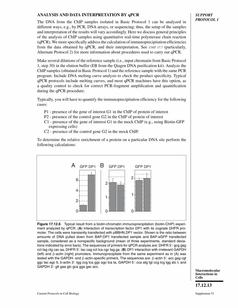
Typically, you will have to quantify the immunoprecipitation efficiency for the followingcases:
P1 - presence of the gene of interest G1 in the ChIP of protein of interestP2 - presence of the control gene G2 in the ChIP of protein of interestC1 - presence of the gene of interest G1 in the mock ChIP (e.g., using Biotin-GFP
expressing cells)C2 - presence of the control gene G2 in the mock ChIP.
To determine the relative enrichment of a protein on a particular DNA site perform thefollowing calculations:
A BGFP DP1 GFP DP1 GFP DP1
6
4
2
0
2
1
0
2
1
0
Figure 17.12.6 Typical result from a biotin-chromatin immunoprecipitation (biotin-ChIP) experi-ment analyzed by qPCR. (A) Interaction of transcription factor DP1 with its cognate DHFR pro-moter. The cells were transiently transfected with pBBHN.DP1 vector. Shown is the ratio betweenamounts of DNA pulled down from BAP-DP1 transfected sample and BAP-eGFP transfectedsample, considered as a nonspecific background (mean of three experiments, standard devia-tions indicated by error bars). The sequences of primers for qPCR analysis are: DHFR 5′: gcg gagcct tag ctg cac aa, DHFR 3′: tac cag cct tca cgc tag ga. (B) DP1 interaction with irrelevant GAPDH(left) and β-actin (right) promoters. Immunoprecipitate from the same experiment as in (A) wastested with the GAPDH- and β-actin-specific primers. The sequences are: β-actin 5′: acc gag cgtggc tac agc tt, b-actin 3′: tgg ccg tca ggc agc tca ta, GAPDH 5′: cca atg tgt ccg tcg tgg atc t, andGAPDH 3′: gtt gaa gtc gca gga gac acc.
Use of In VivoBiotinylation
for ChromatinImmuno-
precipitation
17.12.14
Supplement 51 Current Protocols in Cell Biology
1. Calculate the immunoprecipitation efficiency (IE) for each sample (i.e., P1, P2, C1and C2) by dividing the signal obtained from immunoprecipitated material by thesignal obtained from the input chromatin. Ideally, the mean and SD of each ChIPare calculated from at least three independent experiments, and two independentamplifications.
2. For each DNA fragment analyzed, subtract the IE value obtained for the nonrelevantprotein (C1, C2) from the IE value obtained for the protein of interest (P1, P2),respectively. This subtraction removes background noise due to nonspecific bindingof chromatin to beads.
3. Take the ratio between these data for the gene of interest G1 and control gene G2by calculating the enrichment of a protein on a particular DNA site G1relative to aDNA site G2, according to the formula: (P1−C1)/(P2−C2). This formula gives usthe relative (x-fold) enrichment of a protein on a particular site as compared to anirrelevant DNA site.
Typical results from the biotin-ChIP procedure and qPCR analysis are shown inFigure 17.12.6. The high ratio between the ChIP samples in Figure 17.12.6A signifies aspecific interaction of the protein (DP1) with DNA (DHFR promoter).
SUPPORTPROTOCOL 2
TESTING THE EFFICIENCY OF BIOTINYLATION USING ASTREPTAVIDIN GEL-SHIFT
Due to the strong interaction between streptavidin and biotin, the addition of strep-tavidin to the biotinylated protein of interest before loading onto an SDS-PAGE gelleads to an easily detectable decrease in the mobility of the biotinylated protein(∼50 kDa). Comparing the intensities of the immunoblot signals corresponding to theshifted (streptavidin-bound) and nonshifted (unbound, and hence, unbiotinylated) formsof the protein provides an estimate of the percentage of the total BAP-fusion protein thatwas biotinylated.
The estimation of biotinylation efficiency with a streptavidin gel-shift can be done withamounts of material typically much smaller than required for ChIP. In addition, thecross-linking is not performed. Therefore, we recommend performing the streptavidingel-shift as a preliminary experiment with a small number of cells (transiently trans-fected or stable cell lines). Given that only nuclear proteins are concerned when ChIP isperformed, we recommend using nuclei instead of the total lysate, purifying nuclei fromspecific cell line according to your preferred method.
This protocol describes preparation of nuclei from HEK 293 cells and estimation of thebiotinylation efficiency of the BAP-fused protein. Typically, one well from a 6-well plateis sufficient for the experiment.
Materials
5 × 10⁶ HEK 293 cells expressing the biotinylated protein of interest (see BasicProtocol 1, steps 1 to 5) grown in one well of a 6-well tissue culture plate
Phosphate-buffered saline (PBS, APPENDIX 2A)CSK buffer (see recipe)1× NuPAGE LDS sample buffer (Invitrogen)5 mg/ml streptavidin (Sigma)4 to 12% gradient Novex Tris·glycine precast gels (Invitrogen)Antibody for detection of the protein of interest (e.g., anti-GFP)
MacromolecularInteractions inCells
17.12.15
Current Protocols in Cell Biology Supplement 51
1.5-ml microcentrifuge tubeSonicator (e.g., Diagenode), with appropriate tube holder and accessoriesHeating block set at 96◦C
Additional reagents and equipment for performing immunoblot analysis (UNIT 6.2)
Prepare chromatin samples1. Detach and collect ∼5 × 10⁵ cells expressing the biotinylated protein of interest, by
pipetting in 1 ml of PBS, and transfer to a 1.5-ml microcentrifuge tube.
HEK 293 cells can be easily detached by pipetting. However, most of the other cells lineswill require trypsinization.
2. Centrifuge 5 min at 1000 × g, room temperature.
3. Aspirate and discard the supernatant.
4. Lyse the cells by adding 100 μl of CSK buffer. Incubate for 5 min on ice.
5. Centrifuge 5 min at 2000 × g, room temperature.
6. Aspirate and remove the supernatant.
The pellet contains the nuclei. Store the nuclei up to 1 week at −20◦C.
7. Add 100 μl of 1× NuPAGE LDS sample buffer with 10 mM DTT added just beforeuse.
8. Heat the sample 5 min at 96◦C, and vortex intensively for 10 sec.
9. If the viscous pellet remains, homogenize the sample by sonication.
10. Dispense two 10-μl aliquots of the lysate into 1.5-ml microcentrifuge tubes, and add1 μl of 5 mg/ml streptavidin (5 μg) to one of the aliquots.
Carry out immunoblotting11. Incubate the samples for 5 min at room temperature.
12. Load the samples on a 4 to 12% gradient Novex Tris·glycine precast gel, separatethe proteins, and perform immunoblot analysis under the conditions of your choice(see UNIT 6.2).
If the antibodies against the protein are not available, use HRP-conjugated PentaHisantibody (Qiagen cat. no. 34460) according to the manufacturer’s protocol. The vectorwe use includes a 6×His tag.
13. Interpret results.
Typical results are presented in Figure 17.12.2. Biotinylated GFP is shown in the presenceand absence of streptavidin. Both a cis (both BirA and BAP-GFP expressed from onemRNA) and trans (expressed from two independent, cotransfected plasmids) biotinylationare shown.
The addition of streptavidin to the sample before loading leads to an easily detectable de-crease in the mobility of the biotinylated protein (∼50 kDa). Comparing the intensities ofthe immunoblot signals corresponding to the shifted (streptavidin-bound) and nonshifted(unbound, and hence, unbiotinylated) forms of the protein provides an estimate of whatpercent of the total BAP-fusion protein was biotinylated.
BASICPROTOCOL 2
UNBIASED AMPLIFICATION OF ChIP SAMPLE BY ISOTHERMALMULTIPLE STRAND DISPLACEMENT AMPLIFICATION (IMDA)
Most of the currently available techniques for amplifying small DNA fragments intro-duce sequence-dependent bias in the amplified mixture. We describe here a method,useful for the analysis of complex mixtures of DNA fragments, typically generated in
Use of In VivoBiotinylation
for ChromatinImmuno-
precipitation
17.12.16
Supplement 51 Current Protocols in Cell Biology
1 2
+
B1
2 3
4 5
A
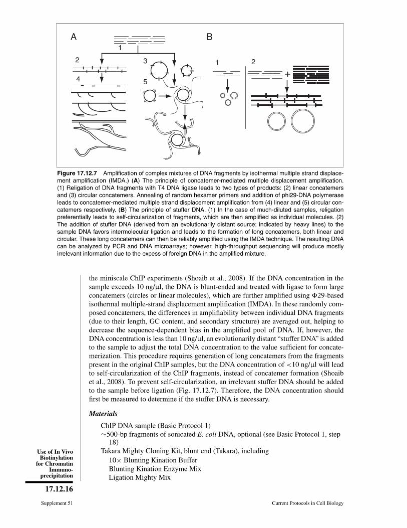
Figure 17.12.7 Amplification of complex mixtures of DNA fragments by isothermal multiple strand displace-ment amplification (IMDA.) (A) The principle of concatemer-mediated multiple displacement amplification.(1) Religation of DNA fragments with T4 DNA ligase leads to two types of products: (2) linear concatemersand (3) circular concatemers. Annealing of random hexamer primers and addition of phi29-DNA polymeraseleads to concatemer-mediated multiple strand displacement amplification from (4) linear and (5) circular con-catemers respectively. (B) The principle of stuffer DNA. (1) In the case of much-diluted samples, religationpreferentially leads to self-circularization of fragments, which are then amplified as individual molecules. (2)The addition of stuffer DNA (derived from an evolutionarily distant source; indicated by heavy lines) to thesample DNA favors intermolecular ligation and leads to the formation of long concatemers, both linear andcircular. These long concatemers can then be reliably amplified using the IMDA technique. The resulting DNAcan be analyzed by PCR and DNA microarrays; however, high-throughput sequencing will produce mostlyirrelevant information due to the excess of foreign DNA in the amplified mixture.
the miniscale ChIP experiments (Shoaib et al., 2008). If the DNA concentration in thesample exceeds 10 ng/μl, the DNA is blunt-ended and treated with ligase to form largeconcatemers (circles or linear molecules), which are further amplified using �29-basedisothermal multiple-strand displacement amplification (IMDA). In these randomly com-posed concatemers, the differences in amplifiability between individual DNA fragments(due to their length, GC content, and secondary structure) are averaged out, helping todecrease the sequence-dependent bias in the amplified pool of DNA. If, however, theDNA concentration is less than 10 ng/μl, an evolutionarily distant “stuffer DNA” is addedto the sample to adjust the total DNA concentration to the value sufficient for concate-merization. This procedure requires generation of long concatemers from the fragmentspresent in the original ChIP samples, but the DNA concentration of <10 ng/μl will leadto self-circularization of the ChIP fragments, instead of concatemer formation (Shoaibet al., 2008). To prevent self-circularization, an irrelevant stuffer DNA should be addedto the sample before ligation (Fig. 17.12.7). Therefore, the DNA concentration shouldfirst be measured to determine if the stuffer DNA is necessary.
Materials
ChIP DNA sample (Basic Protocol 1)∼500-bp fragments of sonicated E. coli DNA, optional (see Basic Protocol 1, step
18)Takara Mighty Cloning Kit, blunt end (Takara), including
10× Blunting Kination BufferBlunting Kination Enzyme MixLigation Mighty Mix
MacromolecularInteractions inCells
17.12.17
Current Protocols in Cell Biology Supplement 51
25:24:1 (v/v/v) phenol/chloroform/isoamyl alcohol24:1 (v/v) chloroform/isoamyl alcohol3 M sodium acetate20 mg/ml glycogen70% (v/v) and 100% ethanol, coldTE (Tris/EDTA) buffer (APPENDIX 2A)5 U/μl Ø29-DNA polymerase: dilute 1000 U/μl Ø29-DNA polymerase (Epicentre
Biotechnologies) 1:200 with the supplied buffer and store up to 3 months at−20◦C
2× IMDA reaction buffer (see recipe)200 μM random hexamer primers (NEB)Mineral oil, biotechnology grade
NanoDrop spectrophotometer (NanoDrop)1.5-ml microcentrifuge tubes16◦C, 30◦C, 37◦C, 65◦C, and 70◦C cooling/heating blocks or thermal cycler
Prepare sample DNA1. Use 1 μl of each sample to measure the DNA concentration using a NanoDrop
spectrophotometer according to the manufacturer’s directions.
2. If the DNA concentration is lower than 10 ng/μl, add sonicated E.coli DNA (stufferDNA) to a final concentration of 15 ng/μl.
3. Using components of the Takara Mighty Cloning Kit, prepare the following 20-μlreaction mixture in a 1.5-ml microcentrifuge tube:
17 μl sample DNA2 μl of 10× Blunting Kination Buffer1 μl Blunting Kination Enzyme Mix.
Incubate 10 min at 37◦C.
4. Mix 80 μl of distilled water and 100 μl of phenol/chloroform/isoamyl alcohol.Centrifuge 5 min at 16,000 × g, room temperature, and transfer the upper layer to anew tube.
5. Add an equal amount of chloroform/isoamyl alcohol and mix. Centrifuge 5 min at16,000 × g, room temperature, and transfer the upper layer to new a tube.
6. Add 10 μl of 3 M sodium acetate, 2 μl of 20 mg/ml glycogen, and 250 μl of chilled100% ethanol, and mix. Incubate 20 min at −80◦C.
7. Centrifuge at 10 min at 16,000 × g, 4◦C.
8. Remove the supernatant. Wash the precipitate with chilled 70% ethanol. Centrifuge5 min at 16,000 × g, 4◦C.
9. Remove the supernatant, and dry the precipitate on the bench at room temperature(5 to 10 min).
10. Dissolve the precipitate in 10 μl TE buffer.
Perform ligation11. Add 10 μl Ligation Mighty Mix, and mix gently. Incubate 1 hr at 16◦C.
12. Inactivate the enzymes by heating 5 min at 65◦C.
Use of In VivoBiotinylation
for ChromatinImmuno-
precipitation
17.12.18
Supplement 51 Current Protocols in Cell Biology
Perform IMDA procedure13. Prepare the Ø29 reaction mix immediately before setting up the reactions. For a
10-μl of reaction mix, combine the following:
6.3 μl of 2× buffer1.2 μl of 5 units/μl Ø29-DNA polymerase (0.6 units/μl final concentration)2.5 μl of 200 μM random hexamer primers (50 μM final concentration).
The volume of the reaction mix may be adjusted as necessary, for the number of samples.
14. Combine 1 μl of DNA concatemers with 2 μl of Q29 reaction mix.
15. Overlay the sample with 10 μl of biotechnology grade mineral oil to prevent evap-oration, and briefly microcentrifuge to make sure that the aqueous phase forms asmall sphere at the bottom of the tube.
16. Incubate 6 hr at 30◦C, and then heat for 10 min at 70◦C to inactivate the enzyme.
17. Store the samples up to 3 months at −20◦C or up to 1 year at −80◦C, or useimmediately for further analysis.
REAGENTS AND SOLUTIONSUse deionized, distilled water in all recipes and protocol steps. For common stock solutions, seeAPPENDIX 2A; for suppliers, see SUPPLIERS APPENDIX.
ChIP buffer
0.3% (w/v) SDS1% (v/v) Triton X-1002 mM EDTA (APPENDIX 2A)30 mM Tris·Cl, pH 8 (APPENDIX 2A)150 mM NaClStore up to several months at 4◦C. Just before use add 25× protease inhibitor stock
(see recipe) to a final concentration of 1×.
CSK buffer
100 mM NaCl300 mM sucrose10 mM Tris·Cl, pH 7.5 (see APPENDIX 2A)3 mM MgCl21 mM EGTA1.2 mM phenymethylsulfonyl fluoride (PMSF, APPENDIX 2A)0.5% (v/v) Triton X-100Store up to several months at 4◦C. Just before use, add 1.2 mM (final concentration)
phenylmethylsulfonyl fluoride (PMSF, APPENDIX 2A) and 25× protease inhibitorstock (see recipe) to a final concentration of 1×.
IMDA reaction buffer, 2×Prepare 10× IMDA reaction buffer containing the following components:
370 mM Tris·Cl, pH 7.5 (see APPENDIX 2A)100 mM MgCl2500 mM KCl50 mM (NH4)2SO4
2% (v/v) Tween 20continued
MacromolecularInteractions inCells
17.12.19
Current Protocols in Cell Biology Supplement 51
1 μg/μl bovine serum albumin (BSA)Store up to 6 months at −20◦C.
Prepare 2× buffer from the 10× buffer, by adding 10 mM dNTP mix (Promega;2 mM final concentration), DTT (2 mM final concentration), and water.
Protease inhibitor stock, 25×Dissolve a 50-ml size of Complete Protease Cocktail Tablet (Roche) in 2 ml water,and dispense 100- to 200-μl aliquots into microcentrifuge tubes. Store up to 6months at −20◦C.
COMMENTARY
Background InformationAll genomic processes in eukaryotes—
transcription, replication, repair, andrecombination—occur in the context ofchromatin, the hierarchically organized anddynamic complex of DNA and histone andnonhistone proteins. The systematic study ofthe time-dependent association among thesecomponents of chromatin requires robust andeffective approaches to characterizing protein-DNA interactions. Methods such as the elec-trophoretic gel mobility shift assay (EMSA;Revzin 1989; Molloy, 2000), systematicevolution of ligands by exponential enrich-ment (SELEX; Gold et al., 1995), and others(Veenstra, 1999) have been developed tostudy binding of protein to DNA. However, inthese approaches, the interactions are studiedin vitro, out of the context of chromatinand the nuclear environment, and thus anindependent confirmation is required to assessthe relevance of the data obtained. Chromatinimmunoprecipitation (ChIP; Kuo and Allis1999; Orlando, 2000) has emerged in thelast decade as part of a standard toolboxfor analyzing protein-DNA interactions ina more physiological context, as it detectsthem directly in their native environments.In the past several years, it has been widelyused to study the in vivo association of aparticular DNA sequence with regulatoryproteins and various forms of histones (i.e.,their post-translationally modified forms andreplacement variants).
Despite its conceptual simplicity, the ChIPprotocol requires fine tuning for each individ-ual protein and cell type. Compared to affin-ity purifications from cellular and nuclear ex-tracts, the most common problem in ChIP ishigh nonspecific background, caused by theeasy aggregation of chromatin and its bind-ing to surfaces. Accordingly, the success ofChIP crucially depends on the quality of an-tibodies used. ChIP grade antibodies have to
tolerate stringent binding and washing condi-tions, and also recognize the parts of the pro-tein that are (1) not involved in the interactionunder question and (2) minimally affected bythe formaldehyde cross-linking.
Some of the limitations of the native anti-body approach are overcome by using epitope-tagging, i.e., by expression in the cells ofthe protein of interest fused to an epitopetag recognized by a well characterized anti-body (Jarvik and Telmer, 1998; Wang, 2009).Among the advantages of this method are stan-dardization and commercial availability of rel-atively inexpensive epitope-tagging systems.Unlike using antibody against a native protein,the epitope-tagging approach lends itself to aneasy and efficient negative control, where thenonspecific signal is detected by a parallel pu-rification from a sample that does not expressan epitope-tagged protein (i.e., mock purifica-tion). Epitope tagging is a preferred methodfor comparing closely related proteins whenthe antibodies for distinguishing between thedifferent protein forms are not readily avail-able (e.g., mutant or alternative splicing vari-ants of the same protein). Finally, the bind-ing of the tag to antibody usually does notcompete with the protein-DNA interaction un-der study, as compared to the native antibody.Nonetheless, given that epitope-tagging mightaffect the protein function, the use of severalversions of epitope tagged protein of interest,typically with their N- or C- termini modified,is recommended.
The interaction between biotin and strepta-vidin (Kd of 10−15 M) is one of the strongestknown noncovalent interactions (Lindqvistand Schneider, 1996). Many commerciallyavailable reagents for purification and detec-tion of biotinylated macromolecules have beendeveloped. Accordingly, to take advantage ofthis interaction, we (Viens et al., 2004) andothers (van Werven and Timmers, 2006; Ooiet al., 2010) have developed biotin-ChIP, a
Use of In VivoBiotinylation
for ChromatinImmuno-
precipitation
17.12.20
Supplement 51 Current Protocols in Cell Biology
variant of the epitope-tagging approach toChIP, based on protein biotinylation in vivo.Such an approach, in its various forms, hasbeen applied to a broad variety of experi-mental systems, including mammalian cells,yeast, insects, and worms (van Werven andTimmers, 2006; Kim et al., 2009; Kolodziejet al., 2009; Lausen et al., 2010; Ooi et al.,2010). The main advantage of this method forChIP is the more stringent washing conditions,as well as more efficient (i.e., more quantita-tive) recovery of the protein-DNA complexes(Figure 17.12.4). Note that the in vivo biotiny-lation of histones has been also used for na-tive ChIP (NChIP) format (i.e., without cross-linking, Ooi et al., 2010), which takes ad-vantage of the exceptionally strong and spe-cific binding between biotin and streptavidinand allows one to reduce the volume of affin-ity medium, and thus decrease nonspecificbackground. Moreover, although not testedyet, one would expect that in vivo biotinyla-tion can be equally useful for many modifica-tions of ChIP developed to date, e.g., 3C-5C(Vassetzky et al., 2009) and RNAChIP (Gilbertand Svejstrup, 2006; Percipalle and Obrdlik2009).
Critical Parameters
Transfection efficiency (Basic Protocol 1,steps 1 to 5)
When using calcium phosphate transfec-tion, the pH of the HeBS buffer is crucial.The precipitate should be barely visible undermicroscope. For control of transfection effi-ciency, we recommend transfecting a separatecell well with BAP-GFP (or any other fluo-rescent protein); this sample can also be laterused as a negative control in ChIP.
Cross-linking (Basic Protocol 1,steps 6 to 16)
Excessive cross-linking can negatively af-fect the efficiency of chromatin fragmentation.The empirically chosen dose of formaldehyde(1% final solution, 10 min) might require opti-mization, depending on the cell type and pro-tein used. Several samples with different cross-linking times should be tested. In most cases,8 to 10 min for cross-linking is sufficient forChIP with mammalian cells.
Fragmentation (Basic Protocol 1,steps 17 to 20)
The desired size of chromatin fragments de-pends on the downstream analysis. Generally,it should be ∼400 to 500 bp. Longer frag-ments will diminish resolution and increase
nonspecific signal; therefore, they are not rec-ommended for high-throughput analysis (mi-croarray analysis or sequencing). Fragmentsthat are too short are generally not good forPCR detection, as it becomes challenging tofind suitable primers for amplification.
The sonication parameters (pulse duration,intensity, and number) vary, depending on theextent of cross-linking and cell type; thus, theymust be optimized for each experiment. Ide-ally, the least amount of input energy that givessatisfactory fragmentation should be used.
Affinity purification (Basic Protocol 1, 33 to41)
After cell lysis (steps 9 to 16), the sam-ple will contain biotin, which will competewith the biotinylated chromatin for binding tostreptavidin in the downstream pull-down step.Thorough washing of medium from the cellsbefore cell lysis is therefore critical.
The affinity between biotinylated BAP andstreptavidin (steps 33 to 41) can be weakenedby steric hindrance, depending on whether thetagged part of the protein (e.g., its N or C ter-minus) is well exposed or buried. Accordingly,the incubation times and washing stringencycould be adjusted. Typically, a 3-hr incuba-tion is sufficient for successful pull-down ofbiotinylated chromatin. If longer incubationtimes are used, the length of the preclearingincubation step (step 30) should be increasedaccordingly.
IMDA (Basic Protocol 2)A DNA concentration of <10 ng/μl will
lead to self-circularization of the fragmentsobtained in ChIP, instead of the formation ofconcatemers. As the result, the fragments willbe amplified individually, and their sequenceand size-dependent bias in amplification effi-ciency will not be averaged out. Therefore, thefirst step in applying ligation-IMDA is measur-ing DNA concentration, which will determineif the stuffer DNA is necessary.
Troubleshooting
Insufficient fragmentation of DNA aftersonication
When monitoring the size of sheared DNAafter sonication, one can observe insufficientfragmentation. There could be several causesfor this problem: (a) material is overcross-linked (Basic Protocol 1, steps 6 to 16),(b) cells are insufficiently lysed (step 17),or (c) sonication is inefficient (steps 18 to20). Accordingly, take the following step:(a) Cross-linking time could be reduced.
MacromolecularInteractions inCells
17.12.21
Current Protocols in Cell Biology Supplement 51
(b) If there are reasons to suspect that too manycells in a small volume of ChIP buffer wereused for lysis, additional ChIP buffer should beadded. Alternatively, cells can be snap-frozenin liquid nitrogen and thawed. The success oflysis can be estimated visually by inspectingthe sample under the microscope. (c) In thecase of insufficient sonication, the duration,energy, and/or number of rounds of sonicationshould be increased.
Low or absent PCR signal after ChIPLow or no PCR signal after ChIP could
have several causes: (a) insufficient amountof chromatin for immunoprecipitation (BasicProtocol 1, steps 33 to 37), (b) failure of im-munoprecipitation, or (c) failure of PCR. Ac-cordingly, (a) the amount of cells or chro-matin should be increased. Note that it may bedifficult to extract all chromatin from certainprimary cell types. (b) The biotinylation effi-ciency of the target protein should be tested,as well as the quality of streptavidin-beads. Inaddition, one should make sure that the cellswere washed thoroughly before lysis, as biotinin the medium will compete with the biotiny-lated protein for binding (step 16). (c) Oneshould set up a control qPCR with the sameprimers on genomic DNA, and optimize PCRconditions.
Elevated background signalInstead of low PCR signal, one may ob-
serve an elevated background signal. This canbe caused by (a) nonspecific immunoprecipi-tation or (b) insufficient washes. Accordingly,one should (a) reduce duration of incubationof streptavidin-beads with chromatin (step 33)and ensure the tube-switch step (steps 35 and39) is performed and (b) increase the numberand/or stringency of the washes after IP (steps38 to 41).
Anticipated ResultsWe usually obtain ∼15 to 20 ng/μl of final
ChIP DNA from biotin-ChIP (total 30 μl). Intotal, 5% of biotin-ChIP material is used inPCR to test the quality of the sample. In mostof the cases, we observe enrichment of 5 to100-fold from the target loci.
The use of DNA amplification (e.g., IMDA)should allow the analysis of more genomicsites and of the whole genome.
Time ConsiderationsFor the biotin-ChIP protocol, transient
transfection takes 2 days before cell harvest.The cell lysis, preclearing, binding, and wash-ing procedures take 1 full day, with the DNA
eluted from beads and ready for PCR analysisthe next day. If necessary, the preclearing andbinding to streptavidin beads can take longerthan 3 hr, e.g., overnight for both steps. Withpractice, ∼10 samples can be processed in par-allel.
The IMDA procedure takes 1 day, start-ing from DNA measurement and ligation. Thelongest part is amplification, which can takea minimum of 6 hr, but can also be extendedovernight.
The streptavidin gel-shift experiment typ-ically takes 1 day, as it includes SDS-PAGEseparation, transfer to PVDF (or nitrocellu-lose) membrane, blocking, incubation withprimary and secondary antibodies, and en-hanced chemiluminescence (ECL) detection.
Literature CitedDahl, J.A. and Collas, P. 2008. A rapid micro chro-
matin immunoprecipitation assay (microChIP).Nat. Protoc. 3:1032-1045.
de Boer, E., Rodriguez, P., Bonte, E., Krijgsveld,J., Katsantoni, E., Heck, A., Grosveld, F., andStrouboulis, J. 2003. Efficient biotinylation andsingle-step purification of tagged transcriptionfactors in mammalian cells and transgenic mice.Proc. Natl. Acad. Sci. U.S.A. 100:7480-7485.
Gilbert, C. and Svejstrup, J.Q. 2006. RNA immuno-precipitation for determining RNA-protein as-sociations in vivo. Curr. Protoc. Mol. Biol.75:27.4.1-27.4.11.
Gilmour, D.S. and Lis, J.T. 1984. Detecting protein-DNA interactions in vivo: Distribution of RNApolymerase on specific bacterial genes. Proc.Natl. Acad. Sci. U.S.A. 81:4275-4279.
Gold, L., Polisky, B., Uhlenbeck, O., and Yarus,M. 1995. Diversity of oligonucleotide functions.Annu. Rev. Biochem. 64:763-797.
Hager, G.L., Elbi, C., and Becker, M. 2002. Pro-tein dynamics in the nuclear compartment. Curr.Opin. Genet. Dev. 12:137-141.
Jarvik, J.W. and Telmer, C.A. 1998. Epitope tag-ging. Annu. Rev. Genet. 32:601-618.
Kim, J., Cantor, A.B., Orkin, S.H., and Wang,J. 2009. Use of in vivo biotinylation to studyprotein-protein and protein-DNA interactions inmouse embryonic stem cells. Nat. Protoc. 4:506-517.
Kolodziej, K.E., Pourfarzad, F., de Boer, E.,Krpic, S., Grosveld, F., and Strouboulis, J. 2009.Optimal use of tandem biotin and V5 tags inChIP assays. BMC Mol. Biol. 10:6.
Kuo, M.H. and Allis, C.D. 1999. In vivo cross-linking and immunoprecipitation for studyingdynamic Protein:DNA associations in a chro-matin environment. Methods 19:425-433.
Lausen, J., Pless, O., Leonard, F., Kuvardina, O.N.,Koch, B., and Leutz, A. 2010. Targets of the Tal1transcription factor in erythrocytes: E2 ubiqui-tin conjugase regulation by Tal1. J. Biol. Chem.285:5338-5346.
Use of In VivoBiotinylation
for ChromatinImmuno-
precipitation
17.12.22
Supplement 51 Current Protocols in Cell Biology
Lindqvist, Y. and Schneider, G. 1996. Protein-biotininteractions. Curr. Opin. Struct. Biol. 6:798-803.
Mechold, U., Gilbert, C., and Ogryzko, V. 2005.Codon optimization of the BirA enzyme geneleads to higher expression and an improved effi-ciency of biotinylation of target proteins in mam-malian cells. J. Biotechnol. 116:245-249.
Molloy, P.L. 2000. Electrophoretic mobility shiftassays. Methods Mol. Biol. 130:235-246.
Negre, N., Lavrov, S., Hennetin, J., Bellis, M., andCavalli, G. 2006. Mapping the distribution ofchromatin proteins by ChIP on chip. MethodsEnzymol. 410:316-341.
O’Neill, L.P. and Turner, B.M. 2003. Immunopre-cipitation of native chromatin: NChIP. Methods31:76-82.
Ooi, S.L., Henikoff, J.G., and Henikoff, S. 2010. Anative chromatin purification system for epige-nomic profiling in Caenorhabditis elegans. Nu-cleic Acids Res. 38:e26.
Orlando, V. 2000. Mapping chromosomal proteinsin vivo by formaldehyde-crosslinked-chromatinimmunoprecipitation. Trends Biochem. Sci.25:99-104.
Park, P.J. 2009. ChIP-seq: Advantages and chal-lenges of a maturing technology. Nat. Rev.Genet. 10:669-680.
Pashev, I.G., Dimitrov, S.I., and Angelov, D. 1991.Crosslinking proteins to nucleic acids by ul-traviolet laser irradiation. Trends Biochem. Sci.16:323-326.
Percipalle, P. and Obrdlik, A. 2009. Analy-sis of nascent RNA transcripts by chromatinRNA immunoprecipitation. Methods Mol. Biol.567:215-235.
Phair, R.D. and Misteli, T. 2000. High mobility ofproteins in the mammalian cell nucleus. Nature404:604-609.
Revzin, A. 1989. Gel electrophoresis assays forDNA-protein interactions. Biotechniques 7:346-355.
Shoaib, M., Baconnais, S., Mechold, U., Le Cam,E., Lipinski, M., and Ogryzko, V. 2008. Mul-
tiple displacement amplification for complexmixtures of DNA fragments. BMC Genomics9:415.
van Werven, F.J. and Timmers, H.T. 2006.The use of biotin tagging in Saccharomycescerevisiae improves the sensitivity of chro-matin immunoprecipitation. Nucleic Acids Res.34:e33.
Vassetzky, Y., Gavrilov, A., Eivazova, E.,Priozhkova, I., Lipinski, M., and Razin, S. 2009.Chromosome conformation capture (from 3C to5C) and its ChIP-based modification. MethodsMol. Biol. 567:171-188.
Veenstra, T.D. 1999. Electrospray ionization massspectrometry: A promising new technique in thestudy of protein/DNA noncovalent complexes.Biochem. Biophys. Res. Commun. 257:1-5.
Viens, A., Mechold, U., Lehrmann, H., Harel-Bellan, A., and Ogryzko, V. 2004. Use of proteinbiotinylation in vivo for chromatin immunopre-cipitation. Anal. Biochem. 325:68-76.
Viens, A., Harper, F., Pichard, E., Comisso, M.,Pierron, G., and Ogryzko, V. 2008. Use ofprotein biotinylation in vivo for immunoelec-tron microscopic localization of a specific pro-tein isoform. J. Histochem. Cytochem. 56:911-919.
Voytas, D. 2001. Agarose gel electrophoresis. Curr.Protoc. Mol. Biol. 51:2.5A.1-2.5A.9.
Wang, Z. 2009. Epitope tagging of endogenous pro-teins for genome-wide chromatin immunopre-cipitation analysis. Methods Mol. Biol. 567:87-98.
Weinmann, A.S. and Farnham, P.J. 2002. Identifi-cation of unknown target genes of human tran-scription factors using chromatin immunopre-cipitation. Methods 26:37-47.
Wolffe, A. 1999. Chromatin: Structure and Func-tion. Academic Press, London.
Zeng, P.Y., Vakoc, C.R., Chen, Z.C., Blobel, G.A.,and Berger, S.L. 2006. In vivo dual cross-linking for identification of indirect DNA-associated proteins by chromatin immunopre-cipitation. Biotechniques 41:694-698.