current trends in preoperative, intraoperative, and ... · pdf filecurrent trends in...
TRANSCRIPT

Contents lists available at ScienceDirect
Current Problems in Surgery
Current Problems in Surgery 52 (2015) 531–569
http://d0011-38
journal homepage: www.elsevier.com/locate/cpsurg
Current trends in preoperative,intraoperative, and postoperative care of theadult cardiac surgery patient
Cardiac surgery, now widely pr
acticed in most parts of the world, came from humblebeginnings. As late as the mid-20th century, the stigma surrounding cutting into the heart, aswell as the inability to control bleeding, prevented many surgeons from exploring the field. Theearly operations were not without consequence. Many surgeons experienced failure, and movedon to other areas of study. But those who continued to pursue the work, would go on to makehistory. Today, cardiac surgery continues to grow, and encompasses many unique areas of study.It is by continuing to adapt to the needs of a changing population of patients that the specialtycontinues to secure its role in the future.Current figures
It is not mandatory to report to the Society of Thoracic Surgeons (STS) National Database, buta program seeking continued recognition and reimbursement will choose to participate. From2004 through 2013, the STS reported 2,735,459 cardiac procedures from the reporting sites.1,2
Although overall case volume reached a peak in 2009 and began to decline the following year,there has been an increase in both isolated aortic valve replacement (AVR) and mitral valvereplacement (MVR).1,2 The unadjusted mortality rate for isolated CABG procedures over the past10 years is approximately 2% in centers that report to the STS.1 Isolated AVR carries anunadjusted mortality rate between 3% and 3.5%.1 More complicated operations continue to havea higher mortality rate, with combined AVR-MVR and combined MVR-CABG procedures carryingmortality rates from approximately 8%-11% over the same 10-year period.1
Changes in practice, regulation, and reimbursement
Financial reimbursement for cardiac procedures has declined over the past decade. In 2003,Medicare began its pay-for-performance program, the Premier Hospital Quality IncentiveDemonstration. Cardiac surgeons have been particularly affected by the Centers for Medicare andMedicaid Services cost-savings measures.3 In the past, programs would receive the samepayments for a certain procedure regardless of the outcome. Now there is a demand on hospitalsto decrease complication rates. Initial studies on the Hospital Quality Incentive Demonstrationdid not show a reduction in risk-adjusted mortality for myocardial infarction (MI), pneumonia,
x.doi.org/10.1067/j.cpsurg.2014.10.00140/& 2015 Elsevier Inc. All rights reserved.
L.A. Szelkowski et al. / Current Problems in Surgery 52 (2015) 531–569532
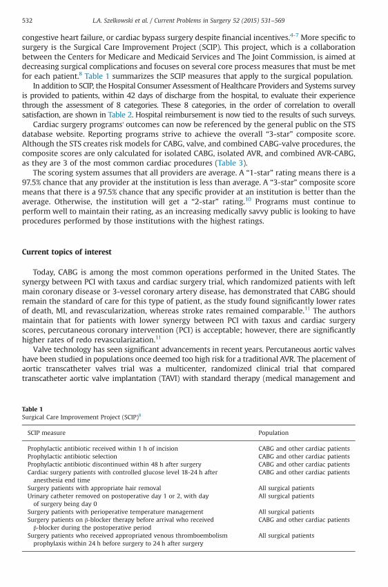
congestive heart failure, or cardiac bypass surgery despite financial incentives.4-7 More specific tosurgery is the Surgical Care Improvement Project (SCIP). This project, which is a collaborationbetween the Centers for Medicare and Medicaid Services and The Joint Commission, is aimed atdecreasing surgical complications and focuses on several core process measures that must be metfor each patient.8 Table 1 summarizes the SCIP measures that apply to the surgical population.
In addition to SCIP, the Hospital Consumer Assessment of Healthcare Providers and Systems surveyis provided to patients, within 42 days of discharge from the hospital, to evaluate their experiencethrough the assessment of 8 categories. These 8 categories, in the order of correlation to overallsatisfaction, are shown in Table 2. Hospital reimbursement is now tied to the results of such surveys.
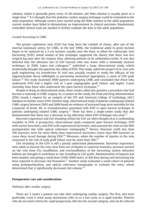
Cardiac surgery programs' outcomes can now be referenced by the general public on the STSdatabase website. Reporting programs strive to achieve the overall “3-star” composite score.Although the STS creates risk models for CABG, valve, and combined CABG-valve procedures, thecomposite scores are only calculated for isolated CABG, isolated AVR, and combined AVR-CABG,as they are 3 of the most common cardiac procedures (Table 3).
The scoring system assumes that all providers are average. A “1-star” rating means there is a97.5% chance that any provider at the institution is less than average. A “3-star” composite scoremeans that there is a 97.5% chance that any specific provider at an institution is better than theaverage. Otherwise, the institution will get a “2-star” rating.10 Programs must continue toperform well to maintain their rating, as an increasing medically savvy public is looking to haveprocedures performed by those institutions with the highest ratings.
Current topics of interest
Today, CABG is among the most common operations performed in the United States. Thesynergy between PCI with taxus and cardiac surgery trial, which randomized patients with leftmain coronary disease or 3-vessel coronary artery disease, has demonstrated that CABG shouldremain the standard of care for this type of patient, as the study found significantly lower ratesof death, MI, and revascularization, whereas stroke rates remained comparable.11 The authorsmaintain that for patients with lower synergy between PCI with taxus and cardiac surgeryscores, percutaneous coronary intervention (PCI) is acceptable; however, there are significantlyhigher rates of redo revascularization.11
Valve technology has seen significant advancements in recent years. Percutaneous aortic valveshave been studied in populations once deemed too high risk for a traditional AVR. The placement ofaortic transcatheter valves trial was a multicenter, randomized clinical trial that comparedtranscatheter aortic valve implantation (TAVI) with standard therapy (medical management and
Table 1Surgical Care Improvement Project (SCIP)8
SCIP measure Population
Prophylactic antibiotic received within 1 h of incision CABG and other cardiac patientsProphylactic antibiotic selection CABG and other cardiac patientsProphylactic antibiotic discontinued within 48 h after surgery CABG and other cardiac patientsCardiac surgery patients with controlled glucose level 18-24 h afteranesthesia end time
CABG and other cardiac patients
Surgery patients with appropriate hair removal All surgical patientsUrinary catheter removed on postoperative day 1 or 2, with dayof surgery being day 0
All surgical patients
Surgery patients with perioperative temperature management All surgical patientsSurgery patients on β-blocker therapy before arrival who receivedβ-blocker during the postoperative period
CABG and other cardiac patients
Surgery patients who received appropriated venous thromboembolismprophylaxis within 24 h before surgery to 24 h after surgery
All surgical patients
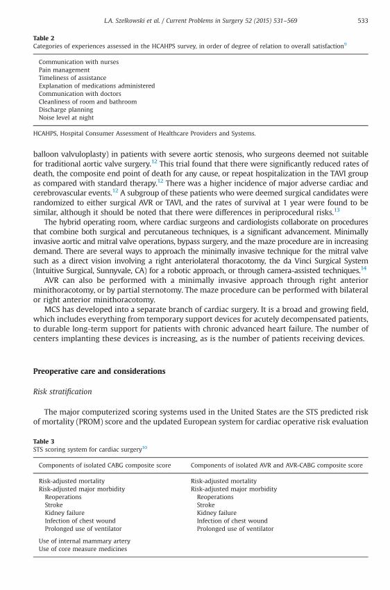
Table 2Categories of experiences assessed in the HCAHPS survey, in order of degree of relation to overall satisfaction9
Communication with nursesPain managementTimeliness of assistanceExplanation of medications administeredCommunication with doctorsCleanliness of room and bathroomDischarge planningNoise level at night
HCAHPS, Hospital Consumer Assessment of Healthcare Providers and Systems.
L.A. Szelkowski et al. / Current Problems in Surgery 52 (2015) 531–569 533
balloon valvuloplasty) in patients with severe aortic stenosis, who surgeons deemed not suitablefor traditional aortic valve surgery.12 This trial found that there were significantly reduced rates ofdeath, the composite end point of death for any cause, or repeat hospitalization in the TAVI groupas compared with standard therapy.12 There was a higher incidence of major adverse cardiac andcerebrovascular events.12 A subgroup of these patients who were deemed surgical candidates wererandomized to either surgical AVR or TAVI, and the rates of survival at 1 year were found to besimilar, although it should be noted that there were differences in periprocedural risks.13
The hybrid operating room, where cardiac surgeons and cardiologists collaborate on proceduresthat combine both surgical and percutaneous techniques, is a significant advancement. Minimallyinvasive aortic and mitral valve operations, bypass surgery, and the maze procedure are in increasingdemand. There are several ways to approach the minimally invasive technique for the mitral valvesuch as a direct vision involving a right anteriolateral thoracotomy, the da Vinci Surgical System(Intuitive Surgical, Sunnyvale, CA) for a robotic approach, or through camera-assisted techniques.14
AVR can also be performed with a minimally invasive approach through right anteriorminithoracotomy, or by partial sternotomy. The maze procedure can be performed with bilateralor right anterior minithoracotomy.
MCS has developed into a separate branch of cardiac surgery. It is a broad and growing field,which includes everything from temporary support devices for acutely decompensated patients,to durable long-term support for patients with chronic advanced heart failure. The number ofcenters implanting these devices is increasing, as is the number of patients receiving devices.
Preoperative care and considerations
Risk stratification
The major computerized scoring systems used in the United States are the STS predicted riskof mortality (PROM) score and the updated European system for cardiac operative risk evaluation
Table 3STS scoring system for cardiac surgery10
Components of isolated CABG composite score Components of isolated AVR and AVR-CABG composite score
Risk-adjusted mortality Risk-adjusted mortalityRisk-adjusted major morbidity Risk-adjusted major morbidityReoperations ReoperationsStroke StrokeKidney failure Kidney failureInfection of chest wound Infection of chest woundProlonged use of ventilator Prolonged use of ventilator
Use of internal mammary arteryUse of core measure medicines

L.A. Szelkowski et al. / Current Problems in Surgery 52 (2015) 531–569534
score (EuroSCORE II).15 The original EuroSCORE, as it has been used for the past 15 years, is nolonger applicable in contemporary practice. The EuroSCORE II has been developed and has beenshown to have better calibration and discrimination.16 It retains many of the core risk factors, butincludes the redefinition of the symptomatic status, and the incorporation of the creatinineclearance.16 In addition, procedures formally listed under the umbrella “other than isolatedcoronary artery bypass grafting” are individually listed.16 This gives the more complicatedprocedures greater weight, as is seen in the STS model.
Despite efforts to keep the scoring systems current, studies have shown their deficiencies inthe current population of cardiac surgery patients. In transcatheter valve patients, the risk ofmortality and morbidity is overpredicted by the EuroSCORE II.16 Transcatheter valve patients areoften older and have more comorbidities than typical cardiac surgery populations, which hasdriven the need for updated scoring systems. There are data to suggest that factors outside therisk models play a role in mortality and morbidity after cardiac surgery. Frailty, which is based onthe speed at which an individual can walk a 5-m length, has been found to be an independentpredictor of mortality after cardiac surgery and is not included in either the STS PROM score orthe EuroSCORE II.17 Chronic lung disease is part of the STS PROM risk model, but clinicaljudgment, rather than spirometry testing, is used to document the diagnosis.18 A studyevaluating the effect of chronic lung disease on mortality and morbidity found that chronic lungdisease was better documented with spirometry testing and that this technique more accuratelycaptured the risk associated with this disease process.18
The preoperative evaluation
Patients should undergo a thorough evaluation before undergoing cardiac surgery. Severalpreoperative conditions should be investigated before entering the operating room to avoidpreventable complications (Table 4).19
There are numerous laboratory and diagnostic studies, which may be performed beforeentering the operating room (Table 5).19
Intraoperative care and considerations
Current trends in cardiac anesthesia
Cardiac anesthesiologists have adapted their skill set to care for the aging cardiac surgerypopulation with more comorbidities. They have become proficient in ultrasound-guidedtechniques for central line placement, the use of videolarygoscopy for difficult intubations, andinterpreting 3-dimensional (3D) transesophageal echocardiography (TEE).20 A significant changeover the past decade has been the withdrawal of the drug aprotinin. Cardiac anesthesiologists usedthis serine protease inhibitor–type antifibrinolytic for nearly all patients before 2007. A multicenter,double-blinded study called the blood conservation using antifibrinolytics in a randomizedcontrolled trial compared full-dose aprotinin with standard dose epsilon aminocaproic acid ortranexamic acid in high-risk cardiac surgery.21 This study was stopped early because there was ahigher risk of death in the patients receiving aprotinin.22 Controversy still remains as to whetherthe drug was withdrawn from the market too quickly, before a thorough evaluationwas performed.
Two-dimensional TEE has been a mainstay in cardiac anesthesia, but the past 2 decades haveseen an emergence of 3D echocardiographic technology. Current technology allows for morereal-time imaging and better resolution.23 The data are not clear as to whether 3D TEE addsclinical value or decreases mortality.23,24 What is known, is that 3D TEE has superior ability toassess left and right ventricular (RV) function and is especially useful in imaging the mitral valveand in diagnosing complex mitral valve lesions.23 It is also useful to image the left atrialappendage, as traditional 2D techniques can overread the incidence of thrombus in thisstructure.23

Table 4Preoperative evaluation
Preoperative condition or finding Possible effect on surgical outcome
Aortic calcification Increased risk of stroke from calcium emboli dislodgedwith aortic cross-clamp
Previous chest radiation Possible adhesions in the chest, making entry difficultHepatic failure Possibility of coagulopathy after surgeryRenal failure Possibility of need of temporary or permanent hemodialysisNutritional status Poor wound healingBleeding disorders Increased need for transfusion of blood and blood productsAlcohol abuse Possible hepatic failureTobacco abuse COPD may require prolonged ventilatory supportDiabetes Poor wound healingNeurologic symptoms Symptoms may be magnified after surgery increasing time to recoveryRecent infections Possibility of sepsis postoperatively or seeding of prosthetic
valves with bacteriaAllergies Systemic allergic reaction and circulatory collapseCurrent medications May be at risk of increased bleeding from antiplatelet agents,
or hypotension from antihypertensivesVein stripping Will need to find alternate conduits in CABG surgeryDifference in the blood pressurebetween upper extremities
Internal mammary artery may not be suitable for a conduitin CABG patients
Poor dentition May cause seeding of prosthetic valves with bacteriaPresence of bruits May indicate the presence of undiagnosed peripheral vascular disease
L.A. Szelkowski et al. / Current Problems in Surgery 52 (2015) 531–569 535
Cardiopulmonary bypass
There have been several areas of focus where advances are being made in cardiopulmo-nary bypass (CPB). These advances are driven by the increase in minimally invasiveoperations, which have required a change in CPB technique, the continued focus on theinflammatory response associated with CPB, the subsequent efforts to decrease it, and an
Table 5Preoperative laboratory and diagnostic studies
Preoperative laboratory anddiagnostic studies
Rationale
Complete blood count Detect chronic anemia, thrombocytopenia, or infectionChemistry Detect kidney disease, electrolyte abnormalities, or hyperglycemiaHgB A1C Undiagnosed diabetesLiver function tests Diagnose liver disease that could affect coagulation after surgeryCoagulation studies Detect blood coagulation disordersType and cross withcold screen
Ensure blood is ready for the patient if needed and check for cold agglutinins(especially important in circulatory arrest cases, where the patient temperaturewill get quite low)
Arterial blood gas Diagnose underlying hypoxia or lung diseaseChest x-ray (PA and lateral inredo surgeries)
Evaluate lung fields, pleural effusions, and calcified aorta (especiallyimportant in a redo to note the number of sternal wires)
CT scan of chest Helpful if calcified aorta is suspected on chest x-rayEKG Evaluate preoperative rhythmSpirometry or pulmonaryfunction testing
Detect undiagnosed lung disease and plan for possible difficulty weaningfrom the ventilator
Carotid Doppler scans Evaluate for carotid stenosisUrinalysis Diagnose urinary tract infection or detect glucose in urineCT angiogram of aorta with3D reconstruction
Helpful in some large aortic cases
CT, computed tomography; EKG, electrocardiogram; PA, posteroanterior.

L.A. Szelkowski et al. / Current Problems in Surgery 52 (2015) 531–569536
effort by surgeons and anesthesiologists to decrease the amount of blood and bloodproduct transfusions.
CPB changed cardiac surgery for the better, but it is also a known contributor to the systemicinflammatory response syndrome (SIRS). SIRS can be described as an inflammatory process,which is no longer focused on the specific site of injury but is, instead, disseminated throughoutthe body.25 SIRS can cause fever, leukocytosis, capillary leak syndrome, and multiorgan failureafter cardiac surgery.26 CPB creates several situations, which set off SIRS. The trauma of thesurgery sets off cellular and humoral mediators of inflammation.25 This is augmented by theinterface of blood with the CPB circuit and the activation of compliment by CPB.25 Myocardialischemia, from cross-clamping and cardiac arrest, further contributes to the process.25 Methodsare being sought to decrease the activation of these processes. A multipronged approach can beused starting by testing patients preoperatively to see whether they are at risk for developing alife-threatening inflammatory response. Intraoperatively, techniques such as minimally invasiveapproach or off-pump CABG can be used.25 On a cellular level, interest is being directed towardinhibiting neutrophils, platelets, and complement activation, and leukocyte depletion.25 Mini-CPB systems are being studied, with the thought that less of the machine will interface with apatient's blood. These circuits do not include a reservoir or cardiotomy suction, have no blood-airinterface, and have a smaller heparin-coated circuit.27 The great disadvantage of these systems isthat without the reservoir, they are without an air removal system, which can result in airemboli. Medical literature does not support the claim that these systems can improve patientoutcomes, and they are not widely used in the United States.27
Retrograde autologous priming (RAP) of the CPB pump has been a major advancement in CPBtechnique. The principle of RAP is that the crystalloid volume of the CPB circuit is replaced withthe patient's blood volume just before commencing CPB.28 This technique has been studied bymany and, although it is not new, there is new interest in it because of its role in bloodconservation. As the CPB circuit is primed with the patient's blood, instead of crystalloid, there isless hemodilution, which reduces the need for subsequent blood transfusion. A meta-analysis ofrandomized controlled trials on the technique of RAP revealed that, although it seemed to haveno effect on clinical outcomes, it did create a significant reduction in the perioperativetransfusion requirements in adults.29
Acute normovolemic hemodilution is another technique designed to reduce the need forblood transfusion. It involves removing 1 or 2 units of the patient's blood before starting CPB toexpose less of the blood volume to the CPB circuit. After terminating CPB, this blood can then begiven back to the patient. Sarin and colleagues30 demonstrated that acute normovolemichemodilution patients received fewer intraoperative blood products, regardless of the surgerythey underwent, and that the decrease in blood exposure afforded patients decreased ventilatortimes and postoperative mortality.
Cardioplegia used during CPB has gone through an evolution from its introduction in the1950s.31 Initially, it was composed mainly of crystalloid but in the 1970s, the use of bloodcardioplegia gained popularity owing to the theory that it was better suited to deliver nutrientsand oxygen to the ischemic heart.31 It also reduces the edema associated with crystalloidcardioplegia and appears to offer less delay of myocardial recovery.31 A traditional crystalloidbased cardioplegia uses an 8:1 crystalloid-to-blood ratio, but newer blood cardioplegia, ormicroplegia, formulations have a much higher blood to crystalloid ratio. Both formulations havebeen studied, and the results are inconclusive. Three meta-analyses done between 2006 and2012 are not in overwhelming favor of the use of blood cardioplegia.32-34 Jacob and colleagues32
reviewed 18 randomized clinical trials and found that 10 of them reported statisticallysignificant clinical differences that favored the use of blood cardioplegia. Another studydemonstrated no difference in the risk of death, MI, or low cardiac output syndrome, regardlessof whether blood cardioplegia or crystalloid cardioplegia was used.32
The pediatric cardiac surgery community is familiar with the use of del Nido solution, whichis used in several centers for myocardial protection during cardiac surgery. This solution, which ismore dilute (1:4 ¼ blood:crystalloid) than standard solutions, has less calcium and containsmagnesium, mannitol, sodium bicarbonate, and lidocaine.35 As opposed to standard cardioplegia
L.A. Szelkowski et al. / Current Problems in Surgery 52 (2015) 531–569 537
solution, which is generally given every 15-20 minutes, del Nido solution is usually given as asingle dose.35 It is thought that this pediatric cardiac surgery technique could be translated to theadult population. Although centers have started using del Nido solution in the adult population,current studies have failed to demonstrate an improvement in clinical outcomes. Randomized,controlled clinical trials are needed to further evaluate the data in the adult population.
Conduit harvesting in CABG
The greater saphenous vein (GSV) has long been the conduit of choice, after use of theinternal mammary artery, for CABG. In the late 1990s, the traditional ankle to groin incisionbegan to be replaced by a 2-cm incision, usually near the knee, to allow for endoscopic veinharvesting (EVH). Initial studies of this technique supported the theory that there was lesssurgical leg pain after the surgery, thus, allowing patients to be ambulatory earlier. It was alsobelieved that the infection rate of GSV harvest sites was lower with a minimally invasivetechnique. In 2009, Lopes and colleagues37 published a large observational study, whichincidentally brought into question the safety of EVH. This study, called the project of ex-vivo veingraft engineering via transfection IV trial was actually created to study the efficacy of theoligonucleotide decoy edifoligide in preventing neointimal hyperplasia, a cause of GSV graftfailure.36 The study examined 3000 patients undergoing CABG and concluded that those whounderwent EVH had a higher risk of 1-year angiographic graft failure, and higher 3-yearmortality than those who underwent the open harvest technique.37
Despite it being an observational study, these results called into question a procedure that hadbecome a mainstay in CABG surgery. As a counter to this study, the Food and Drug Administration(FDA) issued a request for an analysis of the STS and American College of Cardiology (ACC)Database to further assess EVH. Another large, observational study of patients undergoing isolatedCABG surgery between 2003 and 2008 found no evidence of increased long-termmortality for thecomposite of death, MI, or revasularization associated with EVH vs open harvest technique inpatients undergoing isolated CABG surgery.38 There has been a single-center study that alsodemonstrated that there was a decrease in leg infections when EVH technique was used.39
Harvester experience and clot stranding within the GSV are often thought of as 2 confoundingvariables in EVH. A prospective, observational study compared open harvest technique, EVHwith novice harvesters, and EVH with experienced harvesters, and assessed the veins on the fifthpostoperative day with optical coherence tomography.40 Novice clinicians (with less than100 harvests) were far more likely than experienced harvesters (more than 900 harvests) tocause deep vessel damage during EVH.40 Moreover, when the number of injuries to the veinsurpassed 4, the risk of early graft failure rose by more than 50%.40
Clot stranding in the GSV is still a poorly understood phenomenon. Harvester experience,time taken to harvest the vein, heat from use of bipolar to cauterize branches, pressure exertedon the vein from CO2 insufflation, and overinsufflation of the harvesting system obturatorballoon are thought to contribute to clot stranding in GSV grafts. Administration of heparin hasbeen studied, and giving a small dose (2500-5000 units), or full dose during vein harvesting hasbeen reported to decrease clot formation.41 Another study evaluated a small cohort of patientsusing preheparinization, and optical coherence tomography to evaluate the veins, whichdetermined that it significantly decreased clot volume.42
Postoperative care and considerations
Pathways after cardiac surgery
There are 3 routes a patient can take after undergoing cardiac surgery. The first, and mostpreferable, track is what many institutions refer to as a fast track, or a rapid transfer. Patientswho do not meet criteria for rapid progression, fall into the second category, and can be referred
L.A. Szelkowski et al. / Current Problems in Surgery 52 (2015) 531–569538
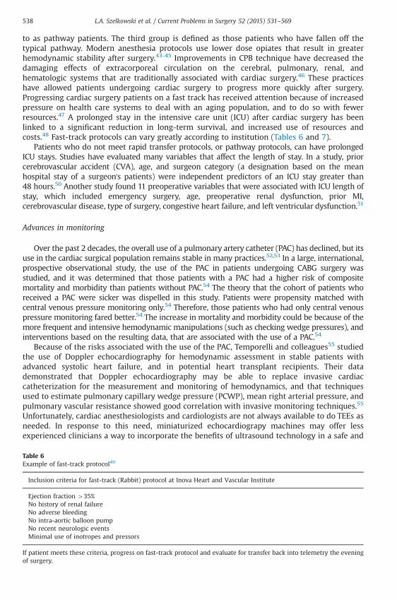
to as pathway patients. The third group is defined as those patients who have fallen off thetypical pathway. Modern anesthesia protocols use lower dose opiates that result in greaterhemodynamic stability after surgery.43-45 Improvements in CPB technique have decreased thedamaging effects of extracorporeal circulation on the cerebral, pulmonary, renal, andhematologic systems that are traditionally associated with cardiac surgery.46 These practiceshave allowed patients undergoing cardiac surgery to progress more quickly after surgery.Progressing cardiac surgery patients on a fast track has received attention because of increasedpressure on health care systems to deal with an aging population, and to do so with fewerresources.47 A prolonged stay in the intensive care unit (ICU) after cardiac surgery has beenlinked to a significant reduction in long-term survival, and increased use of resources andcosts.48 Fast-track protocols can vary greatly according to institution (Tables 6 and 7).
Patients who do not meet rapid transfer protocols, or pathway protocols, can have prolongedICU stays. Studies have evaluated many variables that affect the length of stay. In a study, priorcerebrovascular accident (CVA), age, and surgeon category (a designation based on the meanhospital stay of a surgeon's patients) were independent predictors of an ICU stay greater than48 hours.50 Another study found 11 preoperative variables that were associated with ICU length ofstay, which included emergency surgery, age, preoperative renal dysfunction, prior MI,cerebrovascular disease, type of surgery, congestive heart failure, and left ventricular dysfunction.51
Advances in monitoring
Over the past 2 decades, the overall use of a pulmonary artery catheter (PAC) has declined, but itsuse in the cardiac surgical population remains stable in many practices.52,53 In a large, international,prospective observational study, the use of the PAC in patients undergoing CABG surgery wasstudied, and it was determined that those patients with a PAC had a higher risk of compositemortality and morbidity than patients without PAC.54 The theory that the cohort of patients whoreceived a PAC were sicker was dispelled in this study. Patients were propensity matched withcentral venous pressure monitoring only.54 Therefore, those patients who had only central venouspressure monitoring fared better.54 The increase in mortality and morbidity could be because of themore frequent and intensive hemodynamic manipulations (such as checking wedge pressures), andinterventions based on the resulting data, that are associated with the use of a PAC.54
Because of the risks associated with the use of the PAC, Temporelli and colleagues55 studiedthe use of Doppler echocardiography for hemodynamic assessment in stable patients withadvanced systolic heart failure, and in potential heart transplant recipients. Their datademonstrated that Doppler echocardiography may be able to replace invasive cardiaccatheterization for the measurement and monitoring of hemodynamics, and that techniquesused to estimate pulmonary capillary wedge pressure (PCWP), mean right arterial pressure, andpulmonary vascular resistance showed good correlation with invasive monitoring techniques.55
Unfortunately, cardiac anesthesiologists and cardiologists are not always available to do TEEs asneeded. In response to this need, miniaturized echocardiograpy machines may offer lessexperienced clinicians a way to incorporate the benefits of ultrasound technology in a safe and
Table 6Example of fast-track protocol49
Inclusion criteria for fast-track (Rabbit) protocol at Inova Heart and Vascular Institute
Ejection fraction 435%No history of renal failureNo adverse bleedingNo intra-aortic balloon pumpNo recent neurologic eventsMinimal use of inotropes and pressors
If patient meets these criteria, progress on fast-track protocol and evaluate for transfer back into telemetry the eveningof surgery.
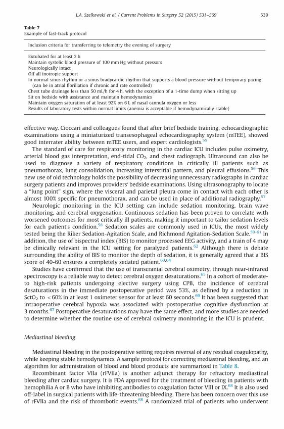
Table 7Example of fast-track protocol
Inclusion criteria for transferring to telemetry the evening of surgery
Extubated for at least 2 hMaintain systolic blood pressure of 100 mm Hg without pressorsNeurologically intactOff all inotropic supportIn normal sinus rhythm or a sinus bradycardic rhythm that supports a blood pressure without temporary pacing(can be in atrial fibrillation if chronic and rate controlled)
Chest tube drainage less than 50 mL/h for 4 h, with the exception of a 1-time dump when sitting upSit on bedside with assistance and maintain hemodynamicsMaintain oxygen saturation of at least 92% on 6 L of nasal cannula oxygen or lessResults of laboratory tests within normal limits (anemia is acceptable if hemodynamically stable)
L.A. Szelkowski et al. / Current Problems in Surgery 52 (2015) 531–569 539
effective way. Cioccari and colleagues found that after brief bedside training, echocardiographicexaminations using a miniaturized transesophageal echocardiography system (mTEE), showedgood interrater ability between mTEE users, and expert cardiologists.55
The standard of care for respiratory monitoring in the cardiac ICU includes pulse oximetry,arterial blood gas interpretation, end-tidal CO2, and chest radiograph. Ultrasound can also beused to diagnose a variety of respiratory conditions in critically ill patients such aspneumothorax, lung consolidation, increasing interstitial pattern, and pleural effusions.56 Thisnew use of old technology holds the possibility of decreasing unnecessary radiographs in cardiacsurgery patients and improves providers' bedside examinations. Using ultrasonography to locatea “lung point” sign, where the visceral and parietal pleura come in contact with each other isalmost 100% specific for pneumothorax, and can be used in place of additional radiography.57
Neurologic monitoring in the ICU setting can include sedation monitoring, brain wavemonitoring, and cerebral oxygenation. Continuous sedation has been proven to correlate withworsened outcomes for most critically ill patients, making it important to tailor sedation levelsfor each patient's condition.58 Sedation scales are commonly used in ICUs, the most widelytested being the Riker Sedation-Agitation Scale, and Richmond Agitation-Sedation Scale.59-61 Inaddition, the use of bispectral index (BIS) to monitor processed EEG activity, and a train of 4 maybe clinically relevant in the ICU setting for paralyzed patients.62 Although there is debatesurrounding the ability of BIS to monitor the depth of sedation, it is generally agreed that a BISscore of 40-60 ensures a completely sedated patient.63,64
Studies have confirmed that the use of transcranial cerebral oximetry, through near-infraredspectroscopy is a reliable way to detect cerebral oxygen desaturations.65 In a cohort of moderate-to high-risk patients undergoing elective surgery using CPB, the incidence of cerebraldesaturations in the immediate postoperative period was 53%, as defined by a reduction inSctO2 to o60% in at least 1 oximeter sensor for at least 60 seconds.66 It has been suggested thatintraoperative cerebral hypoxia was associated with postoperative cognitive dysfunction at3 months.67 Postoperative desaturations may have the same effect, and more studies are neededto determine whether the routine use of cerebral oximetry monitoring in the ICU is prudent.
Mediastinal bleeding
Mediastinal bleeding in the postoperative setting requires reversal of any residual coagulopathy,while keeping stable hemodynamics. A sample protocol for correcting mediastinal bleeding, and analgorithm for administration of blood and blood products are summarized in Table 8.
Recombinant factor VIIa (rFVIIa) is another adjunct therapy for refractory mediastinalbleeding after cardiac surgery. It is FDA approved for the treatment of bleeding in patients withhemophilia A or B who have inhibiting antibodies to coagulation factor VIII or IX.68 It is also usedoff-label in surgical patients with life-threatening bleeding. There has been concern over this useof rFVIIa and the risk of thrombotic events.68 A randomized trial of patients who underwent

Table 8Mediastinal bleeding algorithm
Corrective action Rationale
Keep MAP at 65 mm Hg Hypertension will exacerbate surgical bleedingControl shivering with meperidine or paralyticand keep patient sedated
Decrease metabolic demand when HCT may already be lowand decrease blood pressure
Check laboratory data, including PT, PTT,fibrinogen, CBC, functional platelet count
Diagnose and correct coagulopathy
Evaluate chest x-ray Look for widened mediastinum or pleural effusionsMonitor hemodynamics including CVP, PAdiastolic, SVO2, and cardiac output
May help diagnose impending cardiac tamponade
Monitor urine output May help diagnose impending cardiac tamponadeKeep patient warm (371C) Hypothermia impairs clotting and platelet function
CBC, complete blood cell count; CVP, central venous pressure; HCT, hematocrit; INR, international normalized ratio; MAP,mean arterial pressure; PA, pulmonary artery; PT, prothrombin time; PTT, partial thromboplastin time; SVO2, mixedvenous oxygen saturation.
L.A. Szelkowski et al. / Current Problems in Surgery 52 (2015) 531–569540
cardiac surgery, and were bleeding, demonstrated that rFVIIa provides benefit in the treatmentof post–cardiac surgery bleeding, but that an increased number of adverse thrombotic events,including stroke, occured.69 A meta-analysis of 35 randomized clinical trials found that the useof rFVIIa in an off-label setting significantly increased the risk of arterial, but not venous,thrombotic events, especially in elderly patients.68 These studies serve as a caution, to reserverFVIIa for life-threatening bleeding, and not incorporate it into daily practice.
Mediastinal re-exploration in the ICU
If the corrective efforts do not produce a cessation in mediastinal bleeding, re-exploration atthe bedside or the operating room is warranted. Re-exploration for bleeding increases operativemortality and morbidity, often owing to the delay in getting the patient back to the operatingroom, necessitating open chest resuscitation in the ICU.70,71 Re-exploration in the ICU is associatedwith a high survival rate compared with emergency thoracotomy for lethal arrhythmia, or MI.72
There is evidence that early re-exploration for bleeding may reduce blood transfusions, the risk ofrespiratory insufficiency, and the rate of infection for undrained hematoma.73-75
Mechanical circulatory support
Outside the operating room, many centers are developing protocols to provide MCS devicesto the sickest patients. The intra-aortic balloon pump (IABP) continues to be a mainstay oftherapy. Temporary and durable MCS devices require additional team members to support thegrowing number of patients living with this technology. The fifth Interagency Registry forMechanically Assisted Circulatory Support annual report gives data on almost 7000 patientsliving with durable MCS devices in the United States.76
This number, as well as the number of centers implanting devices, continues to grow.Outcomes for survival on MCS devices continue to improve. Continuous flow pumps in bridge-to-transplant and destination therapy patients have an overall expected survival rate of at least80% at 1 year, and almost 70% at 2 years.77
Intra-aortic balloon pump
IABP is the most commonly used mechanical support device in cardiac patients. It is used in70,000 patients annually in the United States, and specifically in 5%-10% of cardiac surgerypatients.78 Complications of insertion range from the minor (eg, thrombocytopenia) to the moresevere (eg, limb ischemia). Common major complications are listed in Table 9.
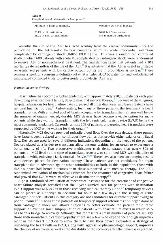
Table 9Complications of intra-aortic balloon pump78
All cause in-hospital mortality Mortality with IABP in place
20.1% In US institutions 10.8% In US institutions28.7% In non-US institutions 18% In non-US institutions
L.A. Szelkowski et al. / Current Problems in Surgery 52 (2015) 531–569 541
Recently, the use of the IABP has faced scrutiny from the cardiac community since thepublication of the Intra-aortic balloon counterpulsation in acute myocardial infarctioncomplicated by cardiogenic shock (IABP-SHOCK II) trial. This was a randomized controlledstudy in which 600 patients with acute MI, complicated by cardiogenic shock, were randomizedto receive IABP or nonmechanical treatment. The trial demonstrated that patients had a 30%mortality rate regardless of the use of the IABP.79 It is intuitive that the IABP is useful in unstablerevascularized patients with low cardiac output, but its use in prophylaxis is unclear.80 Thereremains a need for a consensus definition of what a high-risk CABG patient is, and well-designedrandomized controlled trials to better guide prophylactic IABP use.
Ventricular assist devices
Heart failure has become a global epidemic, with approximately 250,000 patients each yeardeveloping advanced heart failure, despite maximal medical therapy.81 Because of these figures,hospital admissions for heart failure have surpassed all other diagnoses, and have created a hugenational financial burden.82 Unfortunately, for many of these patients, the treatment is hearttransplantation. With a limited pool of hearts acceptable for transplant that remains well belowthe number of organs needed, durable MCS devices have become a viable option for manypatients while they wait for transplant, with the left ventricular assist device (LVAD) being themost commonly implanted. Currently, almost 30% of patients undergoing heart transplant weresupported by MCS while waiting for their organ.83
Historically, MCS devices provided pulsatile blood flow. Over the past decade, these pumpshave, largely, been replaced with continuous flow pumps that provide either axial or centrifugalflow. Devices are used for several different indications depending on the needs of the patient.Devices placed as a bridge-to-transplant allow patients waiting for an organ to experience abetter quality of life. Two prospective multicenter trials demonstrated that nearly 80% ofpatients on MCS lived to the time of transplant, recovery, or continued MCS with eligibility oftransplant, while enjoying a fairly normal lifestyle.84,85 There have also been encouraging resultswith devices placed for destination therapy. These patients are not candidates for organtransplant due to advanced age or other comorbidities or both. In this population, those withLVAD support had better outcomes than those supported with medical therapy. The 2001randomized evaluation of mechanical assistance for the treatment of congestive heart failuretrial proved that LVADs were as effective as destination therapy.86
A post–randomized evaluation of mechanical assistance for the treatment of congestiveheart failure analysis revealed that the 1-year survival rate for patients with destinationLVAD support was 61% vs 25% in those receiving medical therapy alone.87 Temporary devicescan be placed as a “bridge to decision” for hours to a few days in the setting of acutecardiogenic shock. Patients in this category are not candidates for durable support owing topoor outcomes.81 Placing these patients on temporary support attenuates end-organ damagefrom cardiogenic shock and allows clinicians to better evaluate the patient for durablesupport. An exciting small subpopulation of patients with heart failure exists in which MCShas been a bridge to recovery. Although this represents a small number of patients, usuallythose with nonischemic cardiomyopathy, there are a few who experience enough improve-ment in their heart function to allow them to have their device explanted. A strategy ofunloading the heart with an LVAD, along with aggressive pharmacologic support, improvesthe chances of recovery, as well as the durability of the recovery after the device is explanted.

L.A. Szelkowski et al. / Current Problems in Surgery 52 (2015) 531–569542
Studies have shown that the best results are achieved in younger patients with a shorthistory of heart failure.88 Patients continue to have a better quality of life, when comparedwith transplant patients, 9 years after explantation, which shows that recovery is also longlasting.89
The ICU note and the electronic medical record
A system-by-system review of a patient is essential in the ICU because overlookedinformation can quickly become detrimental to these fragile patients. Highlighting eachsubsystem, and addressing any complications related to each is the most efficient way tohandle the note on an ICU patient. It has been the hope of the government that theimplementation of electronic medical records (EMRs) would be a way to further aidpractitioners in keeping track of the large amounts of information necessary to adequatelycare for ICU patients. The US Department of Health and Human Services has stated that EMRsare an integral component of any future health care delivery model, and that they believethat their adoption will be a solution to system safety failures and medical errors.90,91 Thereis still conflicting information regarding whether this statement is true. In the ICU setting,the adoption of an EMR has been thought to have created an information overload forclinicians, because the large amount of available data must then be processed, and therelevant information must be separated from the large amounts of extraneous data thataccompany it.92 This has led to an increased amount of research on different interfacesdesigned to aid practitioners in quickly and accurately processing large amounts ofinformation.
Despite the government's interest in promoting EMRs, there have been several barriers to theacceptance of this new tool. The perception is that the adoption of an EMR requires a largelearning curve and takes the clinician away from the bedside, and puts him in front of acomputer.93 In addition, because reasonable skill with typing and working with a computer areneeded for success, these are also factors that must be taken into consideration. A review of22 studies concerning barriers to physician acceptance of an EMR showed that cost, technicalreasons, and time were the most commonly identified barriers.94 EMRs are expensive to start upand maintain, and this can be a barrier in smaller institutions, or private practices. Physiciansfeel that EMRs take away from a patient visit because of the need to enter data into a computer.When this is done at the bedside, or in office settings, physicians feel that time spent enteringdata prolongs the visit, and leads to less eye contact with the patient. Although an EMR issupposed to cut down on errors, a study in the aforementioned review suggested that typos willbecome the new medical errors.95 Although financial constraints and lack of computerknowledge are tangible, the ideas that many physicians have about EMRs, such as fear of recordloss, and the disbelief that an EMR can improve patient care or clinical outcomes are often notbacked up with statistical data.94
Cardiac surgery workforce and critical care
Cardiac surgery, similar to other branches of medicine, is facing a physician scarcity crisis.96
The crisis is unique because of the extensive length of time it takes to train cardiac surgeons, theadvanced age of much of the cardiac surgery workforce, and relative recent lack of popularity ofthe specialty.97,98 This shortfall has already been felt in ICUs where a multitude of clinicalproviders care for postoperative cardiac surgery patients (Table 10).
Often, care is provided by a combination of providers, raising important questions abouttraining and competency.99 Surgical patients now are older and have more comorbidities thanprior generations and may need prolonged critical care.100 Innovative technology including MCSand novel surgical techniques add a further complexity to postoperative care. The medicalliterature is mixed on the benefit of intensivists with a single-center study citing decreased

Table 10Providers of postoperative care
Providers providing postoperative care to cardiac surgery patients
Operative and nonoperative cardiac surgeonAnesthesia, surgical, or medically trained intensivistsPhysician assistants or acute care nurse practitionersCardiac surgery, general surgery, medical critical care, or anesthesia fellowsAnesthesia, surgical, or emergency medicine residents
L.A. Szelkowski et al. / Current Problems in Surgery 52 (2015) 531–569 543
blood product usage, decreased ICU complications, and decreased total hospital length of staywhen 24-hour intensivist coverage is employed, but no change in 30-day mortality.101 Anotherstudy showed that replacing resident coverage with advanced practice providers did notsignificantly alter mortality in a cardiac surgery program.102 The combination of providers whoprovide care for cardiac surgery patients has changed in the last decade, but the roles ofproviders are being more carefully scrutinized along with their competency.103
Cardiac system
Hemodynamics
The stress of cardiac surgery, as well as the inflammatory process associated with CPB, canhave profound effects on the hemodynamics of a patient. Low cardiac output syndrome isdefined as low cardiac output with evidence of end-organ dysfunction, and it is known toincrease mortality and morbidity following CABG surgery.104,105 Initial goals of treatment shouldfocus on maintaining a cardiac index of at least 2.0 L/min/m2 or a mixed venous oxygensaturation of at least 60%, a PCWP or pulmonary artery diastolic pressure less than 20 mm Hg,and a heart rate less than 100 beats per minute through the correction of abnormalhemodynamics and laboratory values (Table 11).106
Vasoactive medications
An analysis of the literature on the use of inotropic and vasopressor support in the earlypostoperative period after cardiac surgery shows a lack of randomized prospective data to guideclinical practice.104 In addition, there is no superior pattern of vasoactive drug use whencomparing outcomes data.104 Choices should be tailored to a patient's hemodynamics (Table 12).
Table 11Approach to hemodynamics
Cause of hemodynamic abnormality Corrective action
Bradycardia Increase heart rate with temporary pacing or medical therapyLow CVP, or PA diastolic, and lowstroke volume
Replace volume
Hypothermia Warm the patientElevated SVR Replace volume or reduce afterload with medical therapyHypotension Replace volume, medical therapy, or titrate vasopressorsTachyarrhythmias Cardioversion, defibrillation, or medical therapyPump failure Review surgical procedure for possible causes, medical therapy,
and IABP or mechanical circulatory supportPericardial tamponade Reoperation for evacuation of tamponade, or pericardiocentesisAcidemia Determine and correct underlying cause, and adjust ventilator
CVP, central venous pressure; PA, pulmonary artery; SVR, systemic vascular resistance.
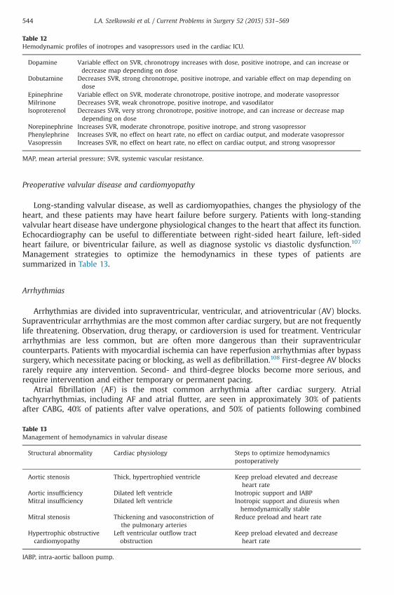
Table 12Hemodynamic profiles of inotropes and vasopressors used in the cardiac ICU.
Dopamine Variable effect on SVR, chronotropy increases with dose, positive inotrope, and can increase ordecrease map depending on dose
Dobutamine Decreases SVR, strong chronotrope, positive inotrope, and variable effect on map depending ondose
Epinephrine Variable effect on SVR, moderate chronotrope, positive inotrope, and moderate vasopressorMilrinone Decreases SVR, weak chronotrope, positive inotrope, and vasodilatorIsoproterenol Decreases SVR, very strong chronotrope, positive inotrope, and can increase or decrease map
depending on doseNorepinephrine Increases SVR, moderate chronotrope, positive inotrope, and strong vasopressorPhenylephrine Increases SVR, no effect on heart rate, no effect on cardiac output, and moderate vasopressorVasopressin Increases SVR, no effect on heart rate, no effect on cardiac output, and strong vasopressor
MAP, mean arterial pressure; SVR, systemic vascular resistance.
L.A. Szelkowski et al. / Current Problems in Surgery 52 (2015) 531–569544
Preoperative valvular disease and cardiomyopathy
Long-standing valvular disease, as well as cardiomyopathies, changes the physiology of theheart, and these patients may have heart failure before surgery. Patients with long-standingvalvular heart disease have undergone physiological changes to the heart that affect its function.Echocardiography can be useful to differentiate between right-sided heart failure, left-sidedheart failure, or biventricular failure, as well as diagnose systolic vs diastolic dysfunction.107
Management strategies to optimize the hemodynamics in these types of patients aresummarized in Table 13.
Arrhythmias
Arrhythmias are divided into supraventricular, ventricular, and atrioventricular (AV) blocks.Supraventricular arrhythmias are the most common after cardiac surgery, but are not frequentlylife threatening. Observation, drug therapy, or cardioversion is used for treatment. Ventriculararrhythmias are less common, but are often more dangerous than their supraventricularcounterparts. Patients with myocardial ischemia can have reperfusion arrhythmias after bypasssurgery, which necessitate pacing or blocking, as well as defibrillation.108 First-degree AV blocksrarely require any intervention. Second- and third-degree blocks become more serious, andrequire intervention and either temporary or permanent pacing.
Atrial fibrillation (AF) is the most common arrhythmia after cardiac surgery. Atrialtachyarrhythmias, including AF and atrial flutter, are seen in approximately 30% of patientsafter CABG, 40% of patients after valve operations, and 50% of patients following combined
Table 13Management of hemodynamics in valvular disease
Structural abnormality Cardiac physiology Steps to optimize hemodynamicspostoperatively
Aortic stenosis Thick, hypertrophied ventricle Keep preload elevated and decreaseheart rate
Aortic insufficiency Dilated left ventricle Inotropic support and IABPMitral insufficiency Dilated left ventricle Inotropic support and diuresis when
hemodynamically stableMitral stenosis Thickening and vasoconstriction of
the pulmonary arteriesReduce preload and heart rate
Hypertrophic obstructivecardiomyopathy
Left ventricular outflow tractobstruction
Keep preload elevated and decreaseheart rate
IABP, intra-aortic balloon pump.

L.A. Szelkowski et al. / Current Problems in Surgery 52 (2015) 531–569 545
CABG-valve repair or replacement surgery.109 There are several factors that contribute to theoccurrence of atrial arrhythmias including trauma, stretch, or ischemia, of the atrium, epicardialinflammation, hypoxia, acidosis, electrolyte disturbances, and electrophysiological changes thataccompany activation of the sympathetic nervous system.109 The incidence of AF also increaseswith patients' age, to approximately 5% of people older than 65 years, and 10% of people 80 yearsor older.110 It most often occurs in the second to fourth postoperative days, and has beenassociated with hypotension, heart failure, palpitations, and fatigue.111 The decision as towhether to control a patient's rhythm or rate is often determined by whether the AF is newonset or permanent. Rhythm management and use of medical therapy to encourage conversionto sinus rhythm should be attempted in new-onset AF to avoid having to anticoagulate thepatient. The literature does not provide the best way to accomplish this in the postoperativepatient. Permanent AF has been well studied, and rate control in these patients is the first-linetreatment, although there is debate as to the best therapy to accomplish this.112,113
Depending on the institution, first-line drugs for prevention or treatment of postoperative AFcan include calcium channel blockers, β-blockers, or antiarrhythmics such as amiodarone orsololol. β-Blockers are the most extensively studied drugs in the treatment and prevention of AF,but their efficacy in the prevention of AF is only moderate in cardiac surgery patients.114 Inaddition, postoperative cardiac surgery patients may be hypotensive and bradycardic, contra-indicating their use. A small prospective randomized trial demonstrated that resumingβ-blockers in the postoperative period, in patients undergoing CABG and who had been takingthem preoperatively, decreased the incidence and severity of AF.115 This supports the theory thatpatients who stop taking β-blockers before surgery will have a rebound effect of heart rate.Clinical trials have not proven that prevention of atrial tachyarrhythmias with β-blockersreduces hospital length of stay or use of resources.114,116 Despite the lack of definitive studies, thecurrent American Heart Association (AHA)-ACC-European Society of Cardiology guidelines give βblocker therapy a class IA recommendation in the prevention of postoperative AF.117 Sotalol, aclass III antiarrhythmic, is an alternative drug for treatment and prevention of AF. A meta-analysis by Kerin and Jacob111 found sotalol to be more effective in preventing postoperative AF,than either placebo or β-blockers. Its use is limited by hypotension associated with bradycardia.It is a short-acting drug, so time to load the patient is less than other alternatives such asamiodarone.111 The AHA-ACC-European Society of Cardiology guidelines state that sotalol hasnot been proven effective in the conversion of new-onset or persistent AF, but that it can aid inrate control.117 Its use is also limited by its ability to prolong the QT interval on electrocardio-gram, causing ventricular tachycardia and torsades de pointes.117
Amiodarone is a class III antiarrhythmic used in the treatment and prevention of AF. It caninduce bradycardia, AV block, and prolong the QT interval, and carries many other extracardiacside effects.117 A randomized, double-blind, placebo-controlled trial found amiodarone to be aseffective as sotalol in converting patients into sinus rhythm.118 The randomized controlled trial ofrevascularization prophylactic oral amiodarone for the prevention of arrhythmias that beginearly after revascularization, valve replacement, or repair found that a 13-day perioperativecourse of oral amiodarone was associated with reducing the postoperative incidence of atrialtacchyarrhythmias by one half.119 This finding applied to patients both older than 65 years andyounger than 65 years, in those undergoing CABG, valve repair or replacement with or withoutCABG, and those receiving or not receiving β-blockers.119 Despite these studies proving the abilityof amiodarone to treat AF, its side-effect profile causes many institutions to use it as a last resort.
Calcium channel blockers, such as diltiazem, are a reasonable choice for the rate control of AF,especially in patients with left ventricular dysfunction, as they have less of a negative inotropiceffect than β-blockers. There is no evidence to support the antiarrhythmic effects of calciumchannel blockers; however, they will aid in rate control.117 There is a study that showed thatdiltiazem decreased the amount of AF episodes in a 3-month period by 50%.120
Ventricular arrhythmias after cardiac surgery are worrisome, and require promptintervention. Coronary artery disease, structural heart disease, and left ventricular dysfunctionwere found to predispose patients to malignant ventricular arrhythmias.121 In 2012, the largestanalysis to date of the incidence, predictors, and outcomes of postoperative ventricular
L.A. Szelkowski et al. / Current Problems in Surgery 52 (2015) 531–569546
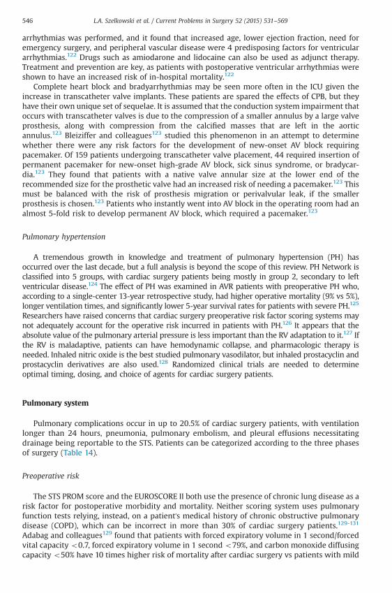
arrhythmias was performed, and it found that increased age, lower ejection fraction, need foremergency surgery, and peripheral vascular disease were 4 predisposing factors for ventriculararrhythmias.122 Drugs such as amiodarone and lidocaine can also be used as adjunct therapy.Treatment and prevention are key, as patients with postoperative ventricular arrhythmias wereshown to have an increased risk of in-hospital mortality.122
Complete heart block and bradyarrhythmias may be seen more often in the ICU given theincrease in transcatheter valve implants. These patients are spared the effects of CPB, but theyhave their own unique set of sequelae. It is assumed that the conduction system impairment thatoccurs with transcatheter valves is due to the compression of a smaller annulus by a large valveprosthesis, along with compression from the calcified masses that are left in the aorticannulus.123 Bleiziffer and colleagues123 studied this phenomenon in an attempt to determinewhether there were any risk factors for the development of new-onset AV block requiringpacemaker. Of 159 patients undergoing transcatheter valve placement, 44 required insertion ofpermanent pacemaker for new-onset high-grade AV block, sick sinus syndrome, or bradycar-dia.123 They found that patients with a native valve annular size at the lower end of therecommended size for the prosthetic valve had an increased risk of needing a pacemaker.123 Thismust be balanced with the risk of prosthesis migration or perivalvular leak, if the smallerprosthesis is chosen.123 Patients who instantly went into AV block in the operating room had analmost 5-fold risk to develop permanent AV block, which required a pacemaker.123
Pulmonary hypertension
A tremendous growth in knowledge and treatment of pulmonary hypertension (PH) hasoccurred over the last decade, but a full analysis is beyond the scope of this review. PH Network isclassified into 5 groups, with cardiac surgery patients being mostly in group 2, secondary to leftventricular disease.124 The effect of PH was examined in AVR patients with preoperative PH who,according to a single-center 13-year retrospective study, had higher operative mortality (9% vs 5%),longer ventilation times, and significantly lower 5-year survival rates for patients with severe PH.125
Researchers have raised concerns that cardiac surgery preoperative risk factor scoring systems maynot adequately account for the operative risk incurred in patients with PH.126 It appears that theabsolute value of the pulmonary arterial pressure is less important than the RV adaptation to it.127 Ifthe RV is maladaptive, patients can have hemodynamic collapse, and pharmacologic therapy isneeded. Inhaled nitric oxide is the best studied pulmonary vasodilator, but inhaled prostacyclin andprostacyclin derivatives are also used.128 Randomized clinical trials are needed to determineoptimal timing, dosing, and choice of agents for cardiac surgery patients.
Pulmonary system
Pulmonary complications occur in up to 20.5% of cardiac surgery patients, with ventilationlonger than 24 hours, pneumonia, pulmonary embolism, and pleural effusions necessitatingdrainage being reportable to the STS. Patients can be categorized according to the three phasesof surgery (Table 14).
Preoperative risk
The STS PROM score and the EUROSCORE II both use the presence of chronic lung disease as arisk factor for postoperative morbidity and mortality. Neither scoring system uses pulmonaryfunction tests relying, instead, on a patient's medical history of chronic obstructive pulmonarydisease (COPD), which can be incorrect in more than 30% of cardiac surgery patients.129-131
Adabag and colleagues129 found that patients with forced expiratory volume in 1 second/forcedvital capacity o0.7, forced expiratory volume in 1 second o79%, and carbon monoxide diffusingcapacity o50% have 10 times higher risk of mortality after cardiac surgery vs patients with mild

Table 14Pulmonary considerations
Preoperative (riskassessment)
Intraoperative (injury may occur,creating a complication)
Postoperative (complications arerecognized and treated)
Identify patients at risk Anesthetic effects AtelectasisDocument preoperativelung disease
Surgical technique Pleural effusionsEffect of surgically induced hypothermia PneumoniaEffect of cardiopulmonary bypass Pulmonary embolism
ARDSVentilator weaning
L.A. Szelkowski et al. / Current Problems in Surgery 52 (2015) 531–569 547
pulmonary function tests abnormalities. A randomized controlled trial comparing CABGoutcomes in patients with COPD using CPB vs off-pump CABG showed that patients had similar30-day and 1-year mortality rates. After propensity matching, the study found that patients withCOPD required more reintubations and stayed on the ventilator longer than 48 hours more oftenthan patients without COPD.132
Intraoperative injury
During anesthesia, functional residual capacity decreases by as much as 20% in healthyvolunteers, and patients with lung disease have more risk of developing pulmonary morbiditiespostoperatively.133 The choice of surgical site can affect postoperative respiratory functionbecause postoperative pain can diminish pulmonary inspiratory effort. Median sternotomieschange pulmonary mechanics, which can decrease patients' vital capacity for up to 4 monthspostoperatively.134,135 Anterior lateral thoracotomies are reportedly more painful than mediansternotomies and require more postoperative analgesia, but neither site appears to affect long-term pulmonary outcomes for CABG patients.136
The incidence of phrenic nerve injury is unclear, with studies citing between 10% and 73%,likely owing to the sensitivity of diagnostic testing. Many patients fully recover the function ofthe nerve with time.137 The injury appears to be caused by hypothermia during CPB, as the nervecourses over the pericardium and is subjected to cold cardioplegia and ice. Postoperatively, thediagnosis of phrenic nerve injury can be made using bedside ultrasound, fluoroscopy, orelectrophysiology.138 Left lower lobe atelectasis, which is common after cardiac surgery, may besecondary to phrenic nerve injury, inadequate pulmonary clearance, lung collapse, lack of use ofpositive end-expiratory pressure during surgery, pulmonary endothelial damage due tocardioplegia solution, and longer duration of CPB.139
Inflammation from CPB can lead to postoperative pulmonary morbidity. The ischemic lunghypothesis postulates that the lungs are not part of the systemic circulation during CPB, and thatblood flow in the lungs is static as bronchial circulation only accounts for a maximum of 5% ofblood flow to the lungs. As a result, an increase in pulmonary lactate occurs, signifying theischemia.140 In the SIRS, monocytes, macrophages, lymphocytes, and endothelial cells areactivated. The lungs withstand the worst of the inflammatory cascade set off by CPB, as they arefilters for the body's cardiac output.140
Strategies to limit lung injury during CPB are summarized in Table 15. The mechanism oflung injury remains an active area of research.141 Before 2012, a significant practice differenceexisted between cardiac surgery centers owing to a lack of literature about the use ofprophylactic steroids to blunt the inflammatory cascade associated with CPB.142 This ispartially due to the dexamethasone for cardiac surgery (DECS) trial, which was a randomizedcontrolled study using a single dose of prophylactic dexamethasone (1 mg/kg), which did notshow statistically significant benefit in the primary end points of death, MI, or respiratoryfailure within 30 days.143 Despite these findings, there were fewer patients who requiredpostoperative ventilation greater than 24 hours in the steroid group vs the placebo group(4.9% vs 3.4%).

Table 15Strategies to limit lung injury during CPB
Intervention Mechanism of action
Off-pump surgery Reduced cytokine and SIRS responseDrugs (steroids) Reduced proinflammatory cytokine releaseBiocompatible circuits Mimics endothelial surface and reduces complement activation
and inflammatory responseLeukocyte filters Preferentially removes activated leukocytes, and attenuates
ischemia and reperfusion injuryUltrafiltration Removes destructive and inflammatory substances, and reduces SIRS
responseProtective ventilation strategies Prevents atelectasis and pulmonary ischemiaPulmonary perfusion techniques(ie, Drew-Anderson technique)
Continuously perfuses the lungs
Meticulous myocardial protection Avoid use of oxygenator, reduced proinflammatory cytokines, andlimit ischemia-reperfusion injury to lungs
L.A. Szelkowski et al. / Current Problems in Surgery 52 (2015) 531–569548
Complications in the postoperative period
The most common postoperative complication is atelectasis, occurring in 54%-92% ofpatients.144 Treatment includes encouraging good pulmonary hygiene, incentive spirometry,chest physiotherapy, noninvasive ventilation, and high-flow nasal cannula oxygen.144,145 Pleuraleffusions requiring drainage (independent of technique) occur in 3.4% of isolated CABG patients,4.9% of isolated AVR patients, and 6.9% of combined CABG and AVR patients.2 The risk factors foreffusions that persist longer are not clear, but have been studied. A study found a significantrelationship between pleural effusion and sex (women), preoperative diagnosis of heart failure, AF,preoperative diagnosis of peripheral vascular disease, and receiving therapy with an anticoagulantor antiarrhythmic agent.146 However, many of these relationships disappeared when amultivariate analysis was performed.146 The use of bedside point-of-care lung ultrasound bymedical providers decreases patients' radiation exposure from radiographs, reduces complicationrates from drainage procedures, and decreases the associated risk of transport.147-149
The most significant postoperative pulmonary complication is acute respiratory distresssyndrome (ARDS), owing to its mortality of up to 40%.150 The definition for ARDS was changed in2012 to the Berlin criteria, eliminating the term “acute lung injury” (ALI) and, instead, creatingcategories defining the severity of ARDS (Table 16).151 The need for the PCWP and theincorporation of positive end-expiratory pressure have been eliminated. This change reducesthe clinical subjectivity in the diagnosis of ARDS and allows accumulated clinical data since theprevious American-European Consensus Conference 1994 definition to be incorporated into thenew definition.152
An 8-year retrospective analysis of more than 6000 cardiac surgery patients found anincidence of ARDS of 0.61%, with transfusion of greater than 3 packed red blood cells, complexcardiac surgery, and previous cardiac surgery as the only risk factors for developing thedisease.150 The cornerstone of treatment for ARDS remains low tidal volume ventilation withgoal plateau pressures less than 30 mm Hg, but recent progress has been made in decreasingmortality with other interventions including the proning, or turning face down, of patients.153-155
Transfusion-related ALI occurs within 6 hours of infusion of plasma containing transfusion andcauses ALI.156 The incidence in a cardiac surgery population in a study was 2.4%, but is likelyhigher owing to underreporting.157 This complication has an associated mortality rate of 13%.157
This association is important, because the transfusion requirements after cardiac surgery trial, a500-patient randomized controlled trial showed no benefit to a liberal packed red blood celltransfusion strategy in a cardiac surgery population.158
Amiodarone toxicity is a rare cause of ARDS, but can cause pulmonary toxicity in 5%-10% ofthe general population, with a mortality rate of 10%-23%.159 The risk factors of lung toxicity havebeen reported to be elderly age, duration of treatment, and total cumulative dosing of

Table 16The Berlin definition of acute respiratory distress syndrome152
Components of thediagnosis
Needed to fit the Berlin definition
Timing Within a week of a new insult, or increased respiratory symptomatology.Chest x-ray or CT scan Must show opacities bilaterally not explained by effusions, atelectasis, or nodules.Origin of edema Edema or respiratory symptoms not fully explained by a cardiac source, or by fluid overload.
Objective assessment needed.
Oxygenation (at altitudes o1000 m)Mild 200 mm Hg o PaO2/FiO2 r 300 mm Hg with PEEP or CPAP Z 5 cm H2OModerate 100 mm Hg o PaO2/FiO2 r 200 mm Hg with PEEP Z 5 cm H2OSevere PaO2/FiO2 r 100 mm Hg with PEEP Z 5 cm H2O
CPAP, continuous positive airway pressure; FiO2, fraction of inspired oxygen; PaO2, partial pressure of arterial oxygen;PEEP, positive end-expiratory pressure.
L.A. Szelkowski et al. / Current Problems in Surgery 52 (2015) 531–569 549
amiodarone. Treatment involves supportive care with possible benefit from treatment withsteroids, as well as withdrawal of the drug itself.160
Gastrointestinal system
Routine care of the gastrointestinal (GI) system after cardiac surgery consists of an orogastrictube for medication administration and management of gastric secretions in the intubatedpatient. A literature review reveals an incidence of GI complications of less than 1%-4%.161-163
Mortality rates vary from 13.9%-63%.164-166 There has been speculation that off-pump surgeryreduces GI complications; however, several studies have shown similar rates of mesenterichypoperfusion, as well as complication and mortality rates.167-169 Vasoconstriction, decreasedperfusion from low cardiac output or hypotension, and hypoxia during CPB or in thepostoperative period predispose the bowel to dysfunction.170,171 Impaired small intestinaltransport, increased gut permeability, preoperative fasting, and use of anesthetics and opioidsare also contributors.172-174
Dysphagia
Dysphagia after cardiac surgery is often related to the use of TEE.175,176 Additionalindependent risk factors are age, diabetes, stroke, impaired myocardial function, longerintubation, tracheostomy, and duration of surgery.176 TEE is also an independent risk factorfor the development of postoperative dysphagia. A prospective randomized trial studying TEEexamination postulated that removing the TEE probe after initial examination and replacing itafter CPB would decrease postoperative dysphagia.177 The study demonstrated that the length oftime the TEE probe was in contact with the esophagus was an independent predictor ofpostoperative dysphagia, and that dysphagia could be reduced by removing the TEE probe forthe duration of the surgery.177 In this series, reinsertion of the probe was not associated with anyesophageal complications.
Upper GI bleeding
GI bleeding is one of the most common complications of the GI system after cardiacsurgery.178 Risk factors include advanced age, diabetes, renal failure, cerebrovascular disease,postoperative low cardiac output, and CPB time greater than 98 minutes.179,180 Most institutionsinclude GI prophylaxis in their postoperative regimen. Studies on the efficacy of prophylacticagents, including mucosal protectants, H2 receptor antagonists, and proton pump inhibitors(PPIs), are conflicting. PPIs have been shown to work better in high-risk patients, but they carry
L.A. Szelkowski et al. / Current Problems in Surgery 52 (2015) 531–569550
the association of possible Clostridium difficile infection and an increase in ventilator-associatedconditions (VACs). Routine prophylaxis with sucralfate during the initial intubation period is lowrisk, and may provide protection against decreased perfusion to end organs and coagulopathyduring low cardiac output states.181
Treatment of bleeding includes a nasogastric tube and administration of PPIs in intravenous(IV) bolus or continuous infusion.182 Administration of this class of medicine has been found tohave better results than H2 receptor blockers.183 Major bleeding will require transfusion ofpacked red blood cells. Anticoagulation should be held and reversed, in the setting of significantbleeding. Upper endoscopy should be used in patients not responsive to medical therapy.
Lower GI bleeding
Risk factors specific to lower GI bleeding are diverticulosis, cancer, ischemic bowel, anorectaldisorders, C. difficile, and angiodysplasia.184 Initial treatment involves diagnosing the underlyingcause, replacing lost blood, and reversing anticoagulation. Refractory bleeding should beexplored with colonoscopy.185 If colonoscopy fails to provide a source, or cannot be performed,angiography, and then radioisotope scans, should then be employed in a stepwise fashion.184
Embolization of the bleeding source, injection, or thermocoagulation during endoscopy willlikely be able to alleviate bleeding.184 Surgical consultation for colectomy is a last resort.
Ischemic bowel
Despite advances in imaging and critical care management, ischemic bowel is still associatedwith a mortality rate of 50%-100%.178 Risk factors include advanced age, prolonged time on CPB,use of IABP, high level of inotropic support, peripheral vascular disease, emergency surgery, andpostoperative renal failure.186-188 Patients with extensive bowel infarct will have severemetabolic acidosis, and this diagnosis must be excluded in those who present this way.178
Aggressive treatment with mesenteric angiogram or exploratory laparotomy should occur ifperitonitis, sepsis, or end-organ failure occur in a patient with clinical suspicion for intestinalischemia.189
Hepatic dysfunction and failure
Asymptomatic transaminitis, elevation of the total bilirubin level, and alkaline phosphataselevel are common after cardiac surgery. As many as 25% of patients will experience temporaryhyperbilirubinemia, as defined by a total bilirubin greater than 3 mg/dL, but less than 1% of thesepatients will experience significant hepatocellular dysfunction that will progress to chronichepatitis or hepatic failure.190,191 Although the mortality rate of hyperbilirubinemia is 4%, aliterature review found the mean mortality rate of actual liver failure to be 56%.178 Causes ofperioperative liver dysfunction include preoperative heart failure and resulting liver congestion;hemolysis from CPB, the use of operative field suction, or prosthetic valves; blood transfusion;hypoxia; or reperfusion injury.178
Management of patients with liver dysfunction will depend on the clinical picture. Theasymptomatic patient with mild elevation of liver function tests can be managed conservativelyby withdrawing hepatotoxic drugs such as statins and acetaminophen, and monitoringlaboratory values. Patients with elevated enzyme levels on liver function tests and whoseabdominal examination have abnormal findings should be further evaluated for biliarypathology with a right upper quadrant ultrasound. For patients with perioperative cardiogenicshock and resulting shock liver, further treatment may be required. These patients will oftenhave coagulation abnormalities that may need to be treated with the supplementation ofclotting factors, if there is significant bleeding postoperatively.
Research on patients undergoing cardiac surgery with known liver failure from cirrhosis islimited. These patients represent a population at significant risk of perioperative complications.

L.A. Szelkowski et al. / Current Problems in Surgery 52 (2015) 531–569 551
Liver failure is often staged using the Child-Pugh score. The Model for End-stage Liver Diseasescore is used to predict 3-month mortality (Table 17).
A small but interesting retrospective cohort study sought to evaluate clinical outcomes onpatients with advanced liver cirrhosis, as defined by Child-Pugh class B or C, who underwentcardiac surgery using a liver protective strategy.192 The cohort had a Model for End-stage LiverDisease score of 3-19. This group preoperatively treated malnutrition, sodium retention,ascites, and hyperammonemia, as well as managed preoperative coagulopathy with vitamin K,platelets, and fresh frozen plasma. Intraoperatively, they were treated with steroids, andprostaglandin, avoided low flow states, kept a hematocrit of 25%-30%, ensured appropriateddrainage, treated tricuspid regurgitation, and used ultrafiltration to remove excess fluid. Withthis protocol in place, the authors postulated that, although redo surgery carried a highmortality rate of 50%, cardiac surgery could safely be performed on select patients withadvanced liver failure.192
Renal system
Postoperative acute kidney injury (AKI) is a poorly understood phenomenon, but one that canhave dramatic consequences for patients. The incidence of AKI in cardiac surgery patients iswide, occurring in 3%-30% of patients, with dialysis required in 1%-5% of patients, and associatedmortalities in more than 60%.193 The incidence of AKI after CABG is less, approximately 1%-2%,with lower need for dialysis, and significantly less associated mortality.194 Independent of thetype of cardiac surgery, AKI portends increased morbidity and mortality, but treating the diseasehas been difficult due to variable definitions of AKI and different predictive scoring systems.193
In an effort to standardize the disease process the AKI Network was created to evaluate renalfunction within 48 hours of injury, and to encourage researchers and clinicians to reduce thevariability in describing renal dysfunction.195
The variability in describing renal dysfunction is almost rivaled by the number of predictivepost–cardiac surgery AKI scoring systems. The most widely validated system is from a singlehigh-volume cardiac surgery center in which more than 30,000 patients were examined.193 Theauthors used a weighted point system for the risk factors and higher scores were associated withan increased incidence of renal failure (Table 18).196
Considerable effort has gone into trying to prevent renal failure, with much attention focusedon N-acetylcysteine. N-acetylcysteine blunts the inflammatory cascade produced by hemody-namic changes incurred during cardiac surgery, but it does not reduce the incidenceof postoperative AKI.197 Fenoldapam has shown promise in reducing the incidence of
Table 17Classification schemes for liver failure192
Scoring system Components Scoring
Child-Pugh Degree of ascites, serum total bilirubin level,INR, and degree of encephalopathy
1-3 Points for each component depending onseverityClass A ¼ 5-6 pointsClass B ¼ 7-9 pointsClass C ¼ 10-15 points
MELD Serum total bilirubin level, INR, and serumcreatinine level
X Loge (bilirubin) þ 11.2 X Loge (INR) þ 9.6 X Loge(creatinine)o9 ¼ 1.9% Mortality10-19 ¼ 6% Mortality20-29 ¼ 19.6% Mortality30-39 ¼ 52.6% Mortality40þ ¼ 71.3% Mortality
INR, international normalized ratio, MELD, model for end-stage liver disease.

Table 18Acute renal failure scoring system (minimum score ¼ 0 and maximum score ¼ 17)196
Risk factor Points
Female sex 1Congestive heart failure 1Left ventricular ejection fraction o35% 1Preoperative use of IABP 2COPD 1Insulin-requiring diabetes 1Previous cardiac surgery 1Emergency surgery 2Valve surgery only (reference to CABG) 1CABG and valve surgery (reference to CABG) 2Other cardiac surgery 2Preoperative creatinine 1.2-2.1 mg/dL (reference to 1.2) 2Preoperative creatinine 42.1 mg/dL (reference to 1.2) 5
IABP, intra-aortic balloon pump; CABG, coronary artery bypass graft; COPD, chronicobstructive pulmonary disease.
L.A. Szelkowski et al. / Current Problems in Surgery 52 (2015) 531–569552
dialysis-dependent AKI in postoperative cardiac surgery patients by vasodilating the renalvasculature, but more data are required.198,199 No pharmacologic therapy has been proventhrough a well-designed randomized controlled trial to be effective in preventing postoperativeAKI.198 A beneficial nonpharmacologic measure is the postponement of cardiac surgery afteradministering contrast dye for coronary angiography (and also computed tomography scan IVcontrast agent) in patients with preoperative AKI, as this decreases the incidence ofpostoperative AKI.200 CPB has well-known effects on the kidneys including the release ofcytokines and vasoactive hormones, which may influence AKI. Coronary artery bypass patientswith preoperative AKI have a slightly better chance of avoiding dialysis if the operation ifperformed off-pump.201 The only 2 known modifiable variables that can decrease the incidenceof postoperative AKI are CPB time and the administration of a contrast agent.202
Once AKI has occurred, patients may benefit from a nephrologist's evaluation to determinethe cause of their dysfunction and the optimal treatment. If dialysis is needed, patients have asignificant increase in mortality rate.203 Patients who are hemodynamically unstable are oftentreated with continuous renal replacement therapy, but no benefit in mortality rate has beenobserved in the general critical care population, with the recognition that studies suffer fromsignificant heterogeneity.204
Neurologic system
Neurologic concerns after cardiac surgery can be divided into stroke, peripheral neuropathies,and encephalopathy. Defining the actual rate of stroke after cardiac surgery can be difficult as thereis wide variation depending on the patient's risk factors, type and length of surgery performed, andthe criteria used to diagnose the infarct. When radiographic evidence of infarct is included in thediagnosis criteria, the incidence of stroke increases by a factor of 10, with inclusion of magneticresonance imaging yielding the highest rates.205 Preoperative risk factors such as advanced age,prior stroke, hypertension, diabetes, anemia, and postoperative AF also play a role.205-207 A recentstudy reported an overall stroke rate to be 1.6% after isolated CABG.208 The STS 2013 data (Q1-Q3)breaks down the postoperative stroke rate according to procedure (Table 19).
Stroke
The exact cause of stroke after surgery is poorly understood. Microembolization of aorticplaque after cross-clamping certainly plays a role, but there are also data to suggest thathypoperfusion and the systemic inflammatory response may also be sources of neurologic
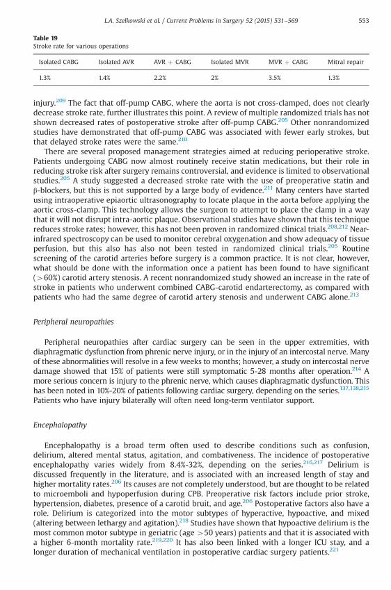
Table 19Stroke rate for various operations
Isolated CABG Isolated AVR AVR þ CABG Isolated MVR MVR þ CABG Mitral repair
1.3% 1.4% 2.2% 2% 3.5% 1.3%
L.A. Szelkowski et al. / Current Problems in Surgery 52 (2015) 531–569 553
injury.209 The fact that off-pump CABG, where the aorta is not cross-clamped, does not clearlydecrease stroke rate, further illustrates this point. A review of multiple randomized trials has notshown decreased rates of postoperative stroke after off-pump CABG.205 Other nonrandomizedstudies have demonstrated that off-pump CABG was associated with fewer early strokes, butthat delayed stroke rates were the same.210
There are several proposed management strategies aimed at reducing perioperative stroke.Patients undergoing CABG now almost routinely receive statin medications, but their role inreducing stroke risk after surgery remains controversial, and evidence is limited to observationalstudies.205 A study suggested a decreased stroke rate with the use of preoperative statin andβ-blockers, but this is not supported by a large body of evidence.211 Many centers have startedusing intraoperative epiaortic ultrasonography to locate plaque in the aorta before applying theaortic cross-clamp. This technology allows the surgeon to attempt to place the clamp in a waythat it will not disrupt intra-aortic plaque. Observational studies have shown that this techniquereduces stroke rates; however, this has not been proven in randomized clinical trials.208,212 Near-infrared spectroscopy can be used to monitor cerebral oxygenation and show adequacy of tissueperfusion, but this also has also not been tested in randomized clinical trials.205 Routinescreening of the carotid arteries before surgery is a common practice. It is not clear, however,what should be done with the information once a patient has been found to have significant(460%) carotid artery stenosis. A recent nonrandomized study showed an increase in the rate ofstroke in patients who underwent combined CABG-carotid endarterectomy, as compared withpatients who had the same degree of carotid artery stenosis and underwent CABG alone.213
Peripheral neuropathies
Peripheral neuropathies after cardiac surgery can be seen in the upper extremities, withdiaphragmatic dysfunction from phrenic nerve injury, or in the injury of an intercostal nerve. Manyof these abnormalities will resolve in a fewweeks to months; however, a study on intercostal nervedamage showed that 15% of patients were still symptomatic 5-28 months after operation.214 Amore serious concern is injury to the phrenic nerve, which causes diaphragmatic dysfunction. Thishas been noted in 10%-20% of patients following cardiac surgery, depending on the series.137,138,215
Patients who have injury bilaterally will often need long-term ventilator support.
Encephalopathy
Encephalopathy is a broad term often used to describe conditions such as confusion,delirium, altered mental status, agitation, and combativeness. The incidence of postoperativeencephalopathy varies widely from 8.4%-32%, depending on the series.216,217 Delirium isdiscussed frequently in the literature, and is associated with an increased length of stay andhigher mortality rates.206 Its causes are not completely understood, but are thought to be relatedto microemboli and hypoperfusion during CPB. Preoperative risk factors include prior stroke,hypertension, diabetes, presence of a carotid bruit, and age.206 Postoperative factors also have arole. Delirium is categorized into the motor subtypes of hyperactive, hypoactive, and mixed(altering between lethargy and agitation).218 Studies have shown that hypoactive delirium is themost common motor subtype in geriatric (age 450 years) patients and that it is associated witha higher 6-month mortality rate.219,220 It has also been linked with a longer ICU stay, and alonger duration of mechanical ventilation in postoperative cardiac surgery patients.221
L.A. Szelkowski et al. / Current Problems in Surgery 52 (2015) 531–569554
Postoperatively, the use of narcotics and benzodiazepines can have a negative effect on delirium.Patients receiving benzodiazepines before admission to a medical ICU developed delirium withinthe first 48 hours of stay.222 Haloperidol is frequently used in the ICU setting to treat delirium. Itsuse is based on limited data.223,224 No randomized placebo-controlled trial exists to support the useof haloperidol in critically ill patients or prove that it decreases the symptoms or duration ofdelirium.224 There is promising research on the drug dexmedetomidine. A randomized clinical trialcompared the use of dexmedetomidine with propofol in cardiac surgery patients and found thatpatients sedated with dexmedetomidine had a significantly lower rate (3% vs 50%) of delirium thanthose given propofol.225 The randomized controlled trial DEXmedetomidine COmpated toMorphine compared dexmedetomidine with a morphine regimen and found that it reduced theduration, but not the incidence, of delirium after cardiac surgery.226
Endocrine and metabolic system
Diabetes and glucose control
It is known that patients with diabetes are at risk for coronary artery disease; however,diabetic patients make up less than 50% of the population in most cardiac surgery centers.227
Hyperglycemia due to the stress of surgery is commonly seen in the cardiac ICU. It has beenassociated with increased perioperative morbidity and mortality.227
Initially, recommendations for glucose control were largely based on a study that advocatedstrict glucose control (90-120 mg/dL) in the ICU.228 The Portland Diabetic Project, a large,prospective, observational study, found that hyperglycemia in the first 72 hours after cardiacsurgery was an independent predictor of mortality, deep sternal wound infection, and anincreased length of stay.229 The authors also advocated a 3-day IV insulin infusion after cardiacsurgery, after finding that it eliminated the increased risks of these 3 complications in thediabetic population.229 A 2004 study of diabetic patients demonstrated that a glucose rangebetween 120 and 180 mg/dL was safer than strict control in diabetic patients undergoing cardiacsurgery, but that this more liberal range did not lead to improved outcomes.230 In 2009, thenormoglycemia in intensive care evaluation-survival using glucose algorithm regulation trialactually found that strict glucose control (81-108 mg/dL) increased 90-day mortality rate whencompared with a liberal glucose (o180 mg/dL) range.231 However, this study was mainlycomprised of nonoperative patients with sepsis or multisystem organ dysfunction and notcardiac surgery patients.
The consensus is that hyperglycemia should be avoided, and that hypoglycemia is harmful,but the optimal glucose in a postoperative cardiac surgery patient has been difficult to define. Toanswer this question, Desai and colleagues227 conducted a prospective, randomized, controlledtrial to test the theory that a liberal glucose range (121-180 mg/dL) was not inferior to a strictglucose range (90-120 mg/ddL) for outcomes in patients after first-time isolated CABG.229 Theirresearch demonstrated that the liberal glucose range led to similar outcomes when comparedwith the strict range, and that the liberal range was superior in glucose control and target rangemanagement.227 Based on a series of clinical trials, the STS currently recommends this sameliberal range for postoperative cardiac surgery patients in the ICU, and that the most efficientway to achieve a glucose target range is with an insulin infusion (level A).227,230,232
Hematologic system
Aspirin therapy
It is well known that aspirin decreases mortality and ischemic events in coronary arterybypass surgery patients. Aspirin is also shown to improve coronary artery bypass graft patency,and decrease the incidence of stroke, renal failure, and bowel infarction.233,234 Despite this
L.A. Szelkowski et al. / Current Problems in Surgery 52 (2015) 531–569 555
evidence on the benefits of aspirin therapy, conflicting opinions remain regarding discontinua-tion of aspirin before cardiac surgery. Multiple major medical societies, including the AHA, STS,ACC, and the European Association for Cardiothoracic Surgery, have not agreed on a stop time forpreoperative aspirin. A reason for the controversy is that several randomized clinical trials haveshown that aspirin given before coronary artery surgery with CPB causes increased post-operative bleeding and the need for blood transfusion.235 The STS practice guidelinesrecommend stopping aspirin therapy 3-5 days before elective CABG surgery.235 This is basedon a review of the literature which found that 5 of the 6 randomized controlled trialsdetermined that aspirin increased blood loss as measured by chest tube drainage, increasedblood transfusion rates, or increased the frequency of mediastinal re-exploration.235 Addition-ally, in randomized trials of patients undergoing CABG, preoperative aspirin administrationresulted in 200-400 mL of increased chest tube drainage, and between 0.5 and 1 unit ofincreased packed red blood cell transfusion, when compared with controls.235
Clopidogrel
The thienopyridine, clopidogrel, inhibits platelet aggregation by blocking the effects ofadenosine diphosphate at its receptor, which inhibits adenosine diphosphate–mediatedactivation of the glycoprotein IIb/IIIa receptor. Despite the development of new novel oralanticoagulants, clopidogrel maintains a role in the treatment of cardiac surgery patients. It isFDA approved to decrease thrombotic events after acute MI, cerebrovascular accident,established peripheral arterial disease, non-ST segment elevation MI, and acute coronarysyndrome in ST segment elevation MI. Most studies demonstrate that when clopidogrel is givenwithin 5 days of surgery, there is an increased risk of bleeding, an increase in blood transfusions,and an increase in the need for re-exploration for bleeding.236-239
Clopidogrel has 2 important roles in the postoperative setting. It should be given to patientswho cannot take aspirin, starting with a load of 300 mg, and then 75 mg daily to preventinadequate platelet inhibition in the first several days.240 It is also important in patientsfollowing off-pump surgery, and its early institution reduces cardiac events, and does notincrease the risk of bleeding.241
Continued role of unfractionated heparin and low-molecular-weight heparin
Unfractionated heparin (UFH) and low-molecular-weight heparin (LMWH) are bothantithrombin activators that inhibit factors Xa and IIa. They are frequently used in thepostoperative setting for venous thromboembolism (VTE) prophylaxis, and bridging therapy invalve replacement patients. Owing to its short half-life and ease of titration, UFH is still the drugof choice for patients on extracorporeal membrane oxygenation, and for bridging therapy inpatients with other MCS devices. Some would advocate that IV UFH is best monitored by anti-Xaheparin assay, rather than the traditional monitoring with activated partial thromboplastin time.Guervil and colleagues242 found that monitoring the anti-Xa heparin assay achieved therapeuticanticoagulation more rapidly and maintained the values within the desired goal range for alonger period of time.
A controversial topic is routine prophylaxis against VTE following cardiac surgery.243
A conventional thought is that VTE may be less clinically significant in the postoperative cardiacsurgery patient, as these patients have been exposed to intraoperative heparin, and that theresultant platelet dysfunction and hemodilution following surgery provide a protective effectagainst VTE.244 This theory remains unsubstantiated, as there are currently no randomizedclinical trials that specifically address the effect of thromboprophylaxis in patients after cardiacsurgery.237 The American College of Chest Physicians recommends that patients undergoingCABG receive VTE prophylaxis with LMWH, low-dose UFH, or intermittent pneumaticcompression devices.245 CABG patients who are at high risk for bleeding should receiveprophylaxis with intermittent pneumatic compression devices. A change in the American

L.A. Szelkowski et al. / Current Problems in Surgery 52 (2015) 531–569556
College of Chest Physicians 2012, 9th edition guidelines addressing perioperative managementof antithrombotic therapy, is the recommendation of subcutaneous prophylactic dose UFH, orsubcutaneous prophylactic or therapeutic dose LMWH as bridging therapy after valvereplacement, rather than the traditional intravenous infusion of UFH.246 This may be a concernas LMWH cannot be reversed in the setting of sudden bleeding.
New oral anticoagulants
There are several newer oral anticoagulants, sometimes referred to as novel oralanticoagulants, on the market. The most commonly used agents in the cardiac surgerypopulation are dabigatran, rivaroxaban, and apixaban. Much of the rationale behind thedevelopment of these agents was to replace the vitamin K antagonist warfarin, which requiresclose monitoring of levels. The direct thrombin inhibitor dabigatran has the ability to inactivateclot bound thrombin, which heparin cannot do.247 Direct thrombin inhibitors are antithrombinindependent, and their sites for binding thrombin are not masked by fibrin, as seen in heparin.This is an important feature because clot bound thrombin is a stimulant of thrombogenesis.Dabigatran is FDA approved for stroke prevention in patients with AF. It has also been used forVTE prevention after hip and knee replacement, but these other indications are not currentlyFDA approved.
Initially, there was interest in using dabigitran as a warfarin alternative in patients withmechanical heart valves, as monitoring the international normalized ratio is not required, as inwarfarin administration. The results of The Randomized, phase II study to Evaluate the sAfetyand pharmacokinetics of oraL dabIGatran etexilate in patients after heart valve relpacemeNt trialdemonstrated that the drug was not safe in this population.248 In this trial, patients withmechanical aortic or mitral valves implanted in the last 7 days were randomized to eitherdabigatran or warfarin. The trial was stopped early because of reports of excess thromboembolicand bleeding events in the dabigatran arm. This study, as well as anecdotal reports, has ledclinicians to believe that new oral anticoagulants are associated with more bleeding events. InNovember 2012, the FDA issued the statement that bleeding rates did not seem to be any higherfor dabigatran users as compared with warfarin users when either drug was used for the firsttime.249 This statement was in line with the randomized evaluation of long-term anticoagulanttherapy trial which compared dabigatran with warfarin in patients with AF, and found that the2 drugs carried a similar risk of bleeding.250
Despite the FDA statements and supporting medical literature, there is still apprehension inthe cardiac surgery community surrounding the use of novel oral anticoagulants in theperioperative period, owing to the lack of a reversal agent. A case report by Wanek andcolleagues251 claims the safe and effective use of hemodialysis for dabigatran clearance beforecardiac surgery, but this has not been studied in clinical trials. The use of new oral anticoagulantsis further limited by the fact that they are not recommended in patients with renal or hepaticfailure. An antidote for dabigatran, called aDabi-Fab, is currently in development and has beenshown to bind the drug in a way similar to the way thrombin binds to it.252 It is made ofhumanized dabigatran-specific (Fab) antibody fragments, which work in a similar fashion tothose used to treat digoxin toxicity. The antidote was shown to completely reverse theanticoagulation of dabigatran in a rat model.252 Further studies regarding the safety and efficacyof this antidote in humans are awaited.
The factor Xa inhibitors rivaroxaban and apixaban have also been designed not to requireroutine monitoring, with the aim of making them a more desirable choice than warfarin.Rivaroxaban is FDA approved for VTE prophylaxis after hip and knee surgery, risk reduction forVTE occurrence in those with current VTE, treatment of VTE, and for risk reduction of stroke innonvalvular AF. Apixaban is FDA approved for the treatment of nonvalvular AF. It has also beenused off-label for deep vein thrombosis prophylaxis after hip or knee replacement surgery, but iscurrently not FDA approved in this setting. Currently, there is no reversal agent for either ofthese drugs, but there is an antifactor Xa antidote currently in development. The agent, called

L.A. Szelkowski et al. / Current Problems in Surgery 52 (2015) 531–569 557
r-Antidote PRT064445, is a catalytically inactive, truncated form of factor Xa that does not bindto cell membranes.253 It binds to factor Xa inhibitors at subnanomolar affinity. The antidote hasbeen tested, and it was able to reverse the effects of factor Xa inhibitors in clotting tests in vitroand restore hemostasis of a liver laceration in a rat model.253
Thrombocytopenia and a summary of heparin-induced thrombocytopenia
Many patients experience thrombocytopenia in the period following CPB. A decrease in theplatelet count of 40%-50% in the first 72 hours after cardiac surgery occurs in almost allpatients.254 This is thought to be due to the exposure of blood to the CPB circuit. It is difficult todistinguish the patient with an expected reduction in platelet count from a patient who hasdeveloped heparin-induced thrombocytopenia (HIT). Studies have shown that 25%-70% ofpatients will develop antiplatelet factor 4 heparin antibodies as detected by immunoassay,and 4%-20% will test positive by platelet activation assay in the first 10 days after cardiacsurgery.255-259 Only a small number of these patients will go on to develop clinically significantHIT. Although it is not universal, it seems that a secondary reduction in the platelet count thatoccurs 5-10 days after cardiac surgery is highly predictive for HIT.260 Thrombocytopenia in the first72 hours after cardiac surgery is usually caused by factors other than HIT, even when results ofantibody tests are positive.260,261 This information may dissuade clinicians from ordering antibodytesting in the first 72 hours after surgery to avoid confusing the clinical picture with a positive test.Warkentin has also proposed a system to estimate the probability of a patient having HIT usingwhat he calls “The Four T’s.”262 The system includes using Thrombocytopenia, Timing of drop inplatelets or other signs of HIT, Thrombosis, and oTher causes of a drop in platelet count to create ascoring system that predicts the probability of a patient being diagnosed with HIT.262
Testing for HIT includes the serotonin release assay (SRA), the heparin-induced plateletaggregation test, and the solid phase enzyme-linked immunosorbent assay immunoassay. Thecarbon 14 (14C)-SRA is still the gold standard for diagnosing HIT.263 This test has been studied ina prospective randomized trial and has a sensitivity and specificity of more than 95% whenperformed by an experienced laboratory technician.264-266 The platelet aggregation assay, whichis usually faster than the SRA, has better than 90% specificity, but lacks sensitivity.267 The solidphase enzyme-linked immunosorbent assay immunoassay is not a functional assay like theother 2 tests. The test has a sensitivity of 91%-97%, meaning a negative result of the test is astrong indicator that a patient does not have HIT, but its positive predictive value varies.268 Theclinical utility of this test is yet to be determined.
Once HIT has been diagnosed, all heparin products, including heparin-coated catheters, mustbe withdrawn. Owing to the risk of thrombosis associated with the disease, patients must beanticoagulated. Initial anticoagulation should be obtained with continuous IV infusion. The IVdirect thrombin inhibitor bivalirudin is FDA approved in patients with, or at risk of, HIT who areundergoing PCI. Although it is used in other populations, it is not FDA approved in otherpopulations. The IV direct thrombin inhibitor argatroban is FDA approved for prophylaxis andtreatment of HIT in all populations, but dosing should be adjusted in patients with heart failure,multiorgan failure, anasarca, liver dysfunction, and in patients after cardiac surgery. A transitionto warfarin, with an international normalized ratio goal of 2-3 should only happen once thepatient has been stably anticoagulated with a thrombin-specific inhibitor, and has a plateletcount that is at least 150,000 per mL, at which time a minimum of 5 days of overlap should takeplace before the thrombin inhibitor is discontinued.269-271 There is no specified length of therapy.There is a high risk of thrombosis in the 30 days after diagnosis of HIT; therefore, therapy shouldbe continued past that time, and for 3-6 months if a thrombotic event has already occurred.272-274
Blood transfusion
Historically, patients with a hemoglobin level of less than 10 g/dL, or a hematocrit less than 30%were given a transfusion of packed red blood cells. This meant that many patients undergoing

L.A. Szelkowski et al. / Current Problems in Surgery 52 (2015) 531–569558
cardiac surgery were receiving blood transfusions. Of all blood transfusions in the United States,20% were associated with cardiac surgery, with 13% of those going to patients undergoingcombined CABG and valve replacement surgery.275,276 Newer research has shown that bloodtransfusion in the perioperative period actually has deleterious effects. Blood transfusion hasbeen associated with a decrease in survival rates, with some studies citing up to a 70% increasein late mortality.277,278 There are no prospective randomized trials in cardiac surgery patientson this topic; however, a randomized trial in critical care patients found that transfusion wasassociated with a higher in-hospital mortality rate, and that using a transfusion trigger ofhemoglobin o7 g/dL was associated with an increase in survival over a transfusion trigger ofhemoglobin o10 g/dL.279 A large, multicenter study sought to determine whether an effort byall participating institutions to reduce the use of blood products would affect postoperativeevents after cardiac surgery, and whether perioperative transfusion influenced risk-adjustedoutcomes.280
A blood product use protocol was put into place across all institutions for a period of 2 yearsfor nonemergency, isolated CABG patients. The protocol used an intraoperative hemoglobino6 g/dL, or a hematocrit o18%, and a postoperative hemoglobin o7 g/dL, or hematocrit o21%as transfusion triggers. The patient also needed to have 1 of 4 clinical signs of decline to receivetransfusion (Table 20).
LaPar and colleagues280 demonstrated that implementing such a protocol improvedpostoperative morbidity, mortality, and resource utilization, and decreased adverse post-operative events and health care costs. Importantly, there were also significant cost reductionsbecause of the conservation initiative. Over the 2-year study period, the total hospitalizationcosts were reduced by $49 million dollars statewide, with a median reduction of $4000 perpatient hospitalization.280
Institutions have been slow to adopt this new way of thinking. The current STS and Society ofCardiovascular Anesthesiologists clinical practice guidelines give a few recommendations:patients who have lost more than 1500 mL or more than 30% of total blood volume shouldreceive blood transfusion regardless of hemoglobin or hematocrit. Patients with chroniccardiovascular or pulmonary disease, or hemoglobin less than 7 g/dL (or o6 g/dL if on CPB)should receive transfusion. Patients with hemoglobin level greater than 10 g/dL fare worse whengiven blood, and there is no clear benefit to transfusing at a threshold of a hemoglobin level of7 g/dL vs 10 g/dL.281,282 Despite this current research, only 20% of programs have actually evenacknowledged these guidelines.283
Infectious disease
The nosocomial infection rate in cardiac surgery is unclear, as some reviews have cited ratesbetween 10% and 20%, but the STS data report from 2013 has a much lower incidence ofsepticemia, pneumonia, and mediastinits.194 Bacteremia due to central line–related bloodstream infections (CLABSI), urinary tract infections due to catheters, and C. difficile colitis are notspecifically reported in the STS database, but are an important source of morbidity and mortalityin all critically ill patients. Our review focuses on ventilator-associated events (VAEs) andmediastinitis, which are the 2 infections with the greatest morbidity.
Table 20Clinical signs favoring blood transfusion280
Intraoperative triggers Postoperative triggers
1. Low oxygen saturation (o60 mm Hg) 1. Elevated oxygen requirement2. Lactate 42.2 mmol/L 2. Systemic hypotension3. Base deficit 43 mEq/L 3. Evidence of end-organ dysfunction4. Serum bicarbonate o22 mEq/L 4. Evidence of persistent bleeding
L.A. Szelkowski et al. / Current Problems in Surgery 52 (2015) 531–569 559
Ventilator-associated events
One of the most significant complications of cardiac surgery is postoperative pneumonias,which is associated with longer length of hospitalization, and associated mortality rate between20% and 49%.284,285 The incidence of postoperative pneumonia is 2.8% in isolated CABG, 2.4% inisolated AVR, and 4.3% in combined AVR-CABG cases.2 A recent attempt has been made toidentify patients who will develop pneumonias postoperatively, with a risk scoring system, butit was not used extensively.286 Another recent change is the diagnostic classification ofventilator-associated pneumonia (VAP), which has changed to VAC, owing to concern over thesubjectivity involved in making the diagnosis.285 All pneumonias after operation are not VAEs,but our discussion focuses on this branch of pneumonias because of its high mortality, andrecent important change in definition.
A surveillance algorithm now exists, provided by the Centers for Disease Control, in whichpatients can have a VAE that could be infectious and is then known as an infectious VAC.287
Ventilator-associated conditions are associated with increased ICU length of stay, increasedlength of mechanical ventilation, but not increased ICU mortality in a study.288 Another studyretrospectively applied the VAC definition to a prospective VAP study and determined that VACsare associated with significant morbidity and mortality, but VAP had poor correlation withVAC.289 Yet, the ventilator bundle used to reduce VAPs also lead to a decrease in VACs. Althoughacronyms are confusing, it is important for cardiac surgery programs to be aware of VAE, as theseare reported publically. More importantly, the condition has adverse effects on critically illpatients. Ventilator bundles are still followed, along with ventilator-weaning protocols. Both ofthese measures are important in cardiac surgery patients to decrease time to extubation.
Mediastinitis
Deep sternal wound infection, or mediastinitis, occur in 0.2% of isolated AVR patients, 0.3% ofisolated CABG patients, and 0.3% of combined CABG-AVR procedures.2 In-hospital mortality ratesvary between 10% and 47%, and in a retrospective case–control series of 18,532 CABG patients,this infection decreased 10-year survival significantly.290 Often, bacteria do not grow, but the2 most common organisms implicated are Staphloccocus aureus and Staphylococcus epidermi-dis.290,291 Risk factors for mediastinitis include diabetes, excess blood transfusions, activesmoking, obesity (body mass index 430 kg/m2), COPD, prolonged CPB time, reoperation,prolonged intubation time, surgical re-exploration, and the use of bilateral internal mammaryarteries.194,290,292 Efforts to prevent surgical site infections have benefited from the use ofpreoperative antibiotics within 60 minutes before surgery, with the use of broad-spectrumantibiotics for patients with methicillin-resistant S. aureus.293 The prevention of methicillin-resistant S. aureus is a priority, and institution-specific protocols have been developed.Best practices have been recommended by the ACC Foundation-AHA, and include reduction ofpostoperative infections, use of perioperative antibiotics, insulin infusion to keep the glucoselevel less than 180 mg/dL, and the use of leukocyte-reduced blood when transfusion isnecessary.194
Catheter-associated or central line–associated and C. difficile infections
Hospital-acquired infections are both costly and lethal. In a 2-institution retrospective reviewof 8405 cardiac surgery patients over 7 years, C. difficile infection had an incidence of 0.79%.294 Thediagnosis of C. difficile infection was associated with a longer ICU and overall hospital stay, but nodifference in 30-day mortality rate.294 Both urinary tract infections due to catheters and CLABSIcause significant health burdens, as CLABSI alone have been implicated in costing the UnitedStates health care system anywhere between $960 million and $18.2 billion. Although associatedmortalities and morbidity exist, and their incidence is difficult to assess, the amount of hospital-acquired infections are publically reported quality indicators. This reporting serves as a stark

L.A. Szelkowski et al. / Current Problems in Surgery 52 (2015) 531–569560
reminder to cardiac surgery programs that the complications of postoperative care are publicallyreported, are costly, and, most importantly, can negatively affect their patient's outcomes.295
Nutrition
Preoperative evaluation of nutritional status has not been well defined. Historically,nutritional status has been studied using the serum albumin concentration. This may not bean entirely accurate marker, as it has a long half-life, is affected by hydration and renal function,and is not reflective of recent nutritional changes.296 By contrast, the serum prealbuminconcentration is not affected by hydration and has a short half-life. A recent study by Yu andcolleagues296 evaluated the use of the prealbumin level in patients undergoing cardiac surgeryand found that patients with a level r20 mg/dL had an increased risk of postoperative infection,and a need for longer mechanical ventilation. These results indicate that it may be prudent tomaximize a patient's nutritional support before cardiac surgery.
Critically ill patients are often in a catabolic state, and suboptimal feeding will eventually havenegative effects on a patient's condition. Historically, total parenteral nutritionwas used to providea patient with nutrition when the gut could not be used. Current research has shown that the useof total parenteral nutrition is a strong predictor of candidemia and that avoiding its use isprotective against developing this infection.297 It is commonly thought that postpyloric feeding isthe answer to this problem and that patients will be able to tolerate higher volumes of enteralfeeding, as well as have a reduced incidence of VAP from aspiration. A prospective, randomized,controlled trial found that mechanically ventilated patients receiving nasogastric feeding, who hadelevated gastric residuals, did not have a reduction in pneumonia, when early nasojejunal feedingwas used.298 Furthermore, these patients did not experience an increase in the level of energydelivery.298 Trophic or “trickle” feeding is often used in the critical care setting. This has beenstudied in a large randomized trial of medical patients with ALI, a population with manycharacteristics seen in the cardiac critical care setting. The EDEN randomized trial (initial trophicvs full enteral feeding in patients with ALI) found that in patients with ALI, some of whom werealso receiving vasopressors, initial trophic enteral feeding for as many as 6 days did not improveventilator-free days, mortality rate at 60 days, or infectious complications, but was associated witha decrease in gastric intolerance.299 The study did not show an increase mortality in either group,which points toward early feeding, either trophic or full enteral, in a critical care population.
References
1. Bridgewater B. Almanac 2012—adult cardiac surgery: the national society journals present selected research thathas driven recent advances in clinical cardiology. Heart. 2012;98(19):1412–1417.
2. Anonymous. STS executive summary. 2013.3. Sade RM. Are surgeons ethically obligated to treat Medicare patients despite substantial reductions in
reimbursement? Introduction. J Thorac Cardiovasc Surg. 2013;145(1):37.4. Epstein AM. Will pay for performance improve quality of care? The answer is in the details. N Engl J Med. 2012;
367(19):1852–1853. http://dx.doi.org/10.1056/NEJMe1212133. [Accessed 08.11.12].5. Jha AK, Joynt KE, Orav EJ, Epstein AM. The long-term effect of premier pay for performance on patient outcomes.
N Engl J Med. 2012;366(17):1606–1615.6. Ryan AM. Effects of the premier hospital quality incentive demonstration on Medicare patient mortality and cost.
Health Serv Res. 2009;44(3):821–842.7. Glickman SW, Ou F, DeLong ER, et al. Pay for performance, quality of care, and outcomes in acute myocardial
infarction. J Am Med Assoc. 2007;297(21):2373–2380.8. The Joint Commission. Core measure sets. http://www.jointcommission.org/core_measure_sets.aspx; Accessed
01.08.14.9. Boulding W, Glickman SW, Manary MP, Schulman KA, Staelin R. Relationship between patient satisfaction with
inpatient care and hospital readmission within 30 days. Am J Manag Care. 2011;17(1):41–48.10. Anonymous. Explanation of quality rating, composite score, and star ratings. http://www.sts.org/quality-research-
patient-safety/sts-public-reporting-online/explanation-quality-rating-composite-sco; Accessed 25.06.14.11. Head SJ, Davierwala PM, Serruys PW, et al. Coronary artery bypass grafting vs. percutaneous coronary intervention
for patients with three-vessel disease: final five-year follow-up of the SYNTAX trial. Eur Heart J. 2014;35(40):2821–2830.

L.A. Szelkowski et al. / Current Problems in Surgery 52 (2015) 531–569 561
12. Leon MB, Smith CR, Mack M, et al. Transcatheter aortic-valve implantation for aortic stenosis in patients whocannot undergo surgery. N Engl J Med. 2010;363(17):1597–1607.
13. Smith CR, Leon MB, Mack MJ, et al. Transcatheter versus surgical aortic-valve replacement in high-risk patients.N Engl J Med. 2011;364(23):2187–2198.
14. Massimiano PS, Yanagawa B, Henry L, Holmes SD, Pritchard G, Ad N. Minimally invasive fibrillating heart surgery:a safe and effective approach for mitral valve and surgical ablation for atrial fibrillation. Ann Thorac Surg. 2013;96(2):520–527.
15. Wendt D, Osswald BR, Kayser K, et al. Society of thoracic surgeons score is superior to the EuroSCORE determin-ing mortality in high risk patients undergoing isolated aortic valve replacement. Ann Thorac Surg. 2009;88(2):468–474.
16. Barili F, Pacini D, Capo A, et al. Does EuroSCORE II perform better than its original versions? A multicentrevalidation study. Eur Heart J. 2013;34(1):22–29.
17. Bagnall NM, Faiz O, Darzi A, Athanasiou T. What is the utility of preoperative frailty assessment for riskstratification in cardiac surgery? Interact Cardiovasc Thorac Surg. 2013;17(2):398–402.
18. Ad N, Henry L, Halpin L, et al. The use of spirometry testing prior to cardiac surgery may impact the society ofthoracic surgeons risk prediction score: a prospective study in a cohort of patients at high risk for chronic lungdisease. J Thorac Cardiovasc Surg. 2010;139(3):686–691.
19. Bojar RM. Manual of Perioperative Care in Adult Cardiac Surgery. Hoboken, NJ: John Wiley & Sons; 2011.20. Ramakrishna H, Reidy C, Riha H, et al. The year in cardiothoracic and vascular anesthesia: selected highlights from
2012. J Cardiothorac Vasc Anesth. 2013;27(1):86–91.21. Fergusson DA, Hébert PC, Mazer CD, et al. A comparison of aprotinin and lysine analogues in high-risk cardiac
surgery. N Engl J Med. 2008;358(22):2319–2331.22. Milas BL. Con: the practice of cardiac anesthesia has not significantly changed after the withdrawal of aprotinin.
J Cardiothorac Vasc Anesth. 2010;24(5):879–882.23. Mackensen GB, Swaminathan M, Mathew JP. PRO editorial: PRO: three-dimensional transesophageal echocardiog-
raphy is a major advance for intraoperative clinical management of patients undergoing cardiac surgery. AnesthAnalg. 2010;110(6):1574–1578.
24. D'Ambra MN. CON editorial: CON: three-dimensional transesophageal echocardiography is a major advance forintraoperative clinical management of patients undergoing cardiac surgery. Anesth Analg. 2010;110(6):1579–1580.
25. Punjabi PP, Taylor K. The science and practice of cardiopulmonary bypass: from cross circulation to ECMO and SIRS.Global Cardiol Sci Pract. 2014;2013(3):249–260.
26. Ben‐Abraham R, Weinbroum AA, Dekel B, Paret G. Chemokines and the inflammatory response followingcardiopulmonary bypass—a new target for therapeutic intervention?—a review. Pediatr Anesth. 2003;13(8):655–661.
27. Kiaii B, Fox S, Swinamer SA, et al. The early inflammatory response in a mini-cardiopulmonary bypass system:a prospective randomized study. Innovations (Phila). 2012;7(1):23–32.
28. Vandewiele K, Bove T, De Somer FM, et al. The effect of retrograde autologous priming volume on haemodilutionand transfusion requirements during cardiac surgery. Interact Cardiovasc Thorac Surg. 2013;16(6):778–783.
29. Sun P, Ji B, Sun Y, et al. Effects of retrograde autologous priming on blood transfusion and clinical outcomes inadults: a meta-analysis. Perfusion. 2013;28(3):238–243.
30. Sarin E, Speir A, Henry L, et al. Acute normovolemic hemodilution decreases blood product use and postoperativemortality and morbidity following cardiac surgery. J Am Coll Cardiol. 2012;59(13s1):E1843-E1843.
31. Algarni KD, Weisel RD, Caldarone CA, Maganti M, Tsang K, Yau TM. Microplegia during coronary artery bypassgrafting was associated with less low cardiac output syndrome: a propensity-matched comparison. Ann ThoracSurg. 2013;95(5):1532–1538.
32. Jacob S, Kallikourdis A, Sellke F, Dunning J. Is blood cardioplegia superior to crystalloid cardioplegia? InteractCardiovasc Thorac Surg. 2008;7(3):491–498.
33. Sa MP, Rueda FG, Ferraz PE, Chalegre ST, Vasconcelos FP, Lima RC. Is there any difference between blood andcrystalloid cardioplegia for myocardial protection during cardiac surgery? A meta-analysis of 5576 patients from36 randomized trials. Perfusion. 2012;27(6):535–546.
34. Guru V, Omura J, Alghamdi AA, Weisel R, Fremes SE. Is blood superior to crystalloid cardioplegia? A meta-analysisof randomized clinical trials. Circulation. 2006;114(suppl 1):I331–I338.
35. Govindapillai A, Hua R, Rose R, Friesen CH, O'Blenes SB. Protecting the aged heart during cardiac surgery: use of delNido cardioplegia provides superior functional recovery in isolated hearts. J Thorac Cardiovasc Surg. 2013;146(4):940–948.
36. Mehta RH, Ferguson TB, Lopes RD, et al. Saphenous vein grafts with multiple versus single distal targets in patientsundergoing coronary artery bypass surgery: one-year graft failure and five-year outcomes from the project of ex-vivo vein graft engineering via transfection (PREVENT) IV trial. Circulation. 2011;124(3):280–288.
37. Lopes RD, Hafley GE, Allen KB, et al. Endoscopic versus open vein-graft harvesting in coronary-artery bypasssurgery. N Engl J Med. 2009;361(3):235–244.
38. Williams JB, Peterson ED, Brennan JM, et al. Association between endoscopic vs open vein-graft harvesting andmortality, wound complications, and cardiovascular events in patients undergoing CABG surgery. J Am Med Assoc.2012;308(5):475–484.
39. Ad N, Henry L, Hunt S, et al. Endoscopic versus direct vision for saphenous vein graft harvesting in coronary arterybypass surgery. J Cardiovasc Surg (Torino). 2011;52(5):739–748.
40. Desai P, Kiani S, Thiruvanthan N, et al. Impact of the learning curve for endoscopic vein harvest on conduit qualityand early graft patency. Ann Thorac Surg. 2011;91(5):1385–1392.
41. Manchio JV, Gu J, Romar L, et al. Disruption of graft endothelium correlates with early failure after off-pumpcoronary artery bypass surgery. Ann Thorac Surg. 2005;79(6):1991–1998.
42. Brown EN, Kon ZN, Tran R, et al. Strategies to reduce intraluminal clot formation in endoscopically harvestedsaphenous veins. J Thorac Cardiovasc Surg. 2007;134(5):1259–1265.

L.A. Szelkowski et al. / Current Problems in Surgery 52 (2015) 531–569562
43. Engelman RM, Rousou JA, Flack III JE, et al. Fast-track recovery of the coronary bypass patient. Ann Thorac Surg.1994;58(6):1742–1746.
44. Karski JM. Practical aspects of early extubation in cardiac surgery. J Cardiothorac Vasc Anesth. 1995;9(5 suppl 1):30–33.
45. Higgins TL. Safety issues regarding early extubation after coronary artery bypass surgery. J Cardiothorac Vasc Anesth.1995;9(5 suppl 1):24–29.
46. Chong JL, Pillai R, Fisher A, Grebenik C, Sinclair M, Westaby S. Cardiac surgery: moving away from intensive care.Br Heart J. 1992;68(4):430–433.
47. Kiessling AH, Huneke P, Reyher C, Bingold T, Zierer A, Moritz A. Risk factor analysis for fast track protocol failure.J Cardiothorac Surg. 2013;8(47):47.
48. Ettema RG, Peelen LM, Schuurmans MJ, Nierich AP, Kalkman CJ, Moons KG. Prediction models for prolongedintensive care unit stay after cardiac surgery: systematic review and validation study. Circulation. 2010;122(7):682–689. [7 p following p 689].
49. Halpin LS, Gilliam P, Barnett SD. Gender differences for rapid after bypass back into telemetry tract followingcardiac surgery. J Nurs Care Qual. 2006;21(3):277–282.
50. Najafi M, Goodarzynejad H, Sheikhfathollahi M, Adibi H. Role of surgeon in length of stay in ICU after cardiacbypass surgery. J Tehran Univ Heart Cent. 2010;5(1):9–13.
51. Tuman K, McCarthy R, March R, Najafi H, Ivankovich A. Morbidity and duration of ICU stay after cardiac surgery.A model for preoperative risk assessment. Chest. 1992;102(1):36–44.
52. Jacka MJ, Cohen MM, To T, Devitt JH, Byrick R. The use of and preferences for the transesophagealechocardiogram and pulmonary artery catheter among cardiovascular anesthesiologists. Anesth Analg. 2002;94(5):1065–1071.
53. Jacka MJ, Cohen MM, To T, Devitt JH, Byrick R. The appropriateness of the pulmonary artery catheter incardiovascular surgery. Can J Anesth. 2002;49(3):276–282.
54. Schwann M, Hoeft M, Barash M, et al. Lack of effectiveness of the pulmonary artery catheter in cardiac surgery.Anesth Analg. 2011;113(5):994–1002.
55. Temporelli PL, Scapellato F, Eleuteri E, Imparato A, Giannuzzi P. Doppler echocardiography in advanced systolicheart failure: a noninvasive alternative to Swan-Ganz catheter. Circ Heart Fail. 2010;3(3):387–394.
56. Lichtenstein DA, Meziere GA. Relevance of lung ultrasound in the diagnosis of acute respiratory Failure: the BLUEprotocol. Chest. 2008;134(1):117–125.
57. Moore CL, Copel JA. Point-of-care ultrasonography. N Engl J Med. 2011;364(8):749–757.58. Kollef MH, Levy NT, Ahrens TS, Schaiff R, Prentice D, Sherman G. The use of continuous iv sedation is associated
with prolongation of mechanical ventilation. Chest. 1998;114(2):541–548.59. Sessler CN, Gosnell MS, Grap MJ, et al. The Richmond Agitation-Sedation scale: validity and reliability in adult
intensive care unit patients. Am J Respir Crit Care Med. 2002;166(10):1338–1344.60. Khan BA, Guzman O, Campbell NL, et al. Comparison and agreement between the Richmond Agitation-Sedation
scale and the Riker Sedation-Agitation scale in evaluating patients’ eligibility for delirium assessment in the ICUcorrelation of sedation scales in delirium. Chest. 2012;142(1):48–54.
61. Ely EW, Truman B, Shintani A, et al. Monitoring sedation status over time in ICU patients: reliability and validity ofthe Richmond Agitation-Sedation scale (RASS). J Am Med Assoc. 2003;289(22):2983–2991.
62. Crippen D. Role of bedside electroencephalography in the adult intensive care unit during therapeutic neuro-muscular blockade. Crit Care. 1997;1(1):15–24.
63. Avidan MS, Zhang L, Burnside BA, et al. Anesthesia awareness and the bispectral index. N Engl J Med. 2008;358(11):1097–1108.
64. Roberts DJ, Haroon B, Hall RI. Sedation for critically ill or injured adults in the intensive care unit. Drugs. 2012;72(14):1881–1916.
65. Calderon-Arnulphi M, Alaraj A, Slavin KV. Near infrared technology in neuroscience: past, present and future.Neurol Res. 2009;31(6):605–614.
66. Greenberg SB, Murphy G, Alexander J, Fasanella R, Garcia A, Vender J. Cerebral desaturation events in the intensivecare unit following cardiac surgery. J Crit Care. 2013;28(3):270–276.
67. Slater JP, Guarino T, Stack J, et al. Cerebral oxygen desaturation predicts cognitive decline and longer hospital stayafter cardiac surgery. Ann Thorac Surg. 2009;87(1):36–45.
68. Levi M, Levy JH, Andersen HF, Truloff D. Safety of recombinant activated factor VII in randomized clinical trials.N Engl J Med. 2010;363(19):1791–1800.
69. Gill R, Herbertson M, Vuylsteke A, et al. Safety and efficacy of recombinant activated factor VII: a randomizedplacebo-controlled trial in the setting of bleeding after cardiac surgery. Circulation. 2009;120(1):21–27.
70. Unsworth-White MJ, Herriot A, Valencia O, et al. Resternotomy for bleeding after cardiac operation: a marker forincreased morbidity and mortality. Ann Thorac Surg. 1995;59(3):664–667.
71. Moulton MJ, Creswell LL, Mackey ME, Cox JL, Rosenbloom M. Reexploration for bleeding is a risk factor for adverseoutcomes after cardiac operations. J Thorac Cardiovasc Surg. 1996;111(5):1037–1046.
72. Anthi A, Tzelepis GE, Alivizatos P, Michalis A, Palatianos GM, Geroulanos S. Unexpected cardiac arrest after cardiacsurgery incidence, predisposing causes, and outcome of open chest cardiopulmonary resuscitation. Chest. 1998;113(1):15–19.
73. Karthik S, Grayson AD, McCarron EE, Pullan DM, Desmond MJ. Reexploration for bleeding after coronary arterybypass surgery: risk factors, outcomes, and the effect of time delay. Ann Thorac Surg. 2004;78(2):527–534.
74. Fiser SM, Tribble CG, Kern JA, Long SM, Kaza AK, Kron IL. Cardiac reoperation in the intensive care unit. Ann ThoracSurg. 2001;71(6):1888–1893.
75. McKowen RL, Magovern GJ, Liebler GA, Park SB, Burkholder JA, Maher TD. Infectious complications and cost-effectiveness of open resuscitation in the surgical intensive care unit after cardiac surgery. Ann Thorac Surg.1985;40(4):388–392.

L.A. Szelkowski et al. / Current Problems in Surgery 52 (2015) 531–569 563
76. Kirklin JK, Naftel DC, Kormos RL, et al. Fifth INTERMACS annual report: risk factor analysis from more than 6,000mechanical circulatory support patients. J Heart Lung Transplant. 2013;32(2):141–156.
77. Kirklin JK, Naftel DC, Kormos RL, et al. The fourth INTERMACS annual report: 4,000 implants and counting. J HeartLung Transplant. 2012;31(2):117–126.
78. Cohen M, Urban P, Christenson JT, et al. Intra-aortic balloon counterpulsation in US and non-US centres: results ofthe benchmark registry. Eur Heart J. 2003;24(19):1763–1770.
79. Thiele H, Zeymer U, Neumann F, et al. Intra-aortic balloon counterpulsation in acute myocardial infarctioncomplicated by cardiogenic shock (IABP-SHOCK II): final 12 month results of a randomised, open-label trial.Lancet. 2013;382(9905):1638–1645.
80. Kyle RA, Shampo MA, Steensma DP. Sir Brian Gerald Barratt-Boyes—pioneer cardiac surgeon. Mayo Clin Proc.2012;87(9):e65.
81. Slaughter MS, Singh R. The role of ventricular assist devices in advanced heart failure. Rev Esp Cardiol. 2012;65(11):982–985.
82. RosamondW, Flegal K, Friday G, et al. Stroke Statistics Subcommittee. AHA statistical update. Circulation. 2007;115:e69–e171.
83. Lund LH, Edwards LB, Kucheryavaya AY, et al. The registry of the international society for heart and lungtransplantation: thirtieth official adult heart transplant report—2013; focus theme: age. J Heart Lung Transplant.2013;32(10):951–964.
84. Miller LW, Pagani FD, Russell SD, et al. Use of a continuous-flow device in patients awaiting heart transplantation.N Engl J Med. 2007;357(9):885–896.
85. Pagani FD, Miller LW, Russell SD, et al. Extended mechanical circulatory support with a continuous-flow rotary leftventricular assist device. J Am Coll Cardiol. 2009;54(4):312–321.
86. Rose EA, Gelijns AC, Moskowitz AJ, et al. Long-term use of a left ventricular assist device for end-stage heart failure.N Engl J Med. 2001;345(20):1435–1443.
87. Long JW, Kfoury AG, Slaughter MS, et al. Long‐Term destination therapy with the HeartMate XVE left ventricularassist device: improved outcomes since the REMATCH study. Congest Heart Fail. 2005;11(3):133–138.
88. Goldstein DJ, Maybaum S, MacGillivray TE, et al. Young patients with nonischemic cardiomyopathy havehigher likelihood of left ventricular recovery during left ventricular assist device support. J Card Fail. 2012;18(5):392–395.
89. George RS, Yacoub MH, Bowles CT, et al. Quality of life after removal of left ventricular assist device for myocardialrecovery. J Heart Lung Transplant. 2008;27(2):165–172.
90. Wu S, Chaudhry B, Wang J, et al. Systematic review: impact of health information technology on quality, efficiency,and costs of medical care. Ann Intern Med. 2006;144(10):742–752.
91. Jha AK, DesRoches CM, Campbell EG, et al. Use of electronic health records in US hospitals. N Engl J Med. 2009;360(16):1628–1638.
92. Ahmed A, Chandra S, Herasevich V, Gajic O, Pickering BW. The effect of two different electronic health record userinterfaces on intensive care provider task load, errors of cognition, and performance. Crit Care Med. 2011;39(7):1626–1634.
93. DesRoches CM, Campbell EG, Rao SR, et al. Electronic health records in ambulatory care—a national survey ofphysicians. N Engl J Med. 2008;359(1):50–60.
94. Boonstra A, Broekhuis M. Barriers to the acceptance of electronic medical records by physicians from systematicreview to taxonomy and interventions. BMC Health Serv Res. 2010;10(1):231.
95. Pizziferri L, Kittler AF, Volk LA, et al. Primary care physician time utilization before and after implementation of anelectronic health record: a time-motion study. J Biomed Inform. 2005;38(3):176–188.
96. Green LV, Savin S, Lu Y. Primary care physician shortages could be eliminated through use of teams, nonphysicians,and electronic communication. Health Aff (Millwood). 2013;32(1):11–19.
97. Grover A, Gorman K, Dall TM, et al. Shortage of cardiothoracic surgeons is likely by 2020. Circulation. 2009;120(6):488–494.
98. Vanderby SA, Carter MW, Latham T, et al. Modeling the cardiac surgery workforce in Canada. Ann Thorac Surg.2010;90(2):467–473.
99. Sherif HM, Cohn LH. Certification in cardiothoracic surgical critical care. J Thorac Cardiovasc Surg. 2014;147(5):1454–1455.
100. Bardakci H, Cheema FH, Topkara VK, et al. Discharge to home rates are significantly lower for octogenariansundergoing coronary artery bypass graft surgery. Ann Thorac Surg. 2007;83(2):483–489.
101. Kumar K, Singal R, Manji RA, et al. The benefits of 24/7 in-house intensivist coverage for prolonged-stay cardiacsurgery patients. J Thorac Cardiovasc Surg. 2014;148(1):290–297.
102. Skinner H, Skoyles J, Redfearn S, Jutley R, Mitchell I, Richens D. Advanced care nurse practitioners can safely providesole resident cover for level three patients: impact on outcomes, cost and work patterns in a cardiac surgeryprogramme. Eur J Cardiothorac Surg. 2013;43(1):19–22.
103. Markewitz A. Nurse practitioners replacing young doctors: it works, but does it make sense? Eur J CardiothoracSurg. 2013;43(1):23–24.
104. Williams JB, Hernandez AF, Li S, et al. Postoperative inotrope and vasopressor use following CABG: outcome datafrom the CAPS‐Care study. J Card Surg. 2011;26(6):572–578.
105. Maganti MD, Rao V, Borger MA, Ivanov J, David TE. Predictors of low cardiac output syndrome after isolated aorticvalve surgery. Circulation. 2005;112(suppl 9):I448–I452.
106. Griffin MJ, Hines RL. Management of perioperative ventricular dysfunction. J Cardiothorac Vasc Anesth. 2001;15(1):90–106.
107. Mebazaa A, Pitsis AA, Rudiger A, et al. Clinical review: practical recommendations on the management ofperioperative heart failure in cardiac surgery. Crit Care. 2010;14(2):201.
108. Chua J, Schwarzenberger J, Mahajan A. Optimization of pacing after cardiopulmonary bypass. J Cardiothorac VascAnesth. 2012;26(2):291–301.
L.A. Szelkowski et al. / Current Problems in Surgery 52 (2015) 531–569564
109. Mitchell LB. Incidence, timing and outcome of atrial tachyarrhythmias after cardiac surgery. Atrial Fibrillation AfterCardiac Surgery. New York, NY: Springer; 2000; p 37–50.
110. Miyasaka Y, Barnes ME, Gersh BJ, et al. Secular trends in incidence of atrial fibrillation in Olmsted county,Minnesota, 1980 to 2000, and implications on the projections for future prevalence. Circulation. 2006;114(2):119–125.
111. Kerin NZ, Jacob S. The efficacy of sotalol in preventing postoperative atrial fibrillation: a meta-analysis. Am J Med.2011;124(9):875. (e1-9).
112. Wann LS, Curtis AB, January CT, et al. 2011 ACCF/AHA/HRS focused update on the management of patientswith atrial fibrillation (updating the 2006 guideline): a report of the American college of cardiologyFoundation/American heart association task force on practice guidelines. J Am Coll Cardiol. 2011;57(2):223–242.
113. European Heart Rhythm Association, European Association for Cardio-Thoracic Surgery, Camm AJ, et al. Guidelinesfor the management of atrial fibrillation: the task force for the management of atrial fibrillation of the Europeansociety of cardiology (ESC). Eur Heart J. 2010;31(19):2369–2429.
114. Connolly SJ, Cybulsky I, Lamy A, et al. Double-blind, placebo-controlled, randomized trial of prophylactic metoprololfor reduction of hospital length of stay after heart surgery: the β-blocker length of stay (BLOS) study. Am Heart J.2003;145(2):226–232.
115. Ali IM, Sanalla AA, Clark V. Beta-blocker effects on postoperative atrial fibrillation. Eur J Cardiothorac Surg. 1997;11(6):1154–1157.
116. Crystal E, Connolly SJ, Sleik K, Ginger TJ, Yusuf S. Interventions on prevention of postoperative atrial fibrillation inpatients undergoing heart surgery: a meta-analysis. Circulation. 2002;106(1):75–80.
117. Fuster V, Rydén LE, Cannom DS, et al. ACC/AHA/ESC 2006 guidelines for the management of patients with atrialfibrillation: full text: a report of the American College of Cardiology/American Heart Association Task Force onpractice guidelines and the European Society of Cardiology Committee for practice guidelines (writingcommittee to revise the 2001 guidelines for the management of patients with atrial fibrillation) developed incollaboration with the European Heart Rhythm Association and the Heart Rhythm Society. Europace. 2006;8(9):651–745.
118. Singh BN, Singh SN, Reda DJ, et al. Amiodarone versus sotalol for atrial fibrillation. N Engl J Med. 2005;352(18):1861–1872.
119. Mitchell LB, Exner DV, Wyse DG, et al. Prophylactic oral amiodarone for the prevention of arrhythmias that beginearly after revascularization, valve replacement, or repair: PAPABEAR: a randomized controlled trial. J Am MedAssoc. 2005;294(24):3093–3100.
120. Tse H, Lau C, Wang Q, et al. Effect of diltiazem on the recurrence rate of paroxysmal atrial fibrillation. Am J Cardiol.2001;88(5):568–570.
121. Huikuri HV, Castellanos A, Myerburg RJ. Sudden death due to cardiac arrhythmias. N Engl J Med. 2001;345(20):1473–1482.
122. El-Chami MF, Sawaya FJ, Kilgo P, et al. Ventricular arrhythmia after cardiac Surgery incidence, predictors, andoutcomes. J Am Coll Cardiol. 2012;60(25):2664–2671.
123. Bleiziffer S, Ruge H, Hörer J, et al. Predictors for new-onset complete heart block after transcatheter aortic valveimplantation. JACC: Cardiovasc Interv. 2010;3(5):524–530.
124. Simonneau G, Robbins IM, Beghetti M, et al. Updated clinical classification of pulmonary hypertension. J Am CollCardiol. 2009;54(1s1):S43–S54.
125. Melby SJ, Moon MR, Lindman BR, Bailey MS, Hill LL, Damiano Jr RJ. Impact of pulmonary hypertension on outcomesafter aortic valve replacement for aortic valve stenosis. J Thorac Cardiovasc Surg. 2011;141(6):1424–1430.
126. Kennedy JL, LaPar DJ, Kern JA, et al. Does the society of thoracic surgeons risk score accurately predict operativemortality for patients with pulmonary hypertension? J Thorac Cardiovasc Surg. 2013;146(3):631–637.
127. Denault A, Deschamps A, Tardif JC, Lambert J, Perrault L. Pulmonary hypertension in cardiac surgery. Curr CardiolRev. 2010;6(1):1–14.
128. King C, May CW, Williams J, Shlobin OA. Management of right heart failure in the critically ill. Crit Care Clin. 2014;30(3):475–498.
129. Adabag AS, Wassif HS, Rice K, et al. Preoperative pulmonary function and mortality after cardiac surgery. Am HeartJ. 2010;159(4):691–697.
130. Han MK, Kim MG, Mardon R, et al. Spirometry utilization for COPD: how do we measure up? Chest. 2007;132(2):403–409.
131. Granton J, Cheng D. Risk stratification models for cardiac surgery. Semin Cardiothorac Vasc Anesth. 2008;12(3):167–174.
132. Almassi GH, Shroyer AL, Collins JF, et al. Chronic obstructive pulmonary disease impact upon outcomes: theVeterans Affairs randomized on/off bypass trial. Ann Thorac Surg. 2013;96(4):1302–1309.
133. Westbrook PR, Stubbs SE, Sessler AD, Rehder K, Hyatt RE. Effects of anesthesia and muscle paralysis on respiratorymechanics in normal man. J Appl Physiol. 1973;34(1):81–86.
134. Braun S, Birnbaum M, Chopra P. Pre-and postoperative pulmonary function abnormalities in coronary arteryrevascularization surgery. Chest. 1978;73(3):316–320.
135. Westerdahl E, Lindmark B, Bryngelsson I, Tenling A. Pulmonary function 4 months after coronary artery bypassgraft surgery. Respir Med. 2003;97(4):317–322.
136. Rogers CA, Pike K, Angelini GD, et al. An open randomized controlled trial of median sternotomy versusanterolateral left thoracotomy on morbidity and health care resource use in patients having off-pump coronaryartery bypass surgery: the sternotomy versus thoracotomy (STET) trial. J Thorac Cardiovasc Surg. 2013;146(2):306–316. [e9].
137. Tripp L, Bolton L, Randolph CJ. Phrenic nerve injury following cardiac surgery: a review. J Card Surg. 1998;13(3):218–223.

L.A. Szelkowski et al. / Current Problems in Surgery 52 (2015) 531–569 565
138. Canbaz S, Turgut N, Halici U, Balci K, Ege T, Duran E. Electrophysiological evaluation of phrenic nerve injury duringcardiac surgery—a prospective, controlled, clinical study. BMC Surg. 2004;4(2):2.
139. Wilcox P, Baile E, Hards J, Müller NM, Dunn L, Pardy R. Phrenic nerve function and its relationship to atelectasisafter coronary artery bypass surgery. Chest. 1988;93(4):693–698.
140. Apostolakis E, Filos KS, Koletsis E, Dougenis D. Lung dysfunction following cardiopulmonary bypass. J Card Surg.2010;25(1):47–55.
141. Kilpatrick B, Slinger P. Lung protective strategies in anaesthesia. Br J Anaesth. 2010;105(suppl 1):i108–i116.142. Whitlock RP, Chan S, Devereaux PJ, et al. Clinical benefit of steroid use in patients undergoing cardiopulmonary
bypass: a meta-analysis of randomized trials. Eur Heart J. 2008;29(21):2592–2600.143. Dieleman JM, Nierich AP, Rosseel PM, et al. Intraoperative high-dose dexamethasone for cardiac surgery: a
randomized controlled trial. J Am Med Assoc. 2012;308(17):1761–1767.144. Pasquina P, Merlani P, Granier JM, Ricou B. Continuous positive airway pressure versus noninvasive pressure support
ventilation to treat atelectasis after cardiac surgery. Anesth Analg. 2004;99(4):1001–1008. [table of contents].145. Pasquina P, Tramer MR, Walder B. Prophylactic respiratory physiotherapy after cardiac surgery: systematic review.
Br Med J. 2003;327(7428):1379.146. Labidi M, Baillot R, Dionne B, Lacasse Y, Maltais F, Boulet L. Pleural effusions following cardiac surgery prevalence,
risk factors, and clinical features. Chest. 2009;136(6):1604–1611.147. Piccoli M, Trambaiolo P, Salustri A, et al. Bedside diagnosis and follow-up of patients with pleural effusion by a
hand-carried ultrasound device early after cardiac surgery. Chest. 2005;128(5):3413–3420.148. Xirouchaki N, Magkanas E, Vaporidi K, et al. Lung ultrasound in critically ill patients: comparison with bedside
chest radiography. Intensive Care Med. 2011;37(9):1488–1493.149. Mayo PH, Goltz HR, Tafreshi M, Doelken P. Safety of ultrasound-guided thoracentesis in patients receiving
mechanical ventilation. Chest. 2004;125(3):1059–1062.150. Kogan A, Preisman S, Levin S, Raanani E, Sternik L. Adult respiratory distress syndrome following cardiac surgery.
J Card Surg. 2014;29(1):41–46.151. Force ADT. Acute respiratory distress syndrome. J Am Med Assoc. 2012;307(23):2526–2533.152. Ferguson ND, Fan E, Camporota L, et al. The Berlin definition of ARDS: an expanded rationale, justification, and
supplementary material. Intensive Care Med. 2012;38(10):1573–1582.153. Stephens RS, Shah AS, Whitman GJ. Lung injury and acute respiratory distress syndrome after cardiac surgery.
Ann Thorac Surg. 2013;95(3):1122–1129.154. Guérin C, Reignier J, Richard J, et al. Prone positioning in severe acute respiratory distress syndrome. N Engl J Med.
2013;368(23):2159–2168.155. Brussel T, Hachenberg T, Roos N, Lemzem H, Konertz W, Lawin P. Mechanical ventilation in the prone position for
acute respiratory failure after cardiac surgery. J Cardiothorac Vasc Anesth. 1993;7(5):541–546.156. Borst HG, Mohr FW. The history of coronary artery surgery—a brief review. Thorac Cardiovasc Surg. 2001;49(4):
195–198.157. Vlaar AP, Hofstra JJ, Determann RM, et al. The incidence, risk factors, and outcome of transfusion-related acute lung
injury in a cohort of cardiac surgery patients: a prospective nested case–control study. Blood. 2011;117(16):4218–4225.158. Hajjar LA, Vincent J, Galas FR, et al. Transfusion requirements after cardiac surgery: the TRACS randomized
controlled trial. J Am Med Assoc. 2010;304(14):1559–1567.159. Ernawati DK, Stafford L, Hughes JD. Amiodarone‐induced pulmonary toxicity. Br J Clin Pharmacol. 2008;66(1):
82–87.160. Papiris SA, Triantafillidou C, Kolilekas L, Markoulaki D, Manali ED. Amiodarone: review of pulmonary effects and
toxicity. Drug Saf. 2010;33(7):539–558.161. Zacharias A, Schwann TA, Parenteau GL, et al. Predictors of gastrointestinal complications in cardiac surgery.
Tex Heart Inst J. 2000;27(2):93–99.162. Negargar S, Kianfar A, Panahipour A, Broomandan F. Gastrointestinal complications after cardiac surgery. J Iran Soc
Anesthesiol Intensive Care. 2003;24(43):66–74.163. Geissler HJ, Fischer UM, Grunert S, et al. Incidence and outcome of gastrointestinal complications after
cardiopulmonary bypass. Interact Cardiovasc Thorac Surg. 2006;5(3):239–242.164. Demirkilig U, Ozal E, Kuralay E, Bingiil H, Tatar H. Gastrointestinal complications after cardiac surgery. Eur J
Cardiothorac Surg. 1996;10(9):763–767.165. Huddy S, Joyce W, Pepper J. Gastrointestinal complications in 4473 patients who underwent cardiopulmonary
bypass surgery. Br J Surg. 1991;78(3):293–296.166. Lazar H, Hudson H, McCann J, et al. Gastrointestinal complications following cardiac surgery. Vascular. 1995;3(3):
341–344.167. Croome KP, Kiaii B, Fox S, Quantz M, McKenzie N, Novick RJ. Comparison of gastrointestinal complications in on-
pump versus off-pump coronary artery bypass grafting. Can J Surg. 2009;52(2):125–128.168. Velissaris T, Tang A, Murray M, El-Minshawy A, Hett D, Ohri S. A prospective randomized study to evaluate
splanchnic hypoxia during beating-heart and conventional coronary revascularization. Eur J Cardiothorac Surg.2003;23(6):917–924.
169. Musleh GS, Patel NC, Grayson AD, et al. Off-pump coronary artery bypass surgery does not reduce gastrointestinalcomplications. Eur J Cardiothorac Surg. 2003;23(2):170–174.
170. Ohri SK, Velissaris T. Gastrointestinal dysfunction following cardiac surgery. Perfusion. 2006;21(4):215–224.171. McSweeney ME, Garwood S, Levin J, et al. Adverse gastrointestinal complications after cardiopulmonary bypass:
can outcome be predicted from preoperative risk factors? Anesth Analg. 2004;98(6):1610–1617.172. Ohri SK, Bjarnason I, Pathi V, et al. Cardiopulmonary bypass impairs small intestinal transport and increases cut
permeability. Ann Thorac Surg. 1993;55(5):1080–1086.173. Schwartz J, Lindsey DE, Khabiri H, Stawicki SP. Gastrointestinal complications in cardiothoracic surgery: a synopsis.
In: Firstenberg Michael S, editor. Principles and Practice of Cardiothoracic Surgery. Rijeka, Croatia: InTech; 2013.

L.A. Szelkowski et al. / Current Problems in Surgery 52 (2015) 531–569566
174. Kraft MD. Methylnaltrexone, a new peripherally acting mu-opioid receptor antagonist being evaluated for thetreatment of postoperative ileus. Expert Opin Investig Drugs. 2008;17(9):1365–1377.
175. Côté G, Denault A. Transesophageal echocardiography-related complications. Can J Anesth. 2008;55(9):622–647.176. Rousou JA, Tighe DA, Garb JL, et al. Risk of dysphagia after transesophageal echocardiography during cardiac
operations. Ann Thorac Surg. 2000;69(2):486–489.177. Chin JH, Lee EH, Choi DK, Choi IC. A modification of the trans-oesophageal echocardiography protocol can reduce
post-operative dysphagia following cardiac surgery. J Int Med Res. 2011;39(1):96–104.178. Rodriguez R, Robich MP, Plate JF, Trooskin SZ, Sellke FW. Gastrointestinal complications following cardiac surgery: a
comprehensive review. J Card Surg. 2010;25(2):188–197.179. Bhat M, Larocque M, Amorim M, et al. Prediction and prevention of upper gastrointestinal bleeding after cardiac
surgery: a case control study. Can J Gastroenterol. 2012;26(6):340–344.180. Yoshida K, Matsumoto M, Sugita T, et al. Gastrointestinal complications in patients undergoing coronary artery
bypass grafting. Ann Thorac Cardiovasc Surg. 2005;11(1):25–28.181. van der Voort, Peter HJ, Zandstra DF. Pathogenesis, risk factors, and incidence of upper gastrointestinal bleeding
after cardiac surgery: is specific prophylaxis in routine bypass procedures needed? J Cardiothorac Vasc Anesth.2000;14(3):293–299.
182. Lau JY, Sung JJ, Lee KK, et al. Effect of intravenous omeprazole on recurrent bleeding after endoscopic treatment ofbleeding peptic ulcers. N Engl J Med. 2000;343(5):310–316.
183. Huggins RM, Scates AC, Latour JK. Intravenous proton-pump inhibitors versus H2-antagonists for treatment of GIbleeding. Ann Pharmacother. 2003;37(3):433–437.
184. Barnert J, Messmann H. Diagnosis and management of lower gastrointestinal bleeding. Nat Rev GastroenterolHepatol. 2009;6(11):637–646.
185. Green BT, Rockey DC, Portwood G, et al. Urgent colonoscopy for evaluation and management of acute lowergastrointestinal hemorrhage: a randomized controlled trial. Am J Gastroenterol. 2005;100(11):2395–2402.
186. Filsoufi F, Rahmanian PB, Castillo JG, Scurlock C, Legnani PE, Adams DH. Predictors and outcome of gastrointestinalcomplications in patients undergoing cardiac surgery. Ann Surg. 2007;246(2):323–329.
187. Chaudhuri N, James J, Sheikh A, Grayson AD, Fabri BM. Intestinal ischaemia following cardiac surgery:a multivariate risk model. Eur J Cardiothorac Surg. 2006;29(6):971–977.
188. Ghosh S, Roberts N, Firmin R, Jameson J, Spyt T. Risk factors for intestinal ischaemia in cardiac surgical patients.Eur J Cardiothorac Surg. 2002;21(3):411–416.
189. Caleb MG. Acute bowel ischemia after coronary bypass surgery—a catastrophic event. Singapore Med J. 2001;42(1):33–37.
190. An Y, Xiao Y, Zhong Q. Hyperbilirubinemia after extracorporeal circulation surgery: a recent and prospective study.World J Gastroenterol. 2006;12(41):6722.
191. Mastoraki A, Karatzis E, Mastoraki S, Kriaras I, Sfirakis P, Geroulanos S. Postoperative jaundice after cardiac surgery.Hepatobiliary Pancreat Dis Int. 2007;6(4):383–387.
192. Morimoto N, Okada K, Okita Y. Results of cardiac surgery in advanced liver cirrhosis. Gen Thorac Cardiovasc Surg.2013;61(2):79–83.
193. Huen SC, Parikh CR. Predicting acute kidney injury after cardiac surgery: a systematic review. Ann Thorac Surg.2012;93(1):337–347.
194. Hillis LD, Smith PK, Anderson JL, et al. 2011 ACCF/AHA guideline for coronary artery bypass graft Surgery. A report ofthe American College of Cardiology Foundation/American Heart Association Task Force on practice guidelinesdeveloped in collaboration with the American Association for Thoracic Surgery, Society of CardiovascularAnesthesiologists, and Society of Thoracic Surgeons. J Am Coll Cardiol. 2011;58(24):e123–e210.
195. Mehta RL, Kellum JA, Shah SV, et al. Acute kidney injury network: report of an initiative to improve outcomes inacute kidney injury. Crit Care. 2007;11(2):R31.
196. Thakar CV, Arrigain S, Worley S, Yared JP, Paganini EP. A clinical score to predict acute renal failure after cardiacsurgery. J Am Soc Nephrol. 2005;16(1):162–168.
197. Adabag AS, Ishani A, Bloomfield HE, Ngo AK, Wilt TJ. Efficacy of N-acetylcysteine in preventing renal injury afterheart surgery: a systematic review of randomized trials. Eur Heart J. 2009;30(15):1910–1917.
198. Patel NN, Rogers CA, Angelini GD, Murphy GJ. Pharmacological therapies for the prevention of acute kidney injuryfollowing cardiac surgery: a systematic review. Heart Fail Rev. 2011;16(6):553–567.
199. Landoni G, Biondi-Zoccai GG, Marino G, et al. Fenoldopam reduces the need for renal replacement therapy and in-hospital death in cardiovascular surgery: a meta-analysis. J Cardiothorac Vasc Anesth. 2008;22(1):27–33.
200. Raja SG, Fida N. Should angiotensin converting enzyme inhibitors/angiotensin II receptor antagonists be omittedbefore cardiac surgery to avoid postoperative vasodilation? Interact Cardiovasc ThoracSurg. 2008;7(3):470–475.
201. Shroyer AL, Grover FL, Hattler B, et al. On-pump versus off-pump coronary-artery bypass surgery. N Engl J Med.2009;361(19):1827–1837.
202. Brochard L, Abroug F, Brenner M, et al. An official ATS/ERS/ESICM/SCCM/SRLF statement: prevention andmanagement of acute renal failure in the ICU patient: an international consensus conference in intensive caremedicine. Am J Respir Crit Care Med. 2010;181(10):1128–1155.
203. Dasta JF, Kane-Gill SL, Durtschi AJ, Pathak DS, Kellum JA. Costs and outcomes of acute kidney injury (AKI) followingcardiac surgery. Nephrol Dial Transplant. 2008;23(6):1970–1974.
204. Karvellas CJ, Farhat MR, Sajjad I, et al. A comparison of early versus late initiation of renal replacement therapyin critically ill patients with acute kidney injury: a systematic review and meta-analysis. Crit Care. 2011;15(1):R72.
205. Selnes OA, Gottesman RF, Grega MA, Baumgartner WA, Zeger SL, McKhann GM. Cognitive and neurologic outcomesafter coronary-artery bypass surgery. N Engl J Med. 2012;366(3):250–257.
206. McKhann GM, Grega MA, Borowicz Jr LM, Baumgartner WA, Selnes OA. Stroke and encephalopathy after cardiacsurgery: an update. Stroke. 2006;37(2):562–571.
L.A. Szelkowski et al. / Current Problems in Surgery 52 (2015) 531–569 567
207. Bucerius J, Gummert JF, Borger MA, et al. Stroke after cardiac surgery: a risk factor analysis of 16,184 consecutiveadult patients. Ann Thorac Surg. 2003;75(2):472–478.
208. Tarakji KG, Sabik JF, Bhudia SK, Batizy LH, Blackstone EH. Temporal onset, risk factors, and outcomes associatedwith stroke after coronary artery bypass grafting. J Am Med Assoc. 2011;305(4):381–390.
209. Raja SG, Berg GA. Impact of Off‐Pump coronary artery bypass surgery on systemic inflammation: current bestavailable evidence. J Card Surg. 2007;22(5):445–455.
210. Nishiyama K, Horiguchi M, Shizuta S, et al. Temporal pattern of strokes after on-pump and off-pump coronaryartery bypass graft surgery. Ann Thorac Surg. 2009;87(6):1839–1844.
211. Bouchard D, Carrier M, Pellerin M, et al. Statin in combination with beta-blocker therapy reduces post-operativestroke following coronary artery bypass graft surgery. Can J Cardiol. 2010;26(Suppl D):47D–47D.
212. Nakamura M, Okamoto F, Nakanishi K, et al. Does intensive management of cerebral hemodynamics andatheromatous aorta reduce stroke after coronary artery surgery? Ann Thorac Surg. 2008;85(2):513–519.
213. Li Y, Walicki D, Mathiesen C, et al. Strokes after cardiac surgery and relationship to carotid stenosis. Arch Neurol.2009;66(9):1091–1096.
214. Mailis A, Umana M, Feindel CM. Anterior intercostal nerve damage after coronary artery bypass graft surgery withuse of internal thoracic artery graft. Ann Thorac Surg. 2000;69(5):1455–1458.
215. Dimopoulou I, Daganou M, Karakatsani A, et al. Phrenic nerve dysfunction after cardiac operations electro-physiologic evaluation of risk factors. Chest. 1998;113(1):8–14.
216. Bucerius J, Gummert JF, Borger MA, et al. Predictors of delirium after cardiac surgery delirium: effect of beating-heart (off-pump) surgery. J Thorac Cardiovasc Surg. 2004;127(1):57–64.
217. Rolfson DB, McElhaney JE, Rockwood K, et al. Incidence and risk factors for delirium and other adverse outcomes inolder adults after coronary artery bypass graft surgery. Can J Cardiol. 1999;15(7):771–776.
218. Lipowski Z. Transient cognitive disorders (delirium, acute confusional sates) in the elderly. In: PsychosomaticMedicine and Liaison Psychiatry. New York, NY: Springer; 1985; p 289–306.
219. Pandharipande P, Cotton BA, Shintani A, et al. Motoric subtypes of delirium in mechanically ventilated surgical andtrauma intensive care unit patients. Intensive Care Med. 2007;33(10):1726–1731.
220. Robinson TN, Raeburn CD, Tran ZV, Brenner LA, Moss M. Motor subtypes of postoperative delirium in older adults.Arch Surg. 2011;146(3):295–300.
221. Stransky M, Schmidt C, Ganslmeier P, et al. Hypoactive delirium after cardiac surgery as an independent risk factorfor prolonged mechanical ventilation. J Cardiothorac Vasc Anesth. 2011;25(6):968–974.
222. Pisani MA, Murphy TE, Van Ness PH, Araujo KL, Inouye SK. Characteristics associated with delirium in older patientsin a medical intensive care unit. Arch Intern Med. 2007;167(15):1629–1634.
223. Jacobi J, Fraser GL, Coursin DB, et al. Clinical practice guidelines for the sustained use of sedatives and analgesics inthe critically ill adult. Crit Care Med. 2002;30(1):119–141.
224. Skrobik Y, Bergeron N, Dumont M, Gottfried S. Olanzapine vs haloperidol: treating delirium in a critical care setting.Intensive Care Med. 2004;30(3):444–449.
225. Maldonado JR, Wysong A, van der Starre PJA, et al. Dexmedetomidine and the reduction of postoperative deliriumafter cardiac surgery. Psychosomatics. 2009;50(3):206–217.
226. Shehabi Y, Grant P, Wolfenden H, et al. Prevalence of delirium with dexmedetomidine compared with morphinebased therapy after cardiac surgery: a randomized controlled trial (DEXmedetomidine COmpared to morphine-DEXCOM study). Anesthesiology. 2009;111(5):1075–1084.
227. Desai SP, Henry LL, Holmes SD, et al. Strict versus liberal target range for perioperative glucose in patientsundergoing coronary artery bypass grafting: a prospective randomized controlled trial. J Thorac Cardiovasc Surg.2012;143(2):318–325.
228. Van Den Berghe G, Wouters P, Weekers F, et al. Intensive insulin therapy in critically ill patients. N Engl J Med.2001;345(19):1359–1367.
229. Furnary AP, Wu Y. Clinical effects of hyperglycemia in the cardiac surgery population: the Portland diabetic project.Endocr Pract. 2006;12(Suppl 3):22–26.
230. Lazar HL, Chipkin SR, Fitzgerald CA, Bao Y, Cabral H, Apstein CS. Tight glycemic control in diabetic coronary arterybypass graft patients improves perioperative outcomes and decreases recurrent ischemic events. Circulation.2004;109(12):1497–1502.
231. The NICE-SUGAR Study Investigators. Intensive versus conventional glucose control in critically ill patients. N Engl JMed. 2009;360:1283–1297.
232. Lazar HL, McDonnell M, Chipkin SR, et al. The society of thoracic surgeons practice guideline series: blood glucosemanagement during adult cardiac surgery. Ann Thorac Surg. 2009;87(2):663–669.
233. Mangano DT. Aspirin and mortality from coronary bypass surgery. N Engl J Med. 2002;347(17):1309–1317.234. Dacey LJ, Munoz JJ, Johnson ER, et al. Effect of preoperative aspirin use on mortality in coronary artery bypass
grafting patients. Ann Thorac Surg. 2000;70(6):1986–1990.235. Ferraris VA, Ferraris SP, Moliterno DJ, et al. The society of thoracic surgeons practice guideline series: aspirin and
other antiplatelet agents during operative coronary revascularization (executive summary). Ann Thorac Surg.2005;79(4):1454–1461.
236. Cannon CP, Mehta SR, Aranki SF. Balancing the benefit and risk of oral antiplatelet agents in coronary artery bypasssurgery. Ann Thorac Surg. 2005;80(2):768–779.
237. Dunning J, Versteegh M, Fabbri A, et al. Guideline on antiplatelet and anticoagulation management in cardiacsurgery. Eur J Cardiothorac Surg. 2008;34(1):73–92.
238. Filsoufi F, Rahmanian PB, Castillo JG, Kahn RA, Fischer G, Adams DH. Clopidogrel treatment before coronary arterybypass graft surgery increases postoperative morbidity and blood product requirements. J Cardiothorac Vasc Anesth.2008;22(1):60–66.
239. Herman CR, Buth KJ, Kent BA, Hirsch GM. Clopidogrel increases blood transfusion and hemorrhagic complicationsin patients undergoing cardiac surgery. Ann Thorac Surg. 2010;89(2):397–402.

L.A. Szelkowski et al. / Current Problems in Surgery 52 (2015) 531–569568
240. Lim E, Cornelissen J, Routledge T, et al. Clopidogrel did not inhibit platelet function early after coronary bypasssurgery: a prospective randomized trial. J Thorac Cardiovasc Surg. 2004;128(3):432–435.
241. Halkos ME, Cooper WA, Petersen R, et al. Early administration of clopidogrel is safe after off-pump coronary arterybypass surgery. Ann Thorac Surg. 2006;81(3):815–819.
242. Guervil DJ, Rosenberg AF, Winterstein AG, Harris NS, Johns TE, Zumberg MS. Activated partial thromboplastin timeversus antifactor Xa heparin assay in monitoring unfractionated heparin by continuous intravenous infusion.Ann Pharmacother. 2011;45(7-8):861–868.
243. Geerts WH, Bergqvist D, Pineo GF, et al. Prevention of venous thromboembolism: American College of ChestPhysicians evidence-based clinical practice guidelines. Chest. 2008;133(suppl 6):381S–453S.
244. Schwann TA, Kistler L, Engoren MC, Habib RH. Incidence and predictors of postoperative deep vein thrombosis incardiac surgery in the era of aggressive thromboprophylaxis. Ann Thorac Surg. 2010;90(3):760–768.
245. Kearon C, Kahn SR, Agnelli G, Goldhaber S, Raskob GE, Comerota AJ. Antithrombotic therapy for venousthromboembolic disease: American College of Chest Physicians evidence-based clinical practice guidelines.Chest. 2008;133(suppl 6):454S–545S.
246. Douketis JD, Spyropoulos AC, Spencer FA, et al. Perioperative management of antithrombotic therapy:antithrombotic therapy and prevention of thrombosis, 9th ed: American College of Chest Physicians evidence-based clinical practice guidelines. Chest. 2012;141(suppl 2):. (e326S-50S).
247. Weitz JI, Hudoba M, Massel D, Maraganore J, Hirsh J. Clot-bound thrombin is protected from inhibition by heparin-antithrombin III but is susceptible to inactivation by antithrombin III-independent inhibitors. J Clin Invest. 1990;86(2):385–391.
248. Van de Werf F, Brueckmann M, Connolly SJ, et al. A comparison of dabigatran etexilate with warfarin inpatients with mechanical heart valves: the randomized, phase II study to evaluate the safety and pharmacokineticsof oral dabigatran etexilate in patients after heart valve replacement (RE-ALIGN). Am Heart J. 2012;163(6):931–937. (e1).
249. Southworth MR, Reichman ME, Unger EF. Dabigatran and postmarketing reports of bleeding. N Engl J Med.2013;368(14):1272–1274.
250. Connolly SJ, Ezekowitz MD, Yusuf S, et al. Dabigatran versus warfarin in patients with atrial fibrillation. N Engl JMed. 2009;361(12):1139–1151.
251. Wanek MR, Horn ET, Elapavaluru S, Baroody SC, Sokos G. Safe use of hemodialysis for dabigatran removal beforecardiac surgery. Ann Pharmacother. 2012;46(9):e21.
252. Schiele F, van Ryn J, Canada K, et al. A specific antidote for dabigatran: functional and structural characterization.Blood. 2013;121(18):3554–3562.
253. Lu G, DeGuzman FR, Hollenbach SJ, et al. A specific antidote for reversal of anticoagulation by direct and indirectinhibitors of coagulation factor Xa. Nat Med. 2013;19(4):446–451.
254. Nader ND, Khadra WZ, Reich NT, Bacon DR, Salerno TA, Panos AL. Blood product use in cardiac revascularization:comparison of on-and off-pump techniques. Ann Thorac Surg. 1999;68(5):1640–1643.
255. Bauer TL, Arepally G, Konkle BA, et al. Prevalence of heparin-associated antibodies without thrombosis in patientsundergoing cardiopulmonary bypass surgery. Circulation. 1997;95(5):1242–1246.
256. Pouplard C, May MA, Iochmann S, et al. Antibodies to platelet factor 4-heparin after cardiopulmonary bypass inpatients anticoagulated with unfractionated heparin or a low-molecular-weight heparin: clinical implications forheparin-induced thrombocytopenia. Circulation. 1999;99(19):2530–2536.
257. Schallmoser K, Drexler C, Rohde E, et al. The particle gel immunoassay as a rapid test to rule out heparin-inducedthrombocytopenia? J Thorac Cardiovasc Surg. 2009;137(3):781–783.
258. Visentin G, Malik M, Cyganiak K, Aster R. Patients treated with unfractionated heparin during open heart surgeryare at high risk to form antibodies reactive with heparin: platelet factor 4 complexes. J Lab Clin Med. 1996;128(4):376–383.
259. Warkentin TE, Sheppard JA, Horsewood P, Simpson PJ, Moore JC, Kelton JG. Impact of the patient population on therisk for heparin-induced thrombocytopenia. Blood. 2000;96(5):1703–1708.
260. Selleng S, Malowsky B, Strobel U, et al. Early‐onset and persisting thrombocytopenia in post‐cardiac surgerypatients is rarely due to heparin‐induced thrombocytopenia, even when antibody tests are positive. J ThrombHaemost. 2010;8(1):30–36.
261. Warkentin TE, Aird WC, Rand JH. Platelet-endothelial interactions: sepsis, HIT, and antiphospholipid syndrome.Hematology Am Soc Hematol Educ Program. 2003;1:497–519.
262. Warkentin TE. Heparin-induced thrombocytopenia: diagnosis and management. Circulation. 2004;110(18):e454–e458.
263. Sheridan D, Carter C, Kelton JG. A diagnostic test for heparin-induced thrombocytopenia. Blood. 1986;67(1):27–30.264. Warkentin TE, Levine MN, Hirsh J, et al. Heparin-induced thrombocytopenia in patients treated with low-
molecular-weight heparin or unfractionated heparin. N Engl J Med. 1995;332(20):1330–1336.265. Warkentin TE. Platelet count monitoring and laboratory testing for heparin-induced thrombocytopenia:
recommendations of the College of American Pathologists. Arch Pathol Lab Med. 2002;126(11):1415–1423.266. Napolitano LM, Warkentin TE, Almahameed A, Nasraway SA. Heparin-induced thrombocytopenia in the critical
care setting: diagnosis and management. Crit Care Med. 2006;34(12):2898–2911.267. Chong BH, Burgess J, Ismail F. The clinical usefulness of the platelet aggregation test for the diagnosis of heparin-
induced thrombocytopenia. Thromb Haemost. 1993;69(4):344–350.268. Pouplard C, Amiral J, Borg J, Laporte-Simitsidis S, Delahousse B, Gruel Y. Decision analysis for use of platelet
aggregation test, carbon 14-serotonin release assay, and heparin-platelet factor 4 enzyme-linked immunosorbentassay for diagnosis of heparin-induced thrombocytopenia. Am J Clin Pathol. 1999;111(5):700–706.
269. Warkentin TE, Greinacher A, Koster A, Lincoff AM. Treatment and prevention of heparin-induced thrombocyto-penia American college of chest physicians evidence-based clinical practice guidelines. Chest. 2008;133(suppl 6):340S–380S.

L.A. Szelkowski et al. / Current Problems in Surgery 52 (2015) 531–569 569
270. Warkentin TE, Elavathil LJ, Hayward CP, Johnston MA, Russett JI, Kelton JG. The pathogenesis of venous limbgangrene associated with heparin-induced thrombocytopenia. Ann Intern Med. 1997;127(9):804–812.
271. Wallis DE, Workman DL, Lewis BE, Steen L, Pifarre R, Moran JF. Failure of early heparin cessation as treatment forheparin-induced thrombocytopenia. Am J Med. 1999;106(6):629–635.
272. Warkentin TE, Kelton JG. A 14-year study of heparin-induced thrombocytopenia. Am J Med. 1996;101(5):502–507.
273. Alving BM. How I treat heparin-induced thrombocytopenia and thrombosis. Blood. 2003;101(1):31–37.274. Watson HG, Keeling DM. The management of heparin‐induced thrombocytopenia. Br J Haematol. 2006;135(2):269-
269.275. Shander A, Moskowitz D, Rijhwani TS. The safety and efficacy of “bloodless” cardiac surgery. Semin Cardiothorac
Vasc Anesth. 2005;9(1):53–63.276. Stover PE, Siegel LC, Parks R, et al. Variability in transfusion practice for coronary artery bypass surgery persists
despite national consensus guidelines: a 24‐institution study. Anesthesiology. 1998;88(2):327–333.277. Surgenor SD, Kramer RS, Olmstead EM, et al. The association of perioperative red blood cell transfusions and
decreased long-term survival after cardiac surgery. Anesth Analg. 2009;108(6):1741–1746.278. Shander A, Javidroozi M, Ozawa S, Hare GM. What is really dangerous: anaemia or transfusion? Br J Anaesth.
2011;107(suppl 1):i41–i59.279. Hébert PC, Wells G, Blajchman MA, et al. A multicenter, randomized, controlled clinical trial of transfusion
requirements in critical care. N Engl J Med. 1999;340(6):409–417.280. LaPar DJ, Crosby IK, Ailawadi G, et al. Blood product conservation is associated with improved outcomes and
reduced costs after cardiac surgery. J Thorac Cardiovasc Surg. 2013;145(3):796–804.281. Ferraris VA, Ferraris SP, Saha SP, et al. Perioperative blood transfusion and blood conservation in cardiac surgery:
the society of thoracic surgeons and the society of cardiovascular anesthesiologists clinical practice guideline.Ann Thorac Surg. 2007;83(5):S27–S86.
282. Ferraris VA, Brown JR, Despotis GJ, et al. Update to the society of thoracic surgeons and the society of cardiovascularanesthesiologists blood conservation clinical practice guidelines. Ann Thorac Surg. 2011;91(3):944–982.
283. Varghese R, Myers ML. Blood conservation in cardiac surgery: let's get restrictive. Semin Thorac Cardiovasc Surg.2010;22(2):121–126.
284. Tamayo E, Álvarez FJ, Martínez-Rafael B, et al. Ventilator-associated pneumonia is an important risk factor formortality after major cardiac surgery. J Crit Care. 2012;27(1):18–25.
285. Timsit JF, Zahar JR, Chevret S. Attributable mortality of ventilator-associated pneumonia. Curr Opin Crit Care.2011;17(5):464–471.
286. Allou N, Bronchard R, Guglielminotti J, et al. Risk factors for postoperative pneumonia after cardiac surgery anddevelopment of a preoperative risk score*. Crit Care Med. 2014;42(5):1150–1156.
287. Gross C. VAE training. CDC Surveillance for ventilator associated events web site. ⟨http://www.cdc.gov/nhsn/acute-care-hospital/vae/⟩; 2014 Accessed 04.08.14.
288. Hayashi Y, Morisawa K, Klompas M, et al. Toward improved surveillance: the impact of ventilator-associatedcomplications on length of stay and antibiotic use in patients in intensive care units. Clin Infect Dis. 2013;56(4):471–477.
289. Muscedere J, Sinuff T, Heyland DK, et al. The clinical impact and preventability of ventilator-associated conditionsin critically ill patients who are mechanically ventilated impact of ventilator-associated conditions. Chest. 2013;144(5):1453–1460.
290. Risnes I, Abdelnoor M, Almdahl SM, Svennevig JL. Mediastinitis after coronary artery bypass grafting risk factorsand long-term survival. Ann Thorac Surg. 2010;89(5):1502–1509.
291. Singh K, Anderson E, Harper JG. Overview and management of sternal wound infection. Semin Plast Surg. 2011;25(1):25–33.
292. Dohmen PM. Post-sternotomy mediastinitis after cardiac surgery. Med Sci Monit. 2014;20:59–60.293. Lador A, Nasir H, Mansur N, et al. Antibiotic prophylaxis in cardiac surgery: systematic review and meta-analysis.
J Antimicrob Chemother. 2012;67(3):541–550.294. Crabtree T, Aitchison D, Meyers BF, et al. Clostridium difficile in cardiac surgery: risk factors and impact on
postoperative outcome. Ann Thorac Surg. 2007;83(4):1396–1402.295. Umscheid CA, Rajender Agarwal MDM, Kendal Williams MDM, Brennan PJ. Estimating the proportion of
healthcare-associated infections that are reasonably preventable and the related mortality and costs. Infect ControlHosp Epidemiol. 2011;32(2):101–114.
296. Yu PJ, Cassiere HA, Dellis SL, Manetta F, Kohn N, Hartman AR. Impact of preoperative prealbumin on outcomes aftercardiac surgery. JPEN J Parenter Enteral Nutr. 2014.
297. Pasero D, De Rosa FG, Rana NK, et al. Candidemia after cardiac surgery in the intensive care unit: an observationalstudy. Interact Cardiovasc Thorac Surg. 2011;12(3):374–378.
298. Davies AR, Morrison SS, Bailey MJ, et al. A multicenter, randomized controlled trial comparing early nasojejunalwith nasogastric nutrition in critical illness. Crit Care Med. 2012;40(8):2342–2348.
299. National Heart, Lung, and Blood Institute Acute Respiratory Distress Syndrome (ARDS) Clinical Trials Network, RiceTW, Wheeler AP, et al. Initial trophic vs full enteral feeding in patients with acute lung injury: the EDENrandomized trial. J Am Med Assoc. 2012;307(8):795–803.