cutaneous b-cell pseudolymphoma: report of two cases · cutaneous b-cell pseudolymphoma: report of...
TRANSCRIPT
Cutaneous pseudolymphoma (CPL) refers to het-erogeneous group of benign reactive T or B cell lym-phoproliferative disease of the skin. Cutaneous B cellpseudolymphoma (CBPL) shares many histopatho-logic and clinical features with cutaneous B cell lym-phoma. Therefore, it is often impossible for the der-matologists to make a conclusive diagnosis of CBPL.Recently, Immunoglobulin gene rearrangement us-ing polymerase chain reaction (PCR) or southernblot analysis is considered as a predictive indicator ofmalignant outcome.
We present two cases of CBPL which was ini-tially diagnosed as cutaneous B cell lymphoma,but demonstrated polyclonality in the im-munoglobulin heavy chain (IgH) gene rearrange-ment by PCR method.
CASE REPORT
CCaassee 11.A 30-year-old healthy man presented with a
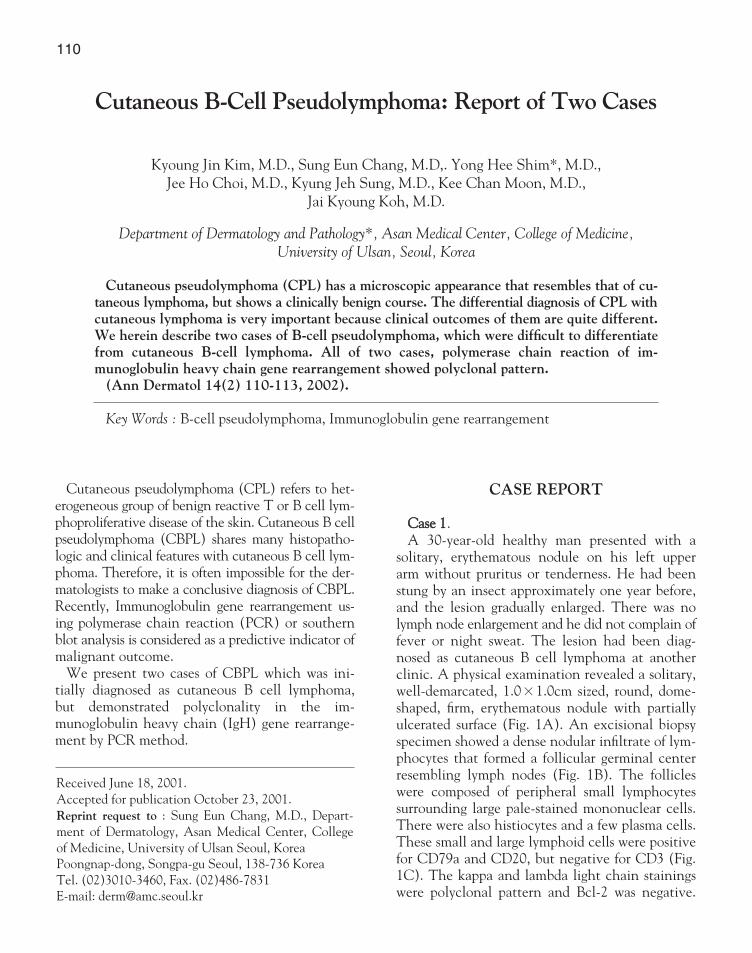
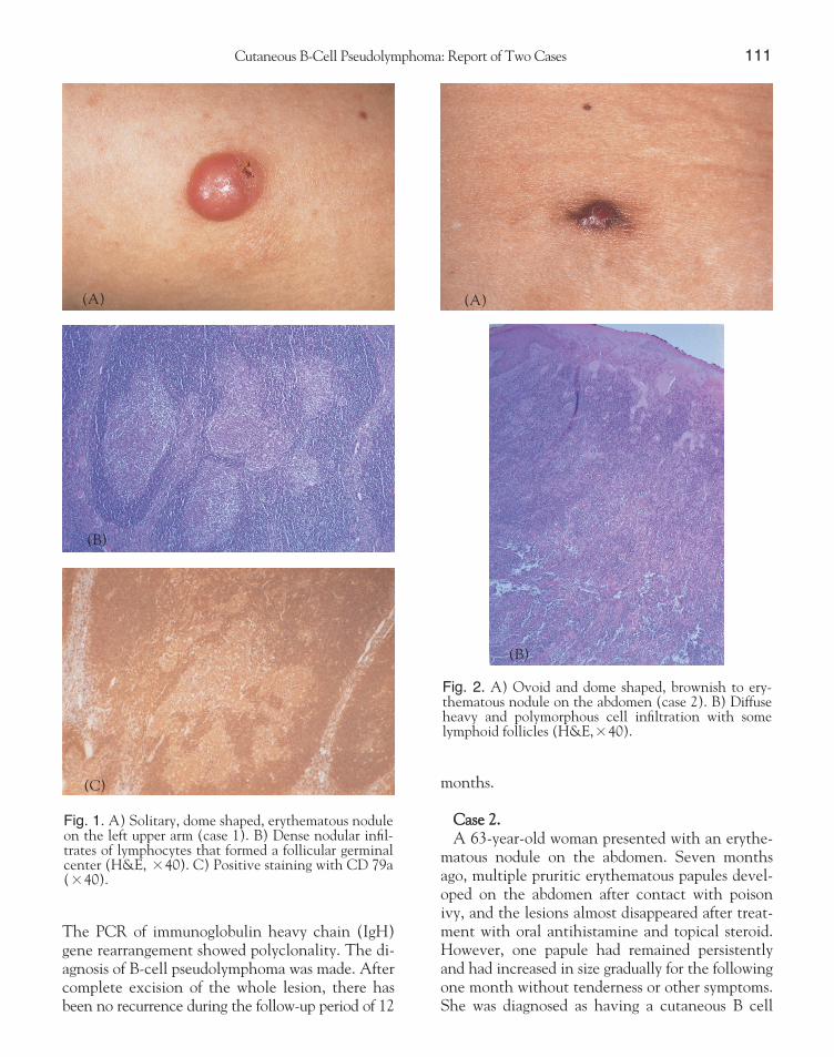
solitary, erythematous nodule on his left upperarm without pruritus or tenderness. He had beenstung by an insect approximately one year before,and the lesion gradually enlarged. There was nolymph node enlargement and he did not complain offever or night sweat. The lesion had been diag-nosed as cutaneous B cell lymphoma at anotherclinic. A physical examination revealed a solitary,well-demarcated, 1.0×1.0cm sized, round, dome-shaped, firm, erythematous nodule with partiallyulcerated surface (Fig. 1A). An excisional biopsyspecimen showed a dense nodular infiltrate of lym-phocytes that formed a follicular germinal centerresembling lymph nodes (Fig. 1B). The follicleswere composed of peripheral small lymphocytessurrounding large pale-stained mononuclear cells.There were also histiocytes and a few plasma cells.These small and large lymphoid cells were positivefor CD79a and CD20, but negative for CD3 (Fig.1C). The kappa and lambda light chain stainingswere polyclonal pattern and Bcl-2 was negative.
Cutaneous B-Cell Pseudolymphoma: Report of Two Cases
Kyoung Jin Kim, M.D., Sung Eun Chang, M.D,. Yong Hee Shim*, M.D.,
Jee Ho Choi, M.D., Kyung Jeh Sung, M.D., Kee Chan Moon, M.D.,
Jai Kyoung Koh, M.D.
Department of Dermatology and Pathology*, Asan Medical Center, College of Medicine, University of Ulsan, Seoul, Korea
Cutaneous pseudolymphoma (CPL) has a microscopic appearance that resembles that of cu-taneous lymphoma, but shows a clinically benign course. The differential diagnosis of CPL withcutaneous lymphoma is very important because clinical outcomes of them are quite different.We herein describe two cases of B-cell pseudolymphoma, which were difficult to differentiatefrom cutaneous B-cell lymphoma. All of two cases, polymerase chain reaction of im-munoglobulin heavy chain gene rearrangement showed polyclonal pattern.
(Ann Dermatol 14(2) 110-113, 2002).
Key Words : B-cell pseudolymphoma, Immunoglobulin gene rearrangement
Received June 18, 2001.
Accepted for publication October 23, 2001.
Reprint request to : Sung Eun Chang, M.D., Depart-
ment of Dermatology, Asan Medical Center, College
of Medicine, University of Ulsan Seoul, Korea
Poongnap-dong, Songpa-gu Seoul, 138-736 Korea
Tel. (02)3010-3460, Fax. (02)486-7831
E-mail: [email protected]
110
Cutaneous B-Cell Pseudolymphoma: Report of Two Cases 111
The PCR of immunoglobulin heavy chain (IgH)gene rearrangement showed polyclonality. The di-agnosis of B-cell pseudolymphoma was made. Aftercomplete excision of the whole lesion, there hasbeen no recurrence during the follow-up period of 12
months.
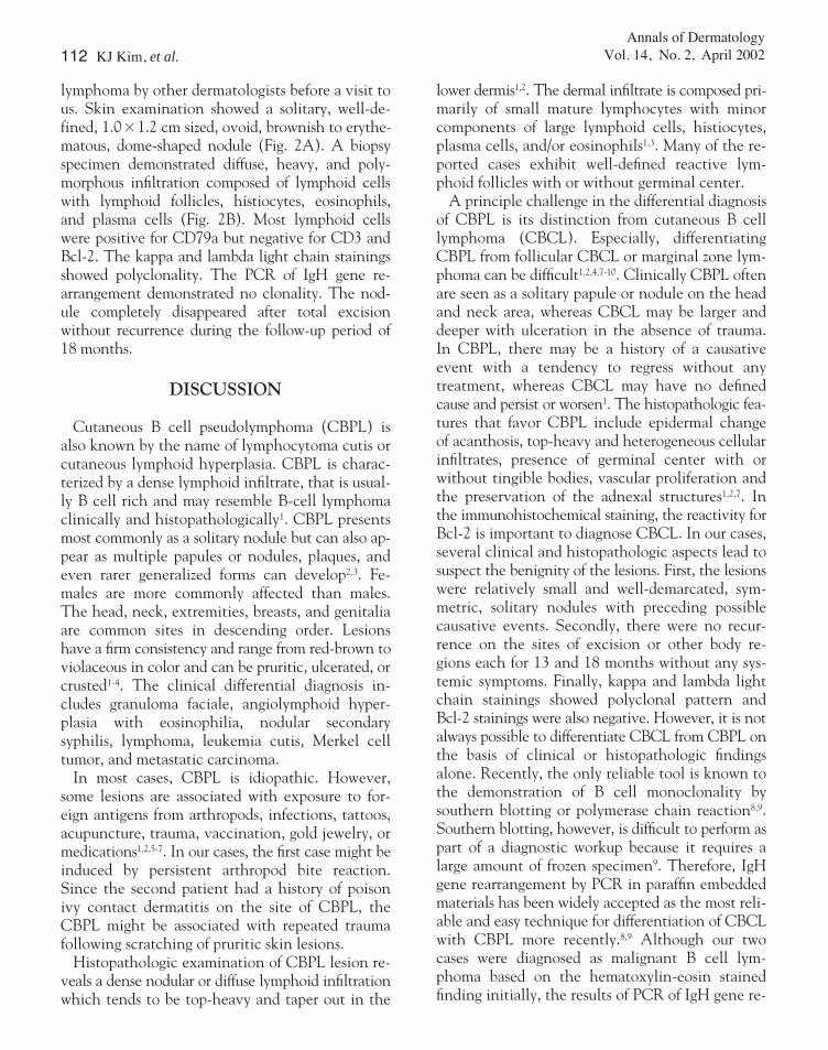
CCaassee 22.. A 63-year-old woman presented with an erythe-
matous nodule on the abdomen. Seven monthsago, multiple pruritic erythematous papules devel-oped on the abdomen after contact with poisonivy, and the lesions almost disappeared after treat-ment with oral antihistamine and topical steroid.However, one papule had remained persistentlyand had increased in size gradually for the followingone month without tenderness or other symptoms.She was diagnosed as having a cutaneous B cell
Fig. 1. A) Solitary, dome shaped, erythematous noduleon the left upper arm (case 1). B) Dense nodular infil-trates of lymphocytes that formed a follicular germinalcenter (H&E, ×40). C) Positive staining with CD 79a(×40).
(A)
(C)
(B)
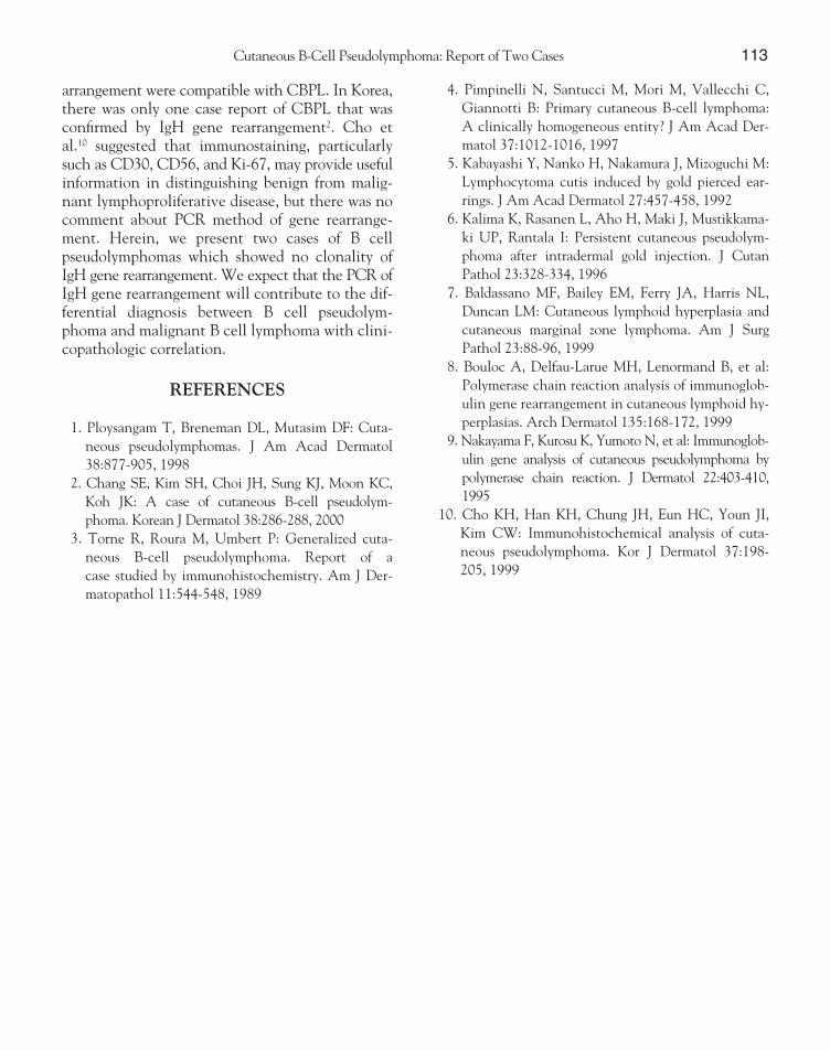
Fig. 2. A) Ovoid and dome shaped, brownish to ery-thematous nodule on the abdomen (case 2). B) Diffuseheavy and polymorphous cell infiltration with somelymphoid follicles (H&E,×40).
(A)
(B)
112 KJ Kim, et al.Annals of Dermatology
Vol. 14, No. 2, April 2002
lymphoma by other dermatologists before a visit tous. Skin examination showed a solitary, well-de-fined, 1.0×1.2 cm sized, ovoid, brownish to erythe-matous, dome-shaped nodule (Fig. 2A). A biopsyspecimen demonstrated diffuse, heavy, and poly-morphous infiltration composed of lymphoid cellswith lymphoid follicles, histiocytes, eosinophils,and plasma cells (Fig. 2B). Most lymphoid cellswere positive for CD79a but negative for CD3 andBcl-2. The kappa and lambda light chain stainingsshowed polyclonality. The PCR of IgH gene re-arrangement demonstrated no clonality. The nod-ule completely disappeared after total excisionwithout recurrence during the follow-up period of18 months.
DISCUSSION
Cutaneous B cell pseudolymphoma (CBPL) isalso known by the name of lymphocytoma cutis orcutaneous lymphoid hyperplasia. CBPL is charac-terized by a dense lymphoid infiltrate, that is usual-ly B cell rich and may resemble B-cell lymphomaclinically and histopathologically1. CBPL presentsmost commonly as a solitary nodule but can also ap-pear as multiple papules or nodules, plaques, andeven rarer generalized forms can develop2,3. Fe-males are more commonly affected than males.The head, neck, extremities, breasts, and genitaliaare common sites in descending order. Lesionshave a firm consistency and range from red-brown toviolaceous in color and can be pruritic, ulcerated, orcrusted1-4. The clinical differential diagnosis in-cludes granuloma faciale, angiolymphoid hyper-plasia with eosinophilia, nodular secondarysyphilis, lymphoma, leukemia cutis, Merkel celltumor, and metastatic carcinoma.
In most cases, CBPL is idiopathic. However,some lesions are associated with exposure to for-eign antigens from arthropods, infections, tattoos,acupuncture, trauma, vaccination, gold jewelry, ormedications1,2,5-7. In our cases, the first case might beinduced by persistent arthropod bite reaction.Since the second patient had a history of poisonivy contact dermatitis on the site of CBPL, theCBPL might be associated with repeated traumafollowing scratching of pruritic skin lesions.
Histopathologic examination of CBPL lesion re-veals a dense nodular or diffuse lymphoid infiltrationwhich tends to be top-heavy and taper out in the
lower dermis1,2. The dermal infiltrate is composed pri-marily of small mature lymphocytes with minorcomponents of large lymphoid cells, histiocytes,plasma cells, and/or eosinophils1,3. Many of the re-ported cases exhibit well-defined reactive lym-phoid follicles with or without germinal center.
A principle challenge in the differential diagnosisof CBPL is its distinction from cutaneous B celllymphoma (CBCL). Especially, differentiatingCBPL from follicular CBCL or marginal zone lym-phoma can be difficult1,2,4,7-10. Clinically CBPL oftenare seen as a solitary papule or nodule on the headand neck area, whereas CBCL may be larger anddeeper with ulceration in the absence of trauma.In CBPL, there may be a history of a causativeevent with a tendency to regress without anytreatment, whereas CBCL may have no definedcause and persist or worsen1. The histopathologic fea-tures that favor CBPL include epidermal changeof acanthosis, top-heavy and heterogeneous cellularinfiltrates, presence of germinal center with orwithout tingible bodies, vascular proliferation andthe preservation of the adnexal structures1,2,7. Inthe immunohistochemical staining, the reactivity forBcl-2 is important to diagnose CBCL. In our cases,several clinical and histopathologic aspects lead tosuspect the benignity of the lesions. First, the lesionswere relatively small and well-demarcated, sym-metric, solitary nodules with preceding possiblecausative events. Secondly, there were no recur-rence on the sites of excision or other body re-gions each for 13 and 18 months without any sys-temic symptoms. Finally, kappa and lambda lightchain stainings showed polyclonal pattern andBcl-2 stainings were also negative. However, it is notalways possible to differentiate CBCL from CBPL onthe basis of clinical or histopathologic findingsalone. Recently, the only reliable tool is known tothe demonstration of B cell monoclonality bysouthern blotting or polymerase chain reaction8,9.Southern blotting, however, is difficult to perform aspart of a diagnostic workup because it requires alarge amount of frozen specimen9. Therefore, IgHgene rearrangement by PCR in paraffin embeddedmaterials has been widely accepted as the most reli-able and easy technique for differentiation of CBCLwith CBPL more recently.8,9 Although our twocases were diagnosed as malignant B cell lym-phoma based on the hematoxylin-eosin stainedfinding initially, the results of PCR of IgH gene re-
arrangement were compatible with CBPL. In Korea,there was only one case report of CBPL that wasconfirmed by IgH gene rearrangement2. Cho etal.10 suggested that immunostaining, particularlysuch as CD30, CD56, and Ki-67, may provide usefulinformation in distinguishing benign from malig-nant lymphoproliferative disease, but there was nocomment about PCR method of gene rearrange-ment. Herein, we present two cases of B cellpseudolymphomas which showed no clonality ofIgH gene rearrangement. We expect that the PCR ofIgH gene rearrangement will contribute to the dif-ferential diagnosis between B cell pseudolym-phoma and malignant B cell lymphoma with clini-copathologic correlation.
REFERENCES
1. Ploysangam T, Breneman DL, Mutasim DF: Cuta-
neous pseudolymphomas. J Am Acad Dermatol
38:877-905, 1998
2. Chang SE, Kim SH, Choi JH, Sung KJ, Moon KC,
Koh JK: A case of cutaneous B-cell pseudolym-
phoma. Korean J Dermatol 38:286-288, 2000
3. Torne R, Roura M, Umbert P: Generalized cuta-
neous B-cell pseudolymphoma. Report of a
case studied by immunohistochemistry. Am J Der-
matopathol 11:544-548, 1989
4. Pimpinelli N, Santucci M, Mori M, Vallecchi C,
Giannotti B: Primary cutaneous B-cell lymphoma:
A clinically homogeneous entity? J Am Acad Der-
matol 37:1012-1016, 1997
5. Kabayashi Y, Nanko H, Nakamura J, Mizoguchi M:
Lymphocytoma cutis induced by gold pierced ear-
rings. J Am Acad Dermatol 27:457-458, 1992
6. Kalima K, Rasanen L, Aho H, Maki J, Mustikkama-
ki UP, Rantala I: Persistent cutaneous pseudolym-
phoma after intradermal gold injection. J Cutan
Pathol 23:328-334, 1996
7. Baldassano MF, Bailey EM, Ferry JA, Harris NL,
Duncan LM: Cutaneous lymphoid hyperplasia and
cutaneous marginal zone lymphoma. Am J Surg
Pathol 23:88-96, 1999
8. Bouloc A, Delfau-Larue MH, Lenormand B, et al:
Polymerase chain reaction analysis of immunoglob-
ulin gene rearrangement in cutaneous lymphoid hy-
perplasias. Arch Dermatol 135:168-172, 1999
9. Nakayama F, Kurosu K, Yumoto N, et al: Immunoglob-
ulin gene analysis of cutaneous pseudolymphoma by
polymerase chain reaction. J Dermatol 22:403-410,
1995
10. Cho KH, Han KH, Chung JH, Eun HC, Youn JI,
Kim CW: Immunohistochemical analysis of cuta-
neous pseudolymphoma. Kor J Dermatol 37:198-
205, 1999
Cutaneous B-Cell Pseudolymphoma: Report of Two Cases 113