cx-5461 is a dna g-quadruplex stabilizer with selective ... · here, we show that cx-5461 is a...
TRANSCRIPT

ARTICLE
Received 1 Jun 2016 | Accepted 28 Dec 2016 | Published 17 Feb 2017
CX-5461 is a DNA G-quadruplex stabilizer withselective lethality in BRCA1/2 deficient tumoursHong Xu1, Marco Di Antonio2,3, Steven McKinney1, Veena Mathew4, Brandon Ho5, Nigel J. O’Neil6,
Nancy Dos Santos7, Jennifer Silvester8, Vivien Wei1, Jessica Garcia1, Farhia Kabeer1, Daniel Lai1, Priscilla Soriano1,
Judit Banath9, Derek S. Chiu1, Damian Yap1, Daniel D. Le2, Frank B. Ye6, Anni Zhang4, Kelsie Thu8, John Soong10,
Shu-chuan Lin10, Angela Hsin Chin Tsai1, Tomo Osako1, Teresa Algara1, Darren N. Saunders1, Jason Wong1,
Jian Xian11, Marcel B. Bally7, James D. Brenton11, Grant W. Brown5, Sohrab P. Shah1, David Cescon8,12,
Tak W. Mak8, Carlos Caldas11, Peter C. Stirling4, Phil Hieter6, Shankar Balasubramanian2,3 & Samuel Aparicio1
G-quadruplex DNAs form four-stranded helical structures and are proposed to play key roles
in different cellular processes. Targeting G-quadruplex DNAs for cancer treatment is a very
promising prospect. Here, we show that CX-5461 is a G-quadruplex stabilizer, with specific
toxicity against BRCA deficiencies in cancer cells and polyclonal patient-derived xenograft
models, including tumours resistant to PARP inhibition. Exposure to CX-5461, and its related
drug CX-3543, blocks replication forks and induces ssDNA gaps or breaks. The BRCA and
NHEJ pathways are required for the repair of CX-5461 and CX-3543-induced DNA damage
and failure to do so leads to lethality. These data strengthen the concept of G4 targeting as a
therapeutic approach, specifically for targeting HR and NHEJ deficient cancers and other
tumours deficient for DNA damage repair. CX-5461 is now in advanced phase I clinical
trial for patients with BRCA1/2 deficient tumours (Canadian trial, NCT02719977, opened
May 2016).
DOI: 10.1038/ncomms14432 OPEN
1 Department of Molecular Oncology, British Columbia Cancer Research Centre, and Department of Pathology and Laboratory Medicine, University of BritishColumbia, 675 West 10th Avenue, Vancouver, British Columbia, Canada V5Z 1L3. 2 Cancer Research UK Cambridge Research Institute, Li Ka Shing Centre,Robinson Way, Cambridge CB2 0RE, UK. 3 Department of Chemistry, University of Cambridge, Cambridge CB2 1EW, UK. 4 Terry Fox Laboratory, BC CancerAgency, 675 West 10th Avenue, Vancouver, British Columbia, Canada V5Z 1L3. 5 Department of Biochemistry and Donnelly Centre, University of Toronto,160 College Street, Toronto, Ontario, Canada M5S 3E1. 6 Michael Smith Laboratories, University of British Columbia, Vancouver, Canada V6T 1Z4. 7 AdvancedTherapeutics, BC Cancer Agency and Department of Pathology and Laboratory Medicine, University of British Columbia, 675 West 10th Avenue, Vancouver,British Columbia, Canada V5Z 1L3. 8 Campbell Family Institute for Breast Cancer Research, Princess Margret Cancer Centre, 610 University Avenue, Toronto,Canada M5G 2M9. 9 Department of Integrative Oncology, BC Cancer Agency, 675 West 10th Avenue, Vancouver, British Columbia, Canada V5Z 1L3.10 Senhwa Biosciences, Inc., 9 F, No.205-1, Section 3, Peihsin Road, Hsintien District, New Taipei City 23143, Taiwan R.O.C. 11 Cancer Research UK CambridgeResearch Institute and Department of Oncology, University of Cambridge, Li Ka Shing Centre, Cambridge CB2 0RE, UK. 12 Division of Medical Oncology andHematology, Department of Medicine, University of Toronto, Toronto, Canada M5S 1A8. Correspondence and requests for materials should be addressed toS.A. (email: [email protected]).
NATURE COMMUNICATIONS | 8:14432 | DOI: 10.1038/ncomms14432 | www.nature.com/naturecommunications 1
Inherited BRCA2 mutations predispose carriers to early onsetbreast, ovarian and other cancers1,2. As an important tumoursuppressor, the key role of BRCA2 is in homologous
recombination (HR)-mediated DNA damage repair bypromoting the formation of RAD51 filaments at DNA breaks3.When BRCA2 is deficient, HR repair efficiency is greatlycompromised, leading to an increased error-prone DNA repairand ultimately, genomic instability. The BRCA2/RAD51 complexis also involved in many other aspects of genome instability, suchas stalled DNA replication fork stabilization4, R-loop resolutionand repairing G-quadruplex (G4) associated DNA damage5,6.
G4 structures can potentially form at over 700,000 sequences inthe human genome7,8, and 10,000 of them have been identifiedfrom ChIP-seq using an antibody that recognises G4 structures9.G4 structures increase the tendency for DNA damage to occur, byimpeding DNA polymerase and DNA damage repair processes10.The importance of the HR pathway in repairing G4-inducedDNA damage has been demonstrated in different organisms11,12.BRCA2-deficient cells display higher sensitivity to toolcompounds such as pyridostatin (PDS)13 and RHS4 (ref. 6),which are both G4 stabilizers, but not medicinal compounds andare structurally unrelated to the fluoroquinolone derived CXseries compounds.
As a general phenomenon in cancer, there is an increasedrequirement for rDNA transcription to meet the greater proteinsynthesis demand in cancer cells14. Some new inhibitors of rDNAtranscription have been synthesized in recent years, such asCX-5461, CX-3543 and BMH-21 (refs 15–17). CX-3543 binds toG4 sequences and disrupts the interaction of rDNA G4 structureswith nucleolin, thereby inhibiting Pol I transcription andinducing apoptotic death in cancer cells16. BMH-21 acts by itsinteraction with the DNA backbone in GC-rich DNA sequences,particularly at rDNA loci, thus inhibiting Pol I transcription andalso promoting degradation of Pol I catalytic subunit RPA194(ref. 18). CX-5461 is an rDNA transcription inhibitor currently inphase I trials for haematologic malignancies. CX-5461 reducesthe binding affinity of the SL1 pre-initiation complex andRNA polymerase I complex to rDNA promoters and conveysp53-dependent anti-tumorigenic activity in hematopoieticmalignancies15,17. Recently, more targets of CX-5461 havebeen discovered, such as the activation of ATM/ATR19 andrapamycin-associated signalling pathway20.
In the present study, we have uncovered a new andunanticipated mechanism of CX-5461 activity in HR andnon-homologous end joining (NHEJ) deficient cancer cells. Weshow that both CX-5461 and the related compound CX-3543induce DNA damage and are dependent on BRCA1/2-mediatedHR and DNA-PK-mediated NHEJ pathway for damage repair.We also discover that CX-5461 (and CX-3543) bind and stabilizeG4 DNA structures in vitro, impede the progression of DNAreplication complexes and result in increased in vivo G4structures. The pattern of activity in polyclonal patient-derivedxenografts (PDX) mirrors that seen in vitro with isogenic cell linepairs, namely sensitivity in BRCA deficient PDX models, in thecontext of pre-treatment with taxane and other standard of careagents. In some cases, superior activity to PARP inhibition isobserved. Our data suggest that the CX drugs, and possibly otherG4 stabilizers have the potential to treat cancers deficient forBRCA1, BRCA2, NHEJ pathway members and some other genesinvolved in DNA damage repair and DNA replication. SinceCX5461 is an advanced phase I medicinal compound, theseobservations have immediate translational significance.
ResultsCX-5461 selectively inhibits cancer cells deficient for BRCA1/2.To identify potential novel drugs for cancers with BRCA2
mutations, we tested a total of 17 commercially available inhibi-tors (Supplementary Table 1) by clonogenic assays inisogenic BRCA2 knockout and wild type (WT) HCT116 cellline pairs published by us21. This clonogenic screen identifiedCX-5461, a previously described RNA pol I inhibitor15,17 to behighly toxic to BRCA2 knockout HCT116 cells as comparedwith isogenic BRCA2 WT cells (Fig. 1a). We extended thequantification of this observation by using a WST-1 metabolic/cell viability assay. As with the clonogenic assay, this revealed a9.0-fold (95% confidence interval (CI), 5.1–16.2) lower IC50 inBRCA2 deficient HCT116 cells than in BRCA2 proficient cells(Fig. 1b, Supplementary Fig. 1a). Importantly, we observed in thisexperiment and those described below, that BRCA2 heterozygouscells displayed similar sensitivity to CX-5461 as BRCA2 proficientwild-type cells (Fig. 1b,d). We also assessed cell death specificallythrough fluorescence-activated cell sorting (FACS) by annexin Vand PI double staining. As shown in Fig. 1c and SupplementaryTable 5, CX-5461 induced more apoptotic cell death in BRCA2knockout cells relative to WT. However, BRCA2þ /þ andBRCA2� /� isogenic cells in HCT116 appeared equallysensitive to actinomycin (an inhibitor for both RNA polymeraseI and II) and cycloheximide (an inhibitor for protein translationelongation) (Supplementary Fig. 1b,c). Together, these dataindicate that BRCA2 deficient cells are not generally sensitiveto transcription and translation inhibition, but show specificsensitivity to CX-5461.
We next sought to determine whether the selective killing effectof CX-5461 in BRCA2 deficient cells could be observed in othercell lines and species backgrounds. We measured CX5461 drugsensitivity in isogenic BRCA2� /� and WT colorectal cancerDLD1 cells; BRCA2 deficient ovarian cancer cell line PEO1 vs theBRCA2 proficient C4-2 (ref. 22); and BRCA2 proficient andknockout breast tumour cells derived from p53 knockout mouse(BRCA2� /� ; p53� /� (KB2P 1.21) vs BRCA2þ /þ ; p53� /�
(KP6.3) cells)23. In all these cell line pairs, BRCA2-deficient cellswere more sensitive to CX-5461 than BRCA2 proficient cells(Fig. 1d,e, Supplementary Figs 1d and 2a–c). When BRCA2 wastransiently knocked down in U2OS, increased sensitivity toCX-5416 was also observed (Supplementary Fig. 2d).
In p53� /� HCT116 cells, drug sensitivity to CX-5461increased significantly (F-test P¼ 0.01) after BRCA2 knockdown(Fig. 1f). This supports the notion that the selective toxicity ofCX-5461 to BRCA2 deficient cells is not dependent on p53 inepithelial cells17. BRCA1 is another critical component of theHR repair pathway and is also involved in replication forkstabilization24,25. Encouragingly, increased drug sensitivity toCX-5461 was observed when BRCA1 was knocked down inp53þ /þ and p53� /� HCT116 (Supplementary Fig. 2e), furthersupporting that synthetic lethality with CX-5461 does notrequire wild type p53. We also note (below) that CX-5461sensitivity is seen in polyclonal p53 deficient PDX tumours(model CFIB-70620).
BRCA2 is not synthetic lethal with rDNA transcription inhibition.We set out to address the possible molecular mechanism bywhich CX-5461 causes synthetic lethality in BRCA2 deficientcells. Since CX-5461 has documented RNA pol I inhibitionactivity, we investigated this notion first, by examining the effectsof a related compound CX-3543 (quarfloxin) and an unrelatedsmall molecule BMH-21. Both of these molecules are reported tosuppress rDNA transcription, but by different mechanisms16,18.In DLD1 cells, BRCA2� /� cells were highly sensitive to CX-3543compared with isogenic BRCA2þ /þ cells (SupplementaryFig. 3d). In contrast, BRCA2� /� cells were not more sensitiveto BMH-21 than isogenic WT cells in either HCT116 or DLD1(Fig. 1h, Supplementary Fig. 3a,b).
ARTICLE NATURE COMMUNICATIONS | DOI: 10.1038/ncomms14432
2 NATURE COMMUNICATIONS | 8:14432 | DOI: 10.1038/ncomms14432 | www.nature.com/naturecommunications

Since BRCA2 deficient cells showed differential response tothe three rRNA transcription inhibitors, we sought to verifythe activity against rDNA transcription in the cells used. Wemeasured the amount of 45S pre-rRNA as a readout for rDNAtranscription by qRT-PCR, as previously described16, at 2 and
24 h after treatment. In these experiments, BMH-21 was at leastas potent an rDNA transcription inhibitor as CX-5461 andCX-3543 (Fig. 1g, Supplementary Fig. 3g), consistent withpublished activity18. However, the same or greater level(median 1–2 fold for CX-5461, CX-3543 at 10� 6 M vs median
WT
B46
B18
HCT116:p53+/+;BRCA2 WT or deficient
Log10(M) CX-5461
WS
T1
inte
nsity
nor
mal
ized
to c
ontr
ol
Control −10 −9 −8 −7 −6 −5
0.0
0.2
0.4
0.6
0.8
1.0
BRCA2proficient
BRCA2deficient
WTB18B46
DLD1:p53S241F/sil;BRCA2 WT or deficient
Log10(M) CX-5461
WS
T1
inte
nsity
nor
mal
ized
to c
ontr
ol
Control −10 −9 −8 −7 −6
0.0
0.2
0.4
0.6
0.8
1.0
BRCA2 proficientBRCA2 deficient
WTHETHOM
PEO1:BRCA2–/–
C4-2: BRCA2–/– (revertant)
WS
T1
inte
nsity
nor
mal
ized
to c
ontr
ol
Control −10 −9 −8 −7 −6
0.0
0.2
0.4
0.6
0.8
1.0
1.2
BRCA2 proficientBRCA2 deficient
C4−2PEO1
0 5×10⁻⁹ 10–8 5×10–810–9
HCT116
Log10(M) BMH-21
WS
T1
inte
nsity
nor
mal
ized
to c
ontr
ol
Control −10 −9 −8 −7 −6 −5
0.0
0.2
0.4
0.6
0.8
1.0
1.2
BRCA2 proficientBRCA2 deficient
WTB+/−B18B46
HCT116:p53–/–
Log10(M) CX-5461
WS
T1
inte
nsity
nor
mal
ized
to c
ontr
ol
Control −9 −8 −7
0.0
0.2
0.4
0.6
0.8
1.0siNTSsiBRCA2
BRCA2 proficientBRCA2 deficient
Control
WT
BR
CA
2–/–
0.039 6.09
5.2388.6
104
104
103
103
102
102
101
101100
104
103
102
101
100
104
103
102
101
100
104
103
102
101
100
100 104103102101100
104103102101100 104103102101100
0.18 4.96
4.1490.7
0.68 10.8
6.0382.5
PI
PI
3.22 17.2
15.7
64.0
Contro
l
CX5461
10–8 M
CX5461
10–7 M
CX5461
10–6 M
0
5
10
15
*** *** ***
BRCA2 proficientBRCA2 deficient
Per
cent
age
of a
popt
otic
cel
ls
Annexin V Annexin V
CX-5461 (M)
Log10(M) CX-5461
Effe
ct o
f dru
g on
45S
tran
scrip
tion,
by
qRT
PC
R
2048
512
128
32
842124
SY
BR
gree
nT
aqm
anS
YB
Rgr
een
SY
BR
gree
n
(Dow
n) fo
ld c
hang
e (U
p)
Drug treatment
BRCA2 proficient, 95% CIBRCA2 deficient, 95% CI
CX-5461 CX-3543 BMH21 Actinomycin
10–8
10–7
10–6
10–5
10–5
SY
BR
gree
nT
aqm
an
SY
BR
gree
nT
aqm
an
SY
BR
gree
nS
YB
Rgr
een
Taq
man
10–8
10–7
10–6
10–5
10–5
SY
BR
gree
nS
YB
Rgr
een
Taq
man
10–8
5×10
–8
5×10
–8
10–7
10–6
(M)
prob
eT
aqm
an
HCT116
10–7 M CX5461
a b
c d
e f
g h
NATURE COMMUNICATIONS | DOI: 10.1038/ncomms14432 ARTICLE
NATURE COMMUNICATIONS | 8:14432 | DOI: 10.1038/ncomms14432 | www.nature.com/naturecommunications 3

16-fold for BMH-21 at 10� 6 M, Fig. 1g, Supplementary Fig. 3g)of rDNA transcription inhibition by BMH-21 did not result inselective toxicity against BRCA2� /� cells, in contrast withCX-5461 and CX-3543. We also noted that the IC50 of CX-5461in the cell lines used is 4.80� 10� 9 M (Fig. 1b, SupplementaryFig. 1a) for BRCA2 knockout HCT116 cells, a concentration atwhich rDNA transcription inhibition was not observed. We thusconcluded that rDNA transcription inhibition alone is notrequired for the selective toxicity of CX-5461 and CX-3543against BRCA2 deficient cells.
We also compromised rDNA transcription with RNAi againstPOLR1B, an essential component in the RNA pol I complex.Upon POLR1B knockdown, rDNA transcription was significantlyreduced (Supplementary Fig. 3c), and cell viability also greatlydecreased by single RNA pol I knockdown, making measurementof synergy with BRCA2 (double knockdown) difficult. In U2OScells, POLR1B and BRCA2 double knockdown reduced cellviability at a level comparable with single POLR1B knockdown(Supplementary Fig. 3f), with non-statistical difference seen,consistent with the absence of a synthetic genetic effect in U2OS.In HCT116, slightly more cell death was observed indouble knockdown cells compared with single knockdown cells(omnibus test Po0.0001) (Supplementary Fig. 3e), however thesingle RNA pol I knockdown induces many fold greater cell deaththan the knockdown of BRCA2, precluding accurate assessmentof synthetic lethality. Taken together with the results of BMH-21,the data disfavour rDNA transcription inhibition as an importantmechanism of CX-5461 toxicity in BRCA2 deficient cells.
When drug sensitivity of CX-5461, CX-3543, and BMH-21 wascompared across 50 breast cancer cell lines, the responsesignature of BMH-21 was different from CX-5461 and CX3543(Fig. 7d, Supplementary Table 4). BMH-21 is only moderatelycytotoxic to a few breast cancer cell lines, while CX-3543 hasresponse pattern very similar to CX-5461 and is clusteredtogether with CX-5461 (Fig. 7d), consistent with a similar modeof action between CX-3543 and CX-5461, but distinct in the caseof BMH-21.
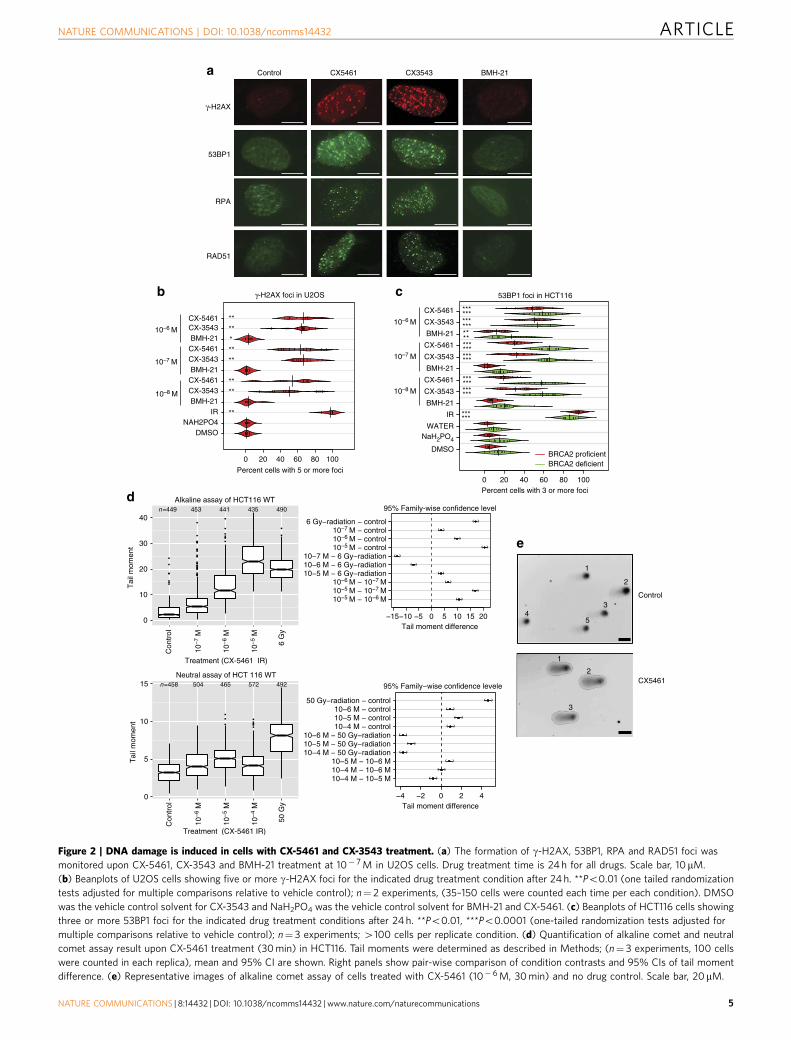
CX-5461 and CX-3543 induce DNA damage in tumour cells.As the major cellular functions of BRCA2 are DNA damagerepair and replication fork stabilization, we investigated DNAdamage response upon exposure to CX-5461, CX-3543 andBMH-21. Strikingly, g-H2AX and 53BP1 DNA damage foci wererobustly induced by CX-5461 and CX-3543 in both HCT116 andU2OS cells when the drug concentration was higher than 10� 8 M(Fig. 2a–c). In marked contrast, no g-H2AX or 53BP1 focusformation was visible with 10� 8 M and 10� 7 M BMH-21,
although 10� 6 M BMH-21 induced a statistically significant levelof damage foci that was slightly higher (Fig. 2b,c). Consistent withthe result of DNA damage protein focus formation, increasedphosphorylation of H2AX was observed after CX-5461 andCX-3543 treatment by Western blotting analysis (Fig. 4a,Supplementary Fig. 4a). As shown above, BMH-21 inhibitedrDNA transcription more potently than CX-5461 and CX-3543(Fig. 1g). However, BMH-21 did not appear to be associated withthe same degree of DNA damage and cell death as CX-5461 andCX-3543 at the same concentration. Moreover, reduction ofrDNA transcription by siRNA for POLR1B did not result in moreg-H2AX and 53BP1 foci formation (Supplementary Fig. 4b).Collectively, these data strongly suggest that CX-5461 andCX-3543 are potent DNA damage inducers, and this mechanismis independent of rDNA transcription inhibition in the cell typesexamined. We also excluded the possibility that the DNA damagephenotype was an indirect effect of nucleolus disruption, becauseDNA damage foci were observed when the concentration ofCX-5461 was not able to disrupt nucleolus (SupplementaryFig. 4e).
In order to better quantify the amount and type of DNAdamage, a comet tail forming assay26 was applied to shortexposure CX-5461 treated cells. After 30 min of drug exposure,statistically increased comet tail-moments were observed underalkaline conditions when the concentration of CX-5461 washigher than 10� 7 M (Po10� 15, w2 test for all drugconcentrations tested) (Fig. 2d,e). Under neutral comet assayconditions, which are more specific for DSBs, we discovered asmall but statistically significant increase in tail moments(Po10� 6, w2 test for all drug concentrations, Fig. 2d). Thus,upon short exposure (30 min) to CX-5461, the initial forms ofDNA damage occurring are mainly SSBs or gaps and a lowerabundance of DSBs.
Remarkably, CX-5461 (Supplementary Fig. 4d) also inducedDNA damage foci in yeast and selectively killed yeast lackingRAD52 (Supplementary Fig. 4h), a functional homologue ofBRCA2, further supporting DNA as the drug target of CX-5461and CX-3543.
CX-5461 and CX-3543 induced DNA damage is replication-dependent. To further investigate the mechanism of CX-5461and CX-3543 induced DNA damage, cell cycle analysis wasperformed on drug treated HCT116 cells by FACS with EdU andPI staining. Shortly after 10� 6 M CX-5461 treatment (2 h),active replication shown by EdU staining decreased significantly(7.1% decrease; 95% CI, 2.6–11.5%) in BRCA2þ /þ cells (Fig. 3a).At a later time point (24 h after), CX-5461 induced a prominent
Figure 1 | BRCA2 deficient cells are highly sensitive to CX-5461 in different human and murine cell types. (a) The colony formation capacity of
BRCA2� /� HCT116 cells was greatly reduced by treatment with CX-5461. Experiments were repeated twice with similar results. Scale bar, 1 cm. (b) The
hypersensitivity of BRCA2� /� cells to CX-5461 in HCT116 validated by WST-1 assay. Representative experiment #3 (see Supplementary Fig. 1a for full
experimental panels, n¼ 9) is displayed as individual data points and as fitted sigmoid dose response curves (green for BRCA2 deficient and red for BRCA2
wild type). Dashed vertical lines are the IC50. (c) CX-5461 induced more apoptosis in BRCA2� /� cells as indicated by FACS analysis. A representative
result is shown on the left panel and the right panel shows mean apoptotic fraction with 95% CI of cells in early apoptosis under different drug
concentrations. ***Po0.0001, t-test; n¼ 3, 2 or more replicates per condition. See Supplementary Table 5 for more statistical analyses. (d) BRCA2� /� in
DLD1 isogenic cell line pairs displayed hypersensitivity to CX-5461 by WST-1 assay. Representative experiment #3 is shown (see Supplementary Fig. 2a for
full experimental panels; n¼4). Green fitted sigmoid curves are for BRCA2 homozygous (HOM) and red for BRCA2 wild type and heterozygous (HET).
(e) BRCA2 deficient ovarian cancer PEO1 cells exhibited increased sensitivity to CX-5461 relative to BRCA2 proficient C4-2 cells by WST-1 assay.
A representative experiment #1 is displayed (see Supplementary Fig. 2b for full experimental panels, n¼ 3). (f) RNAi knockdown of BRCA2 increased
sensitivity to CX-5461 in p53� /� HCT116 cells by WST-1 assay (4 days in drug). The results of all three experiments are summarized by the green (BRCA2
knockdown) and red (non-targeting control) super-smoother fit lines. (g) 45S pre-rRNA level measured by RT-PCR after CX-5461, CX-3543 and BMH-21
treatment in WT and BRCA2� /� HCT116 cells. Drug incubation time was 24 h. Fold change estimates and unadjusted 95% CIs of 45s pre-rRNA levels
under drug treatment condition versus vehicle control are shown. P values (by F-test) are shown in Supplementary Table 7. (h) BRCA2 knockout cells are
not more sensitive to BMH-21 in HCT116 through WST-1 assay. One representative experimental result is shown (more replicates are shown in
Supplementary Fig. 3a).
ARTICLE NATURE COMMUNICATIONS | DOI: 10.1038/ncomms14432
4 NATURE COMMUNICATIONS | 8:14432 | DOI: 10.1038/ncomms14432 | www.nature.com/naturecommunications
BRCA2 proficientBRCA2 deficient
****
*********
**
0
5
10
15
Con
trol
50 G
y
Treatment (CX-5461 IR)
Neutral assay of HCT 116 WT
−4 −2 0 2 4Tail moment difference
−15−10 −5 0 5 10 15 20
95% Family-wise confidence level
Tail moment difference0
10
20
30
40
Con
trol
10−
7 M
10−
5 M
6 G
y
Treatment (CX-5461 IR)
Alkaline assay of HCT116 WT
CX5461
53BP1
RPA
RAD51
Control CX5461 CX3543 BMH-21
Control
53BP1 foci in HCT116
0 20 40 60 80 100
DMSO
NaH2PO4
WATER
IR
BMH-21
CX-3543
CX-5461
BMH-21
CX-3543
CX-5461
BMH-21
CX-3543
CX-5461
10–6 M
10–7 M
10–8 M
**
*********
***
**************
************
10–6 M
10–7 M
10–8 M
0 20 40 60 80 100
DMSONAH2PO4
IRBMH-21
CX-3543CX-5461BMH-21
CX-3543CX-5461BMH-21
CX-3543 CX-5461
******
γ-H2AX foci in U2OS
Percent cells with 3 or more foci
Percent cells with 5 or more foci
n=449 453 441 435 490
n=458 504 465 572 492
γ-H2AX
Tai
l mom
ent
10−
6 M
6 Gy−radiation − control
10−5 M − 10−6 M10−5 M − 10−7 M10−6 M − 10−7 M
10−7 M − control10−6 M − control10−5 M − control
10−7 M − 6 Gy−radiation10−6 M − 6 Gy−radiation10−5 M − 6 Gy−radiation
Tai
l mom
ent
10−
6 M
10−
5 M
10−
4 M
50 Gy−radiation − control10−6 M − control10−5 M − control10−4 M − control
10−6 M − 50 Gy−radiation10−5 M − 50 Gy−radiation10−4 M − 50 Gy−radiation
10−5 M − 10−6 M10−4 M − 10−6 M10−4 M − 10−5 M
95% Family−wise confidence levele
1
1
2
3
2
3
54
a
b c
d
e
Figure 2 | DNA damage is induced in cells with CX-5461 and CX-3543 treatment. (a) The formation of g-H2AX, 53BP1, RPA and RAD51 foci was
monitored upon CX-5461, CX-3543 and BMH-21 treatment at 10� 7 M in U2OS cells. Drug treatment time is 24 h for all drugs. Scale bar, 10mM.
(b) Beanplots of U2OS cells showing five or more g-H2AX foci for the indicated drug treatment condition after 24 h. **Po0.01 (one tailed randomization
tests adjusted for multiple comparisons relative to vehicle control); n¼ 2 experiments, (35–150 cells were counted each time per each condition). DMSO
was the vehicle control solvent for CX-3543 and NaH2PO4 was the vehicle control solvent for BMH-21 and CX-5461. (c) Beanplots of HCT116 cells showing
three or more 53BP1 foci for the indicated drug treatment conditions after 24 h. **Po0.01, ***Po0.0001 (one-tailed randomization tests adjusted for
multiple comparisons relative to vehicle control); n¼ 3 experiments; 4100 cells per replicate condition. (d) Quantification of alkaline comet and neutral
comet assay result upon CX-5461 treatment (30 min) in HCT116. Tail moments were determined as described in Methods; (n¼ 3 experiments, 100 cells
were counted in each replica), mean and 95% CI are shown. Right panels show pair-wise comparison of condition contrasts and 95% CIs of tail moment
difference. (e) Representative images of alkaline comet assay of cells treated with CX-5461 (10� 6 M, 30 min) and no drug control. Scale bar, 20mM.
NATURE COMMUNICATIONS | DOI: 10.1038/ncomms14432 ARTICLE
NATURE COMMUNICATIONS | 8:14432 | DOI: 10.1038/ncomms14432 | www.nature.com/naturecommunications 5

G2 arrest (41% increase; 95% CI, 37–45%), blocking cells beforemitosis (Supplementary Fig. 5b). In HCT116 BRCA2� /� cells,the decrease of S-phase population was more dramatic (10.2%decrease; 95% CI, 5.6–14.9%, 2 vs 0 h, Fig. 3a, SupplementaryFig. 5a, Supplementary Table 6).
To determine whether the DNA damage was occurring inS-phase, we blocked DNA replication with aphidicolin (APH),a DNA polymerase inhibitor. Strikingly, for EdU positive S phasecells, APH pre-treatment remarkably reduced the percentage ofcells with DNA damage foci relative to CX-5461 only treatmentin both WT (decreased 48.6%; 95% CI, 44.2–51.2%) andBRCA2� /� cells (decreased 68.5%; 95% CI, 57.1–75.2%)(Fig. 3b,c). But for EdU negative cells, APH treatment showedno effect. The predominance of DNA damage foci in S-phasepopulation and its dependence on DNA replication were alsoobserved with CX-3543 treatment (Supplementary Fig. 5c,d).Likely, CX-5461 and CX-3543 bind to DNA and impede DNAreplication forks, with the consequence that non-resolved stalledforks give rise early on to ssDNA gaps and breaks. In line withthis model, RPA complex, which specifically accumulates atssDNA tracts, formed foci after CX-5461 and CX-3543 treatment(Fig. 2a, Supplementary Fig. 4f), and the ATR–CHK1 pathwaywas activated as revealed by increased phosphorylation of CHK1and CHK2 with CX-5461 treatment (Supplementary Fig. 4c).Collectively, these data suggest that CX-5461 and CX-3543induce replication-dependent DNA damage.
To better define the nature of this replication defect caused byCX-5461/CX-3543, DNA fibre analysis was utilized to monitorDNA replication at a single molecule level. In this procedure,nascent replication tracks were labelled before (green CIdUlabelling) and after (red IdU labelling) CX-5461 treatmentto evaluate its immediate effect (30 min) on replication.CX-5461 substantially shortened replication track length in bothBRCA2þ /þ and BRCA2� /� HCT116 cells, and this effect wasmore prominent in BRCA2� /� cells (Fig. 3d). Significant forkrate reduction was observed in the presence of CX-5461 at aconcentration 10 times lower in BRCA2� /� cells than inBRCA2þ /þ cells. Thus, CX-5461 blocks replication forkprogression and BRCA2 functions in bypassing the replicationobstacles generated by CX-5461.
CX-5461 and CX-3543 induced DNA damage is repaired byHR pathway. In the absence of functional BRCA2, a higherpercentage of cells exhibited 53BP foci upon CX-5461 andCX-3543 treatment than in BRCA2 proficient cells (Fig. 2c).Furthermore, stronger g-H2AX and RPA phosphorylation wereobserved in BRCA2� /� cells compared to BRCA2þ /þ cells byWestern blotting (Fig. 4a). RPA phosphorylation happened atRPA2-pT21, a site associated with replication stress27. Thesefindings suggest that DNA damage is less efficiently repaired inBRCA2 deficient cells. Moreover, RAD51 foci were detected uponCX-5461 and CX-3543 treatment (Fig. 2a). Most RAD51 focico-localized with g-H2AX foci, implying that the BRCA2/RAD51complex localizes to damaged DNA and is directly involved inrepairing DNA damage generated by CX-5461 and CX-3543(Supplementary Fig. 5e).
To evaluate whether CX5461-induced cell death in BRCA2knockout cells is caused by unrepaired DNA damage, mitoticchromosome spreads were examined to visualize chromosomebreaks and chromosome structure abnormalities. A 48 htreatment with 10� 8 M CX-5461 had no effect on WT cells,but significantly induced more chromosomal abnormalities inBRCA2� /� HCT116 (Fig. 4b,c). Likely, the unrepaired DNAdamage kills BRCA2� /� cells. We further compared the kineticsof repairing CX-5461 associated DNA damage in BRCA2þ /þ
and BRCA2� /� cells. We pulse treated cells with 10� 8 MCX-5461 for 2 h, then washed out drug and examined DNAdamage recovery. In WT, 72 h after drug pulse treatment, thepercentage of cells with 53BP1 foci was significantly reduced incomparison with their initial induction at 2 h (Fig. 4d,e). InBRCA2 knockout cells, 53BP1 foci were not significantly reducedat 72 h and remained higher than no-drug baseline. These resultssuggest the important role of BRCA2 in repairing CX-5461induced DNA damage, and that compromised DNA damagerepair in the absence of BRCA2 will lead to lethality.
CX-5461 is a G-quadruplex stabilizer in the human genome.CX-3543 has been shown to have G4 binding/stabilizingactivity16. This prompted us to examine whether CX-5461, whichhas a related structure, is capable of binding and stabilizing G4forming sequences. We performed a FRET-melting assay withthese compounds using DNA oligonucleotides comprising threedifferent G4 forming sequences (c-MYC, ckit1 and h-Telo)28 anda control dsDNA sequence. In these experiments a known G4binding and stabilizing small molecule, PDS29, was used as apositive control. Both CX-5461 and CX-3543 displayed anincreased melting temperature (DTm) (415 �C) in the presenceof 1 mM of either compound (Fig. 5a). Conversely, poorstabilization and non-specific binding profiles were recordedwhen treating a dsDNA forming sequence with CX-5461 orCX-3543 (Fig. 5a), suggesting that both compounds canselectively bind and stabilize G4 structures over the canonicaldouble helical DNA. In addition, the stabilization of the G4structure by these compounds was not affected in the presence ofcompetitor dsDNA up to 50 mol equiv. of excess (SupplementaryFig. 6a,b), further confirming that CX-5461 and CX-3543 areselective G4 binders. In contrast, BMH-21 revealed no detectableG4 binding or stabilization (Fig. 5a). To assess the ability ofstabilized G4 sequences to stall a DNA polymerase, we performedan in vitro DNA polymerase extension/processivity assay30.Strikingly, same as PDS, both CX-5461 and CX-3543 increasedDNA polymerase stalling selectively at the G4 site in the cKit1template (Fig. 5b).
Next, we investigated whether CX-5461 and CX-3543 have G4stabilization properties in a cellular environment, by performingimmunofluorescence with the G4 selective antibody BG4 (ref. 31)in HCT116 WT cells after incubation with 100 nM CX-5461 orCX-3543 for 24 h. Notably both CX-3543 and CX-5461 showed asignificant increase of nuclear BG4 foci (Fig. 5c), suggesting thatboth compounds can trap and stabilize G4 structures in vivo atnanomolar concentrations. We also measured the co-localizationof DNA damage 53BP1 foci and BG4 foci with andwithout CX-5461/CX-3543, and found significantly increasedco-localization in the presence of CX drugs and PDS in contrastto no drug control and doxorubicin treatment (Fig. 5d).
To test directly whether chromosome destabilization byCX-5461 is dependent on G4 structures, we performed a modifiedgross chromosomal rearrangement (GCR) assay in yeast32, with aknown G4 DNA prone site, or a non-G4 forming G-rich controlsequence inserted near the selectable markers (Fig. 6a). By using asensitized background bearing the pif1-m2 allele, we foundCX-5461 significantly increased GCR events compared to theG-rich but non-quadruplex-forming control (Fig. 6a). Untreatedcells were not significantly different from each other. In a humancell system, we investigated the effect of CX-5461 on the integrityof telomeres, loci enriched with G4 structures. Telomere FISHresults show an increased frequency of telomere defects in bothBRCA2þ /þ and BRCA2� /� HCT116 cells after exposure toCX-5461, and this defect was more prominent in BRCA2� /�
cells (Fig. 6b). Collectively, these data support CX-5461 as a G4
ARTICLE NATURE COMMUNICATIONS | DOI: 10.1038/ncomms14432
6 NATURE COMMUNICATIONS | 8:14432 | DOI: 10.1038/ncomms14432 | www.nature.com/naturecommunications

WT
BR
CA
2–/–
CX5461
APH+CX5461
DAPI EdU
0 h 2 h 24 h
EdU
020
60
100BRCA2 proficientBRCA2 deficient
Con
trol
CX
-546
1
AP
H
AP
H +
CX
-546
1
Con
trol
CX
-546
1
AP
H
AP
H +
CX
-546
1
020
60
100
S−
phas
e (E
dU+
)O
ther
(E
dU−
)
Per
cent
age
of c
ells
with
53B
P1
foci
200 400 600 800 1K 200 400 600 800 1K 200 400 600 800 1K
200 400 600 800 1K 200 400 600 800 1K 200 400 600 800 1K
100
101
102
103
104
100
101
102
103
104
100
101
102
103
104
100
101
102
103
104
100
101
102
103
104
100
101
102
103
104
Con
trol
10–7
M 2
h
10–7
M 4
h
10–7
M 2
4 h
10–6
M 2
h
10–6
M 4
h
10–6
M 2
4 h
0
10
20
30
40
50
60
HCT116 BRCA2 proficient
HCT116 BRCA2 deficientPer
cent
age
of c
ells
in S
pha
se
CX-5461 dose/time
0
1
2
3
4
For
k ra
te (
kbp
min
–1)
MedianTracks
measured
0.99 0.99 0.74 1.07 0.82 0.60
155 186 139 164 125 132
P = 7.6e–05 P = 4.7e–06
P = 2.2e–16
40 kbp
Veh
icle
CX
5461
1 μM
CX
5461
10 μ
M
Veh
icle
CX
5461
1 μM
CX
5461
10 μ
M
WT B18
53BP1
CIdU IdU+/–CX5461
30 min 30 min
PI
WT vehicle B18 vehicle
B18 CX5461 1 μM
B18 CX5461 10 μM
WT CX5461 1 μM
WT CX5461 10 μM
a
b c
d
51.52
G2
G127.52
20.85
S
37.76
G2
G133.86
27.59
S21.08
G2
G144.68
33.45
S
9.42
G2G141.69
48.89
S
41.48S
11.49S
G1
G2
32.75G121.61
G267.1525.45
Figure 3 | CX-5461 and CX-3543 induced DNA damage is replication-dependent. (a) Active replication decreased upon CX-5461 treatment in WT and
BRCA2� /� HCT116. Cells were treated with CX-5461 for the time indicated before incubating with EdU (10 mM) for 1 h. Cells were analysed by FACS with
the intensity of EdU and PI recorded. Left panel shows one representative FACS profile when cells were treated with CX-5461 at 10� 6 M; right panel shows
the mean percentage of cells in S phase (with 95% CIs) under different CX-5461 concentrations at different time points; n¼ 3 experiments. Cell cycle
distributions at more time points and drug concentrations are shown in Supplementary Fig. 5a and Supplementary Table 6. (b) CX-5461 induced 53BP1 foci
enriched in S phase (positive for EdU labelling), and APH greatly suppressed CX-5461 induced DNA damage in HCT116. WT Cells were treated with EdU
(20mM) for 30 min, then EdU was washed out and the cells were treated with CX-5461 (10� 7 M) for 1 h. For APH treatment, after EdU labelling, APH
(5mM) was added for 2 h before incubating with CX-5461 (10� 7 M) for 1 h. Scale bar, 10mM. (c) The percentage of 53BP1 foci positive cells within EdU
positive and EdU negative population with or without APH was quantified in HCT116 cells. Experimental conditions were the same as stated in b. Bars show
the mean of three time course experiments (4100 cells each replica) and 95% CIs. (d) Replication rate is reduced by CX-5461 in BRCA2 deficient cells at
higher level than in BRCA2 proficient cells. CIdU (30 min) treated HCT116 cells were chased with or without CX-5461 for 30 min in the presence of IdU,
then the cells were processed for DNA fibre analysis; n¼ 2. Median fork rate and the number of tracks analysed are shown. The box extends from the 25th
to 75th percentiles. P value was calculated by Mann–Whitney U test.
NATURE COMMUNICATIONS | DOI: 10.1038/ncomms14432 ARTICLE
NATURE COMMUNICATIONS | 8:14432 | DOI: 10.1038/ncomms14432 | www.nature.com/naturecommunications 7

stabilizer and induces genome instability specifically at G4sequences in both human and yeast cells.
In order to identify the targets of CX-5461 at a whole-genomelevel, we performed chromatin immunoprecipitation (ChIP) ofRAD51 in U2OS followed by high throughput sequencinganalysis (ChIP-seq), as RAD51 is able to form chromatin-boundfoci in CX-5461-treated cells (Fig. 2a). We classified the G4overlapping peaks as unique peaks (present in only one biologicalreplicate) and reoccurring peaks (present in more than one
biological replicate). More reoccurring peaks were obtained fromRAD51-ChIP under CX-5461 treatment (mean 2,816 peaks)compared with RAD51-ChIP with vehicle control (mean 65 peaks)or IgG-ChIP (mean 267 peaks) under the same concentration ofCX-5461 (Fig. 6c, Supplementary Tables 8 and 9). We also foundthat the reoccurring peaks for RAD51-ChIP under CX-5461treated condition contained more G4 sites (Fig. 6d, SupplementaryFig. 6d–f) per peak. These results support the notion that CX-5461induced DNA damage is repaired by the RAD51 pathway
b c
0 h 2 h 72 h
WT
BRCA2–/–
Time (hours)
Per
cent
age
of c
ells
with
53B
P1
foci
0 2 24 48 72 0 2 24 48 72
0
10
20
30
40
50
60
P = 0.20
P = 0.037
P = 0.0022
P = 0.13
BRCA2 proficient
BRCA2 deficient
WT
veh
icle
WT
CX
−54
61
B18
veh
icle
B18
CX
−54
61
0
10
20
30
40
50
60
BRCA2 proficient
BRCA2 deficientP < 0.00001
P = 0.75
Per
cent
age
of c
ells
with
ch
rom
osom
al a
bnor
mal
ities
WTvehicle WT
CX5461
B18vehicle
B18 vehicle
Arrow indicates radial chromosome.
a
RPA2RPA2-pT21
γ-H2AXH3
ACTIN
Whole cell lysate Chromatin bound Whole cell lysate Chromatin bound
d e
WT BRCA2–/–
Ve CX PDS
WT BRCA2–/–
Ve CX PDS HU Ve CX PDS Ve CX PDS HU
WT BRCA2–/– WT BRCA2–/–
Ve CX PDS Ve CX PDS HU Ve CX PDS Ve CX PDS HU
37 KD
15 KD
37 KD
15 KD
37 KD
PARP1125 KD
Figure 4 | The repair of CX-5461 and CX-3543 induced DNA damage relies on BRCA pathway. (a) CX-5461 induces higher levels of DNA damage in
BRCA2� /� cells as manifested by the increase of g-H2AX and RPA phosphorylation in BRCA2� /� cells. HCT116 BRCA2þ /þ and BRCA2� /� cells were
incubated with vehicle (Ve), 10mM CX-5461 (CX) or 10uM PDS for 4 h after 1 h release from double thymidine block. Whole-cell lysates or chromatin
bound fractions were analysed by Western blotting. BRCA2þ /þ cells treated with 2 mM HU for 4 h were immunoblotted as a control. Increased g-H2AX
and RPA phosphorylation happened before apoptosis as shown by the absence of Parp1 degradation. Uncropped western blotting pictures are shown in
Supplementary Fig. 11. (b) BRCA2� /� HCT116 cells accumulate more chromosome abnormalities in the presence of CX-5461 (10�8 M 48 h) demonstrated
by mitotic chromosome spread. Scale bar, 10mM. Arrows point to chromosome structure abnormalities. (c) Percentage of cells with chromosome
abnormalities with experimental conditions stated in b. NZ3, 450 cells each replica. 95% CIs are shown for each data point. (d) 53BP foci after pulse
CX-5461 treatment were resolved in WT HCT116 cells after 72 h but not in BRCA2� /� HCT116 cells. Cells were pulse treated with CX-5461 at 10� 8 M for
2 h, and then the drug was washed out. Damage foci were monitored after 24, 48 and 72 h. Scale bar, 10mM. (e) Plot displays the percentage of HCT116
cells with 53BP1 foci with experimental conditions stated in d. At least three independent experiments were done (4100 cells were counted each time).
P values were calculated using two-tailed randomization tests.
ARTICLE NATURE COMMUNICATIONS | DOI: 10.1038/ncomms14432
8 NATURE COMMUNICATIONS | 8:14432 | DOI: 10.1038/ncomms14432 | www.nature.com/naturecommunications

a
0 2 4 6 8 100
10
20
30ΔT
m (K
)ΔT
m (K
)
ΔTm
(K)
H-telo
c-myc
cKIT-1
ds-DNAU
ntre
ated
CX
-354
310
0 nM
CX
-546
110
0 nM
PD
S1
μM
b
c
40-mer
30-mer
100-mer
PD
S
100
nM
CX
3543
1,00
0 nM
CX
5461
1,00
0 nM
DM
SO
cont
rol
WT cKit1 template
0.22 0.16 0.15 0.12
cKIT-1
CX-3543
0 2 4 6 8 100
10
20
30 H-telo
c-myc
ds-DNA
d
BMH21
0 2 4 6 8 100
10
20
30
Concentration (μM)
H-teloc-mycKIT-1ds-DNA
Concentration (μM)
Concentration (μM)
CX-5461
n=738 n=670 n=676n=289
10
20
30
40
50
Control BMH21 CX3543 CX5461
BG
4 fo
ci p
er n
ucle
usDAPI MergeBG4
Untre
ated
CX-354
3 10
0 nM
CX-546
1 10
0 nM
Doxor
ubici
n 10
0 nM
PDS 1 μM
0
5
10
15
20
25
% o
f B
G4
foci
colo
caliz
ing
with
53B
P1
Vehicle
CX5461100 nM
CX3543100 nM
PDS1 μM
DAPI 53BP1 BG4 Merge
Fra
ctio
nal p
eak
are
a
Figure 5 | CX-5461 and CX-3543 stabilize G4 sequences. (a) In vitro FRET melting assay with three different G4 forming DNA fragments and a
non-G4 forming dsDNA control. Vertical axis, changes in melting temperature; horizontal axis, drug concentration (mM). Error bars denote the s.d.;
n¼ 3. The solid lines represent the interpolation of the values with a single binding curve model. (b) Progression of DNA polymerase was stalled
by CX-5461 and CX-3543 when incubating with G4 forming sequence cKit1. Full gel image is displayed in Supplementary Fig. 6c. (c) CX-5461 and CX-3543
bind to and stabilize G4 structure as demonstrated by the increased number of immunofluorescence foci with G4 binding antibody, BG4. Scale bar, 10mM.
Right panel shows the quantification. Median BG4 foci per nucleus is shown. The box extends from the 25th to 75th percentiles. (d) Co-localization
between 53BP1 foci and BG4 foci. Drug treatment time is 24 h, N¼ 2.B500 cells per condition were counted. Scale bar, 10 mM. Right panel shows the
quantification. Error bars denote the s.d.
NATURE COMMUNICATIONS | DOI: 10.1038/ncomms14432 ARTICLE
NATURE COMMUNICATIONS | 8:14432 | DOI: 10.1038/ncomms14432 | www.nature.com/naturecommunications 9

BRCA2+/+
BRCA2–/–
Vehicle 10–8 M CX-5461 10–7 M CX-5461
a
b
Log10(M) CX−5461
Per
cent
age
of c
hrom
osom
esw
ith te
lom
ere
defe
cts
Control −8 −7 Control −8
0
10
20
30
40
50
P < 0.00001
P < 0.00001
P < 0.00001
BRCA2 proficientBRCA2 deficient
c
0
5,000
10,000
15,000
IgG
CH
IPC
X54
6110
–7 M
Rad
51 C
HIP
vehi
cle
Rad
51 C
HIP
CX
5461
10–7
M
Cou
nt
Cou
nt
PeakUniqueReoccuring
Rad
51-C
HIP
vehi
cle
IgG
CH
IPC
X54
61 1
0–7 M
Rad
51-C
HIP
CX
5461
10–7
M
Peak
UniqueReoccuring
d
G4 sites in peak
8,000
6,000
4,000
2,000
08,000
6,000
4,000
2,000
08,000
6,000
4,000
2,000
00.0 2.5 5.0 7.5 10.0
URA3
CEN
CAN1 G-rich / G4 DNA
GCRCX-5461
Loss of URA3 and CAN1 markers
pif1-m2 mutant with G-rich / G4 insert
G4 insert
Control CX5461 (300 μM)
G-rich insert
×10
–8 m
utat
ions
per
gen
erat
ion
0
10
20
30
40
50
60
70
80
90 ***
ns
Figure 6 | CX-5461 induces chromosome instability at G4 sequences in human and yeast systems. (a) The CX-5461 induces increased GCR
rates in yeast. Left panel shows the GCR assay setup. Right panel shows increased GCR rates induced by CX-5461 compared to untreated control
and to a non-G4 forming G-rich control sequence (represented as per 10� 8 mutations/generation). N¼ 3. (b) Effect of CX-5461 on telomere fragility in
BRCA2þ /þ and BRCA2� /�HCT116 cells. Arrows point to telomere defects with either fragile telomeres or missing telomeres. N¼ 3, 4100 cells each
replica. The data were modelled using a logistic regression model. Scale bar, 5 mM. (c) RAD51-ChIP after CX-5461 treatment identified more peaks than
with vehicle control and IgG-CHIP in U2OS cells. Three biological replicates per condition and two for IgG backgrounds. Peaks were classified as unique if
occurred in isolation, or reoccurring if overlapped with at least one other peaks±500 bp. Error bars depict the range of peak numbers for three biological
replicates. The exact peak numbers are shown in Supplementary Tables 8 and 9. (d) Peaks identified from RAD51-ChIP with CX-5461 treatment enrich for
G4 sites. The number of G4 sites in unique and reoccurring peaks are shown for three ChIP conditions. G4 sites normalized by peak length are shown in
Supplementary Fig. 6d. Peak length distribution is shown in Supplementary Fig. 6e. A screen shot of the peak is shown in Supplementary Fig. 6f.
ARTICLE NATURE COMMUNICATIONS | DOI: 10.1038/ncomms14432
10 NATURE COMMUNICATIONS | 8:14432 | DOI: 10.1038/ncomms14432 | www.nature.com/naturecommunications

and the damage loci are enriched at G4 sequences in humangenome.
Genotype specific sensitivity to CX-5461 in human and modelsystems. NHEJ is another important DSB repair pathway parallelto the HR pathway. To clarify the role of the NHEJ pathway inresponse to CX-5461 and other G4 stabilizers, we investigated theeffect of CX-5461 in cells knocked out of DNA-dependent proteinkinase catalytic subunit (DNA-PKcs, encoded by PRKDC), whichis a key component of the NHEJ pathway in mammalian cells.The IC50 for CX-5461 decreased Bseven-fold (95% CI, 2.2–22.0)in PRKDC� /� cells compared with PRKDCþ /þ cells (Fig. 7a,Supplementary Fig. 7a). The involvement of the NHEJ pathway inrepairing G4-associated DNA damage is further strengthened bythe results from LIG4 proficient and deficient isogenic cells. BothCX-5461 and PDS have higher drug sensitivity in LIG4� /�
HCT116 cells compared with LIG4þ /þ HCT116 cells (Fig. 7b).However, consistent with the result from the paper of Zimmeret al.6, knocking down 53BP1 did not affect CX-5461 sensitivity(Supplementary Fig. 7c,d). Thus, the NHEJ pathway is involved inDNA damage repair when treated with G4 stabilizers, but 53BP1is not required for this process. Furthermore, 53BP1 and BRCA1double knockdown cells showed reduced sensitivity to CX-5461than did BRCA1 single knockdown cells, but as compared withnon-targeting control, double knockdown cells were still moresensitive to CX-5461 (Supplementary Fig. 7d). The role ofdifferent genes in NHEJ pathway in regards to G4 resolutionneeds further analysis.
In order to discover additional genotypes important forCX-5461/CX-3543 drug response, we took advantage of the easyhandling of drug screens in the model organism Caenorhabditiselegans. We examined a panel of B100 DNA damage responsemutant strains in C. elegans, using a qualitative chronic exposureassay (Supplementary Table 2), and a quantitative acute exposureassay (Fig. 7c). In addition to brc-2, loss of genes involved inreplication-associated repair and resolution of G4 structures(mus-81, rfs-1, polq-1, helq-1, rtel-1) conferred increasedsensitivity to CX-5461. The Mus81 endonuclease has beenimplicated in the replication fork restart and the resolution ofrecombination intermediates33. The RAD51 paralog rfs-1 and thehelq-1 helicase respond to replication fork blocking lesions butnot to DNA double strand breaks11. polq-1 plays a major rolein microhomology-mediated end-joining and the repair ofreplication associated DSBs34. Taken together, these datasupport the involvement of additional DNA repair pathways inCX-drug response, as has been noted in the context of G4structures in model organisms35.
To further characterize the spectrum of CX-5461/CX-3543cytotoxicity and contrast with Olaparib and BMH-21, wemeasured the anti-proliferative effect of CX-5461/CX-3543,BMH-21 and Olaparib across 50 well-characterised breast cancercell lines36 (Fig. 7d). Triple negative breast cancer (TNBC) celllines with mutation or low expression in HR pathway genes(BRCA1/2 and RAD51) (BT20, CAL51, HCC1806, HCC1395,MDA-MB-436, MDA-MB-468, HCC38)36 (Supplementary Table 4)are more sensitive to CX-5461/CX-3543 (IC50r10� 7 M). TNBCcell lines with deficiency (mutations or low expression) in otherDNA damage repair genes, such as, ATM, ATR and BARD1 alsoexhibit higher sensitivity to CX-5461/CX-3543 (CAL120, HCC1187,HBL100, HCC1143, HS578T, MDA-MB-231, SKBR7, SW527). TwoHer2 subtype cell lines show high sensitivity to CX5461 (HCC1954,CAL-148), however both cell lines harbour a BRCA1 mutationand low expression of FANCL in HCC1954, RAD50, and PTENmutations in CAL-148. These results are consistent with theobservation that CX-5461 is selectively active in BRCA1, BRCA2deficient cancers, and suggest that other DNA damage repair
pathways may also result in increased sensitivity to G4 stabilizers.Furthermore, the sensitivity pattern of these breast cancer cell linesto CX-5461/CX-3543 only partially overlaps that of cisplatinand Olaparib, suggesting CX-5461/CX-3543 may be effective forchemo-resistant cancers.
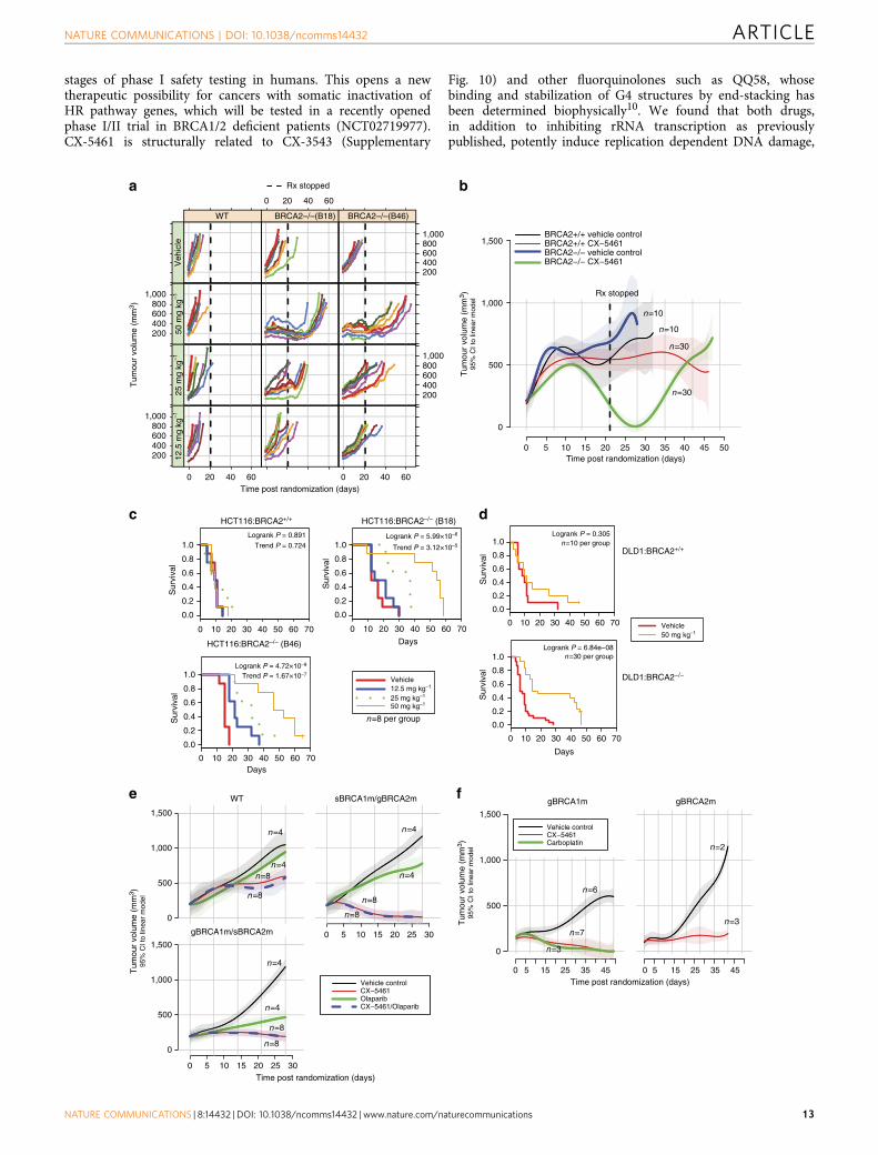
CX-5461 kills BRCA deficient and chemo-resistant PDXtumours. The significant synthetic lethality of CX-5461 withBRCA in vitro motivated us to determine whether a therapeuticwindow could be seen in vivo in xenograft models. The antitumoractivity of CX-5461 was evaluated in NOD/SCID/IL-2g� /�
immunodeficient mice with tumours subcutaneously engraftedfrom HCT116 isogenic cell line pairs. Strikingly,CX-5461 treatment substantially and significantly inhibited thegrowth rate of tumours formed from two BRCA2 deficientHCT116 cell lines (B18 and B46), in a dose-dependent manner(Fig. 8a, Supplementary Fig. 8a). Tumour growth of the BRCA2deficient tumours remained suppressed for 10–20 days afterceasing oral dosing with CX-5461 (Fig. 8a, vertical dashed lines).In marked contrast, no tumour growth inhibition was observedon tumours derived from BRCA2 proficient HCT116 cells underthe highest dosage. As a consequence, CX-5461 greatly extendedthe survival time of the mice bearing tumours derived fromBRCA2 knockout cells (Fig. 8c). At all tested schedules, CX-5461was well tolerated as judged by minimal effect on animal bodyweight (Supplementary Fig. 8a,b). Stronger tumour growthinhibition to BRCA2 deficient than BRCA2 WT cancers was alsoobserved in xenograft model with tumours formed by DLD1isogenic cell line pairs (Fig. 8b,d).
To address the relationship of CX-5461 activity to standardof care chemotherapy in polyclonal tumours, the activity ofCX-5461 was further tested in PDX models, beginning withtaxane resistant TNBC PDX tumours. Three different patienttumours were compared initially: two containing deleteriousBRCA1 or BRCA2 mutations and one wild type status. All threepatients had received a taxane prior to tumour sampling.Compared to vehicle control, CX-5461 reduced the tumourgrowth of all three PDX tumours, (Fig. 8e), but the inhibitionof BRCA1 or BRCA2 deficient PDX tumours (CTG-0012 andCTG-0888) was much greater than on BRCA WT PDX tumour(CTG-1019). We also observed that, CTG-0012 exhibited a weakresponse to Olaparib but was very sensitive to CX-5461, showingthat CX-5461 activity spectrum may in some cases transcend thatof Olaparib. The combination effect of CX-5461 and Olaparib issimilar to CX-5461 alone in these PDX models.
We next evaluated CX-5461 in a platinum-pretreatedTNBC PDX (Fig. 8f). PDX CFIB-NB02 was generated from ametastatic lesion biopsy from a heavily pretreated TNBC patient(including platinum) with BRCA1 germline missense mutation.The administration of CX-5461 resulted in dramatic tumourregression with efficacy comparable with carboplatin. PDXCFIB-70620 was generated from a TNBC patient pretreatedwith anthracycline/taxane chemotherapy with BRCA2 germlinemutation who had minimal response to cisplatin in the metastaticsetting (Supplementary Table 3). Again, CX-5461 significantlyreduced the tumour growth in this PDX model. Taken together,these data show that while CX-5461 activity spectrum partiallyoverlaps that of PARP inhibitors and platinum salts in HRdeficient tumours, CX-5461 exhibits additional activity in sometumours resistant to these agents.
DiscussionWe have discovered two related small molecule drugs CX-5461and CX-3543 that are able to selectively kill BRCA1/2 deficientcancer cells, one of which, CX-5461, is currently in advanced
NATURE COMMUNICATIONS | DOI: 10.1038/ncomms14432 ARTICLE
NATURE COMMUNICATIONS | 8:14432 | DOI: 10.1038/ncomms14432 | www.nature.com/naturecommunications 11

WT
helq-
1
polq-
1
let-4
18
mus
-81
rtel-1
atm
-1cb
p-1
rfs-1
brc-
2/+
0
50
100
% v
iabl
e em
bryo
s
NaH2PO4
100 mM CX-5461
a b
c
600M
PE
EF
M−
19M
DA
−M
B−
330
MC
F−
7M
CF
−7/
LY2
ZR
−75
−1
BT
−47
4M
DA
−M
B−
175−
VII
HC
C14
19O
CU
B−
MK
PL−
1T
−47
DC
AM
A−
1S
K−
BR
−5
MF
M−
223
HC
C21
85H
CC
1428
MD
A−
MB
−13
4−V
IC
AL−
148
HC
C19
54M
DA
−M
B−
361
SK
−B
R−
3A
U56
5E
vsa−
TU
AC
C−
893
HC
C20
2H
CC
1187
SW
527
MD
A−
MB
−46
8H
CC
1806
BT
−20
HC
C38
HC
C11
43H
CC
1937
JIM
T−
1H
CC
70H
CC
3153
MX
−1
MD
A−
MB
−43
6C
AL−
51S
UM
159P
TH
BL−
100
MD
A−
MB
−23
1C
AL−
120
HC
C13
95S
K−
BR
−7
Hs
578T
BT
−54
9M
DA
−M
B−
157
HD
Q−
P1
CX5461
CX3543
BMH21
Olaparib
–10
No mutationHR−mutated
LumALumBHer2BasalCLN
No mutationFunctional mutation
Cisplatin IC50Not doneLowMedHigh
BRCA status
Cisplatin sensitivityDNA repair mutation
Intrinsic subtype BRCA status
Cisplatin sensitivityDNA repair mutation
Intrinsic subtype
MD
A−
MB
−43
6C
AL−
148
600M
PE
CA
L−51
HC
C11
87S
UM
159P
TO
CU
B−
MH
BL−
100
MD
A−
MB
−23
1S
W52
7M
DA
−M
B−
468
EF
M−
19C
AL−
120
HC
C18
06H
CC
1954
HD
Q−
P1
BT
−20
MD
A−
MB
−33
0H
CC
1395
SK
−B
R−
7M
CF
−7
KP
L−1
MC
F−
7/LY
2H
CC
38H
CC
1143
Hs
578T
ZR
−75
−1
MD
A−
MB
−36
1S
K−
BR
−3
BT
−54
9T
−47
DC
AM
A−
1A
U56
5H
CC
1937
SK
−B
R−
5E
vsa−
TJI
MT
−1
MF
M−
223
HC
C21
85U
AC
C−
893
BT
−47
4H
CC
70H
CC
3153
MD
A−
MB
−17
5−V
IIH
CC
1419
MD
A−
MB
−15
7H
CC
1428
MD
A−
MB
−13
4−V
IH
CC
202
MX
−1
CX5461
CX3543
BMH21
Olaparib
HCT116: DNApk WT or KO
Log10(M) CX-5461
WS
T1
inte
nsity
nor
mal
ized
to c
ontr
ol
Control –10 –9 –8 –7 –6 –5
0.0
0.2
0.4
0.6
0.8
1.0
1.2
DNApk proficient
DNApk deficient
WTDNApk−/−
HCT116
Log10(M) CX−5461
WS
T1
inte
nsity
nor
mal
ized
to c
ontr
ol
Control –9 –8 –7 –6 –5
0.0
0.4
0.8
1.2
LIG4proLIG4def
d
HCT116
Log10(M) PDS
WS
T1
inte
nsity
nor
mal
ized
to c
ontr
olControl –9 –8 –7 –6 –5
0.0
0.4
0.8
1.2
LIG4proLIG4def
IC50 log10 (M) –4
Figure 7 | CX-5461 effectively kills tumour cells deficient for a number of DNA damage repair pathways. (a) PRKDC� /� HCT116 cells are more
sensitive to CX-5461 compared with PRKDC wild type HCT116 cells. The dose sensitivity of CX-5461 (6 days in drug) was measured by WST-1 assay with
representative experiment #2 shown (see Supplementary Fig. 7a for all three experiments). Vertical dashed lines indicate the estimated IC50 values. (b)
LIG4� /� HCT116 cells are more sensitive to CX-5461 and PDS compared with LIG4 wild type HCT116 cells. The drug sensitivity (4 days in drug) was
measured by a WST-1 assay. nZ4. IC50 difference for CX-5461 between LIG4þ /þ and LIG4� /� cells is 5.73 (Po10� 10, t-test). Sigmoid fits were
unavailable for PDS treated cells so no IC50 estimates or fold change was available. Assessment via a two-way ANOVA test showed that Lig4 deficient
cells were more sensitive to PDS than Lig4 proficient cells (F-test Po10� 10). Results of drug sensitivity to BMH-21 and cisplatin for LIG4þ /þ and LIG4� /
� cells are shown in Supplementary Fig. 7b. (c) Genotype specific sensitivity to CX-5461 in C. elegans. The percentage of viable embryos observed in the
progeny of animals treated forB20 h with 100mM CX-5461 or carrier (50 mM NaH2PO4). Error bars depict the s.e.m. of at least three experiments.
Statistically significant difference by t-test was discovered for all mutants shown in this figure (Po0.05) comparing carrier-treated to CX-5461-treated
animals. (d) CX-5461/CX-3543 exhibits antiproliferative potency against a panel of breast cancer cell lines by SRB cytotoxicity assay. Heatmap represents
IC50 values for 50 breast cancer cell lines treated with Olaparib, BMH-21, CX-3543 and CX-5461 for 5 days. The ward-linkage method of ‘hclust’ (R
function) was used to compute the dendrogram. The panel on the left is ordered by intrinsic subtype of cell lines, the panel on the right is ordered by ranked
value of CX-5461 sensitivity. The legends indicate the categorical values for intrinsic subtype, BRCA mutation status, DNA repair mutation status, and
cisplatin sensitivity (data from Sanger cell line project).
ARTICLE NATURE COMMUNICATIONS | DOI: 10.1038/ncomms14432
12 NATURE COMMUNICATIONS | 8:14432 | DOI: 10.1038/ncomms14432 | www.nature.com/naturecommunications
stages of phase I safety testing in humans. This opens a newtherapeutic possibility for cancers with somatic inactivation ofHR pathway genes, which will be tested in a recently openedphase I/II trial in BRCA1/2 deficient patients (NCT02719977).CX-5461 is structurally related to CX-3543 (Supplementary
Fig. 10) and other fluorquinolones such as QQ58, whosebinding and stabilization of G4 structures by end-stacking hasbeen determined biophysically10. We found that both drugs,in addition to inhibiting rRNA transcription as previouslypublished, potently induce replication dependent DNA damage,
0 10 20 30 40 50 60 70
HCT116:BRCA2+/+
Sur
viva
l
0.0
0.2
0.4
0.6
0.8
1.0Logrank P = 0.891
Trend P = 0.724
0 10 20 30 40 50 60 70
HCT116:BRCA2–/– (B18)
Days
Sur
viva
l
0.0
0.2
0.4
0.6
0.8
1.0Logrank P = 5.99×10⁻⁶
Trend P = 3.12×10⁻⁵
0 10 20 30 40 50 60 70
HCT116:BRCA2–/– (B46)
Days
Sur
viva
l
0.0
0.2
0.4
0.6
0.8
1.0Logrank P = 4.72×10⁻⁹
Trend P = 1.67×10⁻⁷Vehicle12.5 mg kg–1
25 mg kg–1
50 mg kg–1
gBRCA1m
0 5 15 25 35 45
0
500
1,000
1,500
Vehicle controlCX−5461Carboplatin
gBRCA2m
0 5 15 25 35 45Time post randomization (days)
0 10 20 30 40 50 60 70
0.0
0.2
0.4
0.6
0.8
1.0Logrank P = 0.305
0 10 20 30 40 50 60 70
DLD1:BRCA2−/−
Days
0.0
0.2
0.4
0.6
0.8
1.0Logrank P = 6.84e−08
Vehicle50 mg kg–1
0 5 10 15 20 25 30 35 40 45 50
0
500
1,000
1,500BRCA2+/+ vehicle controlBRCA2+/+ CX−5461BRCA2−/− vehicle controlBRCA2−/− CX−5461
Time post randomization (days)T
umou
r vo
lum
e (m
m3 )
95%
CI t
o lin
ear
mod
el
DLD1:BRCA2+/+
Sur
viva
lS
urvi
val
WT
0
500
1,000
1,500
sBRCA1m/gBRCA2m
0 5 10 15 20 25 30gBRCA1m/sBRCA2m
0 5 10 15 20 25 30
0
500
1,000
1,500
Vehicle controlCX−5461OlaparibCX−5461/Olaparib
Time post randomization (days)
Tum
our
volu
me
(mm
3 )95
% C
I to
linea
r m
odel
n=10
n=10
n=30
n=30
n=10 per group
n=30 per group
n=4
n=4
n=4
n=4n=8
n=8 n=8
n=8
n=4
n=4
n=8
n=8
n=3
n=2
n=7
n=3
n=6
Rx stopped
n=8 per group
Time post randomization (days)
Tum
our
volu
me
(mm
3 )
200400600800
1,000
0 20 40 60
12.5
mg
kg–1
0 20 40 60
25 m
g kg
–1
200400600800
1,000
2004006008001,000
2004006008001,000
50 m
g kg
–1
WT
Veh
icle
0 20 40 60
BRCA2–/–(B18) BRCA2–/–(B46)
Rx stopped
Tum
our
volu
me
(mm
3 )95
% C
I to
linea
r m
odel
a b
c d
e f
NATURE COMMUNICATIONS | DOI: 10.1038/ncomms14432 ARTICLE
NATURE COMMUNICATIONS | 8:14432 | DOI: 10.1038/ncomms14432 | www.nature.com/naturecommunications 13

likely through binding and stabilization of G4 structure formingDNA. We note that a potent, unrelated RNA pol I inhibitor(BMH-21) which does not bind/stabilize G4 sequences, also doesnot induce damage or exhibit synthetic lethality, showing thatRNA pol I transcription inhibition is not required for themechanism.
Upon treatment with CX-5461 and CX-3543, G4 structures aresignificantly induced and accompanied by a dramatic increase ofDNA damage foci in cells. BRCA deficient cells are lesscompetent to bypass drug stabilized G4 structure during DNAreplication and less efficient to repair G4 associated DNAdamage. As a consequence, the accumulated DNA damage inBRCA deficient cells leads to apoptosis (Supplementary Fig. 9).Besides the HR pathway, the repair of CX-5461 and CX-3543generated DNA damage also relies on the NHEJ pathway. Weanalysed CX-5461 response of three genes in the NHEJ pathway:DNA-PK, LIG4 and 53BP1. DNA-PK and LIG4 deficiencyincreases CX-5461 and PDS sensitivity, but 53BP1 knockingdown has no effect. 53BP1 is not strictly required for NHEJ inmany settings. For example, 53BP1 is required for NHEJ inclass-switch recombination, but not required for NHEJ in V(D)J recombination37,38. It is likely that 53BP1 doesn’t contribute toNHEJ of G4 associated DNA damage. In addition, some othergenes in DNA replication and damage response are also involvedin the repair of CX-5461/CX-3543 generated DNA damage.Mutation of ATM, ATR, BARD1, downregulation of genes inFANC pathway are associated with high efficacy to CX drugs inin vitro drug sensitivity assays. These results suggest the potentialapplication of CX-5461 in treating cancers bearing thesemutations.
The specific toxicity of CX-5461 and CX-3543 againstBRCA1/2 deficient cells was seen in a number of cell lines ofdifferent genetic backgrounds (colon, breast, ovary) and differentspecies origins (yeast, mouse and human). This is consistent withrecent data using probe compounds that stabilize G4 sequences,suggesting that selective sensitivity occurs in HR deficientbackgrounds6. CX-5461 has been engineered for superiorin vivo stability and pharmacokinetics and is presently inadvanced phase I trials for haematologic malignancies15.Consistent with the in vitro activities observed, CX-5461exhibited a wide therapeutic index of activity in BRCA2knockout tumour cells in xenograft models, when comparedwith isogenic wild type control cells. Furthermore, CX-5461 isalso effective in PDX models for chemo-resistant breast cancers,including tumours relatively insensitive to PARP inhibitionand/or platinum salts. Our data thus suggest immediatelypractical applications of CX-5461 in BRCA deficienttumours and possibly other tumours deficient for DNA repair.In particular, it is possible that the dose used to treat BRCA
deficient cancers may be lower than that required to inhibit RNApolymerase I and disrupt nucleolus function, because our datasuggest that BRCA deficient cells are killed by CX-5461 at lowdrug concentrations, which are not effective at inhibiting rDNAtranscription.
In summary, our study repurposes the application of CX-5461and CX-3543, and likely other G4 stabilizers, in treating cancerswith deficiencies in BRCA pathway, NHEJ pathway, and othergenes in DNA damage repair and DNA replication.
MethodsHuman cell lines, yeast and C. elegans strains. HCT116 BRCA2þ /þ cells andBRCA2� /� cells were described previously21. Mouse mammary tumour BRCA2knockout cells (K14-Cre; Brca2F11/F11; p53F2-10/F2-10) and control mousemammary tumour BRCA2 proficient cells (K14-Cre; Brca2wt/wt; p53F2-10/F2-10)were from Dr Jos Jonkers’ lab and were cultured according to publication23.DLD1 BRCA2 proficient and BRCA2 knockout cells, HCT116 DNA-PK WT andknockout cells, LIG4 WT and knockout cells were all from Horizon Discovery andwere grown in RPMI140 with 10% FBS and 2 mM L-glutamine. PEO1 and C4-2cells were from Toshiyasu Taniguchi’s lab and were grown in DMEM medium with10% FBS and L-glutamine22. U2OS cells were from ATCC and were grown inMcCoy’s 5 A medium with 10% FBS and L-glutamine. All cell lines are mycoplasmafree and have been authenticated by STR or SNP profiling.
Disease subtypes and mutation status of breast cancer cell line panel in Fig. 7dare extracted from publication36 and Cosmic (http://cancer.sanger.ac.uk/cell_lines),and are summarized in Supplementary Table 4.
Nematode strains were maintained as described previously39. The strains usedare listed in Supplementary Table 2. Some strains were generated by theInternational C. elegans Gene Knockout Consortium and the National BioresourceProject of Japan. The genotypes and background of all the yeast strains used in thisstudy are as previously described40.
Cell line xenograft mouse model. Animal procedures were approved by theUniversity of British Columbia animal protection committee. Six to ten week oldfemale NOD/SCID/IL-2g� /� immunodeficient mice were subcutaneouslyengrafted with 2� 106 tumour cells for BRCA2 proficient and 5� 106 cells forBRCA2 knockout cells. CX-5461 was dissolved in 50 mM NaH2PO4, pH4 forxenograft application. Established tumours were randomized into vehicle andCX-5461-treated groups. Tumour measurement was performed by external caliperand tumour volume was calculated using the formula [V¼ 1/2 (length�width2)].Mouse weight was measured every 3 days. CX-5461 was administered through oralgavage once every three days with three doses: 12.5 mg kg� 1, 25 mg kg� 1 and50 mg kg� 1 for tumours formed from HCT116 cells. For mice bearing tumoursfrom DLD1 cells, CX-5461 was administered orally at 50 mg kg� 1 once every threedays. Mice were sacrificed when tumour size reached 1,000 mm3, or when all othermice in a given genotype/drug dose group had been sacrificed, up to a maximum of90 days post-xenograft.
Patient-derived xenograft model. BRCA status and clinical characteristicof patient tumours in Fig. 8 are listed in Supplementary Table 3. MicePDX experiments for model CFIB-NB02 and CFIB-70620 were approved byUniversity Health Network Research Ethics Board in Toronto #15-9481-CE.PDX experiments for Model CTG-1019, CTG-0012, and CTG-0888 wereapproved by Office of Laboratory Animal Welfare, NIH (A4614-01).
For Model CTG-1019 (WT), vehicle control was administered intravenouslyeach week for 28 days (IV QWx4), Olaparib (50 mg kg� 1 dose� 1)
Figure 8 | CX-5461 selectively suppresses growth of BRCA deficient tumours in murine xenografts and chemo-resistant PDX model. (a,b) The effect of
CX-5461 on tumor growth with xenografted tumours from isogenic WT and BRCA2 knockout HCT116 (a) and DLD1 (b) cells. For HCT116 xenograft model,
three drug doses were administered at 12.5 mg kg� 1, 25 mg kg� 1 and 50 mg kg� 1 together with vehicle control. For DLD1 xenograft model, CX-5461 was
administered at 50 mg kg� 1. Vertical dashed lines show the end of drug treatment. Each coloured line represents individual mouse in a. Solid lines in b
represent the mean tumour volume with 95% CI (shown by shadow around solid lines) from a linear model fitted to the tumour volumes. (c,d) The
administration of CX-5461 greatly extended the survival of mice with tumours from BRCA2 deficient but not BRCA2 proficient in xenograft models with
tumours formed from HCT116 cells (c) and DLD1 cells (d). The survival of mice in experimental panel is shown as moribund-free survival time post
randomization. The significance of survival differences is indicated with the log rank test (See Statistical methods). Dose-dependent trend significance is
indicated with the log rank test for trend. (e) CX-5461 is effective for taxane resistant BRCA1/2 deficient TNBC in PDX model. Growth curve of tumours
grafted from individual patients with BRCA1 and BRCA2 WT (CTG-1019), BRCA1 germline and BRCA2 somatic mutation (CTG-0888, gBRCA1m/sBRCA2m),
and BRCA2 germline and BRCA1 somatic mutation (CTG-0012, sBRCA1m/gBRCA2m). Tumour volume average curves (lines) with pointwise 95% CIs
(shaded regions) are shown. Mice were treated with vehicle, Olaparib, CX-5461 or the combination of Olaparib and CX-5461. See Supplementary Fig. 8c for
body weight and drug dosing schedule. (f) CX-5461 is effective in cisplatin pretreated and BRCA1/2 deficient TNBC in PDX model. Tumour volume average
curves (lines) with pointwise 95% CIs (shaded regions) are shown. Tumours are grafted from patients with BRCA1 (CFIB-NB02), and BRCA2 (CFIB-70620)
germline mutation. See Supplementary Fig. 8d for body weight and drug dosing schedule.
ARTICLE NATURE COMMUNICATIONS | DOI: 10.1038/ncomms14432
14 NATURE COMMUNICATIONS | 8:14432 | DOI: 10.1038/ncomms14432 | www.nature.com/naturecommunications
was administered orally each day for 28 days (PO QDx28), CX-5461(125 mg kg� 1 dose� 1) was administered intravenously each week for 14 days(IV QWx3), and Olaparib (50 mg kg� 1 dose� 1) was administered orally each dayfor 17 days (PO QDx17) in combination with CX-5461 (125 mg kg� 1 dose� 1)administered intravenously each week for 14 days (IV QWx3) to the groupsrespectively.
For Model CTG-0012 (sBRCA1m/gBRCA2m), vehicle control wasadministered intravenously each week for 28 days (IV QWx4), Olaparib(50 mg kg� 1 dose� 1) was administered orally each day for 28 days (PO QDx28),CX-5461 (125 mg kg� 1 dose� 1) was administered intravenously each week for 7days (IV QWx2), and Olaparib (50 mg kg� 1 dose� 1) was administered orally eachday for 10 days (PO QDx10) in combination with CX-5461 (125 mg kg� 1 dose� 1)administered intravenously each week for 7 days (IV QWx2) to the groupsrespectively.
For Model CTG-0888 (gBRCA1m/sBRCA2m), vehicle control wasadministered intravenously each week for 28 days (IV QWx4), Olaparib(50 mg kg� 1 dose� 1) was administered orally each day for 28 days (PO QDx28),CX-5461 (62.5 mg kg� 1 dose� 1) was administered intravenously each week for 28days (IV QWx4) and Olaparib (50 mg kg� 1 dose� 1) was administered orally eachday for 28 days (PO QDx28) in combination with CX-5461 (62.5 mg kg� 1 dose� 1)administered intravenously each week for 28 days (IV QWx4) to the groupsrespectively.
For PDX model CFIB-NB02 (gBRCA1m) and CFIB-70620 (gBRCA2m), vehicle(50 mM NaH2PO4, pH 4) and CX-5461 (50 mg kg� 1) were administered onceevery 3 days through oral gavage. Carboplatin was administered at 7.5 mg kg� 1
through IP once every week.
siRNA sequences. siRNA for BRCA2: Target DNA sequence: AACAA-CAATTACGAACCAAAC, dTdT overhang. Other siRNAs are ordered fromDharmacon.
Drugs and antibodies. Drugs and antibodies used in this study are listed inSupplementary Table 1
WST-1 assay and clonogenic assay. Clonogenic assay was performed aspreviously described21. Drug incubation time for clonogenic assay is 12 days. Forclonogenic assay after siRNA knocking down, the cells were knocked down withsiRNA one day before plating for single cells and treated with drugs. WST-1 assaywas performed according to the protocol provided by manufactory (Roche,Catalogue number 11644807001). Wild type and BRCA2 knockout cells wereplated in 96-well plates either untreated or treated with drug continuously duringthe indicated time before WST-1 assay. Drug incubation time in Fig. 1 is 6 days andin Fig. 7 is 4 days except specified. Absorbance was read with the microplate readerSpectraMax 3 (Molecular Devices, Sunnyvale, CA, USA).
SRB assay. Cells were seeded into 96-well plates and incubated with drugs for 5days before fixation with 10% TCA, and being washed with tap water and air dry.Then the dried cells were stained with SRB reagent and incubated 30 min atroom temperature in the dark. Plates were later washed with 1% acetic acid andcompletely air dry, then solubilised with 10 mM Tris. The intensity of the stainedcells was read at 570 nm on spectrophotometer.
Immunofluorescence of DNA damage foci. For detection of RPA2, RAD51 focicells were extracted with CSK buffer (100 mM NaCl, 300 mM sucrose, 3 mMMgCl2, 10 mM PIPES (pH 6.8), with proteinase inhibitors) for 4 min beforefixation. For g-H2AX and 53BP1 foci visualization, cells were fixed with 4%paraformaldehyde in TBS (50 mM Tris-Cl, pH 7.5, 150 mM NaCl) withoutextraction, and then permeabilized with methanol for 1 min on ice. Cells wereblocked in blocking buffer (2% BSA, 0.2% Tween-20 in TBS) for 30 min beforeincubation with primary antibody overnight. Secondary antibodies were incubatedfor 1 h. Because of background staining, damage foci positive cells were defined ascells with g-H2AX fociZ5, 53BP1 fociZ3, RAD51 fociZ5, and RPA fociZ5.
Immunofluorescence of G4 detection. For detection of G4, HCT116 WT cellswere treated with 100 nM of CX-5461 or CX-3543 for 24 h, then fixed with 4%paraformaldehyde in PBS (15 min) and permeabilized with 0.1% Triton-X(20 min). Cells were blocked with 5% dry milk in PBS for 1 h before incubationwith primary, secondary and ternary antibody for 1 h each at 37 �C.
Western blotting. Cells were directly lysed in RIPA buffer with protease inhibitorsand phosphatases inhibitors. Brief sonication was applied to disrupt DNA, beforeboiling in laemmli sample buffer for 10 min at 90 �C. 10 mg protein was loaded ineach well of SDS–PAGE. For the detection of BRCA1/2 and 53BP1, 1 million cellsper sample were loaded to 6% SDS–PAGE.
Cell fractionation. 1 million cells per sample were washed once with PBS and thenlysed on ice with CSK buffer (100 mM NaCl, 300 mM sucrose, 3 mM MgCl2,
10 mM PIPES (pH 6.8), 0.5% Triton X-100 with proteinase inhibitors, andphosphatase inhibitors) for 15 min. Then the cells were equally separated to threevials: one for whole cell lysate (WC), one for free cytoplasmic and nucleoplasmicproteins (FCN) and one for chromatin-bound proteins (CB). After centrifugation(900 g, 5 min at 4 �C), the FCN fraction is in the suppernant and the CB fraction isin the pellet. The WC and CB fractions were resuspended in RIPA buffer with1� Laemmli sample buffer, then were boiled for 10–15 min before loading toSDS–PAGE.
Apoptosis assay. Apoptosis was assayed via Annexin V-FITC and propidiumiodide (PI) staining according to the manufacturer’s protocol (Invitrogen Carlsbad,CA, USA, Catalogue number, A13201). HCT116 cells were treated with CX-5461or vehicle for 72 h; then the percentage of apoptotic cells was analysed by flowcytometry. Early apoptotic cells are Annexin V positive and PI negative (lowerright quadrant in FACS profile); late apoptotic and dead cells are Annexin V and PIpositive (upper right quadrant). 10,000 cells were counted for each sample.
Cell cycle analysis. Cells were treated with 10 mM EdU, a thymidine analogue, for1 h prior to harvesting. Cells were then fixed with 4% paraformaldehyde for 15 min,permeabilized with 0.5% Triton-X 100 in PBS for 20 min and then incubated withclick-it cocktail according to manufacturers protocol (Invitrogen Carlsbad, CA,USA, Catalogue number, C10420).
Cell cycle synchronization using a double thymidine block was carried outaccording to a published paper41.
Mitotic index. The population of metaphase cells were identified by pH¼ 3 and PIdouble staining and analysed by flow cytometry. Cells were fixed with 70% ethanoland then blocked in TST (50 mM Tris-base, pH 7.6, 0.9% NaCl, 4% FBS, 0.2%Triton X-100) for 10 min before incubation with anti-pH 3 antibody (MilliporeCat. 06-570) for 1 h at room temperature. After washing with TST, secondaryantibody was added and incubated for 1 h. Before flow cytometry, cells wereincubated with 10mg ml� 1 PI and Rnase A (100 mg ml� 1) for 10 min.
Homologous recombination assay. HR was measure by using DR-GFP cell linewith transfected pCBA-(I-SceI)42.
Comet assay. Alkaline comet and neutral comet assays were performed accordingto publication26. Images were collected by fluorescence microscope. Comets wereanalysed from at least 100 cells per each replica using software developed byDr Ralph E Durand for tail moments calculation43.
Chromosome spread. Cells were treated with or without CX-5461 for 2 days, andthen were arrested in mitosis with 3.3 mgml� 1 Colcemid. Cells were arrested for2.5 h, centrifuged for 5 min at 3,500 r.p.m., resuspended in 1 ml hypotonic sodiumcitrate solution (0.5% Na-citrate in ddH2O) at concentration of 2� 105 cells per mland incubated for 20 min. 500ml� 1 ml of swelled cells were spun on positivelycharged slides in a Shandon 4 cytospin (900 r.p.m., high acceleration for 10 min),fixed with methanol and acetic acid mix (3:1) for 10 min and staining wasperformed by PI (1 mg ml) for 30 min. Spread chromosomes were visualized byfluorescence microscope at magnification of � 1,000. Chromosome abnormalitiesinclude breaks, acentric or dicentric chromosome, rings, radial chromosomes.
Telomere FISH. Chromosome spread was first prepared, and then telomere FISHwas performed by following the protocol provided along with the Telomere PNAFISH kit from DAKO (Cat. K5326).
DNA combing analysis. Cells are pulse-labelled with 25 mM CldU for 30 min, thenwashed with pre-warmed PBS and pulsed again with 125 mM IdU for 30 min in thepresence of drug. After trypsinization, cells are resuspended in PBS at 3� 106 cellsper ml. Cells are warmed at 37� for 5 min, then added an equal volume of 1% lowmelting point agarose solution, and poured into plug castor (BIO-RAD #170-3713).The agarose embedded with cells are solidified at 4 �C for 45 min, then are ejectinto ProK solution (1 mg ml� 1 proteinase K in 500 mM EDTA, 1% sarkosyl).Proteinse K pre-digested are performed at 50 �C for 30 min before use.
Each agarose plug was stained with YOYO-1 (1 ml of YOYO-1 in 150ml TE50) atroom temperature for 30 min. Plugs were washed three times in 10 ml TE50 andsubsequently incubated in 2 ml MES (7:3 v/v MES hydrate: MES sodium salt,50 mM, pH 5.7) and melted at 72 �C for 15 min. Silanized coverslips wereincubated in the melted agarose plug solution and DNA fibres were combed ontothe coverslips at 710 mm s� 1. DNA was hybridized at 60 �C for 90 min, anddehydrated sequentially in 70%, 90% and anhydrous ethanol. DNA was denaturedby incubation in 1 M NaOH for 25 min at room temperature. Cover slips withcombed DNA were incubating in block buffer (10% w/v albumin in PBS-T,0.05% Tween-20, 2 mM KH2PO4, 2.7 mM KCl, 137 mM NaCl, pH 7.4), anti-CldU& anti-IdU solution (AbD Serotec MCA2060 and Becton Dickson 347580,respectively, 1:40 dilution in blocking buffer), anti-ssDNA solution (Millipore
NATURE COMMUNICATIONS | DOI: 10.1038/ncomms14432 ARTICLE
NATURE COMMUNICATIONS | 8:14432 | DOI: 10.1038/ncomms14432 | www.nature.com/naturecommunications 15

MAB3034 1:50 dilution in blocking buffer), and anti-secondary solution (AlexaFluor 546 anti-mouse IgG1, Invitrogen A21123, 1:50 dilution, Alexa fluor 648anti-mouse IgG2, Invitrogen A21241, 1:50 dilution, and Alexa Fluor 488 anti-rat,Invitrogen A11006) for 1 h at 37 �C each time, with PBS-T washes between eachstaining. ProLong Gold was added to each cover slip and coverslips were stored at� 20 �C prior to imaging.
GCR assay. Yeast GCR assay was performed as previously described32. CX-5461treatment time was 36–48 h. The GCR rates were calculated using the FALCORweb server and MMS maximum likelihood method. The number of GCR events(m) were used to determine the level of significance (P value) with a studenttwo-tailed t-test44.
Yeast growth and RAD52-YFP imaging. Yeast growth curves in YPD andRad52-YFP imaging in synthetic complete media at 30 �C were performed aspreviously described45. The drugs were diluted in 50 mM NaH2PO4 buffer(CX-5461) and 1% DMSO (CX-3543).
C. elegans CX-5461 chronic sensitivity assays. 5 L1 larvae of each mutant strainwere picked to 50 ml of M9 buffer containing OP50, carbenicillin (50 mg ml� 1),1� nystatin, 500 mM NaH2PO4 with and without 100mM CX-5461. Worms wereincubated for 4–6 days and F1 growth compared between wells with and withoutCX-5461. A strain was scored as sensitive if there was an obvious growth differencebetween wells with and without CX-5461 in three of three separate experiments.
C. elegans CX-5461 acute sensitivity assays. L4 stage animals were picked tofresh plates and aged for 24 h so that the animals were 1-day-old adults on the dayof the experiment. Young adults were incubated in CX-5461 (in NaH2PO4) dilutedin M9 buffer containing OP50, carbenicillin (50mg ml� 1) and 1� nystatin forB20 h. Following treatment, the animals were allowed to recover 1 h on OP50containing NGM plates before being plated at ten per plate on NGM plates for a 4 hinterval (20–24 h post-treatment). The number of embryos laid during the 4 hinterval was counted and the number of arrested embryos versus hatching larvaewas counted 24 h later in order to calculate the percentage of progeny survivingafter treatment. All results were from at least 30 treated animals (three plates with10 animals per plate).
RAD51 ChIP-seq. RAD51-ChIP was performed according to the publishedprotocol46 by using RAD51 antibody (Santa Cruz, Cat. 8349) after 24 h treatmentwith or without CX-5461 (10� 7 M). Paired FASTQ files were obtained and adaptorsequences were removed automatically with Trim Galore! (v0.4.1) [1], then alignedwith bowtie2 (v2.2.7) with default settings against hg19þ rDNA. After alignment,only concordantly aligned pairs with a mapping quality of X30 were kept,removing misaligned and ambiguously aligned reads. Supplementary Table 8shows the number of reads going in, successfully mapped, and remaining afterfiltering.
Biological replicates for treated, untreated, and IgG runs were run throughMACS2 (v2.1.1) using the same replicate background untreated IgG libraries.MACS2 was set to automatically detect fragment size, build the model, and outputall narrow peaks with a q-value of r0.25. We then categorized peaks as those thatwere reoccurring if it overlapped with at least one other peak (within 500 bp up anddownstream) in a biological replicate with the same background library. Peaks thatdid not overlap with any other peaks are then referred to as ‘unique’ peaks. Allpeaks were then filtered by q-value, keeping only those with a q-value of r0.1.
G4 are defined as PDS-induced sites from Chambers’ paper47 on either strandthat fall within the peaks ±500 bps.
Quantitative real-time reverse transcription PCR. RNA isolation and reversetranscription has been described before21. Quantitative PCR was performed on anABI 7900HT system. In total, 45s pre-rRNA level was measured using primersamplifying an internal transcribed spacer region as described in ref. 48 (forwardprimer, GCC TTC TCT AGC GAT CTG AGA G; reverse primer, CCA TAA CGGAGG CAG AGA CA) by sybrgreen analysis (Thermo Scientific, Cat. number,K0221), and also using a published primers-probe set16 by Taqman assay (AppliedBiosystems, Cat. number, 4364338). Relative cDNA amounts were normalized toACTIN B and 18s rRNA. ACTIN B was detected by sybrgreen (forward primer,ccaaccgcgagaagatga; reverse primer, ccagaggcgtacagggatag) and Taqman RT-PCRassay (probe #64 from Roche). In total, 18s rRNA was evaluated by both sybrgreenamplification (forward primer, AAACGGCTACCACATCCA; reverse primer,CCTCCAATGGATCCTCGT, amplifying a non-spacer region in 45s pre-rRNA)and a Taqman primers-probe set (forward primer, cgattggatggtttagtgagg; reverseprimer, agttcgaccgtcttctcagc; prober #81 from Roche). Actinomycin treatment(50 nm, 24 h) was used as a positive control for rDNA transcription inhibition.BRCA2 and polR1B expression were analysed by Taqman approach. BRCA2forward primer: gcaagatggtgcagagcttta; reverse primer: cttaactgctcttcactgaaataacc;probe #84 from Roche. PolR1B forward primer: ttccagctttactgtgcaactt; reverseprimer: acccagccaaccatgaca; probe #18 from Roche.
FRET melting assay. FRET experiments were carried out with a 200 nMoligonucleotide concentration. All DNA oligonucleotides were supplied by IBAGmbH. The donor fluorophore was 6-carboxyfluorescein (FAM) and the acceptorfluorophore was 6-carboxytetramethylrhodamine (TAMRA). The dual-labelledoligonucleotides were annealed at a concentration of 400 nM by heating at 94 �Cfor 10 min followed by slow cooling to room temperature at a controlled rate of0.1 �C min� 1. 96-well plates were prepared by addition of 50 ml of the annealedDNA solution to each well, followed by 50 ml solution of molecules CX-5461,CX-3543 at the appropriate concentration.
Seven dual fluorescently labelled DNA oligonucleotides were used in these ex-periments: H-Telo (50-FAM-GGG TTA GGG TTA GGG TTA GGG-TAMRA-30),C-kit1 (50-FAM-GGG AGG GCG CTG GGA GGA GGG-TAMRA-30), C-myc(50-FAM-TGA GGG TGG GTA GGG TGG GTA A-TAMRA-30), ds-DNA(50-FAM-TAT AGC TAT A-HEG-T ATA GCT ATA-TAMRA-30) which is adual-labelled 20-mer oligonucleotide comprising a self-complementary sequencewith a central polyethylene glycol linker able to fold into a hairpin. The competitorwas an unlabelled 26-mer oligonucleotide (50-CAA TCG GAT CGA ATT CGATCC GAT TG-30). A total of 100 mM stock solutions of oligonucleotides wereprepared in molecular biology grade DNase-free water. Further dilutions werecarried out in 60 mM potassium cacodylate buffer, pH 7.4.
For the competition experiments, competitor was added to the fluorescentlylabelled quadruplex oligonucleotides sequences at an excess of 50 mol equiv. andannealed in the same solution. Measurements were made in triplicate with anexcitation wavelength of 483 nm and a detection wavelength of 533 nm.
Polymerase stop assay. WT cKit1 template: TCT GCT TTG GGA ACC CGAGAG GAG CGC TTA TAG GGA GGG CGC TGG GAG GAG GGA GGA GACTCA GCC GAG CAG CCG AGC ACT CTA GCT CTA G MUT cKit1 template:TCT GCT TTG GGA ACC CGA GAG GAG CGC TTA TAG AGA GAG CGCTGA GAG GAG AGA GGA GAC TCA GCC GAG CAG CCG AGC ACT CTAGCT CTA G
Primer: [Cyanine5] CTA GAG CTA GAG TGC TCG GC8 ml of 1� NEB buffer 1 supplemented with 100 mM KCl containing 500 nM of
template oligo and 50 nM of primer was added to a PCR tube. The solution washeated to 95 �C for 5 min then allowed to cool to room temperature. 1 ml ofG4-stabilizing ligand (in a 1% DMSO/water solution) was added to the annealedmaterial. This solution was incubated at 37 �C for 30 min. Then 1 ml of thepolymerase mix (89 ml of 1� NEB buffer 1, 10ml of 10 mM NEB dNTP mix, and1 ml of NEB Klenow fragmentexo� ) was added. The mixture was incubated at 37 �Cfor 2 h. The reaction was stopped by addition of 20 ml of 2� NEB orange gelloading dye and heated to 95 �C for 5 min. To a 15% TBE-Urea gel (PrecastNovex), was added 7 ml of the quenched solution. Gel running conditions were300 V for 35 min. The gel was scanned on a GE Typhoon imager set to detect Cy5.The lane pixel density quantitation was processed using ImageJ software. Fractionalpeak area was quantified by fluorescence intensity of the dashed box area over thetotal fluorescence on the lane. The appearance of smaller bands indicates the stallof DNA polymerase.
Statistical methods.
R Session information:R version 3.2.0 (2015-04-16) was used for all statistical analysis of data.Platform: x86_64-apple-darwin13.4.0 (64-bit)Running under: OS X 10.10.3 (Yosemite)locale:[1] Cattached base packages:[1] stats graphics grDevices utils datasets methods baseother attached packages:[1] nlme_3.1-120 gdata_2.16.1loaded via a namespace (and not attached):[1] compiler_3.2.0 tools_3.2.0 grid_3.2.0 gtools_3.4.2[5] lattice_0.20-31
Dose response curve fitting for WST-1 assay. Where sigmoid curves wereappropriate for dose response analysis, a four-parameter logistic regression curvewas fitted to the data using the R function ‘nlme’. An IC50 was estimated, thedifference in IC50 values was calculated for conditions with and without drug.The set of differences was tested using the Student’s paired t-test.
Where logistic model fits were not available, typically due to lack of fit when thenumber of drug doses assayed did not adequately cover the descending portion ofthe sigmoid curve, rank regression was used to compare the drug response for themultiple groups comprising the experimental condition. The entire experiment’snormalized intensity values were ranked from smallest to largest, and theirnormalized intensity values replaced by their rank values49. A standard linearmodel of rank versus categorized drug dose was fitted for each experimentalcondition, with differing slope and intercept for each experimental condition,yielding a standard two-way ANOVA design. Drug dose was categorized to allowthe linear model to reasonably fit the non-linear sigmoid shape of the data.Differences in response to drug dose were assessed by testing the experimental
ARTICLE NATURE COMMUNICATIONS | DOI: 10.1038/ncomms14432
16 NATURE COMMUNICATIONS | 8:14432 | DOI: 10.1038/ncomms14432 | www.nature.com/naturecommunications

factor using a standard F-test comparing the ANOVA models with and without theexperimental condition variable. Dose response curves were estimated usingFriedman’s super-smoother50 but IC50s were not estimable.
Survival analysis for xenograft study. Mice showing tumour growth to 200 cubicmillimetres were randomized to treatment groups. After randomization, mice werefollowed until tumour growth reached 1,000 cubic millimetres or until all othermice within each genotype had been sacrificed, up to a maximum of 90 days ofobservation. Thus the survival analysis endpoint was time to sacrifice due tomaximum allowable tumour volume. Mice who lived without reaching maximumallowable tumour volume and were subsequently sacrificed were consideredcensored at their sacrifice date for the purposes of survival analysis.Survival datawere displayed graphically using Kaplan–Meier curves. Time to event differenceswere assessed using the log-rank test51. Log-rank analysis and test for trend werecarried out for the control group and three drug dose groups within each cell linexenograft and genotype. The trend test was conducted using the log-rank test fortrend52. All analyses were conducted using the R statistical software package53.
qPCR assay statistical analysis. qPCR Ct values were modelled using standardGaussian linear models with categorical variables for the experimental conditionsof interest, yielding two-way (for example, experiments comparing effects betweenBRCA2 genotypes) and three-way (for example, experiments comparing BRCA2genotype responses within differing treatment conditions) ANOVA designs.Additional categorical variables indicating experimental plate and multipleloading controls were also included in the models, to allow model adjustment forshifts in Ct values from experiment to experiment and among loading controls.Experimental effects were assessed using F-tests. Effect magnitudes and 95% CIs onthe log2 scale were obtained from delta-delta-Ct calculations from the linearmodels. Fold change estimates were obtained via exponents of the form2� (delta-delta-Ct). When assessing multiple gene targets, resultant F-test P valueswere adjusted for multiple comparisons using the Benjamini–Hochberg method54.All analyses was conducted using the R statistical software package53.
Comet assay statistical analysis. Mixed effects models compare the means of thesets of cells in each experiment and condition and obtain variance estimatesreflecting between experiment and condition means rather than between individualcell measurements, with degrees of freedom reflecting the number of conditionsand experiments rather than the total number of cells assayed.
Raw tail moment values were modelled using a linear mixed effects model55.Family-wise 95% CIs (ie, adjusted for multiple comparisons56 for experimentalcondition contrasts of interest were calculated and displayed graphically.
Data availability. The authors declare that the dataset and computer codegenerated during and/or analysed during the current study are available from thecorresponding author on reasonable request. RAD51-ChIP data have beendeposited in the Genbank GEO database (http://www.ncbi.nlm.nih.gov/geo/) underaccession codes GSE90967. G4 sequences in human genome were obtained fromthe paper by Chamber et al.47, and available under the accession code GSE63874.All other remaining data are available within the Article and Supplementary Files,or available from the authors upon request.
References1. Breast Cancer Linkage, C. Cancer risks in BRCA2 mutation carriers. J. Natl
Cancer Inst. 91, 1310–1316 (1999).2. Wooster, R. et al. Identification of the breast cancer susceptibility gene BRCA2.
Nature 378, 789–792 (1995).3. Davies, A. A. et al. Role of BRCA2 in control of the RAD51 recombination and
DNA repair protein. Mol. Cell 7, 273–282 (2001).4. Schlacher, K. et al. Double-strand break repair-independent role for BRCA2 in
blocking stalled replication fork degradation by MRE11. Cell 145, 529–542(2011).
5. Bhatia, V. et al. BRCA2 prevents R-loop accumulation and associates withTREX-2 mRNA export factor PCID2. Nature 511, 362–365 (2014).
6. Zimmer, J. et al. Targeting BRCA1 and BRCA2 deficiencies withG-quadruplex-interacting compounds. Mol. Cell 61, 449–460 (2016).
7. Huppert, J. L. & Balasubramanian, S. Prevalence of quadruplexes in the humangenome. Nucleic Acids Res. 33, 2908–2916 (2005).
8. Todd, A. K., Johnston, M. & Neidle, S. Highly prevalent putative quadruplexsequence motifs in human DNA. Nucleic Acids Res. 33, 2901–2907 (2005).
9. Hansel-Hertsch, R. et al. G-quadruplex structures mark human regulatorychromatin. Nat. Genet. 48, 1267–1272 (2016).
10. Duan, W. et al. Design and synthesis of fluoroquinophenoxazines that interactwith human telomeric G-quadruplexes and their biological effects. Mol. CancerTher. 1, 103–120 (2001).
11. Ward, J. D., Barber, L. J., Petalcorin, M. I., Yanowitz, J. & Boulton, S. J.Replication blocking lesions present a unique substrate for homologousrecombination. EMBO J. 26, 3384–3396 (2007).
12. Youds, J. L., O’Neil, N. J. & Rose, A. M. Homologous recombination is requiredfor genome stability in the absence of DOG-1 in Caenorhabditis elegans.Genetics 173, 697–708 (2006).
13. McLuckie, K. I. et al. G-quadruplex DNA as a molecular target for inducedsynthetic lethality in cancer cells. J. Am. Chem. Soc. 135, 9640–9643 (2013).
14. Derenzini, M. et al. Nucleolar size indicates the rapidity of cell proliferation incancer tissues. J. Pathol. 191, 181–186 (2000).
15. Bywater, M. J. et al. Inhibition of RNA polymerase I as a therapeutic strategy topromote cancer-specific activation of p53. Cancer Cell 22, 51–65 (2012).
16. Drygin, D. et al. Anticancer activity of CX-3543: a direct inhibitor of rRNAbiogenesis. Cancer Res. 69, 7653–7661 (2009).
17. Drygin, D. et al. Targeting RNA polymerase I with an oral small moleculeCX-5461 inhibits ribosomal RNA synthesis and solid tumor growth. CancerRes. 71, 1418–1430 (2011).
18. Peltonen, K. et al. A targeting modality for destruction of RNA polymerase Ithat possesses anticancer activity. Cancer Cell 25, 77–90 (2014).
19. Negi, S. S. & Brown, P. rRNA synthesis inhibitor, CX-5461, activatesATM/ATR pathway in acute lymphoblastic leukemia, arrests cells in G2 phaseand induces apoptosis. Oncotarget 6, 18094–18104 (2015).
20. Li, L. et al. CX-5461 induces autophagy and inhibits tumor growth viamammalian target of rapamycin-related signaling pathways in osteosarcoma.OncoTargets Ther. 9, 5985–5997 (2016).
21. Xu, H. et al. Up-regulation of the interferon-related genes in BRCA2 knockoutepithelial cells. J. Pathol. 234, 386–397 (2014).
22. Sakai, W. et al. Functional restoration of BRCA2 protein by secondary BRCA2mutations in BRCA2-mutated ovarian carcinoma. Cancer Res. 69, 6381–6386(2009).
23. Evers, B. et al. Selective inhibition of BRCA2-deficient mammary tumor cellgrowth by AZD2281 and cisplatin. Clin. Cancer Res. 14, 3916–3925 (2008).
24. Chen, J. J., Silver, D., Cantor, S., Livingston, D. M. & Scully, R. BRCA1, BRCA2,and Rad51 operate in a common DNA damage response pathway. Cancer Res.59, 1752s–1756s (1999).
25. Schlacher, K., Wu, H. & Jasin, M. A distinct replication fork protection pathwayconnects Fanconi anemia tumor suppressors to RAD51-BRCA1/2. Cancer Cell22, 106–116 (2012).
26. Olive, P. L. & Banath, J. P. The comet assay: a method to measure DNA damagein individual cells. Nat. Protoc. 1, 23–29 (2006).
27. Toledo, L. I. et al. ATR prohibits replication catastrophe by preventing globalexhaustion of RPA. Cell 155, 1088–1103 (2013).
28. Mergny, J. L. & Maurizot, J. C. Fluorescence resonance energy transfer as a probefor G-quartet formation by a telomeric repeat. Chembiochem 2, 124–132 (2001).
29. Rodriguez, R. et al. Small-molecule-induced DNA damage identifies alternativeDNA structures in human genes. Nat. Chem. Biol. 8, 301–310 (2012).
30. Weitzmann, M. N., Woodford, K. J. & Usdin, K. The development and use of aDNA polymerase arrest assay for the evaluation of parameters affectingintrastrand tetraplex formation. J. Biol. Chem. 271, 20958–20964 (1996).
31. Biffi, G., Tannahill, D., Miller, J., Howat, W. J. & Balasubramanian, S. Elevatedlevels of G-quadruplex formation in human stomach and liver cancer tissues.PLoS ONE 9, e102711 (2014).
32. Paeschke, K. et al. Pif1 family helicases suppress genome instability atG-quadruplex motifs. Nature 497, 458–462 (2013).
33. Doe, C. L., Ahn, J. S., Dixon, J. & Whitby, M. C. Mus81-Eme1 and Rqh1involvement in processing stalled and collapsed replication forks. J. Biol. Chem.277, 32753–32759 (2002).
34. Mateos-Gomez, P. A. et al. Mammalian polymerase y promotes alternativeNHEJ and suppresses recombination. Nature 518, 254–257 (2015).
35. Koole, W. et al. A Polymerase Theta-dependent repair pathway suppressesextensive genomic instability at endogenous G4 DNA sites. Nat. Commun. 5,3216 (2014).
36. Marcotte, R. et al. Functional genomic landscape of human breast cancerdrivers, vulnerabilities, and resistance. Cell 164, 293–309 (2016).
37. Ward, I. M. et al. 53BP1 is required for class switch recombination. J. Cell Biol.165, 459–464 (2004).
38. Manis, J. P. et al. 53BP1 links DNA damage-response pathways toimmunoglobulin heavy chain class-switch recombination. Nat. Immunol. 5,481–487 (2004).
39. Brenner, S. Caenorhabditis elegans. Methods (1974).40. Stirling, P. C. et al. Mutability and mutational spectrum of chromosome
transmission fidelity genes. Chromosoma 121, 263–275 (2012).41. Bostock, C. J., Prescott, D. M. & Kirkpatrick, J. B. An evaluation of the double
thymidine block for synchronizing mammalian cells at the G1-S border. Exp.Cell Res. 68, 163–168 (1971).
42. Pierce, A. J., Johnson, R. D., Thompson, L. H. & Jasin, M. XRCC3 promoteshomology-directed repair of DNA damage in mammalian cells. Genes Dev. 13,2633–2638 (1999).
43. Collins, A. R. The comet assay for DNA damage and repair. Mol. Biotechnol.26, 249–261 (2004).
NATURE COMMUNICATIONS | DOI: 10.1038/ncomms14432 ARTICLE
NATURE COMMUNICATIONS | 8:14432 | DOI: 10.1038/ncomms14432 | www.nature.com/naturecommunications 17
44. Rosche, W. A. & Foster, P. L. Determining mutation rates in bacterialpopulations. Methods 20, 4–17 (2000).
45. Stirling, P. C. et al. R-loop-mediated genome instability in mRNA cleavage andpolyadenylation mutants. Genes Dev. 26, 163–175 (2012).
46. Schmidt, D. et al. ChIP-seq: using high-throughput sequencing to discoverprotein–DNA interactions. Methods 48, 240–248 (2009).
47. Chambers, V. S. et al. High-throughput sequencing of DNA G-quadruplexstructures in the human genome. Nat. Biotechnol. 33, 877–881 (2015).
48. Tan, B. C. et al. Epigeneitc silencing of ribosomal RNA genes by Mybbp1a.J. Biomed. Sci. 19, 57 (2012).
49. Iman, R. L. A power study of a rank transform for the two-way classificationmodel when interaction may be present. Can. J. Stat. 2, 227–239 (1974).
50. Friedman, J. H. SMART User’s Guide (1984).51. Kalbfleisch, J. D. & Prentice, R. L. The Statistical Analysis of Failure Time Data
(John Wiley & Sons, 2011).52. TARONE, R. E. Tests for trend in life table analysis. Biometrika 62, 679–690
(1975).53. Kuhn, M., Wing, J., Weston, S., Williams, A. & Keefer, C. The R Core Team
(Benesty, 2015).54. Benjamini, Y. & Hochberg, Y. On the adaptive control of the false discovery
rate in multiple testing with independent statistics. J. Educ. Behav. Stat. 25,60–83 (2000).
55. Bates, D., Maechler, M., Bolker, B. & Walker, S. lme4: Linear Mixed-EffectsModels Using Eigen and S4. R Package Version (2014).
56. Hothorn, T., Bretz, F. & Westfall, P. Simultaneous inference in generalparametric models. Biom. J. 50, 346–363 (2008).
AcknowledgementsWe greatly appreciate Dr Jos Jonkers’ lab for generously distributing mouse mammarytumour BRCA2 knockout cells (K14-Cre; Brca2F11/F11; p53F2-10/F2-10) and control mousemammary tumour BRCA2 proficient cells (K14-Cre; Brca2wt/wt; p53F2-10/F2-10);Toshiyasu Taniguchi’s lab for sharing C4-2 and PEO1 cells; Benjamin G. Neel’s lab forsharing the 50 breast cancer cell lines; Ross D. Hannan, Geraldine Aubert, and TehminaMasud for helpful suggestions to this paper.
This work was supported by the Canadian Breast Cancer Foundation BC/Yukon,BC Cancer Foundation, Stand Up to Cancer Canada (SU2C-AACR-DT-18-15),TFRI Grant 1021, CCSRI Grant 701584, CIHR Grant MOP-126119, Canada Foundationfor Innovation and Cancer Research UK. Grant Brown lab is supported by CCSRI Impact
Grant 702310 (to G.W.B.) and Ontario Government Scholarship (to B.H.). S.A. issupported by a Canada Research Chair in Molecular Oncology. The Balasubramanian labis supported by a programme grant (C14303/A17197) and core funding (C14303/A17197) from Cancer Research UK.
Author contributionsH.X., S.A., S.B., P.H., P.C.S., D.C., T.W.M., C.C., M.B.B., J.B. G.W.B. designedthe experiments. H.X., M.D.A., V.M., B.H., N.J.O., N.D.S., J.S., V.W., J.G., P.C.S.,A.H.C.T., F.K., D.D. Le, D.Y., F.B.Y., J.B., A.Z., K.T., J.S., S.L., T.O., T.A., D.S., J.W.performed the experiments. S.M., D. Lai, and D.S.C. analysed the data. J.X. createdBRCA2� /� HCT116 cell line. H.X., M.D.A., V.M., N.J.O. S.S. and S.A. wrotethe paper.
Additional informationSupplementary Information accompanies this paper at http://www.nature.com/naturecommunications
Competing financial interests: J.S. and S.L. are employees of Senwah Biosciences Ltd.The other authors have no conflicts of interest to declare.
Reprints and permission information is available online at http://npg.nature.com/reprintsandpermissions/
How to cite this article: Xu, H. et al. CX-5461 is a DNA G-quadruplex stabilizerwith selective lethality in BRCA1/2 deficient tumours. Nat. Commun. 8, 14432doi: 10.1038/ncomms14432 (2017).
Publisher’s note: Springer Nature remains neutral with regard to jurisdictional claims inpublished maps and institutional affiliations.
This work is licensed under a Creative Commons Attribution 4.0International License. The images or other third party material in this
article are included in the article’s Creative Commons license, unless indicated otherwisein the credit line; if the material is not included under the Creative Commons license,users will need to obtain permission from the license holder to reproduce the material.To view a copy of this license, visit http://creativecommons.org/licenses/by/4.0/
r The Author(s) 2017
ARTICLE NATURE COMMUNICATIONS | DOI: 10.1038/ncomms14432
18 NATURE COMMUNICATIONS | 8:14432 | DOI: 10.1038/ncomms14432 | www.nature.com/naturecommunications