cxcl17 is a specific diagnostic biomarker for severe
TRANSCRIPT

Washington University School of Medicine Washington University School of Medicine
Digital Commons@Becker Digital Commons@Becker
Open Access Publications
1-1-2021
CXCL17 is a specific diagnostic biomarker for severe pandemic CXCL17 is a specific diagnostic biomarker for severe pandemic
influenza A(H1N1) that predicts poor clinical outcome influenza A(H1N1) that predicts poor clinical outcome
Jose Alberto Choreño-Parra Instituto Politécnico Nacional,
Shabaana A Khader Washington University School of Medicine in St Louis
et al
Follow this and additional works at: https://digitalcommons.wustl.edu/open_access_pubs
Recommended Citation Recommended Citation Choreño-Parra, Jose Alberto; Khader, Shabaana A; and et al, ,"CXCL17 is a specific diagnostic biomarker for severe pandemic influenza A(H1N1) that predicts poor clinical outcome." Frontiers in Immunology.,. . (2021). https://digitalcommons.wustl.edu/open_access_pubs/10291
This Open Access Publication is brought to you for free and open access by Digital Commons@Becker. It has been accepted for inclusion in Open Access Publications by an authorized administrator of Digital Commons@Becker. For more information, please contact [email protected].

CXCL17 Is a Specific DiagnosticBiomarker for Severe PandemicInfluenza A(H1N1) That Predicts PoorClinical OutcomeJose Alberto Choreño-Parra1,2, Luis Armando Jimenez-Alvarez2,Gustavo Ramırez-Martınez2, Montserrat Sandoval-Vega3, Citlaltepetl Salinas-Lara4,Carlos Sanchez-Garibay4, Cesar Luna-Rivero5, Erika Mariana Hernandez-Montiel 2,6,Luis Alejandro Fernandez-Lopez1,2, Marıa Fernanda Cabrera-Cornejo2,6,Eduardo Misael Choreño-Parra7, Alfredo Cruz-Lagunas2, Andrea Domınguez2,6,Eduardo Marquez-Garcıa2, Carlos Cabello-Gutiérrez8,Francina Valezka Bolaños-Morales9, Lourdes Mena-Hernandez10,Diego Delgado-Zaldivar10, Daniel Rebolledo-Garcıa10, Parmenides Guadarrama-Ortiz11,Nora E. Regino-Zamarripa1,2,6, Criselda Mendoza-Milla2, Ethel A. Garcıa-Latorre1,Tatiana Sofia Rodiguez-Reyna12, Diana Cervantes-Rosete12,Carmen M. Hernandez-Cardenas13, Shabaana A. Khader14, Albert Zlotnik15
and Joaquın Zuñiga2,6*
1 Escuela Nacional de Ciencias Biologicas, Instituto Politecnico Nacional, Mexico City, Mexico, 2 Laboratorio deInmunobiologıa y Genetica, Instituto Nacional de Enfermedades Respiratorias “Ismael Cosıo Villegas”, Mexico City, Mexico,3 Facultad de Estudios Superiores Iztacala, Universidad Nacional Autonoma de Mexico, Mexico City, Mexico, 4 Departamentode Neuropatologıa, Instituto Nacional de Neurologıa y Neurocirugıa “Manuel Velasco Suarez”, Mexico City, Mexico,5 Department of Pathology, Instituto Nacional de Enfermedades Respiratorias “Ismael Cosıo Villegas”, Mexico City, Mexico,6 Tecnologico de Monterrey, Escuela de Medicina y Ciencias de la Salud, Mexico City, Mexico, 7 Posgrado en CienciasBiologicas, Universidad Nacional Autonoma de Mexico, Mexico City, Mexico, 8 Department of Virology, Instituto Nacional deEnfermedades Respiratorias Ismael Cosıo Villegas, Mexico City, Mexico, 9 Subdirection of Surgery, Instituto Nacional deEnfermedades Respiratorias Ismael Cosıo Villegas, Mexico City, Mexico, 10 Departments of Dermatology and Education,Instituto Nacional de Ciencias Medicas y Nutricion Salvador Zubiran, Mexico City, Mexico, 11 Centro Especializado enNeurocirugıa y Neurociencias Mexico (CENNM), Mexico City, Mexico, 12 Department of Immunology and Rheumatology,Instituto Nacional de Ciencias Medicas y Nutricion Salvador Zubiran, Mexico City, Mexico, 13 Respiratory Critical Care Unit,Instituto Nacional de Enfermedades Respiratorias Ismael Cosıo Villegas, Mexico City, Mexico, 14 Department of MolecularMicrobiology, Washington University School of Medicine in St Louis, St. Louis, MO, United States, 15 Department ofPhysiology & Biophysics School of Medicine, Institute for Immunology, University of California, Irvine, CA, United States
The C-X-C motif chemokine ligand 17 (CXCL17) is chemotactic for myeloid cells, exhibitsbactericidal activity, and exerts anti-viral functions. This chemokine is constitutivelyexpressed in the respiratory tract, suggesting a role in lung defenses. However, little isknown about the participation of CXCL17 against relevant respiratory pathogens inhumans. Here, we evaluated the serum levels and lung tissue expression pattern ofCXCL17 in a cohort of patients with severe pandemic influenza A(H1N1) from Mexico City.Peripheral blood samples obtained on admission and seven days after hospitalizationwere processed for determinations of serum CXCL17 levels by enzyme-linkedimmunosorbent assay (ELISA). The expression of CXCL17 was assessed byimmunohistochemistry (IHQ) in lung autopsy specimens from patients that succumbedto the disease. Serum CXCL17 levels were also analyzed in two additional comparative
Frontiers in Immunology | www.frontiersin.org February 2021 | Volume 12 | Article 6332971
Edited by:Julia G. Prado,IrsiCaixa, Spain
Reviewed by:Laura Fantuzzi,
National Institute of Health (ISS), ItalyMaria George Ioannou,
University Hospital of Larissa, Greece
*Correspondence:Joaquın Zuñiga
Specialty section:This article was submitted to
Viral Immunology,a section of the journal
Frontiers in Immunology
Received: 25 November 2020Accepted: 20 January 2021
Published: 26 February 2021
Citation:Choreño-Parra JA,
Jimenez-Alvarez LA,Ramırez-Martınez G,
Sandoval-Vega M, Salinas-Lara C,Sanchez-Garibay C, Luna-Rivero C,
Hernandez-Montiel EM,Fernandez-Lopez LA,Cabrera-Cornejo MF,
Choreño-Parra EM, Cruz-Lagunas A,Domınguez A, Marquez-Garcıa E,
Cabello-Gutierrez C,Bolaños-Morales FV,Mena-Hernandez L,Delgado-Zaldivar D,Rebolledo-Garcıa D,Guadarrama-Ortiz P,
Regino-Zamarripa NE,Mendoza-Milla C, Garcıa-Latorre EA,
Rodiguez-Reyna TS,Cervantes-Rosete D,
Hernandez-Cardenas CM, Khader SA,Zlotnik A and Zuñiga J (2021) CXCL17Is a Specific Diagnostic Biomarker forSevere Pandemic Influenza A(H1N1)That Predicts Poor Clinical Outcome.
Front. Immunol. 12:633297.doi: 10.3389/fimmu.2021.633297
ORIGINAL RESEARCHpublished: 26 February 2021
doi: 10.3389/fimmu.2021.633297

cohorts of coronavirus disease 2019 (COVID-19) and pulmonary tuberculosis (TB)patients. Additionally, the expression of CXCL17 was tested in lung autopsy specimensfrom COVID-19 patients. A total of 122 patients were enrolled in the study, from which 68had pandemic influenza A(H1N1), 24 had COVID-19, and 30 with PTB. CXCL17 wasdetected in post-mortem lung specimens from patients that died of pandemic influenza A(H1N1) and COVID-19. Interestingly, serum levels of CXCL17 were increased only inpatients with pandemic influenza A(H1N1), but not COVID-19 and PTB. CXCL17 not onlydifferentiated pandemic influenza A(H1N1) from other respiratory infections but showedprognostic value for influenza-associated mortality and renal failure in machine-learningalgorithms and regression analyses. Using cell culture assays, we also identified thathuman alveolar A549 cells and peripheral blood monocyte-derived macrophages increasetheir CXCL17 production capacity after influenza A(H1N1) pdm09 virus infection. Ourresults for the first time demonstrate an induction of CXCL17 specifically during pandemicinfluenza A(H1N1), but not COVID-19 and PTB in humans. These findings could be ofgreat utility to differentiate influenza and COVID-19 and to predict poor prognosis speciallyat settings of high incidence of pandemic A(H1N1). Future studies on the role of CXCL17not only in severe pandemic influenza, but also in seasonal influenza, COVID-19, and PTBare required to validate our results.
Keywords: influenza A(H1N1), SARS-CoV-2, COVID-19, tuberculosis, chemokines, CXCL17
INTRODUCTION
The C-X-C motif chemokine ligand 17 (CXCL17) is a mucosalchemokine expressed in the respiratory tract under both homeostaticand inflammatory conditions (1–3). Its homeostatic functionsinclude the recruitment of various myeloid cell populations[dendritic cells (DCs), monocytes, and macrophages] to mucosaltissues, including the lung (4). Under inflammatory conditions, itsfunctions are less well characterized, although it has been reported topromote anti-inflammatory activities and likely prevent autoimmunity(2, 4). CXCL17 also exhibits broad antimicrobial activity (2, 4),suggesting that it may play an important role in protective immunityagainst respiratory pathogens. In viral infections, there is only areport suggesting that CXCL17 participates in immunity againstgenitourinary herpes infections (5). However, despite CXCL17 beingone of the most highly expressed chemokines in the human lung, itspotential role in human respiratory infections has not been studied.
Here, we report high levels of CXCL17 in serum from patientswith acute respiratory distress syndrome (ARDS) associated withpandemic influenza A(H1N1). Conversely, serum CXCL17 levelsin severely ill coronavirus disease 2019 (COVID-19) patients andindividuals with pulmonary tuberculosis (PTB) were notelevated, indicating that CXCL17 serum levels are a specificdiagnostic marker that differentiate pandemic influenza A(H1N1) from other infections in patients with respiratoryillness. We also analyzed the pattern of expression of thischemokine in lung specimens from patients with influenza andCOVID-19. Importantly, analyses of CXCL17 serum levels ininfluenza A (H1N1) patients revealed that high levels of CXCL17correlated with poor prognosis, including kidney injury and
death. Finally, we identified that human alveolar A549 cellsand peripheral blood monocyte-derived macrophages produceCXCL17 after an in vitro exposure to the influenza A(H1N1)pdm09 virus.
Our data might be helpful for the clinical decision-makingprocess during the ongoing flu season, which is projected to beone of the most challenging public health crises in recent historydue to the convergence of influenza and COVID-19. Moreover,our results for the first time indicate that CXCL17 might have aprognostic value during severe pandemic influenza A(H1N1).Overall, these preliminary data justify future efforts to address apossible role for CXCL17 during influenza. Also, future studieson the role of CXCL17 in COVID-19 and PTB are warranted.
MATERIALS AND METHODS
Study Design and ParticipantsWe conducted a cohort study in hospitalized patients (N = 68)with laboratory-confirmed influenza A(H1N1)pdm09 virusinfection (hereinafter referred to as influenza) that attendedthe emergency department of the National Institute forRespiratory Diseases (INER) of Mexico during the 2018/19 and2019/20 flu seasons. Patients with influenza-like illness (ILI) thatprogressed to ARDS requiring mechanical ventilation (MV) andadmission to the intensive care unit (ICU) were eligible. Thesesubjects were screened for influenza using a rapid influenzadiagnostic test (RIDT; Fuji dri-chem immuno AG cartridgeFluAB kit, Fujifilm Corp, Tokyo, Japan) in fresh respiratoryswab specimens. Simultaneously, further molecular
Choreño-Parra et al. CXCL17 in Pandemic Influenza A(H1N1)
Frontiers in Immunology | www.frontiersin.org February 2021 | Volume 12 | Article 6332972
characterization of the causative influenza virus subtype wasassessed by RT-PCR, as previously described (6).
In addition, we included a group of patients with COVID-19(N = 24) that attended INER or the National Institute of MedicalSciences and Nutrition (INCMNSZ) in Mexico City, fromMarchto May of 2020. The infection with SARS-CoV-2 was detected byRT-PCR, as described below (7). We also enrolled a third cohortof individuals with active PTB (N = 30) recruited at the“Tuberculosis Clinic” of the INER in Mexico City. Thediagnosis of PTB was performed by direct clinical examination,radiological evaluation, and microbiological analyses of sputumspecimens, as described before (8). None of the patients enrolledin the study had human immunodeficiency virus (HIV)infection. We retrieved clinical and demographic data from theparticipants. Peripheral blood samples were taken from eachpatient within the first 24 h following hospital admission todetermine serum CXCL17 levels. An additional blood samplewas obtained from patients with influenza seven days afteradmission. Healthy volunteer individuals (N = 30) were alsorecruited to participate in the study and served as controls.
RT-PCR for SARS-CoV-2 DetectionBriefly, viral RNA was extracted from clinical samples with theMagNA Pure 96 system (Roche, Penzberg, Germany). The RT-PCR reactions were performed in a total volume of 25 ml,containing 5 ml of RNA, 12.5 ml of 2× reaction buffer providedwith the Superscript III one-step RT-PCR system with PlatinumTaq Polymerase (Invitrogen, Darmstadt, Germany; containing0.4 mM of each deoxyribose triphosphates (dNTP) and 3.2 mMmagnesium sulfate), 1ml of reverse transcriptase/Taq mixturefrom the kit, 0.4 ml of a 50 mM magnesium sulfate solution(Invitrogen), and 1 mg of non-acetylated bovine serum albumin(Roche). Primer and probe sequences, as well as optimizedconcentrations, are shown in Supplementary Table 1. Alloligonucleotides were synthesized and provided by Tib-Molbiol(Berlin, Germany). Thermal cycling was performed at 55°C for10 min for reverse transcription, followed by 95°C for 3 min andthen 45 cycles of 95°C for 15 s, 58°C for 30 s.
In Vitro Infection Assays With Influenza A(H1N1) pdm09 VirusInfluenza A(H1N1) pdm09 virus was isolated from patients withsevere pneumonia in Madin–Darby canine kidney cells(MDCK). Virus infectivity was assessed by titration of tissueculture infection dose 50% (TCID50) in MDCK cells, aspreviously described (9). Human lung adenocarcinomaepithelial cells A549 were purchased from the American TypeCulture Collection (ATCC, Rockville, MD) and cultured inDMEM with 10% fetal bovine serum (Lonza) at 37°C and 5%CO2. Human macrophages were obtained and cultured asdescribed before (9). A549 epithelial cells and macrophageswere infected with influenza A(H1N1) pdm09 virus at amultiplicity of infection (MOI) of 5 for one hour. Mock-treated cells received a virus-free culture medium. Afterincubation, cells were washed twice with phosphate buffer
saline (PBS). Media containing the influenza virus wasreplaced with a virus-free culture medium. Supernatants werecollected after 24, 48, and 72 h, for CXCL17 quantitation.
CXCL17 LevelsLevels of CXCL17 in human serum and culture supernatantswere determined by enzyme-linked immunosorbent assay(ELISA) using a commercial kit (MBS916471, My BioSource,USA) following the manufacturer’s instructions.
Histological Analysis andImmunohistochemistryFormalin-fixed and paraffin-embedded lung autopsy specimensfrom patients who died of influenza or COVID-19 (N = 2patients per group) were obtained from the PathologyDepartment of the INER. Sections of 3–5 mm were processedfor hematoxylin–eosin (H&E) staining for histopathologicalanalysis. For immunohistochemistry (IHQ), lung sections weremounted on silane-covered slides, deparaffinized, and theendogenous peroxidase blocked. Sections were incubatedovernight at room temperature with an optimal dilution (1:100)of the Mouse Anti-Human CXCL17/VCC-1Monoclonal Antibody(Clone # 422208, MAB4207, R&D Systems, Minneapolis, MN).Secondary biotinylated antibodies labeled with peroxidase wereadded, and those attached were revealed with diaminobenzidine(ImmunoCruz™ rabbit ABC Staining System, Santa CruzBiotechnology, Santa Cruz, CA). Slides were counter-stainedwith hematoxylin.
Ethics StatementThe current study was reviewed and approved by theInstitutional Review Boards of the INER (approval number:B04-15, B28-16 and B09-20) and the INCMNSZ (approvalnumber: 3349) in Mexico City. All participants or their legalguardians provided written consent to participate in the study.Serum samples were managed according to the Mexican lawNOM-012-SSA3-2012, which establishes the criteria for theexecution of clinical investigations in humans.
Statistical AnalysisDescriptive statistics were used to characterize the populationunder study clinically. Frequencies and proportions werecalculated for categorical data. Means, medians, standarddeviations (SD), interquartile ranges (IQR), and 95%confidence intervals (CI) were used for continuous variables.Differences in categorical variables between groups were assessedby the Fisher exact or Chi-square test, as appropriate. Forcomparisons of continuous variables between two groups, weused the Mann–Whitney U test. For differences of continuousdata between more than two groups, we used the Kruskal–Wallistest with post hoc Dunn test. Differences in serum levels ofCXCL17 measured in serial samples were determined using theWilcoxon matched-pairs signed-rank test. Multiple linearregression analyses using Spearman rank correlationcoefficients were used to determine correlations between
Choreño-Parra et al. CXCL17 in Pandemic Influenza A(H1N1)
Frontiers in Immunology | www.frontiersin.org February 2021 | Volume 12 | Article 6332973

continuous clinical variables and serum levels of CXCL17. TheK-means algorithm was used for clustering study participantcharacteristics according to their diagnosis (influenza orCOVID-19) and clinical outcome (survival or fatality). Beforedata visualization, clinical features and laboratory parameterswere scaled and centered.
To identify the variables with the highest impact on diseasediagnosis (influenza, COVID-19) and adverse outcomes (death),random forest analyses of 500 classification and regression trees(CARTs) were performed. The diagnostic accuracy of serumCXCL17 levels and other selected variables identified by randomforest logarithm was further evaluated by bivariate logisticregression and Receiver Operating Characteristic (ROC) curve(AUC) analyses. In addition, Kaplan–Meier curves wereconstructed to look for differences in survival according toserum CXCL17 levels dichotomized by the ROC curvethreshold with the highest diagnostic accuracy estimated usingthe Youden index. For random forest and logistic regressionanalyses, patients with any missing value in the variables ofinterest were excluded.
All analyses were conducted using GraphPad Prism 8 (LaJolla, CA) and R Statistical Software (Foundation for StatisticalComputing, Vienna, Austria). Specific analytical tests are alsodescribed in the figure legends. Values of p ≤0.05 wereconsidered as significant: *p ≤ 0.05, **p ≤ 0.01, ***p ≤ 0.001,****p ≤ 0.0001.
RESULTS
Participant CharacteristicsA total of 68 patients infected with influenza were enrolled in thestudy. Their clinical characteristics are summarized in Table 1.Seventy percent of influenza patients were male, with a median ageof 48 years. We also recruited 24 patients with COVID-19, fromwhich most were males (75%), with a median age of 50 years. Mostpatients with influenza presented similar characteristics thanCOVID-19, except for some specific differences. For instance,obesity was more common among influenza patients, while theprevalence of other comorbidities did not differ between bothdiseases. Cough, dyspnea, fever, myalgia, and arthralgia were themost frequent symptoms of respiratory illness in both participantgroups. Interestingly, dyspnea, rhinorrhea, and sputum productionweremore common during influenza, whereas dry cough and vomitwere more frequent among COVID-19 patients (Table 1).
Triage vital signs were similar between groups, except for ahigher blood temperature and heart rate in influenza patients.Also, most laboratory parameters routinely tested in emergencydepartments did not differ between individuals with influenzaand COVID-19. The levels of aspartate aminotransferase (AST),lactate dehydrogenase (LDH), alkaline phosphatase (ALP), andprocalcitonin were increased in influenza patients as comparedto COVID-19 subjects (Table 1). Similarly, severity of illnessscores at admission, including the Sequential Organ FailureAssessment (SOFA), and the Acute Physiology and ChronicHealth Evaluation II (APACHE-II), were higher in influenza
cases. We should note that despite this, COVID-19 patientsshowed higher mortality compared to influenza (41 vs. 23%,p = 0.1151).
An additional cohort of 30 patients with active PTB wereincluded in the study. These individuals were sex-matched withinfluenza patients. However, they presented some differencesregarding their clinical and demographic characteristics. First,PTB patients were younger than influenza subjects, with amedian age of 38 years. Sixty-six percent were males andshowed a lower body mass index (BMI) than influenza andCOVID-19 patients. There were no differences in the prevalenceof diabetes between groups. Finally, although PTB showed amilder and chronic clinical disease with regards to influenza andCOVID-19, they had high degrees of lung damage according totheir scores in a quantitative scale that evaluates changes on chestradiographs (data not shown) (10).
Expression of CXCL17 in the Lung ofPatients With Influenza or COVID-19CXCL17 is constitutively expressed in the respiratory tract andlungs of mice and humans (1, 2). Under inflammatoryconditions, the production of this chemokine is known to befurther upregulated (2, 4, 5, 11, 12). To investigate whetherCXCL17 may participate in immunity against the influenza A(H1N1) virus, we analyzed the tissue expression pattern ofCXCL17 in lung autopsy specimens obtained from patientswho died of influenza. The histological changes induced in thelungs during influenza were mainly characterized by intra-alveolar inflammatory infiltrates consisting of macrophagesand polymorphonuclear cells scattered between areas of edema,hemorrhage, and fibrin deposits (Figure 1A, left upper panel).We also noted that the integrity of alveolar walls and the micro-architecture of the lung were conserved in influenza patients(Supplementary Figure 1). CXCL17 expression was detectedmainly within the cytoplasm of infiltrating macrophages, but notin polymorphonuclear cells. The expression of CXCL17 was alsodetected within alveolar epithelial cells (Figure 1A, left lowerpanel). Blood vessels and pleura did not express CXCL17 in lungautopsy specimens from influenza patients (data not shown).
We also evaluated the tissue-expression pattern of CXCL17 inlungs of patients that succumbed to COVID-19. Interestingly, wefound that SARS-CoV-2 induced distinctive morphologicalchanges in the infected lung, characterized by an intenseinflammatory infiltrate affecting extensive areas of theparenchyma, as well as the thickness of alveolar walls, andpartial loss of the histological architecture of the lung(Supplementary Figure 1 and Figure 1A, right upper panel).These changes are compatible with interstitial pneumonia. Asobserved in influenza infected lung sections, the expression ofCXCL17 in the lungs of deceased COVID-19 patients was alsodetected within macrophages and alveolar epithelial cells (Figure1A, right lower panel). CXCL17 was not expressed in endothelialcells of alveolar capillaries, but pleura showed increased CXCL17expression (data not shown). Collectively, these findingsdemonstrate that CXCL17 can be detected in the lungs duringinfluenza and SARS-CoV-2 infection.
Choreño-Parra et al. CXCL17 in Pandemic Influenza A(H1N1)
Frontiers in Immunology | www.frontiersin.org February 2021 | Volume 12 | Article 6332974

TABLE 1 | Clinical characteristics and laboratory parameters of study participants.
Characteristics Influenza, N = 68A COVID-19, N = 24B p-value, A vs B PTB, N = 30C p-value, A vs C
Age (years), median (range) 48 (20–77) 50 (28–73) 0.6536 39 (20–46) 0.0010Males 48 (70.5) 18 (75) 0.7953 20 (66.6) 0.8126BMI 33.4 (30–38.2) 28.9 (25.2–30.3) <0.0001 20.6 (18.8–23.1) <0.0001ComorbiditiesSmoking 27 (39.7) 5 (20.8) 0.1349 0 (0) <0.0001Diabetes 14 (20.5) 7 (29.1) 0.4063 11 (36.6) 0.1306SAH 18 (26.4) 5 (20.8) 0.7848 2 (6.6) 0.0295OSA 4 (5.8) 0 (0) 0.5695 0 (0) 0.3095COPD 3 (4.4) 1 (4.1) >0.9999 0 (0) 0.5507Symptoms at onsetFever 62 (91.1) 18 (75) 0.0725 – –
Myalgia 56 (82.3) 18 (75) 0.5500 – –
Arthralgia 53 (79.1) 16 (66.6) 0.2847 – –
Headache 33 (48.52) 11 (45.8) >0.9999 – –
Dyspnea 65 (95.5) 16 (66.6) 0.0007 – –
Nasal congestion 13 (19.1) 1 (4.1) 0.1038 – –
Rhinorrhea 26 (38.2) 3 (12.5) 0.0221 – –
Sore throat 24 (35.8) 4 (16.6) 0.1223 – –
Thoracic pain 9 (13.2) 0 (0) 0.1053 – –
Cough 66 (97) 21 (87.5) 0.1093 – –
Sputum 38 (55.8) 2 (8.3) <0.0001 – –
Dry cough 27 (40.2) 19 (79.1) 0.0017 – –
Fatigue 49 (72) 18 (75) >0.9999 – –
Diarrhea 5 (7.35) 5 (20.8) 0.1196 – –
Nausea 4 (5.88) 3 (12.5) 0.3717 – –
Vomit 2 (2.9) 4 (16.6) 0.0382 – –
Illness onset - admission (days) 7.5 (5.2–12) 6 (4.2–10.7) 0.3756 – –
Admission vital signsBody temperature (°C) 38 (37–38) 37 (37–37.08) 0.0018 – –
Respiratory rate (bpm) 25 (20–30) 24 (22–26) 0.3486 – –
Hearth rate (bpm) 96 (85-108) 85 (75–96) 0.0074 – –
MAP (mmHg) 85.4 (75–118.3) 77.5 (71–87) 0.0677 – –
Glucose (mg/dl) 144.9 (114.9–221.4) 115.6 (96.7–200.7) 0.0771 – –
Blood countWhite blood cells (109/L) 7.2 (5.6–10.3) 9.1 (5.2–12.2) 0.4429 – –
Neutrophils (109/L) 5.7 (4.5–8.3) 7.4 (3.6–10.1) 0.7057 – –
Lymphocytes (109/L) 0.8 (0.5–1.1) 0.8 (0.6–1.0) 0.4657 – –
NLR 8.5 (5.4–12.6) 8.7 (3.9–13.4) 0.7927 – –
Hgb (g/dl) 14.9 (13.3–17.3) 14.1 (13.2–15.3) 0.1015 – –
Platelets (109/L) 177.5 (136.5–216.5) 202 (145.8–256.8) 0.2523 – –
Renal functionCr (mg/dL) 1.02 (0.7–1.4) 1.0 (0.8-1.4) 0.8754 – –
BUN (mg/dl) 24.9 (15.5–35.7 18.3 (13.6-26.8) 0.1390 – –
Na (mmol/L) 137.4 (133.4–140.5) 138.6 (136-141.5) 0.2155 – –
K (mmol/L) 4.1 (3.8–4.5) 4.1 (3.9-4.3) 0.9384 – –
Liver functionTotal bilirubin (mg/dl) 0.6 (0.4–0.8) 0.4 (0.3-0.7) 0.0900 – –
AST (U/L) 61.8 (44.7–87) 35.1 (23.6-80.7) 0.0055 – –
ALT (U/L) 41 (26.3–52.2) 32.7 (25.1-48.4) 0.2768 – –
Other biomarkersLDH (U/L) 643.8 (500–877.9) 341.7 (251.5-477.7) <0.0001 – –
ALP (U/L) 121.7 (98.2–161.1) 78 (67.7-88.2) <0.0001 – –
CPK (U/L) 274.4 (108–700.8) 117.8 (64-117.8) 0.4754 – –
Procalcitonin (ng/ml) 0.55 (0.15–1.92) 0.1 (0.05-0.17) <0.0001 – –
PaO2/FiO2 82.5 (59.9–143.5) 127.8 (94.7-198.1) 0.0031 – –
Severity of illnessSOFA 7 (5–9) 5 (3-6) 0.0002 – –
APACHE II 10 (7–16) 7 (4-8) 0.0069 – –
Respiratory supportHigh flow nasal cannula 0 (0) 7 (29.1) <0.0001 0 (0) >0.9999MV 68 (100) 17 (70.8) <0.0001 0 (0) <0.0001Prone position 40 (58.8) 8 (33.3) 0.0359 0 (0) <0.0001
(Continued)
Choreño-Parra et al. CXCL17 in Pandemic Influenza A(H1N1)
Frontiers in Immunology | www.frontiersin.org February 2021 | Volume 12 | Article 6332975

High Serum Levels of CXCL17 DistinguishInfluenza From Other Respiratory InfectionsAlthough CXCL17 is mainly produced at mucosal surfaces,increased serum levels of this chemokine might serve as a readoutof active local immune responses. Thus, we addressed whether theCXCL17 expression found in the lungs of influenza- and SARS-CoV-2-infected patients could be also detected in the serum. Ourresults indicate that CXCL17 levels were significatively elevated inthe serum of influenza cases, but not in healthy donors (HD) orCOVID-19 subjects. The latter group indeed showed low serumCXCL17 levels, similar to the levels observed in HD (Figure 1B).These findings contrast with the expression of CXCL17 detected inthe lung of COVID-19 patients, suggesting that the levels ofCXCL17 in serum and its expression in lung tissue specimens donot correlate during the course of the disease. Nonetheless, our lungimmunohistochemical analyses only focused on the expression ofthis chemokine in the late stages of influenza and COVID-19,whereas serum samples were taken within the first day afterpatients´ hospital admission. We also measured the levels ofCXCL17 in serum samples from PTB patients. Notably, we foundthat the levels of the chemokine in PTB individuals were muchlower compared to those observed in influenza patients (Figure 1B).In contrast, no differences in serum CXCL17 levels were observedbetween COVID-19 and PTB patients. These findings suggestdistinctive serum CXCL17 dynamics during influenza, COVID-19, and PTB that could potentially be harnessed for diagnosticpurposes. This is important since influenza and COVID-19 willconverge at some point during the ongoing winter in the NorthHemisphere. As such, physicians will require novel rapidly testablediagnostic biomarkers to discriminate both diseases, specially atsetting of limited availability of RT-PCR tests.
Hence, next we investigated if CXCL17, along with other clinicalcharacteristics, could have certain diagnostic value to discriminatebetween both viral infections. In an unsupervised clustering analysis,we found that some influenza patients grouped together accordingto their clinical and laboratory parameters, but another cluster wasformed by combined influenza and COVID-19 subjects (Figure1C). This suggests that the differentiation of the two infections byclinical characteristics would be difficult in the emergency room.Nonetheless, in a random forest analysis, CXCL17 was among themost explicative variables of the viral subtype (Figure 1D). Indeed,in a bivariate logistic regression analysis using the variables
identified in the random forest algorithm, only CXCL17, alongwith procalcitonin, showed significant association with influenza(Supplementary Figure 2). LDH and ALP were marginallyassociated with influenza, whereas platelets showed no correlationwith any type of infection. Interestingly, although irrelevant in therandom forest algorithm, symptoms such as dyspnea, rhinorrhea,and sputum production were predictors of influenza, whereas drycough and vomit were associated with COVID-19.
To further estimate the diagnostic value of CXCL17, weperformed a ROC curve analysis with the serum levels ofCXCL17 of influenza and COVID-19 subjects. We observed thatCXCL17 levels could reliably differentiate between influenza andCOVID-19, with an AUC of 0.81 (Figure 1E). Using a cut-offvalue of 841 pg/ml, elevated serum levels of this chemokine have a78.2% sensitivity, 73.5% specificity, 89.2% PPV, 51.3% NPV, andan OR of 8.79 (3.1–26, 95% CI) to distinguish influenza fromCOVID-19. Together, our results point to the diagnostic use ofserum CXCL17 levels in patients with acute respiratory illness toenable distinguishing infection between these viruses, althoughthese findings must be validated in larger cohorts of patients, aswell as in patients infected with seasonal influenza viruses.
Dynamics and Prognostic Value of SerumCXCL17 Levels in InfluenzaNext, we evaluated the dynamics of serum CXCL17 levels duringinfluenza. For this purpose, we grouped influenza patientsaccording to the duration of their illness, defined as the periodfrom symptom onset to hospital admission. Interestingly, we foundthat levels of CXCL17 increased early in influenza patients withinthe first two days following the onset of symptoms, and levelsremained increased during the first two weeks of illness. However,the maximum levels of CXCL17 were observed among influenzapatients seeking medical attention three weeks after the onset ofsymptoms (Figure 2A). In 54 of the 68 influenza patients enrolledin the study, an additional serum sample was obtained seven days(D7) following hospital admission (D0). Although there were nodifferences in CXCL17 between D0 and D7, most patients showedconstant or decreasing chemokine levels (Figure 2B), except forone individual who showed a notable increase in CXCL17 andsuccumbed to the infection. Overall, the dynamics of CXCL17levels post-hospital admission were similar in survivors anddeceased influenza patients (Figure 2C).
TABLE 1 | Continued
Characteristics Influenza, N = 68A COVID-19, N = 24B p-value, A vs B PTB, N = 30C p-value, A vs C
ECMO 7 (10.2) 0 (0) 0.1836 0 (0) 0.1836Renal replacement therapy 16 (23.5) 2 (8.3) 0.1399 0 (0) 0.0022Mortality 16 (23.5) 10 (41.6) 0.1151 0 (0) 0.0022
Data are displayed as n (%) or median (IQR). N is the total number of patients with available data. ALP, alkaline phosphatase; APACHE-II, Acute Physiology And Chronic Health Evaluation II;AST, aspartate aminotransferase; ALT, alanine aminotransferase; BMI, body mass index; bpm, breaths/beats per minute; BUN, blood ureic nitrogen; COPD, chronic obstructivepulmonary disease; CPK, creatine phosphokinase; Cr, creatinine; ECMO, extra-corporeal membrane oxygenation; FiO2, fraction of inspired oxygen; HCO3, bicarbonate; Hgb,hemoglobin; IQR, interquartile range; ICU, intensive care unit; LDH, lactate dehydrogenase; MAP, mean arterial pressure; MV, mechanical ventilation; ND, not determined; NLR, neutrophil/lymphocyte ration; OSA, obstructive sleep apnea syndrome; PaO2, partial pressure of oxygen in arterial blood; PCO2, partial pressure of carbon dioxide in blood; SAH, systemic arterialhypertension; SD, standard deviation; SOFA, Sequential Organ Failure Assessment. Differences in continuous variables were estimated using the Mann–Whitney U test. Differences incategorical variables were calculated using the Fisher’s exact or the Chi square test as appropriate.
Choreño-Parra et al. CXCL17 in Pandemic Influenza A(H1N1)
Frontiers in Immunology | www.frontiersin.org February 2021 | Volume 12 | Article 6332976
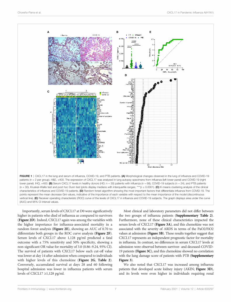
Importantly, serum levels of CXCL17 at D0 were significativelyhigher in patients who died of influenza as compared to survivors(Figure 2D). Indeed, CXCL17 again was among the variables withthe higher importance for influenza-associated mortality in arandom forest analysis (Figure 2E), showing an AUC of 0.70 todifferentiate both groups in the ROC curve analysis (Figure 2F).Serum levels of CXCL17 above 1,128 pg/ml predicted a fataloutcome with a 75% sensitivity and 50% specificity, showing anon-significant OR value for mortality of 3.0 (0.86–9.24, 95% CI).The survival of patients with CXCL17 below such cut-off valuewas lower at day 14 after admission when compared to individualswith higher levels of this chemokine (Figure 2G, Table 2).Conversely, accumulated survival at days 28 and 60 followinghospital admission was lower in influenza patients with serumlevels of CXCL17 ≥1,128 pg/ml.
Most clinical and laboratory parameters did not differ betweenthe two groups of influenza patients (Supplementary Table 2).Furthermore, none of these clinical characteristics impacted theserum levels of CXCL17 (Figure 3A), and this chemokine was notassociated with the severity of ARDS in terms of the PaO2/FiO2values at admission (Figure 3B). These results together suggest thatCXCL17 represents an independent prognostic factor for mortalityin influenza. In contrast, no differences in serum CXCL17 levels atadmission were observed between survivor- and deceased-COVID-19 patients (Figure 3C), and this chemokine showed no correlationwith the lung damage score of patients with PTB (SupplementaryFigure 3).
We also noted that CXCL17 was increased among influenzapatients that developed acute kidney injury (AKIN; Figure 3D),and its levels were even higher in individuals requiring renal
A B
D
E
C
FIGURE 1 | CXCL17 in the lung and serum of influenza, COVID-19, and PTB patients. (A) Morphological changes observed in the lung of influenza and COVID-19patients (n = 2 per group). H&E, ×400. The expression of CXCL17 was analyzed in lung autopsy specimens from influenza (left lower panel) and COVID-19 (rightlower panel). IHQ, ×400. (B) Serum CXCL17 levels in healthy donors (HD; n = 30) patients with influenza (n = 68), COVID-19 subjects (n = 24), and PTB patients(n = 30). Kruskal–Wallis test and post hoc Dunn test (plots display medians with interquartile ranges; ****p ≤ 0.0001). (C) K-means clustering analysis of the clinicalcharacteristics of influenza and COVID-19 patients. (D) Random forest algorithm showing the most important factors that differentiate influenza from COVID-19. Thepoints represent the mean decrease Gini values, indicative of the importance of each variable with respect to the mean importance of the model (discontinuousvertical line). (E) Receiver operating characteristic (ROC) curve of the levels of CXCL17 in influenza and COVID-19 subjects. The graph displays area under the curve(AUC) and 95% CI interval values.
Choreño-Parra et al. CXCL17 in Pandemic Influenza A(H1N1)
Frontiers in Immunology | www.frontiersin.org February 2021 | Volume 12 | Article 6332977

replacement therapy (Figure 3E). Using the same cut-off value of1,128pg/mL, elevated serum levels of CXCL17 showed a 73.6%sensitivity, 51% specificity, and a non-significant OR of 2.91 (0.96–8.28, 95% CI) to predict the development of AKIN (Figure 3F).Similarly, increased levels of CXCL17 showed an 87.5% sensitivity,53.8% specificity, and a significant OR of 8.16 (1.71–38, 95% CI) topredict the need for renal replacement therapy (Figure 3G).
Influenza A(H1N1) pdm09 Virus Inducesthe Production of CXCL17 in Human LungEpithelial Cells and MacrophagesThe higher levels of CXCL17 in sera from influenza patientsprompted us to investigate the possible cellular sources of CXCL17during influenza. To this end, we infected human A549 lungalveolar epithelial cells and peripheral blood monocyte-derivedmacrophages with a clinical isolate of the influenza A(H1N1)
pdm09 virus. Interestingly, while both human cell types producedhigh amounts of CXCL17 at 24 h, 48h, and 72h after the infection,A549 epithelial cells produced lower levels of CXCL17 in responseto influenza as compared to macrophages (Figures 3H, I). Thesefindings are consistent with the expression of CXCL17 in lungmacrophages and alveolar epithelial cells in autopsy specimensfrom our influenza patients. This suggests that the influenza A(H1N1) pdm09 virus stimulates CXCL17 expression in humans,both in vivo and in vitro. However, further analyses are required toconfirm a role for CXCL17 in influenza.
DISCUSSION
The clinical spectrum of ARDS encompasses very severe forms ofthe disease characterized by a profound respiratory failure. These
A B D
E F
G
C
FIGURE 2 | Dynamics and prognostic value of serum CXCL17 levels in influenza (A) Influenza patients (n = 68) were grouped according to their duration ofsymptoms on admission. Levels of CXCL17 were compared to healthy donors (HD; Kruskal–Wallis test and post hoc Dunn test; graph displays means and thestandard error ( ± SE); *p ≤ 0.05, **p ≤ 0.01, ***p ≤ 0.001, ****p ≤ 0.0001). (B) We obtained a second serum sample in 54 influenza subjects seven days (D7) afterhospital admission (D0). Levels of CXCL17 at D0 and D7 were compared with the Mann–Whitney U test. (C) Longitudinal change in CXCL17 levels in survivor anddeceased influenza patients (Wilcoxon test). (D) Initial serum CXCL17 levels in survivor (n = 52) and deceased (n = 16) influenza patients (Mann–Whitney U test; plotsdisplay medians and interquartile ranges). (E) Random forest algorithm showing the most important factors that impact on influenza-associated mortality.(F) Receiver operating characteristic (ROC) curve of the levels of CXCL17 in survivor and deceased influenza subjects. The graph displays area under the curve(AUC) and 95% CI interval values. (G) Survival curves of influenza patients grouped according to their serum CXCL17 levels were compared with the log-rank test.
Choreño-Parra et al. CXCL17 in Pandemic Influenza A(H1N1)
Frontiers in Immunology | www.frontiersin.org February 2021 | Volume 12 | Article 6332978

manifestations are often related to dysregulated immune reactionselicited by different insults. The quality and magnitude of suchresponses may vary according to the causative agent of the illness.
Thus, particular immune profiles of local and circulatingcytokines may serve as readouts to differentiate between specificcauses of ARDS. Here, we demonstrate that CXCL17 can bedetected in post-mortem human lung specimens from patientswith pandemic influenza A(H1N1) and COVID-19. Furthermore,serum levels of CXCL17 are differentially regulated duringinfluenza and COVID-19, pointing to a possible diagnosticvalue for the chemokine. Importantly, we found that CXCL17 isnot elevated in the serum of PTB patients neither. Althoughindividuals with this disease rarely present with ARDS, the factthat only influenza but not PTB or COVID-19 patients showedhigh CXCL17 levels remarks the specific diagnostic potential ofthis chemokine during influenza.
Chemokines can be divided into homeostatic or inflammatorydepending on their expression patterns (13). CXCL17 is considereda “dual” chemokine because it exhibits characteristics of both
TABLE 2 | Cumulative survival rates in patients with influenza according withtheir serum CXCL17 levels.
Time after hospitaladmission
Survival (%, 95% CI)
CXCL17 <1,128 pg/ml CXCL17 ≥ 1,128 pg/ml
7 days 96.66 (78.6–99.52) 94.73 (80.55–98.65)14 days 84.86 (64.38–94.06) 91.776 (76.56–97.27)21 days 84.86 (64.38–94.06) 85.06 (67.5–94.16)28 days 84.86 (64.38–94.06) 63.33 (41.48–78.89)60 days 84.86 (64.38–94.06) 31.66 (1.93-71.53)
Survival rates and their 95% CI were estimated using the Kaplan-Meyer method and thelog rank test.
A B
D E
F G IH
C
FIGURE 3 | Extended predictive value of serum CXCL17 levels in influenza patients and cellular sources. (A) Multiple correlation analysis of the clinical variables andserum CXCL17 levels of influenza patients (n = 68). The heat color map gradient was constructed using Spearman R values. (B) Serum CXCL17 levels in influenzapatients according to the severity of their acute respiratory distress syndrome (ARDS) on admission (mild, PaO2/FiO2 >200, n = 3; moderate, PaO2/FiO2 100–200,n = 27; severe, PaO2/FiO2 <100, n = 38; Kruskal–Wallis test and post hoc Dunn test). (C) Comparison of the levels of CXCL17 between COVID-19 patients thatsurvived (n = 14) or died (n = 10). Mann–Whitney U test. (D) Serum levels of CXCL17 were compared between influenza patients that developed acute kindly injury(AKIN, n = 19), and individuals with normal renal function (n = 49). (E) Differences in CXCL17 levels between influenza patients according to their need for renalreplacement therapy (RRT; n = 52 vs 16). (B–E) Plots display medians and interquartile ranges. Mann–Whitney U test. (F) Receiver operating characteristic (ROC)curve of the CXCL17 levels in influenza patients that developed AKIN and those that maintained normal renal function. The graph display area under the curve (AUC)and 95% CI interval values. (G) ROC curve analysis of the serum CXCL17 levels in patients requiring RRT. (H) Human A549 alveolar epithelial cells and (I) peripheralblood-monocyte derived macrophages were infected with a clinical isolate of the influenza A(H1N1) pdm09 virus for 24, 48, and 72 h n = 6–7 per group per timepoint). Levels of CXCL17 in supernatants from infected and uninfected cells were compared using the unpaired Student’s t test (n = 5 to 9 per group) at each timepoint. Values of p were corrected for multiple comparisons using the Holm–Sidak method. The data represent mean ( ± SE) values from 2 to 3 independentexperiments per time point and experimental condition. *p ≤ 0.05, **p ≤ 0.01, ***p ≤ 0.001, ****p ≤ 0.0001.
Choreño-Parra et al. CXCL17 in Pandemic Influenza A(H1N1)
Frontiers in Immunology | www.frontiersin.org February 2021 | Volume 12 | Article 6332979

homeostatic and inflammatory chemokines. It is constitutivelyexpressed in the mucosa of the respiratory tract with potentialfunctions in lung defenses (14) and is known to chemoattractmacrophages (4). This chemokine was the last member of the C-X-Cmotif chemokine ligand family to be reported (1), and as such, itsrole during respiratory inflammation or infections is not wellunderstood. We should note that bioinformatics analyses ofpublic gene expression databases indicate that CXCL17 is thehighest expressed chemokine in the normal trachea andbronchus and among the highest expressed chemokines in thenormal human lung (2). These observations strongly suggestimportant homeostatic functions for CXCL17 in the respiratorytract. Previous studies have shown that CXCL17 is stronglyupregulated in idiopathic pulmonary fibrosis (2). This, alongwith data from our current study indicates that CXCL17 is verylikely to have important functions in the pathogenesis ofinflammatory/infectious diseases of the lung as well. Past studieshave provided indirect evidence about a possible role of CXCL17 inimmunity against respiratory infections. For instance, it has beenshown that CXCL17 has a potent bactericidal activity over bacteriacausative of respiratory infections (2). The expression of theCXCL17 gene was also found upregulated in group 3 innatelymphoid cells isolated from lung tissues of patients withtuberculosis (TB) (15), suggesting a role for CXCL17 againstMycobacterium tuberculosis (Mtb) infection. However, CXCL17plays redundant activities during anti-Mtb immunity in murinemodels (16). In this context, our study represents the first directevaluation of the expression of CXCL17 during respiratoryinfections in humans.
Our analyses of lung autopsy specimens suggest that theinfluenza A(H1N1) pdm09 virus and SARS-CoV-2 stimulatethe local production of CXCL17 in the pulmonary tissue. Indeed,although these findings were made only in two patients who diedof influenza and two with COVID-19, the pattern of expressionobserved during both infections differ from what it is observed atsteady state. In this regard, using non-infected human lungspecimens, we previously demonstrated that CXCL17 isdetected only at the luminal surface of alveolar epithelial cells(2). Conversely, here we found that lungs infected with influenzaand SARS-CoV-2 express CXCL17 in the whole cytoplasm ofpneumocytes, as well as in infiltrating macrophages. This is furthersupported by a recent paper demonstrating a high induction ofCXCL17 in the BAL of COVID-19 (17). However, we found thatthe magnitude of CXCL17 expression in the serum was also robustduring influenza but minimal in COVID-19 patients. These arecontrasting observations, although we must consider that the lungtissue specimens in our study were obtained post-mortem.Therefore, our immunohistochemical analysis evaluated theexpression of CXCL17 only during the latest phases of viralinfection, whereas serum chemokine determinations capturedearly responses against COVID-19 and influenza. Analysis ofCXCL17 expression in lung biopsy specimens obtained earlyduring the infection would have clarified this point. Despite this,we speculate that lung and serum CXCL17 levels may increase asthe infection with SARS-CoV-2 progresses in patients with severedisease. Whether longitudinal changes in serum CXCL17 levels
could have a prognostic value in COVID-19 should be investigatedin future studies.
Our findings may be also explained by possible different abilitiesof influenza and SARS-CoV-2 virus strains to induce local CXCL17production and translocation to the blood according to theirvirulence. Indeed, previous research showed that the infection ofhuman bronchial epithelial cells, human tracheobronchial epithelialcells, and human alveolar A549 cells with seasonal influenza A(H3N2) virus, promotes robust upregulation of CXCL17. However,the infection with more virulent A(H5N1) and A(H7N9) influenzavirus subtypes induces minor changes in the expression of CXCL17(18). Similarly, in a yet unpublished experiment available from theInfluenza Research Database (IRD), the infection of 2B-4cells/sorted Calu-3 cells with a wild type strain of the 2002-2003SARS-CoV (icSARS CoV) did not stimulate strong expression ofCXCL17 (19). In contrast, the infection with icSARS ExoNI andicSARS dNSP16, which are attenuated mutant strains of SARS-CoV, upregulated the expression of CXCL17.
Notably, we also demonstrated that these differences could beharnessed for clinical applications, as serum CXCL17 levelsdetermined at hospital admission are useful to distinguishbetween influenza and COVID-19 in patients with ARDS. Thisis important, especially at settings of high circulation ofpandemic influenza A(H1N1) virus strains, as it is highly likelythat both diseases will converge during the upcoming flu season.During such a predicted scenario, the differentiation of influenzaand COVID-19 by clinical characteristics may be complicated.Indeed, our results and previous studies show that only a fewnon-specific symptoms and routine laboratory tests are useful forthis diagnostic dilemma (20–22). However, the discrimination ofthe causative pathogen has direct therapeutic implications,including the selection of the adequate anti-viral drug. Thus,novel biomarkers with high diagnostic value to distinguishinfluenza and COVID-19 are urgently needed.
Another striking finding of our study is that serum levels ofCXCL17 impact on pandemic influenza A(H1N1)-associatedmortality. In this sense, it is known that several chemokinesparticipate in the immune response against influenza viruses (23,24). Most of them are produced in high amounts and mediatepathology due to their pro-inflammatory properties (25, 26). Ourdata indicate that the production of CXCL17 is also highlypotentiated during the early stages of pandemic influenza A(H1N1), as the serum levels reported here are as high as thosefound in other inflammatory and human autoimmune disorders(2, 11). Moreover, we demonstrate that both macrophages andlung epithelial cells can become sources of CXCL17 afterinfection with the influenza A(H1N1) pdm09 virus.
CXCL17 normally mediates the recruitment of myeloid cells tothe lung (4). Cell subtypes responding to this chemokine includeDCs, monocytes, and macrophages (1, 4, 27). However, we shouldnote that the nature of the myeloid cells recruited by CXCL17 in vivohas not been established in humans, although the fact that CXCL17is a mucosal chemokine expressed only in the respiratory anddigestive tracts suggests that it is recruiting unique population(s) ofmyeloid cells to mucosal tissues which remain to be functionallycharacterized. Increased and sustained recruitment of myeloid cells
Choreño-Parra et al. CXCL17 in Pandemic Influenza A(H1N1)
Frontiers in Immunology | www.frontiersin.org February 2021 | Volume 12 | Article 63329710

to the lungs is associated with immunopathology and mortality ininfluenza (23, 24). Accordingly, our data indicate that influenzapatients with higher CXCL17 levels have lower survival, suggesting apossible pathogenic role for this chemokine.We should alsomentionthat CXCL17 also mediates anti-inflammatory activities (25, 26).Thus, our findings may reflect a regulatory mechanism for thecytokine storm underlying severe influenza, via the production ofhigh levels of anti-inflammatory mediators.
The role of innate immune cells responding to CXCL17 inCOVID-19 is unknown, but recent studies have found that thenumbers of circulating DCs and monocytes are reduced inpatients with severe SARS-CoV-2 infection as compared topatients with milder forms of the disease (28). In contrast, wefound a high number of macrophages in the lung autopsyspecimens of patients that died of COVID-19. Together, thesedata point to a possible role for chemokines involved in therecruitment of myeloid cells to the lungs during COVID-19, andCXCL17 is an excellent candidate to mediate this recruitment (4).However, whether CXCL17 plays a pathogenic or protective roleduring influenza or COVID-19 is not apparent from our data.
A caveat of our study is that CXCL17 levels were assessed onlyin influenza patients with severe pandemic influenza A(H1N1),but not in individuals with milder forms of the disease nor inpersons with seasonal influenza infections. Hence, future studiesmust validate our results in other cohorts of influenza patients.Also, we measured serum levels of CXCL17 in COVID-19patients only at the time of hospital admission. Thus, theanalysis of additional time points is required to drawconclusions on the prognostic role of this chemokine inCOVID-19. Finally, our findings in PTB patients indicate thatCXCL17 is not highly released from the lung to the serum duringMtb infection. This, along with previous results from our group(16), may indicate a minimal role for this chemokine duringPTB. Nonetheless, more studies are necessary to rule out acontribution of CXCL17 to the immunity against Mtb.
CONCLUSIONS
In conclusion, our study provides preliminary evidence supporting adiagnostic potential of CXCL17 to distinguish severe pandemicinfluenza A(H1N1) from COVID-19 and other respiratoryinfections like PTB. In addition, we show that this chemokine maybe useful as a prognostic biomarker in influenza patients, as serumlevels of CXCL17 are associated with higher risk of renal failure andmortality. Finally, we found some possible cellular sources ofCXCL17 during influenza. Future studies are required to betterunderstand the function of this chemokine in lung defenses againstthe influenza A(H1N1) pdm09 virus and better evaluated a possibleparticipation in immunity against SARS-CoV-2.
DATA AVAILABILITY STATEMENT
The raw data supporting the conclusions of this article will bemade available by the authors, without undue reservation.
ETHICS STATEMENT
The studies involving human participants were reviewed andapproved by IRB of the Instituto Nacional de EnfermedadesRespiratorias Ismael Cosıo Villegas and Instituto Nacional deCiencias Medicas y Nutricion Salvador Zubiran. The patients/participants provided their written informed consent toparticipate in this study.
AUTHOR CONTRIBUTIONS
Collected clinical data and biological samples for the study: JC-P,MS-V, CH-C, EH-M, MC-C, NR-Z, CM-M, AC-L, AD, LM-H,DD-Z, DR-G, TR-R, DC-R and CM-M. Performed in vitroinfections: GR-M, LJ-Á, CC-G, EM-G, and AC-L. PerformedCXCL17 ELISA assays: JC-P, LJ-Á, GR-M, and LF-L. Obtainedand analyzed autopsy lung specimens: CS-L, CS, FB-M, PG-O,and CL-R. Analyzed and discussed data: JC-P, E-CP, MS-V,EG-L, TR-R, SK, and JZ. Drafted the manuscript: JC-P, SK, AZ,and JZ. All authors contributed to the article and approved thesubmitted version.
FUNDING
Institutional research funds of INER supported the current study.This project also received funding from the National Council ofScience and Technology of Mexico (CONACyT) under the researchcontracts: SECTEI/050/2020, Secretarıa de Ciencia, Tecnologıa eInnovacion de la Ciudad de Mexico (SECTEI CDMX);FORDECYT/10SE/2020/05/14-06, CONACYT-Support forscientific research, technological development and innovation inhealth during COVID-19 contingency, with the project number:313517, and FORDECYT/10SE/2020/05/14-07 from the FondoInstitucional de Fomento Regional para el Desarrollo Cientıfico yTecnologico y de Innovacion (FORDECYT). JC-P was supported bya scholarship (CVU 737347) from CONACyT to his Ph.D. degree.Funders did not play any role in the study design and conduction.
ACKNOWLEDGMENTS
To all the patients that participated in the study and theirfamily members.
SUPPLEMENTARY MATERIAL
The Supplementary Material for this article can be found online at:https://www.frontiersin.org/articles/10.3389/fimmu.2021.633297/full#supplementary-material.
Choreño-Parra et al. CXCL17 in Pandemic Influenza A(H1N1)
Frontiers in Immunology | www.frontiersin.org February 2021 | Volume 12 | Article 63329711

REFERENCES
1. Pisabarro MT, Leung B, Kwong M, Corpuz R, Frantz GD, Chiang N, et al.Cutting edge: novel human dendritic cell- and monocyte-attractingchemokine-like protein identified by fold recognition methods. J Immunol(2006) 176(4):2069–73. doi: 10.4049/jimmunol.176.4.2069
2. Burkhardt AM, Tai KP, Flores-Guiterrez JP, Vilches-Cisneros N, Kamdar K,Barbosa-Quintana O, et al. CXCL17 is a mucosal chemokine elevated inidiopathic pulmonary fibrosis that exhibits broad antimicrobial activity.J Immunol (2012) 188(12):6399–406. doi: 10.4049/jimmunol.1102903
3. Weinstein EJ, Head R, Griggs DW, Sun D, Evans RJ, Swearingen ML, et al.VCC-1, a novel chemokine, promotes tumor growth. Biochem Biophys ResCommun (2006) 350(1):74–81. doi: 10.1016/j.bbrc.2006.08.194
4. Burkhardt AM, Maravillas-Montero JL, Carnevale CD, Vilches-Cisneros N,Flores JP, Hevezi PA, et al. CXCL17 is a major chemotactic factor for lungmacrophages. J Immunol (2014) 193(3):1468–74. doi: 10.4049/jimmunol.1400551
5. Srivastava R, Hernandez-Ruiz M, Khan AA, Fouladi MA, Kim GJ, Ly VT,et al. CXCL17 Chemokine-Dependent Mobilization of CXCR8(+)CD8(+)Effector Memory and Tissue-Resident Memory T Cells in the Vaginal MucosaIs Associated with Protection against Genital Herpes. J Immunol (2018) 200(8):2915–26. doi: 10.4049/jimmunol.1701474
6. Castillejos M, Cabello-Gutierrez C, Alberto Choreño-Parra J, Hernandez V,Romo J, Hernandez-Sanchez F, et al. High performance of rapid influenzadiagnostic test and variable effectiveness of influenza vaccines in Mexico. Int JInfect Dis (2019) 89:87–95. doi: 10.1016/j.ijid.2019.08.029
7. Corman VM, Landt O, Kaiser M, Molenkamp R, Meijer A, Chu DK, et al.Detection of 2019 novel coronavirus (2019-nCoV) by real-time RT-PCR. EuroSurveill (2020) 25(3):2000045. doi: 10.2807/1560-7917.ES.2020.25.3.2000045
8. Choreño-Parra JA, Jimenez-Alvarez LA, Muñoz-Torrico M, Ramırez-Martınez G, Jimenez-Zamudio LA, Salinas-Lara C, et al. Antigens ofMycobacterium tuberculosis Stimulate CXCR6+ Natural Killer Cells. FrontImmunol (2020) 11:582414. doi: 10.3389/fimmu.2020.582414
9. Ramırez-Martınez G, Cruz-Lagunas A, Jimenez-Alvarez L, Espinosa E, Ortız-Quintero B, Santos-Mendoza T, et al. Seasonal and pandemic influenza H1N1viruses induce differential expression of SOCS-1 and RIG-I genes andcytokine/chemokine production in macrophages. Cytokine (2013) 62(1):151–9. doi: 10.1016/j.cyto.2013.01.018
10. Monin L, Griffiths KL, Lam WY, Gopal R, Kang DD, Ahmed M, et al.Helminth-induced arginase-1 exacerbates lung inflammation and diseaseseverity in tuberculosis. J Clin Invest (2015) 125(12):4699–713. doi: 10.1172/JCI77378
11. Hernandez-Ruiz M, Zlotnik A, Llorente L, Hernandez-Molina G. Markedlyhigh salivary and lacrimal CXCL17 levels in primary Sjogren’s syndrome.Joint Bone Spine (2018) 85(3):379–80. doi: 10.1016/j.jbspin.2017.05.014
12. Flach C-F, Qadri F, Bhuiyan TR, Alam NH, Jennische E, Lönnroth I, et al. BroadUp-Regulation of Innate Defense Factors during Acute Cholera. Infect Immmun(2007) 75: (5):2343–50. doi: 10.1128/IAI.01900-06
13. Zlotnik A, Yoshie O. The chemokine superfamily revisited. Immunity (2012)36(5):705–16. doi: 10.1016/j.immuni.2012.05.008
14. Choreno-Parra JA, Thirunavukkarasu S, Zuniga J, Khader SA. The protectiveand pathogenic roles of CXCL17 in human health and disease: Potential inrespiratory medicine. Cytokine Growth Factor Rev (2020) 53:53–62.doi: 10.1016/j.cytogfr.2020.04.004
15. Ardain A, Domingo-Gonzalez R, Das S, Kazer SW, Howard NC, Singh A,et al. Group 3 innate lymphoid cells mediate early protective immunity againsttuberculosis. Nature (2019) 570(7762):528–32. doi: 10.1038/s41586-019-1276-2
16. Choreno-Parra JA, Dunlap M, Swanson R, Jimenez-Alvarez LA, Zuñiga J,Khader SA. The C-X-C motif chemokine 17 plays a minimal role during lunginfection with hypervirulent Mycobacterium tuberculosis. J Immunol (2020)204(1 Supplement):85.13–3.
17. Zhou Z, Ren L, Zhang L, Zhong J, Xiao Y, Jia Z, et al. Heightened InnateImmune Responses in the Respiratory Tract of COVID-19 Patients. Cell HostMicrobe (2020) 27(6):883–90.e2. doi: 10.1016/j.chom.2020.04.017
18. Cao Y, Huang Y, Xu K, Liu Y, Li X, Xu Y, et al. Differential responses of innateimmunity triggered by different subtypes of influenza a viruses in human andavian hosts. BMC Med Genomics (2017) 10(Suppl 4):70. doi: 10.1186/s12920-017-0304-z
19. Influenza Research Database. Influenza Research Database (IRD). Available at:https://www.fludb.org/brc/hostFactorExperimentDetails.spg?method=Submi t Fo rm&f romDe t a i l = t r u e&b i o s e t I d s=1320&expS eq Id=178&r e su l tMa t r i xUs e rDe f Id=SCL008_R_N_RM&deco ra t o r=influenza#https://www.fludb.org/brc/hostFactorExperimentDetails.spg?method=SubmitForm&fromDetail=true&biosetIds=1320&expSeqId=178&resultMatrixUserDefId=SCL008_R_N_RM&decorator=influenza#(Accessed July 7th 2020).
20. Tang X, Du R-H, Wang R, Cao T-Z, Guan L-L, Yang C-Q, et al. Comparisonof Hospitalized Patients With ARDS Caused by COVID-19 and H1N1. Chest(2020) 158(1):195–205. doi: 10.1016/j.chest.2020.03.032
21. Jiang C, Yao X, Zhao Y, Wu J, Huang P, Pan C, et al. Comparative review ofrespiratory diseases caused by coronaviruses and influenza A viruses duringepidemic season. Microbes Infect (2020) 22(6-7):236–44. doi: 10.1016/j.micinf.2020.05.005
22. Choreno-Parra JA, Jimenez-Alvarez LA, Cruz Lagunas A, Rodriguez-ReynaTS, Ramirez-Martinez G, Sandoval-Vega M, et al. Clinical and immunologicalfactors that distinguish COVID-19 from pandemic influenza A(H1N1).medRxiv (2020) 2020.08.10.20170761. doi: 10.1101/2020.08.10.20170761
23. Coates BM, Staricha KL, Koch CM, Cheng Y, Shumaker DK, Budinger GRS,et al. Inflammatory Monocytes Drive Influenza A Virus-Mediated LungInjury in Juvenile Mice. J Immunol (2018) 200(7):2391–404. doi: 10.4049/jimmunol.1701543
24. Lin KL, Suzuki Y, Nakano H, Ramsburg E, Gunn MD. CCR2+ monocyte-derived dendritic cells and exudate macrophages produce influenza-inducedpulmonary immune pathology and mortality. J Immunol (2008) 180(4):2562–72. doi: 10.4049/jimmunol.180.4.2562
25. Oka T, Sugaya M, Takahashi N, Takahashi T, Shibata S, Miyagaki T, et al.CXCL17 Attenuates Imiquimod-Induced Psoriasis-like Skin Inflammation byRecruiting Myeloid-Derived Suppressor Cells and Regulatory T Cells.J Immunol (2017) 198(10):3897–908. doi: 10.4049/jimmunol.1601607
26. Hernandez-Ruiz M, Othy S, Herrera C, Nguyen HT, Arrevillaga-Boni G,Catalan-Dibene J, et al. Cxcl17(-/-) mice develop exacerbated disease in a Tcell-dependent autoimmune model. J Leukoc Biol (2019) 105(5):1027–39.doi: 10.1002/jlb.3a0918-345rr
27. Lee WY, Wang CJ, Lin TY, Hsiao CL, Luo CW. CXCL17, an orphanchemokine, acts as a novel angiogenic and anti-inflammatory factor. Am JPhysiol Endocrinol Metab (2013) 304(1):E32–40. doi: 10.1152/ajpendo.00083.2012
28. Wang W, Su B, Pang L, Qiao L, Feng Y, Ouyang Y, et al. High-dimensionalimmune profiling by mass cytometry revealed immunosuppression anddysfunction of immunity in COVID-19 patients. Cell Mol Immunol (2020)17(6):650–2. doi: 10.1038/s41423-020-0447-2
Conflict of Interest: The authors declare that the research was conducted in theabsence of any commercial or financial relationships that could be construed as apotential conflict of interest.
Copyright © 2021 Choreño-Parra, Jimenez-Alvarez, Ramırez-Martınez, Sandoval-Vega,Salinas-Lara, Sanchez-Garibay, Luna-Rivero, Hernandez-Montiel, Fernandez-Lopez,Cabrera-Cornejo, Choreño-Parra, Cruz-Lagunas, Domınguez, Marquez-Garcıa,Cabello-Gutierrez, Bolaños-Morales, Mena-Hernandez, Delgado-Zaldivar,Rebolledo-Garcıa, Guadarrama-Ortiz, Regino-Zamarripa, Mendoza-Milla,Garcıa-Latorre, Rodiguez-Reyna, Cervantes-Rosete, Hernandez-Cardenas, Khader,Zlotnik and Zuñiga. This is an open-access article distributed under the terms of theCreative Commons Attribution License (CC BY). The use, distribution orreproduction in other forums is permitted, provided the original author(s) and thecopyright owner(s) are credited and that the original publication in this journal iscited, in accordance with accepted academic practice. No use, distribution orreproduction is permitted which does not comply with these terms.
Choreño-Parra et al. CXCL17 in Pandemic Influenza A(H1N1)
Frontiers in Immunology | www.frontiersin.org February 2021 | Volume 12 | Article 63329712