cyto-histological aspects in the modified leaf dionaea muscipula … 53/rom.j.biol.-plant biol.,...
TRANSCRIPT

CYTO-HISTOLOGICAL ASPECTS IN THE MODIFIED LEAF OF DIONAEA MUSCIPULA ELLIS
IRINA STANESCU', CONSTANTIN TOMA, IRINA GOSTIN
In the present work, the authors evidence some of the particularities observed in the modified leaf of Dionaea muscipula; the upper epidermis presents a lot of multicellular digestive glands, consisting in a basal cell, located among those of the epidermis, a stalk-cell and a hemispheric multicellular massive. The ultrastructure of the glandular cells confirms that the proteolytic enzymes secretion takes place not in the proper glandular cells, but in the subjacent ones.
Key word: secretory glands, ultrastructure, Dionaea muscipula.
INTRODUCTION
The plant we are referring to, generally called Venus7s flytrap, because of its rapidity of closing the two foliar lobes which form the trap, is one of the most interesting in the world. Dionaea muscipula lives in the peat bogs of North and South Carolina (S.U.A.). It is a perennial plant, having a basal rosette of leaves, from the middle of which the floriferous stem rises up.
The literature dealing with the anatomy of Dionaea muscipula, especially of the carnivorous plants, is quite rich, as it results from the synthesis works referring to the dicotyledons' anatomy (Solereder, 1899; Metcalfe and Chalk, 1972) or to the angiosperms (Napp-Zinn, 1984). Regarding various cyto-histological particularities of Dionaea muscipula, these are mentioned by Juniper et al. (1989). In Romania, only a few articles have been published about carnivorous plants until now (Tarnavschi, 1957; Toma C. and Toma I., 2002); in these synthesis works, the carnivorous plants accommodation to the surroundings was underlined.
Continuing our investigation regarding the anatomy of the carnivorous plants (Stfinescu et al., 2005), in the present work we evidenced a few characteristics of the interesting trap of Dionaea muscipula.
MATERIAL AND METHODS
The material subjected to the histological analysis (the young trap, the petiole and the blade folds of the mature trap of Dionaea muscipula) has been fixed and preserved in 70% ethyl alcohol. The sections were cut by microtome, subsequently
' "Al. 1. Cuza" University - Iagi, Faculty of Biology, 20A Carol I Bvld., 700506, I a~ i , Romania, E-mail: [email protected], [email protected]
ROM. J. BIOL. -PLANT BIOL., VOLUME 53, No 1, P. 3-10, BUCHAREST, 2008
Irina Stgnescu, Constantin Toma. lrina Gostin
coloured with iodine green and alaun-carmine, then mounted in gel and analyzed in a Novex (Holland) light microscope. The light micrographs were performed by means of the same light microscope, using Canon A95 camera.
Regarding the cytological investigations, the material subjected to analysis (the mature trap) had been prepared according to the laboratory techniques for scanning and transmission electron microscopy. The pictures were performed by means of Philips XL30 scanning electron microscope and JEOL 1010 transmission electron microscope from the Electron Microscopy Department of Vigo University (C.A.C.T.I.: Centro de Apoyo Cientifico Tecnologico a Investigacion), Spain.
RESULTS
FOLIAR VAGINA HISTOLOGY
The leaf basis, very wide, with a foliar vagina-like structure, has a semi-lune shape in cross section, with the adaxial face concave and the two wings (margins) narrowed.
The epidermis consists of small cells, having the external wall thicker than the others, covered by a thin cuticle. The cells are isodiametric or tangentially- elongated. The protective trichomes are long, being present only in the lower epidermis. The fundamental parenchyma is of homogeneous-meatus type, having large round cells. It presents a few (5) collateral vascular bundles; the median one is bigger than the others; all of them have the xylem to the upper epidermis. The phloem forms various isles (consisting of sieved tubes and companion cells) separated by large parenchymatic cells. The xylem consists of vessels with thick, but unlignified walls, separated by cellulosic parenchyma.
PETIOLE HISTOLOGY
The petiole is semicircular in cross section, with a blade-like structure: homogeneous mesophyll of lacunary type and a few small vascular bundles.
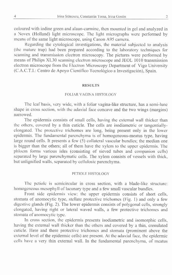
Front side epidermis view: the upper epidermis consists of short cells, stomata of anomocytic type, stellate protective trichomes (Fig. 1) and only a few digestive glands (Fig. 2). The lower epidermis consists of polygonal cells, strongly elongated, having right or lateral waved walls, a few protective trichomes and stomata of anomocytic type.
In cross section, the epidermis presents isodiametric and isomorphic cells, having the external wall thicker than the others and covered by a thin, crenulated cuticle. Here and there protective trichomes and stomata (prominent above the external level of the epidemic cells) are present. At the adaxial face, the epidemic cells have a very thin external wall. In the fundamental parenchyma, of meatus

3 Leaf cyto-histology of Dionaea rnuscipula Ellis 5
type, there are a lot of collateral vascular bundles; the median vascular bundle is very big, the lateral ones (16-1 8) are very small. The phloem is arc-shaped, having a parenchymatic sheath at its exterior, then, other few (4-6) small vascular bundles, with reverse xylem (the xylem is oriented to the lower epidermis).
YOUNG UNOPENED TRAP HISTOLOGY
The young unopened trap has a semicircular shape in cross section, with a deep adaxial riffle delimited by the laterally-adaxial wings, inrolled (Fig. 3).
The epidermis presents large isodiametric cells, having the internal and external walls thicker than the others; the external one is covered by a thin cuticle. Here and there, stomata and protective trichomes are present; the formers, occupying the lateral parts and the latero-adaxial wings of the trap, were numerous. Stomata are at the same level as the epidermic cells.
In the fundamental parenchyma, of meatus type, there is a median vascular bundle and two latero-adaxial vascular bundles; the formers appear longitudinally sectioned, because they are born in the median vascular bundle and then orient themselves through the latero-adaxial wings.
The fundamental parenchyma consists of small cells in the first two hypodermic layers and large cells in the rest, all having thin cellulosed walls. At the adaxial face and, mostly, in the latero-adaxial wings, the parenchyma is more compact, having small cells, regularly disposed, reminding of the structure of a homogeneous mesophyll, weakly differentiated in this ontogenetic stage.
The median vascular bundle is formed of two arcs, both having the phloem at the abaxial face and the xylem at the adaxial one; close to the phloem arc there is a parenchymatic sheath, which has at its exterior part 2-3 groups of phloemic elements and more groups of xylemic vessels. All the xylemic vessels have thin, but lignified walls. Radial rows of large parenchymatic cells come through the xylemic and phloemic arc.
At the adaxial, concave face, among the epidermic cells, there are a lot of small secretory glands (Fig. 4), some of them presenting only 2-3 cells; one epidermic cell divides into two cells; the upper one divides again n-times, resulting the complex structure of the gland.
MATURE TRAP HISTOLOGY
The anatomy of the mature trap is quite similar to that of the young trap; rarely can we see protective trichomes in the epidermis of the abaxial face and in the one present at the margins of the trap. There are lots of glands, having the same structure as those of the upper epidermis of the petiole, but less turgescent.

Irina StBnescu. Constantin Toma. Irina Gostin
The epidermic cells of the abaxial face are tangentially-elongated, having the internal and, mostly, the external wall thicker than the others. The adaxial wings present large cells, strongly elongated, parallel with the epidermis, all having very thin walls.
The cross section through a lateral-rib shows a structure as follows: - The upper (internal) epidermis consists of large, isodiametric or elongated
cells, having the external and internal walls thicker than the others. Here and there, multicellular digestive glands (Figs. 5, 7 and 8) are present, formed of: basal cell, among the epidermic ones, a stalk-cell and a hemispheric multicellular massive; its external cells touch the external wall of the epidermic cells. Stiff trichomes (Fig. 9) are around the edge of the blade folds and become interlocked (intermeshed) when the trap closes. In this region, the digestive glands are present in small depressions, not to be flattened once the trap is closed. Each foliar lobe presents three bristle- like trichomes (Fig. lo), which represent tactile structures, because of their sensitivity to touch. Their mechanical stimulation causes a rapid closure of the blade folds (TronchCt, 1977).
- The lower (external) epidermis consists of smaller cells (having only the external wall thicker than the others and covered by cuticle), multicellular stellate protective trichomes and anomocytic stomata.
The mesophyll is lacunary, homogeneous-type; the hypodermic layers consist in small cells, moderately collenchymatised; the rest of the mesophyll consists in large cells. The vascular tissues form various vascular bundles of different diameters, all of them having the xylem that faces the upper epidermis and the phloem, the lower one. The bigger vascular bundle occupies the midrib (Fig. 6); those occurring in the secondary ribs are smaller (Fig. 5). In cross section through the midrib, the secondary ribs appear longitudinally sectioned; they form in the midrib and end in the stiff trichomes which are present on the edge of the blade folds. The xylem and phloem elements appear parallel elongated with the two epiderrnes; the cells belonging to the mesophyll are elongated, and parallel with the epidermis, too.
As we mentioned before, the upper epidermis of the blade folds presents secretory structures (Fig. 5) consisting of basal cells (where the proteolytic enqmes are formed and stocked) and secretory cells (the upper cells of the gland).
THE ULTRASTRUCTURE OF SECRETORY CELLS
When young, the glandular cells (Fig. 11) have a big, central nucleus, consisting mostly of euchromatine; the vacuoles are filled with opaque inclusions of high density. These were evidenced before by Williams and Mozingo (1971). Buchen et al. (1983) evidenced them, too, in the cells belonging to the sensitive trichomes of the same species.
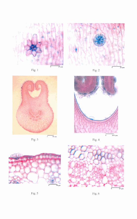
Fig. 1
Fig. 3
Fig. 2
e
Fig. 4
1 3 ~
Fig. 6

Fig. 9
Fig. 11 Fig. 12

5 Leaf cyto-histology of Dionaea muscipula Ellis 7
Fig. 13 Fig. 14
Fig. 15 Fig. 16

8 Irina Stgnescu, Constantin Toma, lrina Gostin 6
The basal cells are isodiametric and result after anticline or pericline divisions. When young, the nucleus is more or less spherical and occupies the central part of the cell; the euchromatic regions are predominant; the hetero- chromatin is in a small quantity, located in the peripheral regions. Sometimes a small nucleolus is visible. There are small and less numerous vacuoles, spherical or tubular mitochondria; the rough endoplasmic reticulum and the ribosomes are present.
During the differentiation process (Fig. 12-14), the nucleus keeps its central position; the nucleo-plasmatic ratio continues to have high values. The cytoplasm is dense, having lots of organelles (correlated with the cells' secretory function). There are small and less numerous vacuoles. In the parietal region the cytoplasm has a high density, consisting in lots of ribosomes and endoplasmic reticulum filaments. The chloroplasts are small, consisting in a few grana-thylakoids, a dense stroma and plastoglobules. The endoplasmic reticulum is dispersed, presenting tubes of various dimensions and cisterns. The ribosomes are free in the cytoplasm or grouped, resulting poly-ribosomes, near the nucleus. The Golgi apparatus is well developed. The mitochondria (Fig. 15) are small, but numerous (the cells have a high energetic activity); some of them are tubular, others are globular, having a lot of cristae. The cells have thin walls, interconnected. As the differentiation process continues, the autophagy phenomenon (the degradation of the membrane) appears.
The external wall, of primary origin, is covered by a thin cuticle, having a homogeneous structure. The wall consists of an external region with disordered cellulosed microfibers, and an internal one, with ordered cellulosed microfibers, parallelly disposed with the cell's external surface. The internal wall (Fig. 16) consists only of the region with ordered cellulosed microfibers. The middle lamella is visible.
The parietal cytoplasm is quite poor, but very dense. There are lots of grouped microvesicles and cisternae, especially in the basal part of the cell; some of them are interconnected with tubes of the endoplasmic reticulum (which are implicated in the trans-cellular transport of the proteolytic enzymes). Many short filaments belonging to the rough endoplasmic reticulum are in the parietal cytoplasm. The central vacuole is very large (Fig. 17), with lots of vesicles inside; the chloroplasts are large, too, but less numerous, containing plastoglobules.
ULTRASTRUCTURE OF THE CELLS BELONGING TO THE ASSIMILATORY PARENCHYMA
The cells belonging to the mesophyll of the blade (Fig. 18) are isodiametric, consisting of a large central vacuole and a less dense cytoplasm. The chloroplasts are elongated, having dense stroma and lots of stacks of grana-thylakoids. Some of the chloroplasts present lots of stroma-thylakoids, parallel with the chloroplast's surface. The transitory starch grains are small, but numerous (4-5/chloroplast);

7 Leaf cyto-histology of Dionaea muscipula Ellis 9
other chloroplasts present plastoglobules. The central vacuole consists of two regions: one of them is less dense, delimited by a thin tonoplast, the other region is compact, proteic, electrono-dense, similar to that occurring in the young glandular cells.
DISCUSSION
The external face of the trap (the lower epidermis of the blade) presents a lot of stomata; the internal (upper) one, concave, consists of various structures able to attract, retain and digest different organisms which represent the prey. The carnivorous plants can use the substances resulted from the decomposed preys only when they are absorbed. Darwin (1965) considers that the same structures, which secrete the digestive enzymes, absorb (assimilate) the nutritive substances resulted.
Dionaea muscipula. The upper epidermis of the petiole in front side view: protective trichome (Fig. 1) and digestive gland (Fig. 2). Cross section through the unopened trap (Fig. 3) and immature digestive gland (Fig. 4). Cross section through the mature trap: digestive gland and secondary ribs (Fig. 5), the vascular bundle of the midrib (Fig. 6).
Dionaea muscipula. Micromorphology of the glands (Figs. 7 and 8), stiff trichomes (Fig. 9) and bristle-like trichome (Fig. 10). Ultrastructure of a young cell (Fig. 11). Ultrastructure of a digestive gland (Fig. 12).
Dionaea muscipula. Utrastructure of secretory cells (Fig. 13 and 14). Mitochondria (Fig. 15). Cell wall (Fig. 16). Vacuole filled with opaque inclusions (Fig. 17). Cell belonging to the assimilatory parenchyma (Fig. 18).
CONCLUSIONS
The bi-lobed blade of Dionaea muscipula is of hypostomatic type; the digestive glands of the upper epidermis have a complex structure, in order to secrete digestive enzymes and absorb the nutritive compounds resulted from the digestion of the captured preys.
Regarding the cytological aspects, the vacuole which occupies more than 90% of the cell's volume, the reduced number of organelles, confirm that the secretion of the proteolytic enzymes takes place not in the proper glandular cells, but in the subjacent ones, then, the proteolytic enzymes are transported and secreted in the exterior of the cell.
REFERENCES
1. Buchen Brigitte, Hensel Dorothea, Sievers A., 1983, Polarity in mechanoreceptor cells of trigger hairs of Dionaea muscipula Ellis. Planta, 158: 458-468.
2. Darwin, Ch., 1965, Plante insectivore. Editura Acad. R.S.R., Bucuregti.

10 Irina Stanescu, Constantin Toma, Irina Gostin 8
3. Juniper, B.E., Robins, R.J., Joel, D. M., 1989, The Carnivorous Plants. Academic Press, San Diego.
4. Metcalfe, C.R., Chalk, L., 1972, Droseraceae (1: 581-585). In: Anatomy of the Dicotyledons. Clarendon Press, Oxford.
5. Napp-Zinn, KI., 1984, Anatomie des Blattes. 11. Blattanatomie der Angiospermen. B. Experimentelle und okologische Anatomie der Angiospemenblattes. Karnivore Angiospermen Ln Handbuch der Pflanzenanatomie, 8: 394-422, Gebriider Bomtraeger, Berlin, Stuttgart.
6. Solereder, H., 1899, Systematische Anatomie der Dicotyledonen. Fr. Enke Verlag, Stuttgart. 7. Sthescu Irina, Toma Irina, Toma C., 2005, Considerations of the stem structure of some
Drosera L. species. Contributii botanice ale Univ. babe^-Bolyai", Cluj-Napoca, 40: 215- 220.
8. Stilnescu Irina, Toma Irina, Toma C., 2005, Adventive root structure considerations of some Drosera L. species. An. St. Univ. "Al. I. Cuza"Ia~i, S. 11, a (Biol. veget.), 51: 15-22.
9. Tarnavschi, I.T., 1957, Adaptilrile morfologice ale plantelor carnivore, Natura, 4: 7692. 10. Toma C., Toma Irina, 2002, Plantele carnivore - un caz particular de adaptare la mediul de viafil.
Prelegeri academice, lagi, 1: 103-130. 11. Tronchet, A., 1977, La sensibilite' desplantes. Ed. Masson, Paris. 12. Williams, M.E., Mozingo H.N., 1971, The fine structure of the trigger hair in Venus' flytrap. Am.
J. Bot., 58: 532-539.