cytochemical responses of the lysosomal system and nadph-ferrihemoprotein reductase in
TRANSCRIPT

l Vol. 46: 81-89, 1988
l MARINE ECOLOGY - PROGRESS SERIES
Mar. Ecol. Prog. Ser. 1 Published June 30
Cytochemical responses of the lysosomal system and NADPH-ferrihemoprotein reductase in
molluscan digestive cells to environmental and experimental exposure to xenobiotics
M. N. Moore
Plymouth Marine Laboratory (West Hoe), Prospect Place. The Hoe, Plymouth PL13DH, United Kingdom
ABSTRACT: Lysosomal characteristics and smooth endoplasmic reticulum (SER) associated NADPH- fernhemoprotein (cytochrome P-450) reductase were measured cytochemically in the digestive cells of mussels A4ytilu.s edulis and periwinkles Littorina littorea exposed to environmental contaminants In Langesundfjord, Norway (PAHs, PCBs, metals) and to xenobiotics in an experimental facility (diesel oil and copper mixture). Lysosomal membrane stability was reduced in both mussels and periwinkles with increasing xenobiotic contamination In the field. Lysosomal and cytoplasmic unsaturated neutral lipid concentration was increased in contaminated mussels in the field, and lysosomal accumulation was associated with enlargement of secondary lysosomes; the latter was inversely correlated with lysosomal membrane stability. hpofuscin content and number of tertiary lysosomes increased in contaminated field mussels. These pathological changes are indicative of enhanced lysosomal autophagy and fatty degeneration. NADPH-ferrihemoprotein reductase activity was elevated in mussels from the 2 most heavily contaminated field sites. Activity of this enzyme was directly correlated with total PAHs. Lysosomal data from experimental exposures were less clear and difficult to interpret. Poor nutritional conditions possibly masked pollutant effects. However, mussels from the highest exposure concen- tration showed elevated lipofuscin. The observed sensitivity of the tests for lysosomal enlargement and lysosomal lipid, combined with their relative simplicity, indicate their potential for the detection of environmentally induced pathology, although better understanding of the mechanisms of toxicity would enhance theu utility.
INTRODUCTION
Digestive cells of the molluscan digestive gland have a highly developed lysosomal system which is involved in intracellular digestion of endocytosed food (Owen 1972). This lysosomal system is sensitive to environ- mental perturbations including many pollutants, and several indices of detrimental biological effect have been based on the responses of lysosomes in the diges- tive cells (review by Moore 1985). Lysosomal responses can be grouped into 3 classes: changes in fusion events, changes in lysosomal components, and changes in membrane permeability (Hawkins 1980). In the molluscan digestive cell, lysosomal alterations have been described that involve all of these categories of response. Increased fusion and lysosomal enlargement occur in cells undergoing pathological degenerative
D Inter-Research/Pnnted in F. R. Germany
processes, while increases in the content of hydrolytic enzymes, lipofuscin and neutral lipid are associated with exposure to xenobiotics such as polycyclic aroma- tic hydrocarbons (Lowe et al. 1981, Moore & Clarke 1982, Moore et al. 1982, 1985, 1986, 1987). Increases in permeability of lysosomal membranes is perhaps the best documented response, being associated with a wide range of environmental stimuli and apparently leading to enhanced protein catabolism and cellular atrophy (Lowe et al. 1981, Moore 1985, Moore & Via- rengo 1987).
NADPH-ferrihemoprotein reductase (EC 1.6.2.4), formally known as NADPH-cytochrome c (P-450 or neotetrazolium) reductase, is a flavoprotein involved in the electron transfer system of the endoplasmic reticulum (Masters & Okita 1980, Strobe1 et al. 1980). The reductase activity can be determined biochemi-

82 GEEP WORKSHOP: CELLUI AR- AND HISTOPATHOLOGY
cally using a variety of electron acceptors and cytochemically using tetrazolium salts, such as neo- tetrazolium, in the absence of intermediate electron acceptors (e.g. menadione), with subsequent quantita- tion of reaction product using microdensitometry (Alt- man 1972, Moore 1979, 1985, Van Noorden & Butcher 1986). This enzyme is apparently induced by a number of organic xenobiotics including certain PAHs (review by Moore 1985).
The objective of the present study was to test respon- ses of the above systems in both an environmental and an experimental situation. Myfilus edulis and Littorina littorea were sampled from a contaminant gradient in Langesundfjord and from the experimental facility at Solbergstrand where these molluscs were exposed to different concentrations of a mixture of diesel oil and copper (Abdullah & Steffenak 1988, Klungs~yr et al. 1988). The responses of the lysosomal system in diges- tive cells of mussels and periwinkles were examined. Lysosomal membrane stability (permeability) was tested in both species; In mussels, additional tests were camed out for evidence of lysosomal swelling or enlargement, as well as lipofuscin and neutral lipid content. NADPH-fernhemoprotein reductase activity was also determined in the digestive cells of mussels.
MATERIAL AND METHODS
Collection and experimental facility. Mytilus edulis (4 to 6 cm shell length) and Littorina littorea (2 to 2.5 cm shell height) were collected from Field sites 1 to 4 along the contaminant gradient in Langesundfjord, and from control (C), low (L), medium (M) and high (H) exposure treatments, as well as a second control for the high- exposure molluscs (CH), in the exposure experiment at Solbergstrand. Full details of sites, animal collection and experimental exposures are given by Bakke et al. (1988) and Follum & Moe (1988).
Concentrations of pollutants In the expenmental basins were, C: 3.0 t 0.7 ppb hydrocarbons (fluores- cence analysis). L: 6.4 k 1.8 ppb hydrocarbons and 0.8 ppb Cu, M: 31.5 +. 22.3 ppb hydrocarbons and 5 ppb Cu, H: 124.5 t 65.3 ppb hydrocarbons and 20 ppb Cu (hydrocarbon values are mean k SD over the dosing period, Cu values are nom~nal levels). Molluscs were exposed for ca 15 wk, with the exception of H mussels and their control (CH), which were exposed for 25 d only, due to high mortality during the longer treatment period.
Tissue preparation for cytochemistry. Digestive glands were dissected from mussels and periwinkles (n = 10 ind. per condition) on their arrival in the labora- tory. Small pieces of digestive gland (ca 5 X 5 X 5 mm) were placed on aluminium cryostat chucks, with 5
pieces of tissue in a straight row across the centre. The chuck was then placed for 1 min in a small bath of hexane (aromatic hydrocarbon-free; boihng range 67 to 70 "C), which had been precooled to -70 "C in liquid nitrogen in order to quench the tissue. The chuck plus the quenched solidified tissues were then sealed by doubly-wrapping in 'Parafilm' and stored at -30 "C until required for sechoning. Tissue sections (10 pm thick) were prepared in a Bright's cryostat (motorised cutting speed setting 50) with a cabinet temperature of less than -25 "C. The knife was cooled by surrounding the haft with solid CO2. Sections were transferred to slides at room temperature, which effectively flash- dried them (Bitensky et al. 1973); the sections were stored in the cryostat until required, but for not longer than 4 h.
Assessment of lysosomal membrane stability. Deter- mination of lysosomal stability was based on acid labili- za t~on characteristics of latent hydrolases, in the responsive primary fraction of latent enzyme in diges- tive cells, as previously described by Moore (1976). The acid hydrolases used in this study included 0-N- acetylhexosaminidase (EC 3.2.1.52) in musseIs and fi-glucuronidase (EC 3.2.1.31) in periwinkles.
(3-N-Acetylhexosaminidase. Serial cryostat sections, prepared as described above, were subjected to acid labilization at 37 "C in 0.1 M citrate buffer (pH 4.5) containing 2.5 '10 NaCl (W : v). The acid labilization sequence involved treating serial sections for 0, 2, 5 , 10, 15, 20, 25, 30 and 35 min respectively. Following this treatment sections were transferred to the substrate incubation medium consisting of 20 mg naphthol AS- BI-N-acetyl-P-D glucosaminide (Sigma) dissolved in 2.5 m1 2-methoxyethanol, and made up to 50 m1 with 0.1 M citrate buffer (pH 4.5) containing 2.5 O/O NaCl ( W : v) and 3.5 g of low viscosity polypeptide (Sigma POLYPEP P51 15) to act as a section stabiliser. Sections were incubated in this medium for 15 nlin at 37 "C, rinsed in 3.0 sI, NaCl at 37 "C for 2 min and then transferred to 0.1 M phosphate buffer (pH 7.4) contaln- ing fast violet B (1 mg ml' l ) , at room temperature for 10 min. Slides were then rinsed in running tap water for 5 min, fixed for 15 min in calcium-formal at 4 "C, rinsed in distilled water and cover glasses were mounted using aqueous mounting medium (Difco UV-free).
(3-Glucuronidase. The method for visualization of (3- glucuronidase was similar to that above, but with the following exceptions: the acid labilizing treatment was canied out in 0.1 M acetate buffer (pH 4.5) containing 2.5 " O NaCl ( W : v), and the substrate incubation used 1.4 mg naphthol AS-BI-(3-D-gl.ucuronide (Slgma) as substrate, dissolved in 0.5 ml dimethylformamide, made up to 50 m1 with 0.1 IM acetate buffer (pH 4.5) containing 2.5 % NaCl ( W . v) and 3.5 g of polypeptide. Sections were incubated for 15 min at 37 "C.

Moore: Cytochemic .a1 responses in molluscs 83
Determination of lysosomal labilization period. The labilization period is the time of acid labilization required to fully labilize the responsive fraction of lysosomal hydrolase in the digestive cells. This was assessed microscopically as the first peak or maximum of reaction product associated with the lysosomes, as related to increasing time of acid labilization (Moore 1976, Moore et al. 1978). Four determinations were made for each animal by dividing each section in the acid labilization sequence into 4 approximately equal segments and assessing the labilization period in each of the corresponding set of segments. A mean value was then derived for each section, corresponding to an individual digestive gland.
Pathological enlargement of lysosomes. Evidence of pathological lysosomal enlargement was determined by microscopical assessment of the digestive cells in sections of mussel digestive gland reacted for p-N- acetylhexosaminidase. The presence of a few enlarged lysosomes was not considered to be abnormal and a section was only considered to represent a pathological condition if the majority of digestive tubules showed evidence of uniform lysosomal enlargement.
Determination of unsaturated neutral lipid. Dupli- cate cryostat sections (10 pm) were post-fixed in cal- cium-formal for 15 min at 4 "C, then rinsed in distilled water and placed in 60 % triethylphosphate in distilled water for 3 min. Sections were stained in a 1 % solution of oil red 0 in 60 % triethylphosphate at 20 OC for 15 min (Bancroft 1967), then washed in 60 O/O triethylphos- phate for 30 S, rinsed in distilled water and mounted in UV-free aqueous mounting medium. Lipid content of digestive cells was determined using a Vickers M85 scanning integrating microdensitometer at 530 nm, with a measuring beam diameter of 0.5 pm and a mask area of 130 pm2. A total of 10 readings was made on each of 2 duplicate sections (i.e. 20 readings per mollusc sampled).
Determination of lipofuscin. Lipofuscin content of tertiary lysosomes was detected using the Schmorl reaction (Pearse 1972). Duplicate cryostat sections (10 vm) were fixed for 15 min in calcium-formal at 4 "C. Sections were then rinsed in distilled water and immersed in the reaction medium. This latter contained 1 % ferric chloride and 1 % potassium ferricyanide in a ratio of 3 : 1. Sections were stained for 5 min in this solution then rinsed in 1 % acetic acid for 1 min, followed by rinsing in distilled water and mounting in aqueous mounting medium. The blue reaction product indicating lipofuscin was measured microdensitometri- cally as for lipid, at a wavelength of 660 nm.
Determination of NADPH-ferrihemoprotein reduc- tase activity. NADPH-fernhemoprotein (neotetrazo- lium) reductase activity was localized in duplicate cryo- stat sections (10 pm), as described by Moore (1979).
The method used 0.1 MHEPES buffer (pH 8.0) contain- ing 20 m M MgC12, 20 % polyvinyl alcohol (Sigma P8136 Type 11, MW about 12 O O O ) , 6 mMNADPH and 5 m M neotetrazolium chloride (Sigma). This medium was purged with 02-free nitrogen for several minutes, and sections were incubated in darkness for 30 min at 37 "C in a nitrogen atmosphere, in an enclosed con- tainer kept moist by a bottom lining of damp tissue paper. The incubation medium was placed on sections surrounded by rubber formers. The sections were washed in running water, rinsed in distilled water and mounted in aqueous medium. Reaction products (blue and red formazans) were measured at the isobestic wavelength of 585 nm (Butcher & Altman 1973), using a Vickers M85 microdensitometer as described above.
Statistical treatment of data. Data were compared using Mann-Whitney U-tests. X2-tests and Fisher exact probability tests were used for comparison of incidences of lysosomal enlargement.
RESULTS
Contaminant concentrations in Mytilus edulis and Littorina littorea were analysed for both field sites and experimental exposures by Abdullah & Steffenak (1988) and Klungsolyr et al. (1988). In summary, these showed an increase in whole-mussel tissue concen- trations through Field sites 1 to 4 for both PAHs (respectively 2240 f 80, 5870 + 130, 11460 k 600 and 15470 t 1430 ng g-' dry wt, mean k semi-range, n =
2) and PCBs (77 + 7, 180 f 10,225 k 15 and 275 f 25 ng dry wt, mean f semi-range, n = 2); concen- trations of trace metals showed less evidence of change (Appendix 1). For experimental exposures, PAH con- centrations in whole-mussel tissue were C: 1080 + 90, CH: 620 k 30, L: 6200 f 180, M: 22740 + 250 and H: 8190 t 860 ng g-l dry wt (mean & semi-range, n = 2); copper concentrations were C: 7.3, CH: 6.6, L: 16.3, M: 26.8 and H: 59.0 pg g-l dry wt.
Field sites: mussels
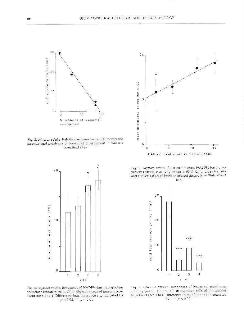
The lysosomal system of digestive cells showed extensive evidence of abnormality in samples from Sites 2, 3 and 4 , compared with those from Site 1. Assessment of lysosomal characteristics included lysosomal enlargement, membrane stability, unsatu- rated neutral lipid and lipofuscin content; results are summarised in Fig. 1. These findings clearly demon- strate that secondary lysosomes were enlarged in molluscs from Sites 2 and 3, and to a lesser extent (though not significantly) at Site 4 (Figs. 1 & 2) . Lysosomal membrane stability was significantly

84 GEEP WORKSHOP: CELLULAR- AND HISTOPATHOLOGY
Fig. 1. Mytilus edulis. Responses of lysosomes in digestive cells of mussels from Field sites 1 to 4 in Langesundfjord (mean + 95 % C[, mean only for [B]). (A) Lysosomal mem- brane stability; (R) incidence of lysosomal enlargement; (C) cytoplasmic content of unsaturated neutral lipid (measured as integrated extinction); (D) lysosomal content of lipofuscin (measured as integrated extlnct~on). Difference from the refer-
ence site (1) indicated by "'::' p < 0.001
reduced in mussels from Sites 2, 3 and 4 in comparison with those from Site 1; Site 4 showed considerable variability (Fig. 1) and its mean value was significantly above that for Sites 2 and 3 (p < 0.01), indicating less severe membrane disturbance. Data for lysosomal sta- bility were inversely correlated with those for incidence of 1ysosom.al enlargement (r = -0.994, df = 2, p < 0.01: Fig. 3).
Increased amounts of unsatu.rated neutral lipid were frequently associated with enlarged secondary lysosomes, in addition to being present as lipid drop- lets, in samples from Sites 2, 3 and 4; this abnormality apparently constituted a form of fatty degeneration, and increased cellular lipid content was confirmed by
microdensitometric measurements (Fig. 1). This latter determination included both cytoplasmic lipid droplets and lysosomally associated lipid (Fig. 2).
Digestive cells generally contain tertiary lysosomes or residual bodies; these are characterised by the pres- ence of lipofuscin detectable as a positive Schmorl reaction. Microdensitometric determination clearly demonstrated significant increases in lipofuscin con- tent in cells from Sites 2, 3 and 4 in comparison with those from Site 1 (Fig. 1). Microscopic assessment of digestive cells revealed an increase in numbers of residual bodies at those sites showing enhanced lipofuscin content (Fig. 2) .
NADPH-ferrihemoprotein reductase activity was present in digestive cells of mussels from all 4 sites; however, the amount of formazan reaction product was significantly higher at Sites 3 and 4 in comparison with Sites 1 and 2 (Fig. 4). In addition, mean values for this enzyme showed a positive linear relation to total PAH concentrations (r = 0.986, df = 2, p < 0.02; Fig. 5) .
Field sites: periwinkles
Lysosomal membrane stability was determined in digestive cells of periwinkles; findings are illustrated in Fig. 6. They demonstrate that lysosomal membrane stability was significantly reduced at Sites 2, 3 and 4 in comparison with Site 1.
Experimental exposures: mussels
Results for lysosomal enlargement, membrane stabil- ity, unsaturated neutral lipid and lipofuscin content are summarised in Fig. 7 There was extensive enlarge- ment of secondary lysosomes in all treatments except the low exposure condition; lysosomal membrane sta- bility was also depressed in all treatments except the low exposure condition. As in the molluscs from the field site, unsaturated neutral lipid was associated with lysosomal enlargement, in addition to being present as lipid droplets; cellular content of lipid was similar for all experimental conditions. Lipofuscin content associated with tertiary lysosomes was significantly elevated in the high exposure condition in comparison with the CH control (Fig. 7).
Experimental exposures: periwinkles
Determinations of lysosomal membrane stability in digestive cells of periwinkles are summarised in Fig. 8. These show that membrane stability was greater in the sample from the medium exposure condition than in

Fig 2 , Mytilus edubs Cryostat tissue sections ( x 400) through digestive tubules showing: (A) normal appearance of lysosomes reacted for 0-N-acetylhexosaminidase mainly in digestive cells (Site 1); (B) abnormally enlarged lysosomes reacted for 0-N- acetylhexosaminidase (Site 3) , (C) lipid droplets localized in digestive cells using oil red 0 (Site 1); (D) unsaturated neutral lipid accumulation in pathologically enlarged lysosomes, together with a general increase in lipid droplets (Site 3); (E) lipofuscin in
secondary and tertiary lysosomes localized using the Schmorl reaction (Site I ) , (F) enhanced lipofuscin content (Site 3)
86 GEEP WORKSHOP: CELLULAR- AND HISTOPATHOLOGY
% inc idence of l y s o s o r n a l enlargemenl
Fig. 3. Mytilus edulis. Relation between lysosomal membrane stability and incidence of lysosomal enlargement in mussels
from field sites
Fig. 4 . Mytilus edulis. Responses of NADPH-fernhemoprotein reductase (mean t 95 "L, CI) in digestive cells of mussels from Field sites I to 4 . Difference from reference site indicated i )y
. p <0.05, . ' p c 0.01
P A H concen t ra t ion in t i s sue ( p p m )
Fig. 5. Mytilus edulis. Relation between NADPH-ferrihemo- protein reductase activity (mean + 95 % CI) in digestive cells and concentration of PAHs in mussel tissues from Field sites 1
to 4
2 3
s i t e
Fig 6 . Littorina Uttorea. Responses of lysosornal membrane stab~lity (mean + 95 ' X , Cl) in digestive cells of periwinkles from Field sites 1 to 4 . Difference from reference site ~ndlcated
by ' . p ~ 0 . 0 0 1

Moore: Cytochemical responses in molluscs 87
C L M C H H C L M C H H
C L M C H H C L M C H H
t r e a t m e n l t r e a t m e n t
Fig. 7. Mytilus edulis. Responses of lysosomes In digestive cells of mussels from experimental exposures C, L, M, CH and H (mean k 95 '/o CI, mean only for [B]) . (A) Lysosomal mem- brane stability; (B) ~ncidence of lysosomal enlargement; (C) cytoplasmic content of unsaturated neutral lipid (~ntegrated extinction); (D) lysosomal content of lipofuscin (integrated extinction). Difference from control basin (C or CH) indicated
by " ' p < 0.001
any of the other experimental conditions, which were consistently low. These data cannot be satisfactorily explained.
DISCUSSION
The results demonstrate that the lysosomal system in digestive cells of Mytilus edulis and Littorina littorea is involved in pathological changes occurring in individu- als exposed to environmental pollutants, which here include polycyclic aromatic hydrocarbons (PAHs) and
C L M H l r e a l r n e n l
Fig. 8. Littorina littorea. Responses of lysosomal membrane stability (mean 2 95 % CI) in digestive cells of periwinkles from C, L, M, and H. Difference from control basin (C) indi-
cated by p < 0.001
polychlorinated biphenyls (PCBs). Similar pathological changes have been described in both mussels and periwinkles following experimental exposure to PAHs, both in the laboratory and in large mesocosms (Moore 1979, 1985, Moore et al. 1985, Livingstone et al. 1986). Further support comes from previous field investiga- tions where evidence of lysosomal dysfunction has been reported (Moore et al. 1982, Moore 1985, Moore et al. 1986).
Evidence for reduced severity of lysosomal pathology in mussels from Field Site 4 - the most impacted site in terms of PAHs and PCBs - may be suggestive of pos- sible selection for more tolerant or resistant types at this location. Obviously the present sample size is too small to reach any conclusions in this respect; however, the mussels at this site could well merit further study.
Unlike the data from the field sites in Langesund- fjord, the results of experimental exposure to diesel oil and copper are more difficult to interpret. In mussels, all samples showed evidence of lysosomally associated fatty degeneration, with a significant increase in lipofuscin in the high exposure animals. Lysosomal membrane stability, however, was greatest in the low- exposure individuals. The food level available to mussels in the experimental exposures was low and this may have contributed to the pathological effects observed in all samples (Widdows & Johnson 1988). In this context, starvation in periwinkles is known to result in abnormal enlargement of the lysosomes (Moore et al. 1986). Certain xenobiotics exert biphasic effects and it is conceivable that the concentration of PAHs present in the low-exposure individuals may have induced a stabilising effect on the lysosomal

88 GEEP WORKSHOP: CELLULAR- AND HISTOPATHOLOGY
membranes, resulting in a marked increase in mem- brane stability over the control value (Szego 1975, Stebbing 1981).
The observed increase in activity of NADPH- ferrihemoprotein reductase in digestive cells of mussels from the 2 field sites with the greatest tissue concen- trations of organic pollutants is entirely consistent with previous data for this enzyme, both from field and experimental investigations (Moore 1979, Moore et al. 1982, 1986, Livingstone et al. 1986, Nott & Moore 1987). However, biochemical determinations of the activity of this enzyme (measured as NADPH-cytochrome c reductase; Livingstone 1988) are not in agreement with the cytochemical evidence. There is no immediate explanation for this apparent discrepancy, as previous investigations have generally shown good agreement (Livingstone et al. 1986). However, as the cytochemical test for this enzyme is performed on the cells in situ (i.e. cryostat tissue sections), the possibility of disrup- tive membrane disturbances resulting from tissue homogenization and preparation of microsomes is eliminated.
A general feature of the results is that fatty degenera- tion and the associated lysosomal accumulation of unsaturated neutral lipid is a useful indicator of pathological alteration in digestive cells of mussels. Support for this statement derives from the evidence that such enlarged and lipid-enriched lysosomes fre- quently showed reduced membrane stability, which is indicative of their increased fragility. These parameters were in fact inversely correlated for the field data and, also, inversely correlated for the combined field and experimental data (r = -0.858, df = 7, p < 0.01). This lysosomal accumulation of neutral lipid appears to be a novel observation in bivalve molluscs and could be descnbed as a chemically-induced lipidosis (Dianzani 1978, Liillmann-Rauch 1979). The cytochemical evi- dence is consistent with an increase in cytoplasmic lipid content, probably in the form of lipid droplets, and transference of part of this lipid to the lysosomal com- partment by autophagic uptake. This phenomenon is indicative of either increased synthesis or decreased utilization of neutral lipid, leading to enhanced auto- phagy of the accumulated lipid in the digestive cells. This is in keeping with other evidence for augmented autophagy being associated with reduced lysosomal membrane stability, probably involving an increase of fluidity of the lysosomal membranes (Pipe & Moore 1985, Storch & Kleinfeld 1985, Moore & Viarengo 1987).
Llpofuscin build-up in digestive cells of mussels showing other evidences of lysosomal pathology is con- sistent with enhanced autophagy of lipoprotein mem- branous components of the cytoplasm (Davies 1983). The latter undergo lipid peroxidation and condensation
within the lysosomes resulting in the formation of the lipofuscin-rich residual bodies (Brunk & Colhns 1981). Elevated intra-cellular or intra-lysosomal concen- trations of metals, such as copper or iron, may contri- bute to the enhanced lipofuscin content, as these met- als are important in mediating the toxic effect of oxy- gen (Halliwell 1981). Exposure of mussels to Cu2+ results in increased lipofuscin formation in the diges- tive cells (Viarengo et al. 1987). In the present field study, the precise role of metals in the pathological alterations associated with lysosomal lipofuscin is unclear, as the levels of metals in the various field samples did not differ greatly (Appendix 1). However, in the experimental exposures, copper concentrations in both whole tissues and digestive glands of mussels from the high exposure condition were an order of magnitude greater than in the controls, and this may have contributed to the elevated lipofuscin in these molluscs.
The results provide additional support to the existing evidence for the utility of lysosomally-based indices of cellular and organismal condition, and underline the high sensitivity of tests for lysosomal enlargement and associated fatty degeneration. Such tests are relatively rapid and easy to apply to environmental samples, yet provide considerable insight into the physiological dys- functions leading to lysosomal pathology. The data showing shmulation of NADPH-ferrihemoprotein reductase activity, linearly related to PAH concen- tration, indicate that this component of the cytochrome P-450 system is also a useful indicator of impact by organic xenobiotics, as has been demonstrated on pre- vious occasions (Moore 1985).
Acknowledgement. Microdensitornetry was carried out by Sue Farrar and this is gratefully acknowledged.
LITERATURE CITED
Abdullah, M. I., Steffenak, I. (1988). The GEEP Workshop: trace metal analyses. Mar. Ecol. Prog. Ser. 46: 27-30
Altman, F P (1972). Quantitahve dehydrogenase his- tochemistry with special reference to the pentose shunt dehydrogenases. Prog. Histochem Cytochem. 4: 225-273
Bakke, T., Follum, 0. A., Moe, K. A., Ssrensen. K. (1988). The GEEP Workshop: mesocosm exposures. Mar Ecol. Prog. Ser. 46: 13-18
Bancroft. J. D. (1967). An introduction to histochemical techni- que. Butterworths, London
Bitensky, L., Butcher, R. S., Chayen, J. (1973). Quantitative cytochemistry in the study of lysosomal function In. Dingle, J. T (ed.) Lysosomes in b~ology and pathology, Vol.. 3. Elsevler, Amsterdam, p. 465-510
Brunk, U. T., Colhns, V P. (1981). Lysosomes and age pig- ments in cultured cells. In: Sohal, R. S. (ed.1 Age pigments. Elsevier, Amsterdam, p. 243-264
Butcher R. S., Altrnan, F. P. (1973). Studies on the reduction of tetrazolium salts. 11. The measurement of the half-reduced

Moore: Cytochemical I .esponses in molluscs 89
and fully reduced farmazans of neotetrazolium chloride in tissue sections. Histochemistry 37: 351-363
Davies, I. (1983). Ageing. Edward Arnold, London Dianzani, M. U. (1978). Biochenlical aspects of fatty liver In:
Slater, T F. (ed.) Blochemica1 mechanisms of liver ~nlury . Academic Press, London, p . 45-95
Follum, 0. A., Moe. K. A. (1988). The GEEP Workshop: field sampling. Mar. Ecol Prog. Ser 46: 7-12
Halliwell, B. (1981). Free radicals, oxygen toxicity and ageing. In: Sohal, R. S. (ed.) Age pigments. Elsev~er, Amsterdam, p. 2-62
Hawkins, H. K. (1980). Reactions of lysosomes to cell injury. In: Trump, B. F., Arstila, A.V. (eds.) Pathobiology of cell membranes, Vol. 2. Academic Press, New York, p. 252-285
Klungs~yr . J., Wilhelmsen, S., Westrheim. K., Saetvedt. E., Palmork, K. H. (1988). The GEEP Workshop: organic chemical analyses. Mar. Ecol. Prog. Ser. 46: 19-26
Livingstone, D. R. (1988). Responses of microsomal NADPH- cytochrome c reductase activity and cytochrome P-450 in digestive glands of Mytilus edulis and Littorina littorea to environmental and experimental exposure to pollutants. Mar. Ecol. Prog. Ser. 46: 37-43
Livingstone. D. R., Moore, M. N., Lowe, D. M., Nasci, C., Farrar. S. V (1986). Responses of the cytochrome P-450 monooxygenase system to diesel oil in the common mussel, Mytilus edulis L, and the periwinkle Littorlna Littorea L. Aquat. Toxlcol. 7: 79-91
Lowe, D. M., Moore, M. N., Clarke, K. R. (1981). Effects of oil on digestive cells in mussels: quantitative alterations in cellular and lysosomal structure. Aquat. Toxicol 1. 213-226
Liillmann-Rauch, R. (1979). Drug-induced lysosomal storage disorders. In: Dingle, J . T., Jacques, P. J., Shaw, I. H. (eds.) Lysosomes in biology and pathology, Vol 6. Elsevier, Amsterdam, p. 49- 130
Masters, B. S. S., Okita. R. T (1980). The history, properties and function of NADPH-cytochrorne P-450 reductase. Pharmac. Ther. 9: 227-244
Moore, M N (1976). Cytochemical demonstration of latency of lysosomal hydrolases in digestive cells of the common mussel, Mytrlus edulis, and changes induced by thermal stress. Cell Tissue Res. 175: 279-287
Moore, M. N. (1979). Cellular responses to polycyclic aromatic hydrocarbons and phenobarbital in Mytilus edulis. Mar. environ. Res. 2: 255-263
Moore, M. N. (1985). Cellular responses to pollutants. Mar. Pollut. Bull. 16: 134-139
Moore, M. N., Clarke, K. R. (1982) Use of microstereology and quantitdtive cytochemistry to determine the effects of crude oil-derived aromatic hydrocarbons on lysosomal structure and function in a marine bivalve mollusc, Mytilus edulis. Histochem. J. 14: 713-718
Moore, M. N., Lowe, D. M., Fieth, P. E. M. (1978). Lysosomal
responses to experlmentally lnjected anthracene in the digestive cells of Mytilus edulis. Mar. Biol. 48: 297-302
Moore, M N., Lowe, D. M., Livingstone, D. R., Dixon, D R . (1986). Molecular and cellular indices of pollutant effects and their use in environmental Impact assessment. Wat. Sci. Tech. 18: 223-232
Moore, M. N., Mayernick, J. A., Giam, C. S. (1985). Lysosomal responses to a polynuclear aromatic hydrocarbon in a marine snail: effects of exposure to phenanthrene and recovery. Mar. environ. Res. 17: 230-233
Moore, M. N., Pipe, R. K., Farrar, S. V. (1982). Lysosomal and microsomal responses to environmental factors in Littorina littorea from Sullom Voe. Mar Pollut. Bull. 13 340-345
Moore, M. N., Pipe, R. K., Farrar, S. V, (1987). Induction of lysosomal lipid accumulation and fatty degeneration by polycyclic aromatic hydrocarbons in molluscan digestive cells. Mar, environ. Res. (Abstract) 24: 352-353
Moore, M. N., Viarengo, A. (1987). Lysosomal membrane fragility and catabolism of cytosolic proteins: evidence for a direct relationship. Experientia 43: 320-323
Nott, J. A.. Moore, M. N. (1987). Effects of polycyclic aromatic hydrocarbons on molluscan lysosomes and endoplasmic reticulum. Histochem. J. 19: 357-368
Owen, G. (1972). Lysosomes, peroxisomes and bivalves. Sci. Prog., Lond. 60: 299-318
Pearse, A. G E. (1972). Histochemistry, theoretical and applied, Vol. 2. Churchill-Livingstone, London
Plpe, R. K., Moore, M. N. (1985). Ultrastructural changes in the lysosomal-vacuolar system in digestive cells of Mytilus edulis as a response to increased salinity Mar. Biol. 87: 157-163
Stebbing, A. R. D. (1981). Hormesis - stimulation of colony growth in Campanulana flexuosa (Hydrozoa) by copper, cadmium and other toxicants. Aquat. Toxicol. 1: 227-238
Storch, J . , Kleinfeld, A. M. (1985). The lipid structure of biological membranes. Trends Biochem. Sci. 10: 418-420
Strobel, H. W., Digmann, J D., Gum, J. R. (1980). NADPH- cytochrome P-450 reductase and its role in the mixed function oxidase reaction. Pharmac. Ther. 8- 525-537
Szego, C. M. (1975). Lysosomal function in nucleocytoplasmic communication. In Dingle, J . T., Dean, R. (eds.) Lysosomes in biology and pathology, Vol. 4. Elsevier, Amsterdam. p. 385-477
Van Noorden, C. J. F.. Butcher, R. G. (1986). A quantitative histochemical study of NADPH-ferrihemoprotein reduc- tase activity. Histochem J. 18: 364-370
Viarengo, A.. Moore, M. N.. Mancinelli, G., Mazzucotelli, A., Pipe, R. K., Farrar, S. V. (1987). Metallothioneins and lysosomes in metal toxicity and homeostasis in marine mussels: the effects of cadmium in the presence and ab- sence of phenanthrene. Mar. Biol. 94: 251-257
Widdows, J., Johnson, D. (1988). Physiological energetics of Mytilus edulis: Scope for Growth. hdar. Ecol. Prog. Ser. 46: 113- 121