cytogenetic abnormalities in acute myeloid leukemia in ... · vladimir lj lazarevic, md, senior...
TRANSCRIPT

LUND UNIVERSITY
PO Box 117221 00 Lund+46 46-222 00 00
Cytogenetic abnormalities in Acute Myeloid Leukemia in Sweden. A population basedstudy.LAZAREVIC, VLADIMIR
Published: 2015-01-01
Link to publication
Citation for published version (APA):Lazarevic, V. (2015). Cytogenetic abnormalities in Acute Myeloid Leukemia in Sweden. A population basedstudy. Stam Cells Centrum (SCC)
General rightsCopyright and moral rights for the publications made accessible in the public portal are retained by the authorsand/or other copyright owners and it is a condition of accessing publications that users recognise and abide by thelegal requirements associated with these rights.
• Users may download and print one copy of any publication from the public portal for the purpose of privatestudy or research. • You may not further distribute the material or use it for any profit-making activity or commercial gain • You may freely distribute the URL identifying the publication in the public portal ?
Take down policyIf you believe that this document breaches copyright please contact us providing details, and we will removeaccess to the work immediately and investigate your claim.
Download date: 10. Aug. 2016

Cytogenetic abnormalities in Acute Myeloid Leukemia in Sweden A population based studyVLADIMIR Lj LAZAREVIC
STAMCELLSCENTRUM (SCC) | FACULTY OF MEDICINE | LUND UNIVERSITY 2015
Lund University, Faculty of Medicine Doctoral Dissertation Series 2016:2
ISBN 978-91-7619-227-6ISSN 1652-8220
Vladimir Lj Lazarevic, MD, senior consultant, member of the Swedish AML group, Department of Hematology and Oncology, Skåne University Hospital, Lund Stem Cell Center, Lund University.
Printed by Media-Tryck, Lund U
niversity 2016
978
9176
1922
76
VLA
DIM
IR Lj LA
ZA
REV
IC
Cytogenetic abnorm
alities in Acute M
yeloid Leukem
ia in Sweden A population based study
2


1
Cytogenetic abnormalities in Acute Myeloid
Leukemia in Sweden

2

3
Cytogenetic abnormalities in Acute Myeloid Leukemia in
Sweden A population based study
Vladimir Lj Lazarević
DOCTORAL DISSERTATION by due permission of the Faculty of Medicine, Lund University, Sweden.
To be defended at Belfragesalen, BMC, Lund. Date 20. January 2016, time 13.00-17.00
Faculty opponent Anthony Moorman, Professor of Genetic Epidemiology, Newcastle University,
Newcastle upon Tyne

4
LUND UNIVERSITY
Stamcellscentrum (SCC)
Medicinska fakulteten / Institutionen för laboratoriemedicin, Lund
Document name: Thesis
Date of issue: 20 januari 2016
Author(s) Vladimir Lj Lazarevic Sponsoring organization
Title and subtitle: Cytogenetic abnormalities in Acute Myeloid Leukemia in Sweden
A population based study
Abstract: The impact of cytogenetic findings in AML was analyzed in the large population-based Swedish AML registry. Karyotypic patterns differed by age: t(8;21), inv(16) and t(11q23) were more common in younger patients, whereas loss of 5q, 7q and 17p, monosomal karyotype (MK) and complex karyotype (CK) were more common in older patients. Patients with ≥5 chromosome abnormalities had worse overall survival than those with fewer abnormalities or normal karyotype in all age groups. Loss of 5q, 7q and/or 17p had, in contrast to MK, a further negative impact on survival. Multivariable analyses on risk factors in patients <80 years with cytogenetic abnormalities and intensive treatment revealed that age and performance status had the most significant impact on survival (both P<0.001), followed by sex (P=0.0135) and a karyotype including -7/del(7q) (P=0.048).
We compared outcome of AHD-AML and tAML, i.e., secondary (sAML) with de novo AML. The CR rates were significantly lower but early death rates similar in sAML vs de novo AML. In a multivariable analysis, AHD-AML (HR 1.51; 95% CI 1.26–1.79) and tAML (1.72; 1.38–2.15) were independent risk factors for poor survival. The negative impact of AHD-AML and tAML on survival was highly age dependent with a considerable impact in younger patients, but without independent prognostic value in the elderly.
The frequencies of unsuccessful cytogenetics (UC) and unperformed cytogenetics (UPC) were 2.1% and 2.0%, respectively. The early death rates differed between the cytogenetic subgroups (P=0.006) with the highest rates in patients with UC (14%) and UPC (12%) followed by high-risk (HR) AML, intermediate risk (IR) and standard risk (SR) cases successfully karyotyped (8.6%, 5.9%, and 5.8%, respectively). The CR rate was lower in UC and UPC and HR compared with the other risk groups (P<0.001). The 5-year OS rates were 25% for UC and 22% for UPC, whereas the corresponding frequencies for SR, IR and HR AML patients without UC and UPC were 64%, 31% and 15%, respectively. Lack of cytogenetic data translates into a poor prognosis.
To ascertain the clinical implications of high hyperdiploid (HH; 49–65 chromosomes) and triploid/tetraploid (TT; >65 chromosomes) adult AML diagnosed 1997-2014, and 68 (1.9%) were HH (n=50)/TT (n=18). The OS was similar between patients with HH/TT and CK AML (median 0.9 years vs. 0.6 years; P=0.082), whereas OS was significantly longer (median 1.6 years; P=0.028) for IR AML. The OS was shorter for cases with HH than with TT (median 0.6 years vs. 1.4 years; P=0.032) and for HH/TT AMLs with adverse abnormalities (median 0.8 years vs. 1.1 years; P=0.044). HH/TT AML is associated with a poor outcome, but chromosome numbers >65 and absence of adverse aberrations seem to translate into a more favorable prognosis.
Also, among 23 patients (0.4 %) with trisomy 13 with a median age of 72 years (44-84), there was a striking male predominance (80%) with AML-M0 subtype in 37% of patients. Therapy-related AML and MDS/MPN/AML were present in 30% of patients. Median OS time was 9.6 months (95 % CI (3.5-13.7), and 13 months for other patients (95% CI 11.7-14.04), which was almost identical as in previously published studies.
Key words: Key words: AML, karyotype, population-based studies, prognosis, chromosomes, hyperdiploidy
Classification system and/or index terms (if any)
Supplementary bibliographical information Language: Eng
ISSN and key title: 1652-8220 ISBN: 978-91-7619-227-6
Recipient’s notes Number of pages Price
Security classification
I, the undersigned, being the copyright owner of the abstract of the above-mentioned dissertation, hereby grant to all reference sourcespermission to publish and disseminate the abstract of the above-mentioned dissertation.
Signature Date

5
Cytogenetic abnormalities in Acute Myeloid Leukemia in
Sweden A population based study
Vladimir Lj Lazarević

6
Cover image (FISH analysis showing deletion of chromosome 7q) kindly provided by Anna Collin, Department of Clinical Genetics and Biobank, Division of Laboratory Medicine, LundCopyright Vladimir Lazarevic
Stamcellscentrum (SCC) | Medicinska fakulteten | Institutionen för laboratoriemedicin, Lund ISSN 1652-8220 ISBN 978-91-7619-227-6 Lund University, Faculty of Medicine Doctoral Dissertation Series 2016:2 Printed in Sweden by Media-Tryck, Lund University Lund 2015
En del av Förpacknings- och Tidningsinsamlingen (FTI)

7
Content
Original articles 9
Brief introduction and aims of the PhD thesis 11
Abstract 13
Abbreviations 15
Historical and general aspects of chromosome analysis in AML 17
Cytogenetic risk classification 19
Epidemiology of AML 26
Incidence of chromosomal abnormalities in AML 28
Gender and chromosomal abnormalities in AML 28
Correlation of FAB (morphology) and chromosomal abnormalities in AML 29
Generally accepted cytogenetically high risk AML 29
Not generally accepted cytogenetically high riskAML 31
Registries, population and methods 33
What we have learned from our studies 35
Age dependent incidence of different chromosomal abnormalities 35
Overlap between poor-risk chromosome abnormalities, complex and monosomal karyotypes 36
Unsuccessful cytogenetics 37
Unperformed cytogenetics 38
Hyperdiploidy 38
Secondary leukemia 39
The role of trisomy 13 in the prognosis of AML 39
Brief summary 39
References 41
Acknowledgments 51

8

9
Original articles
This Thesis is based on the following original articles, referred to in the text by their Roman numerals:
I. Lazarevic V, Hörstedt AS, Johansson B, Antunovic P, Billström R, Derolf A, Hulegårdh E, Lehmann S, Möllgård L, Nilsson C, Peterson S, Stockelberg D, Uggla B, Wennström L, Wahlin A, Höglund M, Juliusson G. Incidence and prognostic significance of karyotypic subgroups in older patients with acute myeloid leukemia: the Swedish population-based experience. Blood Cancer J. 2014 Feb 28;4: e188.
II. Lazarevic V, Hörstedt AS, Johansson B, Antunovic P, Billström R, Rangert-Derolf Å, Lehmann S, Möllgård L, Peterson S, Stockelberg D, Uggla B,
Vennström L, Wahlin A, Höglund M, Juliusson G. Failure matters: unsuccessful cytogenetics and unperformed cytogenetics are associated with a poor prognosis in a population-based series of acute myeloid leukaemia. Eur J Haematol 2015 May;94(5):419-23. doi: 10.1111/ejh.12446. Epub 2014 Oct 3.
III. Hulegårdh E, Nilsson C, Lazarevic V, Garelius H, Antunovic P, Rangert Derolf Å, Möllgård L, Uggla B, Wennström L, Wahlin A, Höglund M, Juliusson G, Stockelberg D, Lehmann S. Secondary AML impacts survival in younger but not in older AML patients: the Swedish population-based experience. Am J Hematol 2015 Mar;90(3):208-14. doi: 10.1002/ajh.23908. Epub 2015 Jan 16.
IV. Lazarevic V, Rosso A, Juliusson G, Antunovic P, Rangert-Derolf Å, Lehmann S, Möllgård L, Uggla B, Wennström L, Wahlin A, Höglund M, Johansson B. Prognostic significance of high hyperdiploid and triploid/tetraploid adult acute myeloid leukemia. Am J Hematol 2015 Sep;90(9):800-5. doi: 10.1002/ajh.24091. Epub 2015 Jul 22.
V. Lazarevic V, Rosso A, Juliusson G. Isolated trisomy 13 in AML from a Swedish population-based perspective. Blood 2014, September 9, E-letter; http://www.bloodjournal.org.ludwig.lub.lu.se/content/124/8/1304.e-letters

10

11
Brief introduction and aims of the PhD thesis
Population-based registries may provide data complementary to that from the basic science and clinical intervention studies, and are helpful for establishing recommendations for the management of patients in the “real” world. Registries with high coverage of the target population reduce the impact of selection on the outcome and the subsequent problem with extrapolating data to non-studied populations. Therefore, data that can help clinical decision-making in the situations that are not well covered by clinical studies can be provided1. We analyzed several aspects of the data from the Swedish AML Registry between 1997-2006 and between 1997-2014 from a population-based perspective in several aspects. The first aim was to describe the incidence and prognostic importance of the known chromosomal abnormalities in AML. Other goals were to test the clinical characteristics and outcome of specific patient groups, such as unsuccessful cytogenetics (UC), unperformed cytogenetics (UPC), high and low hyperdiploidy, as well as isolated trisomy 13. Last aim was to further characterize the prognosis and characteristics of de novo AML versus AML with an antecedent hematological disease (AHD-AML) or therapy related AML (t-AML)

12

13
Abstract
The impact of cytogenetic findings in AML was analyzed in the large population-based Swedish AML registry. Karyotypic patterns differed by age: t(8;21), inv(16) and t(11q23) were more common in younger patients, whereas loss of 5q, 7q and 17p, monosomal karyotype (MK) and complex karyotype (CK) were more common in older patients. Patients with ≥5 chromosome abnormalities had worse overall survival than those with fewer abnormalities or normal karyotype in all age groups. Loss of 5q, 7q and/or 17p had, in contrast to MK, a further negative impact on survival. Multivariable analyses on risk factors in patients <80 years with cytogenetic abnormalities and intensive treatment revealed that age and performance status had the most significant impact on survival (both P<0.001), followed by sex (P=0.0135) and a karyotype including -7/del(7q) (P=0.048).
We compared outcome of AHD-AML and tAML, i.e., secondary (sAML) with de novo AML. The CR rates were significantly lower but early death rates similar in sAML vs de novo AML. In a multivariable analysis, AHD-AML (HR 1.51; 95% CI 1.26–1.79) and tAML (1.72; 1.38–2.15) were independent risk factors for poor survival. The negative impact of AHD-AML and tAML on survival was highly age dependent with a considerable impact in younger patients, but without independent prognostic value in the elderly.
The frequencies of unsuccessful cytogenetics (UC) and unperformed cytogenetics (UPC) were 2.1% and 2.0%, respectively. The early death rates differed between the cytogenetic subgroups (P=0.006) with the highest rates in patients with UC (14%) and UPC (12%) followed by high-risk (HR) AML, intermediate risk (IR) and standard risk (SR) cases successfully karyotyped (8.6%, 5.9%, and 5.8%, respectively). The CR rate was lower in UC and UPC and HR compared with the other risk groups (P<0.001). The 5-year OS rates were 25% for UC and 22% for UPC, whereas the corresponding frequencies for SR, IR and HR AML patients without UC and UPC were 64%, 31% and 15%, respectively. Lack of cytogenetic data translates into a poor prognosis.
To ascertain the clinical implications of high hyperdiploid (HH; 49–65 chromosomes) and triploid/tetraploid (TT; >65 chromosomes) adult AML diagnosed 1997-2014, and 68 (1.9%) were HH (n=50)/TT (n=18). The OS was similar between patients with HH/TT and CK AML (median 0.9 years vs. 0.6 years;

14
P=0.082), whereas OS was significantly longer (median 1.6 years; P=0.028) for IR AML. The OS was shorter for cases with HH than with TT (median 0.6 years vs. 1.4 years; P=0.032) and for HH/TT AMLs with adverse abnormalities (median 0.8 years vs. 1.1 years; P=0.044). HH/TT AML is associated with a poor outcome, but chromosome numbers >65 and absence of adverse aberrations seem to translate into a more favorable prognosis.
Also, among 23 patients (0.4 %) with trisomy 13 with a median age of 72 years (44-84), there was a striking male predominance (80%) with AML-M0 subtype in 37% of patients. Therapy-related AML and MDS/MPN/AML were present in 30% of patients. Median OS time was 9.6 months (95 % CI (3.5-13.7), and 13 months for other patients (95% CI 11.7-14.04), which was almost identical as in previously published studies.
Key words: AML, karyotype, population-based studies, prognosis, chromosomes, hyperdiploidy

15
Abbreviations
AML acute myeloid leukemia
sAML secondary AML
tAML therapy-related AML
AHD-AML AML with an antecedent hematological disease
APL acute promyelocytic leukemia
ATRA all-trans retinoic acid
CK complex karyotype
CML chronic myeloid leukemia
CR complete remission
DFS disease free survival
FISH fluorescent in situ hybridization
MDS myelodysplastic syndrome
MK monosomal karyotype
MPN myeloproliferative neoplasm
NPM1 nucleophosmin 1
PS performance status
FLT3 fms-like tyrosine kinase 3
CEBPA CCAAT/enhancer-binding protein alpha
OS overall survival

16
17
Historical and general aspects of chromosome analysis in AML
A revolution in understanding of chromosome changes in leukemia and in cancer in general started in 1960 with Peter C Nowell and David Hungerford´s discovery of a minute chromosome in chronic granulocytic leukemia, subsequently called the “Philadelphia chromosome”2. Before them, predecessors paved the ground for this at that time accidental finding. Wilhelm von Waldemeyer coined the term “chromosome” (colored body) in the late 1880s, and in 1890 David P. von Hansemann, described multipolar mitoses and other aberrant mitotic figures in carcinoma samples and suggested that these aberrant cell divisions were responsible for the abnormal chromatin content found in cancer cells3. Theodor Boveri proposed that cancer begins within a single cell in which the chromosomal makeup becomes scrambled, permitting cells to proliferate uncontrollably4. At the University in Lund Joe Thin Tjio and Albert Levan contributed with the important finding that the normal number of chromosomes in man is 465. By the end of the 1960s, it seemed as if the Philadelphia (Ph) chromosome was an exception and that such specific chromosomal changes would not be characteristic of other malignancies. Moreover, it was not clear at the time if the Ph chromosome was a simple deletion or if there was a translocation of chromosomal material or not. Janet D Rowley used the newly developed specific staining technique to characterize the Philadelphia chromosome as a balanced translocation t(9;22) in chronic myeloid leukemia (CML), and subsequently t(8;21) in acute myeloid leukemia (AML).
These discoveries opened the pathway for further important investigations of cytogenetic changes in hematological malignancies6,7,8. After that, an explosion of knowledge about the chromosomal abnormalities, especially in AML, help us achieve a better understanding of the disease, with tailoring of the therapy based on the prognostic and predictive value of the chromosomal changes.
The first major classification of AML was that of the French–American–British (FAB) group, who proposed the criteria for defining AML by morphological subtype9. The first prospective study of AML by chromosomal abnormality was the International Workshops on Chromosomes in Leukemia in 1982, from which the Chicago karyotype classification was derived10. The long-term survival of patients identified at

18
this workshop was reported, and multivariate analysis showed that karyotype was an independent predictor of survival for all patients11. Cytogenetic abnormalities are identified in 50–60% of newly diagnosed AML of adult patients12,13,14. Age and chromosomal abnormalities are the most important prognostic factors in AML15,16. Diagnostic karyotype serves as a tool to identify biologically distinct subsets of disease and is widely adopted to provide the framework for risk-adapted treatment approaches14,16.
Furthermore, in newly diagnosed AML patients with abnormal karyotype, cytogenetic analysis is also recommended for documenting complete remission (CR)17. In fact, several data show that the persistence, after induction chemotherapy, of cytogenetic abnormalities present at diagnosis in leukemic blasts determine a high relapse rate of leukemia and a worse clinical outcome with lower disease-free survival (DFS) rate and overall survival (OS)18. Therefore, the International Working Group for Diagnosis, Standardization of Response Criteria, Treatment Outcomes, and Reporting Standards for Therapeutic Trials in Acute Myeloid Leukemia has introduced into standard response criteria for AML the category of cytogenetic CR defined as the absence of any cytogenetic aberrations in bone marrow leukemic blasts after induction chemotherapy in presence of morphologic CR and complete peripheral hematological recovery19.
The presence of some recurrent chromosomal translocation has led to several discoveries relevant for the pathogenesis of leukemia. For example t(8;21)(q22;q22) resulting in the hybrid gene RUNX1-RUNX1T1, is considered a favorable cytogenetic abnormality. It results in an in-frame fusion of two genes, leading to a fusion protein of one N-terminal domain from the AML1 gene and four C-terminal domains from the ETO gene. This leads to altered gene transcription, RNA-dependent mechanisms and ribosomal functions, DNA damage and repair, disrupted cytokine-mediated growth regulation, cell-cycle regulation, regulation of apoptosis and stress responses which all contribute to the leukemogenesis. Furthermore, in adults with t(8;21) AML, the presence of the fusion transcript can serve as pre or post transplant PCR-based RUNX1/RUNX1T1 monitoring of minimal residual disease (MRD)20,21. Since the international cytogenetic community strives for consistency in the descriptive and interpretive reporting of both normal and abnormal karyotypes, regardless of technical evaluation method used, an updated edition of the International System for Human Cytogenetic Nomenclature, (ISCN 2013) is recommended22. The cytogenetic reports should be written according to ISCN, for example the overall chromosome number should be reported, sex chromosomes, affected chromosomes, type of abnormalities, chromosomal band locations, and in brackets, the number of cells with a given karyotype.

19
Cytogenetic risk classification
Although there are some differences in the classification of cytogenetic risk based on karyotype results among the various cooperative international groups, AML patients are generally classified into three groups: high, intermediate and low risk, also called adverse (unfavorable, poor) risk, intermediate risk and favorable risk14,23. It is important to note that the classifications of cytogenetic risk groups in AML patients are based on studies predominantly including younger patients (aged <60 years). Some of these variations are depicted in Table 124.

20
Table 1. Variation in cytogenetic risk group classification across clinical trial groups, (adapted from Grimwade D, Hills RK. 200924); unrel abn indicates unrelated abnormality; abn, abnormal
Original MRC SWOG/ ECOG
CALGB GIMEMA/AML10
German AMLCG
HOVON/SAKK
Refined MRC
Low risk
t(15;17) t(8;21) inv(16)/t(16;16)
t(15;17) t(8;21) [lacking del(9q), complex, ie, ≥ 3 unrel abn] inv(16)/t(16;16)/del(16q)
t(15;17) t(8;21) inv(16)/t(16;16)
t(15;17) t(8;21) inv(16)/
t(16;16)
t(15;17) t(8;21) inv(16)/
t(16;16)
t(15;17) t(8;21) alone inv/del(16) and lacking highrisk abn
t(15;17) t(8;21) inv(16)/t(16;16)
Intermediate risk
Normal, Other non-complex
Normal, +6, +8, -Y, del(12p)
Normal, Other non- complex
Normal, -Y Normal Other non- complex
Normal Other non- complex
Normal,Other non- complex
High risk
abn(3q)
-5/del(5q), -7 complex [≥ 5 unrel abn] Excluding those with low risk changes
abn(3q),(9q),(11q),(21q) abn(17p)
-5/del(5q), -7/del(7q) t(6;9) t(9;22) complex [≥3 unrel abn]
inv(3)/t(3;3) -7, t(6;9), t(6;11) t(11;19),+8 complex
(≥ 3 unrel abn) Excluding those with low risk changes
Other inv(3)/t(3;3) -5/del(5q)
-7/del(7q) abn(11q23) del(12p) abn(17p) complex (≥ 3 unrel abn)
abn(3q),
-5/del(5q) -7/del(7q) abn(11q23) t(6;9) t(9;22) complex (≥ 3 unrel abn)
abn(3q), [excl t(3;5)] inv(3)/t(3;3) add(5q)/del(5q)/-5,-7/add(7q) t(6;11) t(10;11) t(9;22), -17 abn(17p) with other changes Complex (> 3 unrel abn) Excluding
those with low risk changes

21
Large multicenter studies have consistently reported that patients with acute promyelocytic leukemia (APL) with the t(15;17)(q22;q12~21) treated on ATRA- and anthracycline-based protocols together with the core binding factor (CBF) leukemias with t(8;21)(q22;q22) or inv(16)(p13q22)/t(16;16)(p13;q22) treated with intensive chemotherapy involving cytarabine at a range of doses are characterized by relatively favorable prognoses24.
Conversely, adults presenting with AML and abnormalities of 3q [abn(3q)], deletions of 5q [del(5q)], monosomies of chromosome 5 and/or 7 (-5/-7) or complex karyotype have a very poor prognosis with conventional chemotherapy and are therefore considered candidates for allogeneic transplant and experimental treatment approaches24. Patients with normal karyotype at diagnosis are generally classified in the intermediate risk group. However, this group of patients is characterized by a notable heterogeneity in clinical outcome, showing a different response to treatment11. Although karyotype analysis provides a powerful independent prognostic factor for rates of CR, relapse risk and OS in multivariable analyses, there is still uncertainty present concerning a number of miscellaneous cytogenetic abnormalities that together account for ~10% of AML14,24.
The definition of complex karyotype (CK) also varies and is a subject of change. CK comprises the patients with three, four or five chromosomal abnormalities, absence of any of the known recurring balanced abnormalities such as t(8;21), inv(16)/t(16;16), t(15;17), and 11q23/MLL (excluding t(9;11) and t(11;19)); loss of at least one of chromosomal regions 5q, 7q, or 17p; and loss of at least one additional area of regions 18q21q22, 12p13, or 16q22q24 or gain of 11q23q25, 1p33p36, 8q22q24, or 21q11q2225,26. This definition of CK is problematic because chromosomal analysis is subjective, chromosome morphology is often poor and defining independent abnormalities is sometimes difficult to ascertain25.
A study by the HOVON group involving 1975 adults (ages 15-60 years) with AML suggested the existence of a novel adverse-risk group characterized by a presence of an autosomal monosomy in conjunction with at least one other autosomal monosomy or structural abnormality (denoted monosomal karyotype positive, MK+)27. It is important to emphasize that karyotypes with t(8;21)(q22;q22), t(9;11)(p21;q23), t(15;17)(q22;q21) or inv(16)(p13q22)/t(16;16)(p13;q22) should not be classified as MK+ AML27, 28.
One striking observation is an increasing incidence of adverse versus favorable cytogenetic abnormalities with increasing age. The recent WHO classification reflects the fact that an increasing number of AML can be categorized based upon their underlying cytogenetic or molecular genetic abnormalities, and that these genetic changes form clinico-pathologic-genetic entities29. The subgroup “AML with recurrent genetic abnormalities” comprises several primary AML entities. “AML with t(8;21)(q22;q22); RUNX1-RUNX1T1” and “AML with inv(16)(p13.1q22) or

22
t(16;16)(p13.1;q22); CBFB-MYH11” are considered as AML regardless of bone marrow blast counts. In “APL with t(15;17)(q22;q12); PML-RARA,” RARA translocations with other partner genes are recognized separately. The former category “AML with 11q23 (MLL) abnormalities” was redefined into that “AML with t(9;11)(p22;q23); MLLT3-MLL” and is now a unique entity; balanced translocations other than that involving MLLT3 should be specified in the diagnosis. Three new cytogenetically defined entities were incorporated: “AML with t(6;9)(p23;q34); DEK-NUP214”; “AML with inv(3)(q21q26.2) or t(3;3)(q21;q26.2); RPN1-EVI1”; and “AML (megakaryoblastic) with t(1;22)(p13;q13); RBM15-MKL1,” a rare leukemia most commonly occurring in infants.
Two new provisional entities defined by the presence of gene mutations were added, “AML with mutated NPM1 [nucleophosmin (nucleolar phosphoprotein B23, numatrin)],” and “AML with mutated CEBPA [CCAAT/enhancer binding protein (C/EBP), alpha].” There is a growing evidence that these two gene mutations represent primary genetic lesions (so-called class II mutations) that impair hematopoietic differentiation30.
Mutations in the fms-related tyrosine kinase 3 (FLT3) gene are found in many AML subtypes and are considered class I mutations conferring a proliferation and/or survival advantage. AML with FLT3 mutations are not considered a distinct entity, although determining the presence of such mutations is recommended by WHO because of their prognostic significance (Table 2).
The former subgroup termed “AML with multilineage dysplasia” is now designated “AML with myelodysplasia-related changes.” Dysplasia in 50% or more of cells, in 2 or more hematopoietic cell lineages, was the diagnostic criterion for the former subset. However, the clinical significance of this morphologic feature has been questioned31,32. AMLs are now categorized as “AML with myelodysplasia-related changes” if (1) they have a previous history of myelodysplastic syndrome (MDS) or myelodysplastic/myeloproliferative neoplasm (MDS/MPN) and evolve to AML with a marrow or blood blast count of 20% or more; (2) they have a myelodysplasia-related cytogenetic abnormality (listed in a footnote to Table 2); or (3) if 50% or more of cells in 2 or more myeloid lineages are dysplastic.
“Therapy-related myeloid neoplasms” has remained a distinct entity; however, since most patients have received treatment using both alkylating agents and drugs that target topoisomerase II for prior malignancy, a division according to the type of previous therapy is often not feasible. Therefore, therapy-related myeloid neoplasms are no longer subcategorized. Myeloid proliferations related to Down syndrome are now listed as distinct entities.

23
Table 2. Acute myeloid leukemia and related precursor neoplasms, and acute leukemias of ambiguous lineage; (adapted from WHO 2008)
Categories
Acute myeloid leukemia with recurrent genetic abnormalities
AML with t(8;21)(q22;q22); RUNX1-RUNX1T1
AML with inv(16)(p13.1q22) or t(16;16)(p13.1;q22); CBFB-MYH11
APL with t(15;17)(q22;q12); PML-RARA*
AML with t(9;11)(p22;q23); MLLT3-MLL†
AML with t(6;9)(p23;q34); DEK-NUP214
AML with inv(3)(q21q26.2) or t(3;3)(q21;q26.2); RPN1-EVI1
AML (megakaryoblastic) with t(1;22)(p13;q13); RBM15-MKL1
Provisional entity: AML with mutated NPM1
Provisional entity: AML with mutated CEBPA
Acute myeloid leukemia with myelodysplasia-related changes‡
Therapy-related myeloid neoplasms§
Acute myeloid leukemia, not otherwise specified (NOS)
Acute myeloid leukemia with minimal differentiation
Acute myeloid leukemia without maturation
Acute myeloid leukemia with maturation
Acute myelomonocytic leukemia
Acute monoblastic/monocytic leukemia
Acute erythroid leukemia
Pure erythroid leukemia
Erythroleukemia, erythroid/myeloid
Acute megakaryoblastic leukemia
Acute basophilic leukemia
Acute panmyelosis with myelofibrosis (syn.: acute myelofibrosis; acute myelosclerosis)
Myeloid sarcoma (syn.: extramedullary myeloid tumor; granulocytic sarcoma; chloroma)
Myeloid proliferations related to Down syndrome
Transient abnormal myelopoiesis (syn.: transient myeloproliferative disorder)
Myeloid leukemia associated with Down syndrome
Blastic plasmacytoid dendritic cell neoplasm
Acute leukemias of ambiguous lineage
Acute undifferentiated leukemia
Mixed phenotype acute leukemia with t(9;22)(q34;q11.2); BCR-ABL1ǁ
Mixed phenotype acute leukemia with t(v;11q23); MLL rearranged
Mixed phenotype acute leukemia, B/myeloid, NOS
Mixed phenotype acute leukemia, T/myeloid, NOS
Provisional entity: Natural killer (NK)–cell lymphoblastic leukemia/lymphoma

24
For a diagnosis of AML, a marrow blast count of≥ 20% is required, except for AML with the recurrent genetic abnormalities t(15;17), t(8;21), inv(16) or t(16;16) and some cases of erythroleukemia.
* Other recurring translocations involving RARA should be reported accordingly: for example, AML with t(11;17)(q23;q12); ZBTB16-RARA; AML with t(11;17)(q13;q12); NUMA1-RARA; AML with t(5;17)(q35;q12); NPM1-RARA; or AML with STAT5B-RARA (the latter having a normal chromosome 17 on conventional cytogenetic analysis).
† Other translocations involving MLL should be reported accordingly: for example, AML with t(6;11)(q27;q23); MLLT4-MLL; AML with t(11;19)(q23;p13.3); MLL-MLLT1; AML with t(11;19)(q23;p13.1); MLL-ELL; AML with t(10;11)(p12;q23); MLLT10-MLL.
‡ More than 20% blood or marrow blasts AND any of the following: previous history of myelodysplastic syndrome (MDS), or myelodys-plastic/myeloproliferative neoplasm (MDS/MPN); myelodysplasia-related cytogenetic abnormality (see below); multilineage dysplasia; AND absence of both prior cytotoxic therapy for unrelated disease and aforementioned recurring genetic abnormalities; cytogenetic abnormalities sufficient to diagnose AML with myelodysplasia-related changes are:
Complex karyotype (defined as 3 or more chromosomal abnormalities).
Unbalanced changes: −7 or del(7q); −5 or del(5q); i(17q) or t(17p); −13 or del(13q); del(11q); del(12p) or t(12p); del(9q); idic(X)(q13).
Balanced changes: t(11;16)(q23;p13.3); t(3;21)(q26.2;q22.1); t(1;3)(p36.3;q21.1); t(2;11)(p21;q23); t(5;12)(q33;p12); t(5;7)(q33;q11.2); t(5;17)(q33;p13); t(5;10)(q33;q21); t(3;5)(q25;q34).
§ Cytotoxic agents implicated in therapy-related hematologic neoplasms: alkylating agents; ionizing radiation therapy; topoisomerase II inhibitors; others.
BCR-ABL1–positive leukemia may present as mixed phenotype acute leukemia, but should be treated as BCR-ABL1–positive acute lymphoblastic leukemia.
Genotypes defined by the mutational status of NPM1, FLT3, CEBPA, and MLL are associated with the outcome of treatment for patients with cytogenetically normal AML16,33. The consequence of these findings is that the benefit of the transplant was limited to the subgroup of patients with the prognostically adverse genotype FLT3-ITD or the genotype consisting of wild type NPM1 and CEBPA without FLT3-ITD33. European LeukemiaNet proposed a new standardized reporting system for correlation of cytogenetic and molecular genetic data with clinical data (APL not shown)34(Table 3). There is a bulk of information about the significance of other mutations in AML, for example DNMT3A, TET2, IDH1 and IDH2 that have prognostic significance in AML35,36. Some groups propose new classification, which suggests that we do not need to perform karyotype analysis at all at the diagnosis, relying only to specific mutations37. Other groups are trying to integrate and refine

25
the risk-groups including clinical, cytogenetic and molecular markers including microRNA analysis and epigenetics38-41.
Table 3. Standardized reporting for correlation of cytogenetic and molecular genetic data in AML with clinical data (adapted from Döhner et al 201034)
Genetic group Subsets
Favorable t(8;21)(q22;q22); RUNX1-RUNX1T1
inv(16)(p13.1q22) or t(16;16)(p13.1;q22); CBFB-MYH11
Mutated NPM1 without FLT3-ITD (normal karyotype)
Mutated CEBPA (normal karyotype)
Intermediate-I* Mutated NPM1 and FLT3-ITD (normal karyotype)
Wild-type NPM1 and FLT3-ITD (normal karyotype)
Wild-type NPM1 without FLT3-ITD (normal karyotype)
Intermediate-II t(9;11)(p22;q23); MLLT3-MLL
Cytogenetic abnormalities not classified as favorable or adverse†
Adverse inv(3)(q21q26.2) or t(3;3)(q21;q26.2); RPN1-EVI1
t(6;9)(p23;q34); DEK-NUP214
t(v;11)(v;q23); MLL rearranged
−5 or del(5q); −7; abnl(17p); complex karyotype‡
* Includes all AMLs with normal karyotype except for those included in the favorable subgroup; most of these cases are associated with poor prognosis, but they should be reported separately because of the potential different response to treatment.
† For most abnormalities, adequate numbers have not been studied to draw firm conclusions regarding their prognostic significance. ‡ Three or more chromosome abnormalities in the absence of one of the WHO designated recurring translocations or inversions, that is, t(15;17), t(8;21), inv(16) or t(16;16), t(9;11), t(v;11)(v;q23), t(6;9), inv(3) or t(3;3); indicate how many complex karyotype cases have involvement of chromosome arms 5q, 7q, and 17p.
Even though this classification is not thoroughly validated, and based on expert opinion it is just one of the many attempts aiming at simplifying decision making in AML. This area is continually changing and in the USA classifications are pragmatically updated every year42 (Table 4).
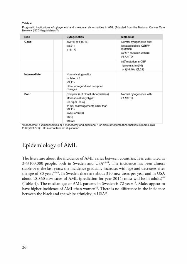
26
Table 4. Prognostic implications of cytogenetic and molecular abnormalities in AML (Adapted from the National Cancer Care Network (NCCN) guidelines42)
Risk Cytogenetics Molecular
Good inv(16) or t(16;16)
t(8;21)
t(15;17)
Normal cytogenetics and:
isolated biallelic CEBPA mutation
NPM1 mutation without
FLT3 ITD
KIT mutation in CBF
leukemia: inv(16)
or t(16;16), t(8;21)
Intermediate Normal cytogenetics
Isolated +8
t(9;11)
Other non-good and non-poor changes
Poor Complex (> 3 clonal abnormalities)
Monosomal karyotype*
-5/-5q or -7/-7q
11q23 rearrangements other than t(9;11)
inv(3) or t(3;3)
t(6;9)
t(9;22)
Normal cytogenetics with:
FLT3 ITD
*monosomal: ≥ 2 monosomies or 1 monosomy and additional 1 or more structural abnormalities (Breems JCO 2008;26:4791) ITD: internal tandem duplication
Epidemiology of AML
The literature about the incidence of AML varies between countries. It is estimated as 3-4/100.000 people, both in Sweden and USA43,44. The incidence has been almost stable over the last years; the incidence gradually increases with age and decreases after the age of 80 years44,45. In Sweden there are about 350 new cases per year and in USA about 18.860 new cases of AML (prediction for year 2014; most will be in adults)46
(Table 4). The median age of AML patients in Sweden is 72 years15. Males appear to have higher incidence of AML than women44. There is no difference in the incidence between the black and the white ethnicity in USA46.

27
Table 5. Incidence of AML from SEERS database in USA (adapted from Howlader et al.43)
Table 6. Incidence of AML (non-APL) in 1997 to 2005 (new cases per 100 000 inhabitants, based on the Swedish population in 2005) according to age and sex (from Juliusson et al.15)

28
There are a number of other reports which confirms that in Spain, France and Europe in general, the incidence of AML is similar47,48,49. There are sparse data about the incidence of AML in Asia, but at least in some regions of Japan the general incidence of AML is roughly similar to Europe and USA (4.2/100.000 inhabitants)50.
Incidence of chromosomal abnormalities in AML
Clonal chromosomal abnormalities are seen in about 50-60% of patients with AML51,52,53. In our study a minority of the patients no karotype analysis was performed or it was not successful54. The presence of normal karyotype was seen in about 40% of patients. Normal karyotype (NK) AML is a very heterogeneous group where additional mutational analyses (FLT3-ITD, NPM1, CEBPA) are nowadays obligatory. There is an association of age and karyotype abnormalities in AML55,56. Chromosomal translocations, such as t(8;21), t(15;17), t(16;16) or inv(16) are more common in younger patients whereas deletion of chromosome 5 is more prevalent in patients older than 60 years. It is important however, to note that the incidence of CBF and APL is constant during life57,58. Some authors proposed cytogenetic classification based on age and incidence according to the type of abnormalities, i.e. “deletional”, “translocational” or “trisomy” karyotype, but these proposals are not generally accepted57.
Gender and chromosomal abnormalities in AML
There is no proven correlation between chromosomal abnormalities and gender, but there was a higher prevalence in females with t(1;22)(p13;q13), t(4;11)(q21;q23), t(8;16)(p11;p13), t(16;21)(q24;q22) and -X (only women)28. More specific for men (younger patients) were der(1;7)(q10;p10), t(6;9)(p22;q34), +13 and -Y (men only). Whether such gender-related differences in frequency reflect a constitutional heterogeneity or a different iatrogenic or environmental exposure is unknown.

29
Correlation of FAB (morphology) and chromosomal abnormalities in AML
Most translocations are usually found within one or two French-American-British (FAB) groups for example: t(15;17) in M3, t(8;21) in M2 or M4, inv(16) in M4 or M5, t(11q23) in M4/5; t(6;9) in M2/4; t(9;22) in M1/2 (or biphenotypical), whereas most deletions or trisomies are not associated with any particular FAB group19. One of the exceptions is trisomy 13, which is correlated to M0 and spliceosome gene mutations and RUNX1 mutation with poor prognosis59. Another translocation with FAB correlation is t(8;16)(p11;p13) associated with M4/M5a/b, hemophagocytosis and poor prognosis60. There are also several recurrent abnormalities, which are not seen often, just to mention t(1;16)(p31;q24) with NFIA/CBFA2T3 fusion gene in very young children associated with acute erythroleukemia; old FAB M661.
Generally accepted cytogenetically high risk AML
Complex karyotype
Definition of complex karyotype differs from group to group14,28,29. It is defined by the presence of ≥3, ≥4, ≥5 cytogenetic abnormalities in bone marrow not including inv(16), t(16;16), t(8;21), t(15;17) and t(9;11). The incidence of complex karyotype increases with age, especially over the age of 60 and is more common in secondary AML. It confers poor prognosis with lower CR rate, and shorter DFS and OS. Complex karyotype is mostly based on the presence of deletions of chromosome 5 and 7 combined with other abnormalities62. There is a strong association between complex karyotype and mutation of the TP53 gene63.
inv(3) and t(3;3)
Rearrangements of the long arm of chromosome 3 as the paracentric inversion of chromosome 3 [inv(3)(q21;q26)] and the translocation between the long arms of both homologous chromosomes 3 [t(3;3)(q21;q26)], are found in 1.0-2.5 % of AML14,17,23,28, 62. The inversion is more common than translocation, but they do not differ clinically28. It is a recognized WHO entity involving RPN1-EVI1 genes (EVI1 gene now called MECOM). It is associated with slightly younger age, normal or high platelet count, previous MDS or present dysplasia, especially dysmegakaryopoiesis, and bone marrow fibrosis (“dry tap”) and a poor prognosis, even with allogeneic stem cell transplantation. Deletion of chromosome 7 is often an additional chromosome abnormality, like complex and monosomal karyotypes63. Data support consideration
30
of MDS with inv(3)(q21q26.2)/t(3;3)(q21;q26.2) as an AML with recurrent genetic abnormalities, irrespective of blast percentage64.
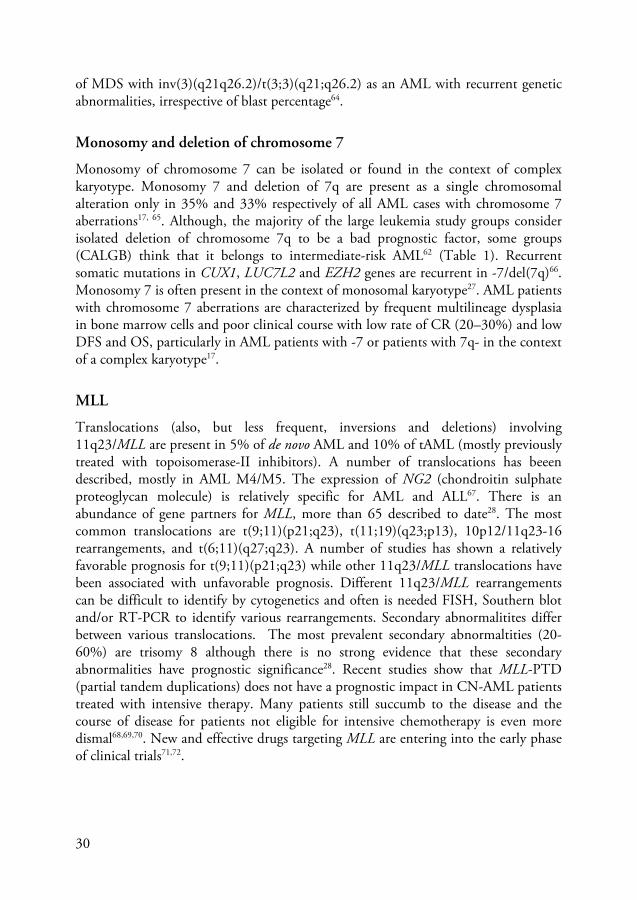
Monosomy and deletion of chromosome 7
Monosomy of chromosome 7 can be isolated or found in the context of complex karyotype. Monosomy 7 and deletion of 7q are present as a single chromosomal alteration only in 35% and 33% respectively of all AML cases with chromosome 7 aberrations17, 65. Although, the majority of the large leukemia study groups consider isolated deletion of chromosome 7q to be a bad prognostic factor, some groups (CALGB) think that it belongs to intermediate-risk AML62 (Table 1). Recurrent somatic mutations in CUX1, LUC7L2 and EZH2 genes are recurrent in -7/del(7q)66. Monosomy 7 is often present in the context of monosomal karyotype27. AML patients with chromosome 7 aberrations are characterized by frequent multilineage dysplasia in bone marrow cells and poor clinical course with low rate of CR (20–30%) and low DFS and OS, particularly in AML patients with -7 or patients with 7q- in the context of a complex karyotype17.
MLL
Translocations (also, but less frequent, inversions and deletions) involving 11q23/MLL are present in 5% of de novo AML and 10% of tAML (mostly previously treated with topoisomerase-II inhibitors). A number of translocations has beeen described, mostly in AML M4/M5. The expression of NG2 (chondroitin sulphate proteoglycan molecule) is relatively specific for AML and ALL67. There is an abundance of gene partners for MLL, more than 65 described to date28. The most common translocations are t(9;11)(p21;q23), t(11;19)(q23;p13), 10p12/11q23-16 rearrangements, and t(6;11)(q27;q23). A number of studies has shown a relatively favorable prognosis for t(9;11)(p21;q23) while other 11q23/MLL translocations have been associated with unfavorable prognosis. Different 11q23/MLL rearrangements can be difficult to identify by cytogenetics and often is needed FISH, Southern blot and/or RT-PCR to identify various rearrangements. Secondary abnormalitites differ between various translocations. The most prevalent secondary abnormaltities (20-60%) are trisomy 8 although there is no strong evidence that these secondary abnormalities have prognostic significance28. Recent studies show that MLL-PTD (partial tandem duplications) does not have a prognostic impact in CN-AML patients treated with intensive therapy. Many patients still succumb to the disease and the course of disease for patients not eligible for intensive chemotherapy is even more dismal68,69,70. New and effective drugs targeting MLL are entering into the early phase of clinical trials71,72.
31
Monosomy or deletion of chromosome 5
Among newly diagnosed AML patients, monosomy of chromosome 5 (-5) and deletion of the long arm of the chromosome 5 (5q-) represents approximately 6–9% of all the chromosomal abnormalities6,23,62. It occurs more often in the patients older than 60 years and is rarely described as an isolated abnormality in AML28. However, del(5q) is, in contrast to monosomy 5, relatively often the sole anomaly; more than 200 such AML cases have been reported. Similarly to the aberrations of chromosome 7, these chromosomal alterations are frequently observed in patients previously exposed to alkylating agent or to other leukemogenic factor favouring multilineage dysplasia in bone marrow cells followed by MDS and finally by a secondary AML28. Similarly with -7 the presence of -5 in the context of a MK confers very poor prognosis (4-year OS: 0%) in newly diagnosed AML patients27.
t(6;9)(p22;q34)
This is a very rare chromosomal abnormality (<1%), but significant as a recognized abnormality in the 2008 WHO classification, involving DEK and NUP214 genes (known also as CAN). These patients are often children or young men (median 23-30 years) and the disease is presented as de novo AML or MDS. It is characterized by basophilia, Auer rods and even multilineage dysplasia, and found in all FAB types. It predicts short survival and these patients are candidates for allogeneic stem cell transplantation28.
Not generally accepted cytogenetically high riskAML
17p abnormalities
These abnormalities are accepted as a marker for high-risk AML in SWOG/ECOG, AMLSG and the revised MRC classification and the ELN classification (Table 1 and Table 3). It is described as 17p deletion or add17p, and is often a part of complex karyotype together with abnormalities of chromosomes 5 and 773. They indicate a resistant disease with short survival and often involvement of tumor supressor gene TP5374. Another abnormality that also leads to deletion of 17p is isochromosome 17q which occurs as the sole change almost always in de novo AML, but similar to other 17p deletions, i(17q) is associated with a poor prognosis75.
t(9;22)
The well-known Philadelphia translocation is seen in <1% of AML, mainly FAB types M1 or M2 (often acute leukemia with ambiguous lineage). Both p190 och p210

32
BCR/ABL1-fusiontranscript is described in AML, as well as in t(9;22)-positive ALL, whereas p210 is typical for CML. It is difficult to establish whether or not a t(9;22) AML is a de novo AML or if it is a blastic phase of a precedent and unknown CML. Cytogenetic classifications of cooperative groups in USA do not always include t(9;22) in high risk AML, neither ELN; however HOVON/SAKK and refined MRC consider AML with this chromosomal aberration a high risk abnormality (Table 1). Philadelphia positive AML cases have to be considered as de novo AML76,77. Despite that, data in the literature are scarce. The patients’ clinical features (no history of abnormal blood counts, lack of an argument for a previous chronic phase and lack of basophilia or splenomegaly), cytogenetic abnormalities (chromosome 7 monosomy, chromosome 16 inversions and chromosome 10 deletions), molecular features (NPM1 mutation and p190 prevalence) and genome signature are different from those with CML78. The t(9;22) allows the use of target therapy (TKI) in association with conventional chemotherapy79.
Trisomy 8
The most common trisomy in AML; about 10% of all AML patients bear this abnormality, isolated trisomy 8 is seen in 5% of cases28. Trisomy 8 is considered by all the international cooperative groups as an intermediate cytogenetic-risk alteration; except in the CALGB 8461 study +8 as an isolated chromosomal aberration was classified in the high-risk category12,13,17 (Table 1). It is frequent in all ages, but prevalence is higher in older age28. Several evidences indicate that +8 occurr in association with other cytogenetic aberrations does not modify the prognosis of the associated alteration; the favorable prognostic impact of t(8;21), inv(16) and t(15;17) is not altered by the presence of an additional +817. There is abundant evidence that trisomy 8 is not sufficient for leukemogenesis. Although individuals with a constitutional +8 mosaicism have an increased risk of AML, only a minority develops this disease, and that after a long latency period80. Secondly, there does not seem to be an increased risk of AML in CML patients with trisomy 8-positive/t(9;22)-negative clones emerging after treatment with imatinib81. Thirdly, the discriminating gene expression pattern of AML with isolated +8 does not depend on the upregulation of chromosome 8 genes alone, concluding that additional genetic changes may be present. In fact, array-based analyses have revealed several cryptic chromosome changes in AML with +8 as a seemingly sole change and mutations of the ASXL1, JAK2, and TET2 genes have been shown to be common82,83,84,85,86.

33
Registries, population and methods
Swedish population registries were introduced in 1686 for taxation and military purposes, with the first report on survival in 1746. Since 1947, all Swedish citizens have a unique personal identification code, which is the same for all registrations, such as taxation, level of education, and medical purposes including causes of death. Thus, all Swedish patients and their medical history are possible to track even after migration within the country or after return from staying abroad. The Swedish Cancer Registry is a compulsory dual-report system developed in 1958. Follow-up of vital status is therefore complete with a minimal loss, and it is possible to perform socioeconomic groupings based on national registries. First, all pathology specimens indicating malignancy are reported by the pathologist to the Regional Tumor Registry; and second, all patients with a newly diagnosed cancer are reported by the clinic; missing data are actively requested. The Swedish Adult Acute Leukemia Registry was founded in 1997 by the Swedish Society of Hematology. It is supported by the Swedish Board for Health and Welfare and run in collaboration with the Regional Tumor Registry in each of the six Swedish healthcare regions, covering a population ranging from 0.9 to 1.9 million people, in total 9 million. Each region has 1 or 2 university hospitals and 3 to 8 county hospitals treating leukemia, and patients are not referred for treatment outside the home region. No patients have been treated at private hospitals. Pediatric patients (<18 years) are reported to the Nordic Society of Pediatric Hematology and Oncology (NOPHO) database, and are not included. Reporting of data on all newly diagnosed patients with acute leukemia, de novo or secondary (blastic phase of chronic myeloid leukemia excluded), has thus been compulsory since 1997. Almost all patients have 3 separate registrations (pathology, clinical report to national cancer registry, and report to leukemia registry), although the reports to the leukemia registry is mostly given retrospectively. The initial registration form for the leukemia registry included patient identification, use of diagnostic procedures, and French-American-British type. Furthermore, the physician was requested to report whether the patient at diagnosis was eligible for intensive combination chemotherapy or not. This decision was based on clinical data and local routine, but not on karyotype, because cytogenetic reports were usually not available when treatment should be initiated. Remission induction always consisted of an anthracycline plus cytosine arabinoside (Ara-C), according to regional protocols and estimated patient status, in general TAD, 3 plus 7, or similar, with possible dose reductions for the elderly. Patients in remission subsequently received consolidation with 1 to 3 courses of combination chemotherapy, usually including Ara-C at more than or equal to 1 g/m2 per dose. Allogeneic stem cell transplantation was also reported. Chemotherapy used with a palliative intent such as single-drug, low-dose

34
Ara-C, hydroxyurea, or thioguanin, was not regarded as a remission induction, despite the potential for myelosuppression and the achievement of remission.
From 2007 the registry has became web-based and modified. Karyotype, mutation analysis and more details on lab data at diagnosis and specifics on primary therapy and transplantation procedures were included. All patients have a yearly follow up until death, with reporting of relapse, relapse treatment and the outcome. The electronic reporting system INCA (informationsnätverk för cancervården; a national IT-platform for managing registering of cancer patients for clinical care and research) is common to all cancer registries in the Swedish National Cancer Programme (http://www.kvalitetsregister.se/sekundarnavigering/inenglish.132.html). Registries are supported by SKL (Sveriges Kommun och Landsting; the Swedish Association of Local Authorities and Regions), and data are monitored by RCC (Regional Cancer Centrum; Comprehensive Regional Cancer Center) in the South region. Since patients diagnosed during the period 1997-2006 did not have reported data on specific karyotype and genetic risk, the Swedish AML group (http://www.sfhem.se/aml-gruppen-1) decided to retrospectively supplement the registry with cytogenetic data.
Specific tasks and background
The first issue was to gather the original data in the paper form from all university regions for all available patients, including the data not done, missing or lost. All the karyotype reports from the different genetic laboratories were sent in paper form. The karyotypes were analyzed and classified or re-classified according to ISCN criteria. For example, karyotypes were first classified as normal karyotype, or more than 30 recurrent abnormalities. Mutations in FLT3, NPM1 or CEBPA were not included since these abnormalities were only sporadically performed during this period.
The second task was to systematize this data in the new computerized system. This was done in collaboration with statistician and IT-technician from RCC South.
Randomized controlled trials (RCT) are regarded as the gold standard in evidence-based medicine and are considered the highest grade of evidence87. Randomization is an effective measure to balance for confounding factors. However, even RCT has limitations and have a selection bias. For example, it is an open question how representative the patients included in the study are, and thus the potential to deduce the results and conclusions of the trial to the general population88. Indeed, studies show under-representation of patients >65 years of age and evidence for race and sex disparities in randomized clinical cancer trials89,90,91. Data from the Swedish Acute Leukemia Registry confirm the value of population-based studies in AML that have the potential to deliver reliable epidemiological data. Registries are also useful as a complement to clinical studies to support decisions about individual patient management, although in retrospect92,93.

35
What we have learned from our studies
Age dependent incidence of different chromosomal abnormalities
Our study has shown that the median age of patients with inv(16) or t(16;16) was 48 years, and with t(8;21) 55 years. Altogether the patients with CBF leukemia were younger than the average AML patient (median age 71 years). The incidence of CBF shows rather insignificant increase during the lifetime (range 1.3-2.4/million/year; p=0.16). Likewise, the patients with inv(3)(q21q26)/t(3;3)(q21;q26), t(6;9)(p22;q34), and 11q23 rearrangements; not t(9;11) were more prevalent in younger patients (median age 58, 36, and 54 years, respectively). In contrast, the incidence of AML with 5q, 7q and/or 17p increases with older age (range 0.9-43/million/years; p<0.0001), as well as patients with complex karyotype. This most likely illustrates different pathophysiologic mechanisms involved in leukemogenesis in different AML groups. The data from one population-based study clearly indicates that the age-dependent increase in incidence of AML substantially differs between the cases with balanced, with normal, and with unbalanced karyotypes, and suggest that mechanisms of leukemogenesis are different and more or less age-dependent94. The results of our study and of others illustrate two different age profiles in AML from the cytogenetic point of view. The first one is characterized by a rather constant incidence over lifetime and is represented by balanced translocations. In contrast, unbalanced aberrations and especially complex aberrant karyotype show a sharp increase of incidence in older age. This is suggestive of different mechanisms in the underlying pathogenesis of AML57,94. At least a proportion of, if not all, balanced translocations of pediatric leukemias already develops in the prenatal period. This was demonstrated by the observation of twins developing acute leukemias with reciprocal gene fusions, e.g. cALL with TEL-AML1, after a latency of up to 14 years95. The retrospective polymerase chain reaction analyses of Guthrie cards of children with AML with t(8;21), t(15;17), and inv(16), who had developed leukemia with a latency of up to 12 years led to the detection of clonotypic sequences of the respective gene fusions AML1-ETO, PML-RARA, and CBFB-MYH1195. On the other hand, unbalanced aberrations lead to genomic imbalances and may occur due to a variety of mechanisms, such as sister chromatid exchange of ring chromosomes, unbalanced

36
distribution of the chromosomes to the daughter cells, or incorrect repair of DNA double strand breaks57,96,97. These genetic alterations seem to occur more frequently in aging cells as aging cells are more likely to acquire such abnormalities due to shortening of telomeres and less efficient DNA repair capacity. On the other hand, unbalanced aberrations lead to genomic imbalances and may occur due to a variety of mechanisms, such as sister chromatid exchange of ring chromosomes, unbalanced distribution of the chromosomes to the daughter cells, or incorrect repair of DNA double strand breaks97,98,99. The age-specific distribution of the molecular markers might be due not only to different mutational mechanisms in dependence on age but also due to age-specific changes in hematopoiesis and to changes in the available pools of hematopoietic precursors as targets for leukemogenesis. Different age profiles of the cytogenetic subtypes and of the recurrent molecular markers indicate different mechanisms of the pathogenesis of AML and point to the need to develop different targeted therapeutic strategies for the different subtypes94. WHO classification from 2008 roughly separates AML in three categories: de novo AML, therapy-related AML and secondary AML (with antecedent MDS or MPN). These categories seem to have a different ontogenesis and age-distribution100. There is accumulating data about the time sequence of events which lead to overt leukemia, where specific gene mutations and even CBFH/MYH translocation in inv(16) occurr in preleukemic stem cells. It supports a model in which mutations in "landscaping" genes, involved in global chromatin changes such as DNA methylation, histone modification, and chromatin looping, occur early in the evolution of AML, whereas mutations in "proliferative" genes occur late101. These findings indicate that preleukemic HSCs can survive induction chemotherapy, identifying these cells as a reservoir for the reevolution of relapsed disease. Cytogenetic data from the Swedish Acute Leukemia Registry can also provide insight into the clonal origin and evolution, especially in the cases with complex karyotype102, 103, 104.
Overlap between poor-risk chromosome abnormalities, complex and monosomal karyotypes
We found a strong overlap between some chromosome abnormalities, especially the changes of chromosomes 5, 7 and 17. This not only indicates that they identify the same subgroup of patients characterized by poor prognosis, but also that they cooperate in the leukemogenic process and/or have similar mechanisms behind their occurrence. Furthermore, the prognostic impact of a complex karyotype or MK was clearly influenced by the presence of 5q, 7q and 17p losses. Adding MK into the risk stratification of Swedish AML patients did not improve survival prediction. We hence conclude that the negative impact of MK seems to be mostly carried out by

37
abnormalities of chromosomes 5, 7 and 17. This is opposed to other studies27, 105, but is in line with the data from the Spanish and German MDS/AML registries106,107. In our series of complex karyotype, monosomal karyotype and changes of chromosomes 5/7/17 overlap in more than 80% of cases. Therefore, we believe that because of the overlap, monosomal karyotype is more “accumulative” and “statistical” than a true “biological entity”. There is now a huge body of evidence on the clear correlation between TP53 mutation and complex karyotype (and most likely monosomal karyotype)63,105,108. Since among 234 complex karyotype AML cases analyzed, TP53 mutations determined by DNA sequencing were more frequent (141/234 cases, i.e., 60%) than TP53 losses determined by array-CGH analysis (94/234 cases, i.e., 40%), it might be concluded that TP53 loss of function indeed causes chromosomal instability (CIN) with subsequent development of complex karyotype alterations, rather than being a consequence of CIN 63,100,104.
Unsuccessful cytogenetics
Unsuccessful (UC) and unperformed cytogenetics (UPC) are often reported together as a “not determined karyotype”109. We separated the UC and UPC groups assuming that their causes and prognostic impact may differ. The incidence of UC of 2.1% is lower than in previously published studies. These patients are >60 years of age (median 66 years) which partly explains a dismal prognosis for these patients. We confirmed the findings of a group from USA with lower CR rate and poor prognosis for AML patients with UC, but we also found a higher early death (ED) rate54. The definition of unsuccessful cytogenetic karyotype (UC) is a lack of analyzable metaphasis. There are several possible explanations of this phenomenon. Some cases with UC are undoubtedly due to insufficient number of cells in the bone marrow aspirates sent for cytogenetic analysis. Furthermore, human errors in taking the bone marrow aspirates cannot be excluded, such as too small volumes or diluting the bone marrow cells with peripheral blood, and technical problems in the laboratory. Finally, there may well be some biological explanations for UC, representing the intrinsic properties of the leukemic clone, such as inability to divide in vitro. In fact, UC is not specific for AML. There are reports of dismal prognosis of ALL cases with UC110,111 as well as of myelodysplastic syndromes with UC112, with the latter suggesting that UC is a property of dysfunctional stem cells. However, the underlying reasons for UC are most likely manifold and heterogeneous, and hence next to impossible to ascertain in a retrospective, registry-based study of this type.

38
Unperformed cytogenetics
The issue of unperformed karyotype is even more controversial than unsuccessful karyotype. This group is almost invisible in the literature, since karyotype is usually mandatory in clinical trials. UPC is often lumped together with unsuccessful karyotype or just classified as not done, not available or not determined109. The population-based AML Registry is an excellent source to identify such a patient group. It is impossible in retrospect to know the reasons for why the karyotype was performed or not from case to case, but it is likely that many were not fit for intensive treatment. We found 364 patients in the Registry, mostly older than 60 years of age, and their outcome was very similar to those with high risk AML, with increased ED rate, decreased CR rate and poor OS. The presence of UPC emphasizes the need for proper genetic analyses of all patients for whom treatment with curative intent is planned, and even in the cases where therapy is not planned in order to have data for future analysis.
Hyperdiploidy
Our report again emphasizes the value of population-based studies, because it is the first population-based study of hyperdiploid AML. We found a male preponderance (71%), and dominance of AML types M2, M4, M5. This is in contrast to childhood AML, where the FAB type M7 was predominant. Furthermore the majority of secondary hyperdiploid AML originated from previous MDS. Tri/tetraploidy AML and high hyperdiploidy had different outcome, since tri/tetraploid AML (>65 chromosomes) had a better survival showing the possibility of different leukemogenic pathways and possibly to be regarded a separate entities. Surprisingly, none of the high hyperdiploidy/tri and tetraploidy AML cases was secondary to a myeloproliferative neoplasm (MPN). The outcome was also different when the hyperdiploidy included chromosome abnormalities with bad prognosis, such as del 5, del 7 or del 17. It is very probable that different diseases with hyperdiploid cytogenetics, have different pathogenetic effect induced by different chromosomes or other mutations113, 114, 115. Our results are in line with the previous studies showing that this group is more heterogeneous than previously described and that the overall survival of this group is a bit closer to HR than IR AML116.

39
Secondary leukemia
The incidence of secondary AML was 26.4%, which is comparable to other population-based studies117. The median latency period between MDS and AML was 1 year, indicating that most MDS patients who progress to AML do so within a short time frame. Median latency times between MPN and AML were between 7 and 8 years, whereas the median latency between the malignancy and tAML was slightly longer compared to most of the previous studies with 5.8 years118,119,120. Median latency between a nonmalignant disease and tAML is seldom reported, but was shown to be 14.3 years in our cohort. CR rates were lower than de novo AML, but not depending on the ED rate. We confirmed the negative prognostic importance of sAML. This was highly significant for younger patients, but did not add to the prognostic information in elderly AML patients. The reason for this difference in younger but not in older patients could potentially be due to the fact that sAML biologically and genetically is more similar to AML in general in older patients. The reason for the poor outcome in secondary AML remains somewhat elusive. Although an increased frequency of high-risk cytogenetics explains some of the treatment resistance, there must clearly be some additional factors conferring the poor prognosis.
The role of trisomy 13 in the prognosis of AML
We specifically analysed the prognosis of patients with trisomy 13 in our AML Registry, including patients diagnosed between 1997-2014. We found that all patients had died, including younger patients as well as two patients who underwent allogeneic stem cell transplantation. The results corresponded to the data from the German group, to which we provided a comment59. This is an obvious example of shortcomings of AML classification. Trisomies are generally regarded as indicators of the intermediate risk AML, but our analysis confirmed that this entity is consistent with a high risk59.
Brief summary
Data from the Swedish AML Registry show the higher incidence of loss of 5q, 7q and 17p, MK and CK in patients >60 years. The patients with ≥5 chromosome abnormalities had very short OS even after intensive chemotherapy. The most significant prognostic factors were age, PS, followed by sex and karyotype (deletion of

40
chromosome 7q). Overall survival of patients with UC and UPC AML were in between IR and HR AML. Triploid/near tetraploid AML (≥65 chromosomes) had a better outcome than high hyperdiploidy (<65 chromosomes). Isolated trisomy 13 had a similar outcome as HR AML. Therefore, hyperdiploid AML, UC, UPC and isolated trisomy 13 should be regarded as HR AML. Secondary AML had an impact on survival in patients <60 years, but not in patients >60 years of age.
New diagnostic techniques have come into the everyday practice of the physicians treating AML, including molecular changes identified through Next Generation Sequencing121,122. Nevertheless, karyotype analysis is still the gold standard for diagnosing AML. By utilizing the population-based perspective we could further describe some of the heterogeneous groups and subgroups of AML and thus humbly contribute to the “old world” of cytogenetics.

41
References
1. Bacher U, Haferlach T. The benefit of population-based studies for older patients with acute myeloid leukemia. Haematologica 2012; 97(12):1781-2.
2. Nowell, P., and Hungerford, D. A minute chromosome in human chronic granulocytic leukemia [abstract]. Science 1960; 132:1497.
3. Nowell PC. Discovery of the Philadelphia chromosome: a personal perspective. J Clin Invest 2007;117(8):2033-5.
4. Boveri, T. Zur Frage der Entstehung maligner Tumoren. Gustav Fischer. Jena, 1914; Germany. 64 pp.
5. Tjio J‐H and Levan A. The chromosome number of man. Hereditas 1956; 42: 1–6.
6. Caspersson T, Gahrton G, Lindsten J, Zech L. Identification of the Philadelphia chromosome as a number 22 by quinacrine mustard fluorescence analysis. Exp Cell Res 1970; 63(1):238-40.
7. Rowley JD. Letter: A new consistent chromosomal abnormality in chronic myelogenous leukaemia identified by quinacrine fluorescence and Giemsa staining. Nature 1973; 243(5405): 290-3.
8. Rowley JD. Identificaton of a translocation with quinacrine fluorescence in a patient with acute leukemia. Ann Genet 1973; 16(2):109-12.
9. Bennett JM, Catovsky D, Daniel MT, Flandrin G, Galton DA, Gralnick HR et al. Proposals for the classification of the acute leukaemias. French-American-British (FAB) co-operative group. Br J Haematol 1976; 33: 451–458.
10. Rowley JD, Golomb HM. The 4th International Workshop on chromosomes in leukemia–a prospective study of acute nonlymphocytic leukemia, Chicago, Illinois, USA, September 2–7 1982. Cancer Genetics and Cytogenetics 1984; 11: 249.
11. Bloomfield CD, Shuma C, Regal L et al. Long-term survival of patients with acute myeloid leukemia: a third follow-up of the Fourth International Workshop on Chromosomes in Leukemia. Cancer 1997; 80 (11 Suppl): 2191–2198.
12. Mrózek K, Heinonen K, Bloomfield CD. Clinical importance of cytogenetics in acute myeloid leukaemia. Best Pract Res Clin Haematol 2001;14:19–47.

42
13. Mrózek K, Heerema NA, Bloomfield CD. Cytogenetics in acute myeloid leukemia. Blood Rev 2004;18:115–36.
14. Grimwade D, Hills RK, Moorman AV et al. Refinement of cytogenetic classification in acute myeloid leukemia: determination of prognostic significance of rare recurring chromosomal abnormalities among 5876 younger adult patients treated in the United Kingdom Medical Research Council trials. Blood 2010; 116, 354-365.
15. Juliusson G, Antunovic P, Derolf A. et al. Age and acute myeloid leukemia: real world data on decision to treat and outcomes from the Swedish Acute Leukemia Registry. Blood 2009; 113, 4179-4187.
16. Löwenberg B. Diagnosis and Prognosis in Acute Myeloid Leukemia — The Art of Distinction. N Engl J Med 2008; 358:1960-1962.
17. Marchesi F, Annibali O, Cerchiara E, Tirindelli MC, Avvisati G. Cytogenetic abnormalities in adult non-promyelocytic acute myeloid leukemia: a concise review. Crit Rev Oncol Hematol 2011; 80(3):331-46.
18. Marcucci G, Mrózek K, Ruppert AS, et al. Abnormal cytogenetics at date of morphologic complete remission predicts short overall and disease-free survival, and higher relapse rate in adult acute myeloid leukemia: results from Cancer and Leukemia Group B study 8461. J Clin Oncol 2004;22:2410–8.
19. Cheson BD, Bennett JM, Kopecky KJ, et al. Revised recommendations of the International Working Group for Diagnosis, Standardization of Response Criteria, Treatment Outcomes, and Reporting Standards for Therapeutic Trials in Acute Myeloid Leukemia. J Clin Oncol 2003; 21:4642–9.
20. Wang Y, Wu DP, Liu QF et al. In adults with t(8;21)AML, posttransplant RUNX1/RUNX1T1-based MRD monitoring, rather than c-KIT mutations, allows further risk stratification. Blood 2014; 124(12):1880-6.
21. Zhu HH, Zhang XH, Qin YZ et al. MRD-directed risk stratification treatment may improve outcomes of t(8;21) AML in the first complete remission: results from the AML05 multicenter trial. Blood 2013; 121(20):4056-62.
22. Shaffer LG, McGowan-Jordan J, Schmid M, (eds). ISCN 2013: An International System for Human Cytogenetic Nomenclature. Basel, Switzerland: Karger, 2013: 16–31.
23. Byrd JC, Mrózek K, Dodge RK, et al. Pretreatment cytogenetic abnormalities are predictive of induction success, cumulative incidence of relapse, and overall survival in adult patients with de novo acute myeloid leukemia: results from Cancer and Leukemia Group B (CALGB 8461). Blood 2002; 100:4325–36.
24. Grimwade D, Hills RK. Independent prognostic factors for AML outcome. Hematology Am Soc Hematol Educ Program. 2009:385-95.

43
25. Schoch C, Kern W, Kohlmann A, Hiddemann W, Schnittger S, Haferlach T. Acute myeloid leukemia with a complex karyotype is a distinct biological entity characterized by genomic imbalances and a specific gene expression profile. Genes Chromosomes Cancer 2005; 43: 227–238.
26. Mitelman Database of Chromosome Aberrations and Gene Fusions in Cancer 2013; In: Mitelman F, Johansson B and Mertens F. (eds), http://cgap.nci.nih.gov/Chromosomes/Mitelman
27. Breems DA, Van Putten WL, De Greef GE, Van Zelderen-Bhola SL, Gerssen-Schoorl KB, Mellink CH, et al. Monosomal karyotype in acute myeloid leukemia: a better indicator of poor prognosis than a complex karyotype. J Clin Oncol 2008; 26(29):4791-7.
28. From SFMG; Swedish Society of Medical Genetics written by Bertil Johansson; http://sfmg.se/riktlinjer/hematologisk-genetik/
29. Steven H, Swerdlow EC, Harris NL et al. WHO classification of tumours of haematopoietic and lymphoid tissues. IARC, France. 2008.
30. Kelly LM, Gilliland DG. Genetics of myeloid leukemias. Annu Rev Genomics Hum Genet 2002; 3:179-198.
31. Haferlach T, Schoch C, Löffler H, et al. Morphologic dysplasia in de novo acute myeloid leukemia (AML) is related to unfavorable cytogenetics but has no independent prognostic relevance under the conditions of intensive induction therapy: results of a multiparameter analysis from the German AML Cooperative Group studies. J Clin Oncol 2003; 21(2):256-265.
32. Wandt H, Schäkel U, Kroschinsky F, et al. MLD according to the WHO classification in AML has no correlation with age and no independent prognostic relevance as analyzed in 1766 patients. Blood 2008; 111(4):1855-1861.
33. Schlenk RF, Döhner K, Krauter J et al. Mutations and treatment outcome in cytogenetically normal acute myeloid leukemia. N Engl J Med 2008; 358(18): 1909-18.
34. Döhner H, Estey EH, Amadori S et al. European LeukemiaNet Diagnosis and management of acute myeloid leukemia in adults: recommendations from an international expert panel, on behalf of the European LeukemiaNet. Blood 2010; 115(3):453-74.
35. Gaidzik VI, Paschka P, Späth D et al. TET2 mutations in acute myeloid leukemia (AML): results from a comprehensive genetic and clinical analysis of the AML study group. J Clin Oncol 2012; 30(12):1350-7.
36. Patel KP, Ravandi F, Ma D et al. Acute myeloid leukemia with IDH1 or IDH2 mutation: frequency and clinicopathologic features. Am J Clin Pathol 2011; 135(1):35-45.
37. Grossmann V, Schnittger S, Kohlmann A et al. A novel hierarchical prognostic model of AML solely based on molecular mutations. Blood 2012 ;120(15):2963-72.

44
38. Patel JP, Gönen M, Figueroa ME et al. Prognostic relevance of integrated genetic profiling in acute myeloid leukemia. N Engl J Med 2012; 366(12):1079-89.
39. Pastore F, Dufour A, Benthaus T et al. Combined molecular and clinical prognostic index for relapse and survival in cytogenetically normal acute myeloid leukemia. J Clin Oncol 2014 ;32(15):1586-94.
40. Marcucci G, Maharry KS, Metzeler KH et al. Clinical role of microRNAs in cytogenetically normal acute myeloid leukemia: miR-155 upregulation independently identifies high-risk patients. J Clin Oncol 2013; 31(17):2086-93.
41. Marcucci G, Yan P, Maharry K et al. Epigenetics meets genetics in acute myeloid leukemia: clinical impact of a novel seven-gene score. J Clin Oncol 2014 ;32(6):548-56.
42. NCCN Clinical Practice Guidelines in Oncology. Acute Myeloid Leukemia. Version 1. 2015. Available from http://www.nccn.org/professionals/physician_gls/PDF/aml.pdf
43. Howlader N NA, Krapcho M, Neyman N, Aminou R, Waldron W, Altekruse SF, Kosary CL, Ruhl J, Tatalovich Z, Cho H, Mariotto A, Eisner MP, Lewis DR, Chen HS, Feuer EJ, Cronin KA, Edwards BK (eds). : SEER Cancer Statistics Review, 1975-2008, National Cancer Institute. Bethesda, MD; Available from: http://seer.cancer.gov/archive/csr/1975_2008/results_merged/sect_13_leukemia.pdf
44. Juliusson G, Lazarevic V, Hörstedt A-S. Nationellt register för akut myeloisk leukemi hos vuxna: Rapport nr 7. Patienter med diagnos 2007-2009 2011. Available from: http://www.sfhem.se/rapporter-blodcancerregistret
45. Deschler B, Lubbert M. Acute myeloid leukemia: epidemiology and etiology. Cancer 2006; 107(9):2099–107.
46. American Cancer Society: Cancer Facts and Figures 2014. Atlanta, Ga: American Cancer Society, 2014, accessed May 21, 2014; Available from: http://www.cancer.org/cancer/leukemia-acutemyeloidaml/overviewguide/leukemia-aml-overview-key-statistics
47. Osca-Gelis G, Puig-Vives M, Saez M, Gallardo D, Lloveras N, Marcos-Gragera R. Population-based incidence of myeloid malignancies: fifteen years of epidemiological data in the province of Girona, Spain. Haematologica 2013; 98(8):e95-7.
48. Maynadié M, Girodon F, Manivet-Janoray I et al. Twenty-five years of epidemiological recording on myeloid malignancies: data from the specialized registry of hematologic malignancies of Cote d'Or (Burgundy, France). Haematologica 2011; 96(1):55-61.
49. Maynadié M, De Angelis R, Marcos-Gragera R et al. HAEMACARE Working Group. Survival of European patients diagnosed with myeloid malignancies: a HAEMACARE study. Haematologica. 2013; 98(2):230-8.

45
50. Ohnishi H, Imataki O, Kawachi Y et al. Age is an independent adverse prognostic factor for overall survival in acute myeloid leukemia in Japan. World J Hematol 2014; 3(3): 105-114.
51. Mauritzson N, Johansson B, Albin M et al. A single-center population-based consecutive series of 1500 cytogenetically investigated adult hematological malignancies: karyotypic features in relation to morphology, age and gender. Eur J Haematol 1999; 62(2):95-102.
52. Mauritzson N, Johansson B, Albin M et al. Survival time in a population-based consecutive series of adult acute myeloid leukemia--the prognostic impact of karyotype during the time period 1976-1993. Leukemia 2000; 14(6):1039-43.
53. Mauritzson N, Albin M, Rylander L, et al. Pooled analysis of clinical and cytogenetic features in treatment-related and de novo adult acute myeloid leukemia and myelodysplastic syndromes based on a consecutive series of 761 patients analyzed 1976-1993 and on 5098 unselected cases reported in the literature 1974-2001. Leukemia 2002; 16(12):2366-2378.
54. Medeiros BC, Othus M, Estey EH et al. Unsuccessful diagnostic cytogenetic analysis is a poor prognostic feature in acute myeloid leukaemia. Br J Haematol 2014 ;164(2):245-50
55. Moorman AV, Roman E, Cartwright RA, Morgan GJ. Age-specific incidence rates for cytogenetically-defined subtypes of acute myeloid leukaemia. Br J Cancer 2002; 86: 1061–1063.
56. Sanderson RN, Johnson PR, Moorman AV et al. Population-based demographic study of karyotypes in 1709 patients with adult acute myeloid leukemia. Leukemia 2006; 20(3):444-50.
57. Moorman AV, Roman E, Willett EV et al. Karyotype and age in acute myeloid leukemia. Are they linked? Cancer Genet Cytogenet 2001; 126(2):155-61.
58. Vickers M, Jackson G, Taylor P. The incidence of acute promyelocytic leukemia appears constant over most of a human lifespan, implying only one rate limiting mutation. Leukemia 2000; 14: 722–726.
59. Herold T, Metzeler KH, Vosberg S et al. Isolated trisomy 13 defines a homogeneous AML subgroup with high frequency of mutations in spliceosome genes and poor prognosis. Blood 2014; 124(8):1304-11.
60. Haferlach T, Kohlmann A, Klein HU et al. AML with translocation t(8;16)(p11;p13) demonstrates unique cytomorphological, cytogenetic, molecular and prognostic features. Leukemia 2009; 23(5):934-43.
61. Micci F, Thorsen J, Panagopoulos I et al. High-throughput sequencing identifies an NFIA/CBFA2T3 fusion gene in acute erythroid leukemia with t(1;16)(p31;q24). Leukemia 2013; 27(4):980-2.

46
62. Slovak ML, Kopecky KJ, Cassileth PA, et al. Karyotypic analysis predicts outcome of preremission and postremission therapy in adult acute myeloid leukemia: a Southwest Oncology Group/Eastern Cooperative Oncology Group Study. Blood 2000; 96:4075–83.
63. Rucker FG, Schlenk RF, Bullinger L et al. TP53 alterations in acute myeloid leukemia with complex karyotype correlate with specific copy number alterations, monosomal karyotype, and dismal outcome. Blood 2012; 119: 2114–2121.
64. Rogers HJ, Vardiman JW, Anastasi J et al. Complex or monosomal karyotype and not blast percentage is associated with poor survival in acute myeloid leukemia and myelodysplastic syndrome patients with inv(3)(q21q26.2)/t(3;3)(q21;q26.2): a Bone Marrow Pathology Group study. Haematologica 2014; 99(5):821-9.
65. Hasle H, Aricò M, Basso G, et al. Myelodysplastic syndrome, juvenile myelomonocytic leukemia, and acute myeloid leukemia with complete or partial monosomy 7. Leukemia 1999; 13:376–85.
66. Hosono N, Makishima H, Jerez A et al. Recurrent genetic defects on chromosome 7q in myeloid neoplasms. Leukemia 2014; 28(6):1348-51.
67. Mauvieux L, Delabesse E, Bourquelot P et al. NG2 expression in MLL rearranged acute myeloid leukaemia is restricted to monoblastic cases. Br J Haematol 1999 ;107(3):674-6.
68. Whitman SP, Caligiuri MA, Maharry K et al. The MLL partial tandem duplication in adults aged 60 years and older with de novo cytogenetically normal acute myeloid leukemia. Leukemia 2012; 26(7):1713-1717.
69. Whitman SP, Ruppert AS, Marcucci G et al. Long-term disease-free survivors with cytogenetically normal acute myeloid leukemia and MLL partial tandem duplication: a Cancer and Leukemia Group B study. Blood 2007; 109(12):5164-5167.
70. Döhner K, Tobis K, Ulrich R et al. Prognostic significance of partial tandem duplications of the MLL gene in adultpatients 16 to 60 years old with acute myeloid leukemia and normal cytogenetics: a study of the Acute Myeloid Leukemia Study Group Ulm. J Clin Oncol 2002; 20(15):3254-3261.
71. Bernot KM, Siebenaler RF, Whitman SP et al. Toward personalized therapy in AML: in vivo benefit of targeting aberrant epigenetics in MLL-PTD-associated AML. Leukemia 2013;27(12):2379-82.
72. Kühn MW, Hadler MJ, Daigle SR et al. MLL partial tandem duplication leukemia cells are sensitive to small molecule DOT1L inhibition. Haematologica 2015; 100(5):e190-3.
73. Lazarevic V, Hörstedt AS, Johansson B et al. Incidence and prognostic significance of karyotypic subgroups in older patients with acute myeloid leukemia: the Swedish population-based experience. Blood Cancer J 2014 Feb 28;4: e188.

47
74. Nahi H, Lehmann S, Bengtzen S et al. Chromosomal aberrations in 17p predict in vitro drug resistance and short overall survival in acute myeloid leukemia. Leuk Lymphoma 2008; 49(3):508-16.
75. Kanagal-Shamanna R, Bueso-Ramos CE, Barkoh B et al. Myeloid neoplasms with isolated isochromosome 17q represent a clinicopathologic entity associated with myelodysplastic/myeloproliferative features, a high risk of leukemic transformation, and wild-type TP53. Cancer 2012; 118(11):2879-88.
76. Nacheva EP, Grace CD, Brazma D et al. Does BCR/ABL1 positive acute myeloid leukaemia exist? Br J Haematol 2013; 161 (4), 541–550.
77. Konoplev S, Yin CC, Kornblau SM et al. Molecular characterization of de novo Philadelphia chromosome-positive acute myeloid leukemia. Leuk Lymphoma 2013; 54 (1),138–144.
78. Reboursiere E, Chantepie S, Gac AC, Reman O. Rare but authentic Philadelphia-positive acute myeloblastic leukemia: Two case reports and a literature review of characteristics, treatment and outcome. Hematol Oncol Stem Cell Ther 2014 Oct 6
79. Lazarevic V, Golovleva I, Nygren I, Wahlin A. Induction chemotherapy and post-remission imatinib therapy for de Novo BCR-ABL-positive AML. Am J Hematol 2006; 81 (6), 470–471.
80. Welborn J. Constitutional chromosome aberrations as pathogenetic events in hematologic malignancies. Cancer Genet Cytogenet 2004; 149:137-153.
81. Baccarani M, Deininger MW, Rosti G et al. European LeukemiaNet recommendations for the management of chronic myeloid leukemia: 2013 Blood; 122: 872-884.
82. Schoch C, Kohlmann A, Dugas M et al. Genomic gains and losses influence expression levels of genes located within the affected regions: a study on acute myeloid leukemias with trisomy 8, 11, or 13, monosomy 7, or deletion 5q. Leukemia 2005; 19:1224-1228.
83. Paulsson K, Heidenblad M, Strömbeck B et al. High-resolution genome-wide array-based comparative genome hybridization reveals cryptic chromosome changes in AML and MDS cases with trisomy 8 as the sole cytogenetic aberration. Leukemia 2006; 20:840-846.
84. Schnittger S, Eder C, Jeromin S et al. ASXL1 exon 12 mutations are frequent in AML with intermediate risk karyotype and are independently associated with an adverse outcome. Leukemia 2013; 27:82-91.
85. Chou W-C, Chou S-C, Liu C-Y et al. TET2 mutation is an unfavorable prognostic factor in acute myeloid leukemia patients with intermediate-risk cytogenetics. Blood 2011; 118:3803-3810.

48
86. Schnittger S, Bacher U, Kern W, Haferlach T, Haferlach C JAK2V617F asprogression marker in CMPD and as cooperative mutation in AML with trisomy 8 and t(8;21): a comparative study on 1103 CMPD and 269 AML cases. Leukemia 2007; 21:1843-1845.
87. Atkins D, Best D, Briss PA, et al. Grading quality of evidence and strength of recommendations. BMJ 2004; 328:1490.
88. Rothwell PM. External validity of randomised controlled trials: "to whom do the results of this trial apply?" Lancet 2005; 365:82-93.
89. Hutchins LF, Unger JM, Crowley JJ, et al. Underrepresentation of patients 65 years of age or older in cancer-treatment trials. N Engl J Med 1999; 341:2061-7.
90. Murthy VH, Krumholz HM, Gross CP: Participation in cancer clinical trials: race-, sex-, and age-based disparities. JAMA 2004; 291:2720-6.
91. Lovisa Vennström. Population-based studies on acute leukemias - lessons from the Swedish Adult Acute Leukemia Registry. University of Gothenburg. Sahlgrenska Academy 2011. https://gupea.ub.gu.se/handle/2077/25488
92. Juliusson G, Lazarevic V, Horstedt AS, Hagberg O, Hoglund M, Swedish Acute Leukemia Registry G. Acute myeloid leukemia in the real world: why population-based registries are needed. Blood 2012; 119(17):3890-9.
93. Preiss BS, Kerndrup GB, Schmidt KG et al. AML Study Group Region of Southern Denmark. Cytogenetic findings in adult de novo acute myeloid leukaemia. A population-based study of 303/337 patients. Br J Haematol 2003 ;123(2):219-34.
94. Bacher U, Kern W, Schnittger S, Hiddemann W, Haferlach T, Schoch C. Population-based age-specific incidences of cytogenetic subgroups of acute myeloid leukemia. Haematologica 2005 ;90(11):1502-10.
95. Wiemels JL, Xiao ZJ, Buffler PA et al. In utero origin of t(8;21) AML1-ETO translocations in childhood acute myeloid leukemia. Blood 2002; 99:3801-5.
96. Mchale CM, Wiemels JL, Zhang LP et al. Prenatal origin of childhood acute myeloid leukemias harboring chromosomal rearrangements t(15;17) and inv(16). Blood 2003; 101:4640-1.
97. Gisselsson D, Pettersson L, Hoglund M, et al. Chromosomal breakage-fusion-bridge events cause genetic intratumor heterogeneity. Proc Natl Acad Sci USA 2004; 97:5357-62.
98. Nicklas RB. How cells get the right chromosomes. Science 1997; 275:632-7.
99. Obe G, Pfeiffer P, Savage JR et al. Chromosomal aberrations: formation, identification and distribution. Mut Res 2002; 504:17-36.

49
100. Lindsley RC, Mar BG, Mazzola E et al . Acute myeloid leukemia ontogeny is defined by distinct somatic mutations. Blood 2014 Dec 30. pii: blood-2014-11-610543. [Epub ahead of print]
101. Corces-Zimmerman MR, Hong WJ, Weissman IL, Medeiros BC, Majeti R. Preleukemic mutations in human acute myeloid leukemia affect epigenetic regulators and persist in remission. Proc Natl Acad Sci U S A 2014; 111(7):2548-53.
102. Medeiros BC, Othus M, Fang M, Appelbaum FR, Erba HP. Cytogenetic heterogeneity negatively impacts outcomes in patients with acute myeloid leukemia. Haematologica 2014 Dec 19.
103. Bochtler T, Stölzel F, Heilig CE et al. Clonal heterogeneity as detected by metaphase karyotyping is an indicator of poor prognosis in acute myeloid leukemia. J Clin Oncol 2013;31(31):3898-905.
104. Bochtler T, Fröhling S, Krämer A. Role of chromosomal aberrations in clonal diversity and progression of acute myeloid leukemia. Leukemia 2015; Feb 12. doi: 10.1038/leu.2015.32. [Epub ahead of print]
105. Kayser S, Zucknick M, Döhner K et al. Monosomal karyotype in adult acute myeloid leukemia: prognostic impact and outcome after different treatment strategies. Blood 2012; 119: 551–558.
106. Valcárcel D, Ademá V, Solé F et al. Complex, not monosomal, karyotype is the cytogenetic marker of poorest prognosis in patients with primary myelodysplastic syndrome. J Clin Oncol 2013; 31:916–922.
107. Schanz J, Tüchler H, Solé F et al. New comprehensive cytogenetic scoring system for primary myelodysplastic syndromes (MDS) and oligoblastic acute myeloid leukemia after MDS derived from an international database merge. J Clin Oncol 2012; 30: 820–829.
108. Middeke JM, Fang M, Cornelissen JJ et al. Outcome of patients with abnl(17p) acute myeloid leukemia after allogeneic hematopoietic stem cell transplantation. Blood 2014; 123(19):2960-7.
109. Löwenberg B, Pabst T, Vellenga E, et al. Cytarabine dose for acute myeloid leukemia. N Engl J Med 2011; 364:1027–36.
110. Moorman AV, Harrison CJ, Buck GA, et al. Karyotype is an independent prognostic factor in adult acute lymphoblastic leukemia (ALL): analysis of cytogenetic data from patients treated on the Medical Research Council (MRC) UKALLXII/ Eastern Cooperative Oncology Group (ECOG) 2993 trial. Blood 2007; 109:3189–97.
111. Nordgren A, Farnebo F, Johansson B, et al. Identification of numerical and structural chromosome aberrations in 15 high hyperdiploid childhood acute lymphoblastic leukemias using spectral karyotyping. Eur J Haematol 2001; 66:297–304.

50
112. Cervera J, Solé F, Haase D, et al. Prognostic impact on survival of an unsuccessful conventional cytogenetic study in patients with myelodysplastic syndromes (MDS). Leuk Res 2009; 33:S75–6.
113. Chretien ML, Corre J, Lauwers-Cances V et al. Understanding the role of hyperdiploidy in myeloma prognosis: which trisomies really matter? Blood 2015 Oct 29. pii: blood-2015-06-650242. [Epub ahead of print]
114. Paulsson K. Chromosome gains drive childhood ALL. Oncotarget 2015 ;6(23):19360-1.
115. Paulsson K, Lilljebjörn H, Biloglav A et al. The genomic landscape of high hyperdiploid childhood acute lymphoblastic leukemia. Nat Genet 2015 Jun;47(6):672-6.
116. Shetty A, Cortes JE, Ravandi F. Hyperdiploidy in AML; Outcomes of acute myelogenous leukemia (AML) patients (pts) with a hyperdiploid karyotype.. J Clin Oncol 32:5s, 2014 (suppl; abstr 7088)
117. Østgård LS, Medeiros BC, Sengeløv H et al. Epidemiology and Clinical Significance of Secondary and Therapy-Related Acute Myeloid Leukemia: A National Population-Based Cohort Study. JClin Onc Nov 1, 2015:3641-3649; DOI:10.1200/JCO.2014.60.0890.
118. Levine EG, Bloomfield CD. Leukemias and myelodysplastic syndromes secondary to drug, radiation, and environmental exposure. Semin Oncol 1992;19:47–84.
119. Smith SM, Le Beau MM, Huo D, et al. Clinical cytogenetic associations in 306 patients with therapy-related myelodysplasia and myeloid leukemia: The University of Chicago series. Blood 2003; 102:43–52.
120. Schoch C, Kern W, Schnittger S, et al. Karyotype is an independent prognostic parameter in therapy-related acute myeloid leukemia (t-AML): An analysis of 93 patients with t-AML in comparison to 1091 patients with de novo AML. Leukemia 2004; 18:120–125.
121. Genomic and epigenomic landscapes of adult de novo acute myeloid leukemia.
Cancer Genome Atlas Research Network. N Engl J Med. 2013; 368(22):2059-74.
122. Klco JM, Miller CA, Griffith M et al. Association Between Mutation Clearance After Induction Therapy and Outcomes in Acute Myeloid Leukemia. JAMA. 2015; 314(8):811-22.

51
Acknowledgments
I am deeply grateful to my supervisor Professor Gunnar Juliusson, an international guru for AML and other hematological diseases for providing me with the opportunity to work on the Swedish AML Registry. Thank you Gunnar for giving me enough time off from clinical work, and for our numerous inspiring discussions about the AML and much more. I have learned so much from You.
To Professor Bertil Johansson from Department of Clinical Genetics in Lund who, although not my official supervisor, was crucial for this work in many ways. Your impressive knowledge of the cytogenetis and molecular genetics of AML, meticulous passion for scientific methodology and language expertise provided this work with the extra qualities it lacked. Without your help this thesis would have never been finished on time or, would probably not have been finished at all.
Thank “my statisticians” Ann-Sofi Hörstedt and Aldana Rosso for your expertise and patience. I am grateful for your reminding me that statistics was not a goal but a tool while I was chasing the statistical significance instead of medical significance. I am even more grateful for your not giving up on me those times I managed to push you to the limits of your patience.
Thank you members of the Swedish AML group for providing me with the opportunity to plunge deeper into the Registry data and review the published papers. Special thanks go to Sören Lehmann, Christer Nilsson from Karolinska Institute, Stockholm and Erik Hullegårdh from Sahlgrenska Hospital in Gothenburg. Many thanks to Mercedes-Marotta Kelly, from the Regional Cancer Center South in Lund for showing good will and enthusiasm for improving the quality of the AML Registry.
To my chefs, colleagues and nurses from the Department of Hematology, Skåne University Hospital, Lund, thank you for taking care of my patients in my absence.
To my closest family, to my love and my best friend Lidija and to my lovely daughters Isidora and Simona: you are here in every moment, every letter and pixel. Thank you for tolerating my absence when, instead of sharing my time with you, I was in a hurry to share it with a computer!
I am grateful to Mark Knopfler, whom I jokingly call “the last of The Mohicans”, for his musical support of my mental work, and to Novak Djokovic for his unintended moral support. His fighting spirit never succumbs, especially not when he is down

52
and under which is how I felt on numerous occasions during the process of producing this work.
Last, but absolutely not the least, thanks to all my patients and their families in Belgrade, Umeå and Lund with a wish of a final AML breakthrough therapy.

Cytogenetic abnormalities in Acute Myeloid Leukemia in Sweden A population based studyVLADIMIR Lj LAZAREVIC
STAMCELLSCENTRUM (SCC) | FACULTY OF MEDICINE | LUND UNIVERSITY 2015
Lund University, Faculty of Medicine Doctoral Dissertation Series 2016:2
ISBN 978-91-7619-227-6ISSN 1652-8220
Vladimir Lj Lazarevic, MD, senior consultant, member of the Swedish AML group, Department of Hematology and Oncology, Skåne University Hospital, Lund Stem Cell Center, Lund University.
Printed by Media-Tryck, Lund U
niversity 2016
978
9176
1922
76
VLA
DIM
IR Lj LA
ZA
REV
IC
Cytogenetic abnorm
alities in Acute M
yeloid Leukem
ia in Sweden A population based study
2