cytokeratin immunoreactivity in ewing sarcoma/ … · 139 malaysian j pathol 2013; 35(2) : 139 –...
TRANSCRIPT
139
Malaysian J Pathol 2013; 35(2) : 139 – 145
Cytokeratin immunoreactivity in Ewing sarcoma/primitive neuroectodermal tumour
Safaa Hassan Ahmed ELBASHIER MBBS, MPath, NAZARINA Abdul Rahman MBBS, MPath and Lai Meng LOOI FRCPath, FRCPA.
Department of Pathology, Faculty of Medicine, University of Malaya
Abstract
Ewing sarcoma (ES)/ primitive neuroectodermal tumour (PNET) is an aggressive malignant neoplasm affecting mainly children and young adults. The tumour is included with other primitive neoplasms under the category of small round cell tumour. Cytokeratin expression in ES/PNET has been described in sporadic case reports as well as a few systemic series. We studied this feature in Malaysian patients diagnosed in University Malaya Medical Centre on the basis of typical morphology and immunohistochemical assays. Immunohistochemical staining for AE1/AE3 and MNF116 were performed in 43 cases. Cytokeratin was expressed in 17 cases (39.5%) in focal, intermediate or diffuse patterns. There was no significant association between cytokeratin immunoreactivity and the following parameters: patient age, sex, skeletal and extraskeletal primary location as well as primary, metastastic or recurrent tumours or chemotherapy treatment. A significant association between cytokeratin and neuron specific enolase (NSE) expression was demonstrated. Our study supports evidence of epithelial differentiation in ES/PNET and emphasizes that the expression of cytokeratin does not exclude ES/PNET in the differential diagnosis of small round cell tumours.
Key words: Ewing sarcoma, primitive neuroectodermal tumours, cytokeratin, epithelial differentiation
Address for correspondence and reprint requests: Associate Professor Dr Nazarina Abdul Rahman, Department of Pathology, Faculty of Medicine, University of Malaya, 50603 Kuala Lumpur, Malaysia. Email: [email protected]
ORIGINAL ARTICLE
INTRODUCTION
The Ewing sarcoma (ES)/ pr imi t ive neuroectodermal tumour (PNET) family represent malignant undifferentiated “small, blue, round cell tumours” arising in an osseous or extra osseous primary site. These tumours are characterized by the presence of balanced translocations resulting in gene fusion of the EWS gene (Ewing sarcoma gene) and members of the ETS family transcription factors, mainly FLI1 or ERG genes.1,2 This family also includes Askin tumour, which is regarded as EW/PNET of the thoracopulmonary region.3 Skin and viscera are also known primary sites of the tumour.4,5 Reaching a definitive diagnosis amongst other small blue round cell tumours6-12 is often challenging to the surgical pathologist because of very similar to almost identical morphological and cytological features. It is believed that immunohistochemical stains provide the diagnosis in about 75% of tumours,13 among which CD99 is the most reliable commonly-used
marker for the diagnosis of ES/PNET.14,15 Other recently recognized markers include FLi114,16-18 and caveolin (CAV1).15,19
The antigenic expression of epithelial markers (especially cytokeratins) of this tumour has been described in several recent studies.14,20-22 This feature is of critical diagnostic importance as pitfalls in the diagnoses has been encountered in many cases, based on strong diffuse or focal immunoreactivity for cytokeratins in these tumours.20,23,24 The evidence for epithelial differentiation was also supported by the demonstration of intermediate filaments and cell junction proteins by several methods including ultrastructural studies.25-27 The prognostic significance of epithelial differentiation is not well studied,27 however, it has been suggested that keratin-expressing tumours may have a more aggressive behaviour.26 To our best knowledge, all related studies were performed in the Western population and there are no available reports involving the
Malaysian J Pathol December 2013
140
Asian populace. We studied this feature in Malaysian patients and analyzed its association with the following parameters: patient age, sex, site of origin (skeletal or extraskeletal), tumour location (primary, metastatic or recurrent), chemotherapy treatment and NSE expression.
MATERIALS AND METHODS
Patients and samplesArchived histopathology reports and slides of EW/PNET cases diagnosed in the Pathology Department, University Malaya Medical Centre between January 1993 and December 2010, including referral and in-house material, were retrieved and reviewed. The selection criteria were confirmed histopathology diagnosis based on histomorphology (Fig. 1) and availability of paraffin-embedded tissue with sufficient tumour for further immunohistochemical studies. For practical reasons and due to the unavailability of molecular genetic techniques in our centre, CD99 positivity was considered an inclusion criterion. In addition, cases with atypical morphology on H&E sections, which essentially require genetic confirmatory studies, were excluded. Cases with ambiguous histopathology reports pointing to other possible differential diagnoses were also excluded. For cases without or with faded H&E slides, 5-µm sections were cut from the paraffin blocks and stained with H&E. The staining pattern for vimentin, neuron-specific enolase (NSE) and CD99 was evaluated from available immunohistochemical slides and histopathology reports. The pattern of CD99 expression was graded as: 3+ (diffuse membranous staining),
2+ (focal membranous staining), 1+ (scanty membranous staining of isolated groups of cells). Information regarding patient age, sex, tumour origin and location and treatment history were extracted from histopathology reports and from patient medical records.
Immunohistochemistry Immunohistochemistry for MNF116 (1:1000, DakoCytomatin, Glostrup, Denmark) and AE1/AE3 (1:100, Dako North America, Carpinteria, CA,USA) were performed on formalin-fixed, paraffin-embedded tissue sections of all cases, using steam heat-induced epitope retrieval and the Dako Envision detection system. Grading of staining results was as follows: 3+ (diffuse staining), >75% of tumour cells staining; 2+ (intermediate staining) 25% to 75% staining; 1+ (focal staining), 1% to < 25% staining; and 0 (negative staining), fewer than 1% staining. Normal or tumour tissue known to express the test antigens (i.e. cytokeratins, CD99) were used as positive controls and included routinely in the immunohistochemical assay.
Statistical analysisData was analysed using SPSS version 16.P-value of <0.05 was considered significant.
RESULTS
Of 75 cases of EW/PNET recorded in the pathology archives, 43 were entered into the study on the basis of adequate documentation and available material for further immunohistochemistry. The 43 tumours arose mainly in children
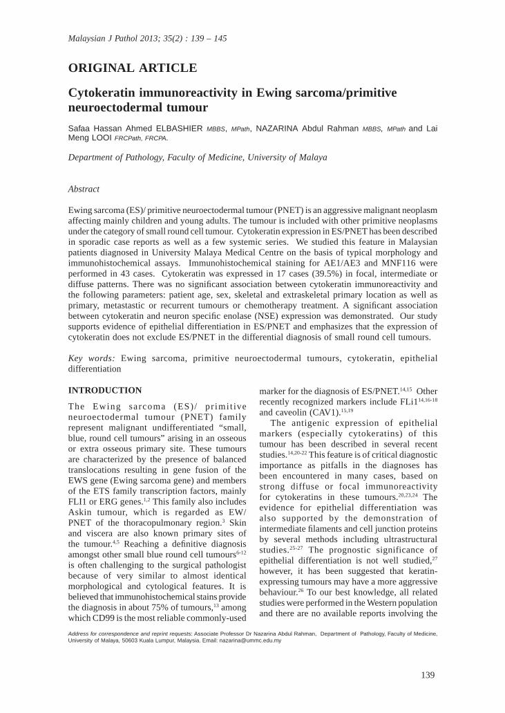
FIG. 1: Diffuse architecture and monotonous small round “blue” cytomorphology of Ewing sarcoma (H&E x40)
141
CYTOKERATIN IN EWING SARCOMA
and young adults (mean age: 26 years; range: 2-67 years) with one patient of unknown age. 27 patients were male and 16 female.
Tumour location and originThere were 37 (86%) primary, 4 (9.3%) recurrent, and 1 (2.3%) metastatic tumours. One was of unknown location. 24(55.8%) arose in extraskeletal sites: 3 each from the leg, lung/chest wall; 2 each from the thigh, shoulder, arm, uterus and pelvic soft tissue and 1 each involving the abdominal wall, buttock, inguinal region, intrabdominal soft tissue, liver, adrenal gland, pancreas and colon. 18 (41.9%) were primary skeletal tumours, including 6 from the femur, 2 each from the pelvis, fibula, maxilla, scapula and 1 each from the humerus, tarsal bones and vertebra. One tumour was from an unknown bone site. 33(76.7%) tumours were sampled
prior to chemotherapeutic treatment. 8(18.6%) were post-chemotherapy samples.
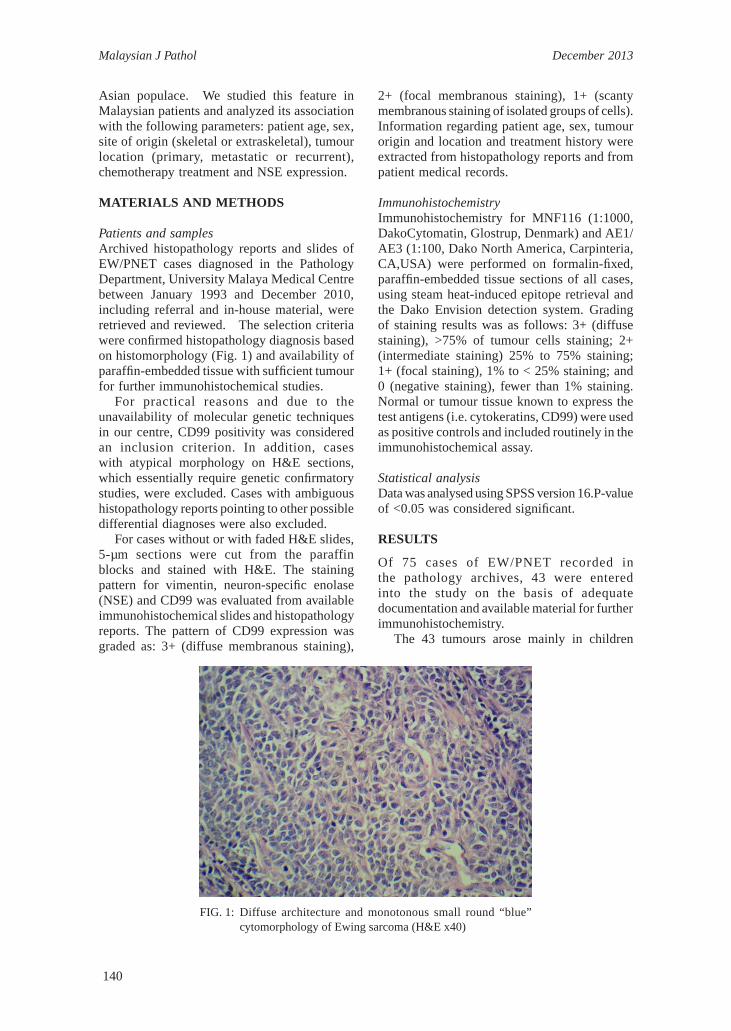
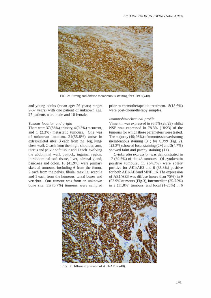
Immunohistochemical profileVimentin was expressed in 96.5% (28/29) whilst NSE was expressed in 78.3% (18/23) of the tumours for which these parameters were tested. The majority (40; 93%) of tumours showed strong membranous staining (3+) for CD99 (Fig. 2). 1(2.3%) showed focal staining (2+) and 2(4.7%) showed faint and patchy staining (1+). Cytokeratin expression was demonstrated in 17 (39.5%) of the 43 tumours. Of cytokeratin positive tumours, 11 (64.7%) were solely positive for AE1/AE3 and 6 (35.3%) positive for both AE1/AE3and MNF116. The expression of AE1/AE3 was diffuse (more than 75%) in 9 (52.9%) tumours (Fig.3), intermediate (25-75%) in 2 (11.8%) tumours; and focal (1-25%) in 6
FIG. 2: Strong and diffuse membranous staining for CD99 (x40).
FIG. 3: Diffuse expression of AE1/AE3 (x40).
Malaysian J Pathol December 2013
142
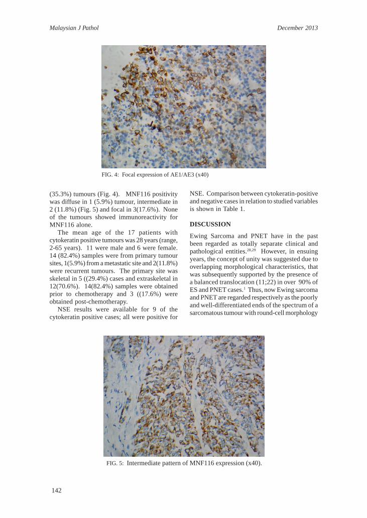
(35.3%) tumours (Fig. 4). MNF116 positivity was diffuse in 1 (5.9%) tumour, intermediate in 2 (11.8%) (Fig. 5) and focal in 3(17.6%). None of the tumours showed immunoreactivity for MNF116 alone. The mean age of the 17 patients with cytokeratin positive tumours was 28 years (range, 2-65 years). 11 were male and 6 were female. 14 (82.4%) samples were from primary tumour sites, 1(5.9%) from a metastatic site and 2(11.8%) were recurrent tumours. The primary site was skeletal in 5 ((29.4%) cases and extraskeletal in 12(70.6%). 14(82.4%) samples were obtained prior to chemotherapy and 3 ((17.6%) were obtained post-chemotherapy. NSE results were available for 9 of the cytokeratin positive cases; all were positive for
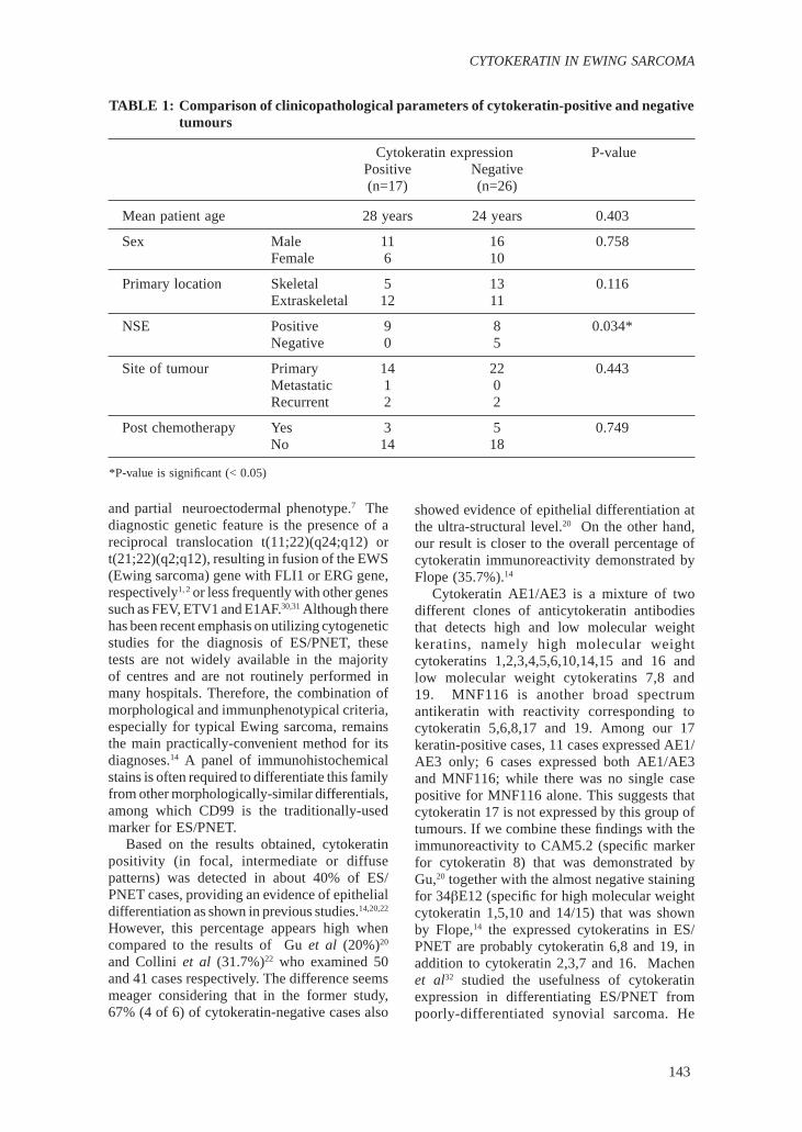
NSE. Comparison between cytokeratin-positive and negative cases in relation to studied variables is shown in Table 1.
DISCUSSION
Ewing Sarcoma and PNET have in the past been regarded as totally separate clinical and pathological entities.28,29 However, in ensuing years, the concept of unity was suggested due to overlapping morphological characteristics, that was subsequently supported by the presence of a balanced translocation (11;22) in over 90% of ES and PNET cases.1 Thus, now Ewing sarcoma and PNET are regarded respectively as the poorly and well-differentiated ends of the spectrum of a sarcomatous tumour with round-cell morphology
FIG. 4: Focal expression of AE1/AE3 (x40)
FIG. 5: Intermediate pattern of MNF116 expression (x40).
143
CYTOKERATIN IN EWING SARCOMA
and partial neuroectodermal phenotype.7 The diagnostic genetic feature is the presence of a reciprocal translocation t(11;22)(q24;q12) or t(21;22)(q2;q12), resulting in fusion of the EWS (Ewing sarcoma) gene with FLI1 or ERG gene, respectively1, 2 or less frequently with other genes such as FEV, ETV1 and E1AF.30,31 Although there has been recent emphasis on utilizing cytogenetic studies for the diagnosis of ES/PNET, these tests are not widely available in the majority of centres and are not routinely performed in many hospitals. Therefore, the combination of morphological and immunphenotypical criteria, especially for typical Ewing sarcoma, remains the main practically-convenient method for its diagnoses.14 A panel of immunohistochemical stains is often required to differentiate this family from other morphologically-similar differentials, among which CD99 is the traditionally-used marker for ES/PNET. Based on the results obtained, cytokeratin positivity (in focal, intermediate or diffuse patterns) was detected in about 40% of ES/PNET cases, providing an evidence of epithelial differentiation as shown in previous studies.14,20,22 However, this percentage appears high when compared to the results of Gu et al (20%)20 and Collini et al (31.7%)22 who examined 50 and 41 cases respectively. The difference seems meager considering that in the former study, 67% (4 of 6) of cytokeratin-negative cases also
showed evidence of epithelial differentiation at the ultra-structural level.20 On the other hand, our result is closer to the overall percentage of cytokeratin immunoreactivity demonstrated by Flope (35.7%).14
Cytokeratin AE1/AE3 is a mixture of two different clones of anticytokeratin antibodies that detects high and low molecular weight keratins, namely high molecular weight cytokeratins 1,2,3,4,5,6,10,14,15 and 16 and low molecular weight cytokeratins 7,8 and 19. MNF116 is another broad spectrum antikeratin with reactivity corresponding to cytokeratin 5,6,8,17 and 19. Among our 17 keratin-positive cases, 11 cases expressed AE1/AE3 only; 6 cases expressed both AE1/AE3 and MNF116; while there was no single case positive for MNF116 alone. This suggests that cytokeratin 17 is not expressed by this group of tumours. If we combine these findings with the immunoreactivity to CAM5.2 (specific marker for cytokeratin 8) that was demonstrated by Gu,20 together with the almost negative staining for 34βE12 (specific for high molecular weight cytokeratin 1,5,10 and 14/15) that was shown by Flope,14 the expressed cytokeratins in ES/PNET are probably cytokeratin 6,8 and 19, in addition to cytokeratin 2,3,7 and 16. Machen et al32 studied the usefulness of cytokeratin expression in differentiating ES/PNET from poorly-differentiated synovial sarcoma. He
TABLE 1: Comparison of clinicopathological parameters of cytokeratin-positive and negative tumours
Cytokeratin expression P-value Positive Negative (n=17) (n=26)
Mean patient age 28 years 24 years 0.403
Sex Male 11 16 0.758 Female 6 10
Primary location Skeletal 5 13 0.116 Extraskeletal 12 11
NSE Positive 9 8 0.034* Negative 0 5
Site of tumour Primary 14 22 0.443 Metastatic 1 0 Recurrent 2 2
Post chemotherapy Yes 3 5 0.749 No 14 18
*P-value is significant (< 0.05)
Malaysian J Pathol December 2013
144
showed immunoreactivity to CK19 in 15% of PNET, while none were positive for CK7. Thus, he concluded that staining for CK7 makes the diagnosis of PNET less likely among differentials. Interestingly, Ewing’s tumour expressing high molecular weight cytokeratin and p63 was recently reported by Weinreb et al.23 Therefore, our emphasis is that cytokeratin expression by ES/PNET is variable and should not be unexpected. This feature has been a source of diagnostic confusion and misdiagnoses, in many reported cases20,23,24 especially when atypical morphology and/ or diffuse cytokeratin staining is encountered. In accordance with the results of Gu et al and Collini et al,20,22 there is no statistically significant association between cytokeratin expression and age and sex of patients or location of the primary tumour (skeletal or extraskeletal). Conversely, we found statistically significant association between cytokeratin and NSE expression. As there is also a well-known immunoreactivity to vimentin by these tumours, the combined expression of epithelial, neural and mesenchymal markers may be a reflection of developmental immaturity rather than a specific line of differentiation as suggested earlier by Sebire et al.33 We found no statistically significant association between cytokeratin expression and site of tumour (primary, metastatic or recurrent) and with chemotherapeutic treatment. A variable frequency of cytokeratin expression in ES/PNET has been shown. Awareness of this fact is of practical importance as diffuse expression of epithelial markers should not rule out a diagnosis of ES/PNET. Clinical and/or histopathological doubts together with the possibility of other differentials such as a poorly differentiated carcinoma may be an indication for molecular genetics confirmation. Because a significant number (40%) of ES/PNET tumours expressed cytokeratin in our limited series, larger-scale studies supported by genetic testing could help to establish the actual prevalence of keratin expression and to establish its prognostic significance.
ACKNOWLEDGMENT
This study was funded by University of Malaya grant UPGP (PPP) P108/2010A and KPT 1053-2011 “Enhancing personalised medicine through pathological profiling of cancer.”
REFERENCES
1. de Alava E, Pardo J. Ewing tumor: tumor biology and clinical applications. Int J Surg Pathol 2001; 9:7-17
2. Dockhorn-Dworniczak B, Schafer KL, Dantcheva R, et al. Diagnostic value of the molecular genetic detection of the t(11;22) translocation in Ewing’s tumours. Virchows Arch 1994; 425:107-12
3. Askin FB, Rosai J, Sibley RK, Dehner LP, McAlister WH. Malignant small cell tumor of the thoracopulmonary region in childhood a distinctive clinicopathologic entity of uncertain histogenesis. Cancer 1979; 43:2438-51
4. Hasegawa SL, Davison JM, Rutten A, Fletcher JA, Fletcher CD. Primary cutaneous Ewing’s sarcoma: immunophenotypic and molecular cytogenetic evaluation of five cases. Am J Surg Pathol 1998; 22:310-8
5. O’Sullivan MJ, Perlman EJ, Furman J, Humphrey PA, Dehner LP, Pfeifer JD.Visceral primitive peripheral neuroectodermal tumours: A clinicopathologic and molecular study. Hum Pathol 2001; 32:1109-15
6. Jayakumar S, Power D. Ewing’s Sarcoma /PNET: A Histopathological Review. Internet J Ortho Surg. 2005; 3 (1).
7. Sabrina R, Antonio GN, Fabio C, Angelo PDT. Small round-cell neoplasm of soft issue: An integrated diagnostic approach. Curr Diagn Pathol 2007; 13:150-63
8. Meis-Kindblom JM, Stenman G, Kindblom LG. Differential diagnosis of small round cell tumours. Semin Diagn Pathol 1996; 13:213-41
9. Hameed M. Small round cell tumours of bone. Arch Pathol Lab Med 2007; 131:192-204
10. Devoe K, Weidner N. Immunohistochemistry of small round-cell tumours. Semin Diagn Pathol 2000; 17:216-24
11. Parham DM, Roloson GJ, Feely M, Green DM, Bridge JA, Beckwith JB. Primary malignant neuroepithelial tumors of the kidney: a clinicopathologic analysis of 146 adult and pediatric cases from the National Wilms’ Tumor Study Group Pathology Center. Am J Surg Pathol 2001; 25:133-46
12. Jimenez RE, Flope A, Lapham RL, et al. Primary Ewing’s sarcoma/primitive neuroectodermal tumor of the kidney: a clinicopathologic and immunohistochemical analysis of 11 cases. Am J Surg Pathol 2002; 26:320-7
13. Leong AS, Wannakrairot P. A retrospective analysis of immunohistochemical staining in identification of poorly differentiated round cell and spindle cell tumours. Pathology 1992; 24(4):245-60
14. Flope AL, Goldblum JR, Rubin BP, et al. Morphologic and immunophenotypic diversity in Ewing family tumors: A study of 66 genetically confirmed cases. Am J Surg Pathol 2005; 29:1025-33
15. Llombart-Bosch A, Machado I, Navarro S, et al. Histological heterogeneity of Ewing’s sarcoma/PNET: an immunohistochemical analysis of 415 genetically confirmed cases with clinical support. Virchows Arch. 2009; 455:397-411
16. Llombart-Bosch A, Navarro S. Immunohistochemical detection of EWS and FLI-1 proteinss in Ewing
145
CYTOKERATIN IN EWING SARCOMA
sarcoma and primitive neuroectodermal tumors: comparative analysis with CD99 (MIC-2) expression. Appl Immunohistochem Mol Morphol 2001; 9:255-60
17. Mhawech-Fauceglia P, Herrmann F, Penetrante R, et al. Diagnostic utility of FLI-1 monoclonal antibody and dual-colour, break-apart probe fluorescence in situ (FISH) analysis in Ewing’s sarcoma/primitive neuroectodermal tumour (EWS/PNET). A comparative study with CD99 and FLI-1 polyclonal antibodies. Histopathology 2006; 49:569-75
18. Folpe AL, Hill CE, Parham DM, O’Shea PA, Weiss SW. Immunohistochemical Detection of FLI-1 Protein Expression: A Study of 132 Round Cell Tumors With Emphasis on CD99-Positive Mimics of Ewing’s Sarcoma/Primitive Neuroectodermal Tumor. Am J Surg Pathol 2000; 24:1657-62
19. Tirado OM, Mateo-Lozano S, Villar J, et al. Caveolin-1 (CAV1) is a target of EWS/FLI-1 and a key determinant of the oncogenic phenotype and tumorigenicity of Ewing’s sarcoma cells. Cancer Res 2006; 66:9937-47
20. Gu M, Antonescu CR, Guiter G, Huvos AG, Ladanyi M, Zakowski MF. Cytokeratin immunoractivity in Ewing’s sarcoma: prevalence in 50 cases confirmed by molecular diagnostic studies. Am J Surg Pathol 2000; 24(3):410-6
21. Vakar-Lopez F, Ayala AG, Raymond AK, Czerniak B. Epithelial Phenotype in Ewing’s Sarcoma/Primitive Neuroectodermal Tumor. Int J Surg Pathol 2000; 8:59-65
22. Collini P, Sampietro G, Bertulli R, et al. Cytokeratin immunoreactivity in 41 cases of ES/PNET confirmed by molecular diagnostic studies. Am J Surg Pathol 2001; 25:273-4
23. Weinreb I, Goldstein D, Perez-Ordonez B. Primary extraskeletal Ewing family tumor with complex epithelial differentiation: a unique case arising in the lateral neck presenting with Horner syndrome. Am J Surg Pathol 2008; 32:1742-8
24. Woestenborghs H, Debiec-Rychter M, Renard M, et al. Cytokeratin-positive meningeal peripheral PNET/Ewing’s sarcoma of the cervical spinal cord: diagnostic value of genetic analysis. Int J Surg Pathol 2005; 13:93-7
25. Moll R, Lee I, Gould VE, Berndt R, Roessner A, Franke WW. Immunocytochemical analysis of Ewing’s tumors. Patterns of expression of intermediate filaments and desmosomal proteins indicate cell type heterogeneity and pluripotential differentiation. Am J Pathol 1987; 127:288-304
26. Srivastava A, Rosenberg AE, Selig M, Rubin BP, Nielsen GP. Keratin-positive Ewing’s sarcoma: an ultrastructural study of 12 cases. Int J Surg Pathol 2005; 13:43-50
27. Schuetz AN, Rubin BP, Goldblum JR, et al.Intercellular junctions in Ewing sarcoma/primitive neuroectodermal tumor: additional evidence of epithelial differentiation. Mod Pathol 2005; 18:1403-10
28. Stout AP. A tumour of the ulnar nerve. Proc NY Pathol Soc 1918; 12:2-12
29. Ewing J. Diffuse endothelioma of bone. Proc NY
Pathol Soc 1921; 21:17-24 30. Barr FG, Womer RB. Molecular diagnosis of Ewing
family of tumors: too many fusions...? J Mol Diagn 2007; 9:437-40
31. Wang L, Bhargava R, Zheng T, et al. Undifferentiated small round cell sarcomas with rare EWS gene fusions: identification of a novel EWS-SP3 fusion and of additional cases with the EWS-ETV1 and EWS-FEV fusions. J Mol Diagn 2007; 9:498-509
32. Machen SK, Fisher C, Gautam RS, Tubbs RR, Goldblum JR. Utility of cytokeratin subsets for distinguishing poorly differentiated synovial sarcoma from peripheral primitive neuroectodermal tumour. Histopathology 1998; 33:501-7
33. Sebire NJ, Gibson S, Rampling D, Williams S, Malone M, Ramsay AD. Immunohistochemical findings in embryonal small round cell tumours with molecular diagnostic confirmation, Appl Immunohistochem Mol Morphol. 2005;13(1):1-5.