cytology and histology - aps home
TRANSCRIPT

Cytology and Histology
Sclerotium Germination and Histopathology of Moniliniavaccinii-corymbosi on Highbush Blueberry
R. D. Milholland
Professor of Plant Pathology, North Carolina State University, Raleigh, NC 27607.Paper No. 5107 of the North Carolina Agricultural Experiment Station.The use of trade names in this publication does not imply endorsement by the North Carolina Agricultural
Experiment Station of the products named, or criticism of similar ones not mentioned.The assistance of Marvin Williams in preparing the illustrations is greatly appreciated.Accepted for publication 31 January 1977.
ABSTRACT
MILHOLLAND, R. D. 1977. Sclerotium germination and histopathology of Monilinia vaccinii-corymbosi on highbush blueberry.Phytopathology 67: 848-854.
Germ tubes from mature ascospores penetrated succulent primary tissue through which the fungus entered the ovary.blueberry leaves either directly through the epidermis or Sclerotium morphology, germination and apotheciumindirectly through stomates. The fungus hyphae in the leaf, development also were studied. Mature sclerotia requiredflower, and fruit tissues grew both intra- and intercellularly chilling below 7 C for a minimum of 900 to 1,200 hr forand caused cellular collapse and necrosis. The stigmatic normal apothecium development.tissue that surrounds the stylar canal appeared to be the
Additional key words: Vaccinium corymbosum, histopathology.
Monilinia vaccindi-corymbosi (Reade) Honey (6) is a 2,160 at a Bladen County farm during the winter of 1968-pathogenic fungus of considerable economic importance 1969 to 1,280 in Pender County during the winter of 1973-in most highbush blueberry (Vaccinium corymbosum 1974. Since apothecium development and ascosporeL.) -producing areas (4, 12, 13). The principal damage on discharge normally coincide with bud break and newhighbush blueberry is a fruit rot resulting in mummified shoot growth, the chilling requirements appear to befruit. However, blighting of twigs and leaves in similar for the host and pathogen.southeastern North Carolina is more devastating on These studies were conducted (i) to determine therabbiteye blueberry (V. ashei) than is the fruit rot. chilling requirements necessary to initiate sclerotium
Longyear (9) first reported the mummy berry disease germination and development of normal apothecia; andoccurring on wild Vaccinium species in Michigan and (ii) to examine the penetration and infection of blueberryidentified the causal organism as Sclerotinia vaccindi leaves, flowers, and fruits by Monilinia vaccinii-Wor. Later Reade (16) reported that the mummy berry corymbosi.fungus differed from S. vaccinii in host range and sizes ofascospores and conidia, and named the fungus S. MATERIALS AND METHODSvaccinii-corymbosi. Honey (6) established the genusMonilinia to include those members of the genus Overwintered sclerotia of M. vaccinii-corymbosi wereSclerotinia commonly possessing a monilioid collected on 21 November, and 22 December 1975,fromamacroconidial stage and a pseudosclerotium, and highbush blueberry farm located in Bladen County,reclassified the organism as Monilinia vaccinii-corymbosi North Carolina, where a planting of the cultivar Croatan(Reade) Honey. was severely infected with the fungus the previous year.
In southeastern North Carolina, overwintered sclerotia Two hundred dormant sclerotia were selected forof M. vaccinii-corymbosi break dormancy around the 1st germination studies each month. A 300-g sample of awk in February and develop mature apothecia about 1 mo nonsterile highbush blueberry soil with a moisturelater (11). Growth of the apothecium initial(s) from the content of 31% was placed in a 150 X 25 mm glass petrisclerotium is favored by a cool temperature (5 C), the dish. Soil was oven-dried and the percent moisturemature apothecium develops at a higher temperature (16 content (SMC) was determined on a dry weight basis (5)C). and recorded. Half of the sclerotia collected each month
Highbush blueberry plants require a minimum of 800 were soaked in distilled water for 72 hr, the other halfto 1,200 hr below 7 C during the winter in order to bloom were not soaked. All the sclerotia (in lots of 25), wereand leaf normally in the spring (3). According to placed in eight dishes and sealed in a 0.025-mm (1-mil)Mainland (10), the number of hours below 7 C in recent polyethelene bag to hold the SMC constant. One dishyears in southeastern North Carolina has ranged from containing soaked sclerotia and one dish with nonsoaked
sclerotia were placed in a Sherer-Gillett CEL 25-7HL
Copyright © 1977 The American Phytopathological Society, 3340 constant temperature chamber at 16 C with a 12 hrPilot Knob Road, St. Paul, MN 55121. All rights reserved. daylength at a light intensity of 9.6 X l03 lx. The
848

July 1977] MILHOLLAND: MONILINIA/HIGHBUSH BLUEBERRY 849
remaining six dishes were placed at 5 C without light. Two C. Inoculations also were made by placing myceliumdishes, one with soaked and one with nonsoaked from a 14-day-old culture of M. vaccinii-corymbosisclerotia, were removed from 5 Cafter 14, 28, and 56 days growing on PDA blocks (2 mm in diameter) onto theand placed at 16 C. Germinated sclerotia and mature stigma of healthy flowers. Inoculated and noninoculatedapothecia were counted 30, 60, and 90 days after pistils from both inoculation procedures were placed incollection. Daily field temperatures were recorded by a FAA after 3, 5, and 7 days. Isolations from infected styleshygrothermograph in a standard U.S. Weather Bureau were made 5 days after inoculation.instrument shelter. The number of hours below 7 C up to The highbush blueberry plants at the Bladen County21 November 1975, 22 December 1975, and 21 January location were in full bloom (80%) on 1 April 1976 and1976, was determined. Sclerotia and apothecia used for infected berries were collected 6 wk later on 15 May 1976.histological examination were fixed in formalin-acetic- Infected leaves and fruit to be sectioned were placed inalcohol (FAA), dehydrated in tertiary butyl alcohol and FAA, embedded in Paraplast Plus, cut at a thickness ofembedded in Paraplast Plus (Sherwood Medical 10 Am, mounted on slides with Haupt's adhesive, andIndustries, St. Louis, MO 63103). Sections 5-10 Aim thick stained with Triarch's quadruple stain (Triarchwere mounted on slides with Haupt's adhesive and Incorporated, Ripon, WS 54971).stained with either safranin and fast green or Harrishematoxylin and Orange G (8). RESULTS
Mature apothecia were produced in the petri dishesmaintained at 16 C. Ascospores were removed with a Germination of sclerotia.-The cumulative number ofcamel's-hair brush from the top of the dish after hours below 7 C up to 21 November, 22 December, and 21discharge and placed on the lower surface of young January was 170, 602, and 1,200, respectively. None of thesucculent leaves of highbush blueberry (cultivar sclerotia collected in November had germinated, whereasCroatan). Rooted cuttings of highbush blueberry 23% of the 392 sclerotia collected in December had(cultivar Croatan) were grown in a peat: sand (1:1, v/v) germinated. Of the 285 sclerotia collected on 21 Januarymixture in 10-cm diameter clay pots in the greenhouse. 1976, 84% had germinated. A few mature apothecia wereInoculated and noninoculated excised leaves were placed observed in the field at this time.on moist filter paper in sterile petri dishes at room Sclerotia that had been exposed to 170 hrbelow7 C didtemperature (20-25 C). Leaves were removed after 1,2,3, not germinate (Table 1). Differences in germination wereand 7 days and placed into FAA. negligible for sclerotia that had been exposed to
Percentage germination of conidia was determined temperatures below 7 C for over 600 hr, and little or noafter 24 hr by counting 100 spores placed on water agar at increase in percent germination was observed after 3025 C. The HC1-Giemsa method (18) was used to stain days. As many as 25 protrusions or stipe initials thatnuclei in the ascospores and in the hyphae growing from ranged from 0.5 to 2.0 mm in length were observed ongerminated conidia. some germinating sclerotia. Apothecium development
The plant parts (leaves, flowers, and fruits) used in was observed within 60 days for sclerotia collected inthese studies were collected from bushes of the highbush December and stored at 5 C for 0, 14, and 28 days prior toblueberry cultivar Croatan. Conidia were collected from being placed at 16 C. Apothecia did not develop at 5 C.naturally infected leaves with a sterile needle and placed Little or no increase in percentage of apothecialin a drop of sterile distilled water on a clean glass slide, development occurred after 60 days, except for sclerotiaInoculum was placed on the stigma of a flower with a stored at 5 C for 56 days. Sclerotia collected in Novembercamel's-hair brush. Several flowers from the same cluster developed mature apothecia after 60 days. Apotheciawere inoculated, the stem was excised, and the flowers produced from sclerotia that had been exposed betweenwere placed on moist filter paper in a petri dish at 20 to 25 602 and 842 hr below 7 C were small, and deteriorated
TABLE 1. Effect of temperature on germination of sclerotia and apothecium development of Monilinia vaccinii-corymbosi
Date sclerotia Total hours Germinated sclerotia (%) Sclerotia with apothecia (%)collected and below
no. days at 5 C 7 C Soaked Nonsoaked Soaked Nonsoaked21 Nov
0 170 0 0 0 014 506 20 0 0 028 842 64 32 20 856 1,514 56 48 60 45
22 Dec0 602 48 45 0 8
14 938 65 45 8 428 1,274 50 56 45 4056 1,946 40 48 50 72
"Germination recorded 30 days after sclerotia were collected from a blueberry field. Apothecium development was recorded 90 daysafter sclerotia were collected.hSclerotia soaked in distilled water for 72 hr.

850 PHYTOPATHOLOGY [Vol. 67
AA
11
O4 r
F
Fig. I-(A to I). Sclerotium germination, apothecium development, and conidium formation of Monilinia vaccinii-corymbosi: A)
Transverse section of mature sclerotium with stipe initial (X50). B) Hymenial layer with paraphyses and developing asci (X400). C)
Mature ascus and ascospores (X530). D) Ascospore penetration through stomate (arrow) of young highbush blueberry (cultivar
Croatan) leaf (X450). E) Transverse section of infected leaf showing necrosis and hyphal accumulation on leaf surface (X450). F)
Transverse section of infected leaf with club-shaped conidiophores (arrow) and conidia (X350). G) Conidia with disjunctors (arrow)
(X500). H) Nuclei in hyphae (Xl,000). I) Hyphae with microconidia (X333).

July 1977] MILHOLLAND: MONILINIA/HIGHBUSH BLUEBERRY 851
XC
t~ 4k
4'0.
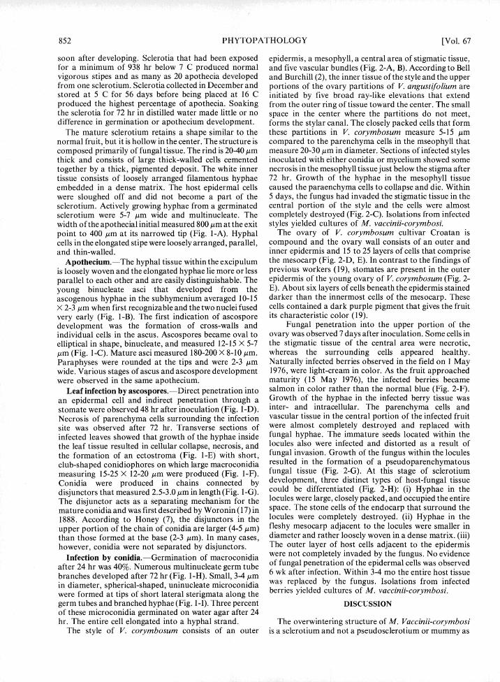
Fig. 2-(A to H). Highbush blueberry fruit infected with Monilinia vaccinii-corymbosi: A) Tangential section of healthy stigma andstyle: s = stigma; st =stigmatic tissue; m = mesophyll (X70). B) Transverse section of healthy style; m = mesophyll; st = stigmatic tissue;sc = style canal; vb = vascular bundle (X 100). C) Transverse section of infected style (X 100). D) Transverse section of healthy ovary; 0= ovule; 1 = locule; p = placentae; m = mesocarp (X50). E) Stomate in ovary wall (X600). F) Healthy and diseased fruit. GH)Transverse sections of 6-wk-old infected fruit. G) Infection of central area of vascular tissue and aborted seed (arrow) in locule (X50).H) Fungus tissue in locule (1), breakdown of endocarp (e), hyphae embedded in matrix (m), and outer mesocarp and epidermis (X50).
852 PHYTOPATHOLOGY [Vol. 67
soon after developing. Sclerotia that had been exposed epidermis, a mesophyll, a central area of stigmatic tissue,for a minimum of 938 hr below 7 C produced normal and five vascular bundles (Fig. 2-A, B). According to Bellvigorous stipes and as many as 20 apothecia developed and Burchill (2), the inner tissue of the style and the upperfrom one sclerotium. Sclerotia collected in December and portions of the ovary partitions of V. angustifolium arestored at 5 C for 56 days before being placed at 16 C initiated by five broad ray-like elevations that extendproduced the highest percentage of apothecia. Soaking from the outer ring of tissue toward the center. The smallthe sclerotia for 72 hr in distilled water made little or no space in the center where the partitions do not meet,difference in germination or apothecium development, forms the stylar canal. The closely packed cells that form
The mature sclerotium retains a shape similar to the these partitions in V. corymbosum measure 5-15 Aimnormal fruit, but it is hollow in the center. The structure is compared to the parenchyma cells in the mseophyll thatcomposed primarily of fungal tissue. The rind is 20-40 4m measure 20-30 Am in diameter. Sections of infected stylesthick and consists of large thick-walled cells cemented inoculated with either conidia or mycelium showed sometogether by a thick, pigmented deposit. The white inner necrosis in the mesophyll tissue just below the stigma aftertissue consists of loosely arranged filamentous hyphae 72 hr. Growth of the hyphae in the mesophyll tissueembedded in a dense matrix. The host epidermal cells caused the paraenchyma cells to collapse and die. Withinwere sloughed off and did not become a part of the 5 days, the fungus had invaded the stigmatic tissue in thesclerotium. Actively growing hyphae from a germinated central portion of the style and the cells were almostsclerotium were 5-7 Am wide and multinucleate. The completely destroyed (Fig. 2-C). Isolations from infectedwidth of the apothecial initial measured 800 Am at the exit styles yielded cultures of M. vaccinii-corymbosi.point to 400 Am at its narrowed tip (Fig. 1-A). Hyphal The ovary of V. corymbosum cultivar Croatan iscells in the elongated stipe were loosely arranged, parallel, compound and the ovary wall consists of an outer andand thin-walled. inner epidermis and 15 to 25 layers of cells that comprise
Apothecium.-The hyphal tissue within the excipulum the mesocarp (Fig. 2-D, E). In contrast to the findings ofis loosely woven and the elongated hyphae lie more or less previous workers (19), stomates are present in the outerparallel to each other and are easily distinguishable. The epidermis of the young ovary of V. corymbosum (Fig. 2-young binucleate asci that developed from the E). About six layers of cells beneath the epidermis stainedascogenous hyphae in the subhymenium averaged 10-15 darker than the innermost cells of the mesocarp. TheseX 2-3 ,m when first recognizable and the two nuclei fused cells contained a dark purple pigment that gives the fruitvery early (Fig. 1-B). The first indication of ascospore its characteristic color (19).development was the formation of cross-walls and Fungal penetration into the upper portion of theindividual cells in the ascus. Ascospores became oval to ovary was observed 7 days after inoculation. Some cells inelliptical in shape, binucleate, and measured 12-15 X 5-7 the stigmatic tissue of the central area were necrotic,gm (Fig. 1-C). Mature asci measured 180-200 X 8-10 Am. whereas the surrounding cells appeared healthy.Paraphyses were rounded at the tips and were 2-3 Am Naturally infected berries observed in the field on I Maywide. Various stages of ascus and ascospore development 1976, were light-cream in color. As the fruit approachedwere observed in the same apothecium. maturity (15 May 1976), the infected berries became
Leaf infection by ascospores.--Direct penetration into salmon in color rather than the normal blue (Fig. 2-F).
an epidermal cell and indirect penetration through a Growth of the hyphae in the infected berry tissue wasstomate were observed 48 hr after inoculation (Fig. l-D). inter- and intracellular. The parenchyma cells andNecrosis of parenchyma cells surrounding the infection vascular tissue in the central portion of the infected fruitsite was observed after 72 hr. Transverse sections of were almost completely destroyed and replaced withinfected leaves showed that growth of the hyphae inside fungal hyphae. The immature seeds located within the
the leaf tissue resulted in cellular collapse, necrosis, and locules also were infected and distorted as a result of
the formation of an ectostroma (Fig. l-E) with short, fungal invasion. Growth of the fungus within the locules
club-shaped conidiophores on which large macroconidia resulted in the formation of a pseudoparenchymatousmeasuring 15-25 X 12-20 Am were produced (Fig. 1-F). fungal tissue (Fig. 2-G). At this stage of sclerotium
Conidia were produced in chains connected by development, three distinct types of host-fungal tissue
disjunctors that measured 2.5-3.0 Am in length (Fig. l-G). could be differentiated (Fig. 2-H): (i) Hyphae in the
The disjunctor acts as a separating mechanism for the locules were large, closely packed, and occupied the entiremature conidia and was first described by Woronin (17) in space. The stone cells of the endocarp that surround the1888. According to Honey (7), the disjunctors in the locules were completely destroyed. (ii) Hyphae in theupper portion of the chain of conidia are larger (4-5 Mm) fleshy mesocarp adjacent to the locules were smaller inthan those formed at the base (2-3 Am). In many cases, diameter and rather loosely woven in a dense matrix. (iii)however, conidia were not separated by disjunctors. The outer layer of host cells adjacent to the epidermis
Infection by conidia.--Germination of macroconidia were not completely invaded by the fungus. No evidence
after 24 hr was 40%. Numerous multinucleate germ tube of fungal penetration of the epidermal cells was observed
branches developed after 72 hr (Fig. 1-H). Small, 3-4 Am 6 wk after infection. Within 3-4 mo the entire host tissue
in diameter, spherical-shaped, uninucleate microconidia was replaced by the fungus. Isolations from infected
were formed at tips of short lateral sterigmata along the berries yielded cultures of M. vaccinii-corymbosi.
germ tubes and branched hyphae (Fig. 1-I). Three percent DISCUSSIONof these microconidia germinated on water agar after 24hr. The entire cell elongated into a hyphal strand. The overwintering structure of M. Vaccinii-corymbosi
The style of V. corymbosum consists of an outer is a sclerotium and not a pseudosclerotium or mummy as

July 1977] MILHOLLAND: MONILINIA/HIGHBUSH BLUEBERRY 853
previously stated (7, 11). By definition (1), "a sclerotium is NC 28429. Little or no fruit developed on thesea firm, frequently rounded, mass of hyphae with or blueberry plants as a result of the primary infections.without the addition of host tissue or soil, normally These infections also provided a source of inoculumhaving no spores in or on it, and may give rise to a fruiting (conidia) for secondary infections of surroundingbody." The overwintering sclerotium is composed almost highbush blueberry blossoms. Therefore, it is importantentirely of fungus tissue; it has an outer rind of melanized that the primary stage of this disease be controlled. Thethick-walled cells cemented together by a thick gelatinous fungicides (Benlate 50W, Ferbam 76W, and Captan 50W)deposit, and a medulla of loosely arranged hyphae that are registered for use on blueberries in Northembedded in a dense matrix. Several apothecia are Carolina do not effectively control the primary infection.usually produced from the sclerotium. Apothecium In the past, mummy berry control in North Carolina hasdevelopment may be initiated either very early or after been achieved best by the elimination of apotheciaextensive growth (15 mm) by the stipe. through the practice of clean cultivation or chemical
Ramsdell et al. (14), reported that the most favorable eradication. The most effective control procedure wouldtemperature for ascospore germination was 15 C, combine the use of an eradicant ground treatment and thealthough ascospores germinated well at 5 to 20 C within 6 application of protectant fungicides (15). Since accuratehr. It appears that ascospores can germinate over a wide weather records are available for different blueberryrange of temperatures, since numerous ascospore germ growing areas in southeastern North Carolina, blueberrytubes were observed on inoculated leaf sections at room growers could be informed about apotheciumtemperature (20-25 C). Mycelium obtained from single- development as it relates to timing of control procedures.conidia isolations grew very well on PDA at 25 C.
The fungus usually gained entrance into the ovary of LITERATURE CITEDinfected pistils through the stigmatic tissue in the centralportion of the style that surrounds the stylar canal. 1. AINSWORTH, G. C. 1971. Ainsworth and Bisby'sTransverse sections of approximately 6-wk-old infected dictionary of the fungi. The Commonwealth Mycologicalberries indicated that the central portion of the fruit was Institute, Kew, Surrey, England. 663 p.colonized initially and the outer mesocarp and epidermis 2. BELL, H. P., and J. BURCHILL. 1955. Flowerwere invaded last. development in the lowbush blueberry. Can. J. Bot.
33:251-258.The primary factors influencing sclerotium 3. DARROW, G. M. 1942. Rest period requirements ofgermination and apothecium development are blueberries. Proc. Am. Soc. Hortic. Sci. 41:189-194.temperature, moisture, and light. Previous studies (11) 4. DEMAREE, J. B., and M. S. WILCOX. 1947. Fungihave shown that without adequate moisture, sclerotia will pathogenic to blueberries in the eastern United States.not germinate and produce apothecia, even if Phytopathology 37:487-506.temperatures are favorable. My results have shown that 5. GARDNER, W. H. 1965. Water content. Pages 92-93 in C.sclerotia will not germinate unless subjected to A. Black, D. D. Evans, J. L. White, L. E. Ensminger, andtemperatures below 7 C for a minimum of 500 hr, even F. E. Clark, eds. Methods of soil analysis. Part 1: Physicalwitempderuat stures bg 7n Ci a and mineralogical properties, including statistics ofwith adequate moisture. Although germination is a maueetadsmln.A.Sc go.Mdsnmeasurement and sampling. Am. Soc. Agron. -Madison,prerequisite, it does not always lead to apothecium Wisconsin. 770 p.development. Sclerotia that had received the minimum 6. HONEY, E. E. 1928. The Monilioid species of Sclerotinia.chilling requirements of 500-600 hr below 7 C germinated Mycologia 20:127-157.but did not produce apothecia. Based upon these and 7. HONEY, E. E. 1936. North American species of Monilinia.previous studies (11), mature sclerotia require adequate I. Occurrence, grouping, and life-histories. Am. J. Bot.moisture (30-40% SMC), a minimum of 900-1,200 hr at 5- 23:100-106.7 C, followed by a temperature of approximately 16 Cfor 8. JOHANSEN, D. A. 1940. Plant microtechnique. McGraw-
Hill, New York. 523 p.normal apothecium development. Light is required for 9. LONGYEAR, B. 0. 1901. A sclerotium disease of thethe development of mature apothecia. huckleberry. Annu. Rep. Mich. Acad. Sci. 3:61-62.
Knowledge of the chilling requirements for sclerotium 10. MAINLAND, C. M. 1975. Blueberry production asgermination and apothecium development would be influenced by weather in southeastern North Carolina.essential for obtaining the inoculum requisite for Proc. 8th Annu. Open House Southeastern Blueberryscreening large numbers of blueberry seedlings for Council. 8:15-27.mummy-berry resistance prior to transplanting into the 11. MILHOLLAND, R. D. 1974. Factors affecting apotheciumfield. This information also would facilitate control of the development of Monilinia vaccinii-corymbosi from
e (twig mummied highbush blueberry fruit. Phytopathologyprimary stage of this disease. The primary infections (64:296-300.and leaf blight) are caused by the ascospores. Conidia are 12. NELSON, J. W., and H. C. BITTENBENDER. 1971.produced within 2-3 wk on the blighted leaves and are Mummy berry disease occurrence in a blueberry selectioncarried by wind or insects to open blossoms. Mummy test planting. Plant Dis. Rep. 55:651-653.berry develops as a result of these secondary infections. 13. PEPIN, H. S., and H. N. W. TOMS. 1969. Economic lossThe fruit rot stage occurs on both the highbush and from mummy berry of highbush blueberry in coastalrabbiteye blueberry; however, damage caused by the British Columbia. Can. Plant Dis. Surv. 49:105-107.primary infections has been much greater on the 14. RAMSDELL, D. C., J. W. NELSON, and R. L. MYERS.rabbiteye species. During the past 3 yr, a severe blighting 1974. An epidemiological study of mummy berry diseaserabiteye spees. Duritsang thoer past 3 f y raeve bbligting of highbush blueberry. Phytopathology 64:222-228.of the leaves, shoots, and flower buds of three rabbiteye 15. RAMSDELL, D. C., J. W. NELSON, and R. L. MYERS.cultivars (Delite, Southland, and Tifblue) was observed at 1976. Interaction of eradicant and protectant treatmentsthe Horticultural Crops Research Station, Castle Hayne, upon the epidemiology and control of mummy berry

854 PHYTOPATHOLOGY [Vol. 67
disease of highbush blueberry. Phytopathology 66:350- 18. YAMASAKI, Y., and H. NIIZEKI. 1965. Studies on354. variation of the rice blast fungus, Piricularia oryzae Cav.:
16. READE, J. M. 1908. Preliminary notes on some species of 1. Karyological and genetical studies on variation. Nat.Sclerotinia. Ann. Mycol. 6:109-115., Inst. Agric. Bull. Ser. D 13:231-274. Tokyo.
17. WORONIN, M. 1888. Ober die Sclerotienkrankheit der 19. YARBROUGH, J. A., and E. B. MORROW. 1947. StoneVacciniee-Beeren. Mem. Acad. Sci. St. Petersburg 36:1- cells in Vaccinium. Proc. Am. Soc. Hortic. Sci. 50:224-49. 228.