cytoplasmic alkalinization induced by insulin through an activation of na+h+ antiporter inhibits...
TRANSCRIPT
Eiochemtcal Phurmacologv. Vol. 41, No. 9. pp. 127P-1282, 1991 Printed zn Great Britain.
ooo62952/91 $3.(x) + 0.00 @ 1991. Pergamon Press plc
CYTOPLASMIC ALKALINIZATION INDUCED BY INSULIN THROUGH AN ACTIVATION OF Na+-H+ ANTIPORTER
INHIBITS TYROSINE HYDROXYLASE ACTIVITY IN STRIATAL SYNAPTOSOMES
SALVATORE AMOROSO. GIANFRANCO DI RENZO, MAURIZIO TAGLIALATELA, LORELLA M. T. CANZONIERO, EDWARD J. CRAGOE JR* and Lucre ANNUNZIATO~~
Department of Pharmacology, 2nd School of Medicine, University of Naples, via S. Pansini, 5, Naples,* P.O. Box 631548 Nacogdoches TX 75963-1548. and
*Institute of Behavioural and Neurological Sciences, University of Chieti. Italy
(Receiued 14 May 1990; accepted 1 October 1990)
Abstract-Insulin dose-dependently inhibited tyrosine hydroxylase (TH) activity and increased intra- synaptosomal pH (pII,) in rat striatal nerve endings. Both these effects of insulin on TH and pH, were prevented by the .5-(N-methyl-N-(guanidinocarbonylmethyl) amiloride (MGCMA), a putative selective inhibitor of the Na+-H’ antiporter. Interestingly when, by changing the extracellular pH (pH,), the pH, was increased. from 7.1 up to 7.5, an equivalent inhibition of TH activity occurred. The inhibitory action exerted from insulin on TH activity disappeared when the hormone was added to synaptosomes whose pH, was lowered to 6.83. Collectively, the results of the present study showed that insulin inhibited TH activity in striatal synaptosomes. This effect seems to involve the activation of the Na+-H’ antiporter. This exchange system once activated. may induce an intrasynaptosomai alkaiinization. a condition in which TH activity is inhibited.
The possibility of a direct effect of insulin on brain physiology has long been the subject of considerable debate. Previously reported studies demonstrated the presence of insulin [l] and of its specific binding sites [2] in the rat brain. This evidence suggests that insulin may influence neuronal function. In fact it is known that insulin modifies catecholamine turnover 131 and release [3,4] from central neurons and that it modulates monoamine uptake in cultured neuronal cells [5]. On the other hand it has been reported that insulin modifies the activity of the membrane Na+-H’ exchanger, which is involved in the modulation of intracellular pH [fi]. Since a variation of the pH,, may modify TH (L-tyrosine 3-mono- oxygenase; EC 1.14.16.2) activity in synaptosomes of rat corpus striatum [7, X], a dopaminergic region where receptor binding sites for insulin have been detected [2], we investigated whether insulin could modify TH activity via an action on Na+-H’ exchange.
i%ATERIALS AND METHODS
Porcine or human reconlbinant insulin (Actrapid) was from Novo Industries. Copenhagen, Denmark. Recombinant insulin-like growth factor-l (IGF-1) was a kind gift of Drs H. H. Peter and K. Scheibli
$ Correspondence to: Prof. Lucia Annunziato, Depart- ment of Pharmacoloev, 2nd School of Medicine, Via S. Pansini. 5, Naples, 8oi31 Italy.
S: Abhreviations: BCECF. 2.7’-bis-(2-carboxvethvll-5.6- , ,* carboxyfluorescein; AM, acetoxymethyl ester; pH,. extra- cellular pH; pH,. intrasynaptosomal pH; TH, tyrosine hydroxylase: IGF- 1, insulin-like growth factor-l ; MGCMA, 5-(N-methyl-N-(guanidinocarbonyl) amiloride.
from Ciba Geigy (Basle, Switzerland) and Chiron Corporation (Emeryville, CA, U.S.A.). 2,7’-Bis- (2-carboxyethyl)-5,6-carboxyfluorescein acetoxy- methyl ester (BCECF-AM§) was from Molecular Probes (Eugene, OR, U.S.A.). MGCMA was synthesized by previously described methods [Y]. All the other drugs were purchased from the Sigma Chemical Co (St Louis, MO, U.S.A.). All chemicals used were from standard commercial sources. L-[ l- carboxyi-“%]tyrosine (sp. act. 51 mCi/mmoI) was obtained from Amersham International.
Male Wistar rats (2~25Og) were decapitated and the striata were dissected out and placed in polyethylene tubes on ice. The tissue was homogenized in 0.32 M sucrose. and centrifuged at 1000 g for 5 min. The supernatant obtained was then centrifuged at 20,OOOg for 20min to sediment the P, fraction. Most of the TH activity in the P? fraction is associated with synaptosomes [lo]. The quality of our synaptosomes preparation was checked by using electron microscopy.
TH activity was assayed by measuring the production of “COz from r_-[ 1-‘“Cltyrosine according to Ill]. The Pz ellet was resuspended in glucose 0.32M 1:l.s (w v). Synaptosomes, 25Opg protein ip assayed by the method of Bradford 1121. were incubated in a standard Tris-buffered medium containing the following composition: 3 mM KCl, 131 mM NaCl. 1.2mM MgSO,. 1.2mM CaC12, 20 mM Tris, 5 mM Na,,HPO, and 1 mM ascorbic acid at pH, 7.4 (final volume 0.5 mL). To obtain the desired pIi,,. small aliquots of 1 M Tris or 1 M HCl were added to the media. Synaptosomes were preincubated for 15min at 37”. Unless otherwise specified the drug examined was added to the incubation medium at the 10th min of preincubation.
1279

1280 S. A~o~oso et al.
Insulin and IGF-1 were added to the incubation medium (pH,, 7.1) in presence of bovine serum albumin (0.1%) plus bacitracin (20 yM), in order to avoid the binding of the insulin to glass tubes and its inactivation, respectively. The reaction was started by adding 6 PL of L-[l-‘JC]tyrosine (0.3 PCi) to give a final concentration of 10pM. The tubes were capped with rubber stoppers and contained a center well with 2OOpL of hyamine hydroxide absorbed into a strip of Whatman No. 1 paper to trap the evolved ‘“CO,. The incubation was performed for 20 min and the reaction was stopped by injecting 200 ,uL of lo%, trichloroacetic acid into the tubes. After a 2 hr incubation at 37” the radioactivity trapped in the paper plus the center well was counted by liquid scintillation spectrometry. Blank samples incubated in the absence of the tissue were 0.03% (200 dpm) of the total radioactivity added. The basal rate of 14C02 formation ranged from 15 to 20 times more than the blank values. Time course analysis indicated that the release of “CO? was linear up to 30 min. pH, was determined by loading synaptosomes with 10pM of the AM- ester of BCECF for 45 min at 37”. pH, 7.4. Synaptosomes were then washed twice to remove the extracellular BCECF-AM and resuspended in the incubation medium. Fluorescence (excitation: SOOnm, emission: 530nm) was recorded in a thermostated quartz cuvette in a Perkin-Elmer LS 5 spectrofluorimeter, equipped with a magnetic stirrer. Calibration of fluorescence signal as a function of pH, was performed by the method of Thomas et al. [13] using the K+-H+ ionophore nigericin (0.5 ,ug/ mL) added to synaptosomes suspended in a medium containing 140mM K+ and 5 mM NaCl. Unless otherwise specified, insulin was used in a medium pH,7.4. The pH, was changed by adding small aliquots of 1 M Tris or HCI and measured with an electrode inserted into the cuvette. Intracellular fluorescence was detected and plotted as a function of the pH,.
The data were analysed by means of the analysis of variance followed by Neuman-Keul’s and Dunnett’s tests.
RESULTS
Dose-dependent inhibition of striatal TH crctivit_y b) insulin
Insulin, added to the incubation medium in concentrations ranging from 21 to 63 PM inhibited TH activity in striatal synaptosomes in a dose- dependent manner (Fig. 1). This effect was not dependent by the presence of zinc ions in the insulin preparation, since this cation (7, 21, 63 PM), which is present in a 1:3 molar ratio with insulin, did not modify TH activity (data not shown). IGF-1 at all the concentrations used, failed to modify TH activity in striatal synaptosomes (6.6 2 0.3. 6.6 * 0.3. 6.7 t 0.2, 6.3 + 0.3. 5.8 F 0.3picomoles 14COz/ min/mg protein in control, 5,50, 500.5000 nM IGF- 1 groups, respectively).
Effect of MGCMA on insulin-inhibited TH activity in striatal synaptosomes
MGCMA at the concentration of 100 and 300 FM,
0 0.7 7 21 63 Insulin concentrations (NM)
Fig. 1. Effects of different concentrations of porcine insulin on basal TH activity in striatal synaptosomes. Columns represent the mean 2 SE of six determinations. *P < 0.01 vs all the other values. **P < 0.01 vs 21 ,uM insulin value.
prevented the inhibitory action exerted by insulin (21 and 63 PM) on TH activity (Table 1).
Effect of insulin on pH,
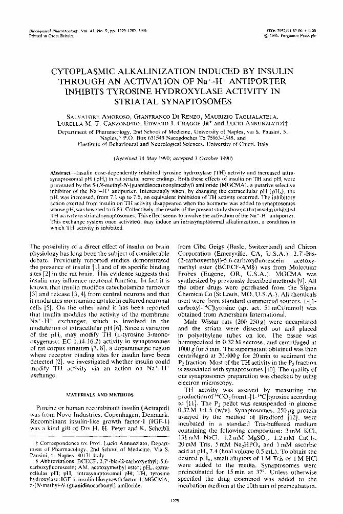
Human recombinant insulin produced a rise in pH, from 7.14 to 7.28 in striatal synaptosomes (Fig. 2), in a dose-dependent manner (EC50 11 PM). Insulin-induced alkalinization was not dependent by the presence of zinc ions since this cation at the concentration of 63 HIM did not cause any variation in pH, (data not shown). MGCMA (1OOpM) prevented the increase in pH, evoked by insulin (Fig.
3).
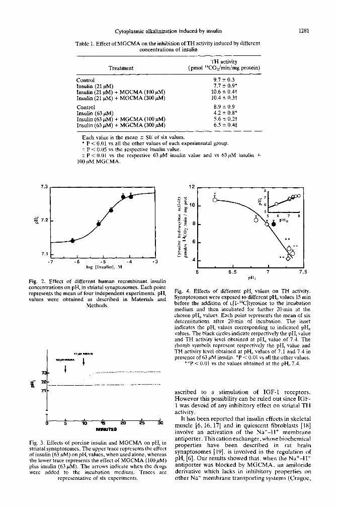
Effect of different pH, values, on TH activity
When striatal synaptosomes were exposed to various external pH values, a change in pHi occurred and the rate of TH activity was modified (Fig. 4). The alkalinization of pHi. obtained by elevating pH,,. to values of 7.3 and 7.38, inhibited TH activity. On the contrary the acidification of pH, to value of 6.26 stimulated TH activity. The inhibitory effect of insulin on TH activity occurred only when the pH, was at a steady state value of 7.14. whereas, when pH, value was 6.83, the hormone did not inhibit TH activity, although it was able to increase pH, (Fig.
4).
DISCUSSION
The results of the present paper showed that insulin inhibited TH activity in striatal synaptosomes in a dose-dependent manner. This effect was not dependent by the presence of zinc ions in the insulin preparation, since this cation (7-63 PM), which is present in a 1:3 molar ratio with insulin, did not modify TH activity in striatal synaptosomes (data not shown). Since it has been reported that insulin may act through an interaction with IGF-1 receptors [14], whose presence in rat brain has been recently detected [15], we examined the possibility that the inhibition of TH activity evoked by insulin could be
7.3
Cytoplasmic alkalinization induced by insulin 1281
Table 1. Effect of MGCMA on the inhibition of TH activity induced by different concentrations of insulin
TH activity Treatment (pm01 “C0,/min/mg protein)
Control 9.1 2 0.3 Insulin (21 PM) 7.1 ‘_ 0.9* Insulin (21 PM) + MGCMA (100 PM) 10.6 k 0.4t Insulin (21 MM) + MGCMA (300 PM) 10.4 + 0.3t
Control 8.9 k 0.9 Insulin (63 PM) 4.2 + 0.8* Insulin (63 MM) + MGCMA (100 PM) 5.6 2 0.2t Insulin (63 ,uM) + MGCMA (300 PM) 6.5 k 0.4$
Each value is the mean + SE of six values. * P < 0.01 vs all the other values of each experimental group. t P < 0.05 vs the respective insulin value. $ P co.01 vs the respective 63nM insulin value and vs 63pM insulin +
100 ,uM MGCMA.
7.1 - cm I I I -7 -6 -5 -4
log [Insulin], hl
Fig. 2. Effect of different human recombinant insulin concentrations on pH, in striatal synaptosomes. Each point represents the mean of four independent experiments. pH, values were obtained as described in Materials and
Methods.
Fig. 3. Effects of porcine insulin and MGCMA on pH, in striatal synaptosomes. The upper trace represents the effect of insulin (63 ,uM) on pH, values, when used alone, whereas the lower trace represents the effect of MGCMA (100 PM) plus insulin (63 PM). The arrows indicate when the drugs were added to the incubation medium. Traces are
representative of six experiments.
*
6 6.5 7 7:5 Pi1i
Fig. 4. Effects of different pH, values on TH activity. Synaptosomes were exposed to different pH, values 15 min before the addition of ~[l-‘%]tyrosine to the incubation medium and then incubated for further 20min at the chosen pH, values. Each point represents the mean of six determinations after 20min of incubation. The inset indicates the pH, values corresponding to indicated pH, values. The black circles indicate respectively the pH, value and TH activity level obtained at pH, value of 7.4. The rhomb symbols represent respectively the pH, value and TH activity level obtained at pH, values of 7.1 and 7.4 in presence of 63 PM insulin. *P < 0.01 vs all the other values.
**P i 0.01 vs the values obtained at the pH,7.4.
ascribed to a stimulation of IGF-1 receptors. However this possibility can be ruled out since IGF- 1 was devoid of any inhibitory effect on striatal TH activity.
It has been reported that insulin effects in skeletal muscle [6. 16,171 and in quiescent fibroblasts [18] involve an activation of the Na+-H+ membrane antiporter. This cation exchanger, whose biochemical properties have been described in rat brain synaptosomes [19]. is involved in the regulation of pH, [6]. Our results showed that, when the Na+-H+ antiporter was blocked by MGCMA. an amiloride derivative which lacks in inhibitory properties on other Na+ membrane transporting systems (Cragoe,

1282 S. Ahno~oso et al.
personal communication), the inhibitory action of insulin on TH activity was prevented. Therefore an activation of Na+-H+ antiporter seems to be involved in the inhibitory effect exerted by insulin on TH activity. The hypothesis that insulin inhibited TH activity via an activation of the Na+-H+ antiporter, which led to a cytoplasmic alkahnization, appears to be also validated by the observation that insulin produced a dose-dependent elevation of pH, and that this increase was prevented by the Na+-H’ antiporter blocker MGCMA. On this regard it should be noted that MGCMA by itself did not lower pH,. however it should be considered that also in cultured cardiac cells [20], when the pH, value is in a steady-state basal condition. the inhibition of the Na+‘-H’ antiporter does not produce a lowering of pH, therefore suggesting that pH, regulation is not due exclusively to the Na+-H+ antiporter. The hypothesis that intracellular alkalinization induced by insulin causes an inhibition of TH activity was further supported by the data showing that when pHi value was increased to values of 7.3 and 7.38, obtained by elevating pH,, an inhibition of TH activity was observed. On the other hand the inhibitory effect of insulin on TH activity can occur only when the pHi reaches the basal steady-state value of 7.14, whereas insulin. when added to synaptosomes, whose pH, was 6.83, did not inhibit TH activity, although it was able to increase pH,.
However. since 100 PM MGCMA. completely prevented insulin-induced alkalinization (Fig. 3) whereas both 100 and 300 PM MGCMA were unable to completely reverse the inhibiting action of insulin on TH activity (Table 1). we cannot exclude that other mechanisms than changes in pH, mav also be involved in insulin action on TH activity. This consideration may also explain the little difference between the haif maximal effects induced by insulin on TH activity and on pH;.
Finally. it should be considered that. since at the concentrations used. insulin may partially exist in a dimeric form, the possibility that insulin effects may be due to a portion of the hormone present in a dimeric form cannot be ruled out. However. the fact that. in other biological systems, insulin modulates the activity of the Na+-H+ antiporter at the concentrations lower (i.e. in a monomeric form) [16, 171 than those utilized in the present study. would suggest that insulin should act in a mon~~mcric form.
Acknowledgements-We are grateful to Dr Giuseppe Galizia for his collaboration in the computerized statistical analysis of the data and Mr L. Scognamiglio and V. Grill0 for their technical assistance. This study was supported by the C.N.R. Target Project on Biotechnology and Bioinstrumentation.
REFERENCES
1. Havrankova J. Schemechel D, Roth J and Brownstein M, Identification of insulin in rat brain. Proc Natl Acud Sci USA 75: _5737-S741, 1978.
2. Havrankova J, Roth J and Brownstein M, insulin
receptors are widely distributed in the central ner\oua system of the rat. Nature 272: X27-82’), lY78.
3. Sauter A. Goldstein M. Engel J and Ueta K. Effect ot insulin on central catecholamines. Bruit1 Res 260: 33(L 333. 1983.
4. Amoroso S, Taglialatela M. Canzoniero LMT. C‘ragoc EG Jr. Di Renzo GF and Annunziato L. Possible involvement of Ca” ions. protein kinase C and Na - H’ antiporter in insulin-induced endogen~~us dopaminc release from tuberoinfundibular neurons. i.<fi ,G-i 46: X85-894, 1990.
5. Boyd FT. Clarke DW. Muther IF and R:urada MK, Insulin receptor5 and insulin modulation 01 norepinephrine uptake in neuronal culture5 from ~;II brain. J Viol Chem 260: 15X8(!-158X4. IYX5.
6. Moore RD. Effects of insulin upon ion transport. Biochim Biophys Acta 737: l-49, 1983.
7. Boarder MR and Fillenz M. Synaptosomal tyrosinc hydroxylation in the rat brain: comparison of activity from hippocampus and hypothalamus with &ivtty from striatum. J Neurochem 31: 14iY-1426. IY7S.
8. Patrick RL and Rendel MT, pH-induced alterations tn dopamine synthesis regulation in rat brain strtatal synaptosomes. J ~effr~~hern 34: l.illt+l.E113. IYXO.
9. Cragoe EJ Jr. Woltersdorf OW Jr. Flicking JB, Kwvttni:
10.
11.
12.
13.
14.
15.
16.
17.
1x.
19.
20.
SF and Jones JH. Pyrazine Diuretics. Il.-,~:-Amidino- 3-amino-5-Suhstituted-h_1_Ialopyr~izinecarh(~x~~mi~i~~. .I Med Chem 10: h6-75. 1967. Coyle JT, Tyrosine hydroxylase in rat brain. Cofactor requirements. regional and subcellular distribution. Biochem Pharmacol 21: 1935-1914. lY72. Waymire JC. Bjur A and Weimer N. Assay of tvro5tnc hydroxylase by coupled dccarboxylation o? dopa formed from I-\‘C-I.-tyrosine. Ar~nl Riockeni 43: 5XX- 600, lY7 1. Bradford MMA, A rapid. and sensitive method for the quantitationof microgramquantities~~fpr~~tein utilizing the principle of protein-dye binding. Art& Rio&etri 72: ‘4x-354, 1976. Thomas JA. Buchst~aum AN. Zimniak A and Racker E. Intracellular pH measurements in Ehriich ascites tumor cells utilizing spectroscopic probes generated itr sifu. Biochemisfrv 18: 7210-2718. 197Y. Goodyear CG. De Stephano L. Lai WH. Guyda HJ and Posner 81, Characterization of insultn-like growth factor receptors in rat anterior pituitary. hypothalamus and brain. Endocrinology 114: 11X7-1 lY.S, IYXJ Arajo DM, Apchak L. Collier B. Chabot I(; and Quiron R. Insulin growth factor 1 (somatomedin (‘1 receptors in the rat brain: distribution and interaction with the hippocampal cholinergic system. Rrrriri lit’\ 484: 13t~l37, IYXY. Klip A, Ramlal T and Cragoe EJ Jr. Inwiin-induced cytoplasmic alkalinizati[)n and g?ucoxe transport in muscle cells. An2 J Phr.%jl 250: C7?_%C7%. iYXt>. Fidelman ML. Serholzer SH, Walsh KB and Moore RD. Intracellular pH mediares action t~i‘ insulin on glycolysis in frog skeletal muscle. ,~tffr I f’!ty\ioi 212: C87-CY3. iY8’. Paris S and Pouyssigur J, Growth factors acti\atc the Na ‘-H T antiporter in quiescent libroblasts by incrca\tng its affinity for intracellular H’ J Rid (‘lrcw 259: IOY89- 10994. 1984. Jean T. Frelin C, Vigne P. Barbry P and Lazdunski M, Biochemical properties of the Na‘-Hi exchange system in rat brain synaptosomes. J Rio/ C’hem 260: 9678-9684, 1985. Frelin C, Vigne P and Lazdunski M, The role of the Na’-H+ exchange system in the regulation of the internal pH in cultured cardiac cells. Eur f B&hem 148: l-4, 1985.